the lipopolysaccharide-activated innate immune … 6: 59-77, 2009 issn 1824-307x review the...
TRANSCRIPT
ISJ 6: 59-77, 2009 ISSN 1824-307X
REVIEW The lipopolysaccharide-activated innate immune response network of the horseshoe crab S Kawabata, T Koshiba, T Shibata Department of Biology, Faculty of Sciences, Kyushu University, Fukuoka 812-8581, Japan
Accepted May 20, 2009
Abstract
Primary stimulation of the horseshoe crab innate immune system by bacterial lipopolysaccharide (LPS) activates a network of responses to ensure host defense against invading pathogens. Granular hemocytes selectively respond to LPS via a G protein-dependent exocytic pathway that critically depends on the proteolytic activity of the LPS-responsive coagulation factor C. In response to stimulation by LPS, the hemocyte secretes transglutaminase (TGase) and several kinds of defense molecules, such as coagulation factors, lectins, antimicrobial peptides, and protein substrates for TGase. LPS-induced hemocyte exocytosis is enhanced by a feedback mechanism in which the antimicrobial peptide tachyplesin serves as an endogenous mediator. The coagulation cascade triggered by LPS or β-1,3-D-glucans results in the formation of coagulin fibrils that are subsequently stabilized by TGase-dependent cross-linking. A cuticle-derived chitin-binding protein additionally forms a TGase-stabilized mesh at sites of injury. Invading pathogens are agglutinated by both hemocyte- and plasma-derived lectins. In addition, the proclotting enzyme and tachyplesin functionally convert hemocyanin to phenoloxidase. In the plasma, coagulation factor C acts an LPS-sensitive complement C3 convertase on the surface of Gram-negative bacteria. In this manner, LPS-induced hemocyte exocytosis leads not only to coagulation but also activates a sophisticated innate immune response network that coordinately effects pathogen recognition, prophenoloxidase activation, pathogen clearance, and TGase-dependent wound healing.
Key Words: horseshoe crab; lipopolysaccharide; innate immunity; hemolymph coagulation; transglutaminase; complement C3
Introduction
Innate immunity, which defends the host against
infectious pathogens, is an ancient and ubiquitous immune system in both vertebrates and invertebrates. Each species employs a variety of environment-specific adaptations to ensure host defense, whereas a generalized recognition strategy against invading pathogens underlies the innate immune reaction. The innate immune system recognizes broadly conserved microbial cell wall components known as pathogen-associated molecular patterns (PAMPs), such as lipopolysaccharides (LPS) of Gram-negative bacteria, peptidoglycans of Gram-positive bacteria, and β-1,3-D-glucans of fungi via pattern-recognition ___________________________________________________________________________
Corresponding author: Shun-ichiro Kawabata 6-10-1 Hakozaki Higashi-Ku, Department of Biology Faculty of Sciences, Kyushu University Fukuoka 812-8581, Japan E-mail: [email protected]
proteins (Janeway, 1989; Hoffmann, 2003; Akira et al., 2006). Innate immune systems in invertebrates consist of pathways that promote recognition of pathogen-associated macromolecules, hemolymph coagulation, phenoloxidase-mediated melanization, cell agglutination, antimicrobial activity, and phagocytosis (Nappi et al., 2004; Theopold et al., 2004; Iwanaga and Lee, 2005; Kurata et al., 2006; Nakanishi and Shiratsuchi, 2006).
The horseshoe crab belongs to the class Merostomata and is phylogenetically more closely related to Arachnoidea than it is to Crustacea. Fossils of horseshoe crabs, such as Mesolimulus walchi and Limulus coffini, have been found in deposits from the Paleozoic era to the Cenozoic era in Europe and North America (Størmer, 1952). Extant horseshoe crabs comprise four species, Limulus polyphemus, Tachypleus tridentatus, T. gigas, and Carcinoscorpius rotundicauda, each having a distinct geographic distribution; L. polyphemus is distributed along the east coast of North America,
59
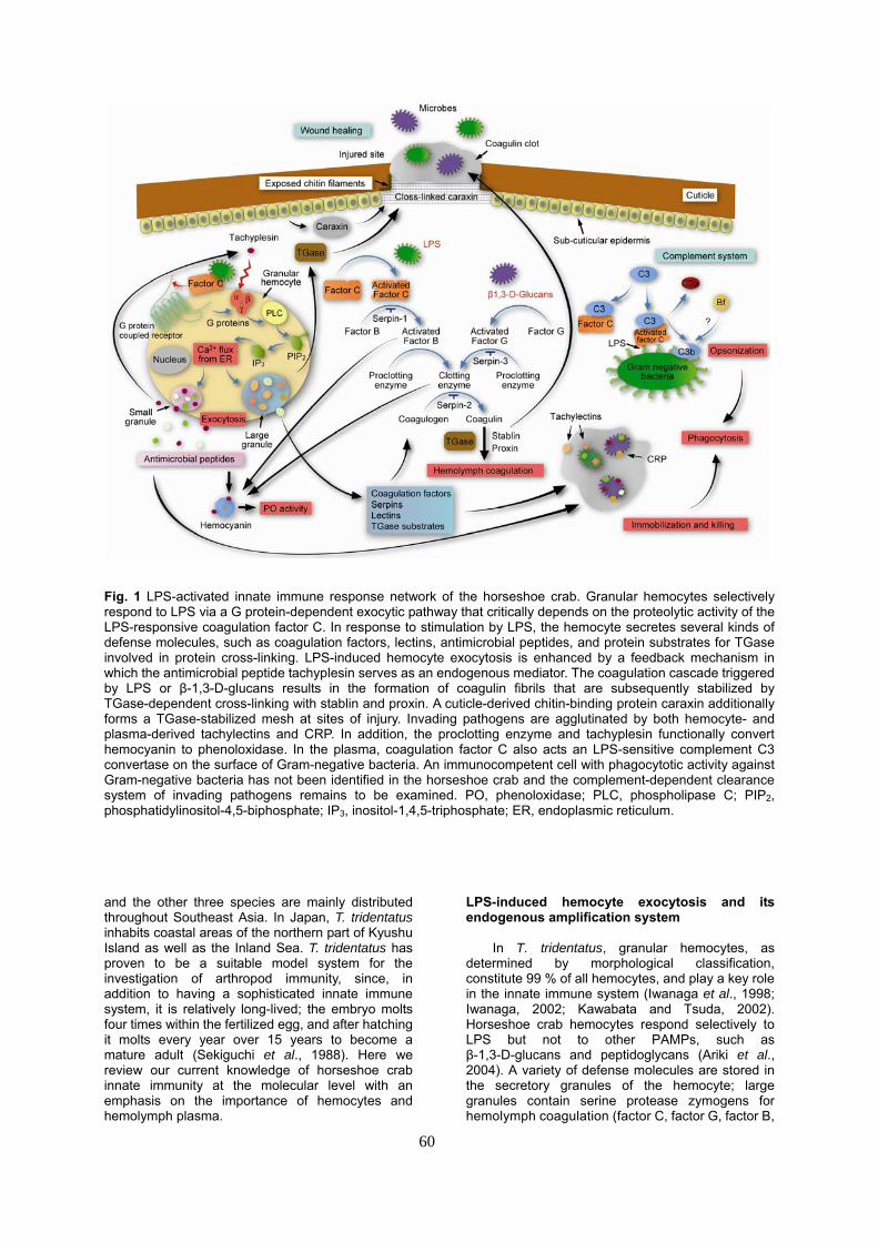
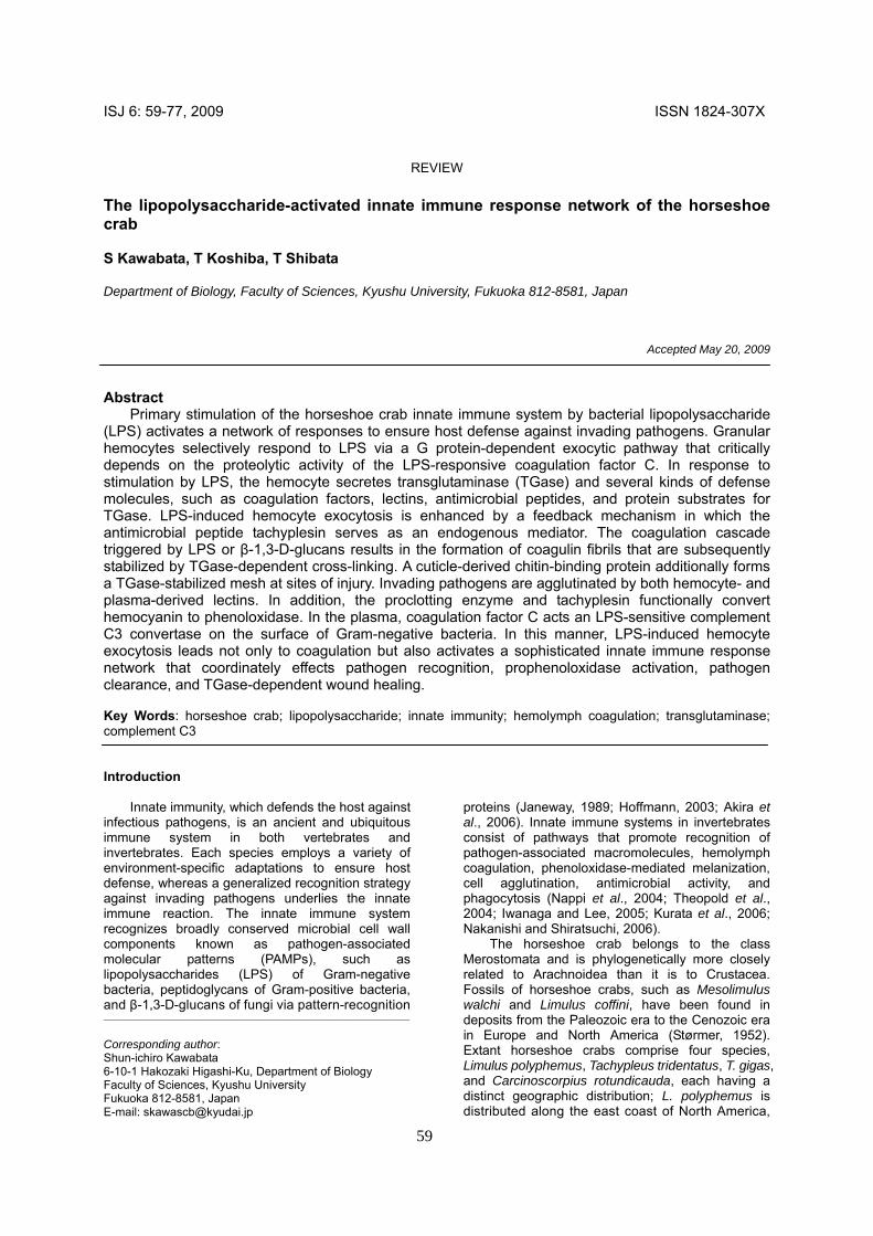
Fig. 1 LPS-activated innate immune response network of the horseshoe crab. Granular hemocytes selectively respond to LPS via a G protein-dependent exocytic pathway that critically depends on the proteolytic activity of the LPS-responsive coagulation factor C. In response to stimulation by LPS, the hemocyte secretes several kinds of defense molecules, such as coagulation factors, lectins, antimicrobial peptides, and protein substrates for TGase involved in protein cross-linking. LPS-induced hemocyte exocytosis is enhanced by a feedback mechanism in which the antimicrobial peptide tachyplesin serves as an endogenous mediator. The coagulation cascade triggered by LPS or β-1,3-D-glucans results in the formation of coagulin fibrils that are subsequently stabilized by TGase-dependent cross-linking with stablin and proxin. A cuticle-derived chitin-binding protein caraxin additionally forms a TGase-stabilized mesh at sites of injury. Invading pathogens are agglutinated by both hemocyte- and plasma-derived tachylectins and CRP. In addition, the proclotting enzyme and tachyplesin functionally convert hemocyanin to phenoloxidase. In the plasma, coagulation factor C also acts an LPS-sensitive complement C3 convertase on the surface of Gram-negative bacteria. An immunocompetent cell with phagocytotic activity against Gram-negative bacteria has not been identified in the horseshoe crab and the complement-dependent clearance system of invading pathogens remains to be examined. PO, phenoloxidase; PLC, phospholipase C; PIP2, phosphatidylinositol-4,5-biphosphate; IP3, inositol-1,4,5-triphosphate; ER, endoplasmic reticulum. and the other three species are mainly distributed throughout Southeast Asia. In Japan, T. tridentatus inhabits coastal areas of the northern part of Kyushu Island as well as the Inland Sea. T. tridentatus has proven to be a suitable model system for the investigation of arthropod immunity, since, in addition to having a sophisticated innate immune system, it is relatively long-lived; the embryo molts four times within the fertilized egg, and after hatching it molts every year over 15 years to become a mature adult (Sekiguchi et al., 1988). Here we review our current knowledge of horseshoe crab innate immunity at the molecular level with an emphasis on the importance of hemocytes and hemolymph plasma.
LPS-induced hemocyte exocytosis and its endogenous amplification system
In T. tridentatus, granular hemocytes, as
determined by morphological classification, constitute 99 % of all hemocytes, and play a key role in the innate immune system (Iwanaga et al., 1998; Iwanaga, 2002; Kawabata and Tsuda, 2002). Horseshoe crab hemocytes respond selectively to LPS but not to other PAMPs, such as β-1,3-D-glucans and peptidoglycans (Ariki et al., 2004). A variety of defense molecules are stored in the secretory granules of the hemocyte; large granules contain serine protease zymogens for hemolymph coagulation (factor C, factor G, factor B,
60
Fig. 2 Factor C is a membrane associated LPS sensor on hemocytes. Hemocytes were treated without (left panel) and with (right panels) FITC-labeled LPS (green), and stained with a monoclonal antibody (2C12) against factor C. For the detection, Cy3-conjugated anti-mouse secondary antibody was used (red). Arrowheads indicate LPS co-localized with membrane bound factor C. Bar = 10 μm. and the proclotting enzyme), the clottable protein coagulogen, serine protease inhibitors (serpins), lectins, and substrates for transglutaminase (TGase), whereas small granules contains antimicrobial peptides. In response to stimulation by LPS, these defense molecules are rapidly secreted by the hemocyte (Fig. 1).
Factor C is a unique LPS-responsive serine protease zymogen that is stored in the large granules of hemocytes and acts as an LPS sensor to potentiate hemocyte exocytosis. Upon activation by LPS, factor C initiates hemocyte exocytosis via a G-protein-dependent exocytic pathway that is dependent upon the proteolytic activity of factor C. In this respect, the activation of hemocytes by factor C is analogous to the thrombin-thrombin receptor (the protease-activated G protein-coupled receptor, PAR) signaling axis in mammalian platelets (Ariki et al., 2004). Hemocyte exocytosis can be quantitatively assayed by ELISA using an antibody against a granular component such as coagulogen in the presence of 50 mM Mg2+ and 10 mM Ca2+, equivalent to the concentrations of these cations in hemolymph plasma. Exclusion of divalent cations from the assay buffer inhibits exocytosis even at high concentrations of LPS. Moreover, in the absence of LPS, hemocyte exocytosis can be
induced by synthetic hexapeptides corresponding to the tethered ligands of mammalian PARs, supporting the notion of a PAR-like receptor on the hemocyte surface.
Immunofluorescence microscopy of the hemocyte using an anti-factor C antibody detects factor C, which is localized in a punctate distribution on the hemocyte surface (Kurata et al., 2006; Koshiba et al., 2007) (Fig. 2). When hemocytes are incubated with FITC-labeled LPS, the factor C antigen co-localizes with FITC-LPS accumulated on the cell surface (Koshiba et al., 2007). The accumulation of FITC-LPS is not observed on hemocytes fixed with formaldehyde, as would be expected with chemical modification and inactivation of cell surface proteins. These results also suggest that factor C is a membrane-bound LPS sensor on the hemocyte surface.
Factor C has no obvious transmembrane domain within in its sequence, whereas it strongly interacts with acidic phospholipids (Nakamura et al., 1988a). Surface plasmon resonance analyses indicate that factor C interacts with phosphatidylserine (Kd = 2.5×10⁻⁹ M) and phosphatidylinositol (Kd = 4.7×10⁻⁹ M) as well as with cholesterol (Kd = 1.4×10⁻⁹ M) (Ariki et al., 2004). The interaction of factor C with LPS (Kd = 7.6×10⁻¹⁰ M) is
61
62
competitively inhibited by the addition of the acidic phospholipids. In contrast, cholesterol does not inhibit the interaction of factor C with LPS, suggesting that factor C interacts with cholesterol through a binding site that is distinct from that for LPS, and raising the possibility that factor C may be localized on cholesterol-rich microdomains or lipid rafts on the hemocyte membrane.
The horseshoe crab hemocyte has an endogenous positive feedback mechanism for LPS-induced hemocyte exocytosis (Ozaki et al., 2005). The hemolymph contains hemocytes at ~106
cells/ml. LPS-induced hemocyte exocytosis is highly dependent on the cell density, namely, an increase in cell density from 0.05×10⁶ to 0.8×10⁶ cells/ml results in a 106-fold change in the apparent LPS sensitivity (from 10-7 to 10-13 g/ml of LPS), suggesting the presence of feedback mechanism for secretion via an unknown secretagogue secreted from hemocytes in response to the stimulation by LPS.
Interestingly, tachyplesin in the exocytosed fluid acts as a secondary secretagogue, thereby dramatically enhancing the sensitivity of the hemocyte to LPS. Tachyplesin (17 residues) is a potent antimicrobial peptide and one of the most abundant components stored in the hemocyte (Nakamura et al., 1988b; Shigenaga et al., 1990). The effective concentration of tachyplesin required for hemocyte exocytosis ranges from 5 to 10 μM, indicating that a high concentration of tachyplesin is required to act as an effective endogenous secretagogue.
Tachyplesin has structural properties in common with mastoparan, a basic tetradecapeptide from wasp venom. Mastoparan interacts directly with G proteins without direct stimulation of the upstream receptor, and induces exocytosis in the mast cell (Higashijima et al., 1988). Consistent with these findings, mastoparan is able to induce hemocyte exocytosis in T. tridentatus (Ariki et al., 2004). Moreover, tachyplesin binds to bovine G protein an equilibrium dissociation constant (Kd) of 8.8×10⁻⁷ M (Ozaki et al., 2005). These data suggest that tachyplesin interacts with G-proteins in the hemocyte in a manner similar to that of mastoparan. In addition, tachyplesin has an ability to bind to hemocyanin (Nagai et al., 2001), the major protein in the hemolymph plasma, suggesting that hemocyanin may serve as a sink for tachyplesin released from hemocytes, thereby spatially restricting its hemocyte-stimulating effect to the site of infection.
On the other hand, insect and mammals conserve a signaling pathway of the innate immune system through cell-surface receptors called Tolls and Toll-like receptors (Hoffmann, 2003; Akira et al., 2006). A Toll-like receptor (tToll) has been identified in T. tridentatus, which is most closely related to Drosophila Toll in both domain architecture and overall length (Inamori et al., 2000, 2004). The two receptors show a significant sequence identity between their TIR domains (39 %). Interestingly, spätzle, a protein ligand for Drosophila Toll, shows significant structurally similarity to horseshoe crab coagulogen (see "Hemolymph coagulation"). Moreover, tToll is nonspecifically expressed in all tissues examined (Inamori et al., 2004), suggesting
that tToll does not act as an LPS receptor on granular hemocytes.
In addition, NF-κB and IκB homologues (CrNF-κB and CrIκB) have been identified in the horseshoe crab C. rotundicauda (Wang et al., 2006). Gram-negative bacteria infection causes degradation of CrIκB and nuclear translocation of CrNF-κB, leading to up-regulation of immune-related gene expression, including nitric oxide synthase and factor C, indicating that the NF-κB/IκB signaling cascade remains well conserved from horseshoe crabs to mammals, playing a fundamental role in regulating the expression of critical immune defense molecules.
Hemolymph coagulation
The hemocyte releases coagulation factors by
LPS-induced exocytosis, leading to the activation of the proteolytic coagulation cascade (Fig. 1). Factor C secreted from hemocytes is autocatalytically activated in the presence of Gram-negative bacteria or LPS. The resulting activated factor C activates coagulation factor B, which in turn converts the proclotting enzyme into the clotting enzyme. The clotting enzyme then promotes the proteolytic conversion of coagulogen to coagulin, which spontaneously forms an insoluble polymer. Alternatively, activated factor G in the presence of β-1,3-D-glucans triggers the activation of the proclotting enzyme to the clotting enzyme. In this manner, factor C and factor G independently serve to couple the recognition of LPS and β-1,3-D-glucans, respectively, to the formation of a physical barrier at the site of microbial invasion.
The vertebrate coagulation system acts locally on the phospholipid surface in cooperation with Ca2+
at the site of vascular injury. In an analogous fashion, hemolymph coagulation in the horseshoe is restricted to the surfaces of invading pathogens, such as Gram-negative bacteria and fungi. This mechanistic similarity between the coagulation cascades of vertebrates and horseshoe crabs may lead to the erroneous assumption of a common evolutionary origin (Fig. 3). In fact, fibrinogen homologues of the horseshoe crab, named tachylectins-5A and -5B, act as non-self recognizing proteins rather than as target proteins of the coagulation cascade (Gokudan et al., 1999). Also coagulogen has no structural similarity or evolutionary relatedness to fibrinogen (Bergner et al., 1996).
A protease cascade in Drosophila has been well characterized as the morphogenetic cascade for determining embryonic dorsal-ventral polarity, leading to the production of the Toll ligand spätzle (Belvin and Anderson, 1996). The Drosophila Toll pathway additionally controls resistance to fungal and Gram-positive bacterial infections (Ferrandon et al., 2007). Spätzle, belongs to nerve growth factor family and possesses a structural similarity to horseshoe crab coagulogen (Smith and DeLotto, 1992; Bergner et al., 1996; Bergner et al., 1997). In addition, a clip-like domain located in the N-terminal region of horseshoe crab coagulation factor B and the proclotting enzyme, originally identified in the proclotting enzyme as a disulfide-knotted domain,
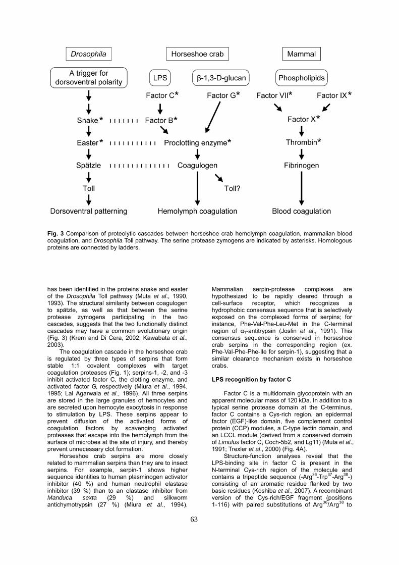
Fig. 3 Comparison of proteolytic cascades between horseshoe crab hemolymph coagulation, mammalian blood coagulation, and Drosophila Toll pathway. The serine protease zymogens are indicated by asterisks. Homologous proteins are connected by ladders. has been identified in the proteins snake and easter of the Drosophila Toll pathway (Muta et al., 1990, 1993). The structural similarity between coagulogen to spätzle, as well as that between the serine protease zymogens participating in the two cascades, suggests that the two functionally distinct cascades may have a common evolutionary origin (Fig. 3) (Krem and Di Cera, 2002; Kawabata et al., 2003).
The coagulation cascade in the horseshoe crab is regulated by three types of serpins that form stable 1:1 covalent complexes with target coagulation proteases (Fig. 1); serpins-1, -2, and -3 inhibit activated factor C, the clotting enzyme, and activated factor G, respectively (Miura et al., 1994, 1995; Lal Agarwala et al., 1996). All three serpins are stored in the large granules of hemocytes and are secreted upon hemocyte exocytosis in response to stimulation by LPS. These serpins appear to prevent diffusion of the activated forms of coagulation factors by scavenging activated proteases that escape into the hemolymph from the surface of microbes at the site of injury, and thereby prevent unnecessary clot formation.
Horseshoe crab serpins are more closely related to mammalian serpins than they are to insect serpins. For example, serpin-1 shows higher sequence identities to human plasminogen activator inhibitor (40 %) and human neutrophil elastase inhibitor (39 %) than to an elastase inhibitor from Manduca sexta (29 %) and silkworm antichymotrypsin (27 %) (Miura et al., 1994).
Mammalian serpin-protease complexes are hypothesized to be rapidly cleared through a cell-surface receptor, which recognizes a hydrophobic consensus sequence that is selectively exposed on the complexed forms of serpins; for instance, Phe-Val-Phe-Leu-Met in the C-terminal region of α1-antitrypsin (Joslin et al., 1991). This consensus sequence is conserved in horseshoe crab serpins in the corresponding region (ex. Phe-Val-Phe-Phe-Ile for serpin-1), suggesting that a similar clearance mechanism exists in horseshoe crabs.
LPS recognition by factor C
Factor C is a multidomain glycoprotein with an
apparent molecular mass of 120 kDa. In addition to a typical serine protease domain at the C-terminus, factor C contains a Cys-rich region, an epidermal factor (EGF)-like domain, five complement control protein (CCP) modules, a C-type lectin domain, and an LCCL module (derived from a conserved domain of Limulus factor C, Coch-5b2, and Lg11) (Muta et al., 1991; Trexler et al., 2000) (Fig. 4A).
Structure-function analyses reveal that the LPS-binding site in factor C is present in the N-terminal Cys-rich region of the molecule and contains a tripeptide sequence (-Arg36-Trp37-Arg38-) consisting of an aromatic residue flanked by two basic residues (Koshiba et al., 2007). A recombinant version of the Cys-rich/EGF fragment (positions 1-116) with paired substitutions of Arg36/Arg38 to
63
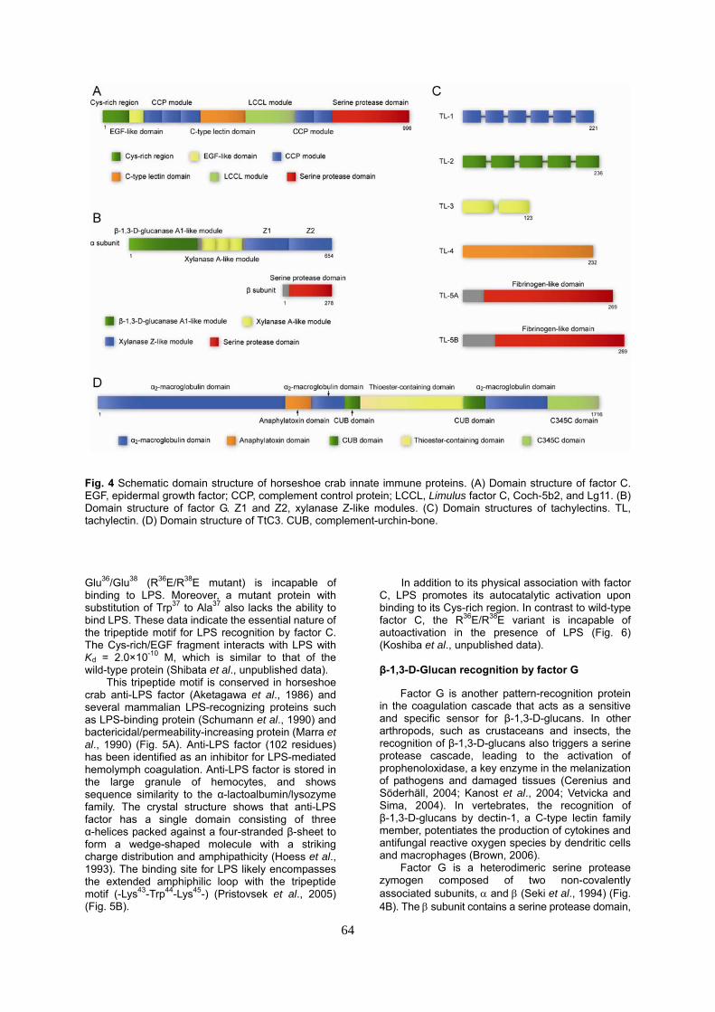
Fig. 4 Schematic domain structure of horseshoe crab innate immune proteins. (A) Domain structure of factor C. EGF, epidermal growth factor; CCP, complement control protein; LCCL, Limulus factor C, Coch-5b2, and Lg11. (B) Domain structure of factor G. Z1 and Z2, xylanase Z-like modules. (C) Domain structures of tachylectins. TL, tachylectin. (D) Domain structure of TtC3. CUB, complement-urchin-bone. Glu36/Glu38 (R36E/R38E mutant) is incapable of binding to LPS. Moreover, a mutant protein with substitution of Trp37 to Ala37 also lacks the ability to bind LPS. These data indicate the essential nature of the tripeptide motif for LPS recognition by factor C. The Cys-rich/EGF fragment interacts with LPS with Kd = 2.0×10⁻¹⁰ M, which is similar to that of the wild-type protein (Shibata et al., unpublished data).
This tripeptide motif is conserved in horseshoe crab anti-LPS factor (Aketagawa et al., 1986) and several mammalian LPS-recognizing proteins such as LPS-binding protein (Schumann et al., 1990) and bactericidal/permeability-increasing protein (Marra et al., 1990) (Fig. 5A). Anti-LPS factor (102 residues) has been identified as an inhibitor for LPS-mediated hemolymph coagulation. Anti-LPS factor is stored in the large granule of hemocytes, and shows sequence similarity to the α-lactoalbumin/lysozyme family. The crystal structure shows that anti-LPS factor has a single domain consisting of three α-helices packed against a four-stranded β-sheet to form a wedge-shaped molecule with a striking charge distribution and amphipathicity (Hoess et al., 1993). The binding site for LPS likely encompasses the extended amphiphilic loop with the tripeptide motif (-Lys43-Trp44-Lys45-) (Pristovsek et al., 2005) (Fig. 5B).
In addition to its physical association with factor C, LPS promotes its autocatalytic activation upon binding to its Cys-rich region. In contrast to wild-type factor C, the R36E/R38E variant is incapable of autoactivation in the presence of LPS (Fig. 6) (Koshiba et al., unpublished data).
β-1,3-D-Glucan recognition by factor G
Factor G is another pattern-recognition protein
in the coagulation cascade that acts as a sensitive and specific sensor for β-1,3-D-glucans. In other arthropods, such as crustaceans and insects, the recognition of β-1,3-D-glucans also triggers a serine protease cascade, leading to the activation of prophenoloxidase, a key enzyme in the melanization of pathogens and damaged tissues (Cerenius and Söderhäll, 2004; Kanost et al., 2004; Vetvicka and Sima, 2004). In vertebrates, the recognition of β-1,3-D-glucans by dectin-1, a C-type lectin family member, potentiates the production of cytokines and antifungal reactive oxygen species by dendritic cells and macrophages (Brown, 2006).
Factor G is a heterodimeric serine protease zymogen composed of two non-covalently associated subunits, α and β (Seki et al., 1994) (Fig. 4B). The β subunit contains a serine protease domain,
64
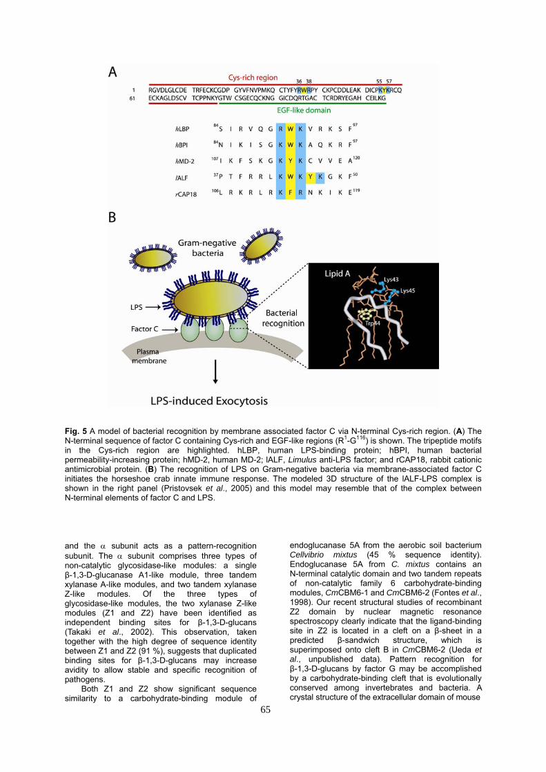
Fig. 5 A model of bacterial recognition by membrane associated factor C via N-terminal Cys-rich region. (A) The N-terminal sequence of factor C containing Cys-rich and EGF-like regions (R1-G116) is shown. The tripeptide motifs in the Cys-rich region are highlighted. hLBP, human LPS-binding protein; hBPI, human bacterial permeability-increasing protein; hMD-2, human MD-2; lALF, Limulus anti-LPS factor; and rCAP18, rabbit cationic antimicrobial protein. (B) The recognition of LPS on Gram-negative bacteria via membrane-associated factor C initiates the horseshoe crab innate immune response. The modeled 3D structure of the lALF-LPS complex is shown in the right panel (Pristovsek et al., 2005) and this model may resemble that of the complex between N-terminal elements of factor C and LPS. and the α subunit acts as a pattern-recognition subunit. The α subunit comprises three types of non-catalytic glycosidase-like modules: a single β-1,3-D-glucanase A1-like module, three tandem xylanase A-like modules, and two tandem xylanase Z-like modules. Of the three types of glycosidase-like modules, the two xylanase Z-like modules (Z1 and Z2) have been identified as independent binding sites for β-1,3-D-glucans (Takaki et al., 2002). This observation, taken together with the high degree of sequence identity between Z1 and Z2 (91 %), suggests that duplicated binding sites for β-1,3-D-glucans may increase avidity to allow stable and specific recognition of pathogens.
65
Both Z1 and Z2 show significant sequence similarity to a carbohydrate-binding module of
endoglucanase 5A from the aerobic soil bacterium Cellvibrio mixtus (45 % sequence identity). Endoglucanase 5A from C. mixtus contains an N-terminal catalytic domain and two tandem repeats of non-catalytic family 6 carbohydrate-binding modules, CmCBM6-1 and CmCBM6-2 (Fontes et al., 1998). Our recent structural studies of recombinant Z2 domain by nuclear magnetic resonance spectroscopy clearly indicate that the ligand-binding site in Z2 is located in a cleft on a β-sheet in a predicted β-sandwich structure, which is superimposed onto cleft B in CmCBM6-2 (Ueda et al., unpublished data). Pattern recognition for β-1,3-D-glucans by factor G may be accomplished by a carbohydrate-binding cleft that is evolutionally conserved among invertebrates and bacteria. A crystal structure of the extracellular domain of mouse
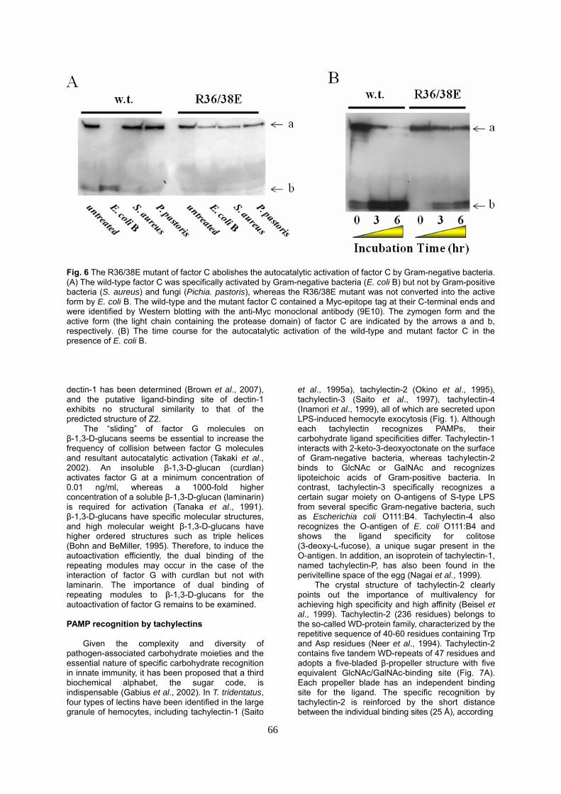
Fig. 6 The R36/38E mutant of factor C abolishes the autocatalytic activation of factor C by Gram-negative bacteria. (A) The wild-type factor C was specifically activated by Gram-negative bacteria (E. coli B) but not by Gram-positive bacteria (S. aureus) and fungi (Pichia. pastoris), whereas the R36/38E mutant was not converted into the active form by E. coli B. The wild-type and the mutant factor C contained a Myc-epitope tag at their C-terminal ends and were identified by Western blotting with the anti-Myc monoclonal antibody (9E10). The zymogen form and the active form (the light chain containing the protease domain) of factor C are indicated by the arrows a and b, respectively. (B) The time course for the autocatalytic activation of the wild-type and mutant factor C in the presence of E. coli B. dectin-1 has been determined (Brown et al., 2007), and the putative ligand-binding site of dectin-1 exhibits no structural similarity to that of the predicted structure of Z2.
The “sliding” of factor G molecules on β-1,3-D-glucans seems be essential to increase the frequency of collision between factor G molecules and resultant autocatalytic activation (Takaki et al., 2002). An insoluble β-1,3-D-glucan (curdlan) activates factor G at a minimum concentration of 0.01 ng/ml, whereas a 1000-fold higher concentration of a soluble β-1,3-D-glucan (laminarin) is required for activation (Tanaka et al., 1991). β-1,3-D-glucans have specific molecular structures, and high molecular weight β-1,3-D-glucans have higher ordered structures such as triple helices (Bohn and BeMiller, 1995). Therefore, to induce the autoactivation efficiently, the dual binding of the repeating modules may occur in the case of the interaction of factor G with curdlan but not with laminarin. The importance of dual binding of repeating modules to β-1,3-D-glucans for the autoactivation of factor G remains to be examined.
PAMP recognition by tachylectins
Given the complexity and diversity of
pathogen-associated carbohydrate moieties and the essential nature of specific carbohydrate recognition in innate immunity, it has been proposed that a third biochemical alphabet, the sugar code, is indispensable (Gabius et al., 2002). In T. tridentatus, four types of lectins have been identified in the large granule of hemocytes, including tachylectin-1 (Saito
et al., 1995a), tachylectin-2 (Okino et al., 1995), tachylectin-3 (Saito et al., 1997), tachylectin-4 (Inamori et al., 1999), all of which are secreted upon LPS-induced hemocyte exocytosis (Fig. 1). Although each tachylectin recognizes PAMPs, their carbohydrate ligand specificities differ. Tachylectin-1 interacts with 2-keto-3-deoxyoctonate on the surface of Gram-negative bacteria, whereas tachylectin-2 binds to GlcNAc or GalNAc and recognizes lipoteichoic acids of Gram-positive bacteria. In contrast, tachylectin-3 specifically recognizes a certain sugar moiety on O-antigens of S-type LPS from several specific Gram-negative bacteria, such as Escherichia coli O111:B4. Tachylectin-4 also recognizes the O-antigen of E. coli O111:B4 and shows the ligand specificity for colitose (3-deoxy-L-fucose), a unique sugar present in the O-antigen. In addition, an isoprotein of tachylectin-1, named tachylectin-P, has also been found in the perivitelline space of the egg (Nagai et al., 1999).
The crystal structure of tachylectin-2 clearly points out the importance of multivalency for achieving high specificity and high affinity (Beisel et al., 1999). Tachylectin-2 (236 residues) belongs to the so-called WD-protein family, characterized by the repetitive sequence of 40-60 residues containing Trp and Asp residues (Neer et al., 1994). Tachylectin-2 contains five tandem WD-repeats of 47 residues and adopts a five-bladed β-propeller structure with five equivalent GlcNAc/GalNAc-binding site (Fig. 7A). Each propeller blade has an independent binding site for the ligand. The specific recognition by tachylectin-2 is reinforced by the short distance between the individual binding sites (25 Å), according
66

Fig. 7 Crystal structures of tachylectins-2 and -5A. (A) The 5-fold β-propeller structure of tachylectin-2 in complex with GlcNAc. (B) Oligomeric arrangement of tachylectin-5A subunits around the 4-fold crystallographic axis. to the pentagonal geometry, required to interact with PAMPs. Tachylectin-1 (221 residues) and tachylectin-3 (123 residues), despite lacking significant overall sequence similarity to tachylectin-2, also consist of WD repeats, with six-WD-repeats for tachyectin-1 and two-WD-repeats for tachylectin-3, suggesting β-propeller structures analogous to that of tachylectin-2. Ultracentrifugation analysis shows that tachylectin-3 is present in dimer in solution. In contrast, tachylectin-4 (232 residues) is an oligomeric glycoprotein of 470 kDa and is homologous to the N-terminal domain of Xenopus pentraxin-1 (Fig. 4C). The function of the N-terminal domain of pentraxin-1 is unknown.
Hemolymph plasma also contains several lectins, such as isoforms of tachylectin-1 (Chiou et al., 2000; Chen et al., 2001) and three types of C-reactive proteins (CRPs), all of which exhibit functional and structural diversity (Iwaki et al., 1999). Human CRP is an acute-phase reactant, its concentration increasing rapidly in response to stress, injury or infection, and it belongs to a protein family of pentraxin (Osmand et al., 1977; Tennent and Pepys, 1994). Horseshoe crab CRP is a predominant LPS-binding protein and is up-regulated at transcript levels by Pseudomonas infection, suggesting the importance of horseshoe crab CRP as a conserved molecule for pathogen recognition (Ng et al., 2004). CRP from the horseshoe crab L. polyphemus forms extended fibrilar structures that encapsulate liposomes in the presence of Ca2+ (Harrington et al., 2009). Furthermore, the membranes of Limulus CRP-treated bacteria exhibit significantly different mechano-elastic properties than those of untreated bacteria, suggesting the protein's role as a primary
defense molecule, acting in the entrapment and killing of potential pathogens.
In addition, tachylectins-5A and -5B, with binding specificity for acetyl-group have been identified in hemolymph plasma (Gokudan et al., 1999). The overall sequence identity between tachylectins-5A (269 residues) and -5B (289 residues) is 45 %. About two thirds of the total hemagglutinating activity in the hemolymph plasma can be attributed to tachylectins-5 A and -5B, which strongly agglutinate all types of human erythrocytes at a minimal concentration of 0.004 μg/ml, and also agglutinate both Gram-negative and Gram-positive bacteria. The concentration of tachylectins-5A or -5B in the plasma is at least 10 μg/ml, suggesting that they play an important role in the recognition of invading pathogens at the forefront of the innate immune system.
Tachylectins-5A and -5B show significant sequence similarity to the C-terminal globular domain of the γ-chain of vertebrate fibrinogens and fibrinogen-related proteins, with pairwise identities ranging from 35 to 51 % and highest for the fibrinogen-like domain of human ficolin. Ficolin is composed of an N-terminal Cys-containing oligomerization segment followed by a collagen-like domain and the C-terminal fibrinogen-like domain, forming an overall structure that resembles a bundle-of-tulips (Matsushita and Fujita, 2001; Fujita, 2002). In plasma, ficolin exists as a complex with specific serine proteases, the mannose-binding lectin-associated serine proteases, through the collagen-like domain, leading to complement activation after the recognition of invading pathogens. An analogous collagenous domain is absent in the corresponding regions of tachylecins-5A and 5B.
67
68
The crystal structure of tachylecitn-5A is readily superimposed onto that of the C-terminal polymerization domain of the γ-chain of fibrinogen (Kairies et al., 2001) (Fig. 7B). There is a dramatic structural similarity between these proteins, not only with respect to the overall topology, but also within their Ca2+-binding sites. During the final stage of mammalian blood coagulation, thrombin cleaves the N-terminal portion of the fibrinogen α-chain, and the newly created N-terminal sequence containing Gly-Pro-Arg-Pro- is recognized by the polymerization pocket on the γ-chain to form fibrin protofibrils (Pratt et al, 1997; Spraggon et al., 1997). The polymerization pocket within the γ-chain structurally corresponds to the acetyl group-binding site of tachylectin-5A, a finding that highlights the evolutionary connection between hemostasis and non-self recognition. Tachylectin-5A is present in oligomer in hemolymph plasma, and its propeller-like arrangement is evident by electron microscopy (Gokudan et al., 1999). Given that tachylectins -2 and -5 exhibit virtually no side- or main-chain conformational changes upon ligand binding, it is likely that the polyvalent nature of these molecules underlies their avidity by allowing them to recognize specific densities or clustering patterns ligands on the surface of pathogens.
Roles of antimicrobial peptides in innate immunity
In arthropods, antimicrobial peptides are widely
recognized to be very important for host defense against, and killing of, invasive microbes. However, under isotonic conditions (0.5 M NaCl concentration for horseshoe crabs), the apparent antimicrobial activities of horseshoe crab antimicrobial peptides are dramatically reduced to those under hypotonic conditions. Therefore, the antimicrobial peptides of horseshoe crabs may act as endogenous mediators and regulators in the innate immune system by enhancing such processes as hemocyte exocytosis and conversion of hemocyanin to phenoloxidase (described below).
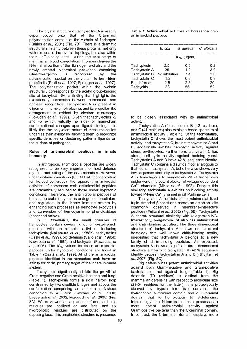
In T. tridentatus, the small granules of hemocytes contain several kinds of cysteine-rich peptides with antimicrobial activities, including tachyplesin (Nakamura et al., 1988b), tachystatins (Osaki et al., 1999), big defensin (Saito et al., 1995b; Kawabata et al., 1997), and tachycitin (Kawabata et al., 1996). The IC50 values for these antimicrobial peptides under hypotonic conditions are listed in Table 1 (Osaki et al., 1999). All of the antimicrobial peptides identified in the horseshoe crab have an affinity for chitin, primary target of the innate immune system.
Tachyplesin significantly inhibits the growth of Gram-negative and Gram-positive bacteria and fungi (Table 1). Tachyplesin forms a rigid hairpin loop constrained by two disulfide bridges and adopts the conformation comprising an antiparallel β-sheet connected to a β-turn (Kawano et al., 1990; Laederach et al., 2002; Mizuguchi et al., 2005) (Fig. 8A). When viewed as a planar surface, six basic residues are localized on one face, and six hydrophobic residues are distributed on the opposing face. This amphiphilic structure is presumed
Table 1 Antimicrobial activities of horseshoe crab antimicrobial peptides --------------------------------------------------------------------- E. coli S. aureus C. albicans --------------------------------------------------------------------- IC50 (μg/ml) Tachyplesin 2.5 0.3 0.2 Tachystatin A 25 4.2 3.0 Tachystatin B No inhibition 7.4 3.0 Tachystatin C 1.2 0.8 0.9 Big defensin 2.5 2.5 20 Tachycitin 33 56 52 --------------------------------------------------------------------- to be closely associated with its antimicrobial activity.
Tachystatins A (44 residues), B (42 residues), and C (41 residues) also exhibit a broad spectrum of antimicrobial activity (Table 1). Of the tachystatins, tachystatin C shows the most potent antimicrobial activity, and tachystatin C, but not tachystatins A and B, additionally exhibits hemolytic activity against sheep erythrocytes. Furthermore, tachystatin C has strong cell lysis activity against budding yeast. Tachystatins A and B have 42 % sequence identity. Tachystatin C contains a disulfide motif analogous to that found in tachystatin A, but otherwise shows very low sequence similarity to tachystatin A. Tachystatin A is homologous to ω-agatoxin-IVA of funnel web spider venom, a potent blocker of voltage-dependent Ca2+ channels (Mintz et al., 1992). Despite this similarity, tachystatin A exhibits no blocking activity toward P-type Ca2+ channel in rat Purkinje cells.
Tachystatin A consists of a cysteine-stabilized triple-stranded β-sheet and shows an amphiphilicity commonly observed in membrane-interactive peptides (Fujitani et al., 2002) (Fig. 8B). Tachystatin A shares structural similarity with ω-agatoxin-IVA. Interestingly, ω-agatoxin-IVA also has antimicrobial and chitin-binding activities. The three dimensional structure of tachystatin A shows no structural homology with well known chitin-binding motifs, suggesting that tachystatin A belongs to a new family of chitin-binding peptides. As expected, tachystatin B shows a significant three dimensional structural similarity to tachystatin A (42 % sequence identity between tachystatins A and B ) (Fujitani et al., 2007) (Fig. 8C).
Big defensin has potent antimicrobial activities against both Gram-negative and Gram-positive bacteria, but not against fungi (Table 1). Big defensin (79 residues) is distinct from the mammalian defensins with respect to molecular size (29-34 residues for the latter). It is proteolytically cleaved by trypsin into two domains, the hydrophobic N-terminal domain and a C-terminal domain that is homologous to β-defensins. Interestingly, the N-terminal domain possesses a more potent antimicrobial activity against Gram-positive bacteria than the C-terminal domain. In contrast, the C-terminal domain displays more

Fig. 8 NMR structures of antimicrobial peptides of the horseshoe crab. (A) Ball-and-stick model of tachyplesin. (B) Ribbon representation of the energy-minimized average structure of tachystatin A. (C) Ribbon representation of the energy-minimized average structure of tachystatin B. (D) Ribbon representation of the energy-minimized average structure of big defensin. (E) Ribbon representation of the energy-minimized average structure of tachycitin. potent antimicrobial activity than the N-terminal domain against Gram-negative bacteria. These data suggest a new sub-class within the defensin family that possesses two discrete functional domains with different antimicrobial activities. It is noteworthy that tachylectins-5A and -5B enhance the antimicrobial activity of big defensin against Gram-positive bacteria (Gokudan et al., 1999).
Structurally, big defensin represents a new class within the defensin family; the C-terminal domain adopts a β-defensin structure, whereas the N-terminal domain forms a unique globular conformation (Kouno et al., 2008) (Fig. 8D). Interestingly, the hydrophobic N-terminal domain, but not the C-terminal domain, undergoes a conformational change in micelle solution, which may be associated with the antimicrobial activity against Gram-positive bacteria.
69
Tachycitin (73 residues) contains five disulfide bridges and has no significant sequence similarity to known antimicrobial peptides. The antimicrobial activity of tachycitin is not strong by itself (Table 1). However, tachycitin synergistically enhances the antimicrobial activity of big defensin as demonstrated by a 50-fold reduction in the IC50 value of big defensin against Gram-negative bacteria when a small amount of tachycitin is present.
The structure of tachycitin is largely divided into the N-terminal domain and the C-terminal domain (Suetake et al., 2000) (Fig. 8E). In the C-terminal domain, tachycitin forms a hairpin loop connecting a two-stranded β-sheet, a structural motif shared by the chitin-binding site of hevein, an antifungal peptide from the rubber tree Hevea brasiliensis (Broekaert et al., 1990). In hevein, the aromatic side chains of the two Trp residues in this loop directly interact with chitin-derived oligosaccharides. The side chain of Tyr and Val residues in the corresponding loop of tachycitin are structurally analogous to these two Trp residues in hevein. Tachyplesin also contains the similar hairpin loop that connects a two-stranded β-sheet. The hydrophobic residues clustered on the one face of their β-hairpin loops possibly participate in chitin-binding sites.
Roles of TGase-dependent protein cross-linking in the innate immune system
In mammals, the blood coagulation cascade
culminates in the proteolytic conversion of soluble fibrinogen into insoluble fibrin. The resulting fibrin fibrils are further stabilized by intermolecular ε-(γ-glutamyl) lysine cross-linking by the plasma
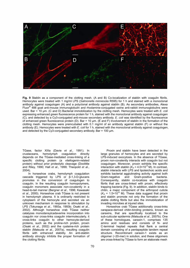
Fig. 9 Stablin as a component of the clotting mesh. (A and B) Co-localization of stablin with coagulin fibrils. Hemocytes were treated with 1 mg/ml LPS (Salmonella minnesota R595) for 1 h and stained with a monoclonal antibody against coagulogen (A) and a polyclonal antibody against stablin (B). As secondary antibodies, Alexa Fluor® 488 goat anti-mouse immunoglobulin and rhodamine-conjugated swine anti-rabbit immunoglobulins were used. Bar = 10 μm. (C and D) Bacterial immobilization by the clotting mesh. Hemocytes were treated with E. coli expressing enhanced green fluorescence protein for 1 h, stained with the monoclonal antibody against coagulogen (C), and detected by a Cy3-conjugated anti-mouse secondary antibody. E. coli was identified by the fluorescence of enhanced green fluorescence protein (D). Bar = 10 μm. (E and F) Involvement of stablin in the formation of the clotting mesh. Hemocytes were preincubated with 0.1 mg/ml of an antibody against stablin (F) or without the antibody (E). Hemocytes were treated with E. coli for 1 h, stained with the monoclonal antibody against coagulogen, and detected by the Cy3-conjugated secondary antibody. Bar = 100 μm. TGase, factor XIIIa (Davie et al., 1991). In crustaceans, hemolymph coagulation directly depends on the TGase-mediated cross-linking of a specific clotting protein (a vitellogenin-related protein) without prior proteolytic cleavage (Doolittle and Riley, 1990; Hall et al., 1999; Theopold et al., 2004).
In horseshoe crabs, hemolymph coagulation cascade triggered by LPS or β-1,3-D-glucans promotes in the conversion of coagulogen to coagulin. In the resulting coagulin homopolymers, coagulin monomers associate non-covalently in a head-to-tail manner (Bergner et al., 1996; Kawasaki et al., 2000). Horseshoe crab TGase is not present in hemolymph plasma. It is ordinarily restricted to cytoplasm of the hemocyte and secreted via an unknown mechanism in response to stimulation by LPS (Tokunaga et al., 1993a, 1993b; Osaki et al., 2002). Although horseshoe crab TGase neither catalyzes monodansylcadaverine incorporation into coagulin nor cross-links coagulin intermolecularly, it cross-links coagulin to other hemocyte-derived proteins, such as the proline-rich protein, proxin (Osaki et al., 2002) and the cysteine-rich protein stablin (Matsuda et al., 2007a), resulting coagulin fibrils with enhanced stability. An anti-stablin antibody strongly inhibits the proper formation of the clotting fibrils.
Proxin and stablin have been detected in the large granules of hemocytes and are secreted by LPS-induced exocytosis. In the absence of TGase, proxin non-covalently interacts with coagulin but not coagulogen. Moreover, proxin exhibits the specific interaction with stablin (Kd = 4.0×10⁻⁹ M). In contrast, stablin interacts with LPS and lipoteichoic acids and exhibits bacterial agglutinating activity against both Gram-negative and Gram-positive bacteria. Consequently, stablin co-localizes with coagulin fibrils that are cross-linked with proxin, effectively trapping bacteria (Fig. 9). In addition, stablin binds to chitin, a major component of the arthropod cuticle (Kd = 1.5×10⁻⁸ M). These data suggest that proxin and stablin promote not only the formation of the stable clotting fibrils but also the immobilization of invading microbes at injured sites.
Horseshoe crab TGase additionally cross-links carapace-derived chitin-binding proteins, known as caraxins, that are specifically localized to the sub-cuticular epidermis (Matsuda et al., 2007b). One of these homologues, caraxin-1, comprises 135 amino acid residues and consists of N- and C-terminal heptad repeats that flank a central domain consisting of a pentapeptide tandem repeat structure. Recombinant caraxin-1 exists as an oligomer (~20-mer) in solution, and these oligomers are cross-linked by TGase to form an elaborate mesh
70
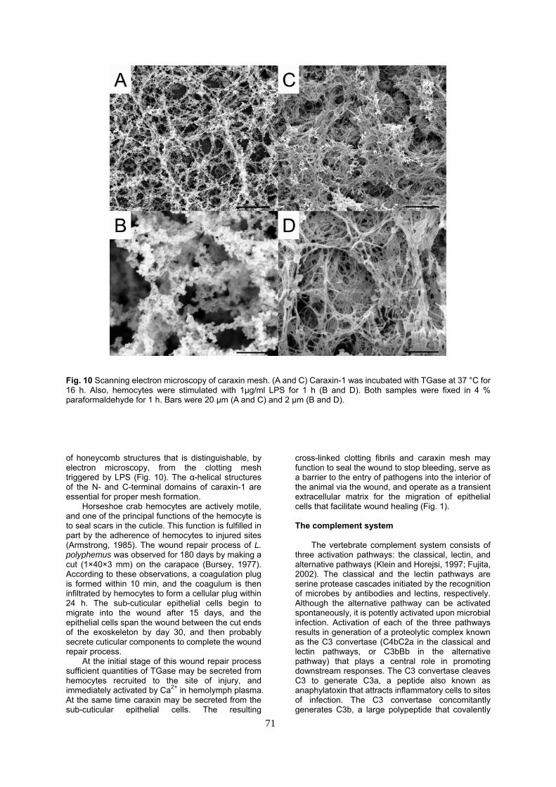
Fig. 10 Scanning electron microscopy of caraxin mesh. (A and C) Caraxin-1 was incubated with TGase at 37 °C for 16 h. Also, hemocytes were stimulated with 1μg/ml LPS for 1 h (B and D). Both samples were fixed in 4 % paraformaldehyde for 1 h. Bars were 20 μm (A and C) and 2 μm (B and D). of honeycomb structures that is distinguishable, by electron microscopy, from the clotting mesh triggered by LPS (Fig. 10). The α-helical structures of the N- and C-terminal domains of caraxin-1 are essential for proper mesh formation.
Horseshoe crab hemocytes are actively motile, and one of the principal functions of the hemocyte is to seal scars in the cuticle. This function is fulfilled in part by the adherence of hemocytes to injured sites (Armstrong, 1985). The wound repair process of L. polyphemus was observed for 180 days by making a cut (1×40×3 mm) on the carapace (Bursey, 1977). According to these observations, a coagulation plug is formed within 10 min, and the coagulum is then infiltrated by hemocytes to form a cellular plug within 24 h. The sub-cuticular epithelial cells begin to migrate into the wound after 15 days, and the epithelial cells span the wound between the cut ends of the exoskeleton by day 30, and then probably secrete cuticular components to complete the wound repair process.
At the initial stage of this wound repair process sufficient quantities of TGase may be secreted from hemocytes recruited to the site of injury, and immediately activated by Ca2+ in hemolymph plasma. At the same time caraxin may be secreted from the sub-cuticular epithelial cells. The resulting
cross-linked clotting fibrils and caraxin mesh may function to seal the wound to stop bleeding, serve as a barrier to the entry of pathogens into the interior of the animal via the wound, and operate as a transient extracellular matrix for the migration of epithelial cells that facilitate wound healing (Fig. 1).
The complement system
The vertebrate complement system consists of
three activation pathways: the classical, lectin, and alternative pathways (Klein and Horejsi, 1997; Fujita, 2002). The classical and the lectin pathways are serine protease cascades initiated by the recognition of microbes by antibodies and lectins, respectively. Although the alternative pathway can be activated spontaneously, it is potently activated upon microbial infection. Activation of each of the three pathways results in generation of a proteolytic complex known as the C3 convertase (C4bC2a in the classical and lectin pathways, or C3bBb in the alternative pathway) that plays a central role in promoting downstream responses. The C3 convertase cleaves C3 to generate C3a, a peptide also known as anaphylatoxin that attracts inflammatory cells to sites of infection. The C3 convertase concomitantly generates C3b, a large polypeptide that covalently
71
72
attaches to microbial surfaces and promotes C3 receptor-dependent phagocytosis by leukocytes as well as the formation of the membrane attack complex.
Vertebrate complement factors D (Df) and B (Bf) are involved in the activation of the alternative pathway. Df is a single-chain serine protease circulating in the blood as an active protease that cleaves Bf, resulting in the formation of C3bBb. Although several homologs of vertebrate complement factors are known to be present in ascidians, cyclostomes, and echinoderms, the functional analog of Df that triggers the alternative pathway has not been identified in protostomes (Endo et al., 2006; Dodds and Matsushita, 2007).
The complement-related protein, α2-macroglobulin, has previously been identified in horseshoe crabs (Iwaki et al., 1996). Recently, a homolog of complement component C3 has been identified in the horseshoe crab C. rotundicauda (CrC3), indicating the presence of a complement system capable of promoting the phagocytosis of invading microbes in protostomes (Zhu et al., 2005). In a phylogenetic tree of reactive thioester-containing proteins from vertebrates and invertebrates, CrC3 shows the highest similarity to C3 sequences from lower deuterostomes and forms a clade with amphioxus C3, cnidaria C3-like protein, and sea urchin C3. However, despite the identification of the key complement component in protostomes, the molecular mechanism underlying C3 activation has remained unknown.
In an effort to elucidate the mechanism of C3 activation in horseshoe crabs, we have isolated and characterized an ortholog of C3 (TtC3) from T. tridentatus (Ariki et al., 2008). TtC3 consists of 1,716 residues with an overall domain structure that is identical to vertebrate C3, including α2-macroglobulin domains, complement-urchin-bone (CUB) domains, a thioester-containing domain, an anaphylatoxin domain, and a C345C domain (Fig. 4D). A thioester motif (-Cys-Gly-Glu-Gln-) at residues 1004 to 1007 and a catalytic His at the position 1116 are conserved in the deduced sequence. The sequence identity between TtC3 and CrC3 (98 %) is considerably higher than that between coagulogens from the two species (90 %) (Srimal et al., 1985). TtC3 forms a disulfide-linked three-chain structure (α-, β-, and γ-chains) in hemolymph plasma, unlike vertebrate C3 which is present in a two-chain form.
The horseshoe crab complement system promotes the deposition of TtC3b on the surface of Gram-negative or Gram-positive bacteria in hemolymph plasma. However, evaluation of the ability of PAMPs to promote the proteolytic conversion of TtC3 to TtC3b revealed that LPS, but not zymosan, peptidoglycan, or laminarin, strongly induces this conversion, highlighting the selective response of the complement system to LPS stimulation. Factor C stored in hemocytes has been identified as an LPS sensor and an initiator of hemolymph coagulation in the horseshoe crab innate immune system. As would be expected from these findings, an anti-factor C antibody inhibits both the proteolytic conversion of TtC3 and the deposition of TtC3b on the surface of Gram-negative bacteria in
hemolymph plasma. Moreover, activated factor C present on the surface of Gram-negative bacteria directly catalyzes the proteolytic conversion of the purified TtC3, thereby promoting TtC3 deposition. These data indicate that factor C acts as an LPS-sensitive C3 convertase on the surface of invading Gram-negative bacteria in the initial phase of horseshoe crab complement activation (Fig. 1).
In the alternative pathway of the vertebrate complement system, the interaction between C3b and Bb is essential to form C3 convertase (C3bBb). A homolog of complement Bf has been identified in C. rotundicauda (Zhu et al., 2005). Although physiological function of this Bf homolog in the horseshoe crab complement system remains unknown, it is likely that it may be responsible for the formation of the second C3 convertase.
TtC3 binds to factor C at Kd = 4.9×10⁻⁸M (Ariki et al., 2008). TtC3 is present at a concentration of at least 300 μg/ml in hemolymph plasma, whereas the amount of factor C in hemolymph plasma is very low (~10 μg/ml). The relatively high concentration of TtC3 and its high affinity to factor C suggest that factor C exists in a complex with TtC3 in hemolymph plasma, and that formation of this complex is a prerequisite for the immediate activation of TtC3 by factor C on the surface of Gram-negative bacteria. Factor C antigen has been shown to be present all the tissues examined by Western blotting. In contrast, factor G and the proclotting enzyme are not detectable in hemolymph plasma.
In contrast to these findings, the anti-factor C antibody exhibits no effect on the deposition of TtC3b on Staphylococcus aureus, suggesting the presence of a factor C-independent pathway to initiate the opsonization of Gram-positive bacteria (Ariki et al., 2008). PAMPs or complement factors in hemolymph plasma required for the deposition of TtC3b on Gram-positive bacteria remain to be examined.
Factor C and complement Bf from C. rotundicauda interact with plasma-derived lectins, including galactose-binding protein, carcinolectin-5, and C-reactive protein (Saux et al., 2008). Also in T. tridentatus, hemocytes and hemolymph plasma contain several lectins responsible for microbial agglutination (Kawabata and Tsuda, 2002). Therefore, the protease-lectin complexes on the surface of Gram-positive bacteria may enhance the deposition of TtC3b. Although the complement-dependent clearance system of invading pathogens in horseshoe crabs remains to be examined, phagocytosis of Gram-positive bacteria by hemocytes both in vivo and in vitro is inhibited by protease inhibitors, raising the possibility that the proteolytic dependence of opsonization by C3b may underlie phagocytosis by hemocytes (Zhu et al., 2005).
Roles of hemocyanin in the innate immune system
In crustaceans and insects, the
prophenoloxidase activation system is an important part of the innate immunity, where it acts to detect and kill invading pathogens as well as to synthesize melanin for wound healing and encapsulation of
73
pathogens (Cerenius and Söderhäll, 2004; Nappi et al., 2004). Arthropod prophenoloxidases are known to be non-enzymatically activated by treatment with detergents, lipids, or organic solvents (Ashida and Yamasaki, 1990). In the horseshoe crab as well, the induction of phenoloxidase activity in hemolymph plasma is evident upon similar treatment (Nellaiappan and Sugumaran, 1996), yet prophenoloxidase has not been identified in horseshoe crabs. Arthropod prophenoloxidases have been sequenced, and show significant homology to hemocyanins (Aspan et al., 1995; Fujimoto et al., 1995; Kawabata et al., 1995).
Both prophenoloxidase and hemocyanin contain two functional copper-binding sites capable of reversibly binding an oxygen molecule (Solomon et al., 1996). Moreover, the origin of arthropod hemocyanins appears to be an ancient prophenoloxidase-like protein (Burmester and Scheller, 1996). Under physiological conditions, arthropod prophenoloxidases require a proteolytic cleavage for activation by a specific protease (Aspan et al., 1995). Interestingly, tarantula hemocyanin is found to express phenoloxidase activity after limited proteolysis with trypsin or chymotrypsin (Decker and Rimke, 1998).
In horseshoe crabs, the clotting enzyme or activated coagulation factor B efficiently produces phenoloxidase activity in hemolymph plasma, whereas activated factor C, activated factor G, or trypsin do not (Nagai et al., 2000). The phenoloxidase activity in hemolymph plasma disappears upon removal of hemocyanin by ultracentrifugation, suggesting that hemocyanin is involved in the prophenoloxidase activation system in horseshoe crabs. Horseshoe crab hemocyanin is composed of six subunits (α, β, γ, δ, ε, and ζ), each of which have a molecular mass of 70 kDa and, in their purified forms, independently express phenoloxidase activity. The clotting enzyme converts the α-subunit of hemocyanin to phenoloxidase in a dose-dependent manner, and the resulting phenoloxidase activity reaches a plateau at a 1:1 molar ratio. The proteolytic cleavage of the hemocyanin subunit is not required for the functional conversion, and the zymogen forms of the coagulation proteases, the proclotting enzyme and coagulation factor B, are effective activators.
A common structural feature of these two coagulation factors is the presence of an N-terminal clip domain (Muta et al., 1990, 1993). Homologous clip domains are present in the N-terminal regions of insect prophenoloxidase-activating enzymes (Jiang et al., 1998; Lee et al., 1998; Satoh et al., 1999). Therefore, the clip domain of the proclotting enzyme or coagulation factor B may promote the interaction of these factors with hemocyanin effect its functional conversion to an active phenoloxidase.
Horseshoe crab hemocyanin is also converted to phenoloxidase by treatment with amphiphilic substances such as SDS and phosphatidylethanolamine (Nagai and Kawabata, 2000). Consistent with the amphiphilic nature of the antimicrobial peptides secreted from the horseshoe crab hemocyte, tachyplesin interacts with the α-subunit of hemocyanin (Kd = 3.4×10⁻⁶ M) and induces its intrinsic phenoloxidase activity. The
phenoloxidase activity induced by tachyplesin is inhibited by phenylthiourea, a typical inhibitor of phenoloxidase.
Although tachyplesin is the most effective activator of hemocyanin, other hemocyte-derived antimicrobial peptides such as tachystatins and tachycitin significantly induce its phenoloxidase activity. Mutation of tachyplesin at Trp2 or Tyr8 and Try13 on its hydrophobic face, but not mutation of basic residues on its cationic face, significantly impair its interaction with the α-subunit, implicating the hydrophobic face of tachyplesin in the functional conversion of hemocyanin to a phenoloxidase.
Horseshoe crab antimicrobial peptides are all capable of binding to chitin, a cell wall component of fungi and also the major structural component of the arthropod cuticle. The antimicrobial peptides likely recognize chitin exposed at sites of injury as well as on the surface of invading microbes. In addition, hemocyanin binds to tachyplesin-coated chitin. Although horseshoe crab hemocyanin present at high concentration in hemolymph plasma (~70 mg/ml) and acts as an oxygen carrier under normal physiological conditions, it may be selectively converted to phenoloxidase by the antimicrobial peptides. We hypothesize that the chitin coated with antimicrobial peptides may serve as a scaffold for the binding of hemocyanin and that the resulting phenoloxidase activity acts as trigger for wound healing in the cuticle (Fig. 1).
In the crayfish Pacifastacus leniusculus, an antibacterial peptide astacidin 1 has been identified (Lee et al., 2003). Interestingly, astacidin 1 is released from the C-terminal part of hemocyanin by a cysteine-like protease and is up-regulated by injection of LPS or β-1,3-D-glucans, indicating that hemocyanin acts as a multifunctional protein in the innate immune system. Recently, horseshoe crab hemocyanin and human hemoglobin have been found to be activated by microbial proteases and to be enhanced by LPS, resulting in the production of reactive oxygen species as an antimicrobial strategy (Jiang et al., 2007).
Acknowledgements
We thank Dr J Kulman (Puget Sound Blood Center, Seattle, USA) for helpful discussion and suggestions on this manuscript.
References Aketagawa J, Miyata T, Ohtsubo S, Nakamura T,
Morita T, Hayashida H, et al. Primary structure of Limulus anticoagulant anti-lipopolysaccharide factor. J. Biol. Chem. 261: 7357-7365, 1986.
Akira S, Uematsu S, Takeuchi O. Pathogen recognition and innate immunity. Cell 124: 783-801, 2006.
Ariki S, Koori K, Osaki T, Motoyama K, Inamori K, Kawabata S. A serine protease zymogen functions as a pattern-recognition receptor for lipopolysaccharides. Proc. Natl. Acad. Sci. USA 101: 953-958, 2004.
Ariki S, Takahara S, Shibata T, Fukuoka T, Ozaki A, Endo Y, Fujita T, Koshiba T, Kawabata S. Factor C acts as a lipopolysaccharide-responsive C3 convertase in horseshoe crab complement activation. J. Immunol. 181: 7794-8001, 2008.
74
Armstrong, PB. Adhesion and motility of the blood cells of Limulus. In: Cohen WD (ed), Blood cells of marine invertebrates: experimental systems in cell biology and comparative physiology, Alan R. Liss, Inc., New York, USA, pp 77-124, 1985.
Ashida M, Yamazaki, H. Biochemistry of the phenoloxidase system in insects: with special reference to its activation. In: Ohnishi E, Ishizaki H (eds), Molting and metamorphosis, Springer-Verlag, Berlin, Germany, pp 239-265,1990.
Aspan A, Huang TS, Cerenius L, Söderhäll K. cDNA cloning of prophenoloxidase from the freshwater crayfish Pacifastacus leniusculus and its activation. Proc. Natl. Acad. Sci. USA 92: 939-943, 1995.
Beisel H-G, Kawabata S, Iwanaga S, Huber R, Bode W. Tachylectin-2: crystal structure of a specific GlcNAc/GalNAc-binding lectin involved in the innate immunity host defense of the Japanese horseshoe crab Tachypleus tridentatus. EMBO J. 18: 2312-2322, 1999.
Belvin MP, Anderson KV. A conserved signaling pathway-the Drosophila toll-dorsal pathway. Annu. Rev. Cell Dev. Biol. 12: 393-416, 1996.
Bergner A, Muta T, Iwanaga S, Beisel H-G, DeLotto R, Bode W. Horseshoe crab coagulation is an invertebrate protein with a nerve growth factor-like domain. Biol. Chem. 378: 283-287, 1997.
Bergner A, Oganessyan V, Muta T, Iwanaga S, Typke D, Huber R, et al. Crystal structure of coagulogen, the clotting protein form horseshoe crab: a structural homologue of nerve growth factor. EMBO J. 15: 6789-6797, 1996.
Bohn JA, BeMiller JN. (l→3)-β-D-glucans as biological response modifiers: a review of structure-functional activity relationships. Carbohydr. Poym. 28: 3-14, 1995.
Broekaert W, Lee HI, Kush A, Chua NH, Raikhel N. Wound-induced accumulation of mRNA containing a hevein sequence in laticifers of rubber tree (Hevea brasiliensis). Proc. Natl. Acad. Sci. USA 87: 7633-7637, 1990.
Brown GD. Dectin-1: a signaling non-TLR pattern-recognition receptor. Nat. Rev. Immunol. 6: 33-43, 2006.
Brown J, O’callaghan CA, Marshall ASJ, Gilbert RJC, Siebold C, Gordon S, et al. Structure of the fungal β-glucan-binding immune receptor dectin-1: Implications for function. Protein Sci. 16: 1042-1052, 2007.
Burmester T, Scheller K. Common origin of arthropod tyrosinase, arthropod hemocyanin, insect hexamerin, and dipteran arylphorin receptor. J. Mol. Evol. 42: 713-728, 1996.
Bursey CR. Histological response to injury in the horseshoe crab, Limulus polyphemus. Can. J. Zool. 55: 1158-1165, 1977.
Cerenius L, Söderhäll K. The prophenoloxidase-activating system in invertebrates. Immunol. Rev. 198: 116-126, 2004.
Chen SC, Yen CH, Yeh MS, Huang CJ, Liu TY. Biochemical properties and cDNA cloning of two new lectins from the plasma of Tachypleus tridentatus-Tachypleus plasma lectin 1 and 2. J.
Biol. Chem. 276: 9631-9639, 2001. Chiou ST, Chen YW, Chen SC, Chao CF, Liu TY.
Isolation and characterization of proteins that bind to galactose, lipopolysaccharide of Escherichia coli, and protein A of Staphylococcus aureus from the hemolymph of Tachypleus tridentatus. J. Biol. Chem. 275: 1630-1634, 2000.
Davie EW, Fujikawa K, Kisiel W. The coagulation cascade: initiation, maintenance, and regulation. Biochemistry 30: 10363-10370, 1991.
Decker H, Rimke T. Tarantula hemocyanin shows phenoloxidase activity. J. Biol. Chem. 273: 25889-25892, 1998.
Dodds AW, Matsushita M. The phylogeny of the complement system and the origins of the classical pathway. Immunobiol. 212: 233-243, 2007.
Doolittle RF, Riley M. The amino-terminal sequence of lobster fibrinogen reveals common ancestry with vitellogenins. Biochem. Biophys. Res. Commun. 167: 16-19, 1990.
Endo Y, Takahashi M, Fujita T. Lectin complement system and pattern recognition. Immunobiol. 211: 283-293, 2006.
Ferrandon D, Imler J-L, Hetru C, Hoffmann, JA. The Drosophila systemic immune response: sensing and signaling during bacterial and fungal infections. Nat. Rev. Immunol. 7: 862-874, 2007.
Fontes CMGA, Clarke JH, Hazlewood GP, Fernandes TH, Gilbert HJ, Ferreira LMA. Identification of tandemly repeated type VI cellulose-binding modules in an endoglucanase from the aerobic soil bacterium Cellvibrio mixtus. Appl. Microbiol. Biotechnol. 49: 552-559, 1998.
Fujita T. Evolution of the lectin-complement pathway and its role in innate immunity. Nat. Rev. Immunol. 2: 346-353, 2002.
Fujimoto K, Okino N, Kawabata S, Iwanaga S, Ohnishi E. Nucleotide sequence of the cDNA encoding the proenzyme of phenol oxidase A1 of Drosophila melanogaster. Proc. Natl. Acad. Sci. USA 92: 7769-7773, 1995.
Fujitani N, Kawabata S, Osaki T, Kumaki Y, Demura M, Nitta K, et al. Structure of the antimicrobial peptide tachystatin A. J. Biol. Chem. 277: 23651-23657, 2002.
Fujitani N, Kouno T, Nakahara T, Takaya K., Osaki T, Kawabata S, et al. The solution structure of horseshoe crab antimicrobial peptide tachystatin B with an inhibitory cystine-knot motif. J. Pep. Sci. 13: 269-279, 2007.
Gabius HJ, Andre S, Kaltner H, Siebert H-C. The sugar code: functional lectinomics. Biochim. Biophys. Acta 1572: 165-177, 2002.
Gokudan S, Muta T, Tsuda R, Koori K, Kawahara T, Seki N, et al. Horseshoe crab acetyl group-recognizing lectins involved in innate immunity are structurally related to fibrinogen. Proc. Natl. Acad. Sci. USA 96: 10086-10091, 1999.
Hall M, Wang R, van Antwerpen R, Sottrup-Jensen L, Söderhäll K. The crayfish plasma clotting protein: a vitellogenin-related protein responsible for clot formation in crustacean blood. Proc. Natl. Acad. Sci. USA 96: 1965-1970, 1999.
75
Harrington JM, Chou H-T, Gutsmann T, Gelhaus C, Stahlberg H, Leippe M, et al. Membrane activity of a C-reactive protein. FEBS Lett. 583: 1001-1005, 2009.
Higashijima T, Uzu S, Nakajima T, Ross EM. Mastoparan, a peptide toxin from wasp venom, mimics receptors by activating GTP-binding regulatory proteins (G proteins). J. Biol. Chem. 263: 6491-6494, 1988.
Hoess A, Watson S, Siber GR, Liddington R. Crystal structure of an endotoxin-neutralizing protein from the horseshoe crab, Limulus anti-LPS factor, at 1.5 Å resolution. EMBO J. 12: 3351-3356, 1993.
Hoffmann, JA. The immune response of Drosophila. Nature 426: 33-38, 2003.
Inamori K, Ariki S, Kawabata, S. A toll-like receptor in horseshoe crabs. Immunol. Rev. 198: 106-115, 2004.
Inamori K, Koori, K, Mishima C, Muta T, Kawabata, S. A horseshoe crab receptor structurally related to Drosophila Toll. J. Endotoxin Res. 6: 397-399, 2000.
Inamori K, Saito T, Iwaki D, Nagira T, Iwanaga S, Arisaka S, et al. A newly identified horseshoe crab lectin with specificity for blood group A antigen recognizes specific O-antigens of bacterial lipopolysaccharides. J. Biol. Chem. 274: 3272-3278, 1999.
Iwaki D, Kawabata S, Miura Y, Kato A, Armstrong PB, Quigley JP, et al. Molecular cloning of Limulus α2-macroglobulin. Eur. J. Biochem. 242: 822-831, 1996.
Iwaki D, Osaki T, Yoshimitsu M, Wai, SN, Iwanaga S, Kawabata, S. Functional and structural diversities of C-reactive proteins present in horseshoe crab hemolymph plasma. Eur. J. Biochem. 264: 314-326, 1999.
Iwanaga S. The molecular basis of innate immunity in the horseshoe crab. Curr. Opin. Immunol. 14: 87-95, 2002.
Iwanaga S, Kawabata S, Muta T. New type of clotting factors and defense molecules found in horseshoe crab hemolymph: their structures and functions. J. Biochem. 123: 1-15, 1998.
Iwanaga S, Lee BL. Recent advances in the innate immunity of invertebrate animals. J. Biochem. Mol. Biol. 38: 128-150, 2005.
Janeway CA Jr. Approaching the asymptote? Evolution and revolution in immunology. Cold Spring Harbor Symp. Quant. Biol. 54: 1-13, 1989.
Jiang H, Wang Y, Kanost MR. Pro-phenol oxidase activating proteinase from an insect, Manduca sexta: A bacteria-inducible protein similar to Drosophila easer. Proc. Natl. Acad. Sci. USA 95: 12220-12225, 1998.
Jiang N, Tan NS, Ho B, Ding JL. Respiratory protein-generated reactive oxygen species as an antimicrobial strategy. Nat. Immunol. 8: 1114-1122, 2007.
Joslin G, Fallon RJ, Bullock J, Adams SP, Perlmutter DH. The SEC receptor recognizes a pentapeptide neodomain of alpha 1-antitrypsin-proteiase complex. J. Biol. Chem. 266: 11282-11288, 1991.
Kairies N, Beisel H-G, Fuentes-Prior P, Tsuda R,
Muta T, Iwanaga S, et al. The 2.0-Å crystal structure of tachylectin 5 provides evidence for the common origin of the innate immunity and the blood coagulation systems. Proc. Natl. Acad. Sci. USA 98: 13519-13524, 2001.
Kanost MR, Jiang H, Yu W-Q. Innate immune response of a lepidopteran insect, Manduca sexta. Immunol. Rev. 198: 97-105, 2004.
Kawabata S, Nagayama R, Hirata M, Shigenaga T, Lal Agarwala K, Saito T, et al. Tachycitin, a small granular component in horseshoe crab hemocytes, is an antimicrobial protein with chitin-binding activity. J. Biochem. 120: 1253-1260, 1996.
Kawabata S, Osaki T, Iwanaga S. Innate immunity in the horseshoe crab. In: Innate Immunity. Ezekowitz RAB, Hoffmann JA (eds), Humana Press, Totowa, USA, pp 109-125, 2003.
Kawabata S, Tsuda R. Molecular basis of non-self recognition by the horseshoe crab tachylectins. Biochim. Biophys. Acta 1572: 414-421, 2002.
Kawabata S, Saito T, Saeki K, Okino N, Mizutani A, Toh Y, et al. cDNA cloning, tissue distribution, and subcellular localization of horseshoe crab big defensin. Biol. Chem. 378: 289-292, 1997.
Kawabata T, Yasuhara Y, Ochiai M, Matsuura S, Ashida M. Molecular cloning of insect pro-phenol oxidase: a copper-containing protein homologous to arthropod hemocyanin. Proc. Natl. Acad. Sci. USA 92: 7774-7778, 1995.
Kawano K, Yoneya T, Miyata T, Yoshikawa K, Tokunaga F, Terada Y, et al. Antimicrobial peptide, tachyplesin I, isolated from hemocytes of the horseshoe crab (Tachypleus tridentatus). J. Biol. Chem. 265: 15365-15367, 1990.
Kawasaki H, Nose T, Muta, T, Iwanaga, S, Shimohigashi, Y, Kawabata S. Head-to-tail polymerization of coagulogen, a clottable protein of the horseshoe crab. J. Biol. Chem. 275: 35297-35301, 2000.
Klein J, Horejsi V. Complement and complement receptors. In: Immunology. Klein J, Horejsi V (eds), Blackwell Science, Oxford, UK, pp 348-392, 1997.
Koshiba T, Hashii T, Kawabata S. A structural perspective on the interaction between lipopolysaccharide and factor C, a receptor involved in recognition of gram-negative bacteria. J. Biol. Chem. 282: 3962-3967, 2007.
Kouno T, Fujitani N, Mizuguchi M, Osaki T, Nishimura S, Kawabata S, et al. A novel β-defensin structure: a potential strategy of big defensin for overcoming resistance by Gram-positive bacteria. Biochemistry 47: 10611-10619, 2008.
Krem M. M., Di Cera E. Evolution of enzyme cascades from embryonic development to blood coagulation. Trends Biochem. Sci. 27: 67-74, 2002.
Kurata S, Ariki S, Kawabata, S. Recognition of pathogens and activation of immune response in Drosophila and horseshoe crab innate immunity. Immunobiol. 211: 237-249, 2006.
Laederach A, Andreotti AH, Fulton DB. Solution and micelle-bound structures of tachyplesin I and its active aromatic linear derivatives. Biochemistry 41: 12359-12368, 2002.
76
Lal Agarwala K, Kawabata S, Miura Y, Kuroki Y, Iwanaga S. Limulus intracellular coagulation inhibitor type 3. Purification, characterization, cDNA cloning, and tissue localization. J. Biol. Chem. 271: 23768-23774, 1996.
Lee S-Y, Cho MY, Hyun JH, Lee KM, Homma K, Natori S, et al. Molecular cloning of cDNA for pro-phenol-oxidase-activating factor I, a serine protease is induced by lipopolysaccharide or 1,3-β-glucan in coleopteran insect, Holotrichia diomphalia larvae. Eur. J. Biochem. 257: 615-621, 1988.
Lee SY, Lee BL, Söderhäll K. Processing of an antibacterial peptide from hemocyanin of the freshwater crayfish Pacifastacus leniusculus. J. Biol. Chem. 278: 7927-7933, 2003.
Marra MN, Wilde CG, Griffith JE, Snable JL, Scott RW. Bactericidal/permeability-increasing protein has endotoxin-neutralizing activity. J. Immunol. 144: 662-666, 1990.
Matsuda Y, Osaki T, Hashii T, Koshiba T, Kawabata S. A cysteine-rich protein from an arthropod stabilizes clotting mesh and immobilizes bacteria at injured sites. J. Biol. Chem. 282: 33545-33552, 2007a.
Matusda Y, Koshiba T, Osaki T, Suyama H, Arisaka F, Toh Y, et al. An arthropod cuticular chitin-binding protein endows injured sites with transglutaminase-dependent mesh. J. Biol. Chem. 282: 37316-37324, 2007b.
Matsushita M, Fujita T. Ficolins and the lectin complement pathway. Immunol. Rev. 180: 78-85, 2001.
Mintz IM, Venema VJ, Swiderek KM, Lee TD, Bean BP, Adam ME. P-type calcium channels blocked by the spider toxin ω-Aga IVA. Nature 355: 827-829, 1992.
Miura Y, Kawabata S, Iwanaga S. A limulus intracellular coagulation inhibitor with characteristics of the serpin superfamily, purification, characterization, and cDNA cloning. J. Biol. Chem. 269: 542-547, 1994.
Miura Y, Kawabata S, Wakamiya Y, Nakamura T, Iwanaga S. A limulus intracellular coagulation inhibitor type 2. Purification, characterization, cDNA cloning, and tissue localization. J. Biol. Chem. 270: 558-565, 1995.
Mizuguchi M, Kamata S, Kawabata S, Fujitani N, Kawano K. In Protein Data Bank (PDB ID 1wo0), RCSB Protein Data Bank (2005).
Muta T, Hashimoto R, Miyata T, Nishimura H, Toh Y, Iwanaga S. Proclotting enzyme from horseshoe crab hemocytes. J. Biol. Chem. 265: 22426-22433, 1990.
Muta T, Miyata T, Misumi Y, Tokunaga F, Nakamura, T, Toh Y, et al. Limulus factor C: an endotoxin-sensitive serine protease zymogen with a mosaic structure of complement-like, epidermal growth factor-like, and lectin-like domains. J. Biol. Chem. 266: 6554-6561, 1991.
Muta T, Oda T, Iwanaga S. Horseshoe crab coagulation factor B. J. Biol. Chem. 268: 21384-21388, 1993.
Nagai T, Kawabata S. A link between blood coagulation and prophenoloxidase activation in arthropod host defense. J. Biol. Chem. 275: 29264-29267, 2000.
Nagai T, Kawabata S, Shishikura F, Sugita H. Purification, characterization, and amino acid sequence of an embryonic lectin in perivitelline fluid of the horseshoe crab. J. Biol. Chem. 274: 37673-37678, 1999.
Nagai T, Osaki T, Kawabata S. Functional conversion of hemocyanin to phenoloxidase by horseshoe crab antimicrobial peptides. J. Biol. Chem. 276: 27166-27170, 2001.
Nakamura T, Tokunaga F, Morita T, Iwanaga S, Kusumoto, S, Shiba T, et al. Intracellular serine-protease zymogen, factor C, from horseshoe crab hemocytes: its activation by synthetic lipid A analogues and acidic phospholipids. Eur. J. Biochem. 176: 89-94, 1988a.
Nakamura T, Furunaka H, Miyata T, Tokunaga, F, Muta T, Iwanaga S, et al. Tachyplesin, a class of antimicrobial peptide from the hemocytes of the horseshoe crab (Tachypleus tridentatus). J. Biol. Chem. 263: 16709-16713, 1988b.
Nakanishi Y, Shiratsuchi A. Mechanisms and roles of phagocytosis in Drosophila and Caenorhabditis elegans. Inv. Surv. J. 3: 89-96, 2006.
Nappi A, Kohler L, Mastore M. Signaling pathways implicated in the cellular innate immune responses of Drosophila. Inv. Surv. J. 1: 5-33, 2004.
Neer EJ, Schmidt CJ, Nambudripad R, Smith TF. The ancient regulatory-protein family of WD-repeat proteins. Nature 371: 297-300, 1994.
Nellaiappan K, Sugumaran M. On the presence of prophenoloxidase in the hemolymph of the horseshoe crab, Limulus. Comp. Biochem. Physiol. 113B: 163-168, 1996.
Ng PML, Jin ZX, Tan SSH, Ho B, Ding JL. C-reactive protein: a predominant LPS-binding acute phase protein responsive to Pseudomonas infection. J. Endotoxin Res. 10: 163-174, 2004.
Okino N, Kawabata S, Saito T, Hirata M, Iwanaga S. Purification, characterization, and cDNA cloning of a 27-kDa lectin (L10) from horseshoe crab hemocytes. J. Biol. Chem. 270: 31008-31015, 1995.
Osaki T, Okino N, Tokunaga F, Iwanaga S, Kawabata S. Proline-rich cell surface antigens of horseshoe crab hemocytes are substrates for protein cross-linking with a clotting protein coagulin. J. Biol. Chem. 277: 40084-40090, 2002.
Osaki T, Omotezako M, Nagayama R, Hirata M, Iwanaga S, Kasahara J, et al. Horseshoe crab hemocyte-derived antimicrobial peptides, tachystatins, with sequence similarity to spider neurotoxins. J. Biol. Chem. 274: 26172-26178, 1999.
Osmand AP, Friedenson B, Gewurz H, Painter RH, Hofmann T, Shelton E. Characterization of C-reactive protein and the complement subcomponent C1t as homologous proteins displaying cyclic pentameric symmetry (pentraxins). Proc. Natl. Acad. Sci. USA 74: 739-743, 1977.
Ozaki A, Ariki S, Kawabata S. An antimicrobial peptide tachyplesin acts as a secondary
77
secretagogue and amplifies lipopolysaccharide-induced hemocyte exocytosis. FEBS J. 272: 3863-3871, 2005.
Pratt KP, Cote HCF, Chung DW, Stenkamp RE, Davie EW. The primary fibrin polymerization pocket: the three-dimensional structure of a 30-kDa C-terminal γ-chain fragment complexed with the peptide Gly-Pro-Arg-Pro. Proc. Natl. Acad. Sci. USA 94: 7176-7181, 1997.
Pristovsek P, Feher K, Szilagyi L, Kidric J. J. Structure of a synthetic of the LALF protein when bound to lipopolysaccharide. Med. Chem. 48: 1666-1670, 2005.
Saito T, Kawabata S, Hirata M, Iwanaga S. A novel type of limulus lectin-L6. J. Biol. Chem. 270: 14493-14499, 1995a.
Saito T, Kawabata S, Shigenaga T, Takayenoki Y, Cho J, Nakajima H, et al. A novel big defensin identified in horseshoe crab hemocytes: isolation, amino acid sequence, and antibacterial activity. J. Biochem. 117: 1131-1137, 1995b.
Saito T, Hatada S, Iwanaga S, Kawabata S. A newly identified horseshoe crab lectin with specificity for blood group A antigen recognizes specific O-antigens of bacterial lipopolysaccharides. J. Biol. Chem. 272: 30703-30708, 1997.
Satoh D, Horri A, Ochiai M, Ashida M. Prophenoloxidase-activating enzyme of the silkworm, Bombyx mori. J. Biol. Chem. 274: 7441-7453, 1999.
Saux AL, Ng PML, Koh JJY, Low DHP, Leong GELL, Ho B, et al. The macromolecular assembly of pathogen-recognition receptors is impelled by serine proteases, via their complement control protein modules. J. Mol. Biol. 377: 902-913, 2008.
Schumann RR, Leong SR, Flaggs GW, Gray PW, Wright SD, Mathison JC, et al. Structure and function of lipopolysaccharide binding protein. Science 249: 1429-1431, 1990.
Seki N, Muta T, Oda T, Iwaki D, Kuma K, Miyata T, et al. Horseshoe crab (1,3)-β-D-glucan-sensitive coagulation factor G. J. Biol. Chem. 269: 1370-1374, 1994.
Sekiguchi K, Yamamichi Y, Seshimo H, Sugita H. Normal development. In: Sekiguchi K (ed), Biology of horseshoe crabs, Science House, Tokyo, Japan, pp133-224,1988.
Shigenaga T, Muta T, Toh Y, Tokunaga F, Iwanaga S. Antimicrobial tachyplesin peptide precursor. J. Biol. Chem. 265: 21350-21354, 1990.
Smith CL, DeLotto R. A common domain within the proenzyme regions of the Drosophila snake and easter proteins and Tachypleus proclotting enzyme defines a new subfamily of serine proteases. Protein Sci. 1: 1225-1226, 1992.
Solomon EI, Sundaram UM, Machonkin TE. Multicopper oxidases and oxygenases. Chem. Rev. 96: 2563-2605, 1996.
Spraggon G, Everse SJ, Doolittle RF. Crystal structure of fragment D from human fibrinogen and its crosslinked counterpart from fibrin. Nature 389: 455-462, 1997.
Srimal S, Miyata T, Kawabata S, Miyata T, Iwanaga S. The complete amino acid sequence of coagulogen isolated from Southeast Asian horseshoe crab, Carcinoscorpius rotundicauda. J. Biochem. 98: 305-318, 1985.
Størmer L. Phylogeny and taxonomy of fossil horseshoe crabs. J. Paleontol. 26: 630-640, 1952.
Suetake T, Tsuda S, Kawabata S, Miura K, Iwanaga S, Hikichi K, et al. Chitin-binding proteins in invertebrates and plants comprise a common chitin-binding structural motif. J. Biol. Chem. 275: 17929-17932, 2000.
Takaki Y, Seki N, Kawabata S, Iwanaga S, Muta T. Duplicated binding sites for (l→3)-β-D-glucan in the horseshoe crab coagulation factor G. J. Biol. Chem. 277: 14281-14287, 2002.
Tanaka S, Aketagawa J, Takahashi S, Shibata Y. Activation of a limulus coagulation factor G by (l→3)-β-D-glucans. Carbohydr. Res. 218: 167-174, 1991.
Tennent GA, Pepys MB. Glycobiology of the pentraxins. Biochem. Soc. Trans. 22: 74-79, 1994.
Theopold U, Schmidt O, Söderhäll K, Dushay MS. Coagulation in arthropod: defence, wound closure and healing. Trends Immunol. 25: 289-294, 2004.
Tokunaga T, Yamada M, Miyata T, Ding Y.-L, Hiranaga-Kawabata M, Muta T, et al. Limulus hemocyte transglutaminase: its purification and characterization, and identification of the intracellular substrates. J. Biol. Chem. 268: 252-261, 1993a.
Tokunaga T, Muta T, Iwanaga S, Ichinose A, Davie EW, Kuma K, et al. Limulus hemocyte transglutaminase: cDNA cloning, amino acid sequence, and tissue localization. J. Biol. Chem. 268: 262-268, 1993b.
Trexler M, Bányai L, Patthy L. Eur. J. Biochem. The LCCL module. 267: 5751-5757, 2000.
Vetvicka V, Sima P. β-glucan in invertebrates. Inv. Surv. J. 1: 60-65, 2004.
Wang XW, Tan NS, Ho B, Ding JL. Evidence for the ancient origin of the NF-B/IB cascade: Its archaic role in pathogen infection and immunity. Proc. Natl. Acad. Sci. USA 103: 4204-4209, 2006.
Zhu Y, Thangamani S, Ho B, Ding JL. The ancient origin of the complement system. EMBO J. 24: 382-394 , 2005.