the human brain: anatomy, functions, · brain anatomy • skull anatomy • interior skull surface...
TRANSCRIPT
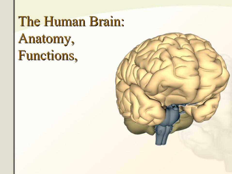
The Human Brain:The Human Brain:Anatomy,Anatomy,Functions,Functions,
Brain AnatomyBrain Anatomy• Skull Anatomy• Interior Skull Surface• Blood Vessels of the
Brain• Arteries of the Brain• The Neuron• The Meninges• External Brain Structures• The Cerebrum• The Cerebrum – The
Cortex• The Neocortex• Lobes of the Cerebrum• Frontal Lobe• Temporal Lobe• Parietal Lobe• Occipital Lobe• Limbic Lobe
•The Limbic System•Cerebellum•Thalamus•Hypothalamus•The Medulla Oblongata•The Pons•The Ventricles•Cerebrospinal Fluid•The Brainstem•Brainstem Components•Brainstem Divisions•The Cranial Nerves
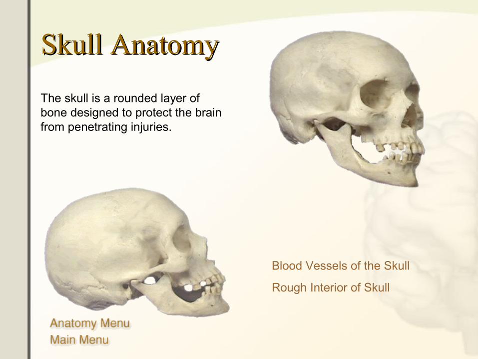
Skull AnatomySkull Anatomy
The skull is a rounded layer of bone designed to protect the brain from penetrating injuries.
Blood Vessels of the Skull
Rough Interior of Skull
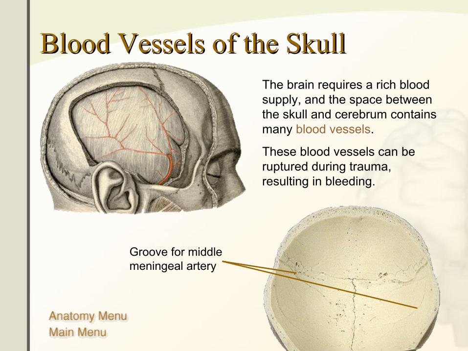
Blood Vessels of the SkullBlood Vessels of the SkullThe brain requires a rich blood supply, and the space between the skull and cerebrum contains many blood vessels.
These blood vessels can be ruptured during trauma, resulting in bleeding.
Groove for middle meningeal artery
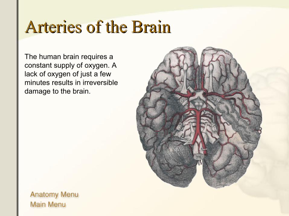
Arteries of the BrainArteries of the Brain
The human brain requires a constant supply of oxygen. A lack of oxygen of just a few minutes results in irreversible damage to the brain.

6
The BrainThe Brain
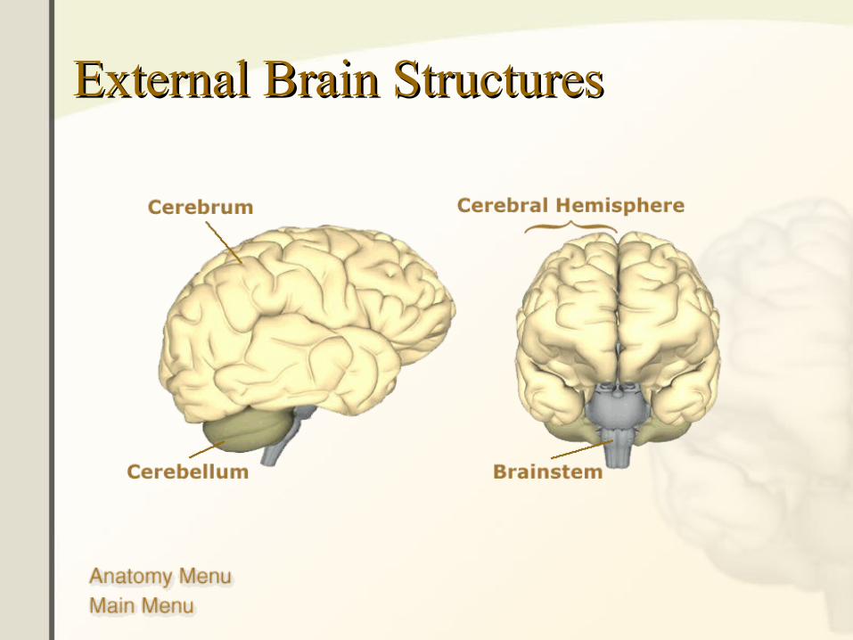
External Brain StructuresExternal Brain Structures
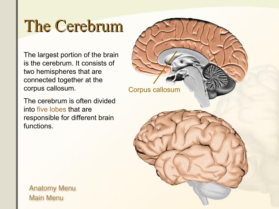
The CerebrumThe Cerebrum
The largest portion of the brain is the cerebrum. It consists of two hemispheres that are connected together at the corpus callosum.
The cerebrum is often divided into five lobes that are responsible for different brain functions.
Corpus callosum
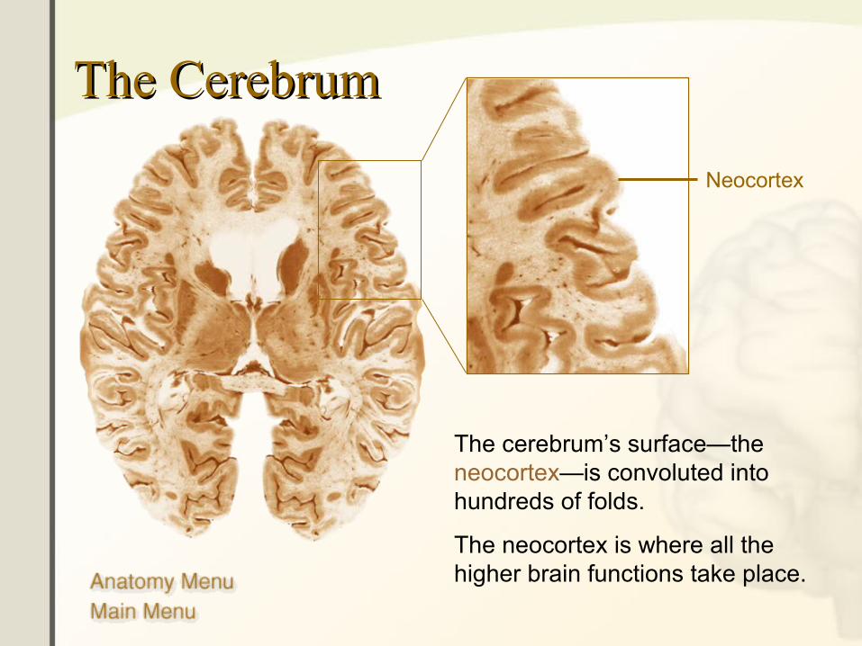
The CerebrumThe Cerebrum
The cerebrum’s surface—the neocortex—is convoluted into hundreds of folds.
The neocortex is where all the higher brain functions take place.
Neocortex
10
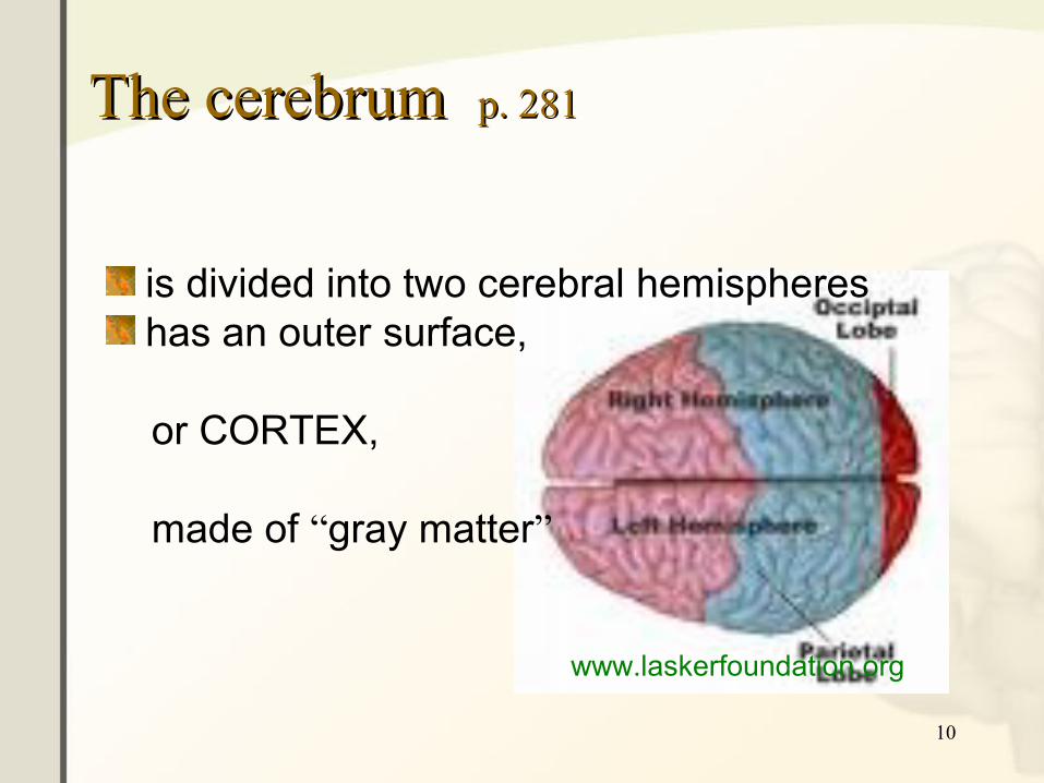
The cerebrum The cerebrum p. 281p. 281
is divided into two cerebral hemisphereshas an outer surface,
or CORTEX,
made of “gray matter”
www.laskerfoundation.org
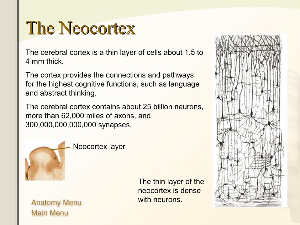
The NeocortexThe NeocortexThe cerebral cortex is a thin layer of cells about 1.5 to 4 mm thick.
The cortex provides the connections and pathways for the highest cognitive functions, such as language and abstract thinking.
The cerebral cortex contains about 25 billion neurons, more than 62,000 miles of axons, and 300,000,000,000,000 synapses.
Neocortex layer
The thin layer of the neocortex is dense with neurons.
12
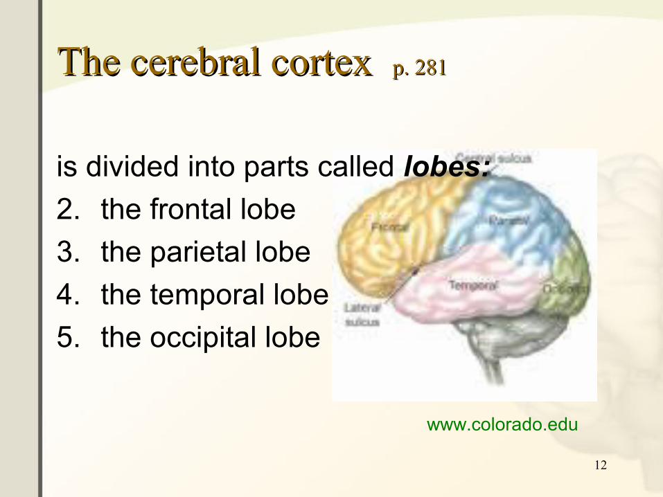
The cerebral cortex The cerebral cortex p. 281p. 281
is divided into parts called lobes:
2. the frontal lobe
3. the parietal lobe
4. the temporal lobe
5. the occipital lobe
www.colorado.edu
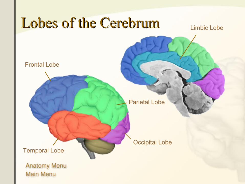
Lobes of the CerebrumLobes of the Cerebrum
Parietal Lobe
Temporal Lobe
Frontal Lobe
Limbic Lobe
Occipital Lobe
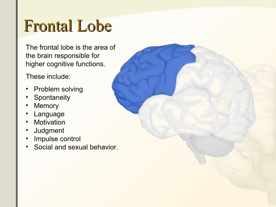
Frontal LobeFrontal LobeThe frontal lobe is the area of the brain responsible for higher cognitive functions.
These include:
• Problem solving• Spontaneity• Memory• Language• Motivation• Judgment• Impulse control• Social and sexual behavior.
15
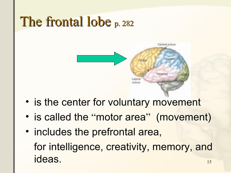
The frontal lobe The frontal lobe p. 282p. 282
• is the center for voluntary movement
• is called the “motor area” (movement)
• includes the prefrontal area,
for intelligence, creativity, memory, and ideas.
16
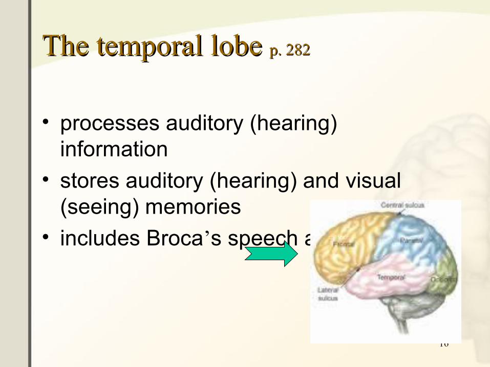
The temporal lobe The temporal lobe p. 282p. 282
• processes auditory (hearing) information
• stores auditory (hearing) and visual (seeing) memories
• includes Broca’s speech area
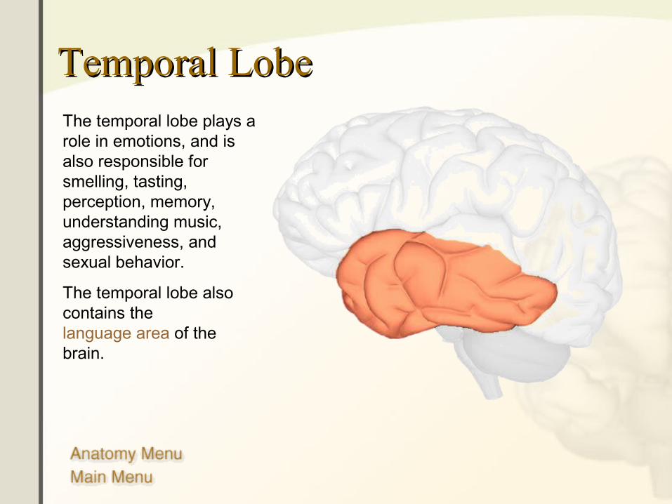
Temporal LobeTemporal LobeThe temporal lobe plays a role in emotions, and is also responsible for smelling, tasting, perception, memory, understanding music, aggressiveness, and sexual behavior.
The temporal lobe also contains the language area of the brain.
18
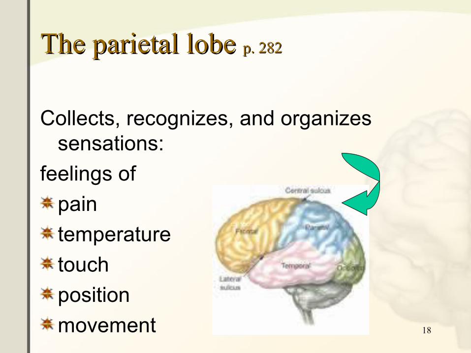
The parietal lobe The parietal lobe p. 282p. 282
Collects, recognizes, and organizes sensations:
feelings of
pain
temperature
touch
position
movement
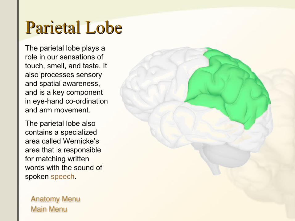
Parietal LobeParietal LobeThe parietal lobe plays a role in our sensations of touch, smell, and taste. It also processes sensory and spatial awareness, and is a key component in eye-hand co-ordination and arm movement.
The parietal lobe also contains a specialized area called Wernicke’s area that is responsible for matching written words with the sound of spoken speech.
20
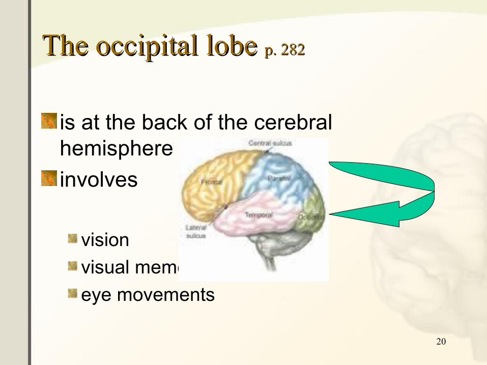
The occipital lobe The occipital lobe p. 282p. 282
is at the back of the cerebral hemisphere
involves
vision
visual memory
eye movements
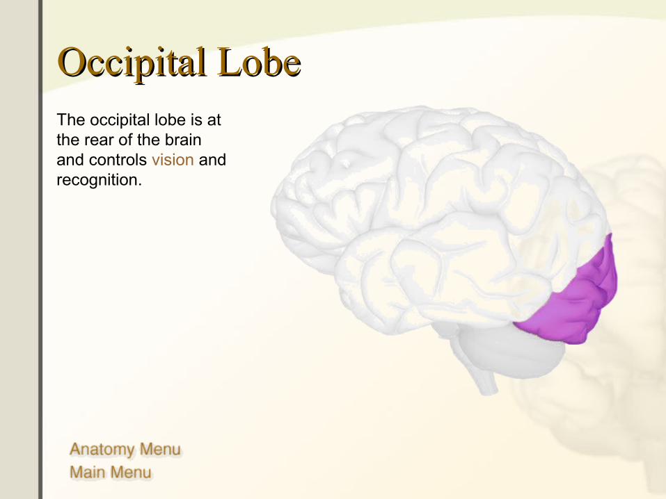
Occipital LobeOccipital LobeThe occipital lobe is at the rear of the brain and controls vision and recognition.
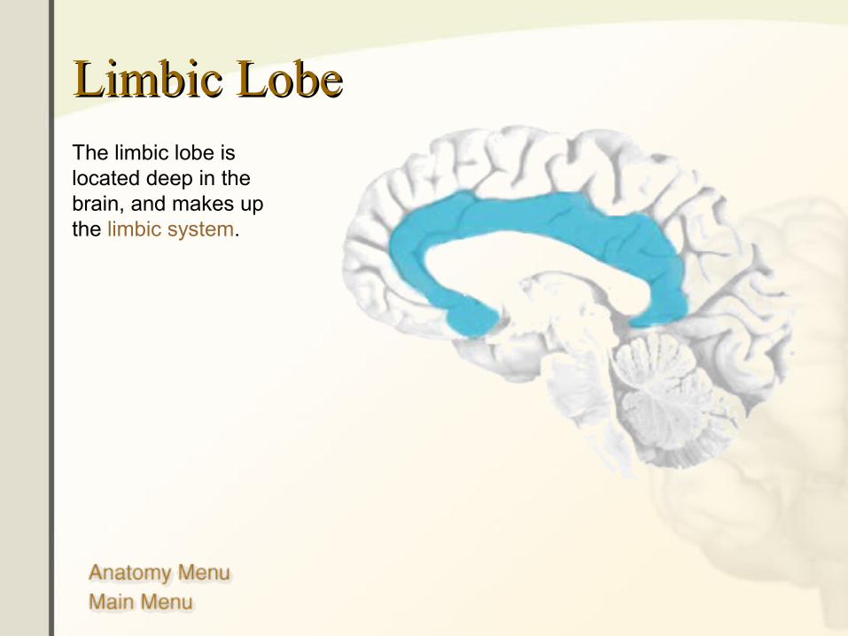
Limbic LobeLimbic LobeThe limbic lobe is located deep in the brain, and makes up the limbic system.
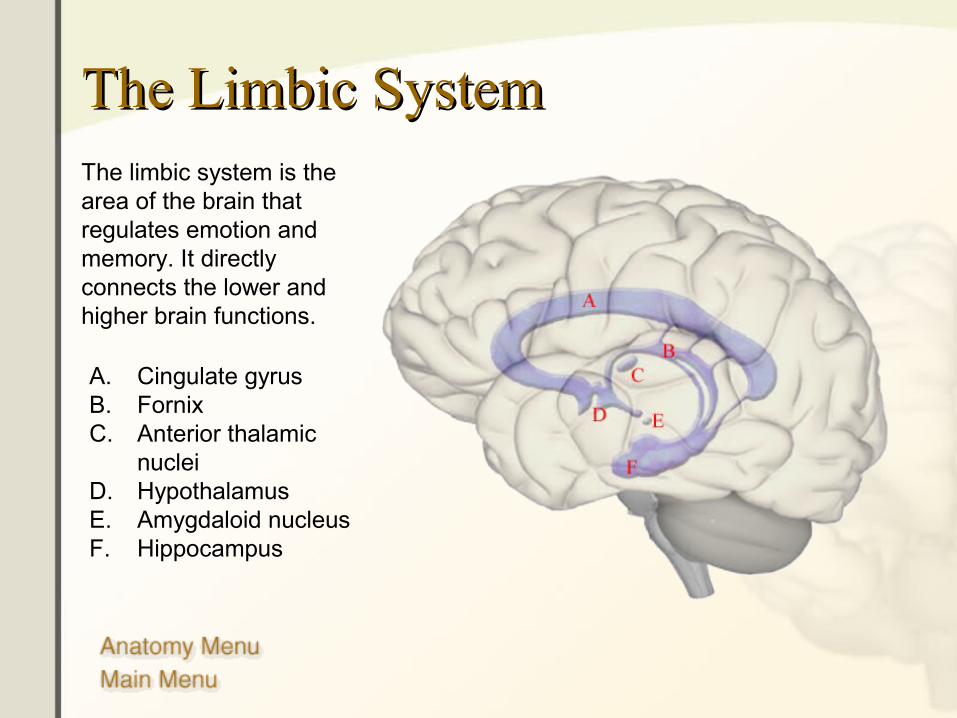
The Limbic SystemThe Limbic System
A. Cingulate gyrusB. FornixC. Anterior thalamic
nucleiD. HypothalamusE. Amygdaloid nucleusF. Hippocampus
The limbic system is the area of the brain that regulates emotion and memory. It directly connects the lower and higher brain functions.
24
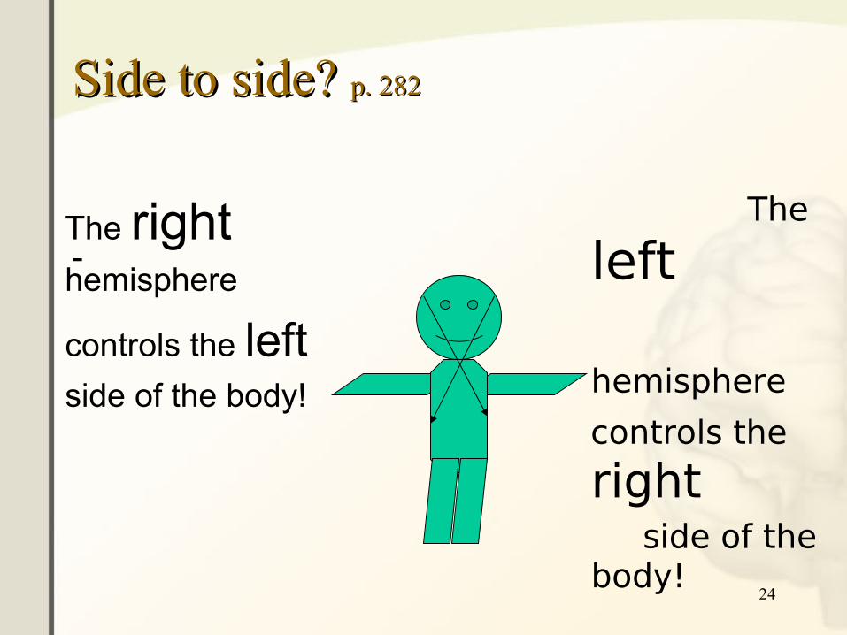
Side to side? Side to side? p. 282p. 282
-The righthemisphere
controls the leftside of the body!
The
left
hemisphere
controls the
right side of the
body!

CerebellumCerebellumThe cerebellum is connected to the brainstem, and is the center for body movement and balance.
Click image to play or pause video
26
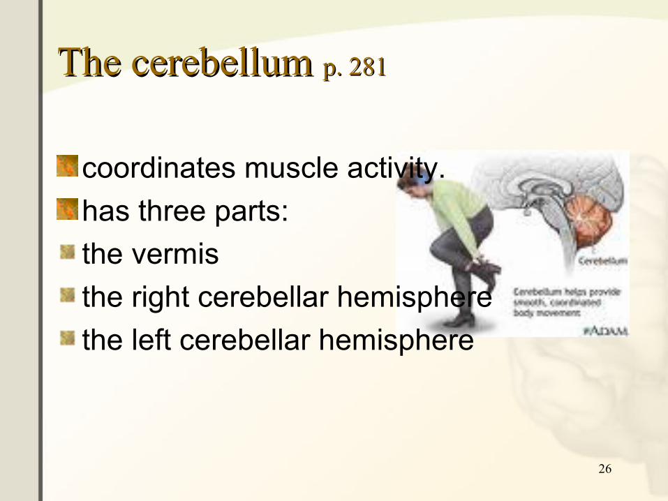
The cerebellum The cerebellum p. 281p. 281
coordinates muscle activity.
has three parts:
the vermis
the right cerebellar hemisphere
the left cerebellar hemisphere

27
The diencephalon The diencephalon p. 281p. 281
• is located between the midbrain and the cerebrum
• has three parts:• the thalamus: receives sensory
information and sends it to the cerebral cortex.
• the epithalamus: contains the pineal body and olfactory centers.
• the hypothalamus: connects the endocrine and nervous systems.
http://www.daviddarling.info/encyclopedia/A/anatomy_and_physiology.html
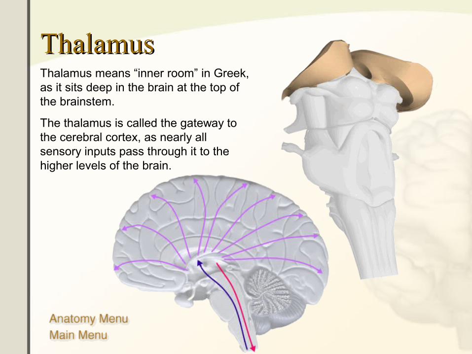
ThalamusThalamusThalamus means “inner room” in Greek, as it sits deep in the brain at the top of the brainstem.
The thalamus is called the gateway to the cerebral cortex, as nearly all sensory inputs pass through it to the higher levels of the brain.
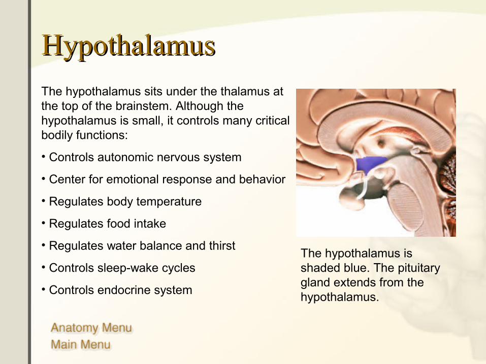
HypothalamusHypothalamus
The hypothalamus sits under the thalamus at the top of the brainstem. Although the hypothalamus is small, it controls many critical bodily functions:
• Controls autonomic nervous system
• Center for emotional response and behavior
• Regulates body temperature
• Regulates food intake
• Regulates water balance and thirst
• Controls sleep-wake cycles
• Controls endocrine system
The hypothalamus is shaded blue. The pituitary gland extends from the hypothalamus.
30
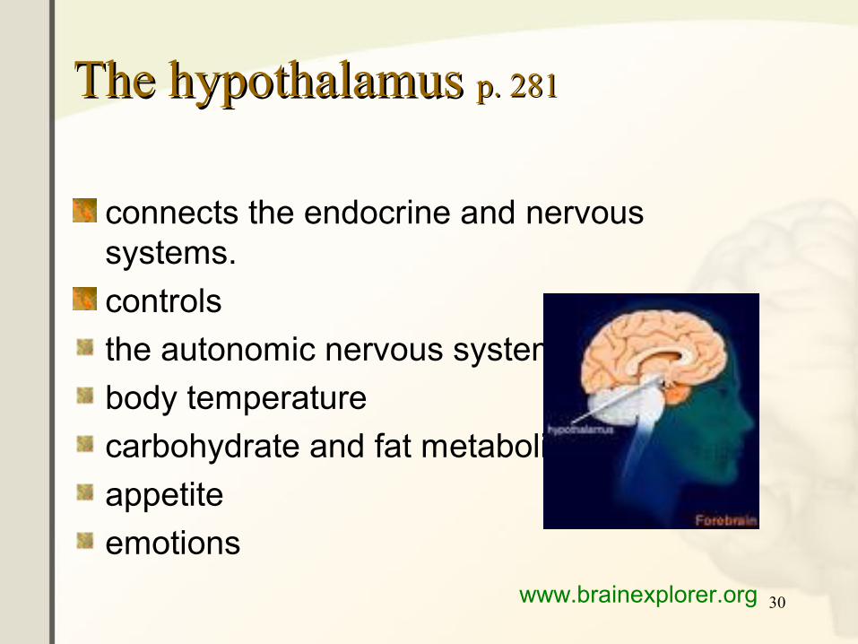
The hypothalamus The hypothalamus p. 281p. 281
connects the endocrine and nervous systems.
controls
the autonomic nervous system
body temperature
carbohydrate and fat metabolism
appetite
emotions
www.brainexplorer.org
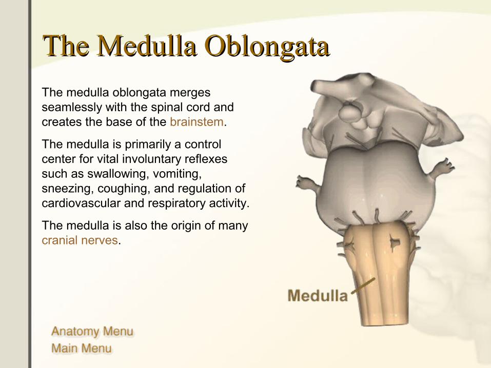
The Medulla OblongataThe Medulla Oblongata
The medulla oblongata merges seamlessly with the spinal cord and creates the base of the brainstem.
The medulla is primarily a control center for vital involuntary reflexes such as swallowing, vomiting, sneezing, coughing, and regulation of cardiovascular and respiratory activity.
The medulla is also the origin of many cranial nerves.
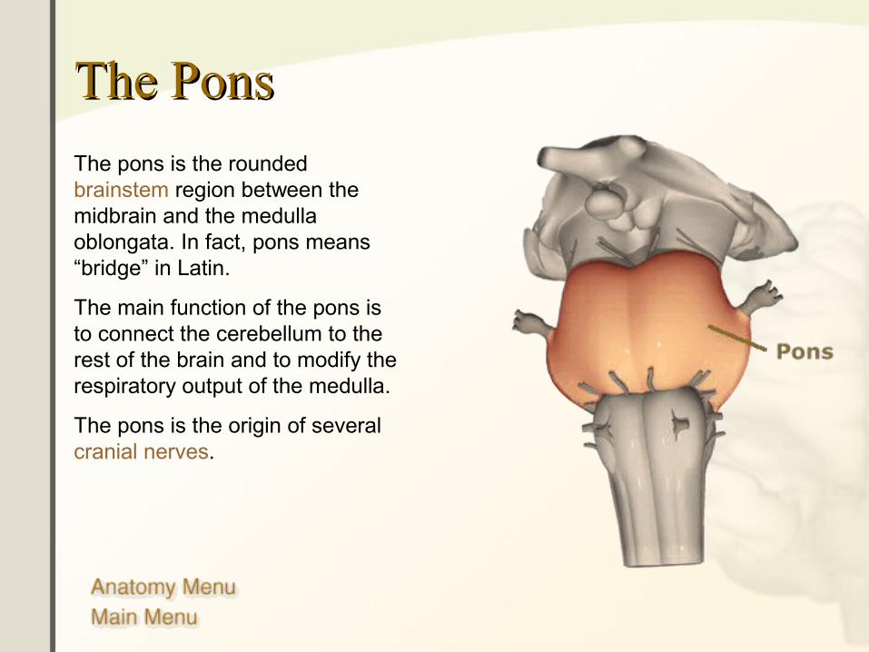
The PonsThe Pons
The pons is the rounded brainstem region between the midbrain and the medulla oblongata. In fact, pons means “bridge” in Latin.
The main function of the pons is to connect the cerebellum to the rest of the brain and to modify the respiratory output of the medulla.
The pons is the origin of several cranial nerves.
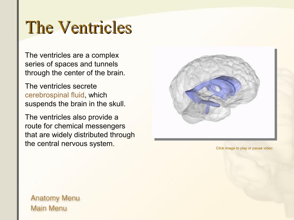
The VentriclesThe Ventricles
Click image to play or pause video
The ventricles are a complex series of spaces and tunnels through the center of the brain.
The ventricles secrete cerebrospinal fluid, which suspends the brain in the skull.
The ventricles also provide a route for chemical messengers that are widely distributed through the central nervous system.
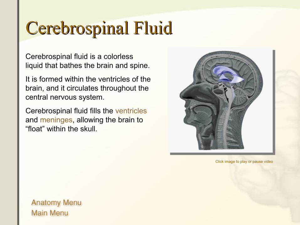
Cerebrospinal FluidCerebrospinal Fluid
Cerebrospinal fluid is a colorless liquid that bathes the brain and spine.
It is formed within the ventricles of the brain, and it circulates throughout the central nervous system.
Cerebrospinal fluid fills the ventricles and meninges, allowing the brain to “float” within the skull.
Click image to play or pause video
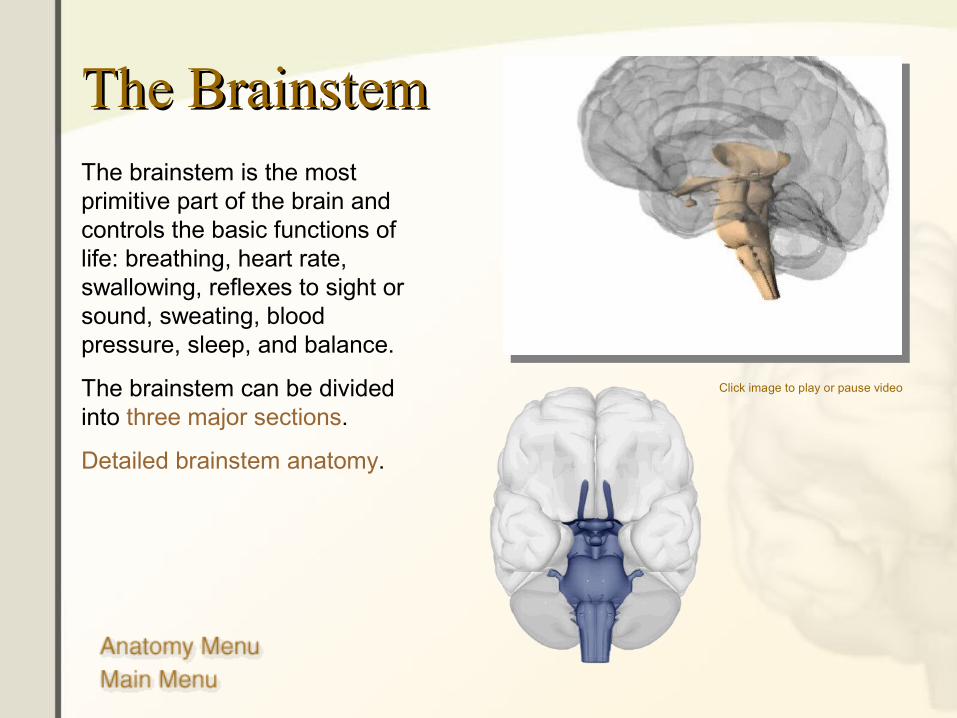
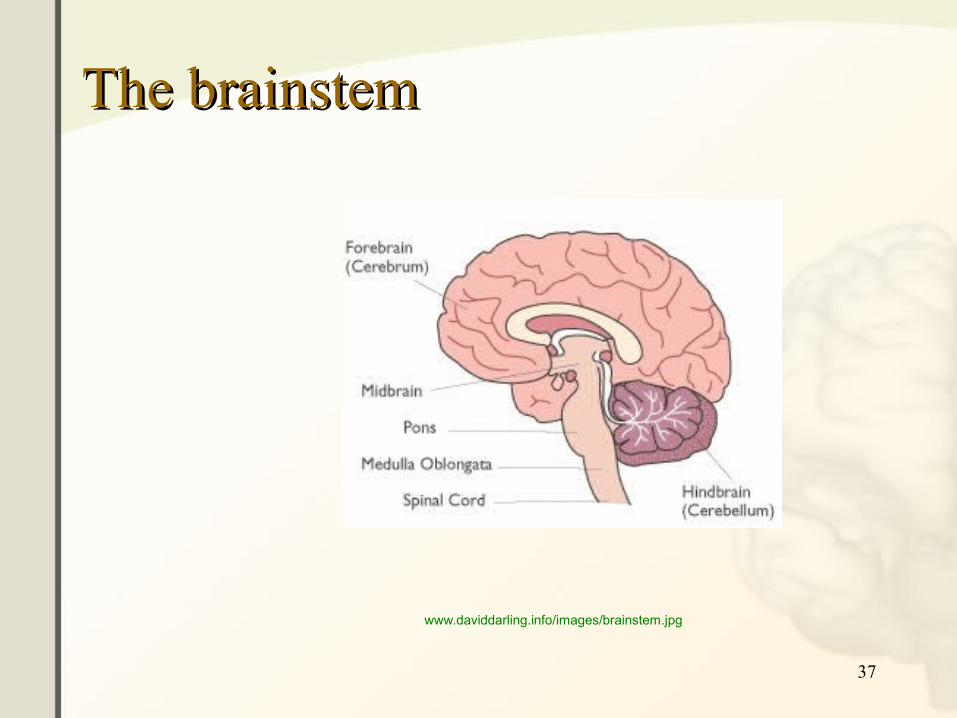
The BrainstemThe BrainstemThe brainstem is the most primitive part of the brain and controls the basic functions of life: breathing, heart rate, swallowing, reflexes to sight or sound, sweating, blood pressure, sleep, and balance.
The brainstem can be divided into three major sections.
Detailed brainstem anatomy.
Click image to play or pause video
36
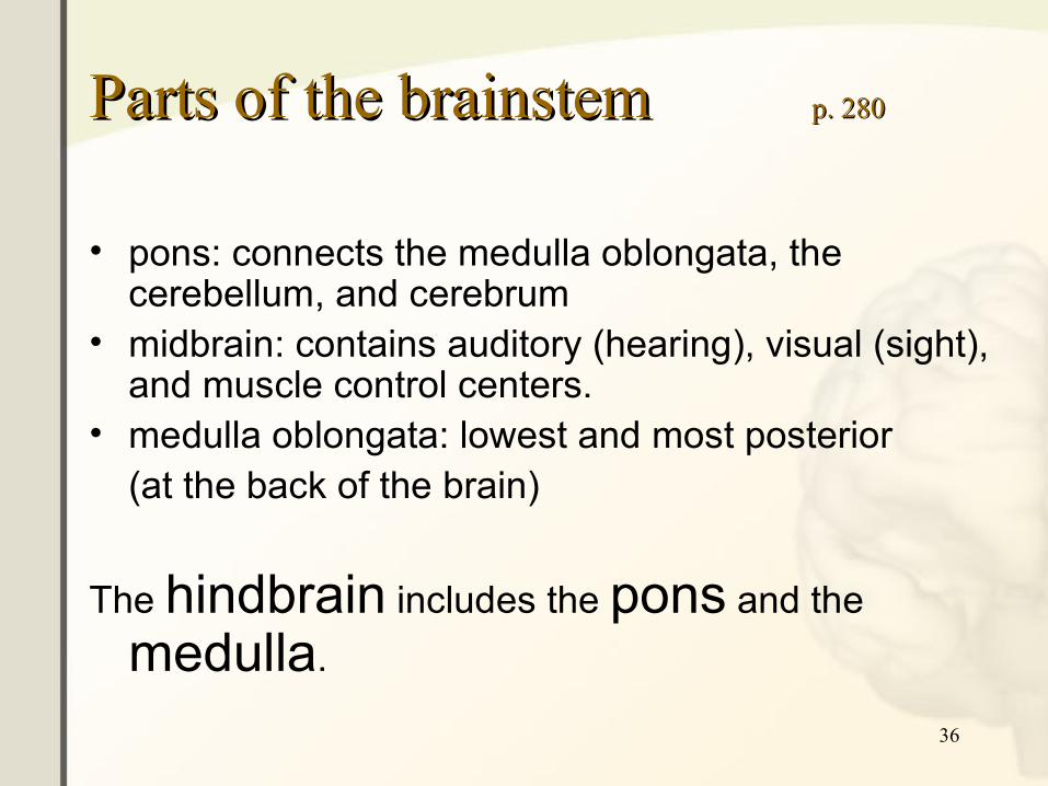
Parts of the brainstem Parts of the brainstem p. 280p. 280
• pons: connects the medulla oblongata, the cerebellum, and cerebrum
• midbrain: contains auditory (hearing), visual (sight), and muscle control centers.
• medulla oblongata: lowest and most posterior(at the back of the brain)
The hindbrain includes the pons and the
medulla.
37
The brainstemThe brainstem
www.daviddarling.info/images/brainstem.jpg
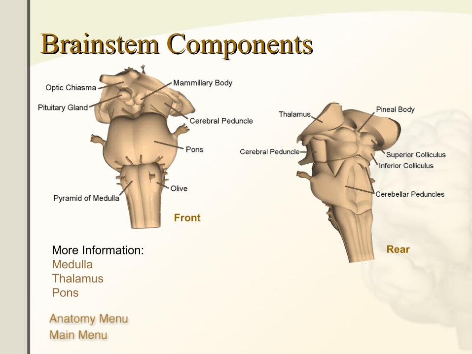
Brainstem Components Brainstem Components
Front
RearMore Information:MedullaThalamusPons
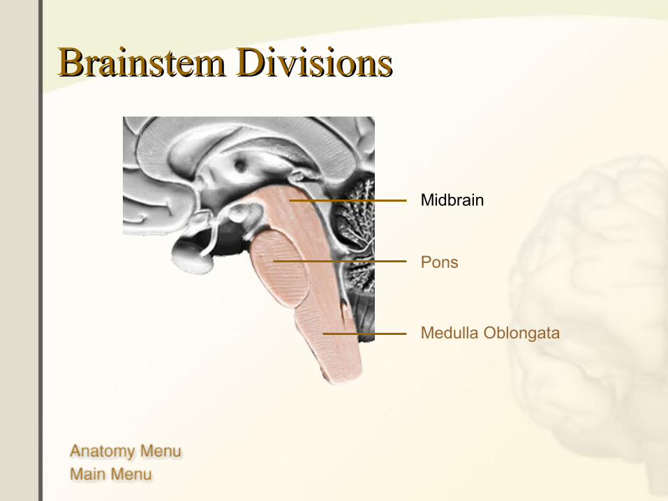
Brainstem DivisionsBrainstem Divisions
Midbrain
Pons
Medulla Oblongata
40
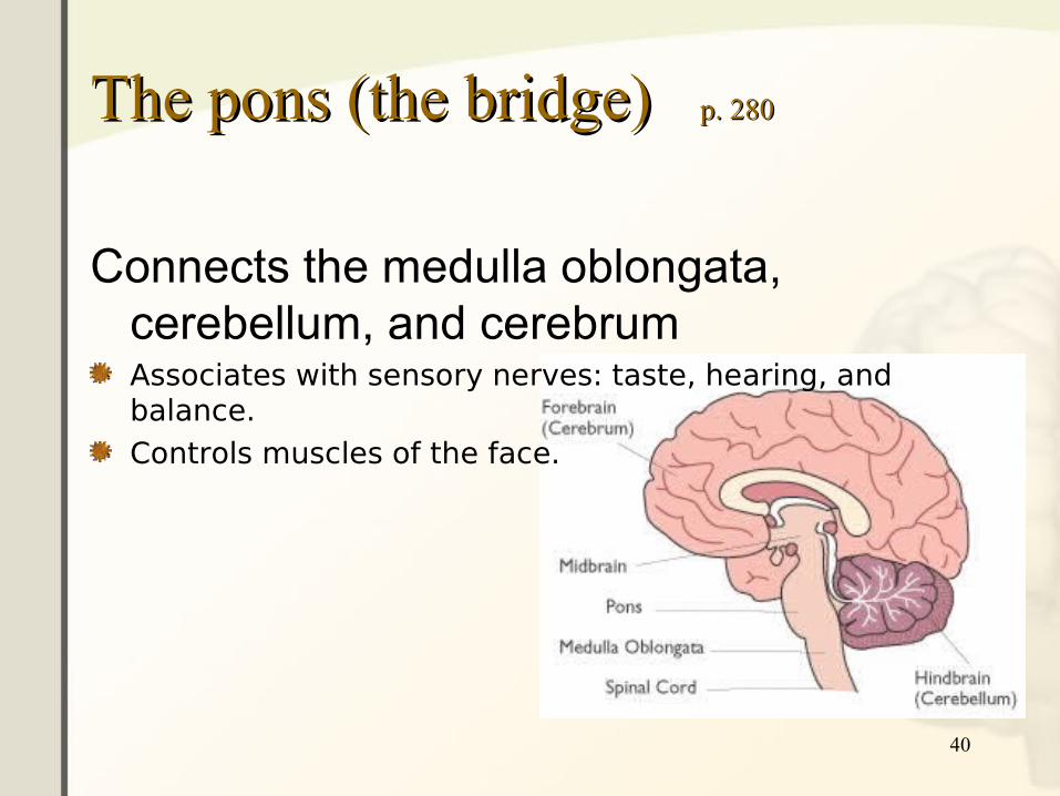
The pons (the bridge) The pons (the bridge) p. 280p. 280
Connects the medulla oblongata, cerebellum, and cerebrumAssociates with sensory nerves: taste, hearing, and balance.Controls muscles of the face.
41
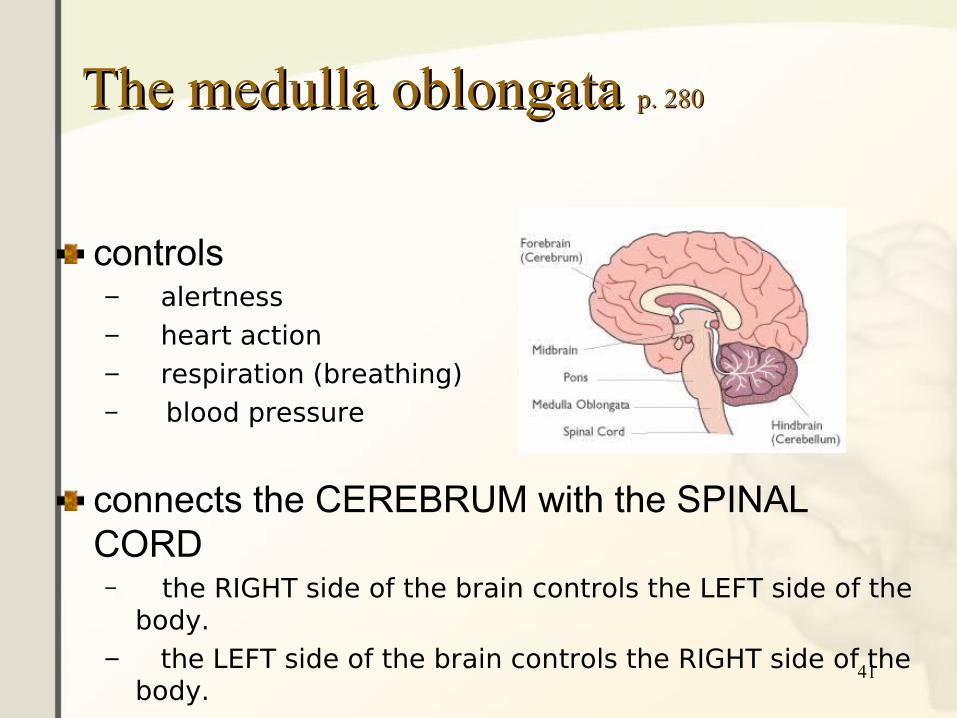
The medulla oblongata The medulla oblongata p. 280p. 280
controls– alertness– heart action– respiration (breathing)– blood pressure
connects the CEREBRUM with the SPINAL CORD– the RIGHT side of the brain controls the LEFT side of the
body.– the LEFT side of the brain controls the RIGHT side of the
body.
42
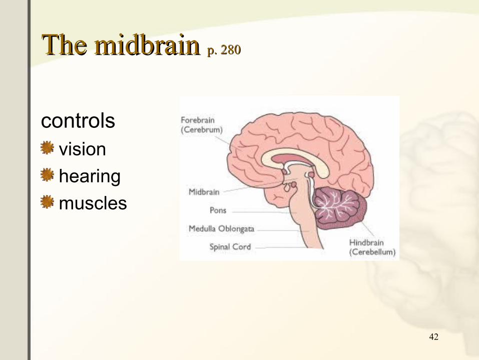
The midbrain The midbrain p. 280p. 280
controlsvision
hearing
muscles
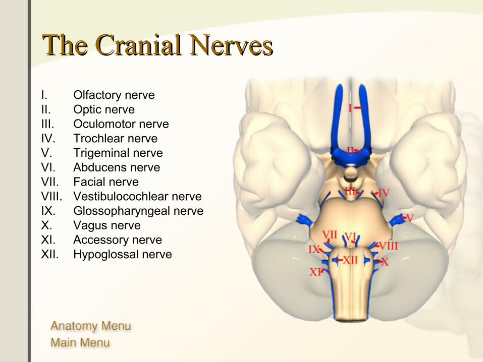
The Cranial NervesThe Cranial Nerves
I. Olfactory nerveII. Optic nerveIII. Oculomotor nerveIV. Trochlear nerveV. Trigeminal nerveVI. Abducens nerveVII. Facial nerveVIII. Vestibulocochlear nerveIX. Glossopharyngeal nerveX. Vagus nerveXI. Accessory nerveXII. Hypoglossal nerve
Brain FunctionsBrain Functions• Vision• Taste• Cognition• Emotion• Speech• Language• Hearing• Motor Cortex• Sensory Cortex• Autonomic Functions
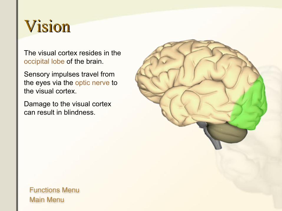
VisionVision
The visual cortex resides in the occipital lobe of the brain.
Sensory impulses travel from the eyes via the optic nerve to the visual cortex.
Damage to the visual cortex can result in blindness.
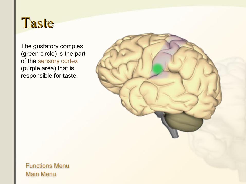
TasteTaste
The gustatory complex (green circle) is the part of the sensory cortex (purple area) that is responsible for taste.
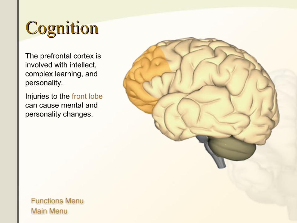
CognitionCognition
The prefrontal cortex is involved with intellect, complex learning, and personality.
Injuries to the front lobe can cause mental and personality changes.
EmotionEmotion
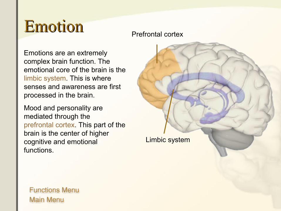
Emotions are an extremely complex brain function. The emotional core of the brain is the limbic system. This is where senses and awareness are first processed in the brain.
Mood and personality are mediated through the prefrontal cortex. This part of the brain is the center of higher cognitive and emotional functions.
Prefrontal cortex
Limbic system
SpeechSpeech
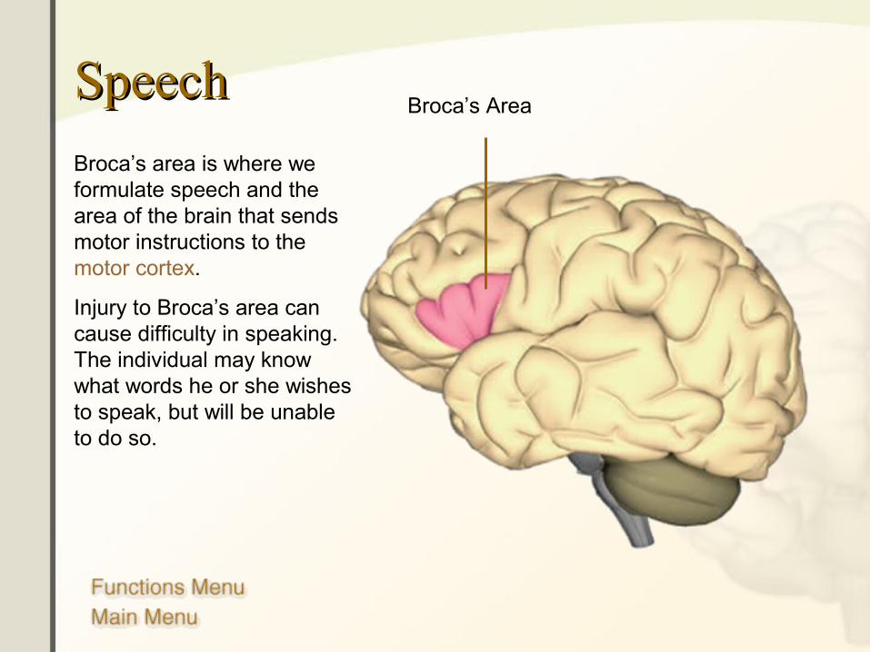
Broca’s area is where we formulate speech and the area of the brain that sends motor instructions to the motor cortex.
Injury to Broca’s area can cause difficulty in speaking. The individual may know what words he or she wishes to speak, but will be unable to do so.
Broca’s Area
LanguageLanguage
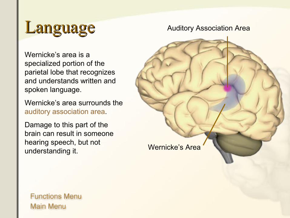
Wernicke’s area is a specialized portion of the parietal lobe that recognizes and understands written and spoken language.
Wernicke’s area surrounds the auditory association area.
Damage to this part of the brain can result in someone hearing speech, but not understanding it. Wernicke’s Area
Auditory Association Area
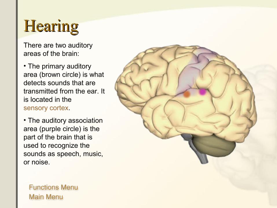
HearingHearingThere are two auditory areas of the brain:
• The primary auditory area (brown circle) is what detects sounds that are transmitted from the ear. It is located in the sensory cortex.
• The auditory association area (purple circle) is the part of the brain that is used to recognize the sounds as speech, music, or noise.
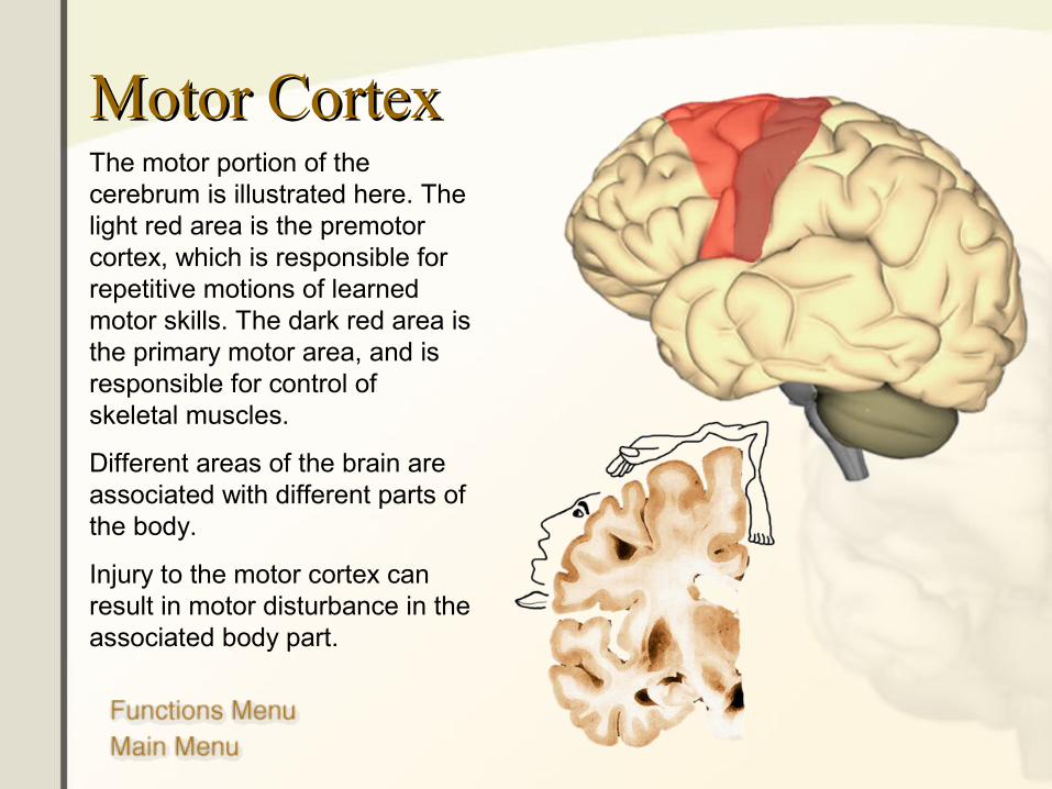
Motor CortexMotor CortexThe motor portion of the cerebrum is illustrated here. The light red area is the premotor cortex, which is responsible for repetitive motions of learned motor skills. The dark red area is the primary motor area, and is responsible for control of skeletal muscles.
Different areas of the brain are associated with different parts of the body.
Injury to the motor cortex can result in motor disturbance in the associated body part.
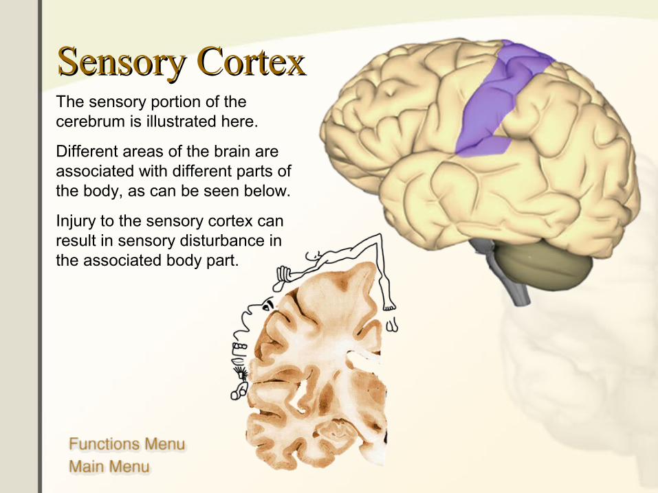
Sensory CortexSensory CortexThe sensory portion of the cerebrum is illustrated here.
Different areas of the brain are associated with different parts of the body, as can be seen below.
Injury to the sensory cortex can result in sensory disturbance in the associated body part.
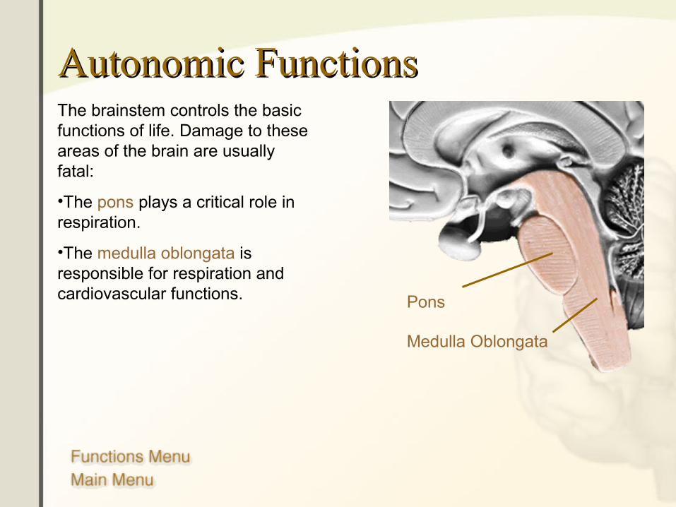
Autonomic FunctionsAutonomic FunctionsThe brainstem controls the basic functions of life. Damage to these areas of the brain are usually fatal:
•The pons plays a critical role in respiration.
•The medulla oblongata is responsible for respiration and cardiovascular functions. Pons
Medulla Oblongata
BibliographyBibliographyThe following are excellent resources and were the basis of the anatomical and functional components of this presentation:
• The Human Brain: An Introduction to Its Functional Anatomy, Fifth Edition. John Nolte, Mosby, 2002. ISBN: 0-323-01320-1 Purchase Here
• Coping with Mild Traumatic Brain Injury. Dr. Diane Stoler, Avery Penguin Putnam, 1998. ISBN: 0895297914 Purchase Here
• Human Anatomy and Physiology, Fifth Edition. Elaine N. Marieb, Benjamin/Cummings, 2000. ISBN: 0805349898. Purchase Here