the electrosurgical method of closed intrapleural pneumolysis in
TRANSCRIPT
The Electrosurgical Method of Closed IntrapleuralPneumolysis in Artificial Pneumothorax
RALPH C. MATSON, M.D.PORTLAND, ORE.
Reprinted from the Archives of SurgeryDecember, 1929, Vol. 19, Part II, pp. 1175-1192
COPYRIGHT, 1929
AMERICAN MEDICAL ASSOCIATION
535 NORTH DEARBORN STREET
CHICAGO

Reprinted from the Archives of SurgeryDecember, 1929, Vol. 19, Part II, pp. 1175-1192
* Read at the Twelfth Annual Meeting of the American Association forThoracic Surgery, held in St. Louis, Mo., April 25-27, 1929.
*From the Department of Thoracic Surgery, Portland Open Air Sanatorium,Milwaukie, Ore., and the Department of Medicine, University of Oregon MedicalSchool.
CLOSED
ARTI-
RALPH C. MATSON, M.D.
PORTLAND, ORE.
THE ELECTROSURGICAL METHOD OF
INTRAPLEURAL PNEUMOLYSIS IN
FICIAL PNEUMOTHORAX *
It is generally admitted that artificial pneumothorax is not only themost widely applicable method of collapsing the lung in the treatmentof pulmonary tuberculosis but also the most valuable. Unfortunately,pleuritic adhesions are almost invariably present in cases in which thepatients require pneumothorax treatment and constitute the greatestobstacle to a satisfactory end-result.
As a result of experience in the treatment of approximately 1,400patients with pulmonary tuberculosis with artificial pneumothoraxduring the past eighteen years, my co-workers and I are convinced ofthe importance of establishing a type of pneumothorax which withina few months will give the diseased lung sufficient functional rest,collapse or compression to render it no longer a source of tuberculotoxemia or tubercle bacilli-laden sputum.
The importance of a satisfactory collapse of the lung is strikinglyshown in the accompanying table. A careful review of the clinicalrecords and stereoroentgenograms in 245 cases in this series revealsthat the primary cause of failure of treatment with pneumothorax in40 per cent of the cases was the presence of adhesions, which prevented a satisfactory collapse of the lung. In most of these cases, atemporary improvement followed pneumothorax treatment in spiteof insufficient collapse of the lung. Consequently, the treatment wascontinued sometimes for prolonged periods, in the hope of stretchingthe offending adhesions and securing a good collapse of the lung.Sooner or later, however, either extension of disease took place tothe opposite lung, intestines or throat, or some complication as empyema,spontaneous pneumothorax or obliterating pneumothorax occurred,compelling discontinuation of pneumothorax treatment. Unfortunately,the cases were then too far advanced to utilize other methods forcollapse of the lung.
While adhesions are present in the majority of patients selectedfor pneumothorax treatment, according to our experience, a satis-
~ iI<I
'I
I
jj
'1III
'1
1
:1
I
2
factory pneumothorax can be established in 40 per cent of the cases.In a similar percentage (40 per cent), however, the character of adhesions will prevent the necessary collapse or compression of the lungto provide adequate functional rest or closure of cavities; and in theremaining 20 per cent, pleuritic adhesions will prevent any introductionof gas. Thus, pneumothorax treatment will prove efficient in considerably less than half the cases wherein it is' indicated.
The phthisiotherapeutist recognizes the value of thoracic surgery.In those cases wherein no gas can be introduced, he now more oftentakes advantage of the surgeon's ability to bring about collapse of thelung by a phrenic neurectomy or thoracoplasty instead of subjecting.the patient to prolonged and unsatisfactory sanatorium care. However, the fate of cases presenting pleuritic adhesions, which preventefficient collapse of the lung, leaves much to be desired.
The usual procedure in cases of this type is to keep up the pneumothorax frequently, month in and out, and sometimes even year in and
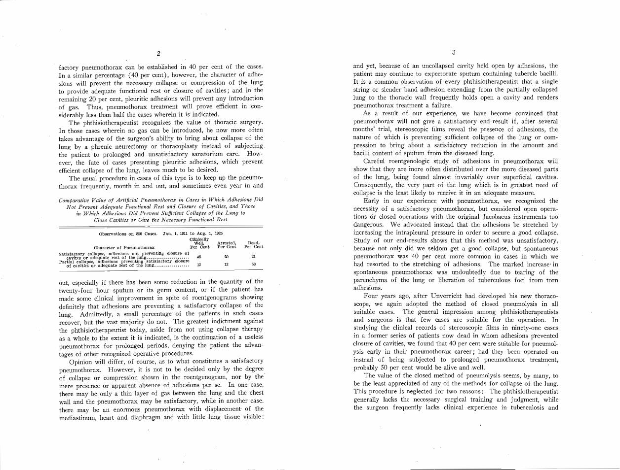
Comparative Value of Artificial Pneumothora.'!; in Cases in Which Adhesions DidN at Prevent Adequate Functional Rest and Closure of Cavities, and Those
in Which Adhesions Did Prevent S~£fficient Collapse of the Lung toClose Cavities or Give the Necessary Functional Rest
Observations on 850 Oases. Jan. 1, 1911 to Aug. 1, 1925Clinically
Well, Arrested, Dead,Character of Pneumothorax Per Cent Per Cent Per Cent
Satisfactory collapse, adhesions not preventing closure ofcavites' or adequate rest of the lung.. 48 20 21
Partial collapse, adhesions preventing satisfactory closureof cavities or adequate rest of the lung.................. 18 13 50
out, especially if there has been some reduction in the quantity of thetwenty-four hour sputum or its germ content, or if the patient hasmade some clinical improvement in spite of roentgenograms showingdefinitely that adhesions are preventing a satisfactory collapse of thelung. Admittedly, a small percentage of the patients in such casesrecover, but the vast majority do not. The greatest indictment againstthe phthisiotherapeutist today, aside from not using collapse therapyas a whole to the extent it is indicated, is the continuation of a uselesspneumothorax for prolonged periods, denying the patient the advantages of other recognized operative procedures.
Opinion will differ, of course, as to what constitutes a satisfactorypneumothorax. However, it is not to be decided only by the degreeof collapse or compression shown in the roentgenogram, nor by the'mere presence or apparent absence of adhesions per se. In one case,there may be only a thin layer of gas between the lung and the chestwall and the pneumothorax may be satisfactory, while in another case.there may be an enormous pneumothorax with displacement of themediastinum, heart and diaphragm and with little ,lung tissue visible;
3
and yet, because of an uncollapsed cavity held open by adhesions, thepatient may continue to expectorate sputum containing tubercle bacilli.It is a common observation of every phthisiotherapeutist that a singlestring or slender band adhesion extending from the partially collapsedlung to the thoracic wall frequently holds open a cavity and renderspneumothorax treatment a failure.
As a result of our experience, we have become convinced thatpneumothorax will not give a satisfactory end-result if, after severalmonths' trial, stereoscopic films reveal the presence of adhesions, thenature of which is preventing sufficient collapse of the lung or compression to bring about a satisfactory reduction in the amount andbacilli content of sputum from the diseased lung.
Careful roentgenologic study of adhesions in pneumothorax willshow that they are more often distributed over the more diseased partsof the lung, being found almost invariably over superficial cavities.Consequently, the very part of the lung which is in greatest need ofcollapse is the least likely to receive it in an adequate measure.
Early in our experience with pneumothorax, we recognized thenecessity of a satisfactory pneumothorax, but considered open operations or closed operations with the original Jacobaeus instruments toodangerous. We advocated instead that the adhesions be stretched byincreasing the intrapleural pressure in order to secure a good collapse.Study of our end-results shows that this method was unsatisfactory,because not only did we seldom get a good collapse, but spontaneouspneumothorax was 40 per cent more common in cases in which wehad resorted to the stretching of adhesions. The marked increase' inspontaneous pneumothorax was undoubtedly due to tearing of theparenchyma of the lung or liberation of tuberculous foci from tornadhesions.
Four years ago, after Unverricht had developed his new thoracoscope, we again adopted the method of closed pneumolysis in allsuitable cases. The general impression among phthisiotherapeutistsand surgeons is that few cases are suitable for the operation. Instudying the clinical records of stereoscopic films in ninety-one casesin a former series of patients now dead in whom adhesions preventedclosure of cavities, we found that 40 per cent were suitable for pneumolysis early in their pneumothorax career; had they been operated oninstead of being subjected to prolonged pneumothorax treatment,probably 50 per cent would be alive and well.
The value of the closed method of pneumolysis seems, by many, tobe the least appreciated of any of the methods for collapse of the lung.This procedure is neglected for two reasons: The phthisiotherapeutistgenerally lacks the necessary surgical training and judgment, whilethe surgeon frequently lacks clinical experience in tuberculosis and

4
knowledge of pneumothorax as a background. Both are unwilling tospend the necessary time to perfect the technic. Thus, the surgeonfavors the open method of pneumolysis, or thoracoplasty, while thephthisiotherapeutist clings to his partial pneumothorax. Both fail toappreciate the improvement in the technic of cutting adhesions, andmany still retain the impression that it is dangerous.
These dangers are largely technical. Formerly, one worked prettymuch in the dark with poor instruments and equipment. We, ourselves,went through this period and gave up the method, as we considered itdangerous and of little utility. Today, however, we have improvedinstruments and a more refined technic, and while the operation may betechnically difficult, it is at least not dangerous when properly done.
I f collapse therapy is indicated, it is our policy to try artificialpneumothorax. If a satisfactory collapse of the lung is not obtainedwithin a few months, and it is shown on serial stereoscopic films thatadhesions are preventing a satisfactory collapse, we consider a pneumolysis at once.
During the past four years, the patients in 45 per cent of our casesin which there was unsatisfactory collapse have proved suitable for theoperation. If not suitable, we consider other methods of collapse insteadof continuing a comparatively useless pneumothorax.
The technic, indications and contraindications for intrapleuralpneumolysis, as well as the selection of cases and end-results, have beencovered in a previous contribution.1 It was pointed out in a formercommunication that the cauterization of adhesions by the galvanocauteryis objectionable because of the heat, smoke, pain and reaction to operation. Perhaps its greatest shortcoming is the character of the cuttingproduced because tissue is destroyed for a short distance around thecautery. Thus, blood vessels are severed without any previous obliteration, and unless a dull red heat is used, undue bleeding may occur. Inaddition to these reactions, if too much heat is used, there is also thedanger of tissue necrosis occurring, which may involve the parenchymaof the lung, tuberculous cavities or tuberculous foci, thus liberatinginfection, or the necrosis may invade blood vessels and cause serioussecondary hemorrhage. Furthermore, while the time required for thecautery to heat and cool after the current is thrown on or off is short, itnevertheless has its disadvantage, as occasionally a moment's delay,rendering the cutting instrument active or inactive, may result in seriousconsequences. Moreover, the.shaft of the cautery sometimes becomesextremely hot and may cause a sloughing at the point where it passesthrough the thoracic wall. The hot cautery shaft may also damage lung
1. Matson, R. c.: Cauterization of Adhesions in Artificial Pneumothorax bythe Jacobaeus-Unverricht Method of Closed Pneumolysis, Am. Rev. Tuberc. 19:233 (March) 1929.
5
tissue or the pericardium on which it may rest, during operation, unobserved by the operator who is giving his attention to the cutting alone.
All of these difficulties and dangers were serious, objectionablefeatures which prevented successful results in many instances..
The successful utilization of electrosurgical methods in operationson the brain and in treatment for cancer stimulated much experimentation on our part to apply these methods to intrathoracic surgery, in aneffort to lessen the difficulties and reduce the hazards incident to theuse of the galvanocautery. vVe applied the electrothermic principle forthe severing of adhesions, and as a result of its advantages over thegalvanocautery, we are utilizing electrocoagulation and cutting in allcases presenting the difficuties that we have outlined.
During the utilization of thif! new principle over a period of the pasttwo years, we have repeatedly performed operations by this methodwhich would not have been attempted with the galvanocautery, and thecomparative safety of the method over the galvanocautery procedure,with the results obtained, have created a strong conviction that theelectrocoagulation and cutting will replace the galvanocautery. The application of the method intrathoracically, however, involves difficulty ofcontrol of bleeding not present in any open operation. vVhile hemostasisin an open operation can be obtained by well known and easily appliedsurgical methods, it is obvious that these same procedures cannot beapplied in such operations as the closed pneumolysis.
Aside from the anatomic relationship of large blood vessels andimportant nerve trunks to pleural adhesions or to the pericardium, it isof vital importance to know whether the adhesion to be cut contains bloodvessels, compressed lung tissue, diseased foci or the prolongation of acavity. Cutting into the prolongation of a cavity or compressed lungtissue is avoidable. Cutting into tuberculous foci in adhesions can beefficiently dealt with; but hemorrhage is one complication not alwaysavoidable, and if it is at all profuse, it is a most unpleasant experiencefor the operator.
It is my prime purpose in this paper to discuss the utilization ofelectrosurgery for cutting adhesions in artificial pneumothorax by theclosed method under thoracoscopic control, with especial reference tocontrol of bleeding.
It is to be hoped that this paper will not encourage those withoutexperience in this new branch of surgery to undertake too difficult operations at first, as the operation might result in failure and thus bringabout unjust criticism of an otherwise valuable procedure. One sometimes hesitates, therefore, to describe a new technic, as it may makeinexperienced operators too venturesome.
The usual source of bleeding during the process of cutting adhesionsis from blood vessels collateral from the intercostals. One meets with

6
two types of vessels: those situated subpleurally, which can usually bedetected by thoracoscopic examination and easily controlled, and thosesituated in the interior of adhesions, from which profuse bleeding occursat times. In the case' of the former, bleeding may be easily controlledby electrocoagulation; or, if the galvanocautery is used, before the bloodvessels are cut they may be thrombosed by the application of the flatsurface of the cautery. Dangerous bleeding, however, comes from bloodvessels situated in the interior of adhesions, particularly in those whichare dense and well organized. This dense type of adhesion is found incases in which the patients have been subjected to prolonged and sometimes even short periods of pneumothorax treatment, particularly if thepatient is of a fibroplastic constitution with a tendency for productivechanges to take place in the tissues following pneumothorax treatment.
The latter type of vascular adhesion is more often found in thecostovertebral gutter, at the apex of the lung and anteriorly near thecostochondral junction. Densely organized adhesions in these areasshould be approached cautiously because blood vessels of considerablesize may be situated in their interior, the presence of which is notalways possible of determination until they have been cut into.
Bleeding occurring during the cutting of cord and band adhesions, ifthe adhesions are not densely organized, usually stops after the adhesionhas been cut through and the stump contracts. In well organized tissue,however, little retraction takes place, and bleeding may be a source ofconsiderable anxiety to the surgeon. Therefore, it is obvious that theideal method of severing adhesions must provide efficient hemostasis.
The objections to the galvanocautery in connection with hemostasishave already been pointed out. A detailed description of the physicalcharacteristics of currents employed in electrosurgery will not be discussed. However, an understanding of the effects of the high frequencycurrents employed in the electrothermic method of cutting adhesions isnecessary to apply this new procedure intelligently.
. Briefly, the cutting effected by the electrothermic method is not a truecutting but a molecular disintegration of the tissues produced by a highfrequency undamped current, an arc being formed at the point of contactbetween the tip of the electrode and the tissue.
It has been found that the form of high frequency electric currentwhich results in the best cutting of tissue with least charring is thatgenerated by the utilization of thermeonic tubes used in radio broadcasting, which give sustained oscillations of uniform amplitude at a rate of600,000 per second. Even with an apparatus generating this form ofcurrent, it will show an impairment in cutting if the rate of oscillationsis considerably increased or decreased. Based on this rate of oscillation,the theory is advanced that the cleavage of the tissue is accomplished notby mechanically cutting or by cauterization, but by a cellular disruption
7
resulting from the reaction of the cells to a sustained vibration transmitted at their own inherent vibration rate. The little sparking andresultant film of coagulation is secondary and subsequent to the cleavageof the tissue. Thus, two separate and distinct functions may be observed.One is the transmission by the operating electrode of the oscillations tocause cleavage, and the other is the slight sparking as the needle breakscontact with the parted tissue. If properly adjusted and handled, thisresults in a slight film or coagulation sufficient to check capillary bleedingbut not sufficient to cause sloughing or charring with the danger ofsubsequent hemorrhage.
The ideal current for cutting, therefore, is recognized as that havinga uniform sustained amplitude of 600,000 oscillations per second.
On the other hand, the best form of high frequency electric currentfor the generation of heat as used in coagulation is that having a dampedoscillation of a long decrement. This current is generated from suitableapparatus containing spark gaps, condensers, etc. An apparatus properlydesigned delivering currents having long decrements will result in aclean blanching and dehydrating of the tissue when properly applied forcoagulation, but apparatus of improper design may have a short decrement of the oscillating currents which will result in a brown or blackburning of the tissue when applied for coagulation. The rate of oscillations of this form of current is usually about 1,250,000, but the rate ofoscillation is not so important as the form of the trains or the decrement.
Two distinctly opposite forms of current are utilized in electrosurgery: One is ideal for cutting and the other for coagulating. To getthe best results, it is essential to employ apparatus furnishing these two
'forms of current obtainable at the will of the operator and in the strengthsuited to the work. There are devices which will deliver cutting characteristics from a generator by means of a spark gap. This current, however, is a compromise between the two ideal currents and consists usuallyof a damped oscillation having short decrement and having the trains ofoscillation crowded together. The peaks of the resultant current maysimulate the oscillations of the undamped oscillations, but there arepresent the superimposed short oscillations which tend to cause undueheating when used for cutting. Similarly, this form of current evenwhen differently adjusted is far from the ideal for coagulating. Inpractice, apparatus delivering the latter form of current is likely to besomewhat erratic and require adjustment from time to time in the courseof operation. In other words, there is no assurance that it will respondto the requirements of the moment. In important surgical work, whenhesitation, delay or trial cannot be tolerated, obviously nothing but theideal currents for cutting and coagulation should be employed.
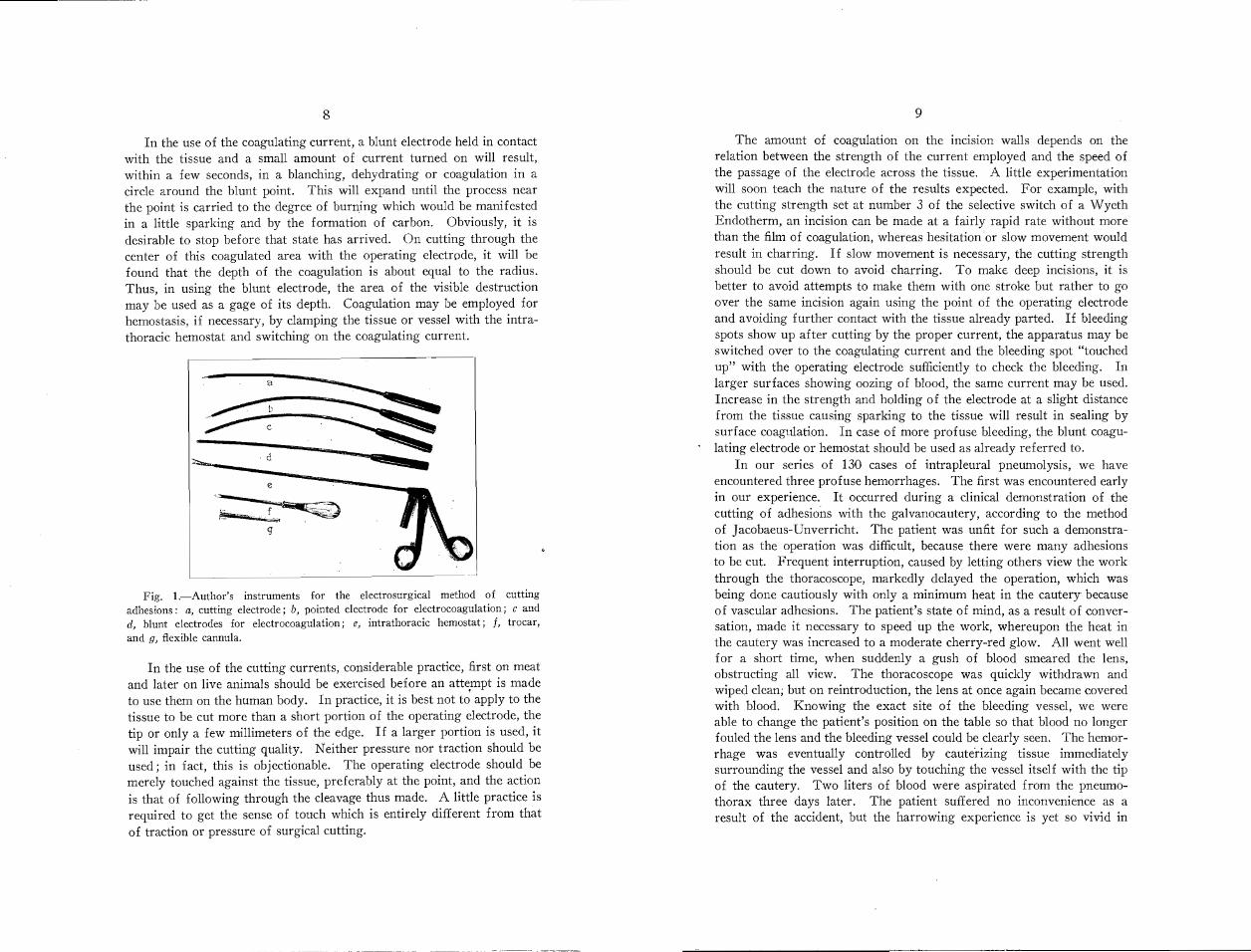
In our work, we have utilized the Wappler 'Wyeth Endotherm withspecial instruments which I have designed for intrathoracic use (fig. 1).
The amount of coagulation on the inClslOn walls depends on therelation between the strength of the current employed and the speed ofthe passage of the electrode across the tissue. A little experimentationwill soon teach the nature of the results expected. For example, withthe cutting strength set at number 3 of the selective switch of a \iVyethEndotherm, an incision can be made at a fairly rapid rate without morethan the film of coagulation, whereas hesitation or slow movement wouldresult in charring. If slow movement is necessary, the cutting strengthshould be cut down to avoid charring. To make deep incisions, it isbetter to avoid attempts to make them with one stroke but rather to goover the same incision again using the point of the operating electrodeand avoiding further contact with the tissue already parted. If bleedingspots show up after cutting by the proper current, the apparatus may beswitched over to the coagulating current and the bleeding spot "touchedup" with the operating electrode sufficiently to check the bleeding. Inlarger surfaces showing oozing of blood, the same current may be used.Increase in the strength and holding of the electrode at a slight distancefrom the tissue causing sparking to the tissue will result in sealing bysurface coagulation. In case of more profuse bleeding, the blunt coagulating electrode or hemostat should be used as already referred to.
In our series of 130 cases of intrapleural pneumolysis, we haveencountered three profuse hemorrhages. The first was encountered earlyin our experience. It occurred during a clinical demonstration of thecutting of adhesions with the galvanocautery, according to the methodof Jacobaeus-Unverricht. The patient was unfit for such a demonstration as the operation was difficult, because there were many adhesionsto be cut. Frequent interruption, caused by letting others view the workthrough the thoracoscope, markedly delayed the operation, which wasbeing done cautiously with only a minimum heat in the cautery becauseof vascular adhesions. The patient's state of mind, as a result of conversation, made it necessary to speed up the work, whereupon the heat inthe cautery was increased to a moderate cherry-red glow. All went wellfor a short time, when suddenly a gush of blood smeared the lens,obstructing all view. The thoracoscope was quickly withdrawn andwiped clean; but on reintroduction, the lens at once again became coveredwith blood. Knowing the exact site of the bleeding vessel, we wereable to change the patient's position on the table so that blood no longerfouled the lens and the bleeding vessel could be clearly seen. The hemorrhage was eventually controlled by caute"rizing tissue immediatelysurrounding the vessel and also by touching the vessel itself with the tipof the cautery. Two liters of blood were aspirated from the pneumothorax three days later. The patient suffered no inconvenience as aresult of the accident, but the harrowing experience is yet so vivid in
9
e
i~t~gl: '
In the use of the cutting currents, considerable practice, first on meatand later on live animals should be exercised before an atte.mpt is madeto use them on the human body. In practice, it is best not to apply to thetissue to be cut more than a short portion of the operating electrode, thetip or only a few millimeters of the edge. If a larger portion is used, itwill impair the cutting quality. Neither pressure nor traction should beused; in fact, this is objectionable. The operating electrode should bemerely touched against the tissue, preferably at the point, and the actionis that of following through the cleavage thus made. A little practice isrequired to get the sense of touch which is entirely different from thatof traction or pressure of surgical cutting.
a
8
Fig. l.-Author's instruments for the electrosurgical method of cuttingadhesions: a, cutting electrode; b, pointed electrode for electrocoagulation; c andd, blunt electrodes for electrocoagulation; e, intrathoracic hemostat; f, trocar,and g, flexible cannula.
In the use of the coagulating current, a blunt electrode held in contactwith the tissue and a small amount of current turned on will result,within a few seconds, in a blanching, dehydrating or coagulation in acircle around the blunt point. This will expand until the process nearthe point is carried to the degree of burning which would be manifestedin a little sparking and by the formation of carbon. Obviously, it isdesirable to stop before that state has arrived. On cutting through thecenter of this coagulated area with the operating electrode, it will befound that the depth of the coagulation is about equal to the radius.Thus, in using the blunt electrode, the area of the visible destructionmay be used as a gage of its depth. Coagulation may be employed forhemostasis, if necessary, by clamping the tissue or vessel with the intrathoracic hemostat and switching on the coagulating current.
10
the mind of the operator that he is convinced that this type of operationis not appropriate for clinical demonstration to groups. Demonstrationsshould be confined to a selected, interested few.
Two other hemorrhages occurred in our recent series, and whileprofuse for a moment, they were easily controlled by electrocoagulation.
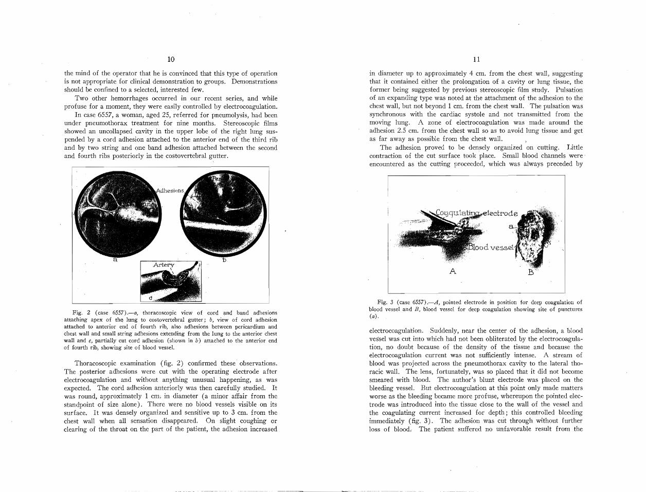
In case 6557, a woman, aged 25, referred for pneumolysis, had beenunder pneumothorax treatment for nine months. Stereoscopic filmsshowed an uncollapsed cavity in the upper lobe of the right lung suspended by a cord adhesion attached to the anterior end of the third riband by two string and one band adhesion attached between the secondand fourth ribs posteriorly in the costovertebral gutter.
Fig. 2 (case 6557).-a, thoracoscopic view of cord and band adhesionsattaching apex of thej lung to costovertebral gutter; b, view of cord adhesionattached to anterior end of fourth rib, also adhesions between pericardium andchest waH and smaH string adhesions extending from the lung to the anterior chestwaH and c, partiaHy cut cord adhesion (shown in b) attached to the anterior endof fourth rib, showing site of blood vessel.
Thoracoscopic examination (fig. 2) confirmed these observations.The posterior adhesions were cut with the operating electrode afterelectrocoagulation and without anything unusual happening, as wasexpected. The cord adhesion anteriorly was then carefully studied. Itwas round, approximately 1 cm~ in diameter (a minor affair from thestandpoint of size alone). There were no blood vessels visible on itssurface. It was densely organized and sensitive up to 3 cm. from thechest wall when all sensation disappeared. On slight coughing orclearing of the throat on the part of the patient, the adhesion increased
11
in diameter up to approximately 4 cm. from the chest wall, suggestingthat it contained either the prolongation of a cavity or lung tissue, theformer being suggested by previous stereoscopic film study. Pulsationof an expanding type was noted at the attachment of the adhesion to thechest wall, but not beyond 1 cm. from the chest wall. The pulsation wassynchronous with the cardiac systole and not transmitted from themoving lung. A zone of electrocoagulation was made around theadhesion 2.5 cm. from the chest wall so as to avoid lung tissue and getas far away as possible from the chest wall.
The adhesion proved to be densely organized on cutting. Littlecontraction of the cut surface took place. Small blood channels were'encountered as the cutting proceeded, which was always preceded by
Fig. 3 (case 6557) .-A, pointed electrode in position for deep coagulation ofblood vessel and B, blood vessel for deep coagulation showing site of punctures(a).
electrocoagulation. Suddenly, near the center of the adhesion, a bloodves'sel was cut into which had not been obliterated by the electrocoagulation, no doubt because of the density of the tissue and because theelectrocoagulation current was not sufficiently intense. A stream ofblood was projected across the pneumothorax cavity to the lateral thoracic wall. The lens, fortunately, was so placed that it did not becomesmeared with blood. The author's blunt electrode was placed on thebleeding vessel. But electrocoagulation at this point only made mattersworse as the bleeding became more profuse, whereupon the pointed electrode was introduced into the tissue close to the wall of the vessel andthe coagulating current increased for depth; this controlled bleedingimmediately (fig. 3). The adhesion was cut through without furtherloss of blood. The patient suffered no unfavorable result from the

13
ba
the costovertebral gutter, were cut, again without bleeding or unfavorablecomplications.
At the third operation, two dense cord adhesions attached to theanterior end of the first and second ribs were cut. All of these operations were technically difficult, but bleeding was perfectly controlled byelectrocoagulation, which alternated with the cutting. Following theoperations, each of which was done at intervals of two weeks, animproved collapse of the lung was noted on stereoscopic films after eachoperation. The quantity of the patient's sputum was gradually reducedto 15 cc. as shown by daily measurement.
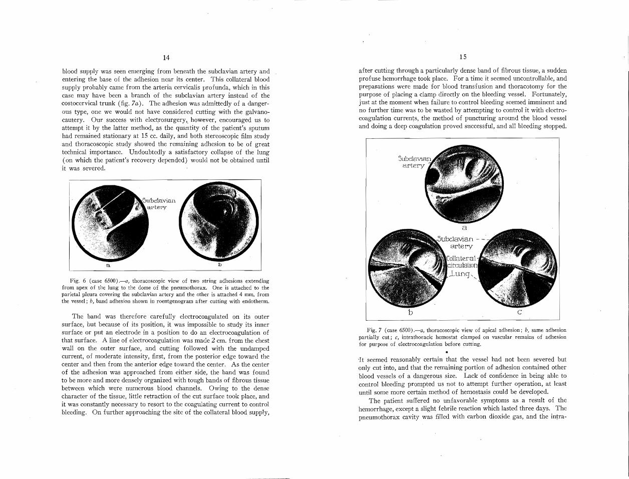
At the time of the previous operations, thoracoscopic study of theremaining adhesion showed that it was attached to the dome of thepneumothorax approximately 1 cm. distant from the subclavian arteryjust before this vessel crossed over the first rib. Furthermore, collateral
Fig. 5 (case 6500) .-a, thoracoscopic view of band adhesion attached to anteriorportion of second rib; b, view of same after cutting adhesion, showing bandattached to the aorta.
above the first rib. There were several cord and band adhesions attachedto the anterior end of the first, second and fourth ribs.
Thoracoscopic examination revealed conditions shown in figure 4. Allof the adhesions were densely organized and contained numerous subpleural blood vessels. At the first operation, a large band adhesionattached to the anterior end of the fourth rib, as well as a band adhesionholding the lung to the aorta (fig. 5) ~ and two string adhesions at theapex were cut. One of the latter was attached to the wall of the subclavian artery, while the other was attached 4 mm. from the vessel(fig. 6n). No bleeding occurred, hemostasis being perfectly controlledby electrocoagulation, and no reaction followed the operation.
At the second operation, two large band adhesions, one attached tothe first intercostal space (fig. 6b) and the second to the third rib in
Subclavianve~~!:l4 ?-TLe
Cavity-_ -. ,.,
.~ .:-"'-Adhesion·· ./
Fig. 4 (case 6500) .-Diagram showing distribution of adhesions between partially collapsed lung and chest wall as revealed by thoracoscopic examination.
bleeding. Two hundred and fifty cubic centimeters of blood was aspirated from the pneumothorax the following day. The operation was acomplete clinical success.
I am certain that the hemorrhage in this case would have been aserious matter if we had been obliged to rely on the galvanocautery forcontrol. As a matter of fact, I do not think we would have attemptedcutting this adhesion with agalvanocautery.
12
In the third case, no. 6500, a woman, aged 24, referred for pneumolysis, had been under pneumothorax treatment for ten months. Atfirst, the quantity of sputum became diminished and then remainedstationary, averaging from 40 to 60 cc. in twenty-four hours during thelast two months of pneumothorax treatment, when she also developedtuberculosis of the larynx and extension of disease to the opposite lung.
Stereoscopic films showed a large cavity in the upper lobe of the leftlung. There were many band and cord adhesions attached posteriorlyin the costovertebral gutter and to the dome of the pneumothorax cavity
cb
after cutting through a particularly dense band of fibrous tissue, a suddenprofuse hemorrhage took place. For a time it seemed uncontrollable, andprepar-ations were made for blood transfusion and thoracotomy for thepurpose of placing a clamp directly on the bleeding vessel. Fortunately,just at the moment when failure to control bleeding seemed imminent andno further time was to be wasted by attempting to control it with electrocoagulation currents, the method of puncturing around the blood vesseland doing a deep coagulation proved successful, and all bleeding stopped.
Fig. 7 (case 6500).-a, thoracoscopic view of apical adhesion; b, same adhesionpartially cut; c, intrathoracic hemostat clamped on vascular remains of adhesionfor purpose of electrocoagulation before cutting.
15
•It seemed reasonably certain that the vessel had not been severed butonly cut into, and that the remaining portion of adhesion contained otherblood vessels of a dangerous size. Lack of confidence in being able tocontrol bleeding prompted us not to attempt further operation, at leastuntil some more certain method of hemostasis could be developed.
The patient suffered no unfavorable symptoms as a result of thehemorrhage, except a slight febrile reaction which lasted three days. Thepneumothorax cavity was filled with carbon dioxide gas, and the intra-
bB.
blood supply was seen emerging from beneath the subclavian artery andentering the base of the adhesion near its center. This collateral bloodsupply probably came from the arteria cervicalis profunda, which in thiscase may have been a branch of the subclavian artery instead of thecostocervical trunk (fig. 7a). The adhesion was admittedly of a dangerous type, one we would not have considered cutting with the galvanocautery. Our success with electrosurgery" however, encouraged us toattempt it by the latter method, as the quantity of the patient's sputumhad remained stationary at 15 cc. daily, and both stereoscopic film studyand thoracoscopic study showed the remaining adhesion to be of greattechnical importance. Undoubtedly a satisfactory collapse of the lung(on which the patient's recovery depended) would not be obtained untilit was severed.
Fig. 6 (case 6500).-a, thoracoscopic view of two string adhesions extendingfrom apex of the lung to the dome of the pneumothorax. One is attached to theparietal pleura covering the subclavian artery and the other is attached 4 mm. fromthe vessel; b, band adhesion shown in roentgenogram after cutting with endotherm.
14
The band was therefore carefully electrocoagulated on its outersurface, but because of its position, it was impossible to study its innersurface or put an electrode in a position to do an electrocoagulation ofthat surface. A line of electrocoagulation was made 2 cm. from the chestwall on the outer surface, and cutting followed with the undampedcurrent, of moderate intensity, first, from the posterior edge toward thecenter and then from the anterior edge toward the center. As the centerof the adhesion was approached from either side, the band was foundto be more and more densely org'anized with tough bands of fibrous tissuebetween which were numerous blood channels. Owing to the densecharacter of the tissue, little retraction of the cut surface took place, andit was constantly necessary to resort to the coagulating current to controlbleeding. On further approaching the site of the collateral blood supply,
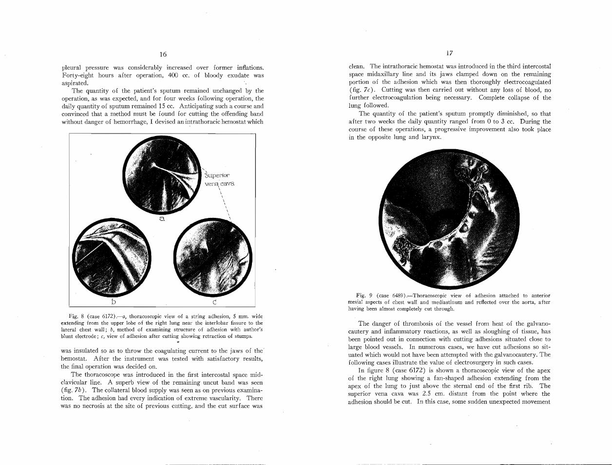
Fig. 9 (case 6489).-Thoracoscopic view of adhesion attached to anteriormesial aspects of chest wall and mediastinum and reflected over the aorta, afterhaving been almost completely cut through.
clean. The intrathoracic hemostat was introduced in the third intercostalspace midaxillary line and its jaws clamped down on the remainingportion of the adhesion which was then thoroughly e1ectrocoagu1ated(fig. 7c). Cutting was then carried out without any loss of blood, nofurther electrocoagulation being necessary. Complete collapse of thelung followed.
The quantity of the patient's sputum promptly diminished, so thatafter two weeks the daily quantity ranged from 0 to 3 cc. During thecourse of these operations, a progressive improvement also took placein the opposite lung and larynx.
17
The danger of thrombosis of the vessel from heat of the galvanocautery and inflammatory reactions, as well as sloughing of tissue, hasbeen pointed out in connection with cutting adhesions situated close tolarge blood vessels. In numerous cases, we have cut adhesions so situated which would not have been attempted with the galvanocautery. Thefollowing cases illustrate the value of electrosurgery in such cases.
In figure 8 (case 6172) is shown a thoracoscopic view of the apexof the right lung showing a fan-shaped adhesion extending from theapex of the lung to just above the sternal end of the first rib. Thesuperior vena cava was 2.5 cm. distant from the point where theadhesion should be cut. In this case, some sudden unexpected movement
c
"-
5uperiorvena cava
\\\\\\\\
\
b
.-.-----__.__~
pleural pressure was considerably increased over former inflations.Forty-eight hours after operation, 400 cc. of bloody exudate wasaspirated.
The quantity of the patient's sputum remained unchanged by theoperation, as was expected, and for four weeks following operation, thedaily quantity of sputum remained 15 cc. Anticipating such a course andconvinced that a method must be found for cutting the offending bandwithout danger of hemorrhage, I devised an intrathoracic hemostat which
16
Fig. 8 (case 6172).-a, thoracoscopic view of a string adhesion, 5 mm. wideextending from the upper lobe of the right lung near the interlobar fissure to thelateral chest wall; b, method of examining structure of adhesion with author'sblunt electrode; c, view of adhesion after cutting showing retraction of stumps.
was insulated so as to throw the coagulating current to the jaws of the'hemostat. After the instrument was tested with satisfactory results,the final operation was decided on.
The thoracoscope was introduced in the first intercostal space midclavicular line. A superb view of the remaining uncut band was seen(fig. 7b). The collateral blood supply was seen as on previous examination. The adhesion had every indication of extreme vascularity. Therewas no necrosis at the site of previous cutting, and the cut surface was

18
or coughing paroxysm on the part of the patient could easily haveresulted in damage with a heated galvanocautery so close to a large bloodvessel, for in spite of shutting off the current at the time the unexpectedact was committed, the cautery could still remain sufficiently hot to dodamage, whereas with the endotherm the instant the current is off, it isinactive and incapable of doing damage.
Figure 9 (case 6489) shows a thoracoscopic view of a dense bandadhesion with its base attached to the anterior mesial aspects of thepneumothorax cavity and reflected over the aorta to which it wasattached, showing cutting effected by the endotherm after electrocoagulation. This operation was executed without the loss of blood, and whileextensive, it was not followed by inflammatory reaction on the part ofthe pleura. Complete collapse of the lung followed, and the patient'sconvalescence was uneventful.
In this case the operation was extremely difficult and prolonged, andwithout electrosurgery it would have been impossible to carry it outsafely. It could not have been done with the galvanocautery except ata great risk.
COMMENT
1. Intrapleural pneumolysis is an operation of great utility. Whenit is properly ·done it is not dangerous and will convert a useless pneumothorax into an efficient one, thus saving the patient from thoracoplasty.
2. My experience with the electrosurgical method has given meconfidence in this method of cutting adhesions. Control of bleeding isthe most dangerous problem and requires thorough knowledge of thecharacter of the currents used. Electrosurgical cutting is accomplishedwithout heat or smoke to disturb the view. There is a minimum of tissuereaction afterward, and while more complicated and technically moredifficult than the galvanocautery method, it is without doubt a notableadvance in this branch of surgery, which is being more widely employed.
3. Intrapleural pneumolysis by the closed method is not a fool-proofprocedure with either the galvanocautery or the electrothermic method.The operator must be familiar with the appearance of the pleural cavityand at all times perfectly orientated regarding the nature of tissue to becut. This training in the use of the thoracoscope in the pleural cavityis just as important to the surgeon or phthisiotherapeutist as a thoroughknowledge of the cystoscopic image is to the urological surgeon. Theoperator should have experience with pneumothorax and must havesurgical training.
Acknowledgment is due Mrs. Henry F. Chaney, who, by a grant to the PortlandOpen Air Sanatorium, aided in this work.
Grateful acknowledgment is hereby made to my associates, Dr. Ray W. Matsonand Dr. Marr Bisaillon, for valuable assistance and many helpful suggestions.
COPYRIGHT, 1929
AMERICAN MEDICAL ASSOCIATION
535 NORTH DEARBORN STREET
CHICAGO