the electrocardiogram of a camel
TRANSCRIPT
The Electrocardiogram of a Camel
K. Braun, M.D., S. Z. Rosenberg, M.D., and L. Be&n, M.D.,* Jerusalem, Israel
INTRODUCTION
The investigations of White and his associates1 on the elephant have brought forward evidence of a relationship between heart size, heart rate, and time in- tervals of the cardiac cycle. They have shown that in the elephant the increase of heart size is accompanied by a slow heart rate and lengthening of the P-R, QRS, and Q-T intervals. The fact that the duration of these intervals was out of proportion to the heart rate was explained by the large mass of heart muscle and by the long paths of impulse conduction. In a recent report King, Jenks and White2 described the electrocardiogram of a beluga whale. Although the heart of this huge animal is much smaller than that of the elephant, its heart rate was found to be about half that of the elephant.
The aim of the present study is to provide additional information on the heart rate and electrocardiographic features in large animals. For this purpose the Camelus dromedarius was chosen.
TECHNIQUE OF RECORDING
During the investigation, the camel, a male, weighing 265 kilograms, was in a quiet, sitting position. The usual electrodes were easily placed on the extremities. The tracings were re- corded on a direct-writing electrocardiograph, with the standardization 1 cm. = 1 mv. Three bipolar standard limb leads and three unipolar augmented limb leads were obtained. Unipolar chest leads were explored from 20 different positions around the chest in a sagittal plane. In addition, several other points on the shoulders and neck base were explored. The effect of exercise on the electrocardiogram was studied after the camel had walked for a half hour carry- ing 120 kilograms of weight on its back. Electrocardiographic tracings were similarly recorded, as mentioned above.
GENERAL DESCRIPTION
The chief feature of the electrocardiogram was the sinus bradycardia. The pulse rate ranged between 24 and 30 per minute. This change in the heart rate was due to sinus arrhy-
From the Cardiovascular Unit and Department of Internal Medicine, Division “B”, Rothschild Hadassah University Hospital, Jerusalem, Israel.
This study was carried out with the aid of a Research Grant from the Hadassah Medical Organiza- tion, Jerusalem, Israel.
Received for publication Jan. 6. 1958. *Fellow in Cardiology. (Formerly of Boston, Mass.)
754
~~*~~~ “5” ELECTROCARDIOGRAM OF A CAMEL 755
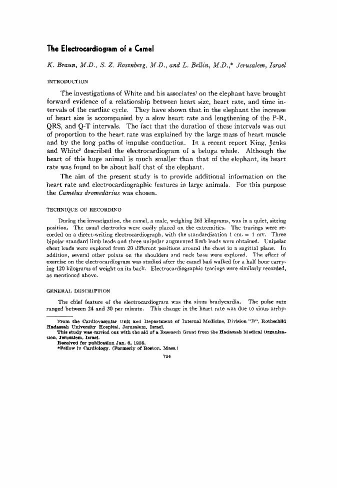
thmia. The P waves were well defined, their maximal width was 0.10 sec., and their maximal height 0.2 mv. The P-R interval ranged from 0.24 to 0.26 sec. The P-R segment was isoelec-
tric. The ventricular complexes exhibited small Q waves in some leads and well-defined R and S waves. The maximal duration of the QRS complex was 0.09 sec. and the maximal height was 1.5 mv. No notching or slurring of the ventricular complexes was noted except in Lead I. The direction of the QRS complex was opposite to that of the P wave. The S-T segment was isoelectric. The Q-T interval ranged from 0.54 to 0.60 sec. The T waves were well defined, maximally measuring 0.9 mv. The direction of the T waves was identical to that of the QRS complexes (Fig. 1).
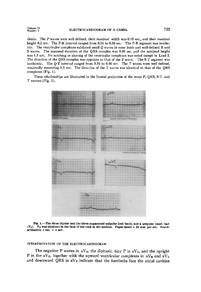
These relationships are illustrated in the frontal projection of the mean P, QRS, S-T, and T vectors (Fig. 2).
Fig. I.-The three bipolar and the three augmented unipolar limb leads, and a unipolar cheat lead (V,). VN was obtained in the base of the neck in the midline. Paper speed = 26 mm. per sec. Stand- ardization 1 cm. = 1 mv.
INTERPRETATION OF THE ELECTROCARDIOGRAM
The negative P waves in aVR, the diphasic, tiny P in aVL, and the upright P in the aVF, together with the upward ventricular complexes in aV= and aVL and downward QRS in aVF indicate that the forelimbs face the atria1 cavities
756 BRAUN, ROSENBERG, AND BELLIN Am. Heart J. May, 195s
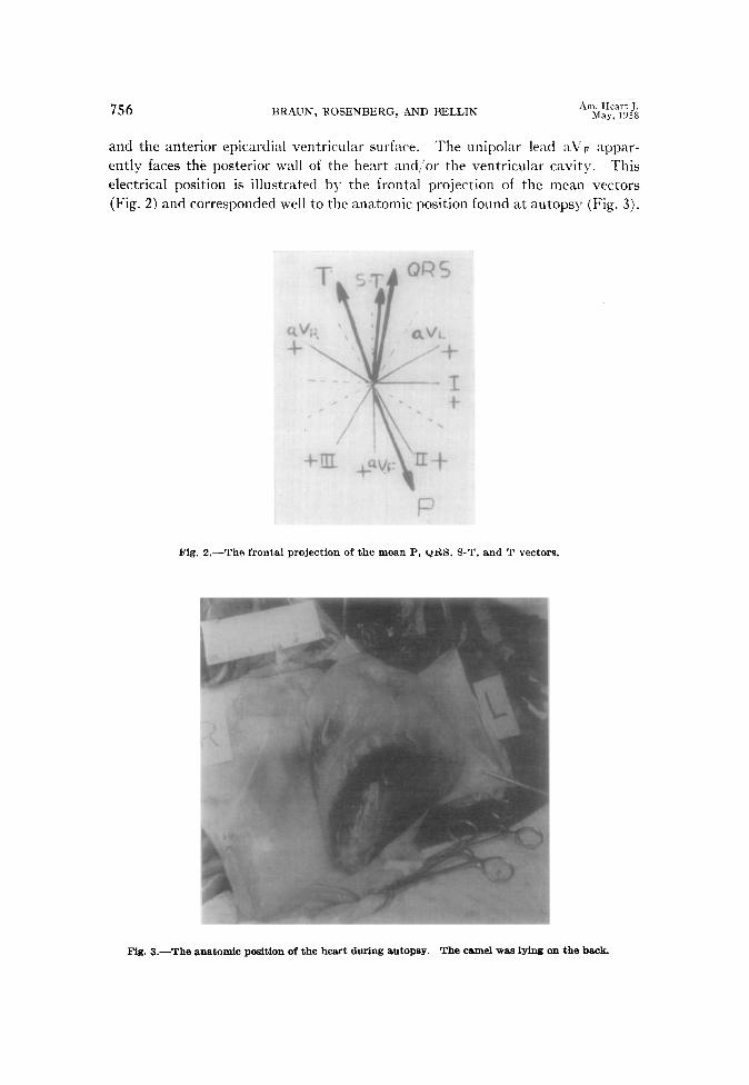
and the anterior epicardial ventricular surface. ‘The unipolar lead aV F “pp jar- ently faces the posterior wall of the heart and,/or the ventricular cavit :y. ‘I ‘his electrical position is illustrated by the frontal projection of the mear 1 vect :ors (Fig. 2) and corresponded well to the anatomic position found at autopsy (Fig. 3).
Fig. 2.-The frontal projection of the moan P. ydS. S-T, and T vectors.
Fig. 3.-The anatomic position of the heart during autogsy. The camel was lying on the back.
Volume 55 Number 5 ELECTROCARDIOGRAM OF A CAMEL 757
COMMENT
Fifty years ago it was stated that the larger the heart, the slower the heart rate.“v4 This is true when comparing a mouse with an elephant, or a hamster with a whale,2y5 but this relationship does not hold true among the large-sized animals. This becomes evident from the reports of White himself.‘r2 He found a rate of 30 per minute in the elephant’s heart weighing about 20 kilograms, while the rate was 15 per minute in a beluga whale’s heart that weighed 2.72 kilograms. The difference was explained by the factor of diving and the consequently in- creased vagal activity in the beluga whale. Our camel’s heart weighed about a twentieth of that of the elephant (1.3 kilograms) and still the heart rate was about the same, or even less. Diving can certainly be excluded as a factor in our camel, and in the elephant as well. An additional noteworthy observation is that the duration of the various time intervals of the cardiac cycle in the camel is not much out of proportion to the slow heart rate as it has been found in the elephant. The width of the QRS complexes was identical with that of a man with much higher heart rate, and the Q-T interval corresponded to the predicted values determined by Bazett’s formula.
After exercise there were no remarkable changes in heart rate, configuration, and time intervals of the electrocardiogram. The possibility exists that the effort was not sufficient to bring about hemodynamic changes which could be reflected in the electrocardiogram.
A more complete investigation of the cardiovascular system of the camel, with special emphasis on the cardiac and renal hemodynamics, is now being carried out in our laboratory.
SUMMARY
Electrocardiograms of a Camelus dromedarius were obtained at rest and after effort. The voltage of P-QRS-T was similar to that of the human electro- cardiogram. The characteristic features were : bradycardia (26 per minute), normal width of QRS complexes, and prolonged P-R and Q-T intervals; the dura- tion of these latter two intervals was proportional to the heart rate. The T waves were in the same direction as the QRS complexes, while the P waves were in the opposite direction. The electrical position of the heart, as obtained by the stand- ard and unipolar limb leads, corresponded well to the anatomic position found at autopsy.
The technical assistance of Mr. Shmuel Werkson is gratefully acknowledged.
REFERENCES
1. 2.
White, P. D., Jenks, J. L., and Benedict, F. G.: AM. HEART J. 16:744, 1938. King, R. L., Jenks, J. L., and White, P. D.: Circulation 8:387, 19.53.
3. Buchanan, F.: by King.4
Trans. Oxford Univ. Junior Scientific Club, 1909, n.s. No. 34, p. 351: Quoted
4. 5.
King, R. L., Burwell, C. S., and White, P. D.: AM. HEART J. 16:734, 1938. Braun, K., Stuczynski, L. A., and Grossowicz, N.:
1949. Proc. Sot. Exper. Biol. & Med. 7258,