the effects of bilateral eye movements on eeg...
TRANSCRIPT
Journal of EMDR Practice and Research Volume 8 Number 3 2014 113copy 2014 EMDR International Association httpdxdoiorg1018911933-319683113
E ye movement desensitization and reprocess-ing (EMDR) has been recognized as an effec-tive and efficient therapeutic approach for the
treatment of effects of traumatic memories (Ameri-can Psychiatric Association 2004 Bisson et al 2007 Cukor Olden Lee amp Difede 2010 Lamprecht et al 2004) These positive endorsements notwithstanding empirical comparisons of EMDR with other popular trauma treatments such as prolonged exposure stress inoculation training cognitive behavior therapy and relaxation therapy have been overall equivocal Some studies have shown EMDR to be more effective oth-ers have shown it to be less effective and others have shown it to be equivalent (eg Davidson amp Parker 2001 Devilly amp Spence 1999 Ironson Freund Stauss amp Williams 2002 Lee Gavriel Drummond Richards amp Greenwald 2002 Power et al 2002 Rothbaum Astin amp Marsteller 2005 Taylor et al 2003) Nevertheless EMDR remains one of the more popular treatments for posttraumatic stress disorder (PTSD Pagani Hogberg Fernandez amp Siracusano
2013) Perhaps a better understanding of the compo-nents of this therapeutic intervention would lead to subtle refinements in the protocol which would pro-duce even better outcomes and improved assistance to trauma victims
A core component of EMDR that distinguishes it from other trauma treatment strategies is the use of bilateral stimulation during the contemplation of traumatic target events (Shapiro 1989 Shapiro amp Maxfield 2002 Solomon amp Shapiro 2008) Following Shapirorsquos adaptive information processing (AIP) model (Shapiro 2001 Solomon amp Shapiro 2008) this bilateral stimulation is posited to activate more remote neural networks to allow the linking of disso-ciated information with the target traumatic events thus facilitating the reprocessing of these events and their eventual desensitization Originally Shapiro (1989) used bilateral visual stimulation through the movement of fingers laterally across the visual field at a rate of approximately two saccadic eye move-ments per second However over the years since her
The Effects of Bilateral Eye Movements on EEG Coherence When Recalling a Pleasant Memory
Brandon KellerLarry StevensColleen Lui
James MurrayMatthew Yaggie
Northern Arizona University
In an investigation of the interhemispheric coherence (IhC) model for eye movement desensitization and reprocessing (EMDR) bilateral eye movement (BEM) effects 30 participants were exposed to a stationary dot a blinking redgreen dot or saccadic BEMs during the contemplation of a positive emotional mem-ory Electroencephalographies (EEGs) were measured afterward during an eyes-closed processing stage Analyses revealed no significant IhC enhancement for the BEM condition but significant increases in Delta and Low Beta EEG intrahemispheric BEM coherence in the right and left frontal areas respectively and a trend increase in Right Frontal Low Beta BEM coherence LORETA neuroimaging was employed to visually present significant amplitude changes corresponding to observed coherence effects The func-tional significance of these intrahemispheric coherence effects is presented and a cortical coherence extension of the IhC model is suggested
Keywords EMDR bilateral eye movements EEG coherence episodic memory PTSD
114 Journal of EMDR Practice and Research Volume 8 Number 3 2014 Keller et al
occur (a) Memory retrieval should improve during or immediately following bilateral stimulation and (b) measures of interhemispheric connection should show an increase following bilateral stimulation These two predictions have received some empirical support from research to date
For example Christman et al (2003) found en-hanced word recognition and autobiographical memory retrieval following a 30-second engage-ment in horizontal saccadic eye movements These outcomes have been supported by earlier studies of handedness (as a representation of interhemispheric interaction) and the effects of a sequential presenta-tion of bilateral visual input on episodic memory (Christman amp Propper 2001) Additional research showing enhanced behavioral measures of interhemi-spheric interaction and creativity following bilateral eye movements (BEMs Shobe Ross amp Fleck 2009) improved memory and accuracy for a visual event narrative after BEMs (Parker Buckley amp Dagnall 2009) enhanced memory retrieval (Christman et al 2003 Lyle Logan amp Roediger 2008) im-paired episodic memory following commissurotomy (Cronin-Golomb Gabrieli amp Keane 1996) and other studies (see Propper amp Christman 2008 for a com-prehensive review of this literature) strongly support the enhancement of episodic-like memory retrieval following the presentation of bilateral saccadic eye movements
The research literature has been more sparse and equivocal however for the effects of bilateral stimulation on direct measures of interhemis pheric connectivity One such measure of functional connectivity is electroencephalography (EEG) inter-hemispheric coherence (IhC) EEG coherence is a quantitative measure of EEG waveform or phase consistency between two disparate sites on the scalp (Nunez et al 1997) Mathematically coherence values represent the EEG waveform cross-spectral density function normalized by the power spectra and are represented by a squared correlation func-tion having a magnitude between 0 and 121 Thus coherence may be interpreted as the functional com-munication or connectivity between two recording sites with higher coherence representing higher co-operation and synchronization between measured brain regions in a specified frequency (Knott LaBelle Jones amp Mahoney 2002 Nunez et al 1997 Weiss amp Mueller 2003) Bergmann (2008) asserts that syn-chronized neuronal oscillations as indexed broadly by cortical EEG coherence are the basis of human per-ception and functioning If the selected recording sites are homologous sites on opposite sides of the cortex
discovery of the contributions of this component to traumatic memory reprocessing bilateral auditory and kinesthetic stimulation has been used as well with equivalent anecdotal effects (Harper 2012)
Although many theories have now been offered to explain the contributions of bilateral stimulation to the processing and depotentiation of traumatic mem-ories (Bergmann 2008) the mechanisms of action of this component have to date not been conclusively explicated One of the more neurobiological mod-els for the effects of bilateral stimulation on PTSD the amygdala-anterior cingulate (ACC)prefron-tal cortical (PFC) coupling model has to do with a growing body of evidence for (a) an overactivation of amygdaloid processes involved in the affective ex-periencing of traumatic events combined with (b) a deactivation or decoupling of ACC and medial PFC functions that would otherwise permit a cognitive processing and depotentiation of such events in PTSD (Francati Vermetten amp Bremner 2007) In an even more reductionistic analysis this model of PTSD symptomatology further hypothesizes that traumatic memories are locked into reverberating synaptic networks of overpotentiated alpha-amino-3-hydroxy-5-methyl-4-isoxazole (AMPA) receptors within the amygdala (Harper Rasolkhani-Kaophorn amp Drozd 2009) (c) This state of pathological processing of trauma is essentially reordered by bilateral sensory stimulation during the reexperiencing of the event by providing the low frequency tetanic stimulation necessary to depotentiate these AMPA receptors and subsequently the retained amygdaloid memories (d) Such a depotentiation of locked neural networks then allows these affective memories to spread into AC and PFC regions where they may be more natu-rally and cognitively reprocessed Components of this model have received some support from animal and human neuroimaging studies (for a thorough review of this literature see Pagani et al 2013)
Shapiro (1989) had early suggested that saccadic bi-lateral visual stimulation in EMDR may recruit neural networks from opposite sides of the brain and allow heretofore dissociated networks to become linked to targeted traumatic events toward their eventual reprocessing Initially proposed by Servan-Schreiber (2000) and empirically elaborated by Christman and colleagues (Christman Garvey Propper amp Phaneuf 2003 Christman Propper amp Brown 2006 Christman Propper amp Dion 2004 Propper amp Christman 2008) this interhemispheric connectivity hypothesis for the effects of bilateral stimulation on episodic memory retrieval has received considerable investigation If this hypothesis is correct then two outcomes should
Journal of EMDR Practice and Research Volume 8 Number 3 2014 115Eye Movements Coherence and Pleasant Memories
initial participant pool had to be rejected from ana-lysis because of noisy unusable EEGs) Second their EEG coherence values particularly for Alpha and Theta were very high approaching 100 even prior to stimulus conditions suggesting a ceiling effect and lessening the likelihood of obtaining significant and meaningful coherence changes And third there was no episodic memory recall task required during the eye movement condition as occurs in EMDR pro-viding no directed task-specific activity as a basis for neural network coordination
The second well-designed and tightly controlled study by Samara Elzinga Slagter and Nieuwenhuis (2011) computed full-scalp EEG phase and ampli-tude coherence prior to participation in a neutral and emotional wordndashrecall task and in the same BEM and control conditions as Propper et al (2007) but using a more powerful within-subjects design In addition these researchers recorded electrooculograms to ver-ify BEMs and painstakingly visually and statistically artifacted their EEG data to remove muscle and noise artifacts Disappointingly for the IhC model and fol-lowing multiple and reduced stringency analyses Samara et al found no consistent or predicted phase or amplitude EEG coherence changes from pre- to post-BEMs or across eye movement conditions They did observe significantly decreased Alpha amplitude coherence bilaterally for the F7ndashF8 electrodes in the BEM condition but an increase in Alpha amplitude coherence for these electrodes in the control condi-tion Although they found a significant improvement in recall of emotional words only for the BEM con-dition there was no significant correlation between coherence and word recall At first glance this study considerably challenges the IhC model for the re-ported effectiveness of EMDR and more specifically for the well-documented improvements in memory retrieval following BEMs
However an important shortcoming of this study acknowledged by the authors was the absence of a true episodic memory retrieval task (Tulving 1985) during the eye movement component of the study Indeed not only was the cognitive task used in this study a semantic memory recall task but also a 30-minute ldquoneutral documentaryrdquo film followed word presentation and occurred before EEGs were recorded and BEMs were prompted As noted earlier the hold-ing of the traumatic event in working memory during the BEMs is an important and unique characteristic of therapeutic EMDR Very few of the published studies of the effects of BEMs on memory retrieval in fact used personally meaningful episodic memory tasks and instructed their participants to contemplate those
(eg electrodes F7 and F8 see Figure 1) then EEG coherence is an ideal measure of interhemispheric neuroelectrical connectivity
Only two studies have been published to date however examining the effects of saccadic horizon-tal eye movements on IhC Propper Pierce Geisler Christman and Bellorado (2007) examined IhC from two frontal bilateral electrode sites (Fp1 and Fp2) before and after a 30-second presentation of either a two-saccadic-eye-movements-per-second moving dot or a stationary redgreen twice per second blinking dot in a between-groups design Their results ob-tained in contradiction to their initial predictions decreased coherence in the Gamma EEG frequency band (35ndash54 Hz) with no effects on Theta (4ndash8 Hz) or Alpha (8ndash13 Hz) bands following the moving stimu-lus relative to the control blinking dot condition The authorsrsquo posthoc interpretation of these surprising outcomes as indicating significant eye movementndashinduced changes in interhemispheric coordination notwithstanding there were several critical problems with this study which render its relevance to EMDR practice and to task-related interhemispheric connec-tivity questionable First they selected two recording sites over the frontalis muscles which are very sen-sitive to residual eye movementndashinduced muscle artifacts and recorded from these sites within 3 sec-onds of stimulus offset potentially contaminating the EEG recordings with muscle artifacts which can be reflected in the Gamma and Delta bands (They failed to report Delta and approximately 20 of their
FIGURE 1 Schematic of international 10ndash20 system for EEG electrode placement
A2C4 T4
P4T6
CZ
PZ
FZ
G
F4F8
Fp2
T3 C3
P3T5
O1 O2
F3F7
Fp1
A1
116 Journal of EMDR Practice and Research Volume 8 Number 3 2014 Keller et al
participant was randomly assigned to one of the three treatment conditions in this between-groups design All participants received course credit for their partici-pation in this study and the study was approved by the Northern Arizona University (NAU) IRB
Instruments
Prior to the EEG portion of the study each participant completed a demographic information form contain-ing relevant identifying information age gender pregnancy status hand preference incidence of neu-rological conditions which could influence the EEG recording and prescribed and recreational medica-tiondrug use In addition each participant completed the Edinburgh Handedness Inventory (Oldfield 1971) to verify right-hand preference A 1ndash10 (10 5 very strong) visual analogue scale (VAS) was used to record memory strength and vividness at baseline and after each stimulus set for each condition
The control visual stimulation conditions consisted of (a) a stationary black dot 3 in in diameter the eye fixation (EF) condition selected to control for effects of alternating visual stimulation in general and (b) an alternating redgreen dot also 3 in in diameter which changed color every 500 milliseconds the Blinking Dot (Blink) condition patterned after the control con-dition reported in Experiment 2 by Christman et al (2004) Both control conditions were presented on a laptop with a 15-in monitor positioned directly in front of the participant at eye level and 30 in away To be as consistent as possible both with EMDR proto-col and across participants bilateral visual stimulation was provided by an EyeScan 2000S Light Bar (1994 NeuroTek Corporation Wheat Ridge Colorado) de-signed for clinical EMDR use Bilateral saccades were set at one leftndashright or rightndashleft saccade every 500 mil-liseconds producing two eye movements per second for 24 seconds The light bar was positioned at eye lev-el 35 cm (approximately 14 in) from the participant
EEG data were recorded using a Lexicor NRS-24C (1989 Lexicor Medical Technology Inc Boulder Colorado) EEG recording system having a 512 Hz dig-ital sampling rate a 128 Hz low-pass antialiasing filter and a fixed 05 Hz high-pass filter The Lexicor NRS-24C used a Neurosearch-24 Acquisition Unit containing 24 channels of differential frontndashend preamplifiers followed by isolation amplifierstransformers ana-log-to-digital (AD) converters and optical isolators for participant protection Resident Neurosearch-24 V41E EEG recording and analysis software was used to record raw EEG data into event files for each treatment condition The 19-channel EEG data were
memories in working memory during bilateral stim-ulation The exceptions to this important omission were the second experiment conducted by Christman et al (2003) which found selective enhancement of true episodic memories following BEMs and the study of contemplation of childhood memories dur-ing BEMs by Christman et al (2006) which found earlier offset of childhood amnesia Neither of these studies however measured EEG coherence
Until IhC is measured during or immediately fol-lowing bilateral stimulation while the participant is contemplating personally meaningful episodic memories the IhC model for the effects of EMDR on the reprocessing of traumatic memories remains untested In addition because the activation of re-mote neural networks proposed by the AIP model can also occur within hemispheres this investigation examined intrahemispheric coherence This study begins this line of investigation by the recording and analysis of multichannel interhemispheric and intra-hemispheric EEG coherence following BEMs and two control conditions during the contemplation of personally meaningful positive episodic memories Positive memories were used in this investigation to avoid potentially retraumatizing our young non-clinical sample to facilitate institutional review board (IRB) approval and because of the practice of install-ing positive memories with bilateral saccades in the development of The Safe Place and Resources during clinical EMDR
Methods
Participants
Participants were 30 right-handed female under-graduate students from a southwestern university re-cruited as nonclinical volunteers from the psychology department subject pool Mean age was 1913 years (SD 5 256) and there was no significant age differ-ence among the three treatment conditions (F 5 854 p 5 437) No participant reported present pregnancy or a history of head injury unconsciousness epilepsy chronic pain psychiatric or PTSD history or neurop-athy Nine reported taking birth control medication 2 were taking asthma medication and 2 were taking an undisclosed other medication medication use was evenly distributed across the three treatment condi-tions (x2 5 5 p 05) Street drug use was minimal and occasional with 1 participant reporting marijuana use 5 reporting alcohol use 3 reporting pain killer use 1 reporting upper use and 3 reporting other drug use no participant reported using amphetamines cocaine benzodiazepines downers or ecstasy Each
Journal of EMDR Practice and Research Volume 8 Number 3 2014 117Eye Movements Coherence and Pleasant Memories
configured the CD for the designated stimulus was started The experimenter remained present through-out the session to operate the EEG equipment and to monitor eye movements
Before presentation of the visual stimulus condi-tion a 5-minute EEG baseline was recorded with eyes closed during which the participants were asked to blank their mind and then to ldquoallow whatever thought feeling or experience comes uprdquo to be considered After this baseline the participants were invited to consider an episodic memory from their childhood which holds very positive emotions for them to signal when this memory had been selected to briefly report the memory and to rate its strength and vividness on the VAS scale Then they were instructed to focus on the visual stimulus (either the stationary black dot the blinking redgreen dot or the bilateral moving dot on the light bar) while contemplating the positive episodic memory for 24 seconds During the BEM con-dition the participants were instructed to move only their eyes from side to side and not their head and their cooperation with this instruction was verified by the researcherrsquos observation of the reflected image in the mirror Following the presentation of the visual stimulus condition the participants were instructed to close their eyes to blank their mind and then to ldquocontemplate whatever thoughts feelings or experi-ences come uprdquo while a 1-minute EEG was recorded At the end of this 1-minute recording period the par-ticipants were asked to again report the ldquostrength and vividnessrdquo of the memory on the 1ndash10 VAS scale This sequence of visual stimulation followed by blanking the mind followed by contemplation during which a 1-minute EEG and memory strength and vividness were recorded was repeated five times for 5 minutes of EEG during contemplation of the positive episodic memory following presentation of the visual stimu-lus After this sequence of recordings was completed the participants were debriefed the Electro-Cap was removed extra credit was awarded and the partici-pants were allowed to leave
Design and Analysis
Following the recording of the 5-minute eyes-closed baseline and the 5 minutes of eyes-closed poststimu-lus EEG data were artifacted and subjected to FFT analysis The mean number of artifact-free 1-second epochsparticipant used in the coherence analyses was 22810 (SD 5 3983) or an average of 380 min-utes (SD 5 066) of artifact-free EEG data for each participant for baseline and for poststimulus analy-ses separately As a part of the Eureka output phase
collected with an AD conversion sampling rate set at 256 Hz high-pass and low-pass filters set at 5 Hz and 60 Hz respectively and notch filter set at 60 Hz The international 10ndash20 EEG electrode placement system was followed for the placement of the 19 monopolar AgAgCl electrodes onto the scalp using the Electro-Cap System (1983 Electro-Cap International Inc Eaton Ohio) with mathematically linkedndashear refer-ence electrodes Electrode impedances were adjusted to less than 5 kohms and to within 1 kohm of each other Data were analyzed and artifacted using Nova Tech EEG Eureka and MHyT data processing and analysis software (2000 Nova Tech EEG Inc Mesa Arizona) Raw EEG data were twice visually artifact-ed by two trained and independent artifactors blind to treatment conditions using precisely written and exacting criteria to remove EMG and other noise arti-fact1 EEG analysis software was employed to conduct fast Fourier transformations (FFT) and power spectral and coherence analyses of raw data and LORETA neuroimaging software (LORETA Low Resolution Electromagnetic Tomographic Analysis Zurich Switzerland) was used to conduct topographic imag-ing and cortical localization of treatment effects FFT analysis employed Hamming time domain tapering Blackman frequency domain smoothing an overlap-ping FFT windows advancement factor of 8 and a moving average smoothing filter of 3 In these anal-yses 10 EEG frequency bins were examined Delta (1ndash399 Hz) Theta (4ndash799 Hz) Low Theta (4ndash599 Hz) High Theta (6ndash799 Hz) Alpha (8ndash1199 Hz) Low Alpha (8ndash999 Hz) High Alpha (10ndash1199 Hz) Beta (12ndash30 Hz) Low Beta (12ndash1999 Hz) and High Beta (20ndash30 Hz)
All data were recorded in a sound attenuated re-search suite with participants seated comfortably and erect in a recliner A mirror was positioned on the wall opposite from and oblique to the participant such that the researcher could observe the presence of eye move-ments and establish whether the eyes were opened or closed without the participant seeing their reflection in the mirror All instructions were standardized and prerecorded to separate CDs for each condition
Procedure
Participants were randomly assigned to scheduled EEG study times and on arrival they completed the requisite informed consent form and relevant questionnaires while the Electro-Cap was fitted and calibrated to the EEG recording system and clean EEG traces were established After the participants were made comfortable and the visual stimulus was
118 Journal of EMDR Practice and Research Volume 8 Number 3 2014 Keller et al
more comparisons than degrees of freedom for effect no adjustment for inflation of family-wise error rate was required (Tabachnick amp Fidell 2013) Alpha for significance was set at 05
In addition to better localize functional brain re-gions potentially affected by visual stimulation during contemplation of positive episodic memories Low Resolution Electromagnetic Tomographic Analysis (LORETA) was used LORETA is a three-dimensional brain imaging software companion to contemporary EEG analyses allowing localization of deep cortical source potentials for recorded surface EEG signals (Pascual-Marqui Esslen Kochi amp Lehmann 2002) LORETA algorithms compute a three-dimensional inverse solution space of cortical gray matter and hippocampi mapped onto a probabilistic Talairach atlas partitioned into 2394 7mm3 volumetric units or voxels Brodmann anatomical labels may be reported for relevant regions of interest using the Montreal Neurological Institute realistic head model For this study LORETA analyses were conducted on the nat-ural log transformation of FFT relative power spectral output for each identified frequency and relevant sta-tistically significant cortical voxels are reported
Results
Effects on Memory Strength and Vividness
Figure 2 presents graphically the changes in memory strength and vividness ratings for each condition from baseline across each of the five visual stimula-tion trials Repeated measures analysis of variance (ANOVA) results show a significant main effect for time (F [5 23] 5 780 p 0001 h2 5 63) but not for condition (F [2 27] 5 59 p 5 56 h2 5 04) and no significant interaction (F [10 46] 5 54 p 5 86 h2 5 10) indicating an increase in memory strength and vividness for the positive memory across time for all three conditions and no differences among condi-tions at any measurement point including at base-line An inspection of these graphs however reveals a different pattern of responses for the BEM condition compared with the two control conditions with the latter tending to plateau at the third trial but mem-ory continuing to increase rather consistently across all trials but one for the BEM condition T-test com-parisons between successive trials support this visual pattern difference with a significant increase for the EF condition only from Trial 2 to 3 (t[9] 5 400 p 5 002) and for the Blink condition only from Trial 1 to 2 (t[9] 5 181 p 5 05) but for the BEM condition from Trial 1 to 2 (t[9] 5 196 p 5 04) Trial 2 to 3 (t[9] 5 181 p 5 05) and Trial 4 to 5 (t[9] 5 181 p 5 05)
coherence values between all possible pairs of elec-trodes for each designated EEG frequency are gener-ated as cross-spectral density functions normalized by individualized power spectra presented as a squared correlation matrix for each frequency Phase cohe-rence analyses used the following formula (Nunez amp Srinivasan 2006)
Coherence ( f ) 5
|Cross 2 Spectrum ( f ) XY|2
(Autospectrum( f )(X))(Autospectrum( f )(Y))
Reference placements for coherence computations were maintained as mathematically linked ears given the suitability of this placement for relatively small electrode arrays (Nunez amp Srinivasan 2006 Thatcher Biver amp North 2004)
From this squared correlation matrix coherence values for electrode pairs of interest were obtained For our interests in IhC and to simplify the analysis by broad functional regions we selected homolo-gous electrode pairs in each hemisphere clustered by frontal (Fp1ndashFp2 F3ndashF4 F7ndashF8) central (C3ndashC4) parietal (P3ndashP4) temporal (T3ndashT4 T5ndashT6) and oc-cipital (O1ndashO2) regions for frontal and temporal regions coherence values for each electrode pair were averaged within each cluster to give five re-gional EEG coherence values for each frequency band (see Figure 1) As an additional exploratory analysis intrahemispheric coherence was examined to inves-tigate whether any of the conditions might increase coordination of neural networks within hemispheres For each EEG frequency left frontal (FzndashFp1 FzndashF3 FzndashF7) and right frontal (FzndashFp2 FzndashF4 FzndashF8) left central (CzndashC3 CzndashT3) and right central (CzndashC4 CzndashT4) left parietal (PzndashP3 PzndashT5) and right parietal (PzndashP4 PzndashT6) and left occipital (PzndashO1) and right occipital (PzndashO2) regional intrahemispheric cohe-rence clusters were compared To reduce the number of separate analyses coherence values within each regional cluster (frontal central parietal) were aver-aged These interhemispheric and intrahemispheric data clusters were then examined for normality and homogeneity of variance assumptions and were found to meet assumptions for further parametric analysis Coherence values for each brain region were then examined among conditions for each of the 10 EEG frequency bands orthogonally for frequency and hemisphere by between-groups analysis of covariance (ANCOVA) with respective baseline values as the covariate Because this was a small-n investigational study with planned comparisons and there were no
Journal of EMDR Practice and Research Volume 8 Number 3 2014 119Eye Movements Coherence and Pleasant Memories
(p 5 055) T-tests comparisons of changes before and after exposure to each of the conditions revealed a significant increase in BEM coherence for Right Fron-tal Delta (t[9] 5 2250 p 5 017) but no significant changes for the EF (t[9] 5 225 p 5 43) or Blink (t[9] 5 233 p 5 38) conditions Importantly there were no significant or trend effects of any of the three conditions for Left Frontal Delta
For Right Frontal Beta there was a statistical trend for condition (ANCOVA F [2 26] 5 3092 p 5 062 h2 5 192) Planned comparisons found the BEM condition to be significantly higher in coherence than the Blink condition ( p 5 022) To tease out the con-tributions of the Low Beta and High Beta frequency bins to this Beta effect analyses of Right Frontal Low Beta (ANCOVA F [2 26] 5 4647 p 5 019 h2 5 263) revealed the BEM ( p 5 008) and EF ( p 5 034) con-ditions to be significantly higher in coherence than the Blink condition and the Right Frontal High Beta (ANCOVA F [2 26] 5 2340 p 5 116 h2 5 153) BEM condition to be significantly higher than Blink ( p 5 044) For Left Frontal Low Beta (ANCOVA F [2 26] 5 2315 p 5 119 h2 5 151) the BEM condi-tion was found to be significantly higher than Blink ( p 5 042) Figures 3ndash5 show the changes in Right Frontal Low Beta Right Frontal High Beta and Left
Interhemispheric Coherence Effects
The ANCOVA interhemispheric analyses obtained no significant condition main effects (all p 05) Simple effects comparisons between each condition for each frequency revealed only one statistically significant condition effect with the Blink condition showing higher coherence than EF for Central Theta ( p 5 028) There were statistical trends for BEM to show higher coherence than EF for Frontal Delta ( p 5 081) and than Blink for Occipital Low Alpha ( p 5 051) and for Blink to show higher coherence than BEM for Cen-tral Alpha ( p 5 054) and for Central ( p 5 066) and Parietal Beta ( p 5 096) No other conditions for any region or frequency reached statistical significance or trend status
Intrahemispheric Coherence Effects
ANCOVA intrahemispheric analyses found several significant and trend condition main effects For Right Frontal Delta a statistical trend was obtained for condition (ANCOVA F[2 26] 5 3161 p 5 059 h2 5 196) Planned simple effects comparisons found the BEM condition to have significantly higher cohe-rence than the Blink condition (p 5 028) and a trend toward higher coherence relative to the EF condition
FIGURE 2 Memory strength and vividness ratings across EEG measurement trials for each condition
Baseline 1 2 3 4 5
Trials
80
75
70
65
60
Memory Strength Across Trials
Eye FixationEye Fixation with BlinkingBilateral Eye Movements
Condition
Rat
ing
(0ndash1
0)
120 Journal of EMDR Practice and Research Volume 8 Number 3 2014 Keller et al
FIGURE 4 Right Frontal High Beta EEG coherence changes across time for each condition
Before After
Time
4600
4400
4200
4000
Right Frontal High Beta Coherence ChangesAcross Time for Each Condition
Eye FixationRedGreen BlinkingBilateral Eye Movements
Condition
EE
G C
oher
ence
FIGURE 3 Right Frontal Low Beta EEG coherence changes across time for each condition
Before After
Time
6400
6200
6000
5800
5600
5400
Right Frontal Low Beta Coherence ChangesAcross Time for Each Condition
Eye FixationRedGreen BlinkingBilateral Eye Movements
Condition
EE
G C
oher
ence
Journal of EMDR Practice and Research Volume 8 Number 3 2014 121Eye Movements Coherence and Pleasant Memories
virtual magnetic resonance imaging (MRI) images of the cortical regions significantly activated during each of the conditions For our interest in this study only the LORETA images for the BEM condition are dis-played Figure 6 shows superior posterior left lateral inferior anterior and right lateral images of Low Beta activation following BEMs The shaded areas reflect activation in Brodmann Areas 10 and 11 in the right superior and middle frontal gyri
Discussion
The outcomes of this study provide little support for an IhC model for the therapeutic effects of EMDR There were only trends for BEMs to show enhanced coherence between hemispheres relative to the EF and alternating redgreen light conditions and these trends were toward slow wave Delta and Low Alpha coherence increases electrocortical frequencies not generally associated with information processing Klimesch et al (Klimesch 1999 Klimesch Sauseng amp Hanslmayr 2007) have suggested that alpha synchro-nization may reflect an inhibitory process in prepara-tion for memory retrieval and subsequent cognitive processing It is tempting to speculate that this Low
Frontal Low Beta coherence descriptively from before to after exposure for each of the conditions revealing increases in BEM coherence decreases in Blink cohe-rence and slight decreases or no remarkable changes in EF coherence T-tests comparisons of these changes before and after exposure to each of the conditions are presented in Table 1 showing a significant in-crease in BEM coherence for Left Frontal Low Beta and a statistical trend toward an increase for Right Frontal Low Beta with a corresponding significant decrease in Blink coherence for Right Frontal Low Beta and a trend toward a decrease for Right Frontal High Beta and with no significant or trend changes in coherence for the EF condition No other cortical regions or frequencies revealed significant effects of BEMs relative to the two control conditions on EEG intrahemispheric coherence2
Localization of Bilateral Eye Movement Effects
LORETA neuroimaging cortical source localization algorithms were applied to the EEG relative power spectral data derived from the five combined 1-min-ute recording epochs following engagement in each of the three conditions This imaging process produces
FIGURE 5 Left Frontal Low Beta EEG coherence changes across time for each condition
Before After
Time
5700
5600
5500
5400
5300
5200
5100
Left Frontal Low Beta Coherence ChangesAcross Time for Each Condition
Eye FixationRedGreen BlinkingBilateral Eye Movements
Condition
EE
G C
oher
ence
122 Journal of EMDR Practice and Research Volume 8 Number 3 2014 Keller et al
the Beta frequency although these observations did not approach significant or trend levels And these Frontal Beta EEG changes were associated with a more continuous and unique pattern of increase in the strength and vividness of the targeted positive epi-sodic memory
LORETA neuroimaging results were also consis-tent with right prefrontal activation in the Low Beta frequency range There is a growing body of research indicating the involvement of the right prefrontal cortex in negative affective states (Davidson 1995 2002) and in empathic responding (Tullett Harmon-Jones amp Inzlicht 2012) It is interesting that our study found increased Low and High Beta coherence and increased Low Beta spectral power in the right pre-frontal cortex during the contemplation of positive affective states These findings would appear to con-tradict those of Davidson however our study also found increased coherence in left prefrontal cortex a
Alpha trend occurring in occipital regions may sug-gest beginning coherent activity in processing areas involved in retrieval of visual components of the elic-ited positive memories This speculation will have to await larger studies for further validation
There was however support from this study for an intrahemispheric coherence model for EMDR effects For higher EEG frequencies and frontal cor-tical regions both well-substantiated as involved in higher order information processing BEMs were as-sociated with enhanced EEG coherence Specifically Left and Right Frontal Low Beta and Right Frontal High Beta frequencies showed increased coherence following BEM stimulation during the contempla-tion of a positive emotional memory relative to decreases in coherence for the Blink condition and no changes for the EF condition Furthermore this pattern of differential effects among the three condi-tions was consistent across other cortical regions for
TABLE 1 Paired Samples (Baseline to Posttreatment) t-Test Results for Frontal Beta Intrahemispheric Coherence Effects
ConditionHemisphereFrequency Difference Mean (SE) t value p value
Eye fixation
Left Frontal Beta 016 (017) 0922 191
Left Frontal Low Beta 003 (013) 0194 426
Left Frontal High Beta 018 (015) 1196 131
Right Frontal Beta 006 (021) 0274 396
Right Frontal Low Beta 2000 (014) 20017 494
Right Frontal High Beta 005 (018) 0252 404
Redgreen blink
Left Frontal Beta 018 (016) 1137 143
Left Frontal Low Beta 017 (014) 1197 131
Left Frontal High Beta 022 (021) 1012 169
Right Frontal Beta 048 (024) 1999 039
Right Frontal Low Beta 042 (017) 2437 019
Right Frontal High Beta 050 (028) 1770 055
Bilateral eye movements
Left Frontal Beta 2026 (020) 21329 108
Left Frontal Low Beta 2028 (015) 21869 047
Left Frontal High Beta 2017 (024) 20710 248
Right Frontal Beta 2023 (016) 21452 090
Right Frontal Low Beta 2017 (011) 21538 079
Right Frontal High Beta 2022 (022) 21001 172
Note All t tests are 1-tailed tests with df 5 9 Negative difference mean rarr coherenceafter treatment was larger than beforep 05
Journal of EMDR Practice and Research Volume 8 Number 3 2014 123Eye Movements Coherence and Pleasant Memories
on both the left and right sides which was not the case in this analysis We believe that another explana-tion of this Delta coherence finding is more tenable Although the Delta EEG rhythm has been historically associated with early developmental stages sleep and certain pathological processes it more recently has been found to be related to brain synchronization with autonomic functions certain reward-based and defensive motivational states and attention to and detection of salient environmental stimuli (Knyazev 2012) Given that our participants had just been treated to a positive and we hope rewarding emo-tional mnemonic experience it seems more plausible that this enhanced Delta coherence could reflect at-tention to this positive emotional state
The prefrontal cortex has long been associated with executive functions more specifically with the planning of complex behavior differentiating positive from negative predicting outcomes deci-sion making personality expression and prosocial behaviormdashmany of the processes involved in the
region reputedly involved in positive affective states as well (Davidson 2002) We can only speculate at the present time that the cognitive task of contempla-tion of positive emotional memories during bilateral stimulation involved a balanced recruitment of both left and right emotional processing regions although not an interconnection of left and right hemispheres as would be seen with increased IhC
We also found increased coherence for the BEM condition in the Delta frequency in the right frontal lobe One may be tempted to interpret this increased slow wave coherence as a residual artifact of the BEM activity occurring prior to the EEG recording There are a couple of reasons for why we believe this not to be the case First we painstakingly visually dou-ble artifacted all EEG files with particular attention to possible eye movement artifacts in frontal elec-trodes using written procedures criteria and training established in over a decade of EEG research in our laboratories In addition if this increased Delta cohe-rence were because of BEM artifacts it would occur
FIGURE 6 LORETA EEG neuroimaging orthographic views for Low Beta spectral power following bilateral eye move-ments during contemplation of a positive emotional memory (shaded areas represent significantly increased power relative to nonshaded areas)
124 Journal of EMDR Practice and Research Volume 8 Number 3 2014 Keller et al
Right Parietal Delta ( p 5 04) and for Left Parietal Theta (p 5 024) with a similar pattern of trends for other leftright central and parietal sites for these low frequencies Indeed the alternating redgreen EF condition appeared to rather consistently increase low frequency coherence in central and parietal corti-cal regions and to decrease high frequency coherence in frontal regions This effect stands in contrast to a tendency for the BEM stimulus to decrease low fre-quency coherence and to increase high frequency coherence in specific cortical regions Given that Delta and Theta frequencies are traditionally associated with sedation sleep and trancelike states and that Beta frequencies are associated with focused atten-tion alertness and associative functions (Bergmann 2008 Stevens et al 2004) the general outcome of this study is consistent with enhanced alertness focus and associations during BEM stimulation and a contradic-tory deactivation of cognitive processing during the Blink condition Although comparing the BEM condi-tion to the Blink condition may increase magnitude of effect using an alternating redgreen blinking dot as a control condition with which to compare BEMs may not be the best choice for a control comparison condi-tion and may have limited external validity
Of course it is possible that our obtained differ-ences between the BEM condition and the controls had somewhat to do with the presentation of the con-trol conditions on a computer screen and the use of a light bar for bilateral stimulation However we feel that the light bar better captures and standardizes the BEMs stimulation more commonly found in clinical settings than the alternating dots appearing on either side of a computer screen used in earlier studies Thus we feel that our results are more externally valid than those obtained from computer-generated dots It is noteworthy in this regard that a constructive replica-tion of this study that used the light bar for all three conditions currently under analysis obtained a simi-lar differential effect among the three conditions
An interesting non-EEG outcome of our study con-cerned the finding of increased memory strength and vividness following all three conditions with a differ-ent pattern of increases for the BEM condition This outcome is a rather glaring contradiction to an exten-sive body of research which finds decreased memory vividness for positive and negative memories follow-ing BEMs (Engelhard van Uijen amp van den Hout 2010 Gunter amp Bodner 2008 Hornsveld et al 2011 Maxfield Melnyk amp Hayman 2008 van den Hout Eidhof Verboom Littel amp Engelhard 2013 van den Hout amp Engelhard 2012 van den Hout Muris Salemink amp Kindt 2001) The typical design for these
selection and contemplation of a positive emotional memory Brodmann Areas 10 and 11 identified from the neuroimaging results are subdivisions of the prefrontal cortex more specifically involved in episodic memory retrieval reward-mediated behav-ior cognitive empathy and cognitive flexibility and originality (Ramnani amp Owen 2004 Trans Cranial Technologies 2012) It is consistent with the task demands of this study that these regions showed significantly enhanced activation and coherence fol-lowing BEMs during the recall of positive episodic memories for the EEG frequency most associated with focus and attentionmdashLow Beta (Bergmann 2008) Furthermore these localization outcomes are consistent with single-photon emission computed to-mography (SPECT) studies showing increased blood flow in limbic and PFC areas following EMDR and may reflect a recoupling of amyg dala-ACCPFC re-gions as described earlier (Pagani et al 2013)
It is important to note that prior EEG studies of coherence effects of BEMs have not done so during memory contemplation as occurs in EMDR therapy We believe this to be an important omission of the few earlier studies because to do so has limited application to the formal process of EMDR Our study employed positive memory contemplation during BEMs The constellation of outcomes is very likely heavily in-fluenced by the specific task used in our study and those outcomes will likely be different or include ad-ditional regions for the contemplation of negative or traumatic memories Indeed the neuroimaging results and localization of cortical coherence effects appeared to be quite specific to the cognitive pro-cesses involved in this unique task We would expect other distinct cortical regions with functions specific to the directed task to show enhanced coherence and activation For example during the contemplation of negative or traumatic memories we might expect these same regions mentioned earlier to be involved with the addition of more frontal medial areas reflect-ing activation of the AC and amygdala subcortices These speculations await further EEG coherence and neuroimaging studies
Regarding such future EEG studies of BEMs ob-servation of the Blink condition outcomes found the alternating redgreen visual fixation control condi-tion used in earlier studies by Propper et al (2007) and by Samara et al (2011) to have rather remarkable effects on EEG coherence In a posthoc IhC analysis Blink Central Theta coherence was found to be sig-nificantly higher than in the EF condition ( p 5 028) and in a similar intrahemispheric analysis Blink cohe-rence was significantly higher than EF coherence for
Journal of EMDR Practice and Research Volume 8 Number 3 2014 125Eye Movements Coherence and Pleasant Memories
supported in research contributions by van den Hout et al (2013 van den Hout amp Engelhard 2012) Maxfield et al (2008) Gunter and Bodner (2008) Andrade et al (1997) and others However this desensitization effect is followed in common EMDR practice by a sec-ond reprocessing stage during which associative links are formed between the now blurred memory trace and related mnemonic experiences resulting in an increase in memory vividness of a more constructive reframe of the original memory Maxfield et al (2008) have suggested just such a sequence of targeted de-terioration of the original memory trace followed by increased vividness through subsequent constructive associative linkages Our enhanced EEG coherence outcomes with positive memories offer very tentative support for this second stage Such a two-stage process can easily be tested by simply extending the present working memory paradigm to include an assessment of memory vividness after a subsequent processing period If this sequence of effects is confirmed by fur-ther studies of this nature they may explain why Dr Shapiro quite perceptively christened this technique Eye Movement Desensitization and Reprocessing
Indeed the support of this study for enhanced in-trahemispheric coherence does not at all negate the numerous other models offered to explain the opera-tive mechanisms for EMDR There are likely multiple mechanisms underlying the efficacy of EMDR for an intervention so clinically powerful and a brain so vir-tually infinite in its potential are likely too complex to be subsumed under the propositions of one model alone This hyperbole notwithstanding we offer here tentative support for an elaboration of one of the early such models for the efficacy of EMDR IhC suggest-ing a broadening of that model to include functional cortical regions specific to the therapeutic process-ing of identified memories Consistent with research outcomes by Lyle et al (Edlin amp Lyle 2013 Lyle amp Jacobs 2010 Lyle amp Martin 2010 Lyle amp Orsborn 2011) suggesting a primarily intrahemispheric mani-festation of saccade-induced cognitive enhancement (SICE) we hypothesize a cortical coherence approach in which diffuse cortical pathways specific to the type of bilateral stimulation employed (visual auditory kinesthetic etc) establish a heightened level of activa-tion pathways which are then more easily recruited during the subsequent processing of the target event (positive negative or traumatic memories) This re-cruitment may involve activation of neural networks across hemispheres (interhemispheric) which would then manifest as increased IhC or within hemispheres (intrahemispheric) which would be reflected in in-creased coherence in more localized cortical regions
studies is to have participants recall a memory to rate its vividness then to recall the memory again during several eye movement or fixation conditions then af-ter a variable waiting period to again rate the memory vividness immediately following recall and condition presentation Most frequently memories following eye movements are rated as less vivid than memo-ries following EF alone (Maxfield et al 2008 van den Hout et al 2013) This basic design and outcomes called ldquoimagination deflationrdquo have been advanced to support a working memory hypothesis for EMDR that being a saturation of the visuospatial sketchpad (Andrade Kavanagh amp Baddeley 1997) or the central executive (Gunter amp Bodner 2008) in working mem-ory by the simultaneous focusing on the memory and on eye movements producing a blurring and subse-quent weakening of memory vividness
Yet as reported in our study memory strength and vividness ratings were found to significantly increase across all conditions but to more consistently increase for the BEM condition How do we explain this con-tradiction to an established body of literature Well there is a fundamental difference between our study design and the designs of the studies reported earlier In our study more consistent with EMDR practice ratings of memory vividness were conducted after each of five sequential 1-minute processing periods following presentation of the stimulus condition In the classic working memory bilateral stimulation research design vividness is assessed immediately following condition presentation with no process-ing period allowed In fact our review of this eye movement literature found only one study (Lee amp Drummond 2008) which reported assessing vividness after Phase 4 of clinical EMDR practice the repeated elicitation of the memory with bilateral stimulation each time followed by a brief processing period until subjective units of disturbance (SUDs) ratings were reduced to 0 (Shapiro 2001) And these authors failed to obtain a decrease in vividness when participants were ldquorelivingrdquo the memory but did find a decrease when they were instructed to distance themselves from the memory and this effect was only found im-mediately after desensitization and not on follow-up a week later We feel that this design difference explains our apparently contradictory results
However rather than challenge the working memory hypothesis on this procedural discrepancy we would like to offer an alternative explanation for the effects of BEMs in EMDR practice We would sug-gest that EMDR works in a 2-stage process In the first stage memories are blurred and deflated via a work-ing memory saturation process well-described and
126 Journal of EMDR Practice and Research Volume 8 Number 3 2014 Keller et al
Andrade J Kavanagh D amp Baddeley A (1997) Eye-movements and visual imagery A working memory approach to the treatment of post-traumatic stress disorder British Journal of Clinical Psychology 36 209ndash223
Bergmann U (2008) The neurobiology of EMDR Ex-ploring the thalamus and neural integration Journal of EMDR Practice and Research 2(4) 300ndash314
Bisson J I Ehlers A Mathews R Pilling S Richards D amp Turner S (2007) Psychological treatments for chronic post-traumatic stress disorder Systematic re-view and meta-analysis British Journal of Psychiatry 190 97ndash104
Christman S D Garvey K J Propper R E amp Pha-neuf K A (2003) Bilateral eye movements enhance the retrieval of episodic memories Neuropsychology 17 221ndash229
Christman S D amp Propper R E (2001) Superior episodic memory is associated with interhemipheric processing Neuropsychology 15 607ndash616
Christman S D Propper R E amp Brown T J (2006) In-creased interhemispheric interaction is associated with earlier offset of childhood amnesia Neuropsychology 20 336ndash345
Christman S D Propper R E amp Dion A (2004) In-creased interhemispheric interaction is associated with decreased false memories in a verbal converging semantic associates paradigm Brain and Cognition 56 313ndash319
Cronin-Golomb A Gabrieli J D E amp Keane M M (1996) Implicit and explicit memory retrieval within and across the disconnected cerebral hemispheres Neu-ropsychology 10 254ndash262
Cukor J Olden M Lee F amp Difede J (2010) Evidence-based treatments for PTSD new directions and special challenges Annals of the New York Academy of Sciences 1208 82ndash89
Davidson P R amp Parker K C H (2001) Eye movement desensitization and reprocessing (EMDR) A meta-analysis Journal of Consulting and Clinical Psychology 69 305ndash316
Davidson R J (1995) Cerebral asymmetry emotion and affective style In R J Davidson amp K Hugdahl (Eds) Brain asymmetry (pp 361ndash387) Cambridge MA MIT Press
Davidson R J (2002) Anxiety and affective style Role of prefrontal cortex and amygdala Biological Psychiatry 51 68ndash80
Devilly G J amp Spence S H (1999) The relative efficacy and treatment distress of EMDR and a cognitive-behav-ior trauma treatment protocol in the amelioration of posttraumatic stress disorder Journal of Anxiety Disorders 13 131ndash157
Edlin J M amp Lyle K B (2013) The effect of repeti-tive saccade execution on the attention network test Enhancing executive function with a flick of the eyes Brain and Cognition 81 345ndash351
Of course this suggestion is based on a rather small-n EEG study with a nonclinical population recalling positive memories and must be tentative at this point However we hope that these outcomes and theo-retical speculations will stimulate follow-up studies to further test our hypotheses
In our study of positive emotional memories as would likely occur during Safe Place or Resource installation in the early stages of EMDR cognitive activities perhaps not requiring additional processing and consequent involvement of dissociated or remote neural networks it appears that rather circumscribed right and left neural networks were recruited An in-vestigation of negative or traumatic memories which have yet to be thoroughly processed and integrated would be expected to see the recruitment of more and remote networks into the targeted memory through these bilateral stimulation pathways and thus both in-ter- and intrahemispheric coherence increases within and across specific cortical regions This extended hypothesis has yet to be more comprehensively ex-amined but research currently being analyzed in our laboratory is hoped to better illuminate these proposed mechanisms
Notes
1 We routinely do not use automated independent component analysis (ICA)principal component analysis (PCA) artifacting procedures in our EEG lab because over a decade of experience has shown us that when we use this software we still must followup with visual artifact-ing to remove remaining noise artifacts We have opted to instead adopt a detailed written protocol for artifacting and the second author (LS) conducts a hands-on workshop with research assistants every year in which these criteria are taught and checked with real data to see that they are being followed In addition all EEG files are blind and dou-ble artifacted to ensure that our data files are clean of any non-EEG noise Our protocol is available on request from the second author (LS)
2 As a further check on the possible contribution of eye movement muscle artifacts to observed frontal pole EEG effects these analyses were run again with the Fp1 and Fp2 electrodes removed The same pattern of significant and trend effects were obtained in this reanalysis
References
American Psychiatric Association (2004) Practice guide-lines for the treatment of patients with acute stress dis-order and posttraumatic stress disorder Arlington VA Author
Journal of EMDR Practice and Research Volume 8 Number 3 2014 127Eye Movements Coherence and Pleasant Memories
Lexicor NRS-24C [Apparatus] (1989) Boulder Colorado Lexicor Medical Technology
LORETA Low Resolution Electromagnetic Tomographic Analysis [Computer software] Zurich Switzerland The KEY Institute for Brain-Mind Research
Lyle K B amp Jacobs N E (2010) Is saccade-induced re-trieval enhancement a potential means of improving eyewitness evidence Memory 18(6) 581ndash594
Lyle K B Logan J M amp Roediger H L (2008) Eye movements enhance memory for individuals who are strongly right-handed and harm it for individuals who are not Psychonomic Bulletin and Review 15 515ndash520
Lyle K B amp Martin J M (2010) Bilateral saccades in-crease intrahemispheric processing but not interhemi-spheric interaction Implications for saccade-induced retrieval enhancement Brain and Cognition 73 128ndash134
Lyle K B amp Orsborn A E (2011) Inconsistent handed-ness and saccade execution benefit face memory with-out affecting interhemispheric interaction Memory 19(6) 613ndash624
Maxfield L Melnyk W T amp Hayman C A G (2008) A working memory explanation for the effects of eye movements in EMDR Journal of EMDR Practice amp Re-search 2(4) 247ndash261
MHyT [Apparatus and software] (2000) Mesa Arizona Nova Tech EEG
Neurosearch-24 V41E [Software] (1989) Boulder Colo-rado Lexicor Medical Technology
Nova Tech EEG Eureka [Apparatus and software] ( 2000) Mesa Arizona Nova Tech EEG
Nunez P L amp Srinivasan R (2006) Electric fields in the brain The neurophysis of EEG (2nd ed) New York NY Oxford University Press
Nunez P L Srinivasan R Westdorp A F Wijesinghe R S Tucker D M Silberstein R B amp Cadusch P J (1997) EEG coherency I Statistics reference electrode volume conduction Laplacians cortical imaging and interpretation at multiple sites Electroencephalography and Clinical Neurophysiology 103 499ndash515
Oldfield R (1971) The assessment and analysis of hand-edness The Edinburgh inventory Neuropsychologia 9 97ndash113
Pagani M Hogberg G Fernandez I amp Siracusano A (2013) Correlates of EMDR therapy in functional and structural neuroimaging A critical summary of recent findings Journal of EMDR Practice and Research 7(1) 29ndash38
Parker A Buckley S amp Dagnall N (2009) Reduced misinformation effects following saccadic bilateral eye movements Brain and Cognition 69 89ndash97
Pascual-Marqui R D Esslen M Kochi K amp Lehmann D (2002) Functional imaging with low resolution brain electromagnetic tomography (LORETA) A review Methods and Findings in Experimental and Clinical Pharma-cology 24(Suppl C) 91ndash95
Power K McGoldrick T Brown K Buchanan R Sharp D amp Swanson V (2002) A controlled comparison of eye movement desensitization and reprocessing versus
Electro-Cap System [Apparatus] (1983) Eaton Ohio Elec-tro-Cap International
Engelhard I M van Uijen S L amp van den Hout M A (2010) The impact of taxing working memory on negative and positive memories European Journal of Psy-chotraumatology 1 1ndash8
EyeScan 2000S Light Bar [Apparatus] (1994) Wheat Ridge Colorado NeuroTek Corporation
Francati V Vermetten E amp Bremner J D (2007) Func-tional neuroimaging studies in posttraumatic stress disorder Review of current methods and findings De-pression and Anxiety 24 202ndash218
Gunter R W amp Bodner G E (2008) How eye move-ments affect unpleasant memories Support for a work-ing-memory account Behaviour Research and Therapy 46 913ndash931
Harper M (2012) Taming the amygdala An EEG analysis of exposure therapy for the traumatized Traumatology 18(2) 61ndash74
Harper M L Rasolkhani-Kaohorn T amp Drozd J F (2009) On the neural basis of EMDR therapy Insights from qEEG studies Traumatology 15 81ndash95
Hornsveld H K Houtveen J H Vroomen M Kapteijn I Aalbers D amp van den Hout M A (2011) Evalu-ating the effect of eye movements on positive memo-ries such as those used in resource development and installation Journal of EMDR Practice and Research 5(4) 146ndash155
Ironson G Freud B Strauss J L amp Williams J (2002) Comparison of two treatments for traumatic stress A community-based study of EMDR and prolonged expo-sure Journal of Clinical Psychology 58 113ndash128
Klimesch W (1999) EEG alpha and theta oscillations re-flect cognitive and memory performance A review and analysis Brain Research Reviews 29 169ndash195
Klimesch W Sauseng P amp Hanslmayr S (2007) EEG alpha oscillations The inhibition-timing hypothesis Brain Research Reviews 53 63ndash88
Knott V J LaBelle A Jones B amp Mahoney C (2002) EEG coherence following acute and chronic clozapine in treatment-resistant schizophrenics Experimental and Clinical Psychopharmacology 10(4) 435ndash444
Knyazev G G (2012) EEG delta oscillations as a correlate of basic homeostatic and motivational processes Neuro-science and Biobehavioral Reviews 36 677ndash695
Lamprecht F Kohnke C Lempa W Sack M Matzke M amp Munte T E (2004) Event-related potentials and EMDR treatment of post-traumatic stress disorder Neu-roscience Research 49 267ndash272
Lee C W amp Drummond P D (2008) Effects of eye movement versus therapist instructions on the process-ing of distressing memories Journal of Anxiety Disorders 22 801ndash808
Lee C Gavriel H Drummond P Richards J amp Green-wald R (2002) Treatment of PTSD Stress inoculation training with prolonged exposure compared to EMDR Journal of Clinical Psychology 58 1071ndash1089
128 Journal of EMDR Practice and Research Volume 8 Number 3 2014 Keller et al
tive efficacy speed and adverse effects of three PTSD treatments Exposure therapy EMDR and relaxation training Journal of Consulting and Clinical Psychology 71 330ndash338
Thatcher R W Biver C J amp North D (2004) EEG cohe-rence and phase delays Comparisons between single reference average reference and current source density Unpublished manuscript NeuroImaging Lab VA Medical Center Bay Pines FL Retrieved from httpwwwappliedneu-rosciencecomCOMPARISONS-COMMONREF-AVE-LAPLACIANpdf 64
Trans Cranial Technologies (2012) Cortical functions Kow-loon Hong Kong China Trans Cranial Technologies Ltd
Tullett A M Harmon-Jones E amp Inzlicht M (2012) Right frontal cortical asymmetry predicts empathic re-actions Support for a link between withdrawal motiva-tion and empathy Psychophysiology 49 1145ndash1153
Tulving E (1985) How many memory systems are there American Psychologist 40(4) 385ndash398
van den Hout M A Eidhof M B Verboom J Littel M amp Engelhard I M (2013) Blurring of emotional and non-emotional memories by taxing working memory during recall Cognition amp Emotion 1ndash10
van den Hout M A amp Engelhard I M (2012) How does EMDR work Journal of Experimental Psychopathology 3(5) 724ndash738
van den Hout M A Muris P Salemink E amp Kindt M (2001) Autobiographical memories become less vivid and emotional after eye movements British Journal of Clinical Psychology 40 121ndash130
Weiss S amp Mueller H M (2003) The contribution of EEG coherence to the investigation of language Brain and Language 85 325ndash343
Acknowledgments This study was conducted by the first author as an undergraduate senior project under the su-pervision of the second author Parts of this study were presented by the first author at the 5th Annual Psi Chi Undergraduate Research Convention Flagstaff Arizona April 10 2010 at the 40th Annual Meeting of the Society for Neuroscience San Diego California November 14 2010 and at the 119th Annual Convention of the American Psychological Association Washington DC August 3 2011 and by the fifth author at the 91st Annual Western Psychological Association Convention Los Angeles Cali-fornia April 30 2011
Correspondence regarding this article should be directed to Dr Larry Stevens Northern Arizona University Depart-ment of Psychological Sciences Box 15106 Flagstaff AZ 86011 E-mail LarryStevensnauedu
exposure plus cognitive restructuring versus waiting list in the treatment of posttraumatic stress disorder Clini-cal Psychology and Psychotherapy 9 299ndash318
Propper R E amp Christman S D (2008) Interhemispheric interaction and saccadic horizontal eye movements Implications for episodic memory EMDR and PTSD Journal of EMDR Practice and Research 2 269ndash281
Propper R E Pierce J Geisler M W Christman S D amp Bellorado N (2007) Effect of bilateral eye move-ments on frontal interhemispheric gamma EEG cohe-rence Implications for EMDR therapy The Journal of Nervous and Mental Disease 195(9) 785ndash788
Ramnani N amp Owen A M (2004) Anterior prefrontal cortex Insights into function from anatomy and neuro-imaging Nature Reviews Neuroscience 5 184ndash194
Rothbaum B O Astin M C amp Marsteller F (2005) Pro-longed exposure versus eye movement desensitization and reprocessing (EMDR) for PTSD rape victims Jour-nal of Traumatic Stress 18 607ndash616
Samara Z Elzinga B M Slagter H A amp Nieuwen-huis S (2011) Do horizontal saccadic eye movements increase interhemispheric coherence Investigation of a hypothesized neural mechanism underlying EMDR Frontiers in Psychiatry 2(4) 1ndash9
Servan-Schreiber D (2000) Eye movement desensitiza-tion and reprocessing Is psychiatry missing the point Psychiatrc Times 17 36ndash40
Shapiro F (1989) Efficacy of the eye movement desensiti-zation procedure in the treatment of traumatic memo-ries Journal of Traumatic Stress Studies 2 199ndash223
Shapiro F (2001) Eye movement desensitization and repro-cessing Basic principles protocols and procedures (2nd ed) New York NY Guilford Press
Shapiro F amp Maxfield L (2002) Eye movement de-sensitization and reprocessing (EMDR) Information processing in the treatment of trauma Journal of Clinical Psychology 58 933ndash946
Shobe E R Ross N M amp Fleck J I (2009) Influence of handedness and bilateral eye movements on creativity Brain and Cognition 71 204ndash214
Solomon R M amp Shapiro F (2008) EMDR and the adap-tive information processing model Potential mecha-nism for change Journal of EMDR Practice and Research 2(4) 315ndash325
Stevens L C Brady B Goon A Adams D Rebarchik J Gacula L Verdugo S (2004) Electrophysiologi-cal alterations during hypnosis for ego-enhancement A preliminary investigation American Journal of Clinical Hypnosis 46(4) 323ndash344
Tabachnick B G amp Fidell L S (2013) Using multivariate sta-tistics (6th ed) Upper Saddle River NJ Pearson Education
Taylor S Thordarson D S Maxfield L Fedoroff I C Lovell K amp Orgodniczuk J (2003) Compara-
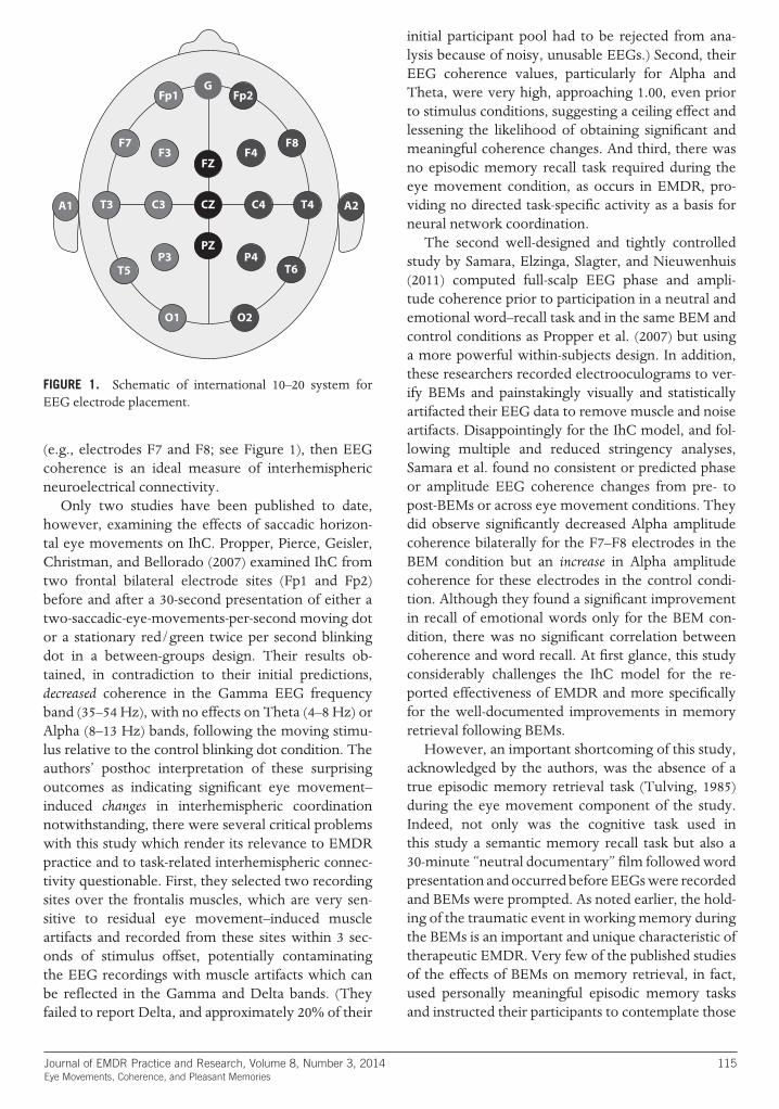
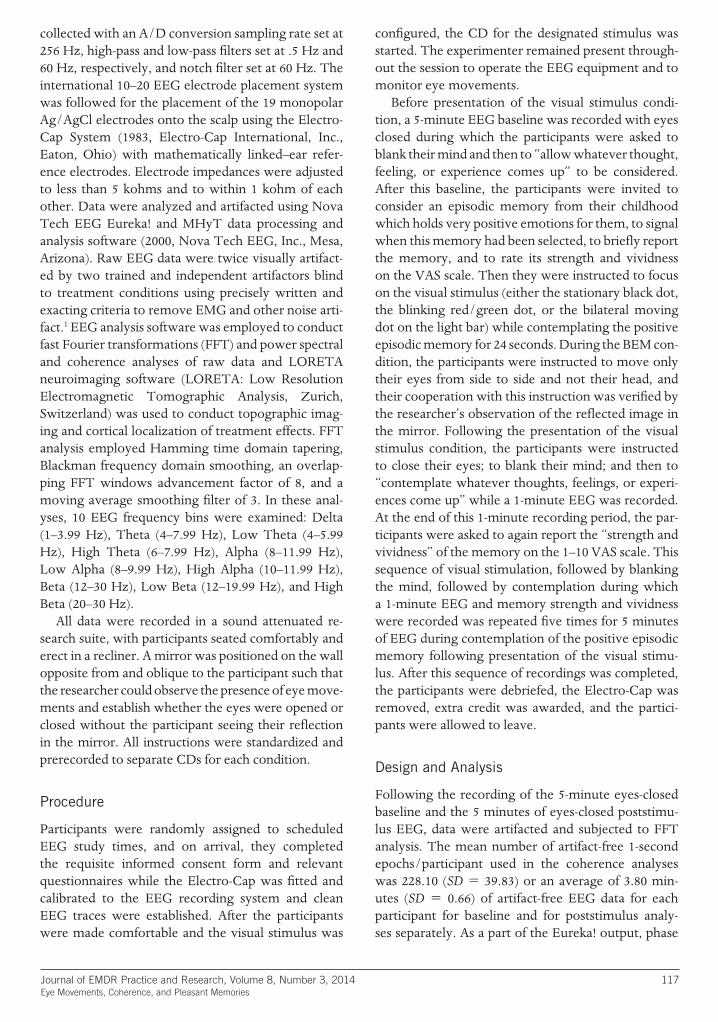
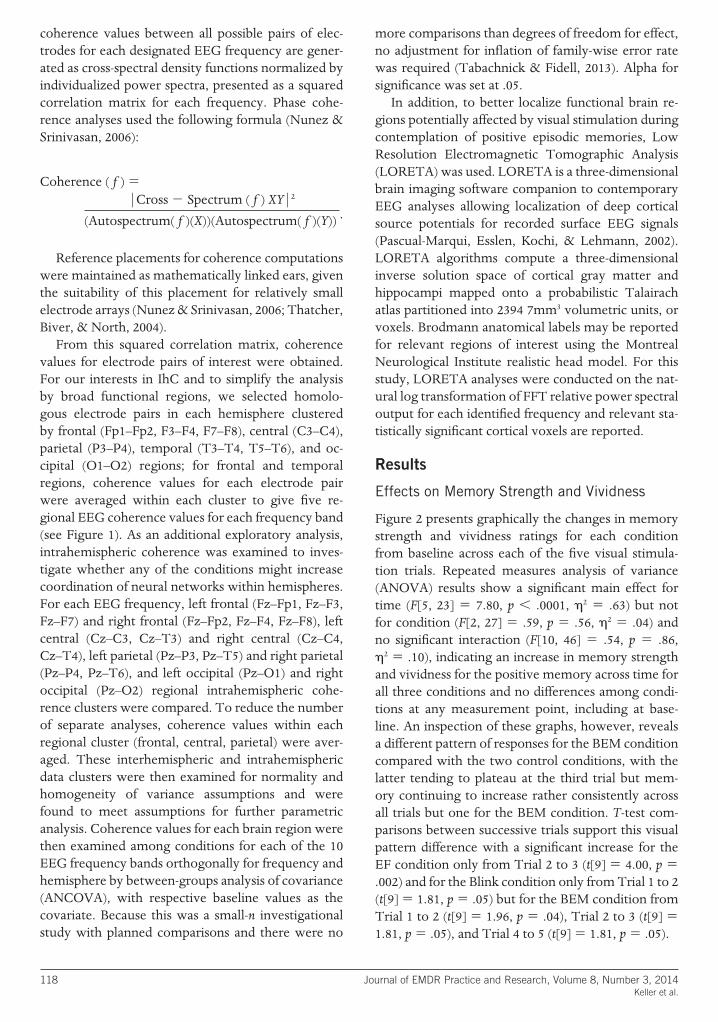
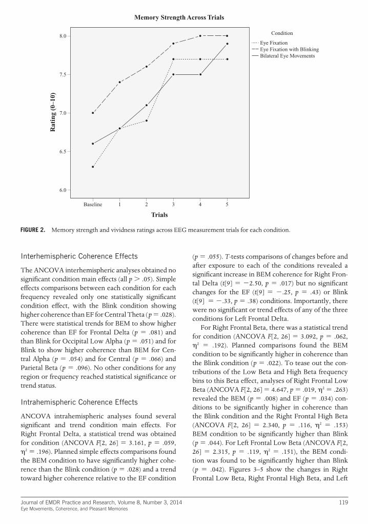
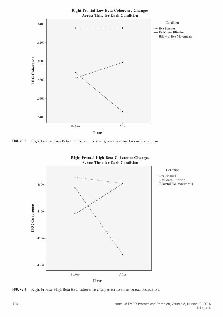
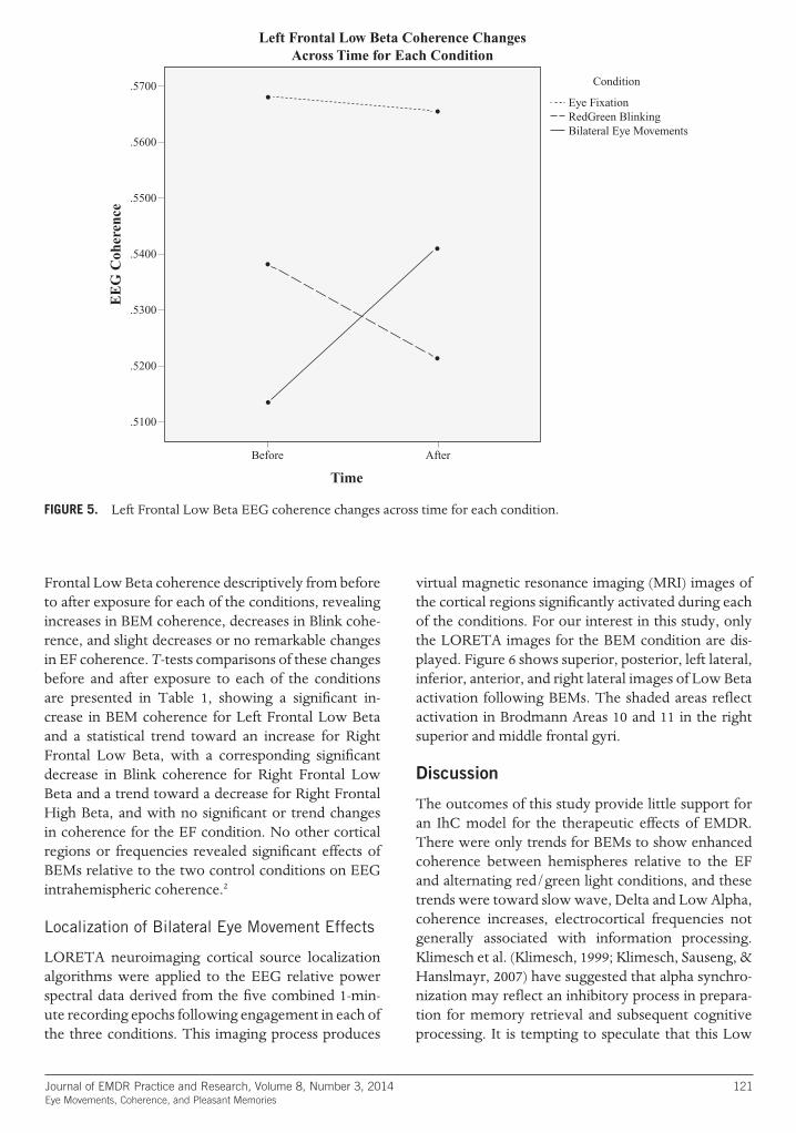
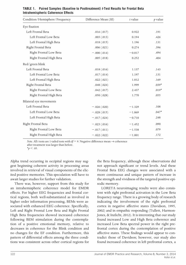
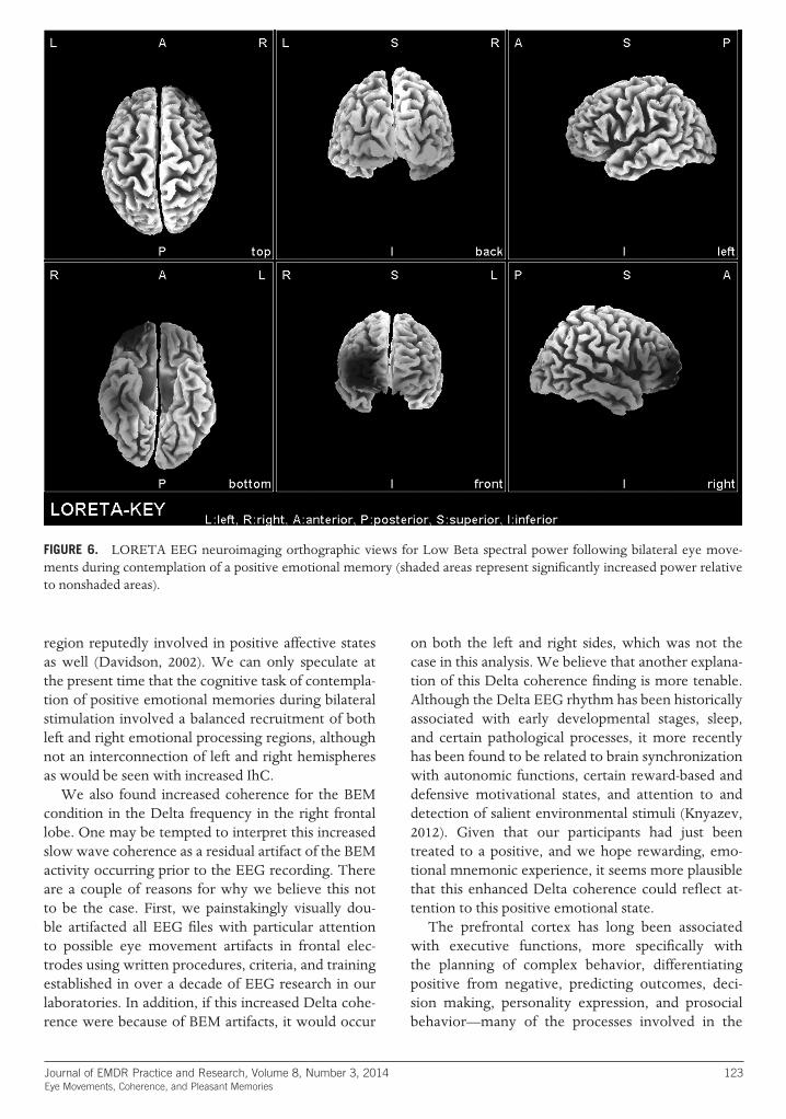
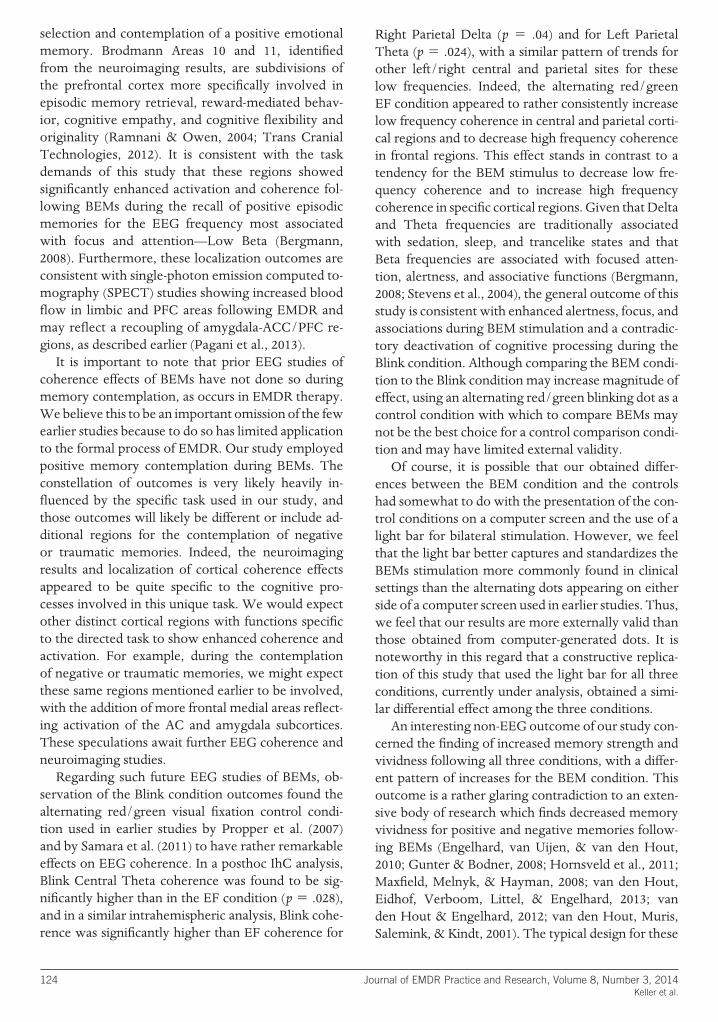
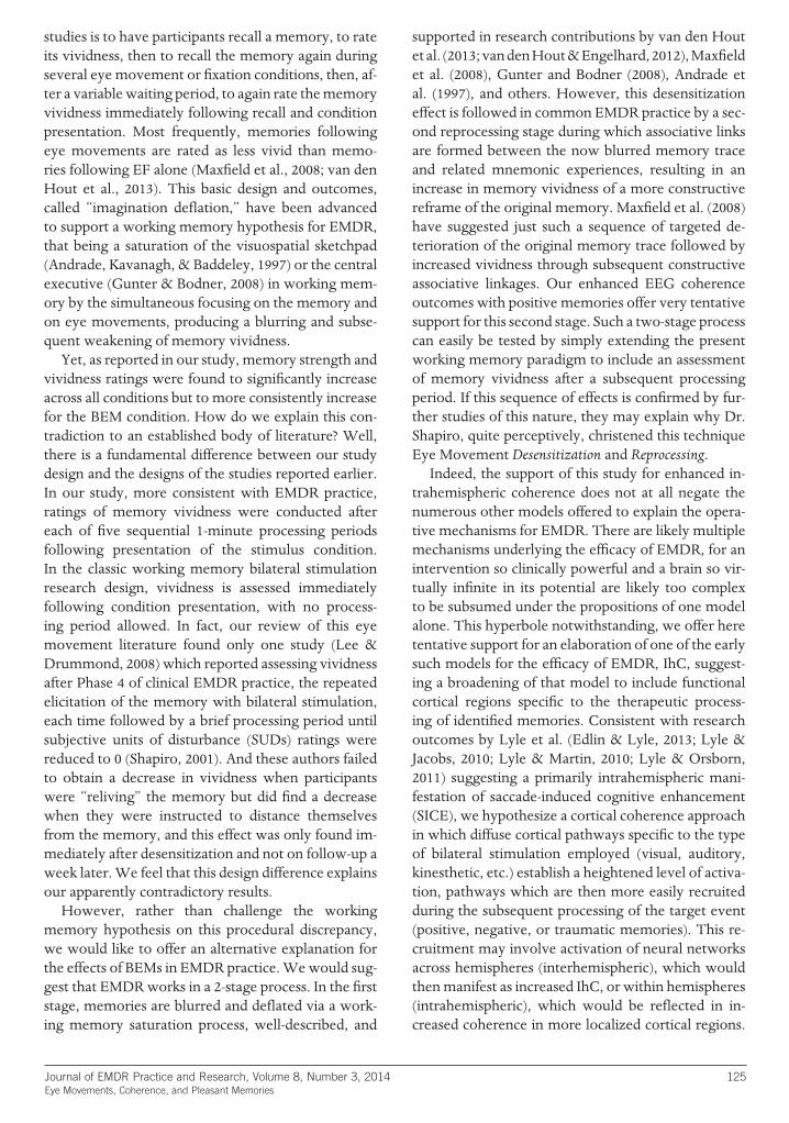
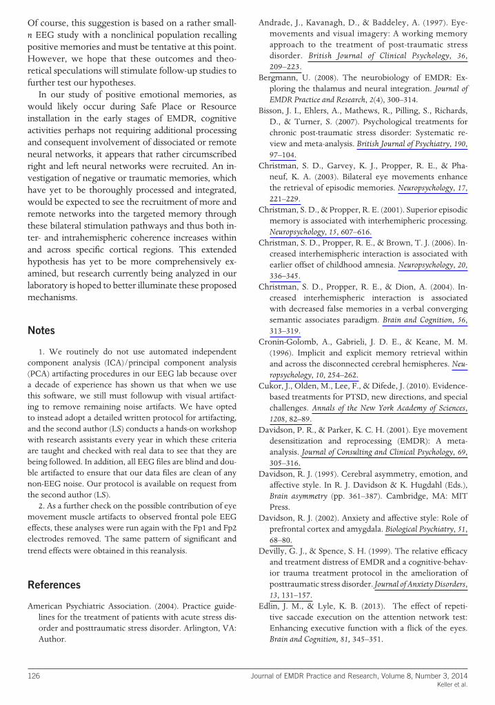
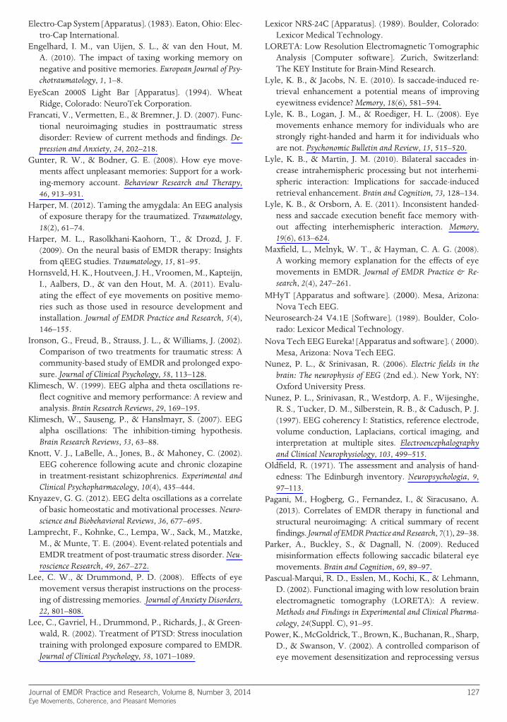
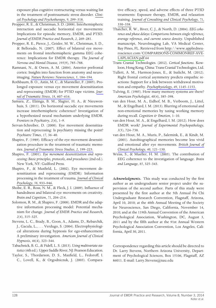