the early diagnosis pheochromocytoma · the early diagnosis of pheochromocytoma case report...
TRANSCRIPT
THE EARLY DIAGNOSIS OF PHEOCHROMOCYTOMABY
LESLIE COLE
From Addenbrook's Hospital, CambridgeReceived October 21, 1949
It is now well known that tumours of the adrenal medulla are a cause of paroxysmal hyperten-sion. It is less generally recognized that these attacks may only occur late or not at all, Whenthis is so, if the risk of surgical treatment is to be reduced to a minimum, diagnosis should be madein the early stages when paroxysmal symptoms are absent and hypertension, if present, closelyresembles the benign or malignant form. The first paroxysm may be the last because after it hasoccurred the patient may be too ill for operation. This paper describes the clinical features of sucha patient from the early stages and serves as a basis for discussion of some of the difficulties ofdiagnosis.
Patients with pheochromocytoma may present in various ways.(1) With recurrent attacks of paroxysmal hypertension and vasomotor instability, with or
without sustained hypertension between attacks.(2) Without paroxysms with sustained hypertension resembling benign or malignant hyper-
tension.(3) In extremis, from some cardiovascular accident when previous symptoms have not been
severe enough to lead them to consult a doctor.(4) Simulating Addison's disease from local pressure on the adrenal cortex (Mackeith, 1944).The classical picture of the adrenal sympathetic syndrome is too well known to need descrip-
tion here. It is characterized by periodic attacks of paroxysmal hypertension with palpitation,anxiety, shakiness, headache, vomiting and vasomotor phenomena. Transient glycosuria may'alsobe present. The similarity of this picture to that produced by the injection of a large dose ofadrenalin is evident.
It is less generally known that pheochromocytoma may produce a picture without paroxysms,closely resembling benign or malignant hypertension, but this had been pointed out by manywriters (Binger and Craig, 1938; Thorn, Hindle, and Sandmeyer, 1944; Palmer and Castleman,1938; McCullagh and Engel, 1942; and Mackeith, 1944). That persistent hypertension is a gooddeal commoner than has been realized, is suggested by Green (1946) who in a recent review of 51cases of pheochromocytoma found that only 14 showed intermittent hypertension, while 37 hadchronic hypertension. Smithwick's (1946) series of 1000 hypertensive patients subjected to lumbo-dorsal sympathectomy showed an incidence of pheochromocytoma of 005 per cent. There is thusconsiderable evidence to show that sustained hypertension of the benign or malignant type may bedue to pheochromocytoma, and it is clear that if the diagnosis is to be made earlier and more fre-quently, it must be considered as a possible cause in every case until it has been excluded. Soffer(1946) has pointed out that success depends first and foremost on the index of suspicion of thephysician. Put more simply, he must think of the possibility in every case of hypertension.
Cases that are first seen in extremis after some cardiovascular accident (Hick, 1933) bearevidence to the fact that paroxysms of hypertension are dangerous and may be fatal and thatdiagnosis should, if possible, be made before they occur. The case described in this paper is anexample of this.
232
on June 1, 2020 by guest. Protected by copyright.
http://heart.bmj.com
/B
r Heart J: first published as 10.1136/hrt.12.3.232 on 1 July 1950. D
ownloaded from
THE EARLY DIAGNOSIS OF PHEOCHROMOCYTOMA
CASE REPORTA married woman, aged 35, who had been previously healthy was admitted to Addenbrooke's
Hospital in October 1946 under Dr. J. R. C. Canney for a severe ante-partem hemorrhage. Herpregnancy had been uneventful up to this time and her blood pressure normal throughout.
Previously she had always been healthy and active, leading an energetic life as a farmer'swife. She had three children alive and well, aged 8, 5, and 3, and her previous confinements hadbeen without incident. Her husband was healthy and the family history did not reveal any tendencyto raised blood pressure, or heart, kidney, or other relevant disease.
On admission she was very anwmic and shocked but no other abnormalities were found, andafter a transfusion a stillborn child of 30 weeks was delivered by CWsarean section. A few dayslater she developed a slight thrombosis in the left leg but she was discharged apparently well onOctober 29th, fifteen days after her hemorrhage.
On November 2nd, in the evening, while she was sitting up in bed, she suddenly developedfrontal headache and this persisted. I saw her for the first time on November 5th. She was thencomplaining of dull continuous headache, frontal and mainly on the right side but spreading over tothe left at times. It was worse in the early morning and was accentuated by movement. Onexamination, there was slight cedema of the left ankle, but there were no other abnormalities.Blood pressure, 130/90; pulse rate, 84; and fundi normal.
During the next four months her general condition improved and she was moderately active,but her headaches continued to trouble her until February 1947 when they cleared up. Sheremained practically free from symptoms until April, when they returned and continued as before.In July, because they were rather worse, she was readmitted to hospital for investigation.
The headaches were still frontal, sometimes on one side, sometimes on the other. They wereworse in the early morning, passed off about noon, and often came back in the evening. They weremade worse by effort or movement but nothing else seemed to affect them. She said that at timesshe used to become very shaky, especially if anything worried her, and attributed this to the shock ofthe operation (for she had never been ill before) and the fact that she had had a good deal of worrythe previous summer. She had no other symptoms.
On examination, her blood pressure was 180/120-130, in both arms. This was taken on manyoccasions at different times of the day and in different postures and hardly varied. Slight aedema ofthe left leg was still present. Her urine occasionally contained a trace of albumen, but this was notconstant. Clinical examination revealed nothing otherwise; she had no fever and her pulse rateremained between 80 and 90, hardly varying with mild effort. Special investigation only showeda slight B. coli bacilluria and a very slight dilatation of the pelvis of the right kidney, which was notconsidered significant.
The hypertension was thought to be benign and did not seem to be enough to account for her head-ache, which was the outstanding symptom; and it seemed likely from her general nervousness thatthere was a considerable functional element present. She was given a course of sulphamezathinefor her B. coli bacilluria.
During the next three months her headaches persisted except for a three-weeks remission whileshe was on holiday in August. They maintained the same character and rhythm but, grew moresevere. In September she began to suffer from short early morning nausea and her appetite beganto fail and in October she was readmitted. She said then that her head did not throb but felt attimes as if someone were sticking knives into her forehead. She began to retch occasionally. Attimes she had " peculiar sensations," during which she felt worked up and excited, and on theseoccasions she looked tremulous and anxious. At times she seemed to be in a state of repressedexcitement and shaking like a patient with exophthalmic goitre but without thyrotoxicosis. Asvomiting became more frequent her nutrition and general condition began to deteriorate for thefirst time. During October her blood pressure taken on many occasions varied between 180/120and 175/115 and her pulse rate was steady between 70 and 80. Clinical examination was otherwisenormal and the following special investigations did not help.
233
on June 1, 2020 by guest. Protected by copyright.
http://heart.bmj.com
/B
r Heart J: first published as 10.1136/hrt.12.3.232 on 1 July 1950. D
ownloaded from
Examination of her pelvis, eyes (fundi, visual fields and refraction), ears, nose, and throat, andrenal function, were all normal. Blood urea 17 and 28 mg. Urea clearance 90 per cent. Fullblood count normal. Sedimentation rate (Westergren) 17 mm. Wassermann and Kahn reactionsnegative. X-rays of her skull, cervical spine, nasal sinuses, and lungs, all normal. Cardioscopyslight ventricular hypertrophy and slight uncoiling of the aorta. Electrocardiogram, left axisdeviation only. The urinary deposit showed epithelial cells only and no albumen or sugar. Thecerebrospinal fluid was under pressure (400/350 mm.) but was otherwise normal, and the Wasser-mann reaction was negative.
On October 31st she was discharged home at her own request pending transfer to a cerebralunit, where her condition remained the same until November 9th. On that afternoon she had somekind of fit, a sudden attack of headache, vomiting, and intense excitement, and her doctor foundthat her blood pressure rose temporarily to 240/160-a typical paroxysm of hypertension. Thesame afternoon, her headache -dramatically disappeared, she stopped vomiting, and was able totake more food. On the morning of the 10th, her blood pressure had fallen to 170/110 and in theafternoon she was suddenly seized with a very severe pain in the right renal region which did notradiate and only gradually eased. On the 11th the pain was still present and much less severe buther abdomen was slightly distended. Her bowels had not been open since the onset of the painbut she had passed flatus. An acute abdominal condition was suspected and she was readmittedto hospital.
On admission, she was anxious and excited with slight tremor of the hands. Her hands and feetwere cold and she sweated profusely at times. Temperature, 101; pulse rate, 110; respiration rate,20. She looked a little dehydrated. Her headache and vomiting had stopped. The abdomen wasslightly distended and there was marked tenderness in the right renal angle and over the right side,but no true rigidity. Vigorous peristalsis was audible and she passed flatus. Rectal examinationwas normal. The blood pressure was 180/130. The urine contained a heavy cloud of albumen.Microscopically there was no deposit and no other abnormalities were found. Enemas yielded verylittle result.. The next morning her general condition was unchanged. Blood count showed aleucocytosis of 29,000 with 89 per cent polymorphs. During the day her abdomen became moredistended and she was seen in consultation with Mr. P. H. R. Ghey. In view of the progressivedistension it was thought that she might have general peritonitis with paralytic ileus or subacuteobstruction and laparotomy was performed.
No evidence of peritonitis or obstruction was found. The whole of the large and small bowelwas distended with gas and the cacum was enormous. There were a few hard fecal masses at thehepatic flexure. A round solid tumour about 3 inches in diameter was felt in the region of the leftsuprarenal which seemed rather fixed. As her general condition was rather poor it was consideredinadvisable to attempt to remove this until the ileus had been controlled, so cxcostomy was per-formed and a de Pezzer catheter inserted.
During the next six days she was given eucortone and glucose saline, plasma or normal salineintravenously at the rate of 4 pints a day, and every effort was made to improve her general con-dition. This hope was not realized and as it threatened to deteriorate, Mr. Ghey performed asecond operation on November 17th. A tumour which subsequently proved to be a typical pheo-chromocytoma was removed from the site of the left suprarenal gland. Section of the last bloodvessels to the tumour was followed by a sudden profound fall in blood pressure. In anticipationof this, an intravenous adrenalin drip (1: 10,000) which had previously been connected, was turnedon at once, but without effect and death took place a few minutes later.
POST-MORTEM REPORTThe following are extracts from the full post-mortem report for which I am indebted to Dr. A. M.
Barrett.Left suprarenal. Thin triangular remnant (sides 3, 2, and 2 cm.) of suprarenal tissue consisting
234 LESLIE COLE
on June 1, 2020 by guest. Protected by copyright.
http://heart.bmj.com
/B
r Heart J: first published as 10.1136/hrt.12.3.232 on 1 July 1950. D
ownloaded from
THE EARLY DIAGNOSIS OF PHEOCHROMOCYTOMA
of two thin layers of yellow cortex without any visible medulla at site of otherwise absent leftsuprarenal. Considerable enlargement of-ligatured and divided left suprarenal blood vessels.
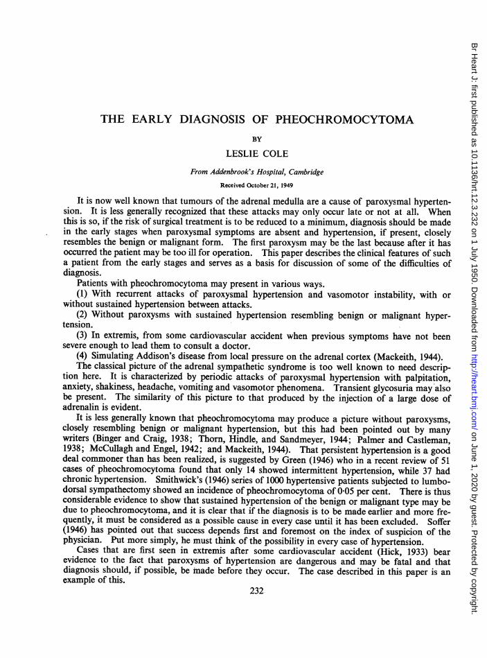
Microscopic sections were normal.Suprarenal tumour. (Fig. 1.) Ovoid tumour (9 5 x 7 5 x 5 7 cm.) enclosed with thin capsule.
Wt. 227 g. Near one pole a slightly raisedr.,e''t*J'yellow ridge (4 x 0-3 cm. wide) apparently con-
sisting ofsuprarenal cortex; small yellow islandsare also present here and there elsewhere on thepale grey-brown outer surface. The consistencyof the tumour is soft, almost cystic, with dullpurplish areas as of hemorrhage which showthrough the capsule here and there. The pre-dominant colour of the bulging cut surface is arich purple-red, but on closer inspection it isseen that there are everywhere close-set purple
[541& dots and streaks (about 01 cm. diameter) set ina greyish white ground.
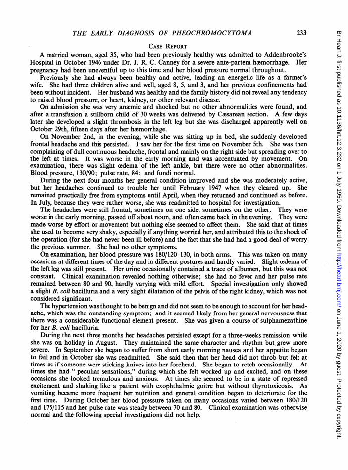
Microscopicalsection. (Fig. 2.) This showschromaffin tumour of suprarenal medulla, withnumerous blood spaces devoid of any lining andalso areas of hemorrhage with necrotic tumour
Right suprarenal. Weight, 9 g. Abundantyellow lipoid, very scanty brown pigment, and
FIG. 1.-Photograph of the suprarenal tumour. The normal grey-white medulla in rather large gland,measurements were 9-5 x 7-5 X 5*7 cm. and the Microscopic sections normal.weight 227 g. Cardiovascular system. Heart weight, 36 g.
Moderate hypertrophy of firm, brown-red myo-cardium of contracted left ventricle (L.V.: R.V., 2 5: 05). No hypertrophy of the rest of theheart. Pale brown ante-mortem thrombosis for the most part only lightly adherent, but in one
FIG. 2.-Photomicrograph, showing the histological appearances of the tumour.
235
on June 1, 2020 by guest. Protected by copyright.
http://heart.bmj.com
/B
r Heart J: first published as 10.1136/hrt.12.3.232 on 1 July 1950. D
ownloaded from
place apparently merging with the underlying muscle lining the apex of left ventricle. Moderateatheroma without narrowing in the coronary arteries. Slight to moderate atheroma of normal-sized aorta. Thrombosis of right and left posterior tibial veins and of left femoral vein.
On section the mural thrombosis was shown to have formed ante-mortem.Respiratory system. About a dozen fragments of ante-mortem clot impacted in branches of the
pulmonary arteries in both lungs. The emboli were about 1 cm. in diameter. Altogether they musthave caused considerable reduction in the arterial "bed " of the lungs.
Urogenital system. Many yellow-white areas of infarction in the right kidney apparentlyinvolving altogether about half its substance. On the outer surface the infarcted areas were neitherraised nor sunken. Scanty fibrinous exudate on the outside of the capsule of the right kidney.This embolus satisfactorily accounted for the largest area of infarction, but there were many othersmaller areas. Slight dilatation of right renal pelvis. No infarction of well developed left kidney.
On section, infarction with fibrinoid degeneration of afferent glomerular arterioles and otherabnormalities of glomerular tufts which are probably related to it.
Special-investigations. The chromaffin reaction (Schmorl) was positive.The test for adrenalin by Craner's method was very suggestive of the presence of much adrenalin.The test by Masson's method showed that argentaffin cells appeared to be present.
DISCUSSIONWhen paroxysmal hypertension is absent a tentative diagnosis of pheochromocytoma would
appear to depend more on careful consideration of the symptoms found in association with hyper-tension than on the physical signs, which are not distinctive unless the tumour is palpable.
The syndrome presented by this patient was quite different from any I had seen before, andduring the three months before her attack of paroxysmal hypertension presented a clinical picturethat once seen could not be forgotten or pass unrecognized again. It is, therefore, worth studyingher symptoms and signs in detail.
The unusual clinical features in the early stages appeared to be as follows: the sudden onsetof headache preceding the rise in blood pressure, the periodicity of the headache in the early mor-ning and evening, the " peculiar sensations " in the upper abdomen with sinking feelings, turmoil,and excitement, and the slow but steady increase in the intensity of the symptoms without anycorresponding rise in blood pressure.
In the later stages the unusual features were the severity of the headache, out of all proportionto the blood pressure, and its character-" like knives being driven into the head "-and the attacksof sweating and cold hands and feet.
It is also worth noting that this syndrome developed in a patient who had been particularly wellbalanced mentally and physically in the past, and had never previously had a headache in her life,and was without any obvious hereditary or environmental causes for her symptoms.
Further special investigations that might have been done to establish the diagnosis earlier mayconveniently be considered under the three following headings.
(1) Tests that suggest hypersecretion of adrenalin. Attempts to demonstrate the presence of apressor substance in the blood have only occasionally been successful, are difficult to perform, andcannot be relied on. Beer, King, and Prinzmetal (1937), by perfusing a denervated rabbit's earwith blood obtained from a patient during an induced paroxysm, were able to demonstrate thepresence of such an active principle. Because of the great practical difficulties that attend thismethod, it cannot often be applied clinically.
Paroxysms can sometimes be induced deliberately, but this is not devoid of risk. Massage ormanipulation of the tumour by change of posture sometimes bring on an attack and indirectly givea clue to its site. Starvation for from 12 to 24 hours may induce one (Soffer, 1946), or the injectionof adrenalin (Mackeith, 1944). Since the greatest difficulties in diagnosis occur in patients in whom
LESLIE COLE236
on June 1, 2020 by guest. Protected by copyright.
http://heart.bmj.com
/B
r Heart J: first published as 10.1136/hrt.12.3.232 on 1 July 1950. D
ownloaded from
THE EARLY DIAGNOSIS OF PHEOCHROMOCYTOMA
the existence and site of the tumour are not known, the injection of adrenalin is the easiest, butbecause of its risk is perhaps better avoided.
Shortly after this patient died, Goldenberg (1947) described a simple test for hypertensioncaused by excess circulating adrenalin and claimed to have established its value in the diagnosis ofpheochromocytoma. Briefly, the test consists in perfusing the patient with benzodioxanes (1 164For 933 F) which given in the appropriate dose are adrenolytic in action and block sympatheticactivity. He found that in patients with pheochromocytomas or with hypertension induced byperfusion with adrenalin, blood pressure readings taken over a period of 15 minutes after theinjection showed a characteristic fall that was in contrast to the rise obtained in those with malig-nant or essential hypertension. Smithwick (1948) confirmed the diagnostic value of this test and iffurther observations confirm Goldenberg's findings, it should be of great benefit in helping todiagnose difficult cases of pheochromocytoma in the early stages. It is clearly easier to localizethe tumour when there is strong evidence that it is present than when its existence is still in doubt.
Another test for pheochromocytoma is by intravenous injection of histamine. It is claimed thatthis is followed immediately by a rise in blood pressure in contrast to the usual fall obtained innormal people. Roth and Kuale (1945) suggest that this test is specific for the condition.
(2) Localization of the tumour. This may be easy or it may be very difficult. It may be palpableor only observable by exploration and may be found by accident in the course of operation forlumbo-dorsal sympathectomy in hypertension or as in the present case for some complication. Itis noteworthy that in the patient described here, while all the signs and symptoms pointed to theright side, the pheochromocytoma was on the left. It should also be remembered that bilateraltumours may occur (Reid and Salm, 1949). If the tumour is not palpable, radiological investiga-tion of the kidneys is the procedure most likely to yield definite evidence. Howard and Barker(1937) estimated that an intravenous pyelogram would have suggested the presence of the tumourin 11 of these 18 cases. If this measure fails a retrogade pyelogram or even perirenal insufflationswith oxygen have proved invaluable (Soffer, 1946). It is important, too, to remember that theposition of these tumours in the abdomen is very varied. They may also be located in the neck andeven in the chest, but do not appear to be active in these situations (Phillips, 1940). Diagnosticprocedures should be as innocuous as possible because these patients stand manipulation badly.This makes it all the more desirable that the tumour should be located early and accurately.
(3) Other investigations. The basal metabolic rate perhaps comes next in order of importancefor this is often above 20 per cent (Binger, 1938, and McCullagh, 1942) and indeed the differentialdiagnosis from hyperthyroidism may be very difficult and has often led to confusion. This is notsurprising, for the clinical resemblance between excessive secretion of adrenalin, chronic anxiety,and hyperthyroidism may be very ,close. Mistakes will be less likely to occur if this resemblance isremembered in typical cases of hyperthyroidism coupled with the fact that the B.M.R. is oftenraised in pheochromocytoma.
Other investigations are useful chiefly for excluding intercurrent disease and for demonstratingchanges that occur in hypertension from any cause but are not specific for pheochromocytoma.Thus cardiographic studies may show changes associated with hypertension; cardioscopy, generalcardiac enlargement, and left ventricular hypertrophy; and renal function tests, a greater or lesserdegree of impairment but nothing diagnostic.
Glycosuria and a raised blood sugar have been found occasionally and although the associationof these with excessive secretion of adrenalin is clear, they do not occur frequently enough to be ofmuch value in diagnosis. Pheochromocytoma has also been found with frank diabetes mellitus(McCullagh, 1942). Retinal changes do not differ from those found in benign or malignant hyper-tension. Blood counts do not show any significant change. The high pressure in the cerebro-spinal fluid found in this patient has also been recorded in another (Palmer, 1938) but might welloccur in hypertension from any other cause. If for any reason lumbar puncture is consideredadvisable, the risk of its provoking a paroxysm must be remembered, and the blood pressure andgeneral condition carefully measured both before and after.
237
on June 1, 2020 by guest. Protected by copyright.
http://heart.bmj.com
/B
r Heart J: first published as 10.1136/hrt.12.3.232 on 1 July 1950. D
ownloaded from
SUMMARYThe symptoms in a patient with sustained hypertension due to pheochromocytoma are described
in detail. The diagnosis is discussed and the importance of early diagnosis is emphasized.
The author wishes to thank Dr. A. M. Barrett for the post-mortem report and Mr. H. P. Hudson for the photo-graph and photomicrograph.
REFERENCESBeer, E., King, F. H., and Prinzmetal, M. (1937). Ann. Surg., 106, 85.Binger, M. W., and Craig, W. McK. (1938). Proc. Staff. Meet., Mayo Clin., 13, 17.Goldenberg, M., Snyder, C. H., and Aranow, H. (1947). J. Amer. med. Assoc., 135, 971.Green, D. M. (1946). Ibid., 131, 1260.Hick, F. K. (1933). Arch. Path., 15, 665.Howard, J. E., and Barker, W. H. (1937). Bull. Johns Hopkins Hosp., 61, 371.Mackeith, R. (1944). Brit. Heart J., 6, 1.McCullagh, E. P., and Engel, W. J. (1942). Ann. Surg., 116, 61.Palmer, R. S., and Castleman, B. (1938). New England J. Med., 219, 793.Phillips, B. (1940). Arch. Path., 30, 916.Platt, R. (1947). Quart. J. Med., N.S., 16, 111.Reid, T. M., and Salm, R. (1949). Brit. med. J., 1, 1116.Roth, G. M., and Kuale, W. F. (1945). Amer. J. Sci., 210, 653.Smithwick, R. H. (1946). Personal communication quoted by Goldenberg (1947). J. Amer. med. Assoc., 135, 971.
, (1948). Lecture at Cambridge.Soffer, Louis J. (1946). Diseases of the Adrenals, (Henry Kimpton), X, 273.Thorn, G. W., Hindle, J. A., and Sandmeyer, J. A. (1944). Ann. intern. Med., 21, 122.
238 LESLIE COLE
on June 1, 2020 by guest. Protected by copyright.
http://heart.bmj.com
/B
r Heart J: first published as 10.1136/hrt.12.3.232 on 1 July 1950. D
ownloaded from