the compactness of ribonuclease a and reduced ribonuclease a
TRANSCRIPT
The compactness of ribonuclease A and reduced ribonuclease A
Jun-Mei Zhoua;*, Ying-Xin Fana, Hiroshi Kiharab, Kazumoto Kimurac, Yoshiyuki Amemiyad
aNational Laboratory of Biomacromolecules, Institute of Biophysics, Academia Sinica, Beijing 100101, PR ChinabPhysics Laboratory, Kansai Medical University, Uyamahigashi, Hirakata, Osaka 573, Japan
cDivision of Medical Electronics, Dokkyo University School of Medicine, Mibu, Tochigi 321-02, JapandFaculty of Engineering, The University of Tokyo, Hongo, Bunkyo-ku, Tokyo 113, Japan
Received 30 March 1998; revised version received 4 May 1998
Abstract The compactness of ribonuclease A with intactdisulfide bonds and reduced ribonuclease A was investigated bysynchrotron small-angle X-ray scattering. The Rg values and theKratky plots showed that non-reduced ribonuclease A maintain acompact shape with a Rg value of about 17.3 Aî in 8 M urea. Thereduced ribonuclease A is more expanded, its Rg value is about20 Aî in 50 mM Tris-HCl buffer at pH 8.1 containing 20 mMDTT. Further expansions of reduced ribonuclease A wereobserved in the presence of high concentrations of denaturants,indicating that reduced ribonuclease A is more expanded and is inneither a random coil [A. Noppert et al., FEBS Lett. 380 (1996)179^182] nor a compact denatured state [T.R. Sosnick and J.Trewhella, Biochemistry 31 (1992) 8329^8335]. The fourdisulfide bonds keep ribonuclease A in a compact state in thepresence of high concentrations of urea.z 1998 Federation of European Biochemical Societies.
Key words: Ribonuclease A; Disul¢de bond; Globularity;Small-angle X-ray scattering; Protein folding
1. Introduction
Ribonuclease A (RNase A) is one of the most extensivelystudied enzymes, with its sequence and three-dimensionalstructure being known completely. The course of its unfoldingduring denaturation by acid [1], heat [2], pressure [3] anddenaturants [4^6], as well as its renaturation during refoldinghave also been well characterized in the literature [7^10]. Inthose refolding studies, RNase A denatured in 8 M urea and6 M GuHCl was used to study the folding process with theimplicit assumption that the denatured protein is completelystructureless. However, the assumption has not yet been de-¢nitively proven. It has been suggested that some proteinsretain considerable residual structure even in the presence ofdenaturants at high concentrations [11,12]. The presence ofany residual ordered structure in a supposedly fully denaturedprotein would be important because the residual structurecould initiate the refolding of the rest of the molecule bymaintaining certain hydrophobic or other interactions, whichwould greatly facilitate the refolding process [13]. Many re-folding studies have been made with disul¢de bond containingproteins, and in such cases it is even more uncertain whetherthese proteins still contain ordered structures in strong dena-turants. Contradictory results have been presented for RNaseA. In an early study, Tanford et al. [14] suggested by intrinsicviscosity measurements that RNase A in 6 M GuHCl is ex-actly as predicted for a randomly coiled polymer chain, butHu and Tsou reported that the RNase A denatured in 6 M
GuHCl with their native disul¢de bonds is not completelystructureless as detected by CD, UV and Fourier transforminfrared spectroscopy [15]. In 1992, Sosnick and Trewhella[16] reported a very small Rg value for reduced-denaturedRNase A. They concluded that reduced-denatured RNase Ais more compact than a random coil and has a signi¢cantamount of regular secondary structure, as deduced fromsmall-angle X-ray scattering measurements and Fourier trans-form infrared spectroscopy. Soon afterwards Noppert and hisco-workers [17] reported that reduced-denatured RNase A isnot in a compact state but that reduction of the four disul¢debonds by DTT at 20³C leads to total unfolding and a subse-quent temperature increase has no further e¡ect on its dimen-sion. The controversy in the literature may best be resolvedusing synchrotron small-angle scattering to measure the over-all dimensions of a protein molecule in solution. In thepresent study, the compactness of disul¢de intact RNase Aand reduced RNase A were investigated using synchrotronsmall-angle X-ray scattering. The result show that reducedRNase A is more expanded than the non-reduced form butit is in neither random coil nor a compact denatured state.
2. Materials and methods
2.1. MaterialsBovine pancreatic RNase A (type III-A) was purchased from Sigma
and used without further puri¢cation. Ultrapure urea was obtainedfrom Nacalai Tesque, Inc., Kyoto, Japan. All chemicals were of an-alytical grade and were used without further puri¢cation.
2.2. Reduction of RNase AReduced RNase A was prepared according to the procedure de-
scribed by Kumar et al. [18]. The RNase A was dissolved in 0.1 MTris-HCl bu¡er, pH 8.6, containing 8 M urea and 1 mM EDTA, to a¢nal protein concentration of about 10 mg/ml. After the addition ofdithiothreitol to a ¢nal concentration of 0.15 M, the solution was£ushed with nitrogen gas for 3 min and reduction was carried outfor 4 h at 25³C. Then, the pH of the mixture was adjusted to 3.0 bythe addition of HCl. The reduced RNase A was then dialyzed thor-oughly against 0.1 M acetic acid. The dialyzed solution was lyophi-lized, and the dried protein was stored in a sealed vial ¢lled withnitrogen. The reduced RNase A contains 7.2 reactive SH groupsmeasured by DTNB. The protein concentrations were determinedspectrophotometrically using a molar extinction coe¤cient of 9200M31 cm31 at 275 nm for both native and reduced RNase A.
2.3. Synchrotron small-angle X-ray scattering measurementsSolution scattering measurements were performed at the beam line
15 small-angle installation of the Photon Factory, National Labora-tory for High Energy Physics, Tsukuba, Japan, where a stable beamof photons with a wavelength of 1.5 Aî was provided by a bent-crystalhorizontally focusing monochromator and a vertically focusing mirror[19]. Samples were irradiated in a quartz cell with 1 mm path lengthfor 300 s for each sample. The temperature of the cell holder was keptat 25³C with circulating water. The background data for the bu¡ersolution and the di¡erent urea concentrations were collected before or
FEBS 20403 6-7-98
0014-5793/98/$19.00 ß 1998 Federation of European Biochemical Societies. All rights reserved.PII: S 0 0 1 4 - 5 7 9 3 ( 9 8 ) 0 0 6 3 9 - 5
*Corresponding author. Fax: (86) (10) 64872026.E-mail: [email protected]
FEBS 20403 FEBS Letters 430 (1998) 275^277
after data collection for the protein solution. The small angle X-rayscattering data were corrected for the di¡erence in contrast betweenthe protein and solvent molecules as well as for X-ray absorption bythe urea according to standard data provided by Semisotnov (unpub-lished data). The Rg values were analyzed by Guinier plot and thecompactness was judged by Kratky plot [20].
3. Results
3.1. The Rg values of native, denatured and reduced RNase AThe solution X-ray scattering of native RNase A at di¡er-
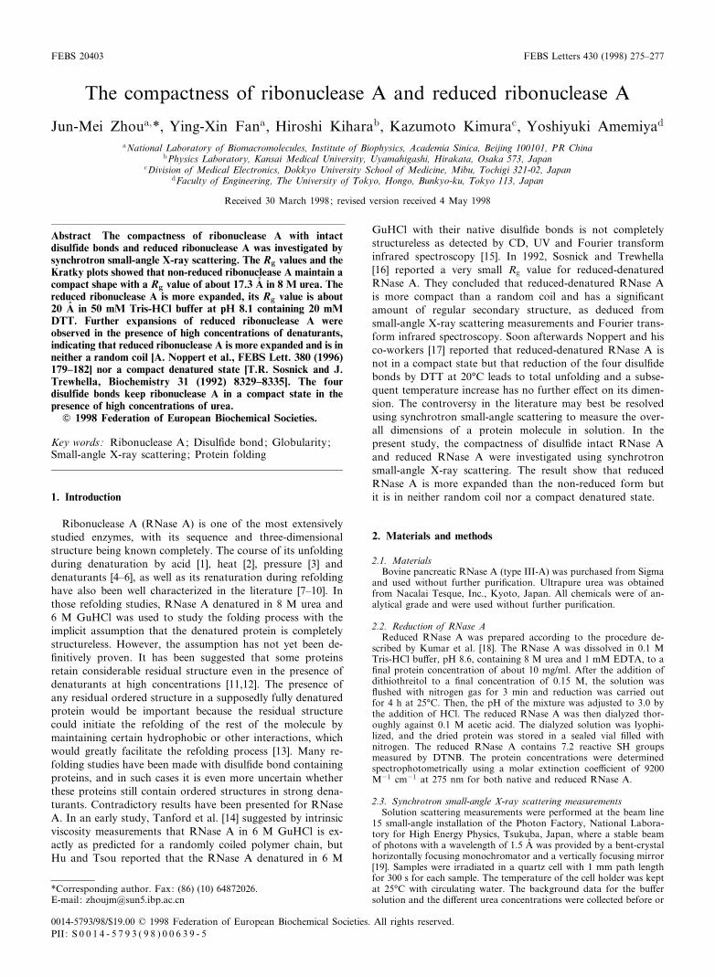
ent concentrations was measured in 50 mM Tris-HCl bu¡er atpH 8.1 and 25³C. The apparent Rg values were calculatedfrom a series of Guinier plots at di¡erent enzyme concentra-tions. The Rg of RNase A was obtained by extrapolatinglinearly the apparent Rg values to zero concentration of theenzyme. Curve 1 of Fig. 1 shows the concentration depend-ence of the Rg values for RNase A. It is clear that there is noobvious change of Rg value as the enzyme concentration wasincreased and that the Rg value at zero concentration is15.4 þ 0.13 Aî . The concentration dependence of the Rg valuesfor RNase A denatured in 6 M urea was also determined asshown in curve 2 of the same ¢gure. The Rg of 6 M urea-denatured RNase A is also almost independent of the proteinconcentration with a value of 15.5 þ 0.11 Aî at zero concen-tration. Fig. 1, curve 3 shows the concentration dependence ofthe Rg value of reduced RNase A in 50 mM Tris-HCl bu¡erpH 8.1 containing 20 mM DTT and 6 M urea. The Rg in-creases slightly with increasing enzyme concentration and theRg value at zero concentration is 24.0 þ 0.1 Aî . This value is ingood agreement with the results reported by Sosnick and Tre-whella [16].
3.2. Changes in compactness of RNase A during denaturationby urea
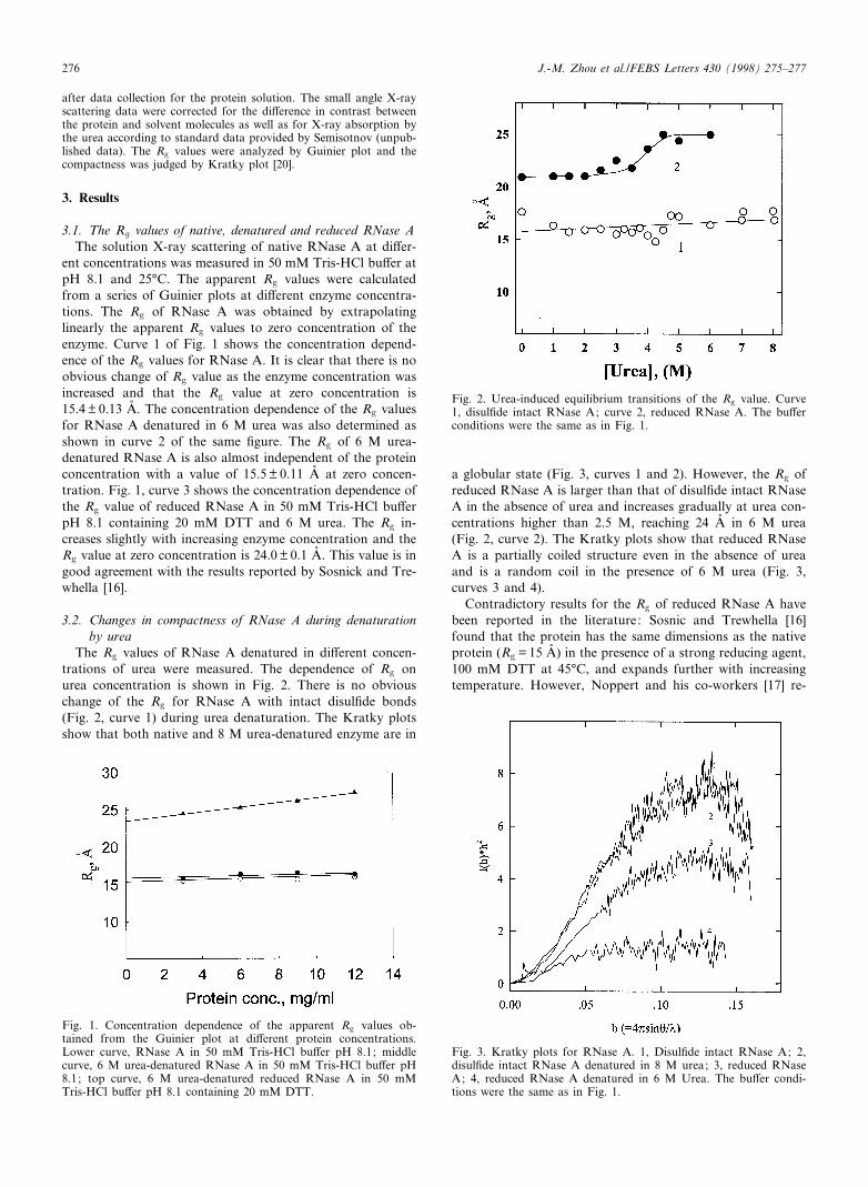
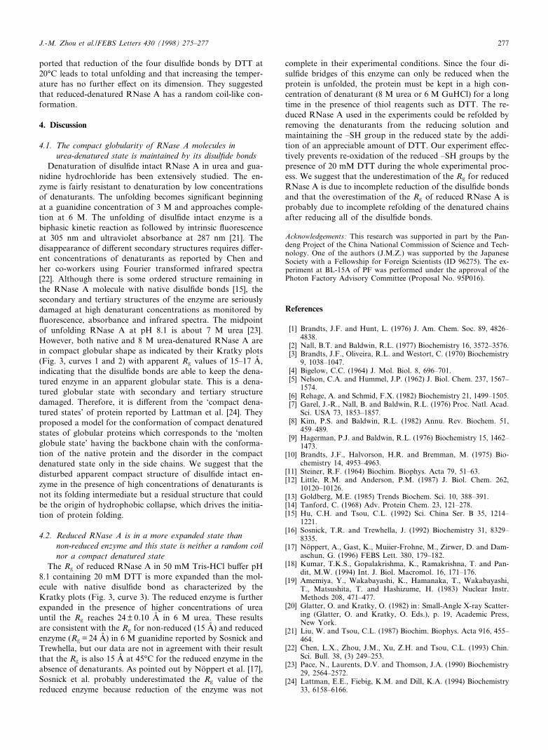
The Rg values of RNase A denatured in di¡erent concen-trations of urea were measured. The dependence of Rg onurea concentration is shown in Fig. 2. There is no obviouschange of the Rg for RNase A with intact disul¢de bonds(Fig. 2, curve 1) during urea denaturation. The Kratky plotsshow that both native and 8 M urea-denatured enzyme are in
a globular state (Fig. 3, curves 1 and 2). However, the Rg ofreduced RNase A is larger than that of disul¢de intact RNaseA in the absence of urea and increases gradually at urea con-centrations higher than 2.5 M, reaching 24 Aî in 6 M urea(Fig. 2, curve 2). The Kratky plots show that reduced RNaseA is a partially coiled structure even in the absence of ureaand is a random coil in the presence of 6 M urea (Fig. 3,curves 3 and 4).
Contradictory results for the Rg of reduced RNase A havebeen reported in the literature: Sosnic and Trewhella [16]found that the protein has the same dimensions as the nativeprotein (Rg = 15 Aî ) in the presence of a strong reducing agent,100 mM DTT at 45³C, and expands further with increasingtemperature. However, Noppert and his co-workers [17] re-
FEBS 20403 6-7-98
Fig. 1. Concentration dependence of the apparent Rg values ob-tained from the Guinier plot at di¡erent protein concentrations.Lower curve, RNase A in 50 mM Tris-HCl bu¡er pH 8.1; middlecurve, 6 M urea-denatured RNase A in 50 mM Tris-HCl bu¡er pH8.1; top curve, 6 M urea-denatured reduced RNase A in 50 mMTris-HCl bu¡er pH 8.1 containing 20 mM DTT.
Fig. 2. Urea-induced equilibrium transitions of the Rg value. Curve1, disul¢de intact RNase A; curve 2, reduced RNase A. The bu¡erconditions were the same as in Fig. 1.
Fig. 3. Kratky plots for RNase A. 1, Disul¢de intact RNase A; 2,disul¢de intact RNase A denatured in 8 M urea; 3, reduced RNaseA; 4, reduced RNase A denatured in 6 M Urea. The bu¡er condi-tions were the same as in Fig. 1.
J.-M. Zhou et al./FEBS Letters 430 (1998) 275^277276
ported that reduction of the four disul¢de bonds by DTT at20³C leads to total unfolding and that increasing the temper-ature has no further e¡ect on its dimension. They suggestedthat reduced-denatured RNase A has a random coil-like con-formation.
4. Discussion
4.1. The compact globularity of RNase A molecules inurea-denatured state is maintained by its disul¢de bonds
Denaturation of disul¢de intact RNase A in urea and gua-nidine hydrochloride has been extensively studied. The en-zyme is fairly resistant to denaturation by low concentrationsof denaturants. The unfolding becomes signi¢cant beginningat a guanidine concentration of 3 M and approaches comple-tion at 6 M. The unfolding of disul¢de intact enzyme is abiphasic kinetic reaction as followed by intrinsic £uorescenceat 305 nm and ultraviolet absorbance at 287 nm [21]. Thedisappearance of di¡erent secondary structures requires di¡er-ent concentrations of denaturants as reported by Chen andher co-workers using Fourier transformed infrared spectra[22]. Although there is some ordered structure remaining inthe RNase A molecule with native disul¢de bonds [15], thesecondary and tertiary structures of the enzyme are seriouslydamaged at high denaturant concentrations as monitored by£uorescence, absorbance and infrared spectra. The midpointof unfolding RNase A at pH 8.1 is about 7 M urea [23].However, both native and 8 M urea-denatured RNase A arein compact globular shape as indicated by their Kratky plots(Fig. 3, curves 1 and 2) with apparent Rg values of 15^17 Aî ,indicating that the disul¢de bonds are able to keep the dena-tured enzyme in an apparent globular state. This is a dena-tured globular state with secondary and tertiary structuredamaged. Therefore, it is di¡erent from the `compact dena-tured states' of protein reported by Lattman et al. [24]. Theyproposed a model for the conformation of compact denaturedstates of globular proteins which corresponds to the `moltenglobule state' having the backbone chain with the conforma-tion of the native protein and the disorder in the compactdenatured state only in the side chains. We suggest that thedisturbed apparent compact structure of disul¢de intact en-zyme in the presence of high concentrations of denaturants isnot its folding intermediate but a residual structure that couldbe the origin of hydrophobic collapse, which drives the initia-tion of protein folding.
4.2. Reduced RNase A is in a more expanded state thannon-reduced enzyme and this state is neither a random coilnor a compact denatured state
The Rg of reduced RNase A in 50 mM Tris-HCl bu¡er pH8.1 containing 20 mM DTT is more expanded than the mol-ecule with native disul¢de bond as characterized by theKratky plots (Fig. 3, curve 3). The reduced enzyme is furtherexpanded in the presence of higher concentrations of ureauntil the Rg reaches 24 þ 0.10 Aî in 6 M urea. These resultsare consistent with the Rg for non-reduced (15 Aî ) and reducedenzyme (Rg = 24 Aî ) in 6 M guanidine reported by Sosnick andTrewhella, but our data are not in agreement with their resultthat the Rg is also 15 Aî at 45³C for the reduced enzyme in theabsence of denaturants. As pointed out by Noëppert et al. [17],Sosnick et al. probably underestimated the Rg value of thereduced enzyme because reduction of the enzyme was not
complete in their experimental conditions. Since the four di-sul¢de bridges of this enzyme can only be reduced when theprotein is unfolded, the protein must be kept in a high con-centration of denaturant (8 M urea or 6 M GuHCl) for a longtime in the presence of thiol reagents such as DTT. The re-duced RNase A used in the experiments could be refolded byremoving the denaturants from the reducing solution andmaintaining the ^SH group in the reduced state by the addi-tion of an appreciable amount of DTT. Our experiment e¡ec-tively prevents re-oxidation of the reduced ^SH groups by thepresence of 20 mM DTT during the whole experimental proc-ess. We suggest that the underestimation of the Rg for reducedRNase A is due to incomplete reduction of the disul¢de bondsand that the overestimation of the Rg of reduced RNase A isprobably due to incomplete refolding of the denatured chainsafter reducing all of the disul¢de bonds.
Acknowledgements: This research was supported in part by the Pan-deng Project of the China National Commission of Science and Tech-nology. One of the authors (J.M.Z.) was supported by the JapaneseSociety with a Fellowship for Foreign Scientists (ID 96275). The ex-periment at BL-15A of PF was performed under the approval of thePhoton Factory Advisory Committee (Proposal No. 95P016).
References
[1] Brandts, J.F. and Hunt, L. (1976) J. Am. Chem. Soc. 89, 4826^4838.
[2] Nall, B.T. and Baldwin, R.L. (1977) Biochemistry 16, 3572^3576.[3] Brandts, J.F., Oliveira, R.L. and Westort, C. (1970) Biochemistry
9, 1038^1047.[4] Bigelow, C.C. (1964) J. Mol. Biol. 8, 696^701.[5] Nelson, C.A. and Hummel, J.P. (1962) J. Biol. Chem. 237, 1567^
1574.[6] Rehage, A. and Schmid, F.X. (1982) Biochemistry 21, 1499^1505.[7] Garel, J.-R., Nall, B. and Baldwin, R.L. (1976) Proc. Natl. Acad.
Sci. USA 73, 1853^1857.[8] Kim, P.S. and Baldwin, R.L. (1982) Annu. Rev. Biochem. 51,
459^489.[9] Hagerman, P.J. and Baldwin, R.L. (1976) Biochemistry 15, 1462^
1473.[10] Brandts, J.F., Halvorson, H.R. and Bremman, M. (1975) Bio-
chemistry 14, 4953^4963.[11] Steiner, R.F. (1964) Biochim. Biophys. Acta 79, 51^63.[12] Little, R.M. and Anderson, P.M. (1987) J. Biol. Chem. 262,
10120^10126.[13] Goldberg, M.E. (1985) Trends Biochem. Sci. 10, 388^391.[14] Tanford, C. (1968) Adv. Protein Chem. 23, 121^278.[15] Hu, C.H. and Tsou, C.L. (1992) Sci. China Ser. B 35, 1214^
1221.[16] Sosnick, T.R. and Trewhella, J. (1992) Biochemistry 31, 8329^
8335.[17] Noëppert, A., Gast, K., Muiier-Frohne, M., Zirwer, D. and Dam-
aschun, G. (1996) FEBS Lett. 380, 179^182.[18] Kumar, T.K.S., Gopalakrishma, K., Ramakrishna, T. and Pan-
dit, M.W. (1994) Int. J. Biol. Macromol. 16, 171^176.[19] Amemiya, Y., Wakabayashi, K., Hamanaka, T., Wakabayashi,
T., Matsushita, T. and Hashizume, H. (1983) Nuclear Instr.Methods 208, 471^477.
[20] Glatter, O. and Kratky, O. (1982) in: Small-Angle X-ray Scatter-ing (Glatter, O. and Kratky, O. Eds.), p. 19, Academic Press,New York.
[21] Liu, W. and Tsou, C.L. (1987) Biochim. Biophys. Acta 916, 455^464.
[22] Chen, L.X., Zhou, J.M., Xu, Z.H. and Tsou, C.L. (1993) Chin.Sci. Bull. 38, (3) 249^253.
[23] Pace, N., Laurents, D.V. and Thomson, J.A. (1990) Biochemistry29, 2564^2572.
[24] Lattman, E.E., Fiebig, K.M. and Dill, K.A. (1994) Biochemistry33, 6158^6166.
FEBS 20403 6-7-98
J.-M. Zhou et al./FEBS Letters 430 (1998) 275^277 277