the cold sensitivity of a mutant of saccharomyces ... · the cold sensitivity of a mutant of...
TRANSCRIPT
The Cold Sensitivity of a Mutant of Saccharomyces cerevisiae Lacking a Mitochondrial Heat Shock Protein 70 Is Suppressed by Loss of Mitochondrial DNA Brenda Schilke, Jeremy Forster, Julie Davis, Philip James, William Walter, Shikha Laloraya, Jill Johnson, Bingjie Miao, and Elizabeth Cra ig Department of Biomolecular Chemistry, University of Wisconsin, Madison, Wisconsin 53706
Abstract. SSH1, a newly identified member of the heat shock protein (hsp70) multigene family of the budding yeast Saccharomyces cerevisiae, encodes a protein lo- calized to the mitochondrial matrix. Delet ion of the SSH1 gene results in extremely slow growth at 23°C or 30°C, but nearly wild-type growth at 37°C. The matrix of the mitochondria contains another hsp70, Sscl, which is essential for growth and required for transloca- tion of proteins into mitochondria. Unlike SSC1 mu- tants, an SSH1 mutant showed no detectable defects in import of several proteins from the cytosol to the ma- trix compared to wild type. Increased expression of Sscl partially suppressed the cold-sensitive growth de-
fect of the SSHI mutant, suggesting that when present in increased amounts, Sscl can at least partially carry out the normal functions of Sshl. Spontaneous suppres- sors of the cold-sensitive phenotype of an SSH1 null mutant were obtained at a high frequency at 23°C, and were all found to be respiration deficient. 15 of 16 sup- pressors that were analyzed lacked mitochondrial DNA, while the 16th had reduced amounts. We suggest that Sshl is required for normal mitochondrial D N A replication, and that disruption of this process in sshl cells results in a defect in mitochondrial function at low temperatures.
H EAT shock proteins of the hsp70 class are found in all organisms and play essential roles as molecular chaperones (for reviews see 7, 18, 20). Related
hsp70s are found in cellular compartments such as the cy- tosol, ER, and mitochondria (5). The cytosol of yeast con- tains two functionally distinct classes of hsp70s, the SSA and SSB proteins (9). The SSA proteins, besides being in- volved in the regulation of expression of heat-inducible genes, are required for normal rates of translocation of some proteins into the ER or mitochondria. The SSB pro- teins associate with translating ribosomes, and they are re- quired for normal protein translation. The presence of both SSA and SSB classes of hsp70s in the same cellular compartment illustrates the evolution of related but func- tionally distinct types of hsp70s.
To date, Sscl has been the only hsp70 identified in the mitochondria matrix, and Kar2 has been the sole family member identified in the ER (8, 30, 36). In the yeast Sac- charomyces cerevisiae, Sscl is required for the transloca- tion of proteins from the cytosol into the matrix and is in- volved in their subsequent folding, maturation, and proteolysis (16, 21, 22, 40, 49). Sscl is proposed to bind to
Address all correspondence to Elizabeth Craig, Department of Biomolec- ular Chemistry, University of Wisconsin, 1300 University Avenue, Madi- son, WI 53706. Tel.: (608) 263-7105. Fax: (608) 262-5253.
an incoming polypeptide, preventing backsliding into the cytosol. Cycles of binding and release provide a driving force for the vectorial movement of polypeptides across the two mitochondrial membranes into the matrix. Simi- larly, Kar2 plays an important role in translocation and folding of proteins into the ER (39, 48). The functions of these organellar hsp70s are critical; both SSC1 and KAR2 are essential genes.
Mitochondria are ubiquitous, complex, and essential or- ganelles (32). Most of the several hundred proteins that comprise mitochondria are encoded by the nuclear ge- nome and transported into the organelle from the cytosol. The mitochondrial genome of yeast encodes eight major proteins; seven polypeptides are components of the respi- ratory apparatus and one is a component of mitochondrial ribosomes. The remainder of the mitochondrial genome encodes ribosomal and transfer RNAs specific for the mi- tochondrial translation machinery and several minor pro- teins involved in intron splicing and gene conversion. The mitochondrial genome is required for respiration compe- tence, but not for the integrity of the structure of mito- chondria, since cells that lack mitochondrial DNA (rho °) are viable, but respiration deficient. Mitochondria are es- sential structures, even when cells are grown on ferment- able carbon sources. Therefore, components of the trans- location apparatus required for the import of cytosolically synthesized mitochondrial proteins are essential genes (2).
© The Rockefeller University Press, 0021-9525/96/08/603/11 $2.00 The Journal of Cell Biology, Volume 134, Number 3, August 1996 603~613 603
on August 21, 2008 www.jcb.org
Downloaded from
Published August 1, 1996
Here, we describe the characterization of a gene encod- ing the second identified hsp70 of yeast mitochondria, SSHI. This hsp70 is required for growth of yeast cells in the lower temperature range, but not at 37°C. Surprisingly, loss of mitochondrial DNA (mtDNA) 1 suppresses this cold- sensitive phenotype.
Materials and Methods
Yeast Strains PJ53 is a member of the W303 isogenic family with the following geno- type: trpl-1/trpl-1 ura3-1/ura3-1 leu2-3,112/leu2-3,112 his3-11,15/his3- 11,15 ade2-1/ade2-1 canl-lOO/canl-lO0 GAL2+/GAL2 + met2-A1/met2-A1 lys2-A2/lys2-A2 (James, P., personal communication). PJ43-2B is an c~- mating type haploid derivative of PJ53 containing a wild-type SSHI gene, and it is referred to as SSH1 throughout the paper. PJ53 was transformed to yield PJ53AH1, which is heterozygous for the sshl-1 allele (this study). Haploid derivatives of PJ53AH1 containing the sshl-1 allele are referred to as sshl throughout the paper. CMV1 is an MATa haploid of PJ53 con- taining the temperature-sensitive allele of SSCI sscl-2::LEU2. The wild- type S. cerevisiae strain D273-10B (ATCC 25657) was used for prepara- tion of mitochondria. Other strains used in this study have the following genotypes: DUL2 is uro3 A lys2 rho +, and DL2rho ° is MA Ta lys2 rho ° (28).
Identification, Isolation, and Manipulation of the SSH1 Gene Yeast sequence databases were searched for additional hsp70 genes using the yeast Ssal protein as a query sequence in the BLASTp search algo- rithm. In addition to the known yeast hsp70s, this search identified an open reading frame on chromosome 12 (GenBank/EMBL/DDBJ acces- sion No. YSCL8039), which is predicted to encode a 657-amino acid pro- tein with a high degree of homology to hsp70 proteins. We have named this gene SSH1.
Based on the sequence in the GenBank/EMBL/DDBJ database, the following oligonucleotide primers were designed to amplify a fragment of the identified gene using the PCR: 5 'TTGGTTGGCGTTCACAAG-3 ' and 5 ' -TTGATGGGCAAGCGGGTG-3 ' . Approximately 4,400 bacterial colonies from a yeast genomic library (37) were grown on LB plates with 100 mg/ml ampicillin and transferred onto nylon filters. The 815-nucle- otide double-stranded PCR product, which spans the region from + 1396 to +2211, with +1 being the A of the initiating ATG, was used to screen the genomic library of yeast chromosomal DNA. The PCR product was labeled using the random priming method and colonies were probed using a standard colony hybridization protocol (1). Colonies generating positive signals were isolated and plasmid DNA was prepared. Plasmid restriction maps were compared to the SSH1 GenBank/EMBL/DDBJ sequence to identify clones that contained the entire coding region.
To construct a strain with a deletion of SSH1, a 2.1-kb HindIII-MunI fragment containing all but the last 100 bp of the coding sequence for SSH1 was replaced with a 4.6-kb HindIII-EcoRI fragment carrying the gene for LYS2. This deletion, which has been given the allele number sshl-1, removed 256 bp of SSH1 upstream of the initiating ATG in addi- tion to the protein-coding region. A linear 6.0-kb ClaI-EcoRI fragment containing the sshl::LYS2 mutation was purified and used to transform the diploid strain PJ53 by the lithium acetate protocol (19). Lys + transfor- mants were subjected to Southern analysis to verify that one of the copies of SSH1 had been replaced with the deletion allele.
Fusions were constructed between SSH1 and SSC1 by using a KpnI site located in a highly conserved region in hsp70 genes at the extreme NH2 terminus (see Fig. 5 A). DNA encoding the promoters and 37 amino acids for Sshl or 39 amino acids for Sscl is 5' of the KpnI site. The carboxyl- encoding end of each gene was isolated as a KpnI-Sac I fragment and cloned behind the promoter of the other gene contained on a derivative of pRS316 (pRS316K; 44), which had its KpnI site destroyed by $1 nuclease treatment (James, P., personal communication). The SSC1-SSH1 fusion
1. Abbreviations used in this paper: CAI, codon adaptation index; DOC, deoxycholate; HA, hemagglutinin; mtDNA, mitochondrial DNA; PK, proteinase K.
was moved to the 2-~m plasmid, pRS426 (44), as a SalI-SaclI fragment. The SSH1-SSC1 fusion was moved as an EcoRI-SacI fragment.
To construct an expression vector for in vitro transcription/translation of SSH1, a 2.6-kb Bst1107I-EcoRI fragment, containing the coding region for SSH1 plus 124 bp of upstream DNA, was cloned into pSP64 (Promega, Madison, WI) restricted with HinclI-EcoRI.
The promoters of SSC1 and SSH1 were translationally fused to the re- porter gene, lacZ. The vector carrying the promoterless lacZ gene was de- rived from pHT102 (46), which had its EcoRI-SacI fragment replaced with the EcoRI-SacI fragment from pMC2010 (6) to generate pBSl-lacZ (this study). The SSC1 promoter was isolated as a 350-bp EcoRI-HindlII (blunted) fragment from pRS316K-C1 (James, P., personal communica- tion) and cloned into pRS316 (44) restricted with EcoRI and SmaI. From this plasmid, an EcoRI-BamHI (blunted) fragment was isolated and cloned into pBSl-lacZ restricted with EcoRI-SmaI. The fusion protein contains the first 12 amino acids of Sscl. The SSH1 promoter was isolated as a 1.4-kb EcoRI-Plel (blunted) fragment and cloned into pBSl-lacZ re- stricted with EcoRI-SmaI. The resulting fusion protein contains the first four amino acids of Sshl.
A modified SSH1 gene capable of expressing Sshl with a hemaggluti- nin (HA) tag on the very carboxyl end was constructed to localize the pro- tein in the cell by immunological techniques. First, a NotI site was gener- ated at the site in the SSH1 gene encoding the extreme carboxyl end of the Sshl protein using standard PCR protocols. Then a l l4-bp NotI fragment carrying three tandem copies of the HA tag was isolated from pGTEPI (35) and cloned into the generated Notl site in SSH1. The clone was se- quenced to determine the correct orientation of the tag and the absence of any mutations caused by PCR. The tagged gene was cloned into pRS316 and transformed into the haploid sshl deletion strain to test for function.
~-Galactosidase Assay Transformants were grown in minimal media lacking tryptophan with ei- ther glucose or galactose as the carbon source (43) to an ODr00 of 0.5-0.8. Cells were collected in 1.5-ml Eppendorf tubes, and the pellets were rap- idly frozen in a dry ice ethanol bath. The pellets were stored at -80°C un- til they were assayed. B-galactosidase activity in Miller units was measured as described previously (45). Miller units were calculated as (OD420 X 1,000)/OD600 × t × v), where t is incubation time in min and v is volume of cell culture harvested in milliliters (25).
Translocation of Proteins into and Fractionation of Mitochondria Mitochondria were isolated from the wild-type yeast strain D273-10B, as described previously (16). [3SS]Methionine-labeled precursor proteins were synthesized in vitro using a coupled rabbit reticulocyte lysate system (Promega). All methods for translocation experiments have been de- scribed previously (16). 50 p.g of mitochondria were preincubated for 5 rain at 23°C, and then incubated at 23°C with lysate containing 35S-labeled Sshl in the presence or absence of a membrane potential for 5 or 30 rain before addition of VAO (final concentration = 0.25 p,M valinomycin, 4 ;tM antimycin A, and 10 IxM oligomycin). Half of each sample was then treated with proteinase K (PK; 100 p.g/ml) for 25 min at 0°C. 25-~g sam- ples were analyzed by SDS-PAGE and autoradiography.
For fractionation of mitochondria, 50 I~g of isolated mitochondria were resuspended in 50 p.l of SEM (250 mM sucrose, 1 mM EDTA, 10 mM Mops, pH 7.2). Mitochondria were swollen to form mitoplasts by diluting 10-fold in 25 mM Hepes pH 7.4 and incubated at 0°C for 25 rain with vor- texing for 10 s every 5 min. Mitoplasts were disrupted by adding sodium deoxycholate (DOC) to 1% in the presence or absence of 75 p,g/ml PK and incubating 15 min at 0°C. Samples were centrifuged for 15 min at 14,000 rpm in an Eppendorf 5415 microfuge (Brinkman Instruments, Inc., Westbury, NY) to separate pellet and supernatant fractions. The superna- tant of the sample not treated with DOC or PK (intermembrane space) was precipitated by the addition of 1/10 vol of 75% TCA. 25-1~g samples were analyzed by SDS-PAGE and autoradiography or Western analysis using the ECL detection kit from Amersham (Arlington Heights, IL).
Analysis of Chromosomal and mtDNA Cellular DNA was isolated and analyzed as previously described (14). Briefly, 250-ml cultures were grown to an ODr00 of 2.0-3.0, and the cells harvested. After treatment with zymolyase, the resulting spheroplasts were washed with 10 ml of 1 M sorbitol and resuspended in 0.25 ml of I M
The Journal of Cell Biology, Volume 134, 1996 604
on August 21, 2008 www.jcb.org
Downloaded from
Published August 1, 1996
sorbitol. 3 ml of 50 mM Tris HCI, pH 8, 50 mM EDTA, pH 8, and 0.25 ml of 20% sarkosyl were added, and the spheroplasts were resuspended. Af- ter a 10-min incubation at room temperature, the broken cell suspension was centrifuged for i0 rain at 4,000 rpm in a Jouan CR412 centrifuge (Winchester, VA). 83 ~1 of 10 mg/ml of bisbenzimide and 3.25 g CsC1 were added to 3.3 ml of the supernatant. The mixture was spun in a TLA100.3 rotor (Beckman Instruments, Fullerton, CA) for 16 h at 80,000 rpm. DNA was visualized with a long wavelength UV light and an image recorded by the LG-3 frame grabber (Scion Corp., Frederick, MD) con- nected to a CCTV camera. To quantify the relative levels of mtDNA com- pared to chromosomal DNA in different strains, serial dilutions of the cell lysates were analyzed as described above. The DNA bands in the re- corded images were quantified using an Apple Macintosh IICi and Scan Analysis software (Biosoft, Cambridge, UK). The mtDNA band was nor- malized to the chromosomal band in tubes that were in the linear range of the dilution series for both mtDNA and chromosomal DNAs.
Microscopy MtDNA was visualized using 4,6-diamidino-2-phenylindole (DAPI) as previously described (34, 50). DAPI was added at a concentration of 1 p~g/ ml to shaking cultures growing in yeast extract/peptone-galactose media at an OD600 of 0.4-0.5 in the dark for 1 h. Equivalent amounts of culture and warm 1% low melting point agarose (Sea Plaque; FMC BioProducts, Rockland, ME) made in yeast extract/peptone-galaclose were mixed, and 10 ixl was spotted on a microscope slide that was then covered with a cov- erslip. The cells were observed using an epifluorescence microscope (Ni- kon Inc., Melville, NY). Photos were taken with a 35-mm camera coupled to the microscope.
Results
Ssh l Is a Mitochondrial Protein Located in the Matrix
A total of 10 genes have been reported from the yeast S. cer- evisiae that encode proteins belonging to the hsp70 family. To determine whether genomic sequencing efforts had iden- tified any new yeast proteins with homology to hsp70's, we searched the publicly available sequence databases using
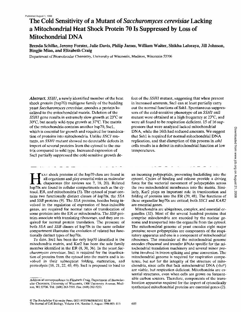
the yeast Ssal protein sequence as a query in the BLASTp search algorithm. In addition to the known yeast hsp70s, this search identified an open reading frame on chromo- some 12 (GenBank/EMBL/DDBJ locus YSCL8039, acces- sion No. U19103) predicted to encode a 657-amino acid protein with a high degree of homology to hsp70 proteins. We compared the sequence to other yeast hsp70s using the DNAstar sequence analysis package. The protein shared the most identity (53%) with the Sscl protein, an hsp70 of the mitochondria. It was 46 and 43% identical to Ssal and Kar2 of the cytosol and lumen of the ER, respectively. The newly identified protein is 49% identical to the hsp70 of Escherichia coli, DnaK, and is therefore more closely re- lated to this bacterial protein than to yeast hsp70 proteins other than Sscl. Because this gene is not a member of a previously described hsp70 family, we have named it SSH1 (stress seventy subfamily H).
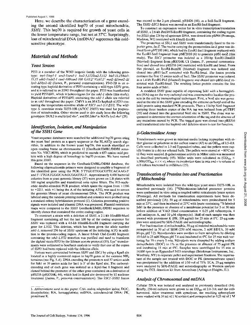
We noted an NH2-terminal extension of the putative Sshl protein relative to many other hsp70s. Most nuclear- encoded mitochondrial proteins are synthesized as precur- sors in the cytosol and upon translocation into rnitochon- dria are processed to a smaller size by cleavage of their NH2-terminal leader sequence. The Sshl extension has characteristics similar to other known mitochondrial pre- sequences, being rich in positively charged and hydrox- ylated residues (Fig. 1). To determine whether Sshl protein is translocated into mitochondria in vitro, radiolabeled Sshl was synthesized in a reticulocyte lysate and added to ener- gized mitochondria, as described in Materials and Meth- ods. Sshl was translocated to a protease-resistant location in a reaction dependent on an electrochemical potential difference across the inner membrane, undergoing cleav- age in the process (Fig. 2 A).
To determine the location of the translocated Sshl pro-
V K ol
1 lip . . . . . . . . . . . . . . . . . . . . . . . . . . . . L~ Iml~lllllletlll~i.ICl~llTi . . . . II~T l iRVL ~ , L I~ IL~ I I I I i I I~YII . . . . . . . . QD!_h~LI DnoK
50 DnaK
138 BlVll l ' i ~1 ] I ~IN ~l l ~oI!l I ~ I I I I~Rml IE~K~ I ~ G T GIR ~ I ElSE VD GE K 711LIIBL I~III]I[H lUll l l D na K
26" ~NVIV~IDTII~NPEITREEITKNmET~I~DvS~R~DmI~HVKK~F~E~m~d~VYK . . . . SI~II~LRVP~EEEIIDNMBLSIII Sshl ''ILL Iil ITI . . . .
228 B~SR I I~LOEE~KDO[ilBI . . . . [ i l lR~PLIBI~ I l I : t I [ I I ; f ; I I : l ; I : I I I l I . ' t . ' ]AOO~OV~WYI~I~L!~T~ I~VIII~KLI!ISL!VEDI!~ DnoK
350 I~IIIII~D I E PEl.m~ll, lml, lt.lt.l~lm:Iml:II RS V~D~S~NSS~II~JITI~)L~'~] ira'S, lllkll'~ II)Nl, lmmm,lt, llU, TI'~I"""~ Sshl 337 K~D~KIT~L~m~'~:~r~:~'|vETm~SIDjS~A~'~m~:~'~`~"`'`~'m"~'~1"~'~T~'~'~|m~'~"~i~-~" Sscl
440 FM~A~s~Vr~V~T~G~AG~D~Km~/~'~'~G~N~1~DLK~T~T|L~Jw~I~';~Yv~I~'~v:~D~||~C~A~ Sshl
404 ~ i 1~AK~Mm~ B H~ .~N~S A ~HF ~L~K RA A ~S L~Q~ iN~D~M ' i ~R~ t .~ |~ 'm lD |~yz~ " | L~K~N ~ DnmK
530 r'~)~PN~I~E~A~L~EE|h~R~NL~R~RL~L~)~M~s~LF~RY)KL~SEK|Y~hmVED~K~RQ~NFKAN Sshl 517 m~DS~AGl~r~I~r~N~E~'KFK~EARK)AImm'~'~.Q~Ah~F)G~VDKA-~A~KVRD~m~m~S~KE-)VA-~Rv Sscl
6 2 0 ENO.S,DV.G,|KAIIo~RILIIrlo . . . . . . . . . mTK)O0)Ol . . . . . . . . . . | 603 0GGE~VNAE~T~E~TSS~E~LYKNDSN . . . . I~NN|~NN~'~SC~T~0. 581KAAI~AKM(~IAOVSOK~MEI~OQOH,~QQTAGADAI~J~N~AKDDDVVD~r"~FE~V~D~K
Sshl Sscl DnaK
Figure 1. Alignment of the predicted amino acid se- quence of Sshl with the amino acid sequences of Sscl and DnaK. The arrows indi- cate the cleavage site of the Sscl presequence and the KpnI site used in fusion gene construction. Identical resi- dues between two or three of the sequences are indicated by the black boxes with white letters. Gaps that were in- serted during the alignment are denoted by dashes. Alignment was performed using MegAlign DNAstar (Madison, WI).
Schilke et al. A Second Mitochondrial Heat Shock Protein 70 605
on August 21, 2008 www.jcb.org
Downloaded from
Published August 1, 1996
Figure 2. Sshl preprotein can be imported into the matrix of mi- tochondria and is cleaved in the process. (A) Sshl preprotein synthesized in a reticulocyte lysate was added to isolated mito- chondria. Translocation was carried out in the presence of a mem- brane potential, Ate, for 5 or 30 min and in the absence of mem- brane potential (-Atb). PK, proteinase K; p, precursor; m, mature; L, reticulocyte lysate containing 35S-labeled Sshl prepro- tein. (B) Fractionation of mitochondria (M) after a 30-min trans- location reaction into mitoplasts (MP) and intermembrane space fractions (IMS). An equivalent portion of mitoplasts was dis- rupted with deoxycholate detergent (MP + DOC). Equivalent amounts of the fractions were either treated with (+PK) or with- out (-PK) proteinase K. Immunoblot analysis was carried out on fractions with antibodies specific for Cytb2 and Mgel after sepa- ration by electrophoresis.
tein, mitochondria were treated with a hypotonic solution that ruptures the outer, but not the inner, mitochondrial membrane to form mitoplasts. Under these conditions, proteins of the intermembrane space, such as cytochrome b2, a re released from the mitochondria and become sensi- tive to exogenously added protease, while matrix proteins, such as Mgel , remain insensitive to protease (Fig. 2 B). Sshl was not sensitive to protease treatment after mito- plast formation, becoming sensitive only after the mito- plasts were treated with detergent to disrupt the inner membrane. This result suggests that Sshl is localized to the matrix.
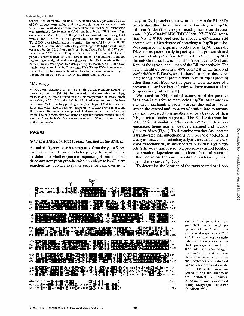
These in vitro translocation experiments demonstrated that Sshl can be imported into the matrix of mitochondria, but did not directly address the question of its normal cel- lular location. Therefore, we constructed a gene that en- coded Sshl with a H A tag at its carboxyl terminus. The tagged protein was recognized by HA-specific antibodies (Fig. 3 A) and was found to be functional, since it rescued the cold-sensitive growth defect of the sshl null mutant (data not shown). Mitochondria, purified from a strain containing the HA-tagged Sshl protein as the only Sshl protein, were subjected to hypotonic treatment and found to contain Sshl. As in the in vitro import experiments, Sshl was completely resistant to protease digestion in mi- toplasts, becoming susceptible only after disruption of the inner membrane with detergent (Fig. 3 B). These results confirm that Sshl is a protein of the mitochondrial matrix.
Figure 3. Sshl is localized to the matrix of mitochondria in vivo. (A) HA-tagged Sshl can be detected by HA-specific antibodies. Whole-cell lysates of cells containing wild-type SSH1 or cells with a disrupted copy of SSH1 and a plasmid carrying an SSH1 gene with a HA tag at the carboxyl end (sshl [SSH1-HA]) were sepa- rated by electrophoresis and subjected to immunoblot analysis using HA- and cytochrome bE (CytbE)-specific antibodies. (B) Mi- tochondria (M) isolated from cells carrying the HA-tagged form of Sshl were separated into mitoplast (MP) and intermembrane space (IMS) fractions. An equivalent portion of mitoplasts was disrupted by addition of deoxycholate detergent (MP + DOC). Equivalent amounts of the fractions were either treated (+PK) or not treated ( -PK) with proteinase K. Immunoblot analysis was carried out on the fractions with antibodies specific for the HA tag (HA), cytochrome b 2 (Cytb2), or Mgel after separation by electrophoresis.
Deletion of SSH1 Results in a Cold-sensitive Phenotype To determine the phenotype of a strain lacking the SSH1 gene, a deletion/insertion mutation was constructed. The 2.1-kb HindI I I -MunI fragment ( - 2 5 6 to +3347, with +1 being the A T G of the initiating methionine) was replaced by the LYS2 gene (Fig. 4 A), and a diploid wild-type strain (PJ53) was transformed as described in Materials and Methods. Transformants were selected based on their lysine prototrophy, and the presence of the mutant allele was confirmed by Southern blot analysis (data not shown). The heterozygous diploid was sporulated. After incuba- tion of the dissection plates at 30°C, the tetrads showed a uniform 2:2 segregation of pinpoint and large colonies af- ter 7 d (Fig. 4 B). The lysine protot rophy segregated 2:2 concomitantly with the small colonies, indicating that they carried the sshl::LYS2 allele. At temperatures of 23°C and below, sshl cells did not form visible colonies. At the higher temperature of 37°C, the growth difference be- tween SSH1 and sshl strains was minimal (Fig. 5 C). The
The Journal of Cell Biology, Volume 134, 1996 606
on August 21, 2008 www.jcb.org
Downloaded from
Published August 1, 1996
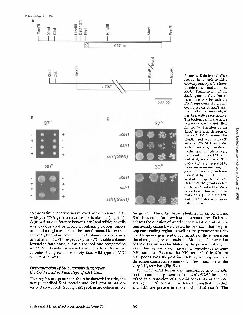
Figure 4. Deletion of SSH1 results in a cold-sensitive growth phenotype. (A) Inser- tion/deletion mutation of SSH1. Transcription of the SSH1 gene is from left to right. The box beneath the DNA represents the protein coding region of SSH1 with the hatched portion indicat- ing the putative presequence. The bottom part of the figure represents the mutant allele formed by insertion of the LYS2 gene after deletion of the SSH1 DNA between the HindlII and MunI sites (B) Asci of PJ53AH1 were dis- sected onto glucose-based media, and the plates were incubated at 30 or 37°C for 7 and 4 d, respectively. The plates were replica plated to lysine omission medium, and growth or lack of growth was indicated by the + and - symbols, respectively. (C) Rescue of the growth defect of the sshl mutant by SSH1 carried on a low copy plas- mid ([SSH1]). Both the 37°C and 30°C plates were incu- bated for 5 d.
cold-sensitive phenotype was relieved by the presence of the wild-type SSH1 gene on a centromeric plasmid (Fig. 4 C). A growth rate difference between sshl and wild-type cells was also observed on medium containing carbon sources other than glucose. On the nonfermentable carbon sources, glycerol or lactate, mutant colonies formed slowly or not at all at 23°C, respectively; at 37°C, visible colonies formed in both cases, but at a reduced rate compared to wild type. On galactose-based medium, sshl cells formed colonies, but grew more slowly than wild type at 23°C (data not shown).
Overexpression o f Ssc l Partially Suppresses the Cold-sensitive Phenotype o f s sh l Cells
Two hsp70s are present in the mitochondrial matrix, the newly identified Sshl protein and Sscl protein. As de- scribed above, cells lacking Sshl protein are cold-sensitive
for growth. The other hsp70 identified in mitochondria, Sscl, is essential for growth at all temperatures. To better address the question of whether these related proteins are functionally distinct, we created fusions, such that the pre- sequence coding region as well as the promoter was de- rived from one gene and the remainder of the fusion from the other gene (see Materials and Methods). Construction of these fusions was facilitated by the presence of a KpnI site in the regions of both genes that encode the extreme NH 2 terminus. Because the NH2 termini of hsp70s are highly conserved, the proteins resulting from expression of the fusion constructs contain only a few alterations at the very NH2 terminus (Fig. 5 A).
The SSCI:SSH1 fusion was transformed into the sshl null mutant. The presence of the SSCI:SSH1 fusion re- sulted in suppression of the cold sensitivity of the sshl strain (Fig. 5 B), consistent with the finding that both Sscl and Sshl are present in the mitochondrial matrix. The
Schilke et al. A Second Mitochondrial Heat Shock Protein 70 607
on August 21, 2008 www.jcb.org
Downloaded from
Published August 1, 1996
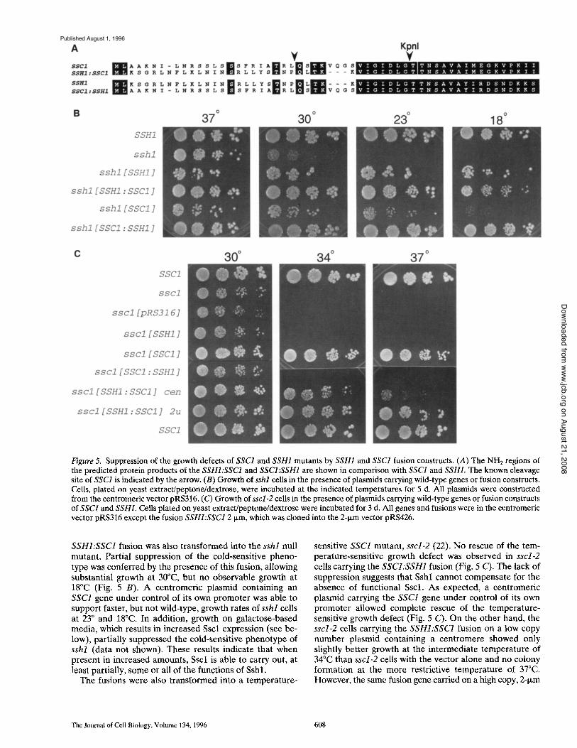
Figure 5. Suppression of the growth defects of SSC1 and SSH1 mutants by SSH1 and SSC1 fusion constructs. (A) The NHE regions of the predicted protein products of the SSHI:SSC1 and SSCI:SSH1 are shown in comparison with SSC1 and SSHI. The known cleavage site of SSC1 is indicated by the arrow. (B) Growth of sshl cells in the presence of plasmids carrying wild-type genes or fusion constructs. Ceils, plated on yeast extract/peptone/dextrose, were incubated at the indicated temperatures for 5 d. All plasmids were constructed from the centromeric vector pRS316. (C) Growth of sscl-2 cells in the presence of plasmids carrying wild-type genes or fusion constructs of SSCI and SSHI. Ceils plated on yeast extract/peptone/dextrose were incubated for 3 d. All genes and fusions were in the centromeric vector pRS316 except the fusion SSHI:SSC1 2 txm, which was cloned into the 2-t~m vector pRS426.
SSHI:SSC1 fusion was also transformed into the sshl null mutant. Partial suppression of the cold-sensitive pheno- type was conferred by the presence of this fusion, allowing substantial growth at 30°C, but no observable growth at 18°C (Fig. 5 B). A centromeric plasmid containing an SSC1 gene under control of its own promoter was able to support faster, but not wild-type, growth rates of sshl cells at 23 ° and 18°C. In addition, growth on galactose-based media, which results in increased Sscl expression (see be- low), partially suppressed the cold-sensitive phenotype of sshl (data not shown), These results indicate that when present in increased amounts, Sscl is able to carry out, at least partially, some or all of the functions of Sshl.
The fusions were also transformed into a temperature-
sensitive SSC1 mutant, sscl-2 (22). No rescue of the tem- perature-sensitive growth defect was observed in sscl-2 cells carrying the SSCI:SSH1 fusion (Fig. 5 C). The lack of suppression suggests that Sshl cannot compensate for the absence of functional Sscl. As expected, a centromeric plasmid carrying the SSC1 gene under control of its own promoter allowed complete rescue of the temperature- sensitive growth defect (Fig. 5 C). On the other hand, the sscl-2 cells carrying the SSHI:SSC1 fusion on a low copy number plasmid containing a centromere showed only slightly better growth at the intermediate temperature of 34°C than sscl-2 cells with the vector alone and no colony formation at the more restrictive temperature of 37°C. However, the same fusion gene carried on a high copy, 2-1xm
The Journal of Cell Biology, Volume 134, 1996 608
on August 21, 2008 www.jcb.org
Downloaded from
Published August 1, 1996
plasmid allowed complete rescue of the temperature sensi- tive phenotype (Fig. 5 C).
The requirement that the SSHI:SSC1 fusion be in high copy number in order to suppress the growth defect of the sscl-2 strain suggests that the SSH1 gene might be ex- pressed at a lower level than the SSC1 gene. Analysis of the codon usage of SSH1 and SSC1 allowed us to make a comparison of the codon adaptation index (CAI), allowing a prediction of the relative levels of expression (42). Genes of S. cerevisiae that are highly expressed, such as riboso- mal protein genes and histones, usually have a CAI be- tween 0.5 and 1.0. Genes that are expressed at very low levels, such as GAL4, have CAIs of 0.098 to 0.12. Using the DNAStrider program, SSCI was calculated to have a CAI of .522 and SSH1 a CAI of 0.148, suggesting that SSH1 is expressed at relatively low levels while, as already known, SSC1 is a relatively highly expressed gene (8).
To attempt to address the question of the level of ex- pression more closely, we constructed fusions between the promoters of SSC1 and SSH1 and the reporter gene, lacZ (see Materials and Methods). Wild-type cells (PJ43-2B) carrying the SSCI:lacZ and SSHI:lacZ fusions on centro- meric vectors were grown at two temperatures, 23 and 30°C, and assayed for [3-galactosidase activity (Table I). The nearly 1,000-fold difference in activity suggests that SSH1 is expressed at much lower levels than SSC1. Ex- pression levels of both SSC1 and SSH1 are twofold higher when cells are grown in media with galactose as the carbon source instead of glucose as expected of genes encoding mitochondrial proteins.
ssh l Mitochondria Show No Obvious Defect in Preprotein Translocation
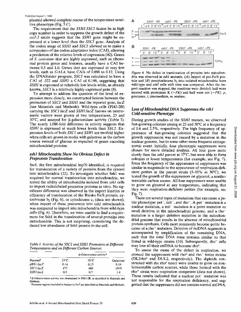
Sscl, the first mitochondrial hsp70 identified, is required for translocation of a variety of proteins from the cytosol into mitochondria (22). To investigate whether Sshl was required for normal translocation into mitochondria, we tested the ability of mitochondria isolated from sshl cells to import radiolabeled precursor proteins in vitro. No sig- nificant difference was observed in the import kinetics or efficiency of translocation of the Rieske Fe/S protein, cy- tochrome b E (Fig. 6), or cytochrome cl (data not shown), when import of these precursors into sshl mitochondria was compared to import into mitochondria from wild-type cells (Fig. 6). Therefore, we were unable to find a require- ment for Sshl in the translocation of several proteins into mitochondria. This is not surprising, considering the de- duced low abundance of Sshl protein in the cell.
Table L Activity of the SSC1 and SSH1 Promoters at Different Temperatures and on Different Carbon Sources
fl-Galactosidase activity*
Plasmid* 23°C 30°C Galactose p B S l - l a c Z 0.14 0.15 0.16 SSC1 :lacZ 477 469 1019 SSHI :lacZ 0.5 0.7 1.4
* [3-Galactosidase activity was determined in PJ43-2B, as described in Materials and Methods. * Promoter regions included in fusions to lacZ are described in Materials and Methods.
Figure 6. No defect in translocation of proteins into mitochon- dria was observed in sshl mutants. (A) Import of pre-Fe/S pro- tein and (B) precytochrome b2 into isolated mitochondria from wild-type and sshl cells with time was compared. After the im- port reaction was stopped, the reactions were divided; half were treated with proteinase K (+PK) and half were not (-PK). p, precursor, i, intermediate, m, mature.
Loss o f Mitochondrial D N A Suppresses the sshl Cold-sensitive Phenotype
During growth studies of the SSH1 mutant, we observed fast-growing colonies arising at 23 and 30°C at a frequency of 0.6 and 2.5%, respectively. The high frequency of ap- pearance of fast-growing colonies suggested that the growth suppression was not caused by a mutation in the nuclear genome, but to some other more frequent extrage- nomic event. Initially, four phenotypic suppressors were chosen for more detailed analysis. All four grew more slowly than the sshl parent at 37°C, but were able to form colonies at lower temperatures (for example, see Fig. 7). Since the frequency of the appearance of suppressors was similar in magnitude to the appearance of respiration-defi- cient petites in the parent strain (5-10% at 30°C), we tested the growth of the suppressors on glycerol, a nonfer- mentable carbon source. All four suppressors were unable to grow on glycerol at any temperature, indicating that they were respiration-deficient petites (for example, see Fig. 7).
There are several types of mutations that can cause a pe- tite phenotype: pet- , mit- , and rho-. A pet- mutation is a nuclear mutation, a mit- mutation is a point mutation or small deletion in the mitochondrial genome, and a rho- mutation is a larger deletion mutation in the mitochon- drial genome that results in the absence of mitochondrial protein synthesis. Cells most commonly become petite be- cause of a rho- mutation. Deletion of mtDNA segments is accompanied by amplification of the remaining DNA, such that the total D N A mass remains similar to that found in wild-type strains (10). Subsequently, rho- cells may lose all their mtDNA to become rho °.
To assess the cause of the defect in respiration, we crossed the suppressors with rho ° and rho + tester strains (DL2rho ° and DUL2, respectively). The diploids con- structed with the rho ° tester were unable to grow on non- fermentable carbon sources, while those formed with the rho + strain were respiration competent (data not shown). These results indicated that a nuclear pet - mutation was not responsible for the respiration deficiency, and sug- gested that the suppressors did not contain normal mtDNA.
Schilke et al. A Second Mitochondrial Heat Shock Protein 70 609
on August 21, 2008 www.jcb.org
Downloaded from
Published August 1, 1996
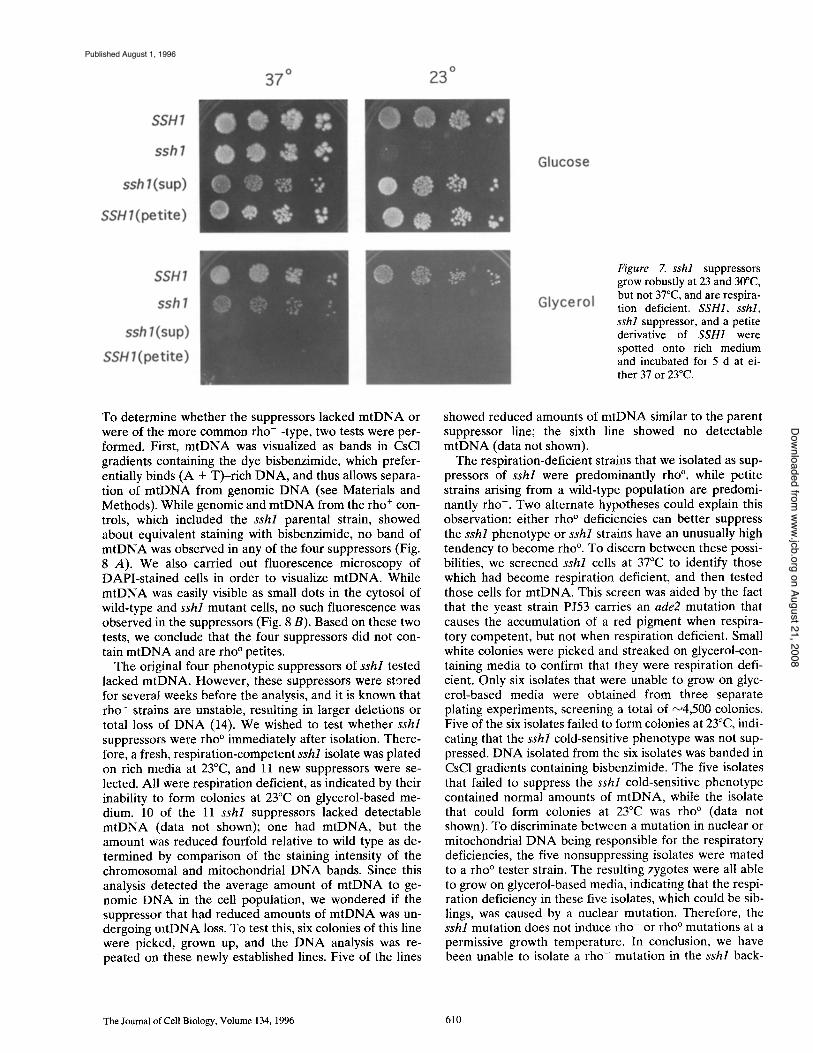
Figure 7. sshl suppressors grow robustly at 23 and 30°C, but not 37°C, and are respira- tion deficient. SSH1, sshl, sshl suppressor, and a petite derivative of SSH1 were spotted onto rich medium and incubated for 5 d at ei- ther 37 or 23°C.
To determine whether the suppressors lacked m t D N A or were of the more common rho- -type, two tests were per- formed. First, m t D N A was visualized as bands in CsCI gradients containing the dye bisbenzimide, which prefer- entially binds (A + T)-r ich D N A , and thus allows separa- tion of m t D N A from genomic D N A (see Materials and Methods). While genomic and m t D N A from the rho ÷ con- trois, which included the sshl parental strain, showed about equivalent staining with bisbenzimide, no band of m t D N A was observed in any of the four suppressors (Fig. 8 A). We also carried out fluorescence microscopy of DAPI-s ta ined cells in order to visualize mtDNA. While m t D N A was easily visible as small dots in the cytosol of wild-type and sshl mutant cells, no such fluorescence was observed in the suppressors (Fig. 8 B). Based on these two tests, we conclude that the four suppressors did not con- tain m t D N A and are rho ° petites.
The original four phenotypic suppressors of sshl tested lacked mtDNA. However, these suppressors were stored for several weeks before the analysis, and it is known that rho- strains are unstable, resulting in larger deletions or total loss of D N A (14). We wished to test whether sshl suppressors were rho ° immediately after isolation. There- fore, a fresh, respiration-competent sshl isolate was plated on rich media at 23°C, and 11 new suppressors were se- lected. All were respiration deficient, as indicated by their inability to form colonies at 23°C on glycerol-based me- dium. 10 of the 11 sshl suppressors lacked detectable m t D N A (data not shown); one had mtDNA, but the amount was reduced fourfold relative to wild type as de- termined by comparison of the staining intensity of the chromosomal and mitochondrial D N A bands. Since this analysis detected the average amount of m t D N A to ge- nomic D N A in the cell population, we wondered if the suppressor that had reduced amounts of m t D N A was un- dergoing m t D N A loss. To test this, six colonies of this line were picked, grown up, and the D N A analysis was re- peated on these newly established lines. Five of the lines
showed reduced amounts of m t D N A similar to the parent suppressor line; the sixth line showed no detectable m t D N A (data not shown).
The respiration-deficient strains that we isolated as sup- pressors of sshl were predominantly rho °, while petite strains arising from a wild-type population are predomi- nantly rho- . Two alternate hypotheses could explain this observation: either rho ° deficiencies can better suppress the sshl phenotype or sshl strains have an unusually high tendency to become rho °. To discern between these possi- bilities, we screened sshl cells at 37°C to identify those which had become respiration deficient, and then tested those cells for mtDNA. This screen was aided by the fact that the yeast strain P J53 carries an ade2 mutation that causes the accumulation of a red pigment when respira- tory competent, but not when respiration deficient. Small white colonies were picked and streaked on glycerol-con- taining media to confirm that they were respiration defi- cient. Only six isolates that were unable to grow on glyc- erol-based media were obtained from three separate plating experiments, screening a total of ~4,500 colonies. Five of the six isolates failed to form colonies at 23°C, indi- cating that the sshl cold-sensitive phenotype was not sup- pressed. D N A isolated from the six isolates was banded in CsC1 gradients containing bisbenzimide. The five isolates that failed to suppress the sshl cold-sensitive phenotype contained normal amounts of mtDNA, while the isolate that could form colonies at 23°C was rho ° (data not shown). To discriminate between a mutation in nuclear or mitochondrial D N A being responsible for the respiratory deficiencies, the five nonsuppressing isolates were mated to a rho ° tester strain. The resulting zygotes were all able to grow on glycerol-based media, indicating that the respi- ration deficiency in these five isolates, which could be sib- lings, was caused by a nuclear mutation. Therefore, the sshl mutation does not induce rho- or rho ° mutations at a permissive growth temperature. In conclusion, we have been unable to isolate a rho- mutation in the sshl back-
The Journal of Cell Biology, Volume 134, 1996 610
on August 21, 2008 www.jcb.org
Downloaded from
Published August 1, 1996
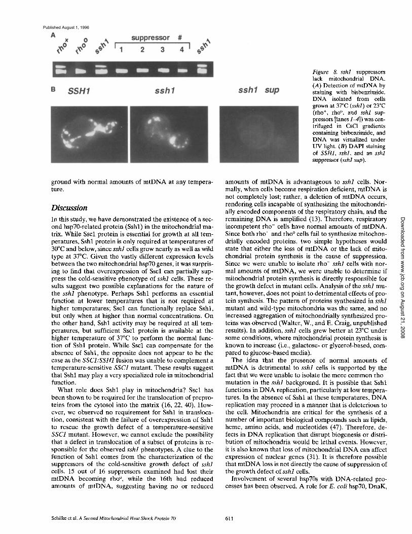
Figure 8. sshl suppressors lack mitochondrial DNA. (A) Detection of mtDNA by staining with bisbenzimide. DNA isolated from cells grown at 37°C (sshl) or 23°C (rho +, rho °, and sshl sup- pressors [lanes 14]) was cen- trifuged in CsC1 gradients containing bisbenzirnide, and DNA was visualized under UV light. (B) DAPI staining of SSH1, sshl, and an sshl suppressor (sshl sup).
ground with normal amounts of mtDNA at any tempera- ture.
Discussion In this study, we have demonstrated the existence of a sec- ond hsp70-related protein (Sshl) in the mitochondrial ma- trix. While Sscl protein is essential for growth at all tem- peratures, Sshl protein is only required at temperatures of 30°C and below, since sshl cells grow nearly as well as wild type at 37°C. Given the vastly different expression levels between the two mitochondrial hsp70 genes, it was surpris- ing to find that overexpression of Sscl can partially sup- press the cold-sensitive phenotype of sshl cells. These re- sults suggest two possible explanations for the nature of the sshl phenotype. Perhaps Sshl performs an essential function at lower temperatures that is not required at higher temperatures; Sscl can functionally replace Sshl, but only when at higher than normal concentrations. On the other hand, Sshl activity may be required at all tem- peratures, but sufficient Sscl protein is available at the higher temperature of 37°C to perform the normal func- tion of Sshl protein. While Sscl can compensate for the absence of Sshl, the opposite does not appear to be the case as the SSCI:SSH1 fusion was unable to complement a temperature-sensitive SSC1 mutant. These results suggest that Sshl may play a very specialized role in mitochondrial function.
What role does Sshl play in mitochondria? Sscl has been shown to be required for the translocation of prepro- teins from the cytosol into the matrix (16, 22, 40). How- ever, we observed no requirement for Sshl in transloca- tion, consistent with the failure of overexpression of Sshl to rescue the growth defect of a temperature-sensitive SSC1 mutant. However, we cannot exclude the possibility that a defect in translocation of a subset of proteins is re- sponsible for the observed sshl phenotypes. A clue to the function of Sshl comes from the characterization of the suppressors of the cold-sensitive growth defect of sshl cells. 15 out of 16 suppressors examined had lost their mtDNA becoming rho °, while the 16th had reduced amounts of mtDNA, suggesting having no or reduced
amounts of mtDNA is advantageous to sshl cells. Nor- mally, when cells become respiration deficient, mtDNA is not completely lost; rather, a deletion of mtDNA occurs, rendering cells incapable of synthesizing the mitochondri- ally encoded components of the respiratory chain, and the remaining DNA is amplified (13). Therefore, respiratory incompetent rho- cells have normal amounts of mtDNA. Since both rho- and rho ° ceils fail to synthesize mitochon- drially encoded proteins, two simple hypotheses would state that either the loss of mtDNA or the lack of mito- chondrial protein synthesis is the cause of suppression. Since we were unable to isolate rho- sshl cells with nor- mal amounts of mtDNA, we were unable to determine if mitochondrial protein synthesis is directly responsible for the growth defect in mutant cells. Analysis of the sshl mu- tant, however, does not point to detrimental effects of pro- tein synthesis. The pattern of proteins synthesized in sshl mutant and wild-type mitochondria was the same, and no increased aggregation of mitochondrially synthesized pro- teins was observed (Walter, W., and E. Craig, unpublished results). In addition, sshl cells grew better at 23°C under some conditions, where mitochondrial protein synthesis is known to increase (i.e., galactose- or glycerol-based, com- pared to glucose-based media).
The idea that the presence of normal amounts of mtDNA is detrimental to sshl ceils is supported by the fact that we were unable to isolate the more common rho- mutation in the sshl background. It is possible that Sshl functions in DNA replication, particularly at low tempera- tures. In the absence of Sshl at these temperatures, DNA replication may proceed in a manner that is deleterious to the cell. Mitochondria are critical for the synthesis of a number of important biological compounds such as lipids, heine, amino acids, and nucleotides (47). Therefore, de- fects in DNA replication that disrupt biogenesis or distri- bution of mitochondria would be lethal events. However, it is also known that loss of mitochondrial DNA can affect expression of nuclear genes (31). It is therefore possible that mtDNA loss is not directly the cause of suppression of the growth defect of sshl cells.
Involvement of several hsp70s with DNA-related pro- cesses has been observed. A role for E. coli hsp70, DnaK,
Schilke et al. A Second Mitochondrial Heat Shock Protein 70 611
on August 21, 2008 www.jcb.org
Downloaded from
Published August 1, 1996
in D N A replication has been known for many years (15). DnaK is required for the replication of phage h DNA, playing an essential role in the disassembly of the initia- tion complex, thus allowing replication to proceed (17). One report has also suggested a role for DnaK in the initi- ation of E. coli D N A replication, based on phenotypic similarities between strains carrying mutations in dnaA, which encodes a protein required for the initiation of DNA replication (38), and a particular allele of dnaK.
For many years, it was thought that procaryotes, the the- orized progenitors of mitochondria, have only a single hsp70, DnaK. Recently, however, a second hsp70, Hsc66 encoded by the hscA gene, which is only 35% identical to DnaK, has been identified in E. coli (23, 24, 41). Both mi- tochondrial proteins, Sscl and Sshl, are similarly related to each of the two procaryotic proteins. Sscl and DnaK share 59% amino acid identity, Sshl and DnaK 49%, Sscl and Hsc66 39%, and Sshl and Hsc66 38%. A role for Hsc66 in DNA metabolism has also been suggested. Muta- tions in the gene encoding Hsc66, HscA, were initially identified as suppressors of mutations of hns-1 (23). hns-1 encodes a major component of the nucleoid (12). One phenotype of hns-1 mutants is a dramatic increase in site- specific D N A inversions that normally occur at low levels. Mutations in hscA are able to suppress this high level of DNA inversion. However, the mechanism by which this suppression occurs is not understood, and the role of ei- ther procaryotic hsp70 in host D N A metabolism therefore remains unresolved. It is also intriguing to note that Sscl has been identified as an essential, noncatalytic subunit of the mitochondrial site-specific endonuclease, Endo.SceI, which is thought to be involved in genetic recombination (27, 29).
A mitochondrial hsp70 has been identified in trypano- somes. This hsp70 has been localized to the site of mtDNA replication within the unusually large mitochondrium called a kinetoplast (11). This location has been an enigma be- cause of the known involvement of the mitochondrial hsp70 from S. cerevisiae, Sscl, in protein translocation. We suggest that the mitochondria of trypanosomes, like those of yeast, are likely to have more than one hsp70, one in- volved in D N A replication and one involved in the trans- location and folding of proteins synthesized in the cytosol.
The fact that loss of mtDNA results in suppression of the sshl cold-sensitive phenotype is unusual, if not unique. This observation may have implications concerning mito- chondrial metabolism in mammalian cells, since alterations in mtDNA are a frequent cause of maternally inherited genetic diseases characterized by defects in respiratory metabolism (for review see 33). Most of these mitochon- drial myopathies are caused by deletions of portions of the mtDNA, but patients with one type have been shown to have reduced amounts of mtDNA (26). Additional re- search with this mutation has shown that over many gener- ations the mitochondria ultimately lose all their mtDNA, becoming rho ° (3, 4). The reduction in mtDNA is caused by a nuclear-encoded mutation whose gene product has not yet been identified. We have shown that mutation of a yeast mitochondrial hsp70 gene is more detrimental in the presence of normal amounts of mtDNA than in its ab- sence. It is intriguing to speculate that a similar mutation in humans would result in selection of cells with lower lev- els of mtDNA.
We thank Tom Fox for generously providing strains and advice, and Bon- nie Baxter for critical comments on the manuscript.
This work was supported by U.S. Public Health Service grant NIH 5 ROI27870 (awarded to E. Craig). B. Schilke and J. Johnson were sup- ported by National Institutes of Health (NIH) postdoctoral fellowship awards 1F32GM17687-01 and 1F32GM17139-01, respectively. J. Davis was supported by an NIH predoctoral training grant (NRSA 5T32GM07215).
Received for publication 20 February 1995 and in revised form 20 May 1995.
References
1. Ausebel, F., R. Brent, R. Kingston, D. Moore, J.G. Seidman, J. Smith, and K. Struhl. 1989. Current Protocols in Molecular Biology. John Wiley and Sons, New York.
2. Baker, K., and G. Sehatz. 1991. Mitochondrial proteins essential for viability mediate protein import into mitochondria. Nature (Lond.). 349:205-208.
3. Bodnar, A.G., J.M. Cooper, I.J. Holt, J.V. Leonard, and A.H.V. Schapira. 1993. Nuclear complementation restores mtDNA levels in cultured cells from a patient with mtDNA depletion. Am. Z Hum. Genet. 53:663-669.
4. Bodnar, A.G., J.M. Cooper, J.V. Leonard, and A.H.V. Schapira. 1995. Re- spiratory-deficient human fibroblasts exhibiting defective mitochondrial DNA replication. Biochem. J. 305:817-822.
5. Boorstein, W.R., T. Ziegelhoffer, and E.A. Craig. 1994. Molecular evolu- tion of the HSP70 multigene family. J. MoL EvoL 38:1-17.
6. Casadaban, M.J., A. Martinez-Arias, S.K, Shapira. and J. Chou. 1983. 13-Galactosidase gene fusions for analyzing gene expression in Escheri- chia coli and yeast. In Methods in Enzymology. R. Wu, L. Grossman, and K. Moldave, editors. Academic Press, New York. 293-308.
7. Craig, E.A., B.D. Gambin, and R.J. Nelson. 1993. Heat shock proteins: mo- lecular chaperones of protein biogenesis. Microbiol. Rev. 57:402--414.
8. Craig, E.A., J. Kramer, J. Shilling, M. Werner-Washburne, S. Holmes, J. Kosic-Smither, and C.M. Nicolet. 1989. SSC1, an essential member of the S. cerevisiae HSPT0 multigene family, encodes a mitochondrial protein. Mol. Cell Biol. 9:3000--3008.
9. Craig, E.A., T. Ziegelhoffer, J. Nelson, S. Laloraya, and J. Halladay. 1995. Complex multigene family of functionally distinct Hsp70s of yeast. In Cold Spring Harbor Symposia on Quantitative Biology: Protein Kinesis: The Dynamics of Protein Trafficking and Stability. Cold Spring Harbor Laboratory Press, Cold Spring Harbor, NY.
10. Dujon, B. 1981. Mitochondrial Genetics and Function. Cold Spring Harbor Laboratory Press, Cold Spring Harbor, NY.
11. Engman~ D.~ L.V. Kirehhoff, and J.E. Donelson. 1989. Molecular cloning of mtp70, a mitochondrial member of the hsp70 family. Mol. Cell Biol. 9: 5163-5168.
12. Falconi, M., M. Gualtieri, M. La Teana, M. Losso, and C.L. Pom. 1988. Pro- teins from the procaryotic nucleoid: primary and quartenary structure of the 15-kD Escherichia coli DNA binding protein H-NS. Mol. Microbiol. 2:323-329.
13. Faye, G., H. Fukuhara, C. Grandchamp, J. Lazowska, M. Rabinowitz, M. Bolotin-Fukuhara, D. Coen, J. Deutsch, P. Netter, and P. Slonimski. 1973. Mitochondrial nucleic acids in petite colony mutants: deletions and repetitions of genes. Biochemie. 55:779-792.
14. Fox, T.D., L.S. Folley, J.J. Mulero, T.W. McMullen, P.E. Thorsness, L.O. Hedin, and M.C. Costanzo. 1991. Analysis and manipulation of yeast mi- tochondrial genes. In Methods in Enzymology. C. Guthrie and G.R. Fink~ editors. Academic Press, New York. 149-165.
15. Friedman, D.I., E.R. Olson, K. Tilly, and C. Georgopolous. 1984. Interac- tion of bacteriophage lambda and host molecules in the growth of bacte- riophage. Microbiol. Rev. 48:299-325.
16. Gambill, B.D., W. Voos, P.J. Kang, B. Miao, T. Langer, E.A. Craig, and N. Planner. 1993. A dual role for mitochondrial heat shock protein 70 in membrane translocation of preproteins. J. Cell Biol. 123:109-117.
17. Georgopoulos, C., D. Ang, K. Liberek, and M. Zylicz. 1991. Properties of the Escherichia coli heat shock proteins and their role in bacteriophage lambda growth. In Stress Proteins in Biology and Medicine. R. Mori- moto, A. Tissieres, and C. Georgopoulos, editors. Cold Spring Harbor Laboratory Press, Cold Spring Harbor, NY. 199-221.
18. Georgopoulos, C., and W. Welch. 1993. Role of the major heat shock pro- teins as molecular chaperones. Annu. Rev. Cell Biol. 9:601-634.
19. Gietz, R.D., R.H. Schiestl, A.R. Willems, and R.A. Woods. 1995. Studies on the transformation of intact yeast cells by the LiAc/SS-DNA/PEG procedure. Yeast. 11:355-360.
20. Hendrick, J., and F.-U. Hartl. 1993. Molecular chaperone function of heat- shock proteins. Annu. Rev. Biochem. 62:349-384.
21. Hermann, J., R. Stuart, E. Craig, and W. Neupert. 1994. Mitochoodrial heat shock protein 70, a molecular chaperone for proteins encoded by mitochondrial DNA. J. Cell Biol. 127:893-902.
22. Kang, P.J., J. Ostermann, J. Shilling, W. Neupert, E.A. Craig, and N. Plan- ner. 1990. Hsp70 in the mitochondrial matrix is required for translocation and folding of precursor proteins. Nature (Lond.). 348:137-143.
The Journal of Cell Biology, Volume 134, 1996 612
on August 21, 2008 www.jcb.org
Downloaded from
Published August 1, 1996
23. Kawula, T., and M. Lelivelt. 1994. Mutations in a gene encoding a new Hsp70 suppress rapid DNA inversion and bgl activation, but not proU derepression in hns-1 mutant Escherichia coli. J. Bacteriol. 176:610-619.
24. Lelivelt, M., and T. Kawula. 1995. Hsc66, an Hsp70 homolog in Escherichia coli, is induced by cold shock but not by heat shock. Z BacterioL 177: 4900-4907.
25. Miller, J.H. 1972. Experiments in Molecular Genetics. Cold Spring Harbor, Cold Spring Harbor, NY. 466 pp.
26. Moraes, C.T., S. Shaske, H. Trischler, J.R. Aprille, F. Andretta, E. Bonilla, E.A. Schon, and S. DiMauro. 1991. mtDNA depletion with variable tis- sue expression: a novel genetic abnormality in mitochondrial diseases. Am. J. Hum. Genet. 48:492-501.
27. Morishima, N., K. Nakagawa, E. Yamamoto, and T. Shibata. 1990. A sub- unit of yeast site-specific endonuclease SceI is a mitochondrial version of the 70-kDa heat shock protein. J. Biol. Chem. 265:15189-15197.
28. Mulero, J.J., J.K. Rosenthal, and T.D. Fox. 1994. Petll2, a Saccharomyces cerevisiae nuclear gene required to maintain rho+ mitochondrial DNA. Curr. Genet. 25:299-304.
29. Nakagawa, K., N. Morishima, and T. Shibata. 1991. A maturase-like sub- unit of the sequence-specific endonuclease Endo.SceI from yeast mito- chondria. J. Biol. Chem. 266:1977-1984.
30. Normington, K., K. Kohno, Y. Kozutsumi, M.J. Gething, and J. Sambrook. 1989. S. cerevisiae encodes an essential protein homologous in sequence and function to mammalian BiP. Cell 57:1223-1236.
31. Parikh, V.S., M. Morgan, R. Scott, L. Clements, and R. Butow. 1987. The mitochondrial genotype can influence nuclear gene expression in yeast. Science (Wash. DC). 235:576-580.
32. Pon, L., and G. Schatz. 1991. Biogenesis of yeast mitoehondria. In The Mo- lecular and Cellular Biology of the Yeast Saccharomyces: Genome Dy- namics, Protein Synthesis and Energetics. J. Broach, J. Pringle, and E. Jones, editors. Cold Spring Harbor Laboratory, Cold Spring Harbor, NY. 333406.
33. Poulton, .1. 1992. Mitochondrial DNA and genetic disease. Bioessays. 14: 763-768.
34. Pringle, J.R., R.A. Preston, A.E.M. Adams, T. Stearns, D.G. Drubin, B.K. Haarer, and E.W. Jones. 1989. Fluorescence microscopy methods for yeast. In Methods in Cell Biology. A.M. Tartakoff, editors. Academic Press, New York. 357435.
35. Roof, D.M., P.B. Meluh, and M.D. Rose. 1992. Kinesin-related proteins re- quired for assembly of the mitotic spindle. J. Cell BioL 118:95-108.
36. Rose, M.D., L.M. Misra, and J.P. Vogel. 1989. KAR2, a karyogamy gene, is
the yeast homolog of the mammalian BiP/GRP78 gene. Cell. 57:1211-1221. 37. Rose, M.D., P. Novick, J.H. Thomas, D. Botstein, and G.R. Fink. 1987. A
Saccharomyces cerevisiae genomic plasmid bank based on a centromere- containing shuttle vector. Gene (Arnst.). 60:237-243.
38. Sakakibara, Y. 1988. The dnaK gene of Escherichia coli functions in initia- tion of chromosome replication. J. Bacteriol. 170: 972-979.
39. Sanders, S.L., K.M, Whitfield, J.P. Vogel, M.D. Rose, and R.W. Schekman. 1992. Sec61p and BiP directly facilitate polypeptide translocation into the ER. Cell 69:353-365.
40. Scherer, P., U. Krieg, S. Hwang, D. Vestweber, and G. Schatz. 1990. A pre- cursor protein partially translocated into yeast mitochondria is bound to a 70kd mitochondrial stress protein. EMBO (Eur. Mol. Biol. Organ.) J. 9: 43154322.
41. Seaton, B.L., and L.E. Vickery. 1994. A gene encoding a DnaK/hsp70 ho- molog in Escherichia coli. Proc. Natl. Acad. Sci. USA. 91:2066-2070.
42. Sharp, P.M. and W.-H. Li. 1987. The codon adaptation index--a measure of directional synonymous codon usage bias, and its potential applica- tions. Nucleic Acid Res. 15:1281-1295.
43. Sherman, F., G.R. Fink, and J.B. Hicks. 1986. Laboratory Course Manual for Methods in Yeast Genetics. Cold Spring Harbor Press, Cold Spring Harbor, NY. 342 pp.
44. Sikorski, R.S., and P. Hieter. 1989. A system of shuttle vectors and yeast host strains designed for efficient manipulation of DNA in Saccharomy- ces cerevisiae. Genetics. 122:19-27.
45. Slater, M., and E.A. Craig. 1987. Transcriptional regulation of an hsp70 heat shock gene in the yeast Saccharomyces cerevisiae. MoL Cell Biol. 7: 1906-1916.
46. Tu, H., and M.J. Casadaban. 1990. The upstream activating sequence for 1-1eucine gene regulation in Saccharomyces cerevisiae, Nucleic Acid Res. 18:3923-3931.
47. Tzagoloff, A. 1982. Mitochondria. Plenum Publishing, NY. 48. Vogel, J.P., L.M. Misra, and M.D. Rose. 1990. Loss of BiP/GRP78 function
blocks translocation of secretory proteins in yeast. J. Cell Biol. 110:1885- 1895.
49. Wagner, I., H. Arlt, L. van Dyck, T. Langer, and T. Neupert. 1994. Molecu- lar chaperones cooperate with PIM1 protease in the degradation of mis- folded protein in mitochondria. EMBO (Eur. MoL Biol. Organ.) J. 13: 5135-5145.
50. Williamson, D.H., and D.J. Fennell. 1979. Visualization of yeast mitochon- drial DNA with the fluorescent stain "DAPI." In Methods in Enzymology. S. Fleischer and L. Packer, editors. Academic Press, New York. 728-733.
Schilke et al. A Second Mitochondrial Heat Shock Protein 70 613
on August 21, 2008 www.jcb.org
Downloaded from
Published August 1, 1996