the cervical vertebrae maturation (cvm) method cannot ... · cohorts of children born between 1961...
TRANSCRIPT
1© The Author 2015. Published by Oxford University Press on behalf of the European Orthodontic Society. All rights reserved. For permissions, please email: [email protected]
Original article
The cervical vertebrae maturation (CVM) method cannot predict craniofacial growth in girls with Class II malocclusionThomas P. Engel*, Anne-Marie Renkema**, Christos Katsaros*, Pawel Pazera*, Nikolaos Pandis* and Piotr S. Fudalej*,***
*Department of Orthodontics and Dentofacial Orthopedics, University of Bern, Switzerland, **Department of Orthodontics and Craniofacial Biology, Radboud University Medical Centre, Nijmegen, The Netherlands, ***Department of Orthodontics, Palacky University, Olomouc, Czech Republic
Correspondence to: Dr Piotr S. Fudalej, Department of Orthodontics and Dentofacial Orthopedics, University of Bern, Freiburgstrasse 7, Bern 3010, Switzerland. E-mail: [email protected]
Summary
Introduction: The cervical vertebrae maturation (CVM) method is used to determine the timing of treatment of Class II malocclusion. Because its performance has not been tested in patients with Class II, the objective of this study was to evaluate the effectiveness of the CVM method in predicting craniofacial growth in Class II malocclusion.Methods: Twenty-nine untreated girls with Class II malocclusion were identified among participants of the Nijmegen Growth Study. Each girl had a series of cephalograms taken semi-annually from 9 to 14 years of age. The CVM status was established by five observers on a cephalogram taken at 9 years; mandibular and maxillary length and anterior face height were assessed on all available cephalograms. Method error was evaluated with kappa statistics and Bland–Altman (BA) plots. Regression analysis was used to determine if CVM grade can predict the amount of facial growth.Results: The mean kappa for intra-rater agreement during grading with CVM was 0.36 (fair agreement). BA plots demonstrated acceptable agreement for cephalometric measurements. The regression analysis demonstrated that the only chronologic age was associated with the facial growth. The largest effect of age was for condylion–gnathion (Cd–Gn) and articulare–gnathion (Ar–Gn)—for every additional 6 months the Cd–Gn increases by 1.8 mm [95 per cent confidence interval (CI): 1.7, 1.9, P < 0.001] and Ar–Gn increases by 1.59 mm (95 per cent CI: 1.52, 1.67, P < 0.001). The CVM grade could not predict the change of cephalometric variables.Conclusions: There is no evidence to support the hypothesis that the CVM method can predict the amount of craniofacial growth in girls with Class II malocclusion.
Introduction
The timing of orthodontic therapy may influence the final outcome. For example, skeletal Class II division 1 malocclusion is frequently associated with hypoplastic mandible, retruded chin (1) and, conse-quently, unaesthetic facial profile. Evidence from a systematic review (2) suggests that initiation of orthodontic Class II correction before the pubertal growth spurt results mainly in dento-alveolar changes
with little alterations of the facial skeleton. This scenario is not opti-mal because the underlying skeletal problem is not actually corrected and the facial profile may not sufficiently improve. Some evidence (3–5) implies, however, that deferring treatment with functional appliances until the commencement of the growth spurt may result in a more favourable skeletal correction.
A practical problem associated with timing of the therapy is the identification of a period of maximum growth. Two popular
European Journal of Orthodontics, 2016, 1–7doi:10.1093/ejo/cju085
Advance Access publication 9 February 2015
methods have been used and are still in use to this end in orthodon-tics: 1. assessment of hand-wrist (HW) radiographs and 2. evalua-tion of cervical vertebrae maturation (CVM). In the HW method, skeletal maturation is determined on the basis of the stages of ossifi-cation of the bones of the hand and the wrist (6, 7), whereas changes in the cervical vertebrae morphology are used in the CVM method (8–10). Both methods relate maturational stages in the correspond-ing areas with general and facial growth. The CVM method does not require an additional radiograph for assessment and for this reason it has been widely used by clinicians worldwide.
A method can be implemented for diagnostic purposes if it is valid and reproducible. Validity refers to accuracy of the method, i.e. how well the method measures what it purports to measure. To be reproducible a measurement should be the same if repeated by the same or different observer (11). Several early studies showed excellent reproducibility of the CVM method (4, 9, 10, 12–14); however, those early findings have been questioned and even refuted in more recent studies (15–17). The primary reason for criticism was that in those studies the assessment of reliability of the CVM method was under ideal conditions which are unat-tainable in every day clinical practice. A number of studies have shown, however, that the reliability of the method can differ when under ideal conditions and when used in everyday practice (18, 19). A recent study by Beit et al. (20) concluded that ‘… assessment of age-dependent changes in the cervical spine offers no advantage over chronologic age, in either assessing skeletal age or predicting the pubertal growth spurt’.
Additionally, it has been suggested that the CVM method can predict the pubertal growth peak in Class II malocclusion (10). None of the previous studies (9, 10, 12, 13) have tested the validity of the CVM method in predicting the pubertal growth peak in Class II patients. It was therefore, the aim of this study to evaluate the per-formance of the CVM method in predicting craniofacial growth in subjects with Class II malocclusion.
Subjects and methods
SubjectsSubjects for this investigation were selected from the Nijmegen Growth Study (NGS). The NGS was a mixed-longitudinal, inter-disciplinary study of the growth and development of 486 normal Dutch children (232 boys and 254 girls). During the study six cohorts of children born between 1961 and 1967 were measured every 3 months. The repeated measurements were taken during the 5 years, 1970–75, covering a total age range of 4–14 years, with some overlapping of the ages over which the various cohorts were followed. During each examination period numerous measurements were taken including anamnestic variables (i.e. changes in medical history), dental variables (cephalograms and dental casts), medi-cal variables (e.g. blood tests, HW X-rays), psychological variables (e.g. intelligence tests), and anthropometric variables (e.g. evalua-tions of stature height, body weight, and leg length). Cephalometric radiographs and dental study casts were made twice a year in each subject (21).
The NGS was terminated when participants turned 14 years of age. At this age most boys were likely still be in the growth spurt stage and we decided to exclude them from the study. In con-trast, many girls had likely completed or were close to comple-tion of pubertal growth spurt by the age of 14. In order to include a more homogenous sample we decided to select from the NGS database only female participants who had a complete series of
cephalograms taken semi-annually from 9 until 14 years of age. Subsequently their dental casts were inspected to identify those girls with Angle Class II malocclusion. For the purpose of this study only girls with at least ½-cusp Class II on both right and left sides were considered. Exclusion criteria were: orthodontic treat-ment during the period of observation, visible pathology of cervical vertebrae, and poor representation of second, third, or fourth cervi-cal vertebrae (C2, C3, C4).
MethodsCephalograms taken at 9 years of age were used to establish a CVM status according to the method proposed by Baccetti et al. (10). The radiographs were scanned at 300 dpi resolution. Then the images were cropped to restrict visualization to the cervical vertebrae; thus, the dentition was not visible on any of the images. Subsequently, the scans were loaded into PowerPoint to prepare a presentation for the CVM rating. A PowerPoint presentation con-sisted of a detailed description of the CVM method along with instructions on how to rate, examples of all stages of skeletal matu-rity, and all images to be assessed. The presentation file was sent to five raters (senior orthodontic residents). Prior to rating a cali-bration session took place during which observers rated 20 other cephalograms and discussed their scores. Raters assessed cephalo-grams at the time of convenience but not later than 1 day after the calibration session. There was no time restriction on the length of rating session, i.e. each rater used as much time as he/she needed for assessment. The raters reassessed cephalograms after at least 4 weeks. The order of images was randomly changed in the second rating session. The raters did not participate in the design or con-struction of the research project.
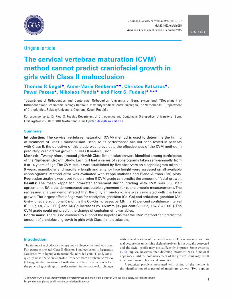
The following measurements were performed on each semian-nual cephalogram: condylion–point A (Cd–A), condylion–gnathion (Cd–Gn), articulare–point A (Ar–A), articulare–gnathion (Ar–Gn), nasion–anterior nasal spine (N–Spa), and anterior nasal spine–men-ton (Spa–Me)—Figure 1. The measurements were completed inde-pendently by two calibrated investigators using the Onyx CEPH 3™ version 3.1.36 (52) software (Image Instruments GmbH, Chemnitz, Germany). The images were adjusted for magnification. Both investi-gators remeasured 30 randomly selected cephalograms after a mini-mum 4 weeks.
Statistical analysisIntra- and inter-observer reliability of scoring with the CVM method was evaluated with kappa statistics. For intra-observer agreement individual kappas were calculated. For inter-rater agreement the mean kappa for 10 pairs of observers (O1 versus O2, O1 versus O3, etc.) was calculated. The interpretation of kappa was as follows (22): kappa values from 0.01 to 0.20 indicate slight agreement, from 0.21 to 0.40—fair agreement, from 0.41 to 0.60—moderate agreement, from 0.61 to 0.80—very good, and from 0.81 to 1—almost perfect agreement.
Moreover the Bland–Altman (BA) limits of agreements were car-ried out in order to assess intra-observer and inter-observer agree-ment during cephalometric measurements (22).
A linear mixed model was fitted in order to determine potential associations between the individual measurements (Cd–A, Cd–Gn, Ar–A, Ar–Gn, N–Spa, and Spa–Me—independent variables) and average across observers CVM scoring after adjusting for age and accounting for the within patient correlations. All analyses were con-ducted using the Stata 13.1 statistical package (Stata Corp, College Station, Texas, USA).
European Journal of Orthodontics, 2016, Vol. 38, No. 12
Results
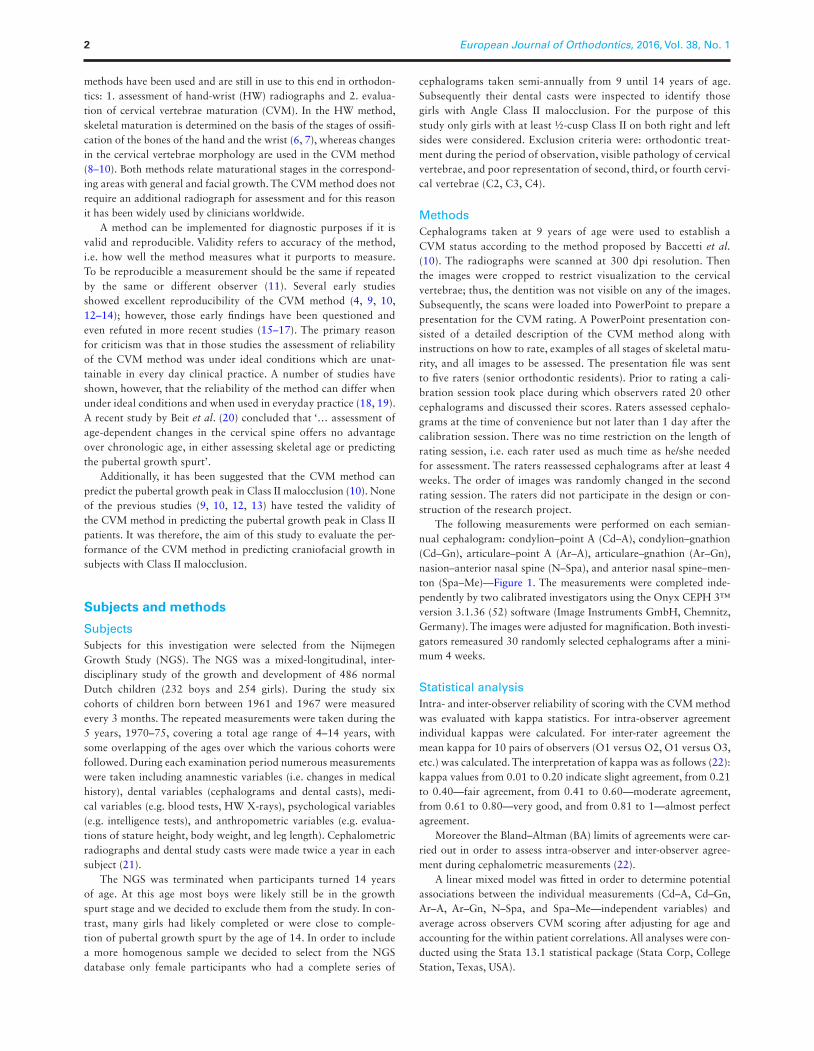
Thirty-nine girls (15.4 per cent) of the 254 females participating in the NGS had bilateral Angle Class II. Twenty-nine of them (74.4 per cent) were not treated orthodontically until the age of 14 years, i.e. until the end of the NGS. All participants had good quality cephalograms with a full representation of cervical ver-tebrae from C2 to C4 and were included in the study (Figure 2). The total number of assessed cephalograms was 302 and all cephalograms were of comparable quality with good representa-tion of facial skeleton. The subjects were born between August and December 1961. They were 9.3 years old (SD = 0.2) at the start of this study. Five girls had bilateral full cusp Class II, 5 had full cusp Class II on one side and ½ cusp on the other side, and 18 girls had ½ cusp Class II on both sides. Two girls had lateral crossbite, 11 girls had deep bite, and 1 girl had anterior open bite.
At 9.3 years of age, the mean CVM grade for all five observers and two rating sessions was 2.4 (SD = 1.4). In 5 subjects the mean CVM grade was 1.5 or below, in 8 subjects—between 1.6 and 1.9, in 4 subjects—between 2.0 and 2.4, in 7—between 2.5 and 2.9, in 5—between 3 and 3.4, and in 2 was 3.5 or more.
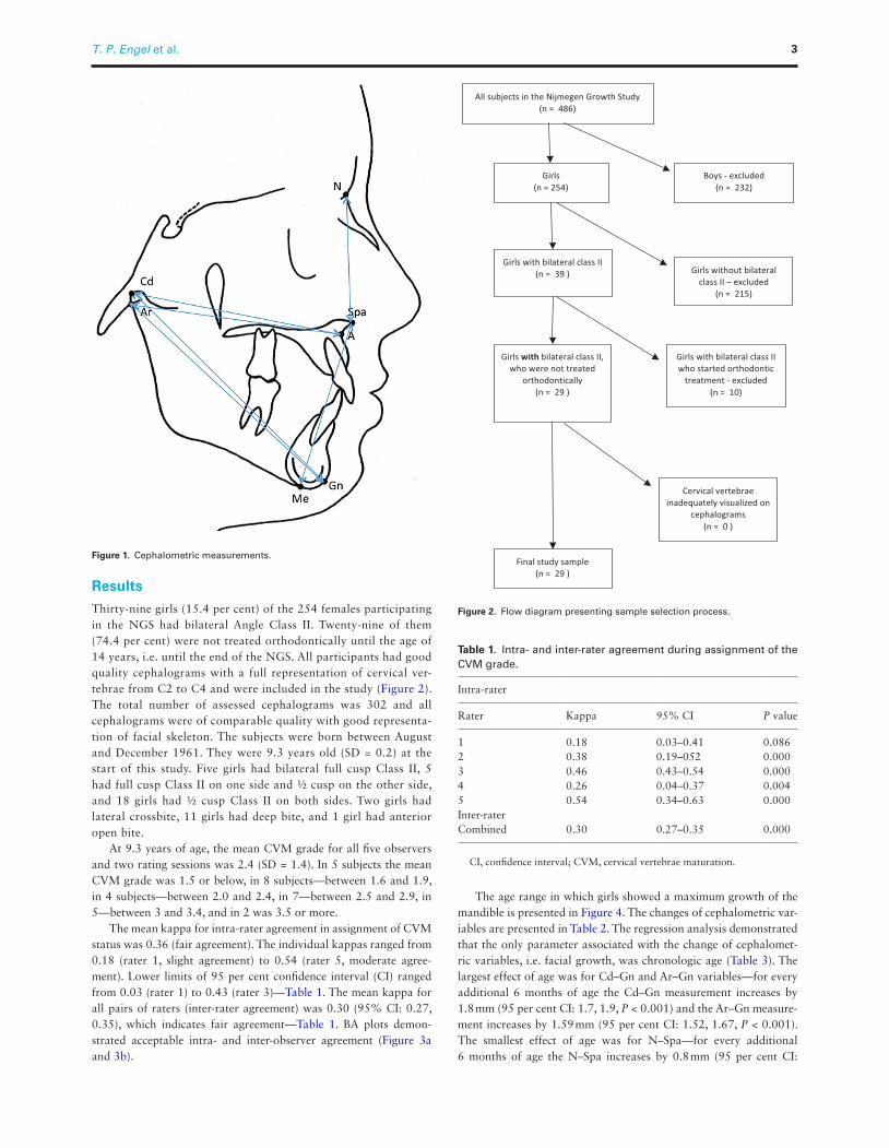
The mean kappa for intra-rater agreement in assignment of CVM status was 0.36 (fair agreement). The individual kappas ranged from 0.18 (rater 1, slight agreement) to 0.54 (rater 5, moderate agree-ment). Lower limits of 95 per cent confidence interval (CI) ranged from 0.03 (rater 1) to 0.43 (rater 3)—Table 1. The mean kappa for all pairs of raters (inter-rater agreement) was 0.30 (95% CI: 0.27, 0.35), which indicates fair agreement—Table 1. BA plots demon-strated acceptable intra- and inter-observer agreement (Figure 3a and 3b).
The age range in which girls showed a maximum growth of the mandible is presented in Figure 4. The changes of cephalometric var-iables are presented in Table 2. The regression analysis demonstrated that the only parameter associated with the change of cephalomet-ric variables, i.e. facial growth, was chronologic age (Table 3). The largest effect of age was for Cd–Gn and Ar–Gn variables—for every additional 6 months of age the Cd–Gn measurement increases by 1.8 mm (95 per cent CI: 1.7, 1.9, P < 0.001) and the Ar–Gn measure-ment increases by 1.59 mm (95 per cent CI: 1.52, 1.67, P < 0.001). The smallest effect of age was for N–Spa—for every additional 6 months of age the N–Spa increases by 0.8 mm (95 per cent CI:
Figure 1. Cephalometric measurements.
Figure 2. Flow diagram presenting sample selection process.
Table 1. Intra- and inter-rater agreement during assignment of the CVM grade.
Intra-rater
Rater Kappa 95% CI P value
1 0.18 0.03–0.41 0.0862 0.38 0.19–052 0.0003 0.46 0.43–0.54 0.0004 0.26 0.04–0.37 0.0045 0.54 0.34–0.63 0.000Inter-raterCombined 0.30 0.27–0.35 0.000
CI, confidence interval; CVM, cervical vertebrae maturation.
T. P. Engel et al. 3
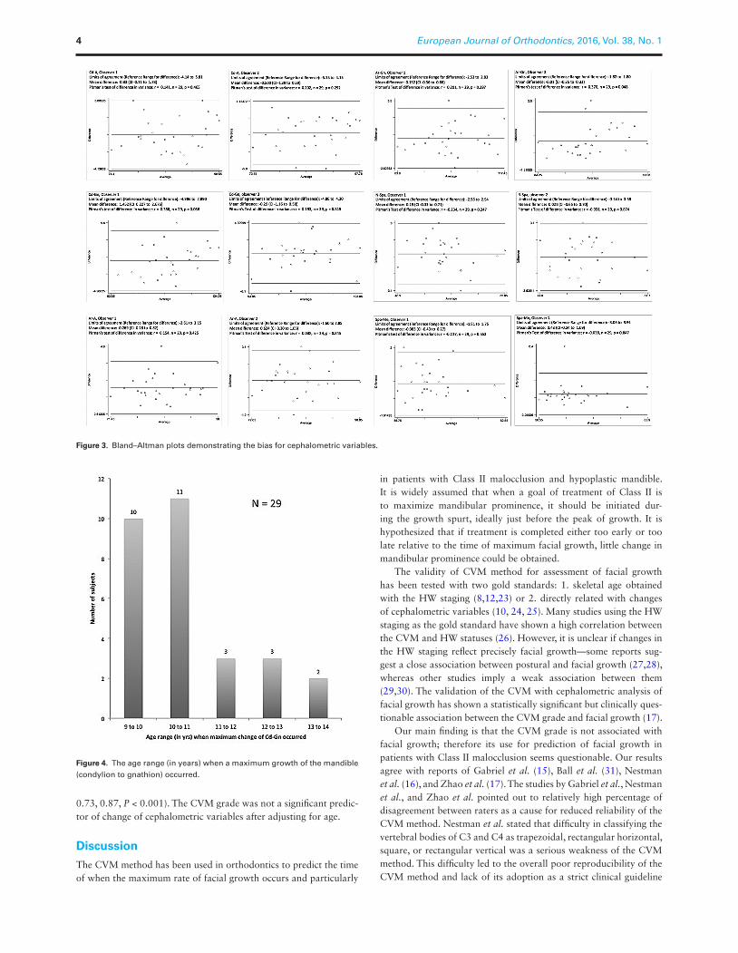
Figure 4. The age range (in years) when a maximum growth of the mandible (condylion to gnathion) occurred.
Figure 3. Bland–Altman plots demonstrating the bias for cephalometric variables.
0.73, 0.87, P < 0.001). The CVM grade was not a significant predic-tor of change of cephalometric variables after adjusting for age.
Discussion
The CVM method has been used in orthodontics to predict the time of when the maximum rate of facial growth occurs and particularly
in patients with Class II malocclusion and hypoplastic mandible. It is widely assumed that when a goal of treatment of Class II is to maximize mandibular prominence, it should be initiated dur-ing the growth spurt, ideally just before the peak of growth. It is hypothesized that if treatment is completed either too early or too late relative to the time of maximum facial growth, little change in mandibular prominence could be obtained.
The validity of CVM method for assessment of facial growth has been tested with two gold standards: 1. skeletal age obtained with the HW staging (8,12,23) or 2. directly related with changes of cephalometric variables (10, 24, 25). Many studies using the HW staging as the gold standard have shown a high correlation between the CVM and HW statuses (26). However, it is unclear if changes in the HW staging reflect precisely facial growth—some reports sug-gest a close association between postural and facial growth (27,28), whereas other studies imply a weak association between them (29,30). The validation of the CVM with cephalometric analysis of facial growth has shown a statistically significant but clinically ques-tionable association between the CVM grade and facial growth (17).
Our main finding is that the CVM grade is not associated with facial growth; therefore its use for prediction of facial growth in patients with Class II malocclusion seems questionable. Our results agree with reports of Gabriel et al. (15), Ball et al. (31), Nestman et al. (16), and Zhao et al. (17). The studies by Gabriel et al., Nestman et al., and Zhao et al. pointed out to relatively high percentage of disagreement between raters as a cause for reduced reliability of the CVM method. Nestman et al. stated that difficulty in classifying the vertebral bodies of C3 and C4 as trapezoidal, rectangular horizontal, square, or rectangular vertical was a serious weakness of the CVM method. This difficulty led to the overall poor reproducibility of the CVM method and lack of its adoption as a strict clinical guideline
European Journal of Orthodontics, 2016, Vol. 38, No. 14
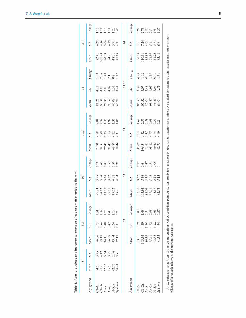
Tab
le 2
. A
bso
lute
val
ues
an
d in
crem
enta
l ch
ang
es o
f ce
ph
alo
met
ric
vari
able
s (i
n m
m).
Age
(ye
ars)
99.
510
10.5
1111
.5
Mea
nSD
Mea
nSD
Cha
nge*
Mea
nSD
Cha
nge
Mea
nSD
Cha
nge
Mea
nSD
Cha
nge
Mea
nSD
Cha
nge
Cd–
A74
.55
3.75
76.0
63.
751.
5177
.84
3.55
1.78
79.8
84.
782.
0481
.26
4.26
1.38
82.4
14.
281.
15C
d–G
n91
.94.
1293
.49
3.66
1.58
96.1
23.
962.
6398
.53.
892.
3810
0.56
4.80
2.06
101.
844.
561.
28A
r–A
72.8
53.
6474
.13.
481.
2675
.96
3.58
1.85
77.4
93.
731.
5378
.93
3.8
1.45
80.0
83.
641.
15A
r–G
n85
.59
3.57
86.9
93.
471.
489
.51
3.62
2.52
91.4
23.
531.
9293
.52
4.08
2.1
94.7
4.39
1.18
N–S
pa42
.75
2.96
43.9
43.
241.
1945
.52
4.01
1.58
46.8
84.
321.
3647
.08
3.36
0.2
48.3
13.
711.
22Sp
a–M
e56
.41
3.8
57.1
13.
80.
758
.44.
041.
2959
.46
4.2
1.07
60.7
34.
431.
2761
.16
4.7
0.42
1212
.513
13.5
`14
Age
(ye
ars)
Mea
nSD
Cha
nge*
Mea
nSD
Cha
nge
Mea
nSD
Cha
nge
Mea
nSD
Cha
nge
Mea
nSD
Cha
nge
Cd–
A83
.33.
790.
8883
.46
3.62
0.17
85.0
93.
851.
6285
.53
4.37
0.45
86.4
94.
80.
96
Cd–
Gn
103.
344.
491.
4910
3.94
5.36
0.6
106.
55.
522.
5510
7.52
5.07
1.02
110.
317.
262.
79A
r–A
80.5
93.
460.
581
.06
3.59
0.47
81.6
33.
550.
5882
.04
3.48
0.41
82.8
54.
040.
81A
r–G
n95
.66
4.72
0.95
97.1
65.
451.
5198
.12
4.97
0.95
99.4
74.
921.
3510
1.97
5.6
2.5
N–S
pa48
.93
3.22
0.63
48.8
73.
21-0
.06
49.4
33.
740.
5549
.83
3.94
0.41
51.2
33.
781.
4Sp
a–M
e61
.53
4.54
0.37
62.5
35.
151
62.7
34.
490.
264
.04
4.52
1.31
65.4
16.
61.
37
Ar–
A, a
rtic
ular
e–po
int A
; Ar–
Gn,
art
icul
are–
gnat
hion
; Cd–
A, c
ondy
lion–
poin
t A; C
d–G
n, c
ondy
lion–
gnat
hion
; N–S
pa, n
asio
n–an
teri
or n
asal
spi
ne; S
D, s
tand
ard
devi
atio
n; S
pa–M
e, a
nter
ior
nasa
l spi
ne–m
ento
n.a C
hang
e of
a v
aria
ble
rela
tive
to
the
prev
ious
reg
istr
atio
n.
T. P. Engel et al. 5
for the timing of orthodontic treatment. In addition, Zhao et al. found that the percentage of complete agreement between observers for assignment of CVM stage 3, i.e. the stage, which is supposed to precede the maximum facial growth, was only 26.8 per cent. A disa-greement of one stage apart was found in 49.5 per cent, two stages apart—in 22.2 per cent, and three stages apart in 1.5 per cent scor-ings. From a clinical point of view, disagreement by one CVM stage roughly corresponds with a 1-year difference in predicted maximum facial growth (8). Because a typical treatment of Class II with func-tional appliance lasts 1 year, inaccuracy of the CVM method may be unacceptable.
In contrast to previous reports, Ball et al. (31) found almost per-fect concordance among raters using the CVM system. Nevertheless the authors stated that the CVM was unable to predict the onset of a peak in mandibular growth because of the duration of particular CVM stages. According to Baccetti et al. (10) the peak in mandibular growth occurs between third and fourth CVM stage and the stages last approximately 1 year. Therefore if a subject has CVM stage 3, the maximum facial growth should occur within 12 months. Ball et al. reported, in turn, that the peak of mandibular growth in their sample occurred in fourth CVM stage and the duration of this stage was on average 3.8 years. As a result identification of the CVM stage 4 in a patient gives time span that is too wide to accurately predict the peak of growth.
Several studies demonstrated a high association between the CVM stage and facial growth (9, 10, 25, 32). Our results are in conflict with them. There are several possible explanations for this. First, it is unclear if researchers performing cephalometric analysis were blinded to the results of CVM staging, and vice-versa. It is par-ticularly important when a team of researchers works on the same records for extended period of time. A developing ‘memory effect’ can introduce bias into the results. Secondly, in the current study we applied a strategy that mimicks a typical clinical situation, when the orthodontist after evaluation of a cephalogram attempts to predict facial growth. O’Reilly and Yanniello (25), Franchi et al. (9), and Baccetti et al. (10, 32) used a different approach—they identified the period of greatest change of cephalometric variable(s) and correlated
it with morphology of cervical vertebrae C2, C3, and C4. These two strategies, although seem complementary, they may look at different aspects of facial growth and for this reason may produce conflict-ing findings. Finally, missing (25) or incomplete (9, 10, 32) method error analysis makes the assessment of accuracy of cephalometric measurements difficult.
The consistency of CVM staging was relatively low in this inves-tigation. The intra- and inter-rater agreement was worse than in most other publications. It can result from the composition of the rater’s panel—we asked senior orthodontic residents to stage CVM status whereas other authors performed CVM staging themselves (9, 10, 12–14). Although our raters were calibrated, their experience in using the CVM system might have been less than that of raters from other studies. On the other hand, the use of a panel comprising residents rather than highly trained experts may resemble better the everyday clinical practice situation.
The use of CVM requires that second, third, and fourth cervical vertebrae are visible on a cephalogram. As a result, if this region of cervical spine is to be visualized no thyroid shield can be worn dur-ing radiographic exposition. However, the thyroid gland is one of the most radiation-sensitive parts of the body. External irradiation to the thyroid, even at low doses, may induce subsequent development of nodules and neoplasmas and is associated with an increased inci-dence of thyroid autoimmune abnormalities and the development of thyroiditis and Graves’ disease (33). Since the use of thyroid shield can considerably reduce the effective dose of the thyroid (34) taking lateral cephalograms without thyroid shield is questionable because of an increased health hazard and a little benefit for a patient.
The participants included in our study were followed until 14 years of age. Ideally a series of examinations should be finished at 17–18 years when the pubertal phase of facial growth is expected to be completed. This was not possible because the NGS had been planned to cover only the years from 4 to 14. However, our aim was to test the performance of the CVM method to predict the peak of facial growth, which very likely occurred within the age range of our participants (35). Buschang et al. using a large French–Canadian sample of girls born in 1960s demonstrated that peak growth
Table 3. Regression models with average CVM grade of all raters and age of the subject as independent variables and change of cephalo-metric variable as dependent variable.
β 95% CI P value
Cd–A CVM grade—average for all raters −0.05 −1.80 to 1.70 0.95 Age 1.23 1.15 to 1.32 <0.001Cd–Gn CVM grade—average for all raters −1.19 −3.14 to 0.76 0.23 Age 1.8 1.71 to 1.90 <0.001Ar–A CVM grade—average for all raters 0.39 −1.26 to 2.03 0.65 Age 1.05 0.99 to 1.11 <0.001Ar–Gn CVM grade—average for all raters −0.76 −2.68 to 1.15 0.44 Age 1.59 1.52 to- 1.67 <0.001Spa–Me CVM grade—average for all raters 1.28 −0.76 to 3.33 0.22 Age 0.87 0.81 to 0.93 <0.001N–Spa CVM grade—average for all raters −1.27 −2.76 to 0.21 0.92 Age 0.8 0.73 to 0.87 <0.001
Ar–A, articulare–point A; Ar–Gn, articulare–gnathion; Cd–A, condylion–point A; Cd–Gn, condylion–gnathion; CI, confidence interval; CVM, cervical vertebrae maturation; N–Spa, nasion–anterior nasal spine; SD, standard deviation; Spa–Me, anterior nasal spine–menton.
European Journal of Orthodontics, 2016, Vol. 38, No. 16
velocities were well before 13 years of age. Thus the sample seems appropriate for this study.
A limitation of our study could be a relatively small sample size. Increasing the number of subjects in the sample would increase the power to detect smaller growth increments; however, any uncer-tainty in our estimates is reflected in the provided CIs.
In conclusion, our study has shown that there is no evidence to support the hypothesis that the CVM method can predict the amount of craniofacial growth in girls with Class II malocclusion.
References 1. McNamara, J.A. (1981) Components of Class II malocclusion in children
8–10 years of age. Angle Orthodontist, 51, 177–202. 2. Thiruvenkatachari B., Harrison, J.E., Worthington, H.V. and O’Brien, K.D.
(2013) Orthodontic treatment for prominent upper front teeth (Class II malocclusion) in children. Cochrane Database of Systematic Reviews, CD003452. doi:10.1002/14651858.CD003452.pub3
3. Pancherz, H. and Hägg, U. (1985) Dentofacial orthopedics in relation to somatic maturation. An analysis of 70 consecutive cases treated with the Herbst appliance. American Journal of Orthodontics and Dentofacial Orthopedics, 88, 273–287.
4. Baccetti T., Franchi, L. and Kim, L.H. (2009) Effect of timing on the out-comes of 1-phase nonextraction therapy of Class II malocclusion. Ameri-can Journal of Orthodontics and Dentofacial Orthopedics, 136, 501–509.
5. Cozza, P., Baccetti, T., Franchi, L., De Toffol, L. and McNamara, J.A. Jr. (2006) Mandibular changes produced by functional appliances in Class II malocclusion: a systematic review. American Journal of Orthodontics and Dentofacial Orthopedics, 129, 599.e1–12.
6. Houston, W.J.B., Miller, J.C. and Tanner, J.M. (1979) Prediction of the tim-ing of the adolescent growth spurt from ossification events in hand-wrist films. British Journal of Orthodontics, 6, 145–152.
7. Fishman, L.S. (1982) Radiographic evaluation of skeletal maturation. A clinically oriented method based on hand-wrist films. Angle Orthodon-tist, 52, 88–112.
8. Lamparski, D.G. (1972) Skeletal Age Assessment Utilizing Cervical Verte-brae. Thesis, University of Pittsburgh.
9. Franchi, L., Baccetti, T. and McNamara, J.A. (2000) Mandibular growth as related to cervical vertebral maturation and body height. American Journal of Orthodontics and Dentofacial Orthopedics, 118, 335–340.
10. Baccetti, T., Franchi, L. and McNamara, J.A. (2005) The cervical vertebral maturation (CVM) method for the assessment of optimal treatment timing in dentofacial orthopedics. Seminars in Orthodontics, 11, 119–129.
11. Dawson, B. and Trapp, R.G. (2004) Basic and Clinical Biostatistics. Lange Medical Books/McGraw-Hill. Medical Publishing Division, New York.
12. Uysal, T., Ramoglu, S.I., Basciftci, F.A. and Sari, Z. (2006) Chronologic age and skeletal maturation of the cervical vertebrae and hand-wrist: is there a relationship? American Journal of Orthodontics and Dentofacial Orthopedics, 130, 622–628.
13. Lai, E.H., Liu, J.P., Chang, J.Z., Tsai, S.J., Yao, C.C., Chen, M.H., Chen, Y.J. and Lin, C.P. (2008) Radiographic assessment of skeletal maturation stages for orthodontic patients: hand-wrist bones or cervical vertebrae? Journal of Formosa Medical Association, 107, 316–325.
14. Soegiharto, B.M., Cunningham, S. J. and Moles, D.R. (2008) Skeletal maturation in Indonesian and white children assessed with hand-wrist and cervical vertebrae methods. American Journal of Orthodontics and Dentofacial Orthopedics, 134, 217–226.
15. Gabriel, D.B., Southard, K.A., Qian, F., Marshall, S.D., Franciscus, R.D. and Southard, T.E. (2009) Cervical vertebrae maturation method: poor reproducibility. American Journal of Orthodontics and Dentofacial Orthopedics, 136, 478–480.
16. Nestman, T.S., Marshall, S.D., Qian, F., Holton, N., Franciscus, R.G. and Southard, T.E. (2011) Cervical vertebrae maturation method morphologic criteria: poor reproducibility. American Journal of Orthodontics and Dentofacial Orthopedics, 140, 182–188.
17. Zhao, X.G., Lin, J., Jiang, J.H., Wang, Q. and Ng, S.H. (2012) Validity and reliability of a method for assessment of cervical vertebral maturation. Angle Orthodontist, 82, 229–234.
18. Cook, J.A. (2009) The challenges faced in the design, conduct and analysis of surgical randomised controlled trials. Trials, 10, 9. doi:10.1186/1745-6215-10-9.
19. Farrokhyar, F., Karanicolas, P.J., Thoma, A., Simunovic, M., Bhandari, M., Devereaux, P.J., Anvari, M., Adili, A. and Guyatt, G. (2010) Randomized controlled trials of surgical interventions. Annals of Surgery, 251, 409–416.
20. Beit, P., Peltomäki, T., Schätzle, M., Signorelli, L. and Patcas, R. (2013) Evaluating the agreement of skeletal age assessment based on hand-wrist and cervical vertebrae radiography. American Journal of Orthodontics and Dentofacial Orthopedics, 144, 838–847.
21. Prahl-Andersen, B. and Kowalski, C.J. (1973) A mixed longitudinal, interdisciplinary study of the growth and development of Dutch children. Growth, 37, 281–295.
22. Bland, J.M. and Altman, D.G. (1986) Statistical methods for assessing agreement between two methods of clinical measurement. Lancet, 327, 307–310.
23. Hassel, B. and Farman, A.G. (1995) Skeletal maturation evaluation using cervical vertebrae. American Journal of Orthodontics and Dentofacial Orthopedics, 107, 58–66.
24. Fleiss, J.L. (1981) Statistical Methods for Rates and Proportions. John Wiley & Sons, New York, pp. 212–236.
25. O’Reilly, M. and Yanniello, G.J. (1988) Mandibular growth changes and mat- uration of cervical vertebrae–a longitudinal cephalometric study. Angle Orthodontist, 58, 179–184.
26. Santiago, R.C., de Miranda Costa, L.F., Vitral, R.W., Fraga, M.R., Bolognese, A.M. and Maia, L.C. (2012) Cervical vertebral maturation as a biologic indicator of skeletal maturity. Angle Orthodontist, 82, 1123–1131.
27. Singh, I.J., Savara, B.S. and Miller, P.A. (1967) Interrelations of selected measurements of the face and body in pre-adolescent and adolescent girls. Growth, 31, 119–131.
28. Bergersen, E.O. (1972) The male adolescent facial growth spurt: its predic-tion and relation to skeletal maturation. Angle Orthodontist, 42, 319–338.
29. Howell, W.W. (1952) A factorial study of constitutional type. American Journal of Physical Anthropology, 10, 91–118.
30. Mitani, H. and Sato, K. (1992) Comparison of mandibular growth with other variables during puberty. Angle Orthodontist, 62, 217–222.
31. Ball, G., Woodside, D., Tompson, B., Hunter, W.S. and Posluns, J. (2011) Relationship between cervical vertebral maturation and mandibular growth. American Journal of Orthodontics and Dentofacial Orthopedics, 139, e455-e461.
32. Baccetti, T., Franchi, L. and McNamara, J.A. Jr. (2002) An improved ver-sion of the cervical vertebral maturation (CVM) method for the assess-ment of mandibular growth. Angle Orthodontist, 72, 316–323.
33. De Groot, L.J. (1993) Effects of irradiation on the thyroid gland. Endocri-nology Metabolism Clinics of North America, 22, 607–615.
34. Patcas, R., Signorelli, L., Peltomäki, T. and Schätzle, M. (2013) Is the use of the cervical vertebrae maturation method justified to determine skeletal age? A comparison of radiation dose of two strategies for skeletal age estimation. European Journal of Orthodontics, 35, 604–609.
35. Buschang, P.H., Jacob, H.B. and Demirjian A. (2013) Female adolescent craniofacial growth spurts: real or fiction? European Journal of Ortho-dontics, 35, 819–825.
T. P. Engel et al. 7