the brazilian consensus on the management of pompe disease
TRANSCRIPT
The Brazilian Consensus on the Management of Pompe DiseaseJuan C. Llerena, Jr., MD, PhD, Dafne Maria Horovitz, MD, PhD, Suely Kazue Nagahashi Marie, PhD, Gilda Porta, MD, PhD,
Roberto Giugliani, MD, PhD, Maria Veronica Munoz Rojas, MD, and Ana Maria Martins, MD, PhD, on behalf of the Brazilian
Network for Studies in Pompe Disease (ReBrPOM)*
The Brazilian Consensus on the Management of PompeDisease was established through collaboration of allBrazilian physicians known to be treating patients
with Pompe disease (PD) in Brazil. It was based on the clinicalpresentation of 7 cases of early-onset PD (EOPD) and 18 casesof late-onset PD (LOPD) presented to a working group in thecity of Rio de Janeiro in 2007. Coordinated by the BrazilianNetwork for Studies in PD (ReBrPOM) with key objectivesof enhancing understanding of the clinical heterogeneity,progression, and natural history of this severe and lethal ge-netic disease, as well as providing an overview of the Brazilianexperience with enzyme replacement therapy with recombi-nant human acid alpha glucosidase (rhGAA; Myozyme; Gen-zyme, Cambridge, Massachusetts). This is not an exhaustiveinvestigation of the issue, but rather preliminary guidelinesto optimize the management of these patients under thecare of the Brazilian public health care system (SUS-Brasil).
PD (MIM 232300), also known as glycogen storage diseasetype II or acid maltase deficiency, is rare, progressive, and, inits early form, often fatal.1 The disease is inherited in an au-tosomal recessive fashion and is caused by a deficiency of theenzyme acid alpha-glucosidase (GAA; 3.2.1.20), which has anintralysosomal action and is responsible for releasing glucoseunits from glycogen. Glycogen buildup occurs within lyso-somes of several tissues, particularly in skeletal and cardiacstriated muscle cells in early-onset PD (EOPD), destroyingthe cells and compromising muscle fiber function. The dis-ease may present in the first year of life, characterized mainlyby hypertrophic cardiomyopathy and generalized muscularhypotonia, or after the first year through the sixth decadeof life, with progressive proximal muscular weakness andrespiratory complaints as the major symptoms.2
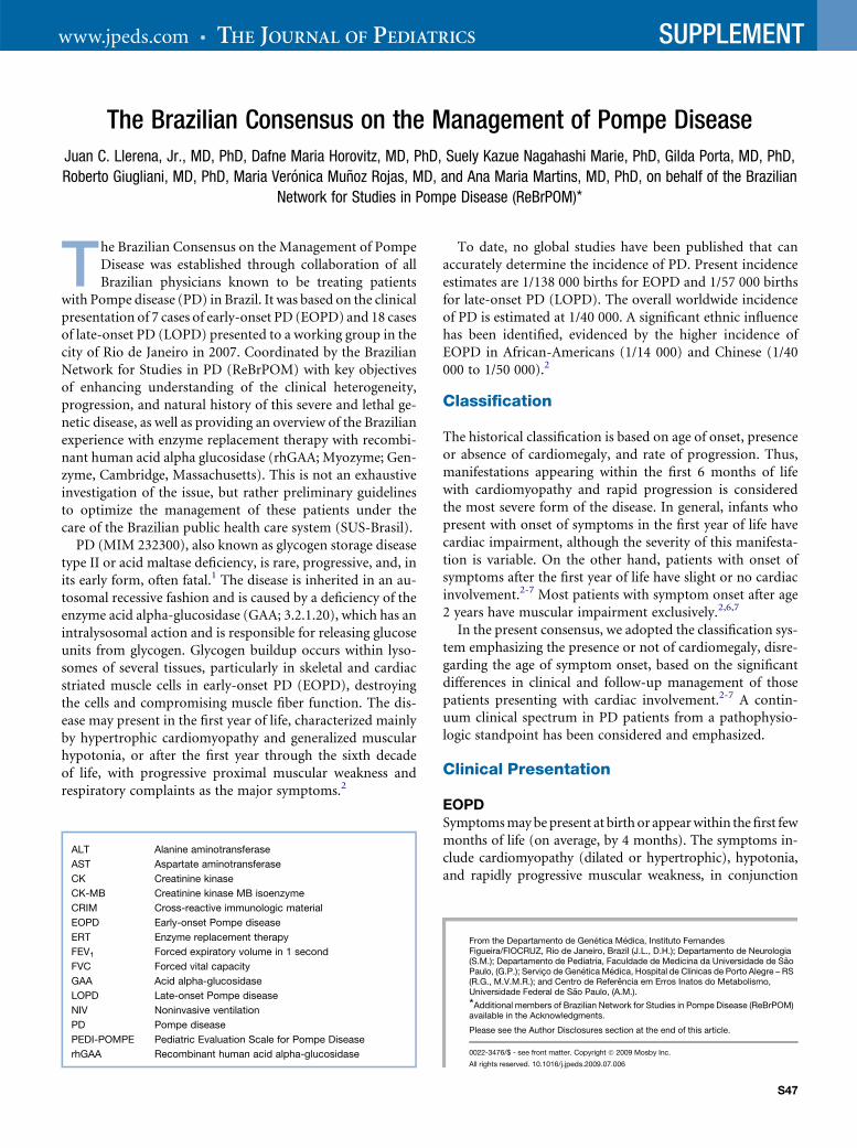
ALT Alanine aminotransferase
AST Aspartate aminotransferase
CK Creatinine kinase
CK-MB Creatinine kinase MB isoenzyme
CRIM Cross-reactive immunologic material
EOPD Early-onset Pompe disease
ERT Enzyme replacement therapy
FEV1 Forced expiratory volume in 1 second
FVC Forced vital capacity
GAA Acid alpha-glucosidase
LOPD Late-onset Pompe disease
NIV Noninvasive ventilation
PD Pompe disease
PEDI-POMPE Pediatric Evaluation Scale for Pompe Disease
rhGAA Recombinant human acid alpha-glucosidase
To date, no global studies have been published that canaccurately determine the incidence of PD. Present incidenceestimates are 1/138 000 births for EOPD and 1/57 000 birthsfor late-onset PD (LOPD). The overall worldwide incidenceof PD is estimated at 1/40 000. A significant ethnic influencehas been identified, evidenced by the higher incidence ofEOPD in African-Americans (1/14 000) and Chinese (1/40000 to 1/50 000).2
Classification
The historical classification is based on age of onset, presenceor absence of cardiomegaly, and rate of progression. Thus,manifestations appearing within the first 6 months of lifewith cardiomyopathy and rapid progression is consideredthe most severe form of the disease. In general, infants whopresent with onset of symptoms in the first year of life havecardiac impairment, although the severity of this manifesta-tion is variable. On the other hand, patients with onset ofsymptoms after the first year of life have slight or no cardiacinvolvement.2-7 Most patients with symptom onset after age2 years have muscular impairment exclusively.2,6,7
In the present consensus, we adopted the classification sys-tem emphasizing the presence or not of cardiomegaly, disre-garding the age of symptom onset, based on the significantdifferences in clinical and follow-up management of thosepatients presenting with cardiac involvement.2-7 A contin-uum clinical spectrum in PD patients from a pathophysio-logic standpoint has been considered and emphasized.
Clinical Presentation
EOPDSymptoms may be present at birth or appear within the first fewmonths of life (on average, by 4 months). The symptoms in-clude cardiomyopathy (dilated or hypertrophic), hypotonia,and rapidly progressive muscular weakness, in conjunction
From the Departamento de Genetica Medica, Instituto FernandesFigueira/FIOCRUZ, Rio de Janeiro, Brazil (J.L., D.H.); Departamento de Neurologia(S.M.); Departamento de Pediatria, Faculdade de Medicina da Universidade de SaoPaulo, (G.P.); Servico de Genetica Medica, Hospital de Clınicas de Porto Alegre – RS(R.G., M.V.M.R.); and Centro de Referencia em Erros Inatos do Metabolismo,Universidade Federal de Sao Paulo, (A.M.).
*Additional members of Brazilian Network for Studies in Pompe Disease (ReBrPOM)available in the Acknowledgments.
Please see the Author Disclosures section at the end of this article.
0022-3476/$ - see front matter. Copyright � 2009 Mosby Inc.
All rights reserved. 10.1016/j.jpeds.2009.07.006
S47

THE JOURNAL OF PEDIATRICS � www.jpeds.com Vol. 155, No. 4, Suppl. 2
with delayed motor milestones, followed by death from cardio-respiratory failure, usually by age 1 year.7 Impairment of respi-ratory muscles (including abdominal and intercostal musclesand the diaphragm), leads to rapid-onset respiratory insuffi-ciency. Swallowing difficulties are common, causing malnutri-tion, repeated aspiration pneumonia, and respiratory tractinfections. Other common findings include macroglossia, he-patomegaly, hearing loss, osteopenia, and scoliosis.7-10 Patientswith EOPD develop cardiomyopathy secondary to the massiveaccumulation of glycogen within the cardiac muscles. Theyhave biventricular hypertrophy, including hypertrophy of theventricular septum, which can be present at birth or developwithin the first year of life. Hypertrophy may result in leftventricular outflow tract obstruction and progress to dilatedcardiomyopathy. Glycogen accumulation also affects thespecialized conduction tissue cells and gross depolarizationand repolarization patterns, resulting in characteristic eletro-cardiographic changes, including shortened PR interval, in-creased QT dispersion, and enlarged QRS complexes.3,11 Thehypertrophic cardiomyopathy and conduction system abnor-malities put these patients at risk for ventricular arrhythmiasand sudden death.12 Two natural history studies delineate theEOPD course. In the first study, which included 20 Dutch casesand 133 cases from the literature, the median age at symptomonset was 1.6 months in both groups, the median age at diag-nosis was 5.3 months in the Dutch group and 4.5 months in theliterature cases, and the median age at death was 7.7 monthsand 6.0 months, respectively.13 In the second study, the naturalhistory of disease progression was determined from a retrospec-tive chart review on a worldwide cohort of 168 patients withEOPD. The median age at symptom onset was 4 months, me-dian age of first ventilator support was 5.9 months, and medianage of death was 8.7 months.7 Despite intervention, mortalityhas remained largely unchanged.7,9,13
In EOPD, laboratory examination usually reveals cardio-megaly on thoracic radiography; electrocardiogram changes,with a high-voltage QRS complex and short PR interval;14,15
elevated serum creatine kinase (CK), CK MB isoenzyme(CK-MB), alanine aminotransferase (ALT), and aspartateaminotransferase (AST) levels; and increases in the level ofenzyme of muscle origin, which is more evident with progres-sion of the disorder.8,16
In our series of 7 patients with EOPD, the first symptomswere recognized from birth to age 4 months. All 7 patientspresented with hypotonia and cardiomegaly, and 4 of the7 presented with hepatomegaly.
The following syndromes share clinical signs and symp-toms with EOPD: spinal muscular atrophy I (MIM253300), Danon disease (MIM 300257), endocardial fibroe-lastosis (MIM 305257), carnitine deficiency (MIM 212140),and glycogen storage disease type III (MIM 232400) and IV(MIM 232500).5,8,17
LOPDSymptoms of LOPD may manifest any time after the first yearof life up to the sixth decade of life.4 Progression is slowerthan in infants but is still relentlessly progressive. By assump-
S48
tion, LOPD does not present with cardiomyopathy; it ischaracterized by predominantly proximal progressive mus-cular weakness, with greater impairment of the lower limbs.8
Usually, paraspinal muscles and lower limb proximal musclesare the first affected, leading to motor impairment and diffi-culty performing daily activities, and possibly scoliosis,kyphosis, or lordosis. Impairment of the diaphragm and aux-iliary respiratory muscles causes chronic respiratory failurealong with fatigue, carbon dioxide retention, respiratoryinsufficiency, and, in many cases, sleep apnea. Repeated aspi-ration pneumonia is common. Other findings include mac-roglossia, palpebral ptosis, cerebral aneurism, hyporeflexia,and osteopenia.18-21
Muscular weakness is the most prominent symptom, de-tectable in around 80% of cases and occuring in 95% of casesat some stage of the disease.19 Respiratory problems resultingfrom compromised respiratory muscles appear as the firstsymptom in approximately 11% of patients, occurring atsome stage of the disease in 44% of cases.19,22-24 In our seriesof 18 confirmed cases of LOPD (9 males and 9 females), thefirst signs of muscular disorder appeared between age 1 and42 years (mean, 15.4 years; median, 11 years), and age at di-agnosis ranged from 2.5 to 48 years (mean, 25.3 years; me-dian, 23.5 years). Occasionally, dysphagia for solids mayresult from weakness in the pharyngeal muscles involved inthe swallowing reflex. A significant adverse impact on activ-ities of daily living is a common finding.22
Laboratory findings include increased CK, AST, andALT values16 and spirometric parameters indicating alteredrespiratory assessment capacity, including forced expiratoryvolume in 1 second (FEV1) and forced vital capacity(FVC) in erect and supine positions,7 with a normal elec-trocardiogram. In our series, 10 of 18 patients with LOPDrequired some sort of respiratory support during thecourse of the disease. Muscle biopsy reveals greatly in-creased deposition of glycogen within muscle vacuoles,with destruction of myofibrils.2
The following syndromes share clinical signs and symp-toms with LOPD: limb girdle muscular dystrophy (MIM253600), Becker/Duchenne muscular dystrophy (MIM300376/310200), polymyositis, rheumatoid arthritis, and gly-cogen storage disease type III (MIM 231400), IV (MIM232500), and V MIM 232600). Further details regarding thedifferential diagnosis of neuromuscular disease are availableat www.musclegenetable.org.25
Diagnosis
Patients with a clinical history compatible with PD must beinvestigated to confirm the diagnosis. Asymptomatic familymembers of a known patient also should be considered forassessment.16,25 Hypotonia and cardiomegaly on radiogra-phy or cardiomyopathy on echocardiogram are importantsigns in the clinical examination of infants that includes PDamong the diagnostic possibilities.4,7
In EOPD, the diagnosis should be made based on measure-ment of GAA enzymatic activity in a blood sample using filter
Llerena Jr. et al
October 2009 SUPPLEMENT
paper.26 Subsequent confirmation should always be donethrough enzyme activity assays in fibroblast cultures or lym-phocyte assays and/or genotyping through DNA analysis.27-30
It should be noted that the sensitivity and specificity of filterpaper testing depend on the method used. Studies comparingacarbose and maltase inhibition have been reported,31-34 anduse of the acarbose method to inhibit the activity of theenzyme maltase-glucoamylase has been recommendedrecently.35 Increased urinary excretion of oligossacharides inurine chromatography also may corroborate the diagnosis.36
In our series of 7 patients with EOPD, all had dried blood spotenzyme assays compatible with the diagnosis of PD; othertests, including leukocyte GAA assay (2 of 7), muscle biopsy(4 of 7), genotyping (7 of 7), and oligossacharide urinarychromatography (4 of 7) confirmed the diagnosis. The GAAassay using skin fibroblasts was once the gold standard, butnow the addition of acarbose to assays prevents false-negativeresults. The reliable, less invasive, more convenient, and fasterGAA assay using blood samples can now be considered themethod of choice for diagnosing PD.35
The assessment of cross-reactive immunologic material(CRIM) status, which detects the presence or absence of na-tive GAA protein on Western blot assay using cultured fibro-blasts, is a sensitive and specific test that may be of great valuein establishing the prognosis and response to enzyme replace-ment therapy (ERT). In our series of EOPD patients, 5 of 6 an-alyzed cases were CRIM-negative. This observation correlateswell with the presence of approximately 2560 C > T null mu-tations in homozygosis or in association with other deleteri-ous mutations in the majority of Brazilian EOPD casesstudied to date.27 Other issues regarding the method and han-dling of samples are important as well, such as the need to col-lect frozen muscle biopsy specimens for histochemicalanalysis, DNA for genotyping, and RNA for gene expressionanalysis.37-39
In older patients, analysis of dry blood spots using filterpaper can be a reliable screening test. Abnormal or suspectedresults should always be confirmed by further testing, includingmeasuring enzymatic activity in lymphocyte or fibroblastassays and/or genotyping by DNA analysis. Muscle biopsyfindings also may suggest PD.27-30,37-39 Increased plasma andurine concentrations of glucose tetrasaccharide, which mayserve as a biomarker for diagnostic purposes, have been found;this test is not currently in widespread use, however.
In our series of 18 LOPD patients, all had dried blood spotenzyme assay results compatible with PD. Seventeen patientshad vacuolar myopathy with glycogen storage on muscle bi-opsy, and genotypes comprehending a series of homozygousmissense mutations or compound heterozygotes with a com-bination of missense and intronic mutations or missense andstop codons mutations were identified.27
Management
Because PD is a multisystem disorder, it is best managed bya multidisciplinary team led by an experienced physician.Optimally, the team should include a metabolic disease spe-
The Brazilian Consensus on the Management of Pompe Diseas
cialist in addition to the specialist dictated by the patient’ssigns and symptoms, possibly a cardiologist, pulmonologist,neurologist/neuromuscular specialist, orthopedist, physicaltherapist, speech therapist, geneticist, and/or metabolic dieti-tian, among others.
Clinical ApproachCardiology. Cardiomyopathy in patients with PD shouldbe treated cautiously, preferably by a pediatric cardiologistwith experience with this disorder. Care must be adjustedto disease stage; inappropriate use of the standard drugsused to treat cardiomyopathy may worsen an already im-paired heart. In the earlier phases of the disease, hyperthro-phy is predominant with or without left ventricularoutflow tract obstruction and normal or even hyperdynamicleft ventricular ejection fraction. In the presence of left ven-tricular outflow tract obstruction, beta-blockers can beused judiciously due to the risk of sudden death.9 Diureticsalso can be used judiciously in patients with pulmonary con-gestion. The use of digoxin, beta-adrenergic agonists, orafterload-reducing agents, such as angiotensin-convertingenzyme inhibitors and diuretics, may exacerbate the left ven-tricular outflow tract obstruction. But these agents generallyare used in the later phases of the disease in the presence ofventricular dysfunction (dilated cardiomyopathy).3 Somepatients present with ventricular dysfunction in the earlierphases of the disease and need to start with these medica-tions. Arrhythmia also may occur, due to the insulator effectof glycogen in conduction tissue40 and to underlying suben-docardial ischemia from inadequate coronary perfusion ofa massively hypertrophied ventricle. Therefore, special caremust be taken in patients with PD to avoid hypotensionif volume depletion occurs or anesthesia is required.41
Pulmonology. Approximately 30% of patients with PDrequire ventilatory support, irrespective of age of symptomonset. Orotracheal intubation must be avoided wheneverpossible in view of the difficulties in weaning from invasiveventilation and resulting complications, such as fistulas.However, when indicated, an aggressive approach is recom-mended, especially in infants, given the extremely high vul-nerability of these patients. Ideally, bilevel positive-pressureairway ventilation support should be used instead of contin-uous positive airway pressure, using various inspiratory andexpiratory pressure levels. Adults tend to respond well tononinvasive ventilation (NIV).42 Children present greateradaptive difficulties, although NIV can be used in the eventof progressive respiratory insufficiency. Despite NIV, pa-tients may have symptoms of sleep apnea. Bilateral positivepressure airway ventilation should be adjusted (with manda-tory frequency during sleep) according to the patient’s needs.Muscular weakness can lead to generation of insufficientstimulus to trigger the next cycle of the ventilator. Thus, sleepapnea can be avoided with less thoracic muscular effort andimproved sensitization to CO2 and O2. It is important thatatelectasis be ruled out as the main cause in any respiratorydecline in infants.23,24
e S49

THE JOURNAL OF PEDIATRICS � www.jpeds.com Vol. 155, No. 4, Suppl. 2
Musculoskeletal. Musculoskeletal involvement in PD ischaracterized by progressive weakness that leads to decreasedmotor function, altered postural tendencies and positioning,and the use of compensatory patterns of movement. Second-ary musculoskeletal manifestations, such as contractures, de-formities, and osteoporosis, can occur and further impairfunction. Rehabilitation management of PD should be com-prehensive and preventive, based on an understanding of thepathokinesiology of disease progression and on individualassessment. It should optimize and preserve motor and phys-iological function, prevent or minimize secondary complica-tions, and promote and maintain the maximum level offunction.9
Speech, Swallowing, and Nutritional Assessment.Muscular weakness may lead to difficulty in phonation andin the completion of safe swallowing. Poor swallowing, mus-cle wasting, increased effort of breathing, and impaired mo-bility should be considered as factors in undernourishment.Malnutrition may exacerbate the muscle weakness. Severalstudies have investigated the possible benefits of a high-pro-tein diet in patients with PD. In general, a high-protein diet isrecommended and should be administered under the super-vision of a metabolic dietician.43,44 The risk of aspirationshould be assessed by a trained speech therapist and also byvideofluoroscopy when necessary.
Anesthetic Risk. Patients with EOPD are at high risk forclinical complications from anesthetics due to significant car-diovascular compromise.41 Surgical procedures should beavoided whenever possible. If anesthesia is necessary, the pre-ferred drug is ketamine, which does not significantly affectpre- and post-cardiac load, thereby avoiding the risk of myo-cardial ischemia. Propofol, particularly at high doses, andsuxamethonium should be avoided. Sedation and local anes-thesia (even for gastrostomy, if possible) should be usedwhenever possible.
ERT. Among the available approaches to treat PD, ERTbased on rhGAA has become a major cornerstone in the im-proved care of these patients.45-49 Several open-label clinicaltrials involving patients with EOPD have shown that ERT sig-nificantly prolongs survival,45,50-56 decreases cardiomegaly,and improves cardiac and skeletal muscle function. In thevast majority of cases, cardiac response appears to be good,irrespective of the stage of the disease at the start of ERT.Skeletal muscle response has been more variable than cardiacmuscle response. The best skeletal muscle response has beennoted in patients treated early, before severe skeletal muscledamage has occurred. There are several EOPD patients onERT who are walking, a milestone that probably would nothave been achieved without ERT. Thus, with the advent ofERT, the natural history of this once-lethal disease haschanged, and additional physical and mental disabilitiesmay be uncovered. Some patients have not a good outcomedespite early treatment, however. Such factors as muscle fibertype, stage of disease at the start of therapy, genotype, and
S50
immune response to rhGAA may play a role in determiningoutcome and merit further investigation. Work on a second-generation recombinant enzyme for PD for more efficienttargeting to muscles is in preclinical stages.9
Gene therapy, small molecules, and chaperones.Progress in the development of gene therapy continues. Studiesin Pompe mice have demonstrated that various gene deliveryvector systems have potential and demonstrate that expressionof the GAA gene within muscle cells decreases the glycogenstorage abnormality in the mice. The use of small moleculesto treat lysosomal storage disorders, either as a single agentor in combination with other therapies, is emerging as a thera-peutic tool. Pharmacologic chaperones that bind to the affectedproteins and restore their shape, proper trafficking, and biolog-ical activity also may become available for those who can makethe protein, most likely those with LOPD.9
Following confirmation of the diagnosis of PD, a series ofprocedures are recommended to establish measurements forboth prognosis and therapeutic response. The first step in thisprocess should be teaching parents of children with EOPDand parents and teenagers/adults with LOPD about PD’s nat-ural history, genetic basis, and pathophysiology, to properlydiscuss treatment options and realistic expectations if ERT isto be started.
Pretreatment AssessmentThis begins with filling out the anamnesis form available atwww.pomperegistry.com. The objective is to create a patientdatabase file based on signs and symptoms before ERT. Suchapproach ensures a combined series of functional scales andevaluations for different ages, such as the Peabody Develop-mental Motor Scale (PDMS-2) and the Pediatric EvaluationScale for PD (PEDI-POMPE).4,22
A family history is obtained to ascertain ancestry and con-sanguinity. The classification by skin color on the PD registry(www.pompe.com) especially for the Brazilian population isopen to erroneous interpretations. The classification used bythe Latin-American Collaborative Study of Congenital Mal-formations is much more accurate in this regard, takinginto account the panethnic characteristics of the Brazilianpopulation based on 7 different ethnic groups: Latin-Euro-pean (Portuguese, Italian, Spanish, French, Romenian),non-Latin-European (Caucasian), Jewish (Ashkenazi orSephardic), Native (Amerindian), Afro-American, Arabic(Turkish, Lebanese, and Syrian), and Oriental (including In-dia).57 Family history should be reviewed to allow geneticcounseling and to identify other cases through the index case.
The following complementary tests are required forpatients with EOPD:
� Laboratory tests: CK, CK-MB, AST, and ALT (they re-main high throughout follow-up), electrolytes (cardiacdysfunction risk), g-glutamyl transpeptidase, lactatedehydrogenase, urea, creatinine, hemosediment rate,blood counts, total protein and fractions (albumin [nu-tritional assessment]) and glycemia. In view of the
Llerena Jr. et al
October 2009 SUPPLEMENT
frequent respiratory complications, arterial gasometryis highly recommended.
� Polysomnography, when possible. Assessment of hyper-capnea peaks during sleep must be assessed, includingsleep apnea. At a minimum, heart rate, overnight oxygensaturation, and capnography should be assessed.
� Electrocardiography, to assess for the presence of high-voltage QRS and duration of PR interval according toage, together with T-wave alterations.
� Echocardiography is essential to follow-up therapeuticresponse. It is important to perform a baseline echocar-diogram as close as possible to the initiation of ERT andto assess septum size, end-diastolic volume, and ejec-tion fraction. Ventricular mass and ventricular mass in-dex should be calculated using Devereaux’s formula.15
� Radiography of the thorax to determine the cardiotho-racic index at baseline and in the event of any clinicaldeterioration.
� Motor assessment to quantify hypotonia and muscularstrength. The PEDI-POMPE scale is useful but appliesonly to patients over age 2 years.
� Swallowing evaluation. Videofluoroscopy is the mostwidely indicated test for infants under age 4 months.
� Nutritional assessment and monitoring: weight, height,skin triceps fold.
� Physiotherapeutic assessment and monitoring.� Neurologic assessment.� Immunologic assessment. Adverse reactions to ERT
must be correlated with the immunologic status inves-tigation in each patient, including the CRIM test andIgG- and IgE-mediated reactions, with complementand tryptase investigations. Baseline serum samplescan be stored frozen should immunologic investigationbe necessary during treatment.
� CRIM test to assess residual enzymatic activity in fibro-blast cultures. Collection should be done with a skinpunch using local anesthesia. Cultures should be setup for fibroblast culture or done simultaneously withmuscle biopsy (with local anesthesia). Biopsy speci-mens should be snap-frozen to allow proper histo-chemical and protein studies. Ideally biopsy and theCRIM test should be performed before the start oftreatment.
The following complementary tests are required for pa-tients with LOPD. Note that the heterogeneity in the pheno-type poses some challenges for standard assessment. Somepatients with LOPD are incapable of completing all of thefunctional tests due to the severity of their condition. Physi-cians should choose the best of the following tests to docu-ment the patient’s severity and pattern of motor,respiratory, and functional impairment:
� Laboratory tests: phosphocreatine kinase, CK-MB, al-dolase, lactate dehydrogenase, g-glutamyl transpepti-dase, urea, creatinine, electrolytes, arterial blood gasanalysis, and hemogram (polycythemia).
The Brazilian Consensus on the Management of Pompe Disease
� Polysomnography for evidence of hypercapnia or sleep-disordered breathing.
� Pulmonary assessment tests. Some 60% of patientspresent with an FVC of < 80% and 30% to 40%with an FVC of < 60% of expected values. Testingpatients in both the sitting and supine positions ismandatory. The following should be noted: percentdecrease of FEV1 in the supine position, percent de-crease of FVC in the supine position, and each param-eter in the erect position, expressed as % predicted,along with maximum expiratory pressure, maximuminspiratory pressure, peak cough flow, and arterialor capillary blood gas estimation (pCO2, pO2, andpH), specifying whether self-ventilating and whetherawake or asleep.
� Motor assessment. Assessment of specific functional ac-tivities includes standing posture and alignment (if ableto stand), observing for lordosis or scoliosis; paraspinaland other muscle wasting; deep tendon reflexes; con-tractures; range of joint movement; and ability towalk, climb stairs, stand from a chair, stand from lyingon the floor (Gower maneuver), reach for an object,and lift hands to the top of the head, ideally also onvideo. The PEDI-POMPE scale is extremely useful inolder patients with PD because it allows assessment ofnot only motor function, but also self-care on 2 scalesof 0-100. Manual muscle testing (MRC 0-5/5 scale)should be applied to document all main muscle groups:left and right proximal and distal, flexors, extensors,abductors, and adductors. The assessment of range ofactive movement at each major joint may reveal func-tional consequences of muscle weakness and may bean early indicator of treatment effect. These tests shouldbe applied systematically to provide quantitative pa-rameters, very important for assessing the therapeuticresponse.
� Bone densitometry and radiography of the thorax andspinal column, to identify osteopenia and bone defor-mities.
� Physiotherapy assessment, using an analog visual scalefor painful symptoms.
� Echocardiography and electrocardiography. Generally,cardiac alterations are secondary to pulmonary alter-ations.
� Nutritional assessment.� Audiometry.� Quality-of-life questionnaire: SF 36, available at www.
pompe.com or www.sf-36.org/.� Electroneuromyography: myotonic discharges in para-
vertebral musculature.� Magnetic resonance spectroscopy of muscle with phos-
phate and/or lactate marker (single-photon emissioncomputed tomography) or magnetic resonance imag-ing, using T2-weighted images of pelvic, paravertebral,and scapular muscles to assess for edema and infiltra-tion. This test is largely for academic purposes.
S51
THE JOURNAL OF PEDIATRICS � www.jpeds.com Vol. 155, No. 4, Suppl. 2
Treatment With ERTA child or adult patient with a confirmed diagnosis of PDassociated with muscle weakness and/or respiratory failure,leading to an impaired quality of life, is a candidate fortreatment. A definitive diagnosis, enzymatic or genetic, ofacid-maltase deficiency is a requirement but should notalone be an indication to initiate treatment. The physicianand family should be aware of the risk of possible complica-tions and the realistic expected benefits, and should be com-mitted and able to follow the recommended protocol formonitoring the response to treatment. The family and phy-sician will have discussed the circumstances under whichthis treatment is to be started and may be withdrawn. Betteroutcomes are associated with early diagnosis and therapeu-tic intervention. However, given the complexity of the dis-ease and the infusion-associated adverse events potentiallyassociated with ERT, particularly in CRIM-negative infants,as demonstrated in all Brazilian-treated patients, it is im-portant to check for the available health care unit infrastruc-ture necessary to safely carry out the infusions. Infants withPD generally are severely ill and more likely to suddenlypresent with complications. A level-3 hospital with a pediat-ric intensive care unit is mandatory. Discussion with fami-lies regarding the efficacy of ERT and possible side effects isalso paramount. Difficulties in predicting response to treat-ment should be addressed, because prognostic factors thatmay influence the response to treatment are currently un-known. The multiprofessional team also should be madeaware of these difficulties. It is important to determine thebaseline measurement, to consider any improvement in orprevention of progression of disease activity indicated bystabilization in clinical condition as an important endpointto achieve. Furthermore, individualized therapeutic goalsshould be set based on a panel of monitoring tests thatbest captures the patient’s state of health.
Medical staff training, along with specific guidance for phy-sicians, nurses, and the pharmacy, is recommended before thefirst therapeutic infusion. Adverse effects must be recordedand forwarded to the pharmaceutical company for pharmaco-vigilance (Genzyme Serious Adverse Experience/Infusion-As-sociated Reaction Report, Medical Affairs Pharmacovigilance675 West Kendall Street Cambridge MA, 02142).
After parental consent, patients diagnosed with EOPD,confirmed by paper filter testing, should be eligible to starttreatment without delay. The recommended dose of rhGAAis 20 mg/kg every 2 weeks. The medication should be recon-stituted in strict adherence with the manufacturer’s instruc-tions. An infusion pump should be set up using a pre-filter(0.2 m), with a gradually increasing infusion rate (startingat 1 mg/kg/hour and increasing by 2 mg/kg/hour up to a max-imum rate of 7 mg/kg/hour). Should the patient exhibit anytype of reaction, the infusion rate should be reduced until thepatient’s condition stabilizes. Discontinuation of infusion isrecommended in patients with evidence of fever, infection,or hemodynamic instability. In such cases, infusion shouldbe postponed until these factors have been controlled, atwhich point the infusion routine should be reestablished as
S52
soon as possible. The recommended treatment for LOPD isrhGAA at a dose of 20 mg/kg every 2 weeks.46,48
The most frequent adverse reactions are infusion-associ-ated reactions, typically tachycardia, sweating, and flushingdue to the protein infusion. These reactions tend to be ofmild to moderate intensity and generally respond to slowingthe infusion rate or stopping and restarting the infusion oncethe symptoms have resolved. Children also may experiencewhat adults have described as a ‘‘sensation of imminentdeath,’’ commonly manifested clinically by rapid oxygen de-saturation, sudden awakening from sleep, perioral cyanosis,sudoresis, malaise, irritability, and acute crying. Althoughno controlled studies are available, our clinical experiencewith nebulization with a b2-agonist has shown that most ofthese symptoms improve or disappear with this therapy. Italso is important to reduce the infusion rate of ERT duringsuch events. The use of premedication is not indicated untilthe initial occurrence of adverse reactions; the choice must bemade on a case-by-case basis. Most adverse reactions occurbetween the 5th and 15th infusions, but there is no generalrule; hence the need for supervision every 30 minutes duringthe infusion by the nursing staff. Physical monitoring is rec-ommended, with special attention to the cardiorespiratorysystems, assessing breath sounds, respiratory rate, heartrate, and blood pressure both before and during infusion.
True IgE-mediated anaphylactic reactions, with cutaneoushypersensitivity, urticaria, respiratory symptoms, especiallybronchospasm and/or oxygen desaturation, may occur.Skin testing for rhGAA allergy is recommended in the eventof moderate to severe infusion-associated reaction or acuteanaphylactoid reaction suggestive of mediation by IgE(ie, persistence of such symptoms such as bronchospasm,hypotension, and/or urticariform reactions).
Older patients with PD, who generally have greater resid-ual enzyme activity and lower antibody levels, tend to expe-rience much fewer, and milder, adverse effects. Nevertheless,if an infusion-associated reaction occurs, regardless of pre-treatment, decreasing the infusion rate, temporarily stoppingthe infusion, and/or administration of antihistamines and/orantipyretics is indicated to ameliorate the symptoms. If thereaction is thought to be due to anaphylaxis, then IgE anti-body, tryptase, and complement levels should be assessedthrough the manufacturer’s pharmacovigilance program.For the next infusion, premedication should not be used ifthe reaction is thought to be truly due to anaphylaxis, becauseit could mask potentially severe symptoms. Once seriousadverse events have been controlled, resumption of infusionis recommended. The only event precluding infusion isanaphylactic reaction.46-48
In our series of 7 EOPD patients, all 7 patients were en-rolled in ERT with rhGAA (age range, 2 to 11 months;mean age, 7 months). The 5 CRIM-negative children experi-enced moderate to severe infusion reactions, including bron-chospasm, severe skin rash, and hypotension. A recent reportsuggests a strong correlation between adverse effects duringERT and a lack of residual enzymatic activity in CRIM-neg-ative patients.12 This may correspond to the immunologic
Llerena Jr. et al

October 2009 SUPPLEMENT
response against rhGAA in patients with an absence of con-stitutional GAA immunologic exposure. In our series, suchCRIM-negative patients carried genotypes characterized byhomozygous nonsense mutations, resulting in prematurestop-codons and lack of residual GAA enzymatic activity.27
Eleven of our 18 LOPD patients were on ERT. Therapystarted between age 4 and 55 years (mean, 23 years). Althoughmost patients received treatment for only several months, allpatients but one reported a positive clinical improvement inthe ability to perform daily activities (eg, pouring boiling waterfrom a kettle without assistance, being able to speak loudlyenough to be heard from the next room, improving sexual per-formance, and other anedoctal reports of various aspects ofdaily life). Importantly, the number of hours without ventila-tory support increased, with some needing it only for sleeping.Objective measurements of improved respiratory functionand muscle strength are in progress.
Treatment Follow-UpIn infants, follow-up should include the following:
� Motor and respiratory physiotherapy42
� Speech therapy: swallowing exercises, even in thosewithout this disturbance
� Nutritional assessment (highly recommended)� Echocardiography every 3 months. Holter monitoring
to detect intermittent arrhythmias, particularly in pa-tients with concentric ventricular hypertrophy, is alsorecommended.11,14
� Monthly clinical assessment conducted outside admis-sion times for ERT, preferably by a physician not di-rectly involved in the infusion routine, aimed ata generalized systematic assessment
� Assessment of motor function based on developmentprotocols for the pediatric age group
� Blood gas analysis every 3 months (highly recommen-ded for detecting chronic hypercapnea)
� Polysomnography, to monitor disease progression andevaluate treatment efficacy in terms of chronic hyper-capnea during sleep
� Tracking of osteopenia using spine radiography (L1 toL5) corroborated by bone densitometry
� Muscle magnetic resonance imaging, if available, to de-tect muscular fibrosis, as well as body mass calculation.
� Audiometry. Given the restrictions imposed on generalanesthetic in cases of EOPD, audiometry or otoacoustictests may be used screen for hearing deficit.
� IgG antibody testing every 3 months (per sponsorguidelines)
For older children and adults, clinical reassessment shouldbe carried out independently every 6 months, and a physio-therapist and/or occupational therapist should be involvedin the assessment. Functional motor assessment should in-clude the ability to move around inside and outside thehome, ability to transfer from a bed to a chair or from a bedto the standing position, functional aids required to achieve
The Brazilian Consensus on the Management of Pompe Diseas
mobility, ability to perform self-care, and quantitative testsmeasuring 6-minute walking and climbing stairs on a stan-dardized form. Pulmonary function tests and sleep testsshould be performed every year and compared with baselinevalues. Annual reassessment entails filling out the SF-36 qual-ity-of-life questionnaire (www.sf-36.org/). Muscle magneticresonance imaging or another quantitative measurement ofthe muscle involvement is recommended. Motor and respira-tory rehabilitation programs, including orthostatic and supineassessment, should be maintained and constantly managed.
A panel of monitoring tests based on the patient’s capacityto perform the tests should be determined as a protocol forthe treatment follow-up. The therapeutic goals should beset according to the patient’s documented functional state,and the consequences of failing to meet these goals shouldbe explained. Stabilization of the condition is considereda therapeutic success. Spirometric assessment of respiratorymuscle strength, manual muscle testing, quantitative func-tional muscle testing, and assessment of activities of dailyliving should be included in the standardized protocol of fol-low-up assessment, which will be carried out every 6 monthsfor clinical assessment and every year for complementaryexaminations. The physician and patient must agree to followthe determined protocol of assessment.
Prognosis and Therapeutic ResponseThere are no known accurate prognostic factors regardingthe response to ERT in patients with PD, in either theEOPD or LOPD form. Preliminary studies suggest that a con-stitutional absence of native GAA protein on a Western blotassay, described as CRIM-negative, can serve as a predictor ofworse treatment prognosis.12,58 Other prognostic factorsshould be considered as well, including genotypes carryingmutations that are expected to introduce stop codons and/or null mutations, leading to a premature halt of protein pro-duction and CRIM-negative status; muscle biopsy specimensdemonstrating extensive impairment of striated muscle withextensive vacuolated fibers; late introduction of ERT; and thepresence of comorbidities, such as gastrostomy and tracheos-tomy. Patients should be evaluated on a case-by-case basis,however. Clinical studies of ERT in infants with PD have fo-cused on ventilator-free survival time as the main treatmentoutcome.12 Secondary outcomes have included greater over-all survival period, changes in left ventricular mass,11,14 im-provements in weight gain and growth, and improvedmotor assessment and function indices.25
In our series of infants with EOPD receiving ERT, thefollowing factors were associated with worse therapeuticresponse: initiation of ERT after age 6 months, CRIM-nega-tive status, severe swallowing disturbance, cardiocirculatoryshock, and evidence of solid vacuolization on muscle biopsy.
Discontinuation of TreatmentThe following variables can serve as a basis for considerationof whether or not to withdraw ERT in EOPD: CRIM-negativestatus, mechanical ventilation, motor and quality-of-lifefunction (ie, ability to perform activities of daily living,
e S53

THE JOURNAL OF PEDIATRICS � www.jpeds.com Vol. 155, No. 4, Suppl. 2
cognition, fine and gross motor development, and languagedevelopment), and associated comorbidities. Patients whoare too sick at the start of treatment, including those alreadyreceiving invasive ventilation for respiratory failure or whohave another life-threatening disease, are expected to re-spond poorly to ERT. All of these aspects should be exhaus-tively discussed before ERT is commenced.
Negative CRIM status, permanent mechanical ventilation,absence of motor and functional gains, or clinical deteriora-tion due to the presence of comorbidities may point to theneed to revise treatment goals. Morphological criteria inmuscle, related to irreversible tissue degeneration and in-creased glycogen deposits despite ERT treatment, also maycontribute to the decision of whether or not to suspend treat-ment. Monitoring functional improvements through clinicalassessment and application of the PEDI-POMPE,4,22 serialechocardiography, and time without mechanical ventilationappear to be the best measures and outcomes currently avail-able to facilitate prognostic assessment.
The development of severe anaphylactic reaction (anaphy-lactic shock) cals for immediate suspension of ERT. This isperhaps the only point on which there is true consensus. De-sensitization has been successfully carried out in some patients,however. A discussion of the case with the patient’s family ismandatory. Discontinuation of therapy should be consideredin patients in whom criteria for a worse prognosis were presentat the start of treatment, patients receiving continuous invasivemechanical ventilation, and patients with an absence of spon-taneous motor activity, as well as in cases where it is the fam-ily’s wish. In this respect, the patient’s family of should be keptinformed and encouraged to actively participate in the deci-sion related to the initiation or withdrawal of ERT.
It should be kept in mind that because PD is a rare geneticdisease involved with a novel treatment proposal, it is impor-tant to have the maximum amount of information pertainingto the patient and his or her response to treatment.
Discontinuation of ERT should be considered in patientswith LOPD when (1) intolerable and unavoidable adverse ef-fects occur; (2) the patient demonstrates a lack of response toERT after a minimum period of observation and treatment,as assessed through basal meaures established from the be-ginning of ERT; (3) the patient wishes to stop ERT; or (4)the patient exhibits insufficient compliance with treatment,where clinical follow-up assessment is not possible.
Conclusion
Despite recent medical advances in the management of PDthat have had a significant impact on the disease’s naturalhistory, PD continues to be disabling and lethal. The rarityof PD has hindered the necessary systematic clinical studiesand establishment of minimal standard clinical care. Brazilis a continental country with a very heterogeneous healthcare system. Continuous medical education is needed tofoster constant improvement in both technology and inhealth care provision, especially in diagnosing and treatingthis rare disorder. In view of the multisystemic and com-
S54
plex pathophysiology of PD, standardization of laboratorytechniques and liaison among different medical teamsshould be optimized. Considering the impact of ERT onthe health of individuals with PD,12 discussions regardingtreatment strategies for PD should be included in the Bra-zilian public health arena.59 Under such circumstances, thisconsensus can offer the minimal health care guidelines tostandardize the clinical management of Brazilian patientswith PD.
Author Disclosures
Ana Maria Martins, MD, PhD has received from Genzymetravel expenses as part of continuous medical educationand grants as coordinator for the Fabry Registry in Brasil(since 2002) and Member of the International Board ofAdvisors for the Fabry Registry (from 2007). Juan C LlerenaJr, MD, PhD received research/grant support from GenzymeBrazil for the diagnosis of Pompe Disease and receivedtravel expenses from Genzyme Brazil as part of continuousmedical education. Dafne Maria Horovitz, MD, PhD andGilda Porta, MD, PhD received travel expenses fromGenzyme Brazil as part of continuous medical education.The following authors have no financial arrangement or affil-iation with a corporate organization or a maufacturer of aproduct discussed in this supplement: Suely Kazue NagahashiMarie, PhD, Roberto Giugliani, MD, PhD and Maria VeronicaMunoz Rojas, MD. n
Acknowledgments
Aline T. Costa and Maria Angelica F. D. Lima, Departamento de Ge-netica Medica, Instituto Fernandes Figueira/FIOCRUZ, Rio de Ja-neiro; Mary Souza de Carvalho, Departamento de Neurologia; CaioCesar Benetti-Filho and Christiane Hashimoto Hirata, SomatusClınica - Botucatu – SP; Veronica Amado, Hospital Universitario deBrasılia - Universidade de Brasılia; Carlo D Marrone, Clınica Mar-rone - Porto Alegre – RS; Acary Souza and Marcela Machado, Servicode Neurologia, Escola Paulista de Medicina, Universidade Federal deSao Paulo; Anderson Kuntz Grzesiuk, Centro de Reabilitacao IntegralDom Aquino Correa – CRIDAC, Cuiaba-MT; Celia Ruth Berditchev-sky and Sandra Pereira, Hospital dos Servidores do Estado, Rio de Ja-neiro; Allan Eduardo da Silva and Sergio Alexandre Goncalves, BabyCor – Rio de Janeiro; Daniela Giovannetti, Hospital e Maternidade deCampinas – SP; Antonio Abılio Santa Rosa, Hospital Geral de Bonsu-cesso – RJ; Helena Pimentel, APAE – Salvador; Claudia Sobreira, Ser-vico de Neurologia, Universidade de Sao Paulo de Ribeirao Preto, SP;Regina Albuquerque, Servico de Neurologia, Faculdade de Medicina deSao Jose do Rio Preto, SP; Heidi Pacheco, Unidade Intermediaria, Hos-pital Geral de Bonsucesso, RJ; and Rogerio Pecchini, Servico de Pedia-tria, Santa Casa Sao Paulo-SP, Brasil.
Reprint requests: Juan Clinton Llerena, Jr, Departamento de Genetica
Medica, Instituto Fernandes Figueira/FIOCRUZ, Av Rui Barbosa, 716 –
Flamengo, CEP 22250-020 Rio de Janeiro RJ, Brazil. E-mail: llerena@iff.
fiocruz.br.
References
1. Online Mendelian Inheritance in Man (OMIM). Available from: http://
www.ncbi.nlm.nih.gov/omim. Accessed July 30, 2008.
Llerena Jr. et al

October 2009 SUPPLEMENT
2. Hirschhorn R, Reuser AJJ, et al. Glycogen storage disease type II: acid al-
pha-glucosidase (acid maltase) deficiency. In: Beaudet A, Scriver C,
Sly W, editors. The Metabolic and Molecular Bases of Inherited Disease.
New York: McGraw-Hill, 2001. p. 3389-410.
3. Kishnani PS, Howell RR. Pompe disease in infants and children. J Pediatr
2004;144:S35-43.
4. Hagemans MLC, Winkel LPF, Hop WCJ, Reuser AJJ, Van Doorn PA,
Van der Ploeg AT. Disease severity in children and adults with Pompe
disease related to age and disease duration. Neurology 2005;64:
2139-41.
5. Wang S-M, Hou J-M, Lin J-L. A retrospective epidemiological and eti-
ological study of metabolic disorders in children with cardiomypathies.
Acta Paediatr Taiwan 2006;47:83-7.
6. Shin YS. Glycogen storage disease: clinical, biochemical, and molecular
heterogeneity. Semin Pediatr Neurol 2006;13:115-20.
7. Kishnani PS, Hwu P, Mandel H, Nicolino M, et al. A retrospective mul-
tinational, multicenter study of the natural history of infantile Pompe
disease. J Pediatr 2006;148:671-6.
8. Howell RR, Byrne B, Darras BT, Kishnani PS, Nicolino M, van der
Ploeg AT. Diagnostic challenges for Pompe disease: an underrecognized
cause of floppy baby syndrome. Genet Med 2006;8:289-96.
9. Kishnani PS, for the ACMG Work Group on Management of Pompe
Disease. Pompe disease diagnosis and management guidelines. Genet
Med 2006;8:267-88.
10. Case LE, Hanna R, Frush DP, Krishnamurthy V, DeArmey S, Mackey J,
et al. Fractures in children with Pompe disease: a potential long-term
complication. Pediatr Radiol 2007;37:437-45.
11. Cook AL, Kishnani PS, Carboni MP, Kanter RJ, Chen YT, Ansong AK,
et al. Ambulatory electrocardiogram analysis in infants treated with re-
combinant human acid-glucosidase enzyme replacement therapy for
Pompe disease. Genet Med 2006;8:313-7.
12. Kishnani PS, Corzo D, Nicolino M, et al. Recombinant human acid
alpha-glucosidase: major clinical benefits in infantile-onset Pompe
disease. Neurology 2007;68:99-109.
13. Marsden D. Infantile-onset Pompe disease: a report of physician narra-
tives from an epidemiologic study. Genet Med 2005;7:147-50.
14. Ansong A, Li J, Nozik-Grayck E, Ing R, Kravitz RM, Idriss SF, et al. Elec-
trocardiographic response to enzyme replacement therapy for Pompe
disease. Genet Med 2006;8:297-301.
15. Bonatto RC, Fioretto JR, Okoshi K, Matsubara BB, Padovani CR,
Manfrin TCR, et al. Percentile curves of normal values of echocardio-
graphic measurements in normal children from the central-southern re-
gion of the state of Sao Paulo, Brazil. Arq Bras Cardiol 2006;87:651-60.
16. Hoeksma M, Boon M, Koning KEN, Gils LO, Spronsen FJ. Isolated ele-
vated serum transaminases leading to the diagnosis of asymptomatic
Pompe disease. Eur J Pediatr 2007;166:871-4.
17. Fernandez C, Maues de Paula A, Figarella-Branger D, Krahn M, Giorgi R,
Chabrol B, et al. Diagnostic evaluation of clinically normal subjects with
chronic hyperCKemia. Neurology 2006;66:1585-7.
18. Hagemans ML, Winkel LP, Van Doorn PA, Hop WJ, Loonen MC,
Reuser AJ, et al. Clinical manifestations and natural course of late-onset
Pompe’s disease in 54 Dutch patients. Brain 2005;1-7.
19. Winkel LPF, Hagemans MLC, van Doorn PA, Loonen MCB, Hop WJC,
Reuser AJJ, et al. The natural course of non-classic Pompe’s disease: a re-
view of 225 published cases. J Neurol 2005;252:875-84.
20. Hagemans SPM, van Schie AC, Janssens JW, van Doorn PA, Reuser AJJ,
van der Ploeg AT. Fatigue: an important feature of late-onset Pompe dis-
ease. J Neurol 2007;254:941-5.
21. Pruszczyk AK, Opuchlik A, Lugowska A, Nadaj A, Bojakowski J,
Szymanska AT, et al. Juvenile-onset acid maltase deficiency presenting
as a rigid spine syndrome. Neuromuscul Disord 2006;16:282-5.
22. Hagemans MLC, Laforet P, Hop WJC, Merkies ISJ, Van Doorn PA,
Reuser AJJ, et al. Impact of late-onset Pompe disease on participation
in daily life activities: evaluation of the Rotterdam Handicap Scale. Neu-
romuscul Disord 2007.
23. Pellegrini N, Laforet P, Orlikowski D, Pellegrini M, Caillaud C,
Eymard B, et al. Respiratory insufficiency and limb muscle weakness
in adults with Pompe’s disease. Eur Respir J 2005;26:1024-31.
The Brazilian Consensus on the Management of Pompe Diseas
24. Hagemans MLC, Hop WJC, Van Doorn PA, Reuser AJJ, Van der
Ploeg AT. Course of disability and respiratory function in untreated
late-onset Pompe disease. Neurology 2006;66:581-3.
25. Amartino H, Painceira D, Pomponio RJ, Niizawa G, Sabio Paz V,
Blanco M, et al. Two clinical forms of glycogen-storage disease type II
in two generations of the same family. Clin Genet 2006;69:187-8.
26. Chamoles NA, Niizawa G, Blanco M, Gaggioli D, Casentini C. Glycogen
storage disease type II: enzymatic screening in dried blood spots on filter
paper. Clin Chim Acta 2004;347:97-102.
27. Oba-Shinjo SM, Silva R, Andrade FA, Palmer RE, Pomponio RJ,
Ciociola KM, et al. Pompe disease in a Brazilian series: clinical and mo-
lecular analysis with identification of nine new mutations. J Neurol 2009
[Epub ahead of print].
28. Palmer RE, Amartino HM, Niizawa G, Blanco M, Pomponio RJ,
Chamoles NA. Pompe disease (glycogen storage disease type II) in Ar-
gentineans: clinical manifestations and identification of 9 novel muta-
tions. Neuromuscul Disord 2007;1:16-22.
29. Kroos MA, Pomponio RJ, Hagemans ML, Keulemans JLM, Phipps M,
DeRiso M, et al. Broad spectrum of Pompe disease in patients with the
same c.-32-13T-G haplotype. Neurology 2007;68:110-5.
30. Park Y-E, Park K-H, Lee C-H, Kim C-M, Kim D-S. Two new missense
mutations of GAA in late onset glycogen storage disease type II. J Neurol
Sci 2006;251:113-7.
31. Jack RM, Gordon C, Scott CR, Kishnani PS, Bali D. The use of acarbose
inhibition in the measurement of acid alpha-glucosidase activity in
blood lymphocytes for the diagnosis of Pompe disease. Genet Med
2006;8307-12.
32. Gelb MH, Turecek F, Scott CR, Chamoles NA. Direct multiplex assay of
enzymes in dried blood spots by tandem mass spectrometry for the new-
born screening of lysosomal storage disorders. J Inherit Metab Dis 2006;
29:397-404.
33. Zhang H, Kallwass H, Young SP, Carr C, Dai J, Kishnani PS, et al.
Comparison of maltose and acarbose as inhibitors of maltase-glucoa-
mylase activity in assaying acid a-glucosidase activity in dried blood
spots for the diagnosis of infantile Pompe disease. Genet Med 2006;
8:302-6.
34. Kallwass H, Carr C, Gerrein J, Titlow M, Pomponio R, Bali D, et al.
Rapid diagnosis of late-onset Pompe disease by Xuorometric assay of
a-glucosidase activities in dried blood spots. Mol Genet Metab 2007;
90:449-52.
35. Winchester B, Bali D, Bodamer OA, et al. Methods for a prompt
and reliable laboratory diagnosis of Pompe disease: report from
an international consensus meeting. Mol Genet Metab 2008;93:
275-81.
36. Ramsay SL, Meikle PJ, Hopwood JJ, Clements PR. Profiling oligosac-
charidurias by electrospray tandem mass spectrometry: quantifying re-
ducing oligosaccharides. Anal Biochem 2005;345:30-46.
37. Umapathysivam K, Hopwood JJ, Meikle PJ. Correlation of acid a-gluco-
sidase and glycogen content in skin fibroblasts with age of onset in
Pompe disease. Clin Chim Acta 2005;361:191-8.
38. Fukuda T, Ahearn M, Roberts A, Mattaliano RJ, Zaal K, Ralston E, et al.
Autophagy and mistargeting of therapeutic enzyme in skeletal muscle in
Pompe disease. Mol Ther 2006;14:831-9.
39. Winkel LPF, Hout JMPVDH, Kamphoven JHJ, Disseldorp JAM,
Remmerswaal M, Willem AFM, et al. Enzyme replacement therapy in
late-onset Pompe’s disease: a three-year follow-up. Ann Neurol 2004;
55:495-502.
40. Metzl JD, Elias ER, Berul CI. An interesting case of infant sudden death:
severe hypertrophic cardiomyopathy in Pompe’s disease. Pacing Clin
Electrophysiol 1999;22:821-2.
41. Ing RJ, Cook DR, Bengur RA, Williams EA, Eck J, Dear GL, et al. Anaes-
thetic management of infants with glycogen storage disease type II:
a physiological approach. Pediatr Anesth 2004;14:514-9.
42. Mellies U, Stehling F, Dohna-Schwake C, Ragette R, Teschler H, Voit T.
Respiratory failure in Pompe disease: treatment with noninvasive venti-
lation. Neurology 2005;64:1465-7.
43. Bodamer OAF, Leonard JV, Halliday D. Dietary treatment in late-onset
acid maltase deficiency. Eur J Pediatr 1997;156(Suppl 1):S39-42.
e S55
THE JOURNAL OF PEDIATRICS � www.jpeds.com Vol. 155, No. 4, Suppl. 2
44. Mundy HR, Williams JE, Cousins AJ, Lee PJ. The effect of L-alanine
therapy in a patient with adult-onset glycogen storage disease type II.
J Inherit Metab Dis 2006;29:226-9.
45. Winkel LPF, Kamphoven JHJ, Hout HJMPD, Severijnen LA,
Doorn PAV, Reuser AJJ, et al. Morphological changes in muscle tissue
of patients with infantile Pompe’s disease receiving enzyme replacement
therapy. Muscle Nerve 2003;27:743-51.
46. Beek VD, Hagemans MLC, Van der Ploeg AT, Doorn PAV. Pompe
disease (glycogen storage disease type II): clinical features and enzyme
replacement therapy. Acta Neurol Belg 2006;106:82-6.
47. Thurberg BL, Maloney CL, Vaccaro C, Afonso K, Tsai ACH, Bossen EH,
et al. Characterization of pre- and post-treatment pathology after enzyme
replacement therapy for pompe disease. Lab Invest 2006;86:1208-20.
48. Wagner KR. Enzyme replacement for infantile Pompe disease: the first
step toward a cure. Neurology 2007;68:88-9.
49. Matalon R, Surendran S, Campbell GA, Matalon KM, Tyring SK,
Grady J, et al. Hyaluronidase increases the biodistribution of acid
a-1,4 glucosidase in the muscle of Pompe disease mice: an approach to
enhance the efficacy of enzyme replacement therapy. Biochem Biophys
Res Commun 2006;359:783-7.
50. Van den Hout H, Reuser AJ, Vulto AG, Loonen MC, et al. Recombinant
human alpha-glucosidase from rabbit milk in Pompe patients. Lancet
2000;356:397-8.
51. Van den Hout JM, Reuser AJ, de Klerk JB, Arts WF, et al. Enzyme therapy
for Pompe disease with recombinant human alpha-glucosidase from
rabbit milk. J Inherit Metab Dis 2001;24:266-74.
S56
52. Amalfitano A, Bengur AR, Morse JM, et al. Recombinant human acid al-
pha-glucosidase enzyme therapy for infantile glycogen storage disease
type II: results of a phase I/II clinical trial. Genet Med 2001;3:132-8.
53. Kishnani P, Spencer C, Byrne B, Nicolino M, et al. Long-term efficacy of
enzyme replacement therapy (ERT) in children with Pompe disease.
Genet Med 2007;146:77-86.
54. Van den Hout JM, Kamphoven JH, Winkel LP, Arts WE, et al. Long-
term intravenous treatment of pompe disease with recombinant human
alpha-glucosidase from milk. Pediatrics 2004;113:e448-57.
55. Klinge L, Straub V, Neudorf U, Schaper J, et al. Safety and efficacy of re-
combinant acid alpha-glucosidase (rhGAA) in patients with classical in-
fantile Pompe disease: results of a phase OO clinical trial. Neuromuscul
Disord 2005;15:24-31.
56. Kishnani PS, Nicolino M, Voit T, Rogers RC, et al. Results from
a phase II trial of Chinese hamster ovary cell-derived recombinant hu-
man acid alpha-glucosidase in infantile-onset Pompe disease. J Pediatr
2006.
57. Castilla EE, Orioli IM. ECLAMC: the Latin-American Collaborative
Study of Congenital Malformations. Community Genet 2004;7:76-94.
58. Pascual-Pascual SI, Rubio P, Albajara L, Gutierrez M, Chabas A,
Alvarado F. Sudden deterioration in nonclassical infantile-onset Pompe
disease responding to alglucosidase alfa infusion therapy: a case report. J
Inherit Metab Dis 2006;29:763.
59. Llerena J Jr. Doencas geneticas: desafio para o SUS. In: Emerick MC,
Karla BM, editors. Novas Tecnologias na Genetica Humana: Avancos
e Impactos para a Saude. Montenegro: Win Degrave; 2007. p. 81-6.
Llerena Jr. et al