the blood. definition definition blood is a connective tissue, not a body fluid, made of fluid...
TRANSCRIPT

The BloodThe Blood

The BloodThe Blood
DefinitionDefinition
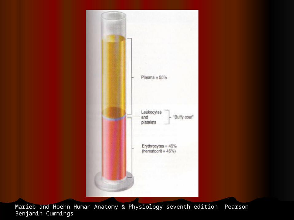
Blood is a connective tissue, not a body fluid, made of fluid Blood is a connective tissue, not a body fluid, made of fluid (plasma) and cellular elements (RBC, WBC, and platelets)(plasma) and cellular elements (RBC, WBC, and platelets)
Its volume is 5-6 L in males and 4-5 L in femalesIts volume is 5-6 L in males and 4-5 L in females
It is slightly alkaline, with a pH of ~ 7.4It is slightly alkaline, with a pH of ~ 7.4
Its color varies from bright to dark redIts color varies from bright to dark red
It has a salty metallic tasteIt has a salty metallic taste

The BloodThe Blood
FunctionsFunctions
The bloodThe blood is vehicular organ that reaches all the other tissuesis vehicular organ that reaches all the other tissues
Transports oxygen and nutrientsTransports oxygen and nutrients
Removes CORemoves CO22 and other by-products of cell activity and other by-products of cell activity
Pivotal in maintaining homeostasis, growth and tissue Pivotal in maintaining homeostasis, growth and tissue repairrepair
Participates in the defense against infectionParticipates in the defense against infection
Participates in hemostasisParticipates in hemostasis
Participates in body heat distribution and regulationParticipates in body heat distribution and regulation
Marieb and Hoehn Human Anatomy & Physiology seventh edition Pearson Benjamin Cummings
The BloodThe Blood
PlasmaPlasma
Straw colored fluid made of water (~90%), other contents include: Straw colored fluid made of water (~90%), other contents include:
Proteins make the bulk of the solutes: Proteins make the bulk of the solutes:
Albumens (60%), manufactured in the liver are the most abundantAlbumens (60%), manufactured in the liver are the most abundant
Globulins (36%) are immune bodiesGlobulins (36%) are immune bodies
Fibrinogen (4%) for blood clottingFibrinogen (4%) for blood clotting
Nutrients: glucose, amino acids, lipids, cholesterolNutrients: glucose, amino acids, lipids, cholesterol
Electrolytes: NaElectrolytes: Na++, K, K++, Ca, Ca++++, Mg, Mg++++, H, H++, Cl, Cl--, HCO, HCO33--, PO, PO44
----, SO, SO44----
Waste: urea, creatinine, uric acid, bilirubinWaste: urea, creatinine, uric acid, bilirubin
Gases: OGases: O22 , CO , CO2 2 , N, N22
Protein bound hormonesProtein bound hormones
Plasma without clotting factors is called “serum”Plasma without clotting factors is called “serum”

Peripheral blood smear
Marieb and Hoehn Human Anatomy & Physiology seventh edition Pearson Benjamin Cummings

Pathophysiology McCance & Huether fifth edition Elsevier Mosby
Electron micrograph of blood smear

The BloodThe Blood
RBCRBC
An RBC is a 7.5 micron disc shaped body with a central An RBC is a 7.5 micron disc shaped body with a central depressiondepression
The cell is without a nucleus or mitochondriaThe cell is without a nucleus or mitochondria
AN RBC contains hemoglobin and filamentous proteins attached AN RBC contains hemoglobin and filamentous proteins attached to the cell wall to impart flexibility on itto the cell wall to impart flexibility on it
Antigens are embedded in the cell membrane, they decide the Antigens are embedded in the cell membrane, they decide the blood groupblood group
The RBC cytoplasm provides energy to maintain intracellular The RBC cytoplasm provides energy to maintain intracellular homostasishomostasis
This energy is generated mostly through anaerobic glycolysisThis energy is generated mostly through anaerobic glycolysis
RBCs function is gas exchange: ORBCs function is gas exchange: O22 to the tissues and CO to the tissues and CO22 to the to the lungslungs
The BloodThe Blood
RBCRBC
Structure of HemoglobinStructure of Hemoglobin
Each hemoglobin molecule is made up of four globin chains and fourEach hemoglobin molecule is made up of four globin chains and four heme moleculesheme molecules Heme molecule is a porphyrin type pigment with a ferrous ion (FeHeme molecule is a porphyrin type pigment with a ferrous ion (Fe++
++)) Globin is a 287 amino acid protein made of two Globin is a 287 amino acid protein made of two αα, and two non , and two non αα
chainschains Adult hemoglobin A is (Adult hemoglobin A is (αααα//ββββ), fetal hemoglobin F is ( ), fetal hemoglobin F is ( αααα//γγγγ)) An enzyme, 2,3 diphosphoglycerate (2,3 DPG) binds to hemoglobin An enzyme, 2,3 diphosphoglycerate (2,3 DPG) binds to hemoglobin molecule, it lowers its affinity to Omolecule, it lowers its affinity to O22
Binding of OBinding of O22 to heme breaks some of the globin bonds exposing to heme breaks some of the globin bonds exposing moremore
heme molecules to bind with Oheme molecules to bind with O22
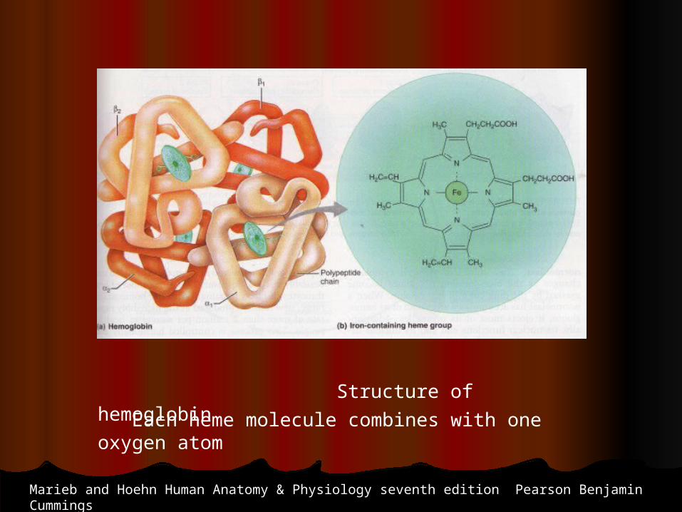
Structure of hemoglobin
Marieb and Hoehn Human Anatomy & Physiology seventh edition Pearson Benjamin Cummings
Each heme molecule combines with one oxygen atom

Davidson’s Principles and Practice of Medicine eighth edition, Churchill Livingstone
Structure of hemoglobin and the oxygen dissociation curve


The BloodThe Blood
RBCRBC
Development of RBCsDevelopment of RBCs Hypoxia Hypoxia → erythropoietin (kidney)→→ erythropoietin (kidney)→ red marrow of long bones red marrow of long bones→→
erythroid stem cell→ erythroblasts cell division→ smaller cells loosing nucleus erythroid stem cell→ erythroblasts cell division→ smaller cells loosing nucleus and gaining hemoglobin → reticulocyte→ mature RBC and gaining hemoglobin → reticulocyte→ mature RBC
Reticulocytes contain remnants of cell organellesReticulocytes contain remnants of cell organelles Their presence in excess in the peripheral blood (>2%) indicatesTheir presence in excess in the peripheral blood (>2%) indicates excessive RBC destructionexcessive RBC destruction The normal number of RBCs is 4.3-5 million/mmThe normal number of RBCs is 4.3-5 million/mm3 3 in the female and 5.1-5.8 million/mm3 in the male The normal values for Hgb are 13-15 gm/dl for females and 14-16 gm/dl in males
Amino acids, lipids, carbohdrates, iron, vitamin B12 and folic acid are essntial for hemoglobin synthesis

The BloodThe Blood
RBCRBC
Hemoglobin has a remarkable ability to bind with oxygen Hemoglobin has a remarkable ability to bind with oxygen
forming oxyhemoglobinforming oxyhemoglobin
It can also release the oxygen to the tissues becoming It can also release the oxygen to the tissues becoming deoxyhemoglobindeoxyhemoglobin
Hemoglobin combines with COHemoglobin combines with CO22 forming carbaminohemoglobin forming carbaminohemoglobin

The BloodThe Blood
RBCRBCDestructionDestruction Life span ~ 120 daysLife span ~ 120 days
RBCs are phagocytosed by the reticulo-endothlial cells of the spleenRBCs are phagocytosed by the reticulo-endothlial cells of the spleen
Globulin is hydrolyzed to amino acids that are recycledGlobulin is hydrolyzed to amino acids that are recycled
Iron is removed from heme and reused to synthesize HgbIron is removed from heme and reused to synthesize Hgb
Heme remnant converted to bilirubin Heme remnant converted to bilirubin
Bilirubin is conjugated with glucoronic acid, and secreted in bileBilirubin is conjugated with glucoronic acid, and secreted in bile
In the intestine most is converted to stercobilin and excreted In the intestine most is converted to stercobilin and excreted
A small amount of stercobilin is absorbed by the kidney andA small amount of stercobilin is absorbed by the kidney and
secreted as urobilinogensecreted as urobilinogen
The BloodThe Blood
RBCRBCDisorders of RBCsDisorders of RBCs
Anemia is reduced RBC countAnemia is reduced RBC count
Anemias can be caused byAnemias can be caused by
RBC loss or reduced production RBC loss or reduced production
Hemorrhage, hemolysis, depressed bone marrowHemorrhage, hemolysis, depressed bone marrow
Reduced hemoglobin content of RBCsReduced hemoglobin content of RBCs
Iron, intrinsic factor, folic acid, or BIron, intrinsic factor, folic acid, or B12 12 deficiencydeficiency
Congenital hemoglobin defectsCongenital hemoglobin defects
Thalassemia, sickle cell anemia, spherocytosisThalassemia, sickle cell anemia, spherocytosis

The BloodThe Blood
RBC RBC
Disorders of RBCsDisorders of RBCs PlolycythemiaPlolycythemia Bone marrow disorder causing an increased number of Bone marrow disorder causing an increased number of circulating RBCs and increased blood viscositycirculating RBCs and increased blood viscosity Aplastic anemiaAplastic anemia Results from bone marrow suppression or destruction Results from bone marrow suppression or destruction
(radiation,(radiation, drugs, chemicals)drugs, chemicals) All the blood elements are deficientAll the blood elements are deficient
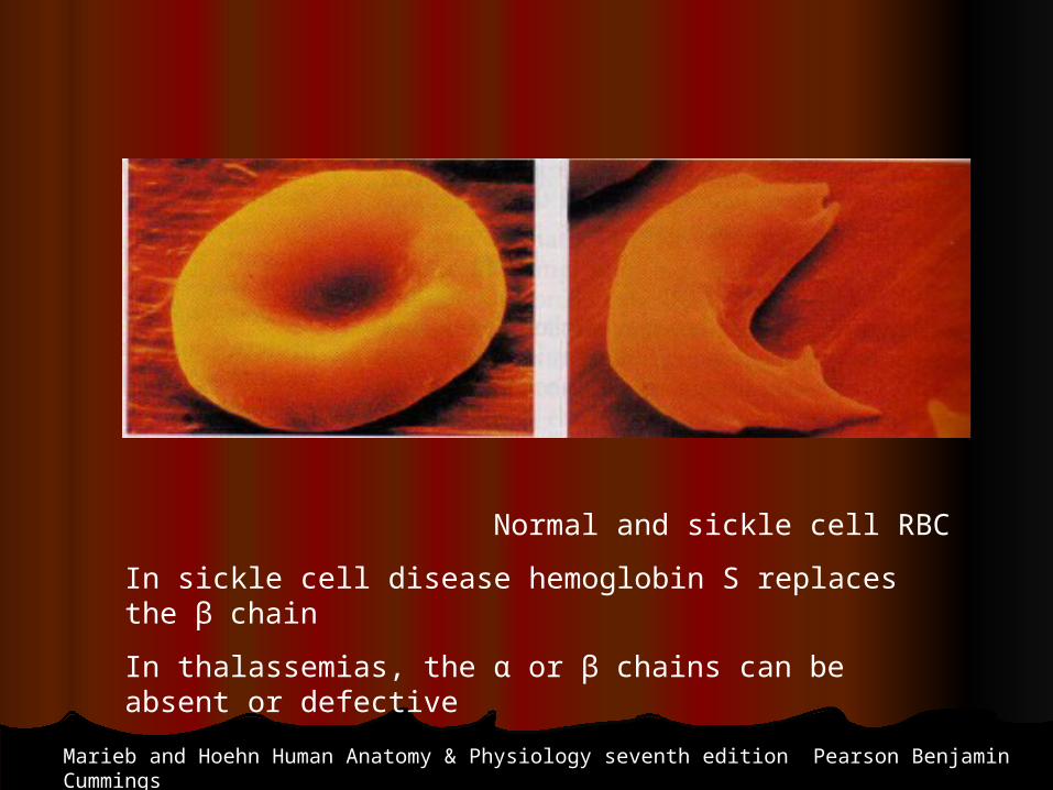
Normal and sickle cell RBC
In sickle cell disease hemoglobin S replaces the β chain
In thalassemias, the α or β chains can be absent or defective
Marieb and Hoehn Human Anatomy & Physiology seventh edition Pearson Benjamin Cummings
The BloodThe Blood
White Blood Cells (WBC)White Blood Cells (WBC)FunctionFunction
Lymphocytes are the effectors of the immune functionLymphocytes are the effectors of the immune function
WBCs main function is to fight bacterial infectionsWBCs main function is to fight bacterial infections WBCs are the only nucleated blood formed elementsWBCs are the only nucleated blood formed elements They exercise their functions in the tissues not the blood streamThey exercise their functions in the tissues not the blood stream Selectines induce the WBC to adhere to capillary endotheliumSelectines induce the WBC to adhere to capillary endothelium WBC migrate to the tissue spaces by diapeditic movement WBC migrate to the tissue spaces by diapeditic movement
between between endothelial cell, diapedesis is initiated by chemical attraction toendothelial cell, diapedesis is initiated by chemical attraction to the infection site initiated by damaged cells “chemotaxis”the infection site initiated by damaged cells “chemotaxis” WBC destroy the bacterial cell wall by oxidation and digestion byWBC destroy the bacterial cell wall by oxidation and digestion by proteins “defensisns”proteins “defensisns”

The BloodThe Blood
White Blood Cells (WBC)White Blood Cells (WBC)
TypesTypes
The total number of WBCs is 4000 to 10,000/mmThe total number of WBCs is 4000 to 10,000/mm33
There two main types of WBCs: granulucytes and agranulocytes
Granulocytes, are of three typesGranulocytes, are of three types
Neutrophils (polymorphs) 50-70%, oxidize bacteria Neutrophils (polymorphs) 50-70%, oxidize bacteria
Eosinophils, 2-4% bilobed nuclei, attack parasites Eosinophils, 2-4% bilobed nuclei, attack parasites
Basophils, 1% in peripheral blood, reside in the tissuesBasophils, 1% in peripheral blood, reside in the tissues, , contain histamine, involved in hypersensitivity reactioncontain histamine, involved in hypersensitivity reaction
The BloodThe Blood
White Blood Cells (WBC)White Blood Cells (WBC)
TypesTypes
Agranulocytes are of two typesAgranulocytes are of two types
Lymphocytes, the smallest and second most abundantLymphocytes, the smallest and second most abundant T cells (80%) mediate cellular immunity, express T cells (80%) mediate cellular immunity, express
CD1,2,3,4,5,7 & 8CD1,2,3,4,5,7 & 8 B cells mediate humoral immunity, express immunoglobulin B cells mediate humoral immunity, express immunoglobulin
light light chains on the surfacechains on the surface Monocytes, the largest, migrate to the tissues and become Monocytes, the largest, migrate to the tissues and become
macrophages involved in cellular immunity, secrete ILmacrophages involved in cellular immunity, secrete IL11 , TNF, , TNF, andand
CSFCSF

Davidson’s The Priciples and Practice of Medicine, eigthteenth edition Churchill Livingstone
White blood cells, the granulcytes

Monocytes and lymphocytes
Davidson’s The Priciples and Practice of Medicine, eighteenth edition Churchill Livingstone
The BloodThe Blood
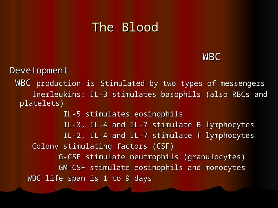
WBCWBCDevelopmentDevelopment WBC WBC productionproduction isis Stimulated by two types of messengersStimulated by two types of messengers
Inerleukins: IL-3 stimulates basophils (also RBCs and platelets) Inerleukins: IL-3 stimulates basophils (also RBCs and platelets)
IL-5 stimulates eosinophilsIL-5 stimulates eosinophils
IL-3, IL-4 and IL-7 stimulate B lymphocytesIL-3, IL-4 and IL-7 stimulate B lymphocytes
IL-2, IL-4 and IL-7 stimulate T lymphocytesIL-2, IL-4 and IL-7 stimulate T lymphocytes
Colony stimulating factors (CSF)Colony stimulating factors (CSF)
G-CSF stimulate neutrophils (granulocytes)G-CSF stimulate neutrophils (granulocytes)
GM-CSF stimulate eosinophils and monocytesGM-CSF stimulate eosinophils and monocytes
WBC life span is 1 to 9 days WBC life span is 1 to 9 days

Vander’s Physiology eighth edition, Mc Graw Hill
Development of blood cells

The BloodThe Blood
WBCWBCLeukemiasLeukemias A single unspecialised WBC precursor cell “clones” itself A single unspecialised WBC precursor cell “clones” itself
uncontrollablyuncontrollably
The resulting “leukemia” is therefore “monclonal”The resulting “leukemia” is therefore “monclonal”
The more undifferentiated cells produce acute (blastic) leukemiasThe more undifferentiated cells produce acute (blastic) leukemias
The more differentiated (cytic) cells produce chronic typesThe more differentiated (cytic) cells produce chronic types
A leukemia is called according to the producing cellA leukemia is called according to the producing cell
Leukemias impair the bone marrow, and other blood cells Leukemias impair the bone marrow, and other blood cells functions functions
Death is inevitable unless treated, it results from bleeding or Death is inevitable unless treated, it results from bleeding or sepsissepsis
The BloodThe Blood
Blood CoagulationBlood Coagulation
Coagulation is a natural mechanism that acts to diminish Coagulation is a natural mechanism that acts to diminish blood loss from hemorrhageblood loss from hemorrhage
Coagulation (thrombosis) especially in the microcirculation is Coagulation (thrombosis) especially in the microcirculation is also a manifestation of inflammation and sepsisalso a manifestation of inflammation and sepsis
Coagulation occurs in three StagesCoagulation occurs in three Stages Platelet plugPlatelet plug The cascade leading to fibrin (clot) formationThe cascade leading to fibrin (clot) formation Clot retraction and repair (PDGF)Clot retraction and repair (PDGF)

The BloodThe Blood
Blood CoagulationBlood Coagulation The PlateletsThe Platelets
Structure and FunctionStructure and Function
Platelets are discoid shaped bodies of 2-4 Platelets are discoid shaped bodies of 2-4 μμm m Derived from megacaryocytes arising from lineage committed stem Derived from megacaryocytes arising from lineage committed stem
cellscells They are fragments of cells, they have no nucleiThey are fragments of cells, they have no nuclei They contain three types of granules in the cytoplasmThey contain three types of granules in the cytoplasm Alpha granules contain fibrinogen and von Willebrand factor (vWF)Alpha granules contain fibrinogen and von Willebrand factor (vWF) Delta (dense) granules store adenosine diphosphate and serotoninDelta (dense) granules store adenosine diphosphate and serotonin Lysosomes, contain acid hydrolasesLysosomes, contain acid hydrolases
The BloodThe Blood
Blood CoagulationBlood Coagulation
The Platelets The Platelets
Platelets are activated by thrombin, collagen, or ADP, they Platelets are activated by thrombin, collagen, or ADP, they discharge discharge
their content which leads to the formation of thrombaxane A2their content which leads to the formation of thrombaxane A2
Platelets adhere to exposed collagen in the presence of Platelets adhere to exposed collagen in the presence of
von Willebrand factor (vWF)von Willebrand factor (vWF)
Their life span is 8-14 days, they are destroyed in the cells of the Their life span is 8-14 days, they are destroyed in the cells of the RERE
systemsystem

The structure of a platelet
Davidson’s The Priciples and Practice of Medicine, eighteenth edition Churchill Livingstone

Initial vasoconstriction and platelet plug
vVander’s Physiology eighth edition Mc Graw Hill
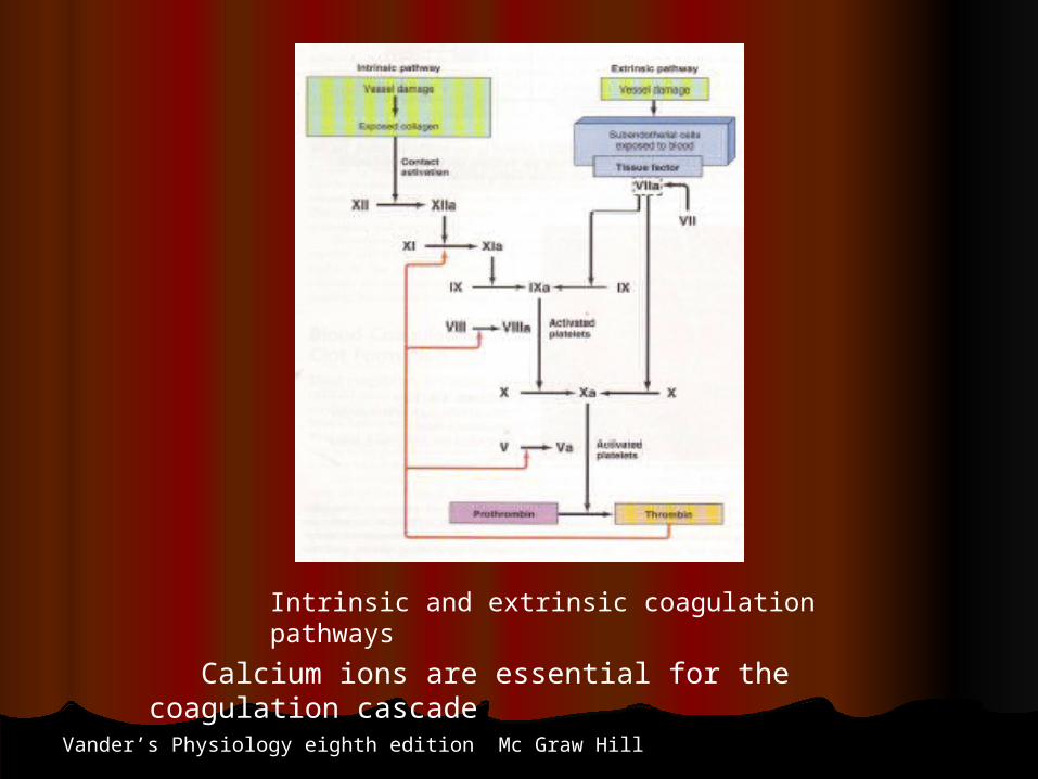
Intrinsic and extrinsic coagulation pathways
Vander’s Physiology eighth edition Mc Graw Hill
Calcium ions are essential for the coagulation cascade
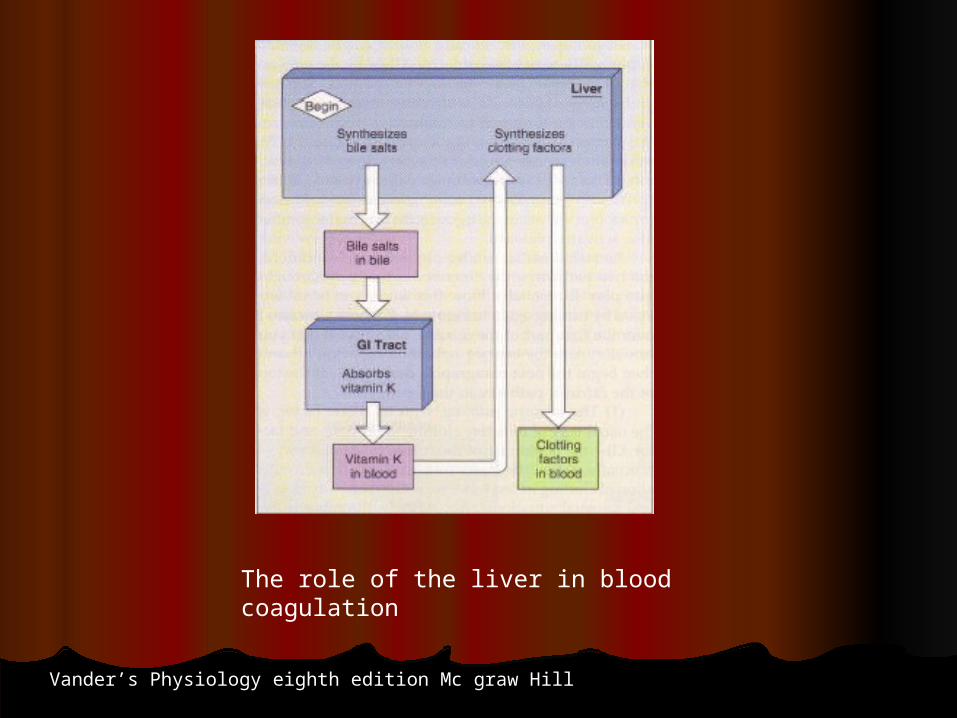
The role of the liver in blood coagulation
Vander’s Physiology eighth edition Mc graw Hill
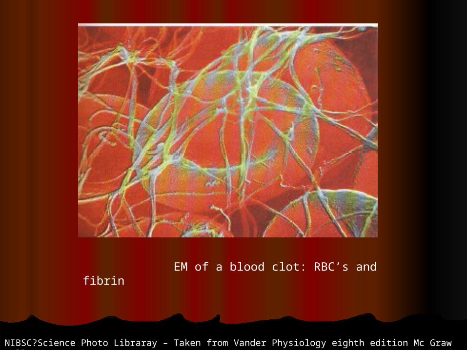
EM of a blood clot: RBC’s and fibrin
NIBSC?Science Photo Libraray – Taken from Vander Physiology eighth edition Mc Graw Hill
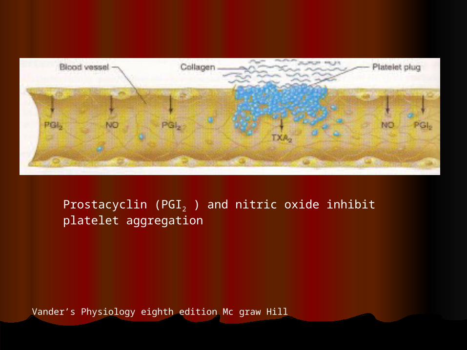
Prostacyclin (PGI2 ) and nitric oxide inhibit platelet aggregation
Vander’s Physiology eighth edition Mc graw Hill
The BloodThe Blood
AnticoagulationAnticoagulation
Natural AnticoagulantsNatural Anticoagulants Tissue factor pathway inhibitor (TFPI)Tissue factor pathway inhibitor (TFPI)
Plasminogen - plasminPlasminogen - plasmin
HeparinHeparin
Antithrombin IIIAntithrombin III
Protein CProtein C
Protein S Protein S
Vitamin E quinoneVitamin E quinone
NaturalNatural anticoagulants also have anti inflammatory activity anticoagulants also have anti inflammatory activity
The BloodThe Blood
AnticoagulationAnticoagulation
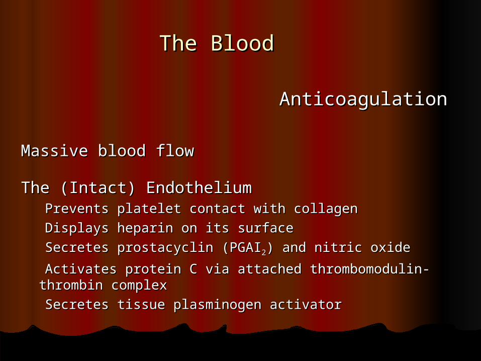
Massive blood flowMassive blood flow
The (Intact) EndotheliumThe (Intact) Endothelium Prevents platelet contact with collagenPrevents platelet contact with collagen
Displays heparin on its surfaceDisplays heparin on its surface
Secretes prostacyclin (PGAISecretes prostacyclin (PGAI22) and nitric oxide) and nitric oxide
Activates protein C via attached thrombomodulin-thrombin Activates protein C via attached thrombomodulin-thrombin complexcomplex
Secretes tissue plasminogen activatorSecretes tissue plasminogen activator

The BloodThe Blood
AnticoagulationAnticoagulation
HeparinHeparin Is a natural anticoagulant found in the bodyIs a natural anticoagulant found in the body
Can be given by IV or subcutaneous routes for anticoagulationCan be given by IV or subcutaneous routes for anticoagulation
Can be easily neutralized (its action reversed) by protamineCan be easily neutralized (its action reversed) by protamine
It has an antithrombin effectIt has an antithrombin effect
Inhibits platelet functionsInhibits platelet functions
Its effect is measured by estimating the partial prothrombin time Its effect is measured by estimating the partial prothrombin time (PTT)(PTT)

The BloodThe Blood
AntigoagulationAntigoagulation
Aspirin (ASA)Aspirin (ASA)
Inhibits cyclooygenase (COX) that catalyzes the formation of Inhibits cyclooygenase (COX) that catalyzes the formation of thrombaxanethrombaxane
Mature platelets can not make new thrombaxane because their COX isMature platelets can not make new thrombaxane because their COX is
blockedblocked
Endothelial cells can synthesize new COX, therefore they are not Endothelial cells can synthesize new COX, therefore they are not affectedaffected
by ASAby ASA
Thrombaxane is important for platelet aggregationThrombaxane is important for platelet aggregation
Platelet function is assessed by the bleeding timePlatelet function is assessed by the bleeding time
New drugs that interfere with fibrinogen/platelet binding are now New drugs that interfere with fibrinogen/platelet binding are now
available available

The BloodThe Blood
AnticoagulationAnticoagulation
Vitamin K antagonistsVitamin K antagonists Warfarin, also known as coumadinWarfarin, also known as coumadin
Interfere with the liver synthesis of coagulation factorsInterfere with the liver synthesis of coagulation factors
Effect measure by checking the protime (PT) now reported asEffect measure by checking the protime (PT) now reported as
international normalized ratio (INR)international normalized ratio (INR)
Vitamin E quinone is a potent anticoagulantVitamin E quinone is a potent anticoagulant
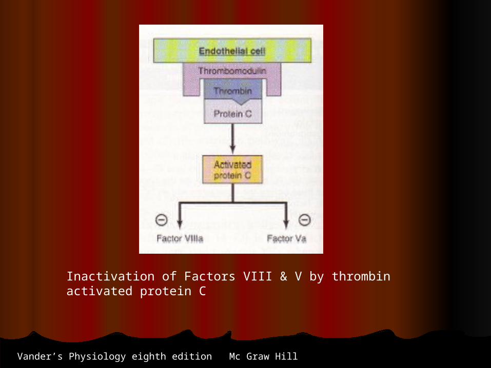
Inactivation of Factors VIII & V by thrombin activated protein C
Vander’s Physiology eighth edition Mc Graw Hill

Fibrinolysis, a mechanism for clot resorption
Vander’s Physiology eighth edition Mc Graw Hill

The BloodThe Blood
ThrombolyticsThrombolytics
Plasminogen activates plasmin, a natural fibrinolytic agentPlasminogen activates plasmin, a natural fibrinolytic agent Tissue Plasminogen Activator (t-PTA) activates plasminogenTissue Plasminogen Activator (t-PTA) activates plasminogen Plasmin dissolves an already formed clot (thrombolytic therapy)Plasmin dissolves an already formed clot (thrombolytic therapy)
Streptokinase and Urokinase Streptokinase and Urokinase Thrombolytic actionThrombolytic action Streptokinase has side effects but less costlyStreptokinase has side effects but less costly Urokinase has less side effects but more expensiveUrokinase has less side effects but more expensive
New drugs that interfere with fibrinogen/platelet binding are now New drugs that interfere with fibrinogen/platelet binding are now availableavailable


The BloodThe Blood
TransfusionTransfusion
The following blood elements can be transfused to cover a The following blood elements can be transfused to cover a deficiencydeficiency
in quantity or quality of one or more of the blood componentsin quantity or quality of one or more of the blood components
Whole blood does not allow time for checking for the presence of Whole blood does not allow time for checking for the presence of
infectious agentsinfectious agents
Packed RBC’c for low HbPacked RBC’c for low Hb
Platelets for thrompcytopenia and bleedingPlatelets for thrompcytopenia and bleeding
Plasma (fresh frozen) to replace deficient intrinsic factorsPlasma (fresh frozen) to replace deficient intrinsic factors
Cryoprecipitate for hemophiliaCryoprecipitate for hemophilia
The BloodThe Blood
TransfusionTransfusion
Stored blood is acidified with citrates to prevent coagulationStored blood is acidified with citrates to prevent coagulation
It looses its plateletsIt looses its platelets
Has more potassium, and accumulates ammoniaHas more potassium, and accumulates ammonia
More hemolysed RBC as the storage is prolongedMore hemolysed RBC as the storage is prolonged
Hemoglobin tends to hold more to OHemoglobin tends to hold more to O22 because of the reduction because of the reduction in in
2,3 diphophoglyceric acid (2.3 DPG)2,3 diphophoglyceric acid (2.3 DPG)
The life span of RBCs stored at 4The life span of RBCs stored at 4oo C is about 28 days C is about 28 days

The BloodThe Blood
Blood TypesBlood Types
RBC’s have surface antigensRBC’s have surface antigens
RBC’s can be grouped according to the presence or absence RBC’s can be grouped according to the presence or absence of certain antigensof certain antigens
There are many RBC antigens but only a A, B, AB and the Rh There are many RBC antigens but only a A, B, AB and the Rh are of are of
clinical significanceclinical significance
Each one of the four types can be Rh positive or negativeEach one of the four types can be Rh positive or negative
Donor blood is mixed with recipient serum to decide Donor blood is mixed with recipient serum to decide compatibility, donor cell clumping indicate incompatibilitycompatibility, donor cell clumping indicate incompatibility
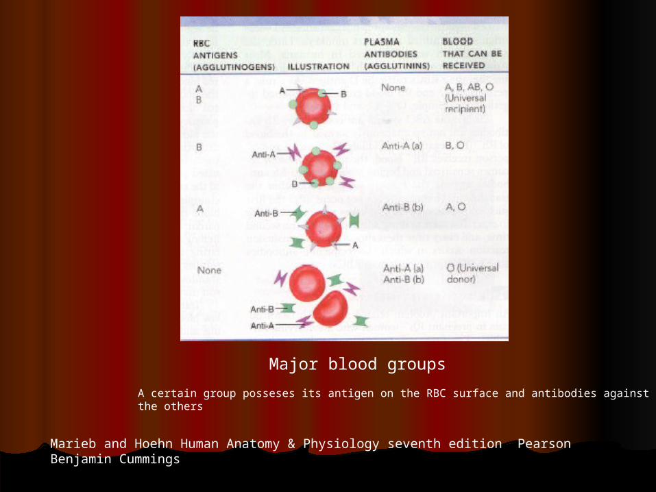
Major blood groups
Marieb and Hoehn Human Anatomy & Physiology seventh edition Pearson Benjamin Cummings
A certain group posseses its antigen on the RBC surface and antibodies against the others

The BloodThe Blood
TransfusionTransfusion
Transfusion of incompatible RBCs results in their hemolysis in the Transfusion of incompatible RBCs results in their hemolysis in the donordonor
This leads to blockage of the renal tubules, hypotension, fever, This leads to blockage of the renal tubules, hypotension, fever, back pain, and may prove fatalback pain, and may prove fatal
This is treated by stopping the transfusion, hydration, and This is treated by stopping the transfusion, hydration, and alkalinization of the urinealkalinization of the urine
Not every reaction to transfusion is due to infused RBC hemolysis, Not every reaction to transfusion is due to infused RBC hemolysis,
other less severe reactions can occur other less severe reactions can occur
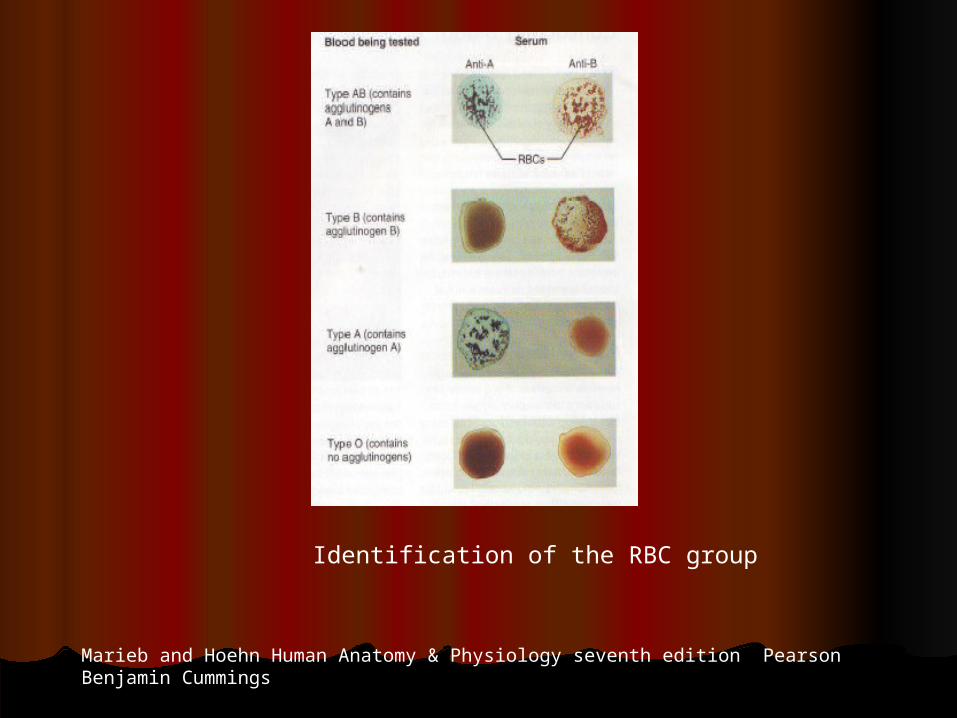
Identification of the RBC group
Marieb and Hoehn Human Anatomy & Physiology seventh edition Pearson Benjamin Cummings
The BloodThe Blood
Rh FactorRh FactorThere are no preformed Rh antibodies in RhThere are no preformed Rh antibodies in Rh-- individuals individuals
They develop after exposure to RhThey develop after exposure to Rh++ factors (antigens) factors (antigens)
This explains the fact that an RhThis explains the fact that an Rh++ fetus of an Rh fetus of an Rh-- mother mother
does not usually sufferdoes not usually suffer
But the RhBut the Rh-- mother can be sensitized to the Rh mother can be sensitized to the Rh++ antigens during antigens during the first pregnancy, especially during deliverythe first pregnancy, especially during delivery
Subsequent fetuses can suffer from the mother’s Rh antibodies Subsequent fetuses can suffer from the mother’s Rh antibodies passedpassed
to it and its blood hemolyzesto it and its blood hemolyzes
This can be prevented by giving the mother serum that blocks This can be prevented by giving the mother serum that blocks the Rhthe Rh++
factors antigenicityfactors antigenicity
The BloodThe Blood
Rh FactorRh Factor
Eighty five percent of the population have Rh antigens (RhEighty five percent of the population have Rh antigens (Rh++))There are several Rh groups (factors, antigens)There are several Rh groups (factors, antigens)Three groups: Rh, C, D, and E, are of clinical importanceThree groups: Rh, C, D, and E, are of clinical importanceAn RhAn Rh-- mother may develop antibodies against her Rh mother may develop antibodies against her Rh++ fetus fetusThe RhThe Rh-- mother antibodies can pass to the Rh mother antibodies can pass to the Rh++ fetus resulting fetus resulting
in hemolysis of its RBCs, a condition know as in hemolysis of its RBCs, a condition know as erythroblastosis fetaliserythroblastosis fetalis
This sequence usually occurs after the first pregnancyThis sequence usually occurs after the first pregnancy