the biological synthesis of hippuric acid · water + benzoic acid + ... the biological synthesis of...
TRANSCRIPT
THE BIOLOGICAL SYNTHESIS OF HIPPURIC ACID IN VITRO*
BY HENRY BORSOOK AND JACOB W. DUBNOFF
(Prom the William G. Kerckhoff Laboratories of the Biological Sciences, California Institute of Technology, Pasadena)
(Received for publication, November 8, 1939)
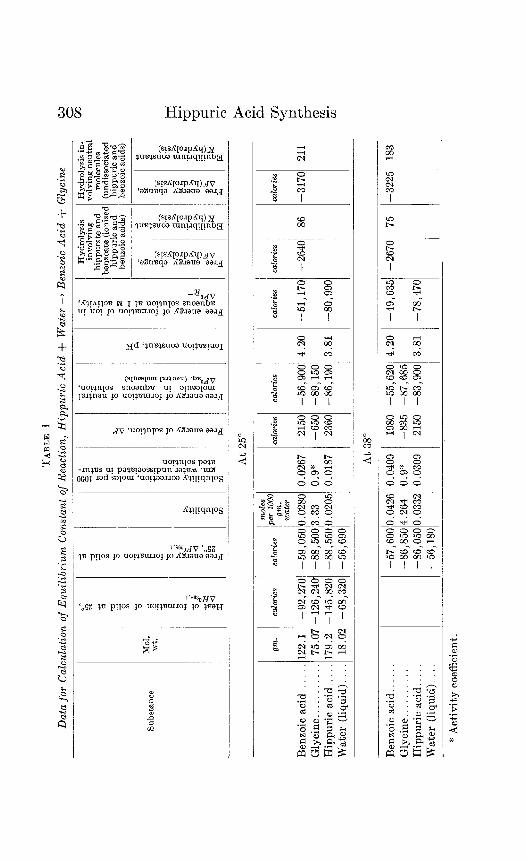
The mechanism of the synthesis of hippuric acid in viva is in- teresting from several points of view. There is the intrinsic interest in a compound found in the urine of many animals, an interest which is heightened by the use of the rate of hippuric acid excretion following the administration of benzoic acid as a clinical test of liver function. This synthesis is interesting also from the point of view of physiological cncrgetics. The formation of hippuric acid from glycine and benzoic acid is attended by a gain in free energy (Table I). In other words the tendency of the reaction, if allowed to proceed spontaneously at 25” or 38”, is not toward synthesis but toward practically complete hydroly- sis of hippuric acid (Table III). Yet when benzoic acid is fed, hippuric acid is rapidly synthesized. This synthesis also occurs and can be measured, as shown below, when liver slices are suspended in Ringer’s solution containing low concentrations of benzoic acid and glycine. More than half the benzoic acid is converted to hippuric acid. From the thermodynamic data it may bc deduced that the enzymatic synthesis of hippuric acid cannot be simply the reverse of its hydrolysis. The hydrolysis can proceed spontaneously; the synthesis must be coupled with an energy-yielding reaction.
Another, possibly more important, point of interest is that the synthesis of hippuric acid resembles in several respects the synthe- sis of the peptidc bond. The group which is formed, the CONH
* An account of most of the work described here was read at the meeting of the American Society of Biological Chemists at Toronto, April, 1939 (Proc. Am. Sot. Biol. Chem., J. Biol. Chem., 128, p. cxiv (1939)).
307
TABL
E I
Data
for
Ca
lcula
tion
of E
quilib
rium
Co
nsta
nt
o.f
Reac
tion,
Hi
ppur
ic Ac
id
+ W
ater
+
Benz
oic
Acid
+
Glyc
ine
Hydr
olysis
inv
olving
hip
pura
te
and
t. mxm
ate
(ioni
sec
hipp
uric
and
benr
oic
acid
s)
Hydr
olysis
in-
m
lving
ne
utra
l m
olec
ules
(u
ndiss
ocia
ted
hipp
uric
and
benz
oic
acid
s)
Mol.
wt
. Su
bsta
nce
At
25”
lc &rie
s
-317
0 21
1
cala
ries
calo
ries
-51,
1701
-2
640
86
-80,
990
122.
1 -9
2,27
0 0.
0267
75.0
7 -1
26,2
40
-59,
050’0
.028
0 -8
8,50
013.
33
0.9*
17
9.2
-145
,820
-8
8,55
0/0.
0205
0.
0187
18
.02
-68,
320,
-5
6,69
01
2150
1 -5
6,90
0 4.
20
-650
-8
9,15
0 23
60
-86,
190
3.81
Benz
oic
acid
. Gl
ycine
Hi
ppur
ic ac
id
Wat
er
(liquid
).
I- At
38
”
-57,
600’0
.042
6 0.
0409
19
80
-55,
620
4.20
-4
9,63
5’ -2
670
75
-86,
8504
.264
0.9
%
-835
-8
7,68
5 -8
6,05
00.0
332
0.03
09
2150
-8
3,90
0 3.
81
-78,
470
-56,
180
-322
51
183
Benz
oic
acid
.. Gl
ycine
Hi
ppur
ic ac
id.
Wat
er
(liquid
).
* Ac
tivity
co
effic
ient
.
H. Borsook and J. W. Dubnoff 309
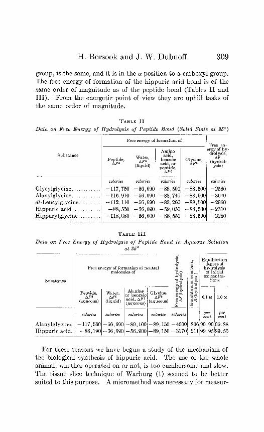
group, is the same, and it is in the Q! position to a carboxyl group. The free energy of formation of the hippuric acid bond is of the same order of magnitude as of the peptide bond (Tables II and III). From the energetic point of view they are uphill tasks of the same order of magnitude.
TABLE II
Data on Free Energy of Hydrolysis of Peptide Bond (Solid State at 25“)
Free energy of formation of Free en-
ytg, A&$$
ergy of hy.
Sul3stance
pel:d" benzdic
dro$is,
(liquid) acid, or G’;$=, (hydkl-
PW~p? ysis)
- calories calories cazories cozories calories
Glycylglycine -117,750 -56,690 -88,500 -88,500 -2560 Alanylglycine. -116,910 -56,690 -88,740 -88,500 -3640 dl-Leucylglycine -112,110 -56,690 -83,260 -88,500 -2960 Hippuric acid. -88,550 -56,690 -59,050 -88,500 -2310 Hippurylglycine . . -118,080 -56,690 -88,550 -88,500 -2280
TABLE III
Data on Free Energy of Hydrolysis of Peptide Bond in Aqueous Solution at 25”
Substance
calories calories calories calories calories
Alanylglycine.. -117,560 -56,690 -89,1001-89,150 -4OOC Hippuric acid.. 1 -86,190 -56,690 -56,900 -89,150 -317(
I I
Equilibrium degree of
hydrolysis of initial
concentra- tions
O.lM l.OM
Pm Pm cent cent
19.99 99.88 19.9q99.55
For these reasons we have begun a study of the mechanism of the biological synthesis of hippuric acid. The use of the whole animal, whether operated on or not, is too cumbersome and slow. The tissue slice technique of Warburg (1) seemed to be better suit,ed to this purpose. A micromethod was necessary for measur-
310 Hippuric Acid Synthesis
ing very small quantities of hippuric acid. One was devised by means of which it is possible to measure as little as 2 X low3 mg. We were then able to measure the synthesis of hippuric acid effected by tissue slices in the course of a few hours from dilute solutions (0.0025 M) of benzoic acid and glycine.
A survey was made of the distribution of the capacity for syn- thesizing hippuric acid among the tissues of several animals. The availability of different amino acids for this synthesis and the effect of poisons and damage to cell structure have been studied. These findings, the thermodynamic data, and a descrip- tion of the technique and analytical method employed are de- scribed in the present communication.
l’hermodynam,ic Data
WC have described in previous communications both the gc’n- era1 principles and the details of calculating equilibrium constants from thermal data (2-4). We shall therefore present here only the necessary data and the final results of the calculations. The values for the free energy of formation at 38” were calculated from the values at 25” by means of the van’t Hoff equation, on the assumption that AH is constant over the temperature interval from 25-38”.
The thermodynamic data in Tables I to III are based on measurements of heat capacities and heats of combustion made in this laboratory by Dr. H. PI. Huffman. The details of this work have not yet been published, though one compilation of part of these data has been published (5). The thermal data in Table I differ from those given in this previous compilation. The differences are formal only, and arise from a change in the ac- cepted value for the heat of combustion of benzoic acid. The values for the heat and free energy of formation of liquid water at 25” are those of Rossini (6).
The solubility of benzoic acid was obtained from Seidell (7). The value at 25” agrees closely with bhat determined later by Kolthoff and Bosch (8). The solubility of glycine at 25” and 38” as well as the activity coefficient in the saturated solution are taken from Schmidt (5). We have assumed that the activity co- efficient at 38” is the same as that determined experimentally at 25”. The possible error involved is negligible for our present
H. Borsook and J. W. Dubnoff 311
purposes. We have ourselves determined the solubility of hip- puric acid at 25” and 38”. Our value at 25” is identical with that given by Kendall (9).
A number of determinations have been reported of the thermo- dynamic ionization constant of benzoic acid. The most reliable and concordant value is that obtained at 25” by Brockman and Kilpatrick (lo), and by Saxton and Meier (11). The value for the thermodynamic ionization constant of hippuric acid we have used is that given by Josephson (12).
There are no reliable values for the ionization constants of these two acids at 38”. The values we have used are the experimental values at 25”. This seemed preferable to guessing the change in the ionization constants with temperature. It is exceedingly improbable that the difference between the ionization constants of the two acids is, for our purpose, significantly different at 38” from what it is at 25”. It is the difference in the ionization con- stants which contributes to the final over-all change in free energy. These ionization constants were used to compute the ionization of the two acids in their saturated solutions.
The equilibrium constant for the hydrolysis of hippuric acid in water has been measured experimentally at 184” and 194” by Ingersoll and Burrows (13). The tempcrat’ure difference is too small, and uncertainties regarding degrees of ionization and activity coefficients at these temperatures too great, for a reliable hxtrapolation of the equilibrium constant from the higher tem- peratures to 25’. Without any corrections, t.he extrapolated value at 25’ for the reaction between neutral molecules is 546, which corresponds to a value of AF of -3735 calories. The more reliable value calculated from the thermal data is -3170 calories. The agreement is surprisingly close.
The findings on the enzymatic hydrolysis of hippuric acid arc in complete accord, as far as they go, with these thermodynamic data. Thus Mutch (14) prepared an extract of hog kidney which hydrolyzed 0.08 N hippuric acid 97 per cent in 255 hours at 37”. The hydrolysis was prevented from being complete by the inhi- bition of the enzyme, demonstrated by the same author, by the accumulating benzoic acid. A saturated solution of sodium benzoate and glycine set away with the enzyme for 1 month at 37” yielded a small amount of impure, unidentified material, which
312 Hippuric Acid Synthesis
at the most, had it been hippuric acid, would have amounted to 0.6 per cent conversion of the bcnzoic acid to hippuric acid. Kimura (15) obtained complete hydrolysis of hippuric acid with a glycerol extract of hog kidney. Takahashi (16) found nearly complete hydrolysis when sodium hippurate was incubated with an aqueous suspension of minced chicken kidney. Ho recovered in one experiment 81 per cent of the theoretical benzoic acid and 90 per cent of the amino nitrogen freed.
Technique
Most of the animals used in these experiments wcrc Wistar Institute adult white rats. When other animals were used, the technique was the same. The animals were all in a normal state of nutrition, and were killed by stunning. The organs were sliced free-hand with a straight edge razor. A well sharpened straight edge razor is a better tool for slicing than any safety razor blade we have tried.
The slices were rinsed in Ringer’s solution prepared according to the formula of Krebs and Henseleit (17), containing 0.2 per cent glucose, and equilibrated at 38” with a gas mixture consist- ing of 95 per cent oxygen and 5 per cent carbon dioxide. Each slice was rinsed immediately after being cut and then trans- ferred to the reaction vessel.
Since we were not interested in the respiration of the slices, we have been using 25 ml. bottles as reaction vessels instead of Warburg respirometers. Into these are fitted a-hole rubber stop- pers provided with inlet and outlet tubes for the passage of the gas mixture. The inlet tube reaches half-way into the bottle. It is connected to a glass manifold, which in turn is connected to a cylinder containing the gas mixture. The flow of gas through each reaction vessel is regulated by means of a screw-clamp. One end of the outlet tube in the reaction bottle is flush with the bottom of the stopper; the other end passes through a ring seal into a glass bulb which contains water and has a hole near the top. The bubbling of the gas through the water in this bulb en- ables one to estimate the rate at which the gas mixture is passing through the reaction vessel.
Twenty-four such reaction vessels are clamped on three strips of wood in such a manner that the lower half of each vessel is
H. Borsook and J. W. Dubnoff 313
immersed in the water bath. These strips are attached at each end to an upright strip which is mounted on the usual rocking device for Warburg respirometers.
When all the reaction vessels are in place and connected, the gas mixture is blown through vigorously for 10 minutes, after which, for the remainder of the experiment, the gas is slowed down so that about one bubble escapes in the traps about every 2 seconds. Throughout the period of gassing and afterwards the vessels are rocked at a rate of about 90 cycles per minute. The temperature of the water bath was 38”.
This arrangement is much cheaper and more convenient than the usual all-glass respirometer assembly; it is more rugged, and serves quite as well.
Analytical Procedure
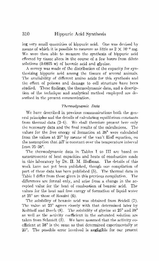
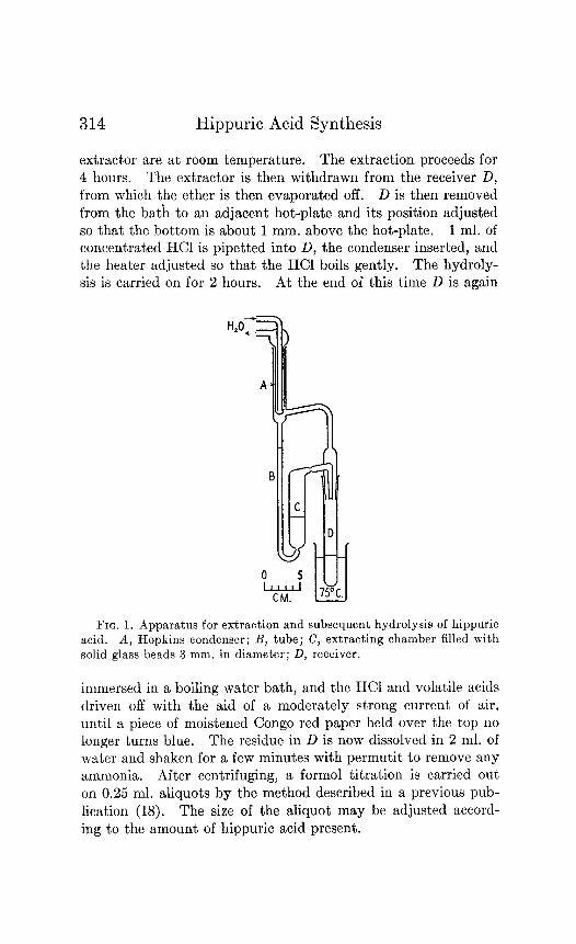
The method employed for the analysis of hippuric acid is in principle the classical method of extraction, hydrolysis, and form01 titration of the liberated glycine. The extraction apparatus is shown in Fig. 1. Bulb C in which the extraction occurs is filled with solid glass beads 3 mm. in diameter to increase the ether- water interface. The receiver D is a test-tube attached by a ground glass joint to the extractor. During the extraction D rests in a water bath at 75”. Fitting into A is a Hopkins con- denser. D and A have the same diameter, so that the condenser will fit as snugly into it as it does into A. D is about 4 cm. longer than A, so that the condenser will not reach too far down when it is used as a reflux condenser in the hydrolysis of the hip- puric acid which has been extracted. The tube B must be tall enough to carry a column of ether which can overcome the hydro- static pressure of the water in the lower part of C and the layer of ether above it.
The analytical procedure is as follows: Proteins are removed by boiling or by precipitation with trichloroacetic acid (final concentration 2 per cent). 2 ml. of protein-free solution acidi- fied to pH 1.5 to 2.0 with HzS04 are pipetted into B. Ether is pipetted into B until enough has overflowed from C to a depth of about 1 inch at the bottom of D. The condenser A with water running through it is then inserted, and the temperature of the bath in which D is held is raised to 75”. The other parts of the
314 Hippuric Acid Synthesis
extra&or are at room temperature. The extraction proceeds for 4 hours. The extractor is then withdrawn from the receiver D, from which the ether is then evaporated off. D is then removed from the bat’h to an adjacent hot-plate and its position adjusted so that the bottom is about 1 mm. above the hot-plate. 1 ml. of concentrated HCl is pipetted into D, the condenser inserted, and the heater adjusted so that the HCl boils gently. The hydroly- sis is carried on for 2 hours. At the end of this time D is again
FIG. 1. Apparatus for extraction and subsequent hydrolysis of hippuric acid. A, Hopkins condenser; B, tube; C, extracting chamber filled with solid glass beads 3 mm. in diameter; D, receiver.
immersed in a boiling water bath, and the HCl and volatile acids driven off with the aid of a moderately strong current of air, until a piece of moistened Congo red paper held over the top no longer turns blue. The residue in D is now dissolved in 2 ml. of water and shaken for a few minutes with permutit to remove any ammonia. After centrifuging, a form01 titration is carried out on 0.25 ml. aliquots by the method described in a previous pub- lication (18). The size of the aliquot may be adjusted accord- ing to the amount of hippuric acid present.
H. Borsook and J. W. Dubnoti 315
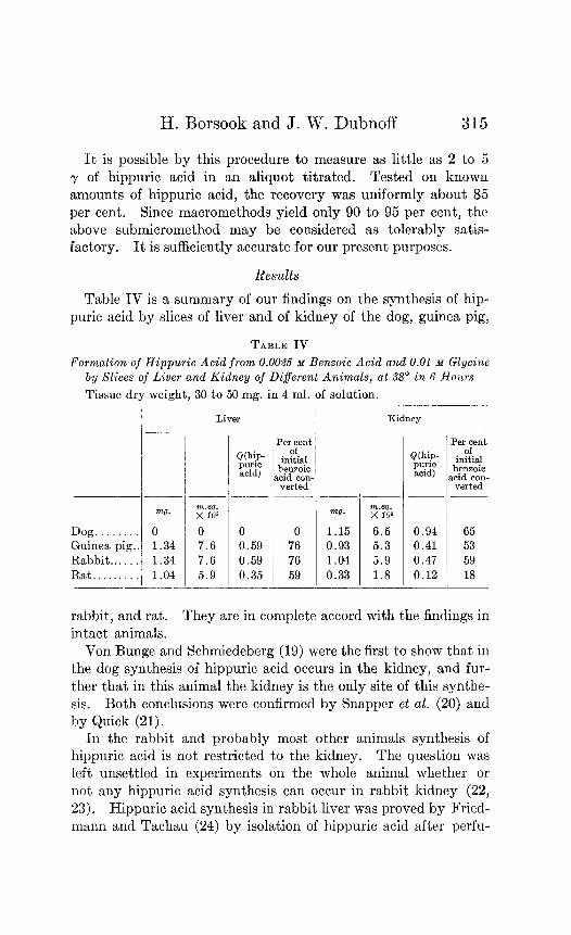
It is possible by this procedure to measure as little as 2 to 5 y of hippuric acid in an aliquot titrated. Tested on known amounts of hippuric acid, the recovery was uniformly about 85 per cent. Since macromethods yield only 90 to 95 per cent, the above submicromethod may be considered as tolerably satis- factory. It is sufficiently accurate for our present purposes.
Results
Table IV is a summary of our findings on the synthesis of hip- puric acid by slices of liver and of kidney of the dog, guinea pig,
TABLE IV
Formation of Hippuric Acid from 0.0026 M Benzoic Acid and 0.01 M Glycine by Slices of Liver ancl Kidney oj Different Animals, at 58” in 6 Hours
Tissue dry weight, 30 to 50 mg. in 4 ml. of solution.
Dog ........ Guinea pig., Rabbit ...... Rat. ........
mg.
0 1.34 1.34 1.04
Liver
;‘I$; 0 7.6 7.6 5.9
-
Per cent &Chip- in~~~)tfial puric acid) bensoic
acid con- verted
0 0 0.59 76 0.59 76 0.35 59
PeroFt initial benzoic
acid con- verted
1.15 6.5 0.94 65 0.93 5.3 0.41 53 1.04 5.9 0.47 59 0.33 1.8 0.12 18
rabbit, and rat. They arc in complete accord with the findings in intact animals.
Von Bunge and Schmiedcberg (19) were the first to show that in the dog synthesis of hippuric acid occurs in the kidney, and fur- ther that in this animal the kidney is the only site of this synthe- sis. Both conclusions were confirmed by Snapper et al. (20) and by Quick (21).
In the rabbit and probably most other animals synthesis of hippuric acid is not restricted to the kidney. The question was left unsettled in experiments on the whole animal whether or not any hippuric acid synthesis can occur in rabbit kidney (22, 23). Hippuric acid synthesis in rabbit liver was proved by Fried- mann and Tachau (24) by isolation of hippuric acid after perfu-
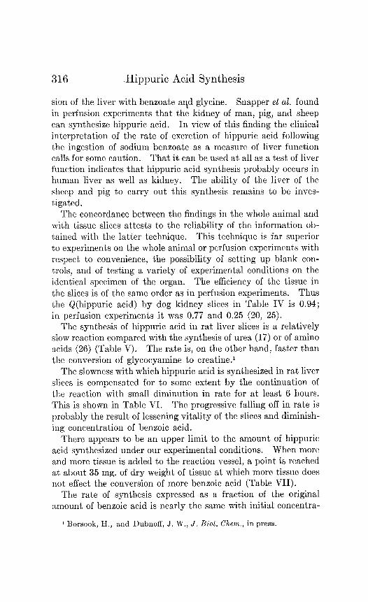
316 .Hippuric Acid Synthesis
sion of the liver with benzoate agd glycine. Snapper et al. found in perfusion experiments that the kidney of man, pig, and sheep can synthesize hippuric acid. In view of this finding the clinical interpretation of the rate of excretion of hippuric acid following the ingestion of sodium benzoate as a measure of liver function calls for some caution. That it can be used at all as a test of liver function indicates that hippuric acid synthesis probably occurs in human liver as well as kidney. The ability of the liver of the sheep and pig to carry out this synthesis remains to be inves- tigated.
The concordance between the findings in the whole animal and with tissue slices attests to the reliability of the information ob- tained with the latter technique. This technique is far superior to experiments on the whole animal or perfusion experiments with respect to convenience, the possibility of setting up blank con- trols, and of testing a variety of experimental conditions on the identical specimen of the organ. The efficiency of the tissue in the slices is of the same order as in perfusion experiments. Thus the Q(hippuric acid) by dog kidney slices in Table IV is 0.94; in perfusion experiments it was 0.77 and 0.25 (20, 25).
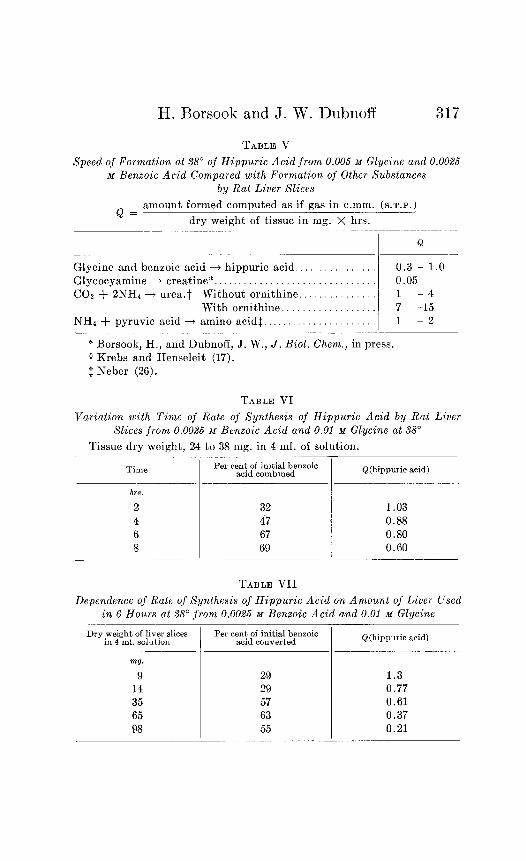
The synthesis of hippuric acid in rat liver slices is a relatively slow reaction compared with the synthesis of urea (17) or of amino acids (26) (Table V). The rate is, on the other hand, faster than the conversion of glycocyamine to creatine.’
The slowness with which hippuric acid is synthesized in rat liver slices is compensated for to some extent by the continuation of the reaction with small diminution in rate for at least 6 hours. This is shown in Table VI. The progressive falling off in rate is probably the result of lessening vitality of the slices and diminish- ing concentration of benzoic acid.
There appears to be an upper limit to the amount of hippuric acid synthesized under our experimental conditions. When more and more tissue is added to the reaction vessel, a point is reached at about 35 mg. of dry weight of tissue at which more tissue does not effect the conversion of more benzoic acid (Table VII).
The rate of synthesis expressed as a fraction of the original amount of benzoic acid is nearly the same with initial concentra-
1 Borsook, H., and Dubnoff, J. W., J. Biol. Chem., in press.
H. Borsook and J. IV. Dubnoff 317
TABLE V
Speed of Formation at 38” of Hippuric Acid from 0.006 M Glycine and 0.0025 M Benzoic Acid Compared with Formation of Other Substances
by Rat Liver Slices
amount formed computed as if gas in c.mm. (s.T.P.) Q=-
- dry weight of tissue in mg. X hrs.
Q
Glycine and benzoic acid ---$ hippuric acid.. 0.3 - 1.0 Glycocyamine --t creatine*. 0.05 COZ + 2NH4 + urea.? Without ornithine.. 1 - 4
With ornithine.. _. 7 -15 NH4 + pyruvic acid + amino acid$. 1 - 2
* Borsook, H., and Dubnoff, J. W., J. Biol. Chem., in press. t Krebs and Henseleit (17). $ Neber (26).
TABLE VI
variation with Time of Rate of Synthesis of Hippuric Acid by Rat Liver Slices from 0.0025 M Benzoic Acid and 0.01 M Glycine at 38”
Tissue dry weight, 24 to 38 mg. in 4 ml. of solution.
Time Per cent of initial benzoic acid combmed
hrs.
2 32 4 47 6 67 8 69
Q(hippuric acid)
1.03 0.88 0.80 0.60
TABLE VII
Dependence oj Rate vj Synthesis of Hippuric Acid 07~ AnLount of Liver Used in 6 Hours at 38” from 0.0025 M Benzoic Acid and 0.01 M Glycinc
Dry weight of liver slices Per cent of initial bensoic in 4 ml. solution acid converted
m!J.
9 14 35 65 98
29 1.3 29 0.77 57 0.61 63 0.37 55 0.21
Q(hippuric acid)
315 Hippuric Acid Synthesis
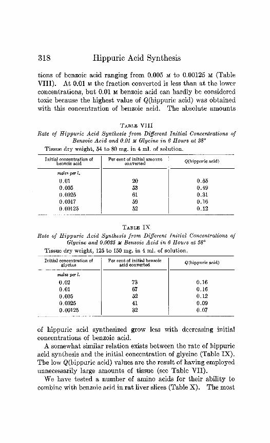
tions of benzoic acid ranging from 0.005 M to 0.00125 M (Table VIII). At 0.01 M the fraction converted is less than at the lower concentrations, but 0.01 M benzoic acid can hardly be considered toxic because the highest value of Q(hippuric acid) was obtained with this concentration of benzoic acid. The absolute amounts
TABLE VIII
Rate of Hippuric Acid Synthesis from Different Initial Concentrations of Benzoic Acid and 0.01 M Glycine in 6 Hours at 38’
Tissue dry weight, 54 to 80 mg. in 4 ml. of solution.
Initial concentration of bensoic acid
Per sent of initial amount converted Q(hippuric acid)
mole8 per 2. 0.01 0.005 0.0025 0.0017 0.00125
20 0.55 53 0.49 61 0.31 59 0.16 52 0.12
TABLE IX
Rate of Hippuric Acid Synthesis from Di$erent Initial Concentrations of Glycine and 0.00% M Benzoic Acid in 6 Hours at 38”
Tissue dry weight, 125 to 150 mg. in 4 ml. of solution.
Initial concentration of Per cent of initial bensoio glycine acid converted
moles per 1. 0.02 0.01 0.005 0.0025 0.00125
73 0.16 67 0.16 52 0.12 41 0.09 32 0.07
Q(hippuric acid)
of hippuric acid synthesized grow less with decreasing initial concentrations of benzoic acid.
A somewhat similar relation exists between the rate of hippuric acid synthesis and the initial concentration of glycine (Table IX). The low Q(hippuric acid) values are the result of having employed unnecessarily large amounts of tissue (see Table VII).
We have tested a number of amino acids for their ability to combine with benzoic acid in rat liver slices (Table X). The most

H. Borsook and J. IV. Dubnoff 319
rapid combination occurred with glycine. A large scale experi- ment was carried out with benzoic acid and glycine and the hip- puric acid formed was isolated. The yield on isolation was 75 per cent of that indicated by titration. It was proved to be hippuric acid by melting point determination, 187” (corrected), mixed melting point, and by electrometric titration. A 0.2805 ml. aliquot of a solution of 44.6 mg. in 25 ml. of water was neu- tralized by 74.1 ml.-3 of 0.0375 N NaOH; theoretical titer, 74.5 ml.-3.
TABLE X
Synthesis oj Hippuric Acid by Rat Liver Slices from Benzoic Acid and Dij- ferent Amino Acids in 6 Hours at 68” Compared with Amount Obtained
with Glycine in Simultaneous Experiments from Same Lobe of Liver
Benzoic acid initially 0.0025 M; amino acid 0.01 M.
Amino acid
d-Alanine .................. d-Arginine ................ l-Aspartic acid. 0 I-Asparagine. 0 d-Citrulline 0 1-Cysteine. 0 l-Cystine. 0 d-Glutamic acid. 0
‘I “ + glycine 80 Glycine..................... 100 Z-Histidine. 0 l-Leucine.. 0
PeroF amour synth6 iaed w glycin
0 0
?- ith e -.
Amino acid
d-Lysine dl-Methionine. d-Ornithine.. dl-Phenylalanine. I-Proline. . I-Hydroxyproline dl-Serine. d-Threonine. I-Tryptophane. . I-Tyrosine d-Valine
*
Perocfent amount synthe- ized with glycine
0 0 0 0 0
40 (?) 50 (?)
0 0 0 0
Of all the other amino acids only hydroxyproline and serine gave a positive titer. In the case of serine we are certain that the product formed is not hippuric acid. All that can be said at present is that under our experimental conditions a product is formed by the liver slices which is extracted by the ether and gives in our analytical procedure a significantly positive form01 titration.
We obtained some evidence that glutamic acid inhibited some- what the formation of hippuric acid both in the blank, i.e. benzoic acid without glycine added to liver slices, and also with glycine.
320 Hippuric Acid Synthesis
From the failure of all the amino acids we have tried (except glycine) to participate in t,hc formation of hippuric acid we may conclude that, at least under the conditions of these experiments, none of the other amino acids is converted to glycine in the liver. The glycine necessary for the hippuric acid which is formed when benzoic acid was added without glycine (it was about 5 per cent of the amount formed with glycine) can easily be ac- counted for by the autolysis of the liver slices in the course of the experiment (27). We may therefore have to look to other organs, or t,o materials other than amino acids for the synthesis of glycinc in the animal body. The possibility remains, of course, that glycine is formed in the liver from amino acids under conditions other than those which existed in the slices in these experiments.
We have investigated the possibility of the synthesis of hippuric acid from benzoic acid and glycine in minced tissues and tissue extracts. So far all the results have been negative. We have tried minced liver and aqueous and glycerol extracts of liver (rat), and glycerol extracts of the kidney (rat and horse).
Here, we have been unable to confirm the observations of Waelsch and Busztin (28). These authors reported that glycerol extracts of horse kidney effected a very rapid synthesis of benzoic acid and glycine to hippuric acid. In 6 hours at 37” more than 60 per cent of the benzoic acid was converted. The hippuric acid formed was isolated and identified. Depending on the con- ditions, there was also a varying amount of benzamide formed. These are the first observations to be reported of the synthesis of hippuric acid from bcnzoic acid and glycine in a tissue extract. They would indicate that in a glycerol extract of kidney the whole syst.cm, consisting of the enzyme acting upon the benzoic acid and glycine, the energy-donating enzyme and substrate, and the mechanism by means of which these two are coupled, is preserved intact. Furthermore the synthesis, according to Waelsch and Busztin, proceeds under what are practically anaerobic conditions. Synthesis through “mass action,” i.e. through reduction of the concentration of water by the glycerol, which is present in a concentration of 30 per cent, is excluded, because the vapor pres- sure of water in 30 per cent glycerol, and therefore the active con- centration of free water, is about 90 per cent of that in pure water (29).
II. Borsook and J. W. Dubnoff 321
Duplicating the conditions of their experiment as exactly as possible, we could find no trace of hippuric acid synthesis. The benxoic acid we added remained uncombined, and was recovered quantitatively. We were also unable to confirm the extraction procedure of Waelsch and Busztin. In their hands shaking six t,imes with ether (amount not specified) was sufficient to remove the benzoic and hippuric acids from the 30 per cent glycerol- water-protein mixture containing a final concentration of 2 N
HzS04. With this procedure we could recover added benzoic acid only incompletely and very little of added hippuric acid. We found it necessary to employ an exhaustive continuous ex- traction for 2 hours in order to extract these two acids completely from the glycerol-water mixture. We are at a loss to account for our inability to confirm the striking observations reported by Waelsch and Busztin.
It is pertinent in this connection that a glycerol extract of kidney-of the pig in most cases-has been used as a source of histozyme; i.e., of the enzyme hydrolyzing hippuric acid (15, 30-32). Thus Kimura (15) reported that glycerol extracts of pig kidney and liver completely hydrolyzed hippuric acid in 68 to 140 hours at 37”. In 24 hours the most potent glycerol ex- tracts effected 50 to 60 per cent hydrolysis.
We have found that synthesis of hippuric acid by liver slices is completely inhibited by 0.001 M KCN and by treatment with toluene. We may conclude therefore that not only intact cell structure but also cell respiration is essential for the synthesis. This is to be expected from the thermodynamic data.
We have found further that liver slices treated with cyanide did not hydrolyze hippuric acid when it was added to the Ringer’s solution. If the synthesis of hippuric acid were simply the re- verse of hydrolysis brought about by a shift of the equilibrium through coupling with an energy-donating reaction, it may be expected that when this coupling is broken by a respiratory poi- son hydrolysis of any hippuric acid present would ensue. This would have been analogous to the hydrolysis of protein in autol- ysis. The finding that there is no hydrolysis in liver slices poisoned with cyanide indicates that the synthesis of hippuric acid under the conditions we have employed, and probably also under those in viva, is not simply the reverse of hydrolysis.
322 Hippuric Acid Synthesis
Quick (33) made the interesting observations that when glycu- ronic acid monobenzoate was injected into the dog some hippuric acid appeared in the urine; and, conversely, after the injection of hippuric acid glycuronic acid monobenzoate appeared in the urine. Hippuric acid can therefore be both hydrolyzed and synthesized in the dog. This does not indicate that the conjugation of ben- zoic acid with glycine in the dog or elsewhere is a (‘reversible” reaction which obeys the law of mass action, in the same sense for example as an ionic reaction, in which the reaction can be moved to the “right” or ‘(left” simply by changing the concentrations on the “left” or “right.” The equilibrium is so far over on the side of hydrolysis of hippuric acid that no conceivable increase in benzoic acid and glycine could per se lead to a significant amount of hippuric acid being synthesized. This negative result was ob- served by Mutch (14). The hydrolysis and synthesis of hippuric acid in the dog is an example of a reaction which is physiologically but not physicochemically “reversible,” as reversibility is ordi- narily understood. The simplest explanation would appear to be that hippuric acid is synthesized in the kidney of the dog and hydrolyzed in other organs.
SUMMARY
1. Thermodynamic data are presented pertaining to the free energy of formation of hippuric acid and of peptides.
2. From these data the equilibrium constants at 25“ and 38” for the reaction hippuric acid F? benzoic acid and glycine are cal- culated.
3. The magnitude of the equilibrium constants so obtained in- dicates that the synthesis of hippuric acid in viva must be a coupled reaction of which one of the components is an energy- donating reaction. The synthesis cannot be simply the reverse of hydrolysis. This deduction is in accord with the experimental findings.
4. A micromethod is dcscribcd for determining 3 y of hippuric acid in 1 to 4 ml. of solution.
5. A technique is described by means of which it is possible to observe the synthesis of hippuric acid effected by slices of liver and kidney.
6. By this technique it was found that in the guinea pig, rab-
H. Borsook and J. W. Dubnoff
bit, and rat the conjugation of benzoic acid with glycine can occur in both the kidney and liver. In the dog it occurs in the kidney, but not in the liver.
7. In rat liver the reaction is relatively slow, one-tenth to one- hundredth that of the synthesis of urea from ammonia.
8. The influence of such factors as time, concentration of ben- zoic acid and of glycine, and of amount of tissue on the rate of synthesis with rat liver slices is described.
9. Of twenty-three amino acids and amides only glycine led to the formation of hippuric acid by rat liver slices.
10. This synthesis does not occur when the cell structure is destroyed by maceration or when the intact tissue is poisoned with toluenc or cyanide. Physically intact rat liver slices poi- soned with cyanide do not hydrolyze hippuric acid. These find- ings are shown to be in accord with deductions from the thermo- dynamic data.
11. An interpretation of the observed hydrolysis and synthesis of hippuric acid in the whole animal (dog) is presented in the light of the above thermodynamic and experimental data.
We are indebted to Dr. W. C. Rose for a generous donation of threonine and to Dr. C. L. A. Schmidt for a loan of amino acids. We are taking this opportunity of thanking them for their kind- ness.
BIBLIOGRAPHY
1. Warburg, O., The metabolism of tumours, translated by Dickens, F., London (1930).
2. Borsook, H., and Schott, H. F., J. Biol. Chem., 92,535 (1931). 3. Borsook, H., and Huffman, H. M., J. Biol. Chem., 99, 663 (1932-33). 4. Borsook, H., in Nord, F. F., and Weidenhagen, R., Ergebnisse der
Enzymforschung, Leipzig, 4, 1 (1935). 5. Borsook, H., and Huffman, H. M., in Schmidt, C. L. A., The chemistry
of the amino acids and proteins, Springfield and Baltimore, 822 (1938). 6. Rossini, F. D., BUT. Standards J. Research, 22, 407 (1939). 7. Seidell, A., Solubilities of inorganic and organic compounds, New
York, 2nd edition, 133 (1919). 8. Kolthoff, I. M., and Bosch, W., J. Physic. Chem., 36, 1685 (1932). 9. Kendall, J., Proc. Roy. Sot. London, Series A, 66, 200 (1911).
10. Brockman, F. G., and Kilpatrick, M., J. Am. Chem. Sot., 66,1483 (1934). II. Saxton, B., and Meier, H. F., J. Am. Chem. Sot., 66, 1918 (1934). 12. Josephson, B. A., Biochem. Z., 267, 74 (1933).
Rippuric Acid Synthesis
13. Ingersoll, C. L., and Burrows, G. H., J. Am. Chem. Xoc., 60, 136 (1938). 14. Mutch, N., J. Physiol., 44, 176 (1912). 15. Kimura, H., J. Biochem., Japan, 10, 207, 225 (192829). 16. Takahashi, M., J. Biochem., Japan, 10, 457 (1929). 17. Krebs, H. A., and Henseleit, K., Z. physiol. Chem., 210, 33 (1932). 18. Borsook, H., and Dubnoff, J. W., J. Biol. Chem., 131, 163 (1939). 19. van Bunge, G., and Schmiedeberg, O., Arch. exp. Path. u. Pharmakol.,
6, 233 (1876-77). 20. Snapper, I., Grtinbaum, A., and Neuberg, J., Biochem. Z., 146, 40
(1924). 21. Quick, A. J., J. Biol. Chem., 96, 73 (1932). 22. Salomon, W., 2. physiol. Chem., 3, 365 (1879). 23. Kanzaki, I., Chem. AM., 28, 2402 (1934). 24. Friedmann, E., and Tachau, H., Biochem. Z., 36, 88 (1911). 25. Sekine, M., 2. physiol. Chem., 164, 226 (1927). 26. Neber, M., 2. physiol. Chem., 234, 83 (1935). 27. Borsook, H., and Jeffreys, C. E. P., J. Biol. Chem., 110, 495 (1935). 28. Waelsch, H., and Busztin, A., 2. physiol. Chem., 249, 135 (1937). 29. Washburn, E. W., International critical tables of numerical data,
physics, chemistry, and technology, New York and London, 3, 291, 293 (1928).
30. So, T., J. Biochem., Japan, 12, 107 (1930). 31. Akizuki, H., J. Biochem., Japan, 26, 43 (1937). 32. Nawa, K., J. Biochem., Japan, 28, 237 (1938). 33. Quick, A. J., J. BioZ. Chem., 96, 189 (1932).