the atg12-conjugating enzyme atg10 is essential for ... · atg12 conjugation pathway specifically,...
TRANSCRIPT

Copyright � 2008 by the Genetics Society of AmericaDOI: 10.1534/genetics.107.086199
The ATG12-Conjugating Enzyme ATG10 Is Essential for AutophagicVesicle Formation in Arabidopsis thaliana
Allison R. Phillips,1 Anongpat Suttangkakul and Richard D. Vierstra2
Department of Genetics, University of Wisconsin, Madison, Wisconsin 53706
Manuscript received December 18, 2007Accepted for publication January 8, 2008
ABSTRACT
Autophagy is an important intracellular recycling system in eukaryotes that utilizes small vesicles to trafficcytosolic proteins and organelles to the vacuole for breakdown. Vesicle formation requires the conjugationof the two ubiquitin-fold polypeptides ATG8 and ATG12 to phosphatidylethanolamine and the ATG5protein, respectively. Using Arabidopsis thaliana mutants affecting the ATG5 target or the ATG7 E1 requiredto initiate ligation of both ATG8 and ATG12, we previously showed that the ATG8/12 conjugation pathwaystogether are important when plants encounter nutrient stress and during senescence. To characterize theATG12 conjugation pathway specifically, we characterized a null mutant eliminating the E2-conjugatingenzyme ATG10 that, similar to plants missing ATG5 or ATG7, cannot form the ATG12-ATG5 conjugate.atg10-1 plants are hypersensitive to nitrogen and carbon starvation and initiate senescence and pro-grammed cell death (PCD) more quickly than wild type, as indicated by elevated levels of senescence- andPCD-related mRNAs and proteins during carbon starvation. As detected with a GFP-ATG8a reporter, atg10-1and atg5-1 mutant plants fail to accumulate autophagic bodies inside the vacuole. These results indicate thatATG10 is essential for ATG12 conjugation and that the ATG12-ATG5 conjugate is necessary to formautophagic vesicles and for the timely progression of senescence and PCD in plants.
AS with other eukaryotes, plants have developedsophisticated mechanisms to recycle intracellular
proteins. Most selective protein turnover occurs by theubiquitin (Ub)/26S proteasome pathway, which directsthe correct removal of short-lived regulatory and abnor-mal proteins (Smalle and Vierstra 2004). Conversely,autophagy is a catabolic process that is largely respon-sible for nonselective bulk turnover of cytosolic compo-nents from individual proteins and protein complexesto the removal of whole organelles (Thompson andVierstra 2005; Bassham 2007). It involves the engulf-ment of cytoplasm in small vesicles followed by theirdeposition into the lytic vacuole (lysosome in animals)where the vesicles and cargo are quickly degraded by acache of vacuolar proteases, peptidases, lipases, andother hydrolytic enzymes.
Thus far, primarily using the yeasts Saccharomycescerevisiae and Pichia pastoris as models, at least two auto-phagic routes have been identified (for reviews seeOhsumi 2001; Thompsonand Vierstra 2005; Klionsky
2007). Microautophagy proceeds by forming tubularinvaginations of cytoplasm into the vacuole, which pinchoff and release vesicles called autophagic bodies into thevacuolar lumen. In contrast, macroautophagy involvesthe de novo formation of small double-membrane-bound
vesicles called autophagosomes within the cytoplasm,which sequester cytosolic constituents. These vesiclesdock with the vacuole, where the outer membrane fuseswith the tonoplast to release the inner compartmentinto the vacuolar lumen as an autophagic body. In addi-tion, a derivative of macroautophagy called the cytoplasm-to-vacuole targeting (CVT) pathway exists to encapsulateand deliver functional proteins such as preamino-peptidase to the vacuole (Klionsky 2007). While theCVT pathway has been confirmed only in S. cerevisiae, itis possible that a similar vacuolar transport pathway isactive in plants (Thompson and Vierstra 2005; Seay
et al. 2006). Both micro- and macroautophagy are essen-tial in yeast for maintaining nitrogen (N) and carbon(C) pools, recycling amino acids, removing unwanted ordamaged organelles, and survival during starvation. Ad-ditional roles in programmed cell death (PCD) and var-ious pathologies have been observed in animals (Bursch
2001; Levine and Klionsky 2004; Ueno et al. 2004;Juhasz et al. 2007).
Through genetic dissection of autophagy in yeastsover the past decade, several groups have discovereda set of autophagy (ATG) proteins common to bothmicro- and macroautophagy (Tsukada and Ohsumi
1993; Thumm et al. 1994; Harding et al. 1995). Inparticular, two Ub-like conjugation pathways were iden-tified as essential for proper formation of autophagicvesicles (Ohsumi 2001). These pathways employ twoUb-fold proteins, ATG8 and ATG12, as tags, whichthrough an ATP-dependent reaction cascade become
1Present address: Carnegie Institute for Plant Biology, 290 Panama St.,Stanford, CA 94305.
2Corresponding author: Department of Genetics, 425-G Henry Mall,University of Wisconsin, Madison, WI 53706. E-mail: [email protected]
Genetics 178: 1339–1353 (March 2008)

conjugated to their respective targets, the lipid phos-phatidylethanolamine (PE) and the ATG5 protein. Bothtags are first activated by the common E1-activating en-zyme ATG7, which couples ATP hydrolysis to the for-mation of ATG8-ATG7 and ATG12-ATG7 thioesterintermediates. Activated ATG8 and ATG12 are thendonated by transesterification to their respective conju-gating enzymes (or E2s), ATG3 and ATG10, which thenform covalent adducts with the targets via an amidebond between the C-terminal glycines of ATG8 andATG12 and the ethanolamine moiety of PE and a specificLys in ATG5, respectively (Mizushima et al. 1998;Ichimura et al. 2000). For ATG8, this Gly becomesexposed after processing of the initial translation prod-uct by the ATG4 protease that removes the amino acidsC-terminal to this residue (Kirisako et al. 2000).
In yeasts, the ATG8-PE conjugate binds to theautophagic membrane via the lipid moiety and appearsto help the membrane to expand during vesicle forma-tion (Kirisako et al. 1999). The ATG12-ATG5 conjugateassembles with ATG16 to form a hetero-octomeric struc-ture that is peripherally associated with the autophagicmembrane (Mizushima et al. 1999; Suzuki et al. 2001;Kuma et al. 2002). Assembly of this ATG12/5/16 proteincomplex appears to precede formation of ATG8-PE andmay enhance this lipidation reaction. Through theconcerted action of both conjugates and other ATGcomponents, an autophagic body is eventually depos-ited in the vacuole.
Orthologous autophagic systems have been identifiedin numerous eukaryotes, including Drosophila melano-gaster, Caenorhabditis elegans, mice, and humans, as well asseveral members of the plant kingdom (Thompson andVierstra 2005; Klionsky 2007). For example, genesencoding many ATG proteins have been detected inArabidopsis thaliana, rice (Oryza sativa), and maize (Zeamays), including most components of the ATG8 andATG12 conjugation pathways (Doelling et al. 2002;Hanaoka et al. 2002; Bassham 2007; A. Suttangkakul,T. Chung and R. D. Vierstra, unpublished results).Whereas the E1, ATG7, the E2s, ATG3 and ATG10, andthe ATG5 target are encoded by single genes inArabidopsis, the polypeptide tags ATG8 and ATG12are encoded by gene families containing nine and twomembers, respectively. This increased complexity cou-pled with our failure to detect obvious Arabidopsis or-thologs for other yeast ATG genes (e.g., ATG16 andseveral encoding components of the yeast ATG1/13 ki-nase complex) suggests that the plant autophagic sys-tem is not identical to that in yeasts and even may haveevolved new components and functions.
Reverse genetic analyses of Arabidopsis mutantsaffecting ATG5 and ATG7 recently revealed that plantsdefective in ATG8/ATG12 conjugation senesce earlierthan wild type and are also hypersensitive to N starvationand limiting light that depresses fixed C availability(referred to here as C limitation) (Doelling et al. 2002;
Thompson et al. 2005). Combined with analyses of otherautophagic proteins, such as ATG4a/4b, ATG6, ATG9,ATG18, and VTI12, it appears that autophagy is essentialfor appropriate C and N recycling in plants (Hanaoka
et al. 2002; Surpin et al. 2003; Yoshimoto et al. 2004;Xiong et al. 2005; Fujiki et al. 2007; Qin et al. 2007).Additionally, Liu et al. (2005) found from analysis of anatg6 mutant that autophagy helps to restrict PCDtriggered by the hypersensitive response (HR) close tothe site of pathogen invasion, although the relationshipbetween PCD and autophagy is unclear. Surprisingly, nogenetic connections to plant development have beenobserved despite the predicted need for autophagy inprocesses such as xylogenesis, sclereid, fiber and aeren-chyma maturation, organ abscission, anther dehiscence,and female gametogenesis and embryogenesis, all ofwhich likely involve the wholesale turnover of cellularconstituents by PCD mechanisms (Bursch et al. 2004;van Doorn and Woltering 2005).
To further describe the functions of the plant ATGsystem during autophagy and its potential involvementin PCD and to define the role(s) of the ATG12 con-jugation pathway more specifically, we initiated a reversegenetic analysis of Arabidopsis ATG10, the E2 predictedto be responsible for ATG12 conjugation. Key questionsincluded the following: Can ATG5 function in theabsence of ATG12 modification? Is ATG5 the sole targetof ATG12 and is ATG10 the sole E2? Is the ATG12 con-jugation pathway individually essential to form auto-phagic bodies decorated with ATG8? Does inactivationof ATG12 conjugation have the same phenotypic con-sequences as does inactivation of ATG8 conjugation?Here, we show that a T-DNA insertion allele disruptingthe ATG10 gene affects the normal response of seed-lings exposed to N- or C-limiting environments. Thesemutant plants cannot form the ATG12-ATG5 conjugateand fail to accumulate autophagic bodies inside thevacuole during nutrient starvation. The plants alsoappear to carry out senescence and PCD much morequickly than wild-type plants, as indicated by elevatedlevels of a collection of senescence- and PCD-relatedtranscripts and proteins under C-limiting conditions.These results indicate that ATG12 conjugation is essen-tial for the proper formation of autophagic vesicles andthat the defects in the ATG system upregulate PCDin addition to attenuating N and C recycling duringstarvation.
MATERIALS AND METHODS
Sequence analysis of ATG10 proteins: ATG10 protein se-quences were identified in the A. thaliana ecotype Columbia(Col-0) (http://www.Arabidopsis.org), rice (O. sativa) (http://www.tigr.org), poplar (Populus trichocarpa) (http://genome.jgi-psf.org/Poptr1_1/Poptr1_1.home), Physcomitrella (Physcomi-trella patens) (http://moss.nibb.ac.jp), Drosophila (D. melano-gaster), and mouse (Mus musculus) (http://www.ncbi.nlm.nih.gov) databases using the yeast ATG10 protein sequence as the
1340 A. R. Phillips, A. Suttangkakul and R. D. Vierstra
query (Ichimura et al. 2000). Intron/exon junctions in A. tha-liana ATG10 were determined by alignment with the full-length cDNA sequence from The Arabidopsis InformationResource (TAIR; http://www.Arabidopsis.org). Coding regionsfor the other plant ATG10 genes were deduced by comparisonto Arabidopsis ATG10 and alignments of genomic sequencesto those available for cDNAs. Amino acid sequence compar-isons were performed using CLUSTALX and MACBOXSHADE(Institute of Animal Health, Pirbright, UK). GenBank, TAIR,and The Institute for Genomic Research accession numbers forthe sequences described in this article are At3g07525 (AtATG10),Os04g41990 (OsATG10a), Os12g32210 (OsATG10b), eugene3.00141226 (PtATG10), YLL042C (ScATG10), FBpp0087919(DmATG10), and Q8R1P4 (MmATG10).
Isolation and complementation of atg10-1: The atg10-1 T-DNA insertion mutant (SALK_084434) was obtained from theSIGnAL T-DNA collection generated in the A. thaliana Col-0ecotype (Alonso et al. 2003). Homozygous mutant plants wereidentified by PCR using the 59- and 39-gene-specific primersATGGATTCAGCTCGAGAGGTCAGCG and ACAGGGATGTAGCTTGAACCATGGCCTGTT, respectively, in combinationwith the left border T-DNA-specific primer TGGTTCACGTAGTGGGCCATCG (Alonso et al. 2003), and by kanamycin re-sistance conferred by the T-DNA. The mutant was backcrossedthree times to wild-type Col-0 to help remove extraneousmutations.
For complementation, the full-length coding region of theATG10 cDNA was amplified by PCR using the primers GGGGACAAGTTTGTACAAAAAAGCAGGCTTCATGGATTCAGCTCGAGAGGTCA and GGGGACCACTTTGTACAAGAAAGCTGGGTTCTAATTCAGCATCTCAAGAGGG designed to in-troduce BP recombination sites at the 59- and 39-ends (under-lined), respectively, for subsequent cloning into the GatewaypDONR221 vector (Invitrogen, Carlsbad, CA). Using theprimer pair CTACATCCCTCTGGGACTGAGGACTG andCAGTCCTCAGTCCCAGAGGGATGTAG (altered nucleotidesunderlined), the active-site Cys178 codon was changed to thatfor serine by the Quickchange method (Stratagene, La Jolla,CA). The ATG10 and ATG10C-S coding regions were trans-ferred to the Gateway pEARLEY201 vector (Earley et al. 2006)by an LR recombination reaction to append the cauliflowermosaic virus (CaMV) 35S promoter and codons for a HAepitope tag to the 59-end.
The resulting 35S:ATG10 and 35S:ATG10C-S transgeneswere introduced into Agrobacterium tumefaciens strain GV3101and then transformed into homozygous atg10-1 plants by thefloral dip method (Clough and Bent 1998). T2 plants homo-zygous for the atg10-1 mutation were confirmed to contain thetransgenes by PCR using primers TGACGTAAGGGATGACGCACAAT and ACTAGTCCCGGGTCTTAATTAACTCTC. PCRproducts from 35S:ATG10C-S plants were sequenced to con-firm the Cys178-Ser mutation. Transgene expression was dem-onstrated by reverse transcription–PCR (RT–PCR) analysisusing 28 amplification cycles with Ex-Taq polymerase (TaKaRa,Madison, WI) and the 59- and 39-ATG10 gene-specific primersATGGATTCAGCTCGAGAGGTCAGCGAT and CAGTCCTCAGTCCCACAGGGATGTAG. The ATG8e 59- and 39-gene-spe-cific primers, GCATCTTTAAGATGGACGACGATTTCGAAand ATGTGTTCTCGCCACTGTAAGTGATGTAA, were usedas an internal RT–PCR control. Because the 59-ATG8e primerspans an intron, ATG8e genomic DNA is not amplified by thisprimer set.
Plant growth conditions: Arabidopsis seeds were vapor-phase sterilized (Clough and Bent 1998), incubated in waterat 4� for 2 days, and germinated on solid Gamborg’s B5 (Sigma,St. Louis) medium containing 0.7% agar or in liquid growthmedium (GM; Sigma) containing 2% sucrose. The plates andliquid cultures were incubated at 21� in a 16-hr light/8-hr dark
photoperiod for long day (LD; fluence rate ¼ 95 mmol m�2
sec�1), an 8-hr light/16-hr dark photoperiod for short day (SD;fluence rate ¼ 95 mmol m�2 sec�1), or in continuous light(fluence rate ¼ 65 mmol m�2 sec�1).
For exposure to N-starvation conditions, 1-week-old seed-lings grown in the LD were transferred to N-deficient liquid orsolid media containing Murashige and Skoog micronutrientsalts (Sigma), 3 mm CaCl2, 1.5 mm MgSO4, 1.25 mm KH2PO4,5 mm KCl, and 2 mm 2-(N-morpholino)ethanesulfonic acid(pH 5.7). After various amounts of time on the N-deficientsolid medium, seedlings were transferred back to GM agar. Forexposure to C-limiting conditions, seedlings grown in solidGM for 3 weeks in SD were transferred to soil and grown for 3more weeks. The plants were then transferred to continuousdarkness for various lengths of time and either collected imme-diately or returned to SD for a 1-week recovery. For confocalmicroscopy, seeds were germinated in liquid GM. After 1 week,the seedlings were transferred to N-deficient medium for 2 days.Twelve to 16 hr prior to examination by fluorescence confocalmicroscopy, concanamycin A (Sigma) was added to the mediumto a final concentration of 0.5 mm. Plants stably expressing35S:GFP-ATG8a in the wild-type and atg7-1 backgrounds wereas described in Thompson et al. (2005). 35S:GFP-ATG8a wasintroduced into the atg10-1 and atg5-1 mutant backgrounds bycrossing. Homozygous atg10-1 and atg5-1 seedlings expressingthe GFP-ATG8a transgene were identified by Basta resistanceand verified by fluorescence microscopy and PCR.
DNA/RNA gel-blot analyses: Total genomic DNA wasisolated from 1 g of leaf tissue as described (Balk and Leaver
2001). Twenty micrograms of DNA per sample was subjected togel electrophoresis using 1.5% agar, the DNA was stained withethidium bromide and then transferred to Hybond XLmembrane (GE Healthcare, Piscataway, NJ) for DNA gel-blotanalysis. The 32P-labeled 18S rRNA riboprobe was synthesizedwith SP6 RNA polymerase using a linearized pGEMT (Prom-ega, Madison, WI) cDNA construction and the Riboprobe sys-tem (Promega). Membranes were hybridized overnight at68� and washed as described (Smalle et al. 2002) prior toautoradiography.
RNA was isolated from liquid-grown and soil-grown plantsusing the Trizol reagent (Invitrogen). RNA for RT–PCR wastreated with DNase RQI (Promega) prior to the synthesis offirst-strand cDNA by Superscript II-reverse transcriptase (In-vitrogen). The first-strand synthesis primers were the ATG10gene-specific primers AAGCCACTCATATGTTAATGAAACTCAAGTT and AGAGATTCATCCTCTGGAATTTCCTC (pri-mers 2 and 3, respectively; Figure 2B) or the H2A 39 gene-specific primer GCAACTTGCTTAGCTCCTCATCATTCCTC(control; Figure 2B). RT–PCR included 35 cycles with Ex-Taqpolymerase, the first-strand synthesis primer, and either theATG10 59 gene-specific primer pair ATGGATTCAGCTCGAGAGGTCAGCGAT and TAGTTTACAGTGCATCATACAAGGTTCCTG (primers 1 and 4, respectively; Figure 2B).
For RNA gel-blot analysis, total RNA was isolated accordingto Smalle et al. (2002). 32P-labeled riboprobes were synthe-sized with T7, SP6, or T3 RNA polymerase using the Riboprobesystem (Promega) and the linearized pGEMT (Promega) orpBluescript (Stratagene) cDNA constructions for ATG8e,ATG12a, ATG12b, SAG12, PED1, GPX2, CSD1, CAT3, NYE1,TUB4, and 18S rRNA. The CAB, SEN1, and ATG8a probes werefrom Doelling et al. (2002). Membranes were hybridizedovernight at 68� and washed as described (Smalle et al. 2002)prior to autoradiography.
Protein isolation and immunoblot analysis: Total proteinwas isolated from liquid- or soil-grown plants by homogeniza-tion in 2:1 (volume to gram fresh weight) SDS–PAGE samplebuffer ½125 mm Tris–HCl (pH 6.8), 5% SDS, 20% glycerol, and10% 2-mercaptoethanol� and extracts were clarified by centri-
Autophagic Recycling in Arabidopsis 1341

fugation at 10,000 3 g. Proteins were subjected to SDS–PAGEin 12–16% acrylamide gels with or without 6 m urea in theseparating gel and either stained with silver or electrophoret-ically transferred onto PVDF membranes (Millipore, Bedford,MA) for immunoblot analysis using alkaline phosphatase-labeled or peroxidase-labeled goat anti-mouse or goat anti-rabbit immunoglobulins (Kirkegaard & Perry Laboratories,Gaithersburg, MD) for detection. Sample sizes were adjustedto reflect either equal protein or equal fresh weight as indi-cated. Changes in total protein content during C starvationwere measured by spotting the SDS-containing crude extractsdirectly on PVDF membranes, staining the membranes withPonceau S, and quantifying the amount of protein densito-metrically using bovine serum albumin as the standard.
Antibodies against Arabidopsis ATG3 were produced inrabbits (Harlan Polyclonal Antibody Service, Madison, WI)using recombinant protein expressed with N-terminal His6and maltose-binding protein (MBP) tags. The full-lengthATG3 coding region was inserted into the Gateway pDONR221vector (Invitrogen), transferred to pVP13 (Center for Eukary-otic Structural Genomics; http://www.uwstructuralgenomics.org) by an LR reaction, and introduced into Escherichia coliBL21 Codon Plus cells (Novagen, Madison, WI). Following a3-hr induction of log-phase cultures by the addition of 1 mm
isopropyl-b-d-thiogalactoside, soluble His6-MBP-ATG3 waspurified by NiNTA chromatography (QIAGEN Sciences,Germantown, MD). The His6 and MBP tags were removedby tobacco etch virus protease (Invitrogen) cleavage and thedigested protein was further purified by SDS–PAGE. Gel frag-ments were injected directly into rabbits. The anti-vacuolarprocessing enzyme (anti-VPEg) and anti-SAG2 antibodieswere as described (Grbic 2003; Rojo et al. 2003). The anti-PBA1, -ATG7, -ATG5, and -ATG8a antibodies were fromDoelling et al. (2002), Smalle et al. (2002), and Thompson
et al. (2005). Anti-H3A antibodies were supplied by Abcam(Cambridge, MA). Antibodies against the large subunit ofspinach RUBISCO were provided by Archie Portis (Universityof Illinois, Champagne, IL).
Leaf staining and fluorescence confocal microscopy: Lac-tophenol blue was used to discriminate between live and deadcells according to Rate et al. (1999). The seventh leaf fromindividual plants was harvested at various times during thedark treatment and immediately boiled in a lactophenol bluesolution ½10 ml lactic acid, 10 ml glycerol, 10 ml liquid phenol,10 ml water, and 10 mg trypan blue (Sigma) for 1 min�, clearedin alcoholic lactophenol (2:1 95% ethanol:lactophenol) for2 min, and incubated in 50% ethanol for 1 day. Leaves werewashed in water before imaging on a Leica MZFLIII micro-scope equipped with an Optronics digital camera.
Fluorescence confocal microscopy of hypocotyl cells ex-pressing either free GFP or GFP-ATG8a was conducted with aZeiss 510-Meta scanning laser confocal microscope using a488-nm light excitation (Thompson et al. 2005). Fluorescencewas monitored 12–16 hr after treatment with 0.5 mm con-canamycin A (Wako Chemicals, Richmond VA) using theBP505–530 (excitation 488 nm, emission 505–530 nm) filterset. Images were processed with LSM510 software and NationalInstitutes of Health ImageJ (http://rsb.info.nih.gov/ij/). Thedensity of fluorescent vesicles within the vacuoles of eachgenetic background was determined by counting their num-ber within a 20- 3 20-mm2 section of the central vacuole fromrepresentative cells. The data for wild type represent the av-erage number in a 100-mm2 area (6SE) from three indepen-dent experiments that each analyzed images captured from 9to 36 different cells. The data for the atg7-1, atg5-1, and atg10-1lines represent the average number in a 100-mm2 area (6SD)from one experiment that analyzed images captured from 12to 30 different cells.
RESULTS
Isolation of a mutant affecting ATG10: To more spe-cifically define the functions of ATG12, we searched formutants affecting the cognate E2 ATG10 (Ichimura
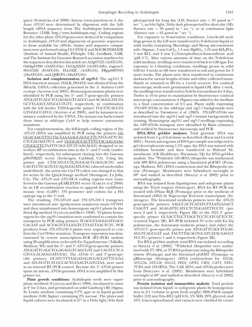
et al. 2000). Genomic database searches by BLASTP iden-tified single ATG10 genes in Arabidopsis (ecotype Col-0;At3g07525) and poplar (P. trichocarpa; eugene3.00141226), and two ATG10 genes in rice (OsATG10a,Os04g41990; OsATG10b, Os12g32210) (Figure 1). Wealso detected several genomic fragments predicted toencode ATG10 in the moss P. patens genome, whichlikely were derived from the same locus. By analysis ofgenomic and full-length cDNA sequences, the Arabi-dopsis ATG10 gene was determined to encode a 225-amino-acid protein with 49 and 61% similarity to its riceand poplar orthologs, respectively. In contrast, the Arabi-dopsis protein shares only 22, 29, and 35% similaritywith its nonplant counterparts in S. cerevisiae, Drosoph-ila, and mice (Figure 1). However, several regions withstrong amino acid conservation are apparent among thegroup, including a block bracketing the presumedactive-site cysteine (residue 178 in AtATG10) that formsthe thioester intermediate with ATG12 prior to itstransfer to ATG5 (Figure 1).
In a screen of the available Arabidopsis T-DNA inser-tion populations prepared with the Col-0 background,we identified a mutant allele of ATG10 designatedatg10-1 in the SIGnAL collection (Figure 2A; Alonso
et al. 2003). The mutant was backcrossed three times tothe wild-type Col-0 ecotype to eliminate possible extra-neous secondary mutations, using kanamycin resistanceassociated with the T-DNA to track the mutation andthen self-fertilized to generate homozygous individuals.Genomic PCR of atg10-1 plants with 59 and 39 gene-specific primer pairs alone or in combination with theT-DNA left border primer confirmed disruption of thewild-type ATG10 gene and the presence of the intro-duced T-DNA (see Figure 7A below). Sequencing theregion flanking the T-DNA revealed that it was insertedas a tandem duplication in the fourth exon and simul-taneously created a 28-bp deletion in the ATG10 codingregion. RT–PCR analysis of homozygous atg10-1 seed-lings failed to amplify the full-length ATG10 mRNA(primers 1 and 2) (Figure 2B). Although a slight amountof RT–PCR product encoding the region upstream of theT-DNA was generated from atg10-1 transcripts (primers1 and 3), amplification of the downstream region was notdetected (primers 2 and 4) (Figure 2B). Given that themissing downstream sequence encodes Cys178, it ishighly likely that atg10-1 is a functionally null allele.
To demonstrate that ATG10 is the sole E2 thatassembles the ATG12-ATG5 conjugate, we performedimmunoblot analysis on crude extracts from homozy-gous atg10-1 seedlings using antibodies against ATG5(Thompson et al. 2005). As shown in Figure 2C, the50-kDa presumed ATG12-ATG5 conjugate (solid arrow-
1342 A. R. Phillips, A. Suttangkakul and R. D. Vierstra
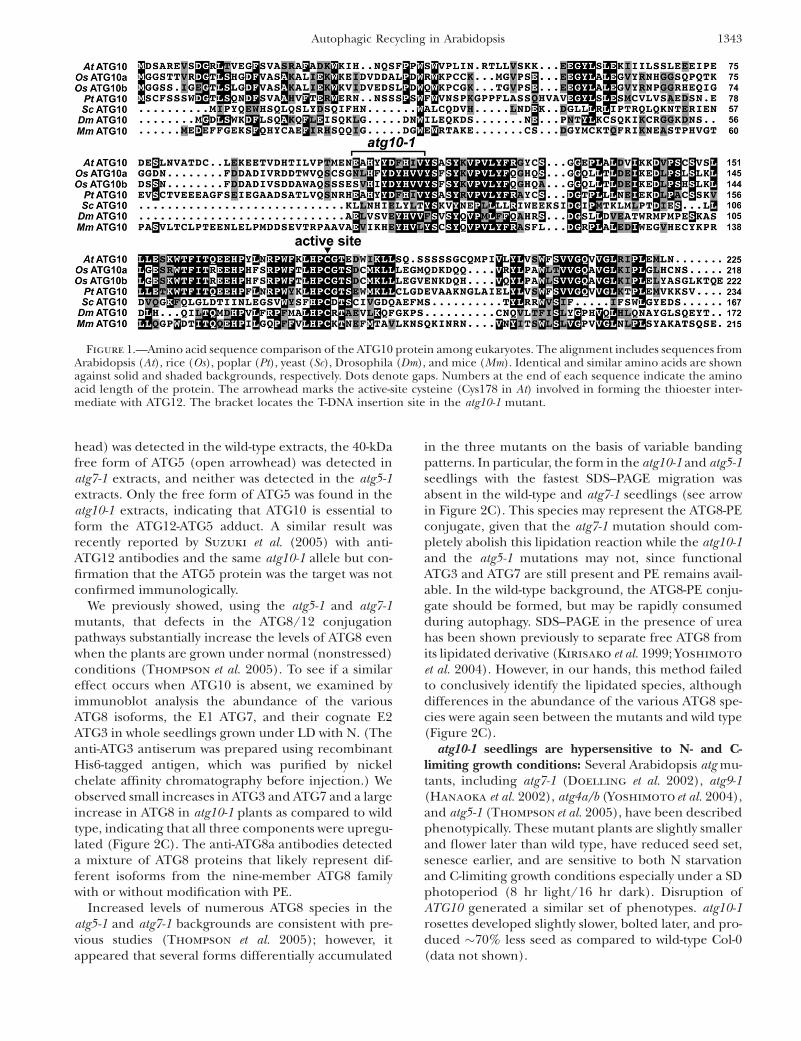
head) was detected in the wild-type extracts, the 40-kDafree form of ATG5 (open arrowhead) was detected inatg7-1 extracts, and neither was detected in the atg5-1extracts. Only the free form of ATG5 was found in theatg10-1 extracts, indicating that ATG10 is essential toform the ATG12-ATG5 adduct. A similar result wasrecently reported by Suzuki et al. (2005) with anti-ATG12 antibodies and the same atg10-1 allele but con-firmation that the ATG5 protein was the target was notconfirmed immunologically.
We previously showed, using the atg5-1 and atg7-1mutants, that defects in the ATG8/12 conjugationpathways substantially increase the levels of ATG8 evenwhen the plants are grown under normal (nonstressed)conditions (Thompson et al. 2005). To see if a similareffect occurs when ATG10 is absent, we examined byimmunoblot analysis the abundance of the variousATG8 isoforms, the E1 ATG7, and their cognate E2ATG3 in whole seedlings grown under LD with N. (Theanti-ATG3 antiserum was prepared using recombinantHis6-tagged antigen, which was purified by nickelchelate affinity chromatography before injection.) Weobserved small increases in ATG3 and ATG7 and a largeincrease in ATG8 in atg10-1 plants as compared to wildtype, indicating that all three components were upregu-lated (Figure 2C). The anti-ATG8a antibodies detecteda mixture of ATG8 proteins that likely represent dif-ferent isoforms from the nine-member ATG8 familywith or without modification with PE.
Increased levels of numerous ATG8 species in theatg5-1 and atg7-1 backgrounds are consistent with pre-vious studies (Thompson et al. 2005); however, itappeared that several forms differentially accumulated
in the three mutants on the basis of variable bandingpatterns. In particular, the form in the atg10-1 and atg5-1seedlings with the fastest SDS–PAGE migration wasabsent in the wild-type and atg7-1 seedlings (see arrowin Figure 2C). This species may represent the ATG8-PEconjugate, given that the atg7-1 mutation should com-pletely abolish this lipidation reaction while the atg10-1and the atg5-1 mutations may not, since functionalATG3 and ATG7 are still present and PE remains avail-able. In the wild-type background, the ATG8-PE conju-gate should be formed, but may be rapidly consumedduring autophagy. SDS–PAGE in the presence of ureahas been shown previously to separate free ATG8 fromits lipidated derivative (Kirisako et al. 1999; Yoshimoto
et al. 2004). However, in our hands, this method failedto conclusively identify the lipidated species, althoughdifferences in the abundance of the various ATG8 spe-cies were again seen between the mutants and wild type(Figure 2C).
atg10-1 seedlings are hypersensitive to N- and C-limiting growth conditions: Several Arabidopsis atg mu-tants, including atg7-1 (Doelling et al. 2002), atg9-1(Hanaoka et al. 2002), atg4a/b (Yoshimoto et al. 2004),and atg5-1 (Thompson et al. 2005), have been describedphenotypically. These mutant plants are slightly smallerand flower later than wild type, have reduced seed set,senesce earlier, and are sensitive to both N starvationand C-limiting growth conditions especially under a SDphotoperiod (8 hr light/16 hr dark). Disruption ofATG10 generated a similar set of phenotypes. atg10-1rosettes developed slightly slower, bolted later, and pro-duced �70% less seed as compared to wild-type Col-0(data not shown).
Figure 1.—Amino acid sequence comparison of the ATG10 protein among eukaryotes. The alignment includes sequences fromArabidopsis (At), rice (Os), poplar (Pt), yeast (Sc), Drosophila (Dm), and mice (Mm). Identical and similar amino acids are shownagainst solid and shaded backgrounds, respectively. Dots denote gaps. Numbers at the end of each sequence indicate the aminoacid length of the protein. The arrowhead marks the active-site cysteine (Cys178 in At) involved in forming the thioester inter-mediate with ATG12. The bracket locates the T-DNA insertion site in the atg10-1 mutant.
Autophagic Recycling in Arabidopsis 1343
As with other autophagy mutants, atg10-1 seedlingswere hypersensitive to N-deficient growth conditions.Wild-type, atg5-1, atg7-1, and atg10-1 seedlings weregrown under a LD photoperiod (16 hr light/8 hr dark)or continuous light in medium containing sucrose andN for 1 week and then were transferred to N-deficientmedium for increasing lengths of time. As shown inFigure 3, A and B, 2 weeks of such N starvation slowedleaf emergence and expansion and enhanced cotyledonchlorosis of atg10-1 seedlings similar to that previouslydescribed for atg7-1 and atg5-1 (Doelling et al. 2002;Thompsonet al. 2005). When the plants were exposed tovarious durations of N starvation and then returned toN-rich medium, the three homozygous mutant popula-tions had dramatically impaired recovery (Figure 3C).Whereas nearly all of the wild-type plants resumedgrowth after 17 days on N-deficient medium, �50% ofatg10-1 and 60% of atg5-1 and atg7-1 plants failed torecover. After 45 days on N-deficient medium, almost allof the mutant seedlings died, while .40% of wild-typeplants resumed growth.
To test the effects of C limitation, plants were grown inSD for 6 weeks to maintain a low level of fixed C,transferred to the dark for various lengths of time, and
then allowed to recover in SD for 1 week. As shown inFigure 3, D and E, the atg10-1 homozygous mutants werehypersensitive to C limitation. Similar to atg5-1 and atg7-1, atg10-1 plants were more chlorotic and more wiltedthan wild type right after the extended dark treatmentsand showed poor recovery after transfer back to thelight. The survival profiles for all three mutants weresimilar. Whereas a majority of the wild-type plantssurvived up to 8 days of darkness, most of the mutantplants died following only 4 days (Figure 3, D and E).
Molecular defects of atg10-1 seedlings under C-limiting conditions: To examine the effects of Climitation at the molecular level, we collected plantsimmediately after various days of dark treatment andassessed the levels of several autophagy and senescence-related proteins and transcripts. Both wild-type andatg10-1 seedlings progressively lost total protein duringextended darkness, with�75% of the total remaining ineach after 8 days of dark treatment (Figure 4A).However, examination of the protein profiles by SDS–PAGE revealed differences, with the atg10-1 plantslosing high-molecular-mass polypeptides and specificlower-molecular-mass species more rapidly than wildtype as the dark treatment continued (Figure 4B).Notable were the more rapid declines of the large andsmall subunits of RUBISCO (�50 and 13 kDa), furtherindicative of reduced photosynthetic capacity. By im-munoblot analyses, we also confirmed an acceleratedloss of the large subunit of RUBISCO and VPEg in theatg10-1 background (Figure 4C), the latter of which isenriched in the lytic vacuoles of senescing vegetativeorgans (Kinoshita et al. 1999). By comparison, histone3A (H3A) disappeared at similar rates in wild-type andatg10-1 seedlings (Figure 4C).
Not all proteins decreased in abundance duringextended darkness. For example, the level of theSENESCENCE-ASSOCIATED GENE (SAG)-2 cysteineprotease (Grbic 2003) increased in the wild-typebackground as the plants remained in the dark forlonger periods of time, implying that wild-type plantsactivated their senescence program (Greenberg 1996;Pennell and Lamb 1997; Figure 4C). In the atg10-1seedlings, SAG2 levels were high even before darktreatment and remained high during prolonged dark-ness, suggesting that inactivation of autophagy consti-tutively induces the senescence program. Likewise,levels of various ATG8 isoforms were constitutivelyupregulated in the atg10-1 seedlings as compared towild type (Figure 4C). These high levels were retainedthroughout the extended darkness, similar to thatobserved for the atg5-1 and atg7-1 mutants (Thompson
et al. 2005; A. R. Phillips, unpublished data). Levels ofATG3, ATG5, and the 26S proteasome subunit PBA1also remained high during the dark treatments in boththe wild-type and atg10-1 plants, which likely reflects anattempt to maintain protein recycling systems during Climitation (Figure 4C). For ATG5, its ATG12 conjugate
Figure 2.—Description of the Arabidopsis atg10-1 mutant.(A) Diagram of the Arabidopsis ATG10 gene. Lines indicateintrons and boxes indicate exons with coding regions shadedand 59 and 39-UTRs as open boxes. The arrowhead locates theactive-site cysteine (Cys178). The position of the T-DNA in theatg10-1 mutant is shown. Half-arrows locate the primer bind-ing sites used in B. (B) RT–PCR analysis of the atg10-1 mutant.Total RNA isolated from wild-type (WT) and atg10-1 seedlingswas subjected to RT–PCR using the 1 1 2, 1 1 3, or 2 1 4primer pairs. PCR amplification of genomic DNA (gDNA)and RT–PCR using a primer pair specific for the histoneH2A gene were included as controls. (C) Immunoblot detec-tion of the ATG12-ATG5 conjugate. Crude protein extractsfrom 10-day-old wild-type, atg10-1, atg5-1, and atg7-1 seedlingswere subjected to SDS–PAGE with or without 6 m urea andimmunoblot analysis with anti-ATG5, anti-ATG7, anti-ATG3,and anti-ATG8 antibodies. Equal protein loads were con-firmed by immunoblot analysis with anti-PBA1 antibodies.Solid and open arrowheads identify the ATG12-ATG5 conju-gate (50 kDa) and free ATG5 (40 kDa), respectively. Arrowidentifies possible ATG8-PE conjugates.
1344 A. R. Phillips, A. Suttangkakul and R. D. Vierstra

was retained in wild type, while the free form wasretained in the atg10-1 background. The ATG7 proteinremained high in wild type but decreased in atg10-1seedlings during the dark treatment. (We note that anincrease in the ATG7 level at day 4 is apparent in Figure4C for the experiment involving atg10-1 plants, but thiseffect was not seen in other trials.)
We then exploited RNA gel-blot analyses to investi-gate changes at the transcript level using 18S rRNA as amarker for equal RNA loading (Figure 5). Similar to pre-vious studies (Weaver and Amasino 2001; Doelling
et al. 2002; Thompson et al. 2005), chlorophyll a/b-binding protein (CAB) mRNA levels dropped rapidlyfollowing incubation in the dark (between 0 and 2 days)in both wild-type and atg10-1 plants, consistent with theinstability of the CAB mRNA in the dark and its light-induced transcription (Figure 5; Giuliano et al. 1988).Levels of b4-tubulin (TUB4) mRNA also decreased inboth backgrounds, which is similar to the reporteddecrease of b9-tubulin mRNA levels during senescence(Swidzinski et al. 2002). The abundance of severalATG8 transcripts were previously shown to increase inresponse to limited nutrient levels (Contento et al.2004; Thompsonet al. 2005; Rose et al. 2006; Osuna et al.2007). Here, we found that mRNA levels for two
different isoforms, ATG8a and ATG8e, were even moreincreased by darkness in the atg10-1 mutant plants. Bycontrast, abundance of the two ATG12 transcripts ap-pears to be differentially regulated by darkness. Whereasthe ATG12a mRNA dropped soon after the dark treat-ment (days 2–4), the ATG12b mRNA slowly increased inabundance over the course of prolonged darkness inboth the wild-type and atg10-1 backgrounds (Figure 5).
The abundance of several senescence- and PCD-related transcripts were also consistently increased inatg10-1 seedlings as compared to wild type (Figure 5).These included SENESCENCE (SEN)-1 and SAG12genes, both markers for dark-induced senescence; thePEROXOSOME DEFECTIVE (PED)-1, which encodes athiolase involved in fatty acid b-oxidation during ger-mination and senescence; and glutathione peroxidase2 (GPX2), which responds to oxidative stress (Oh
et al. 1996; Hayashi et al. 1998; Weaver et al. 1998;Mullineaux et al. 2000; Swidzinski et al. 2002; van der
Graaff et al. 2006). However, not all senescence/PCDmRNAs were affected by the atg10-1 mutation. Tran-scripts from the Cu/Zn superoxide dismutase 1 (CSD1)and the CAT3 catalase genes, which have previouslybeen associated with senescence and oxidative stress(Swidzinski et al. 2002; Contento et al. 2004), were not
Figure 3.—Enhanced sensitiv-ity of atg10-1 plants to N- and C-lim-iting conditions. Lines includewild-type Col-0 (WT) and homozy-gous atg5-1, atg7-1, and atg10-1 mu-tants. (A and B) Representativeplants grown for 1 week on N-richsolid (A) or liquid (B) media andtransferred to N-rich (1N) or N-deficient (�N) media for 2 weeks.(C) Survival on N-deficient me-dium. One-week-old seedlingswere sown on N-rich solid mediumand transferred to N-deficient me-dium for various lengths of timebefore transfer back to N-rich me-dium, all under SD. The graphplots the percentage of plants thatresumed growth after exposure toN-deficient medium. Each pointrepresents the analysis of 45 seed-lings. (D and E) Survival underC-limiting growth conditions in-duced by extended darkness. Six-week-old plants were grown underSD, transferred to darkness for var-ious lengths of time, and thentransferred back to SD. (D) Repre-sentative plants after 1-week recov-ery from 0-, 2-, 4-, 6-, 8-, or 10-daydark treatments. (E) Percentageof plants that survived increasingdays in the dark as determined byresumption of growth after 1 weekin SD. Each point represents theanalysis of 15 seedlings.
Autophagic Recycling in Arabidopsis 1345

upregulated in the atg10-1 background. Whereas theCSD1 mRNA decreased soon after dark treatment inboth backgrounds, the CAT3 mRNA first increased atthe beginning of darkness and then decreased, with thedrop even more rapid in the atg10-1 background. ½Itshould be noted that CAT3 expression is coordinatelyregulated by the circadian clock, the rhythm of whichmay cease after several days in the dark (Zhang et al.2007).� These results, combined with previous studies(Contento et al. 2004; Liu et al. 2005; Thompson et al.2005; Xiong et al. 2007), suggest that autophagy isconnected with some, but not all, aspects of the senes-cence and PCD programs.
Figure 4.—Protein profile of atg10-1 plants exposed to C-limiting conditions induced by extended darkness. Tissue wascollected from wild-type (wt) and atg10-1 seedlings just afterthe indicated days of extended darkness (see Figure 3). (A)Quantification of total protein from wild-type and atg10-1 ex-tracts. (B) Profile of total protein separated by SDS–PAGE andstained with silver. (C) Immunoblot analysis with antibodiesagainst ATG8, ATG5, ATG7, ATG3, the large subunit of RU-BISCO (RBC), SAG2, VPEg, H3A, and the b1-subunit ofthe 26S proteasome (PBA1). Solid and open arrowheads iden-tify the ATG12-ATG5 conjugate (50 kDa) and free ATG5 (40kDa), respectively. Equivalent amounts of tissue fresh weightwere analyzed in each lane.
Figure 5.—RNA profile of atg10-1 plants exposed to C-lim-iting conditions induced by extended darkness. Tissue wascollected from wild-type (WT) and atg10-1 seedlings just afterthe indicated days of extended darkness (see Figure 3). Equalamounts of total RNA (10 mg) were subjected to gel-blot anal-ysis using probes for ATG8a, ATG8e, CAB, SEN1, SAG12, PED1,GPX2, CSD1, CAT3, NYE1, and TUB4. Near equal loading oftotal RNA was confirmed by RNA gel-blot analysis of 18S rRNA(18S) and staining for total rRNA with methylene blue (datanot shown).
1346 A. R. Phillips, A. Suttangkakul and R. D. Vierstra
Presumably, the more rapid chlorosis of plantsmissing ATG7, ATG5, and ATG10 (Doelling et al.2002; Thompson et al. 2005; this report) in the dark iscaused, in part, by increased chlorophyll degradation.This breakdown requires pheophorbide a oxygenase,which is regulated in turn by the NON-YELLOWING(NYE)-1 nuclear-encoded chloroplast protein (Ren
et al. 2007). Levels of NYE1 positively correlate withchlorophyll turnover, with the abundance of the NYE1transcript increasing markedly during senescence, sug-gesting that the NYE1 protein is a major regulator ofchlorophyll turnover. Here, we found that the amount ofthe NYE1 mRNA is dramatically affected by the atg10-1mutation (Figure 5). Whereas the NYE1 transcript wasbarely detectable in wild-type seedlings even after pro-longed darkness, it rose substantially following extendeddarkness in the atg10-1 seedlings, coinciding with in-creased chlorosis of the leaves.
Disruptions of ATG10, ATG5, and ATG7 enhanceprogrammed cell death: As shown in Figures 3–5, thesurvival of atg10-1 seedlings is severely compromisedwhen exposed to extended darkness and many senes-cence and PCD-associated factors are upregulated overthe course of the treatment. It is also likely that theseplants are impaired in normal cell death pathways thatinvolve autophagy (autophagic PCD: Bursch et al. 2004;
van Doorn and Woltering 2005) due to their inabilityto conjugate ATG12 to ATG5. To further investigate howatg mutants undergo PCD in the absence of ATG-dependent autophagy, we tested for the involvement ofother PCD types, such as apoptosis (thought not to occurin plants), nonlysosomal PCD, and necrosis, which maywork exclusively or together (mixed-type PCD: Bursch
et al. 2004; van Doorn and Woltering 2005).Necrosis was observed by staining wild-type, atg7-1,
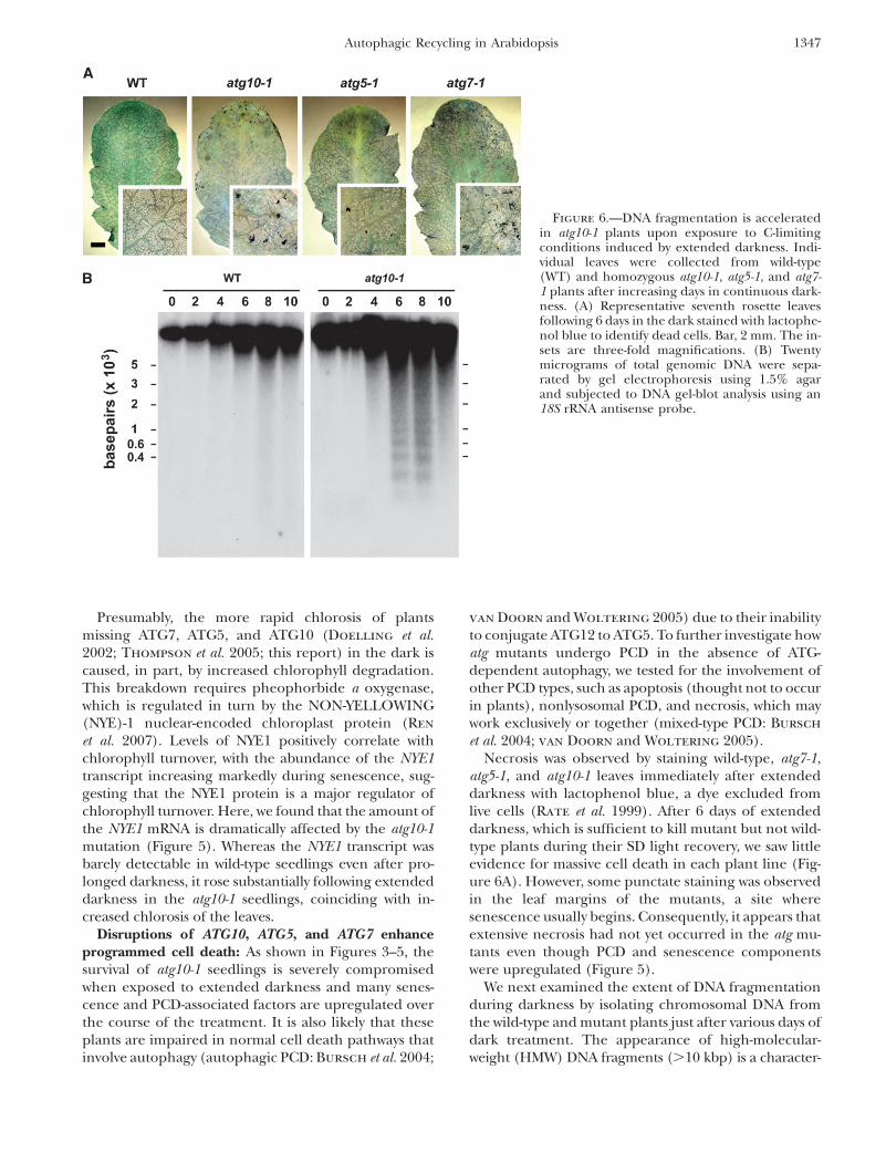
atg5-1, and atg10-1 leaves immediately after extendeddarkness with lactophenol blue, a dye excluded fromlive cells (Rate et al. 1999). After 6 days of extendeddarkness, which is sufficient to kill mutant but not wild-type plants during their SD light recovery, we saw littleevidence for massive cell death in each plant line (Fig-ure 6A). However, some punctate staining was observedin the leaf margins of the mutants, a site wheresenescence usually begins. Consequently, it appears thatextensive necrosis had not yet occurred in the atg mu-tants even though PCD and senescence componentswere upregulated (Figure 5).
We next examined the extent of DNA fragmentationduring darkness by isolating chromosomal DNA fromthe wild-type and mutant plants just after various days ofdark treatment. The appearance of high-molecular-weight (HMW) DNA fragments (.10 kbp) is a character-
Figure 6.—DNA fragmentation is acceleratedin atg10-1 plants upon exposure to C-limitingconditions induced by extended darkness. Indi-vidual leaves were collected from wild-type(WT) and homozygous atg10-1, atg5-1, and atg7-1 plants after increasing days in continuous dark-ness. (A) Representative seventh rosette leavesfollowing 6 days in the dark stained with lactophe-nol blue to identify dead cells. Bar, 2 mm. The in-sets are three-fold magnifications. (B) Twentymicrograms of total genomic DNA were sepa-rated by gel electrophoresis using 1.5% agarand subjected to DNA gel-blot analysis using an18S rRNA antisense probe.
Autophagic Recycling in Arabidopsis 1347

istic of autophagic PCD, while their appearance, alongwith low-molecular-weight (LMW) oligonucleosome-sizedDNA pieces (�200-bp laddering), is characteristic ofapoptotic PCD (Pennell and Lamb 1997; Bursch et al.2004). As can be seen in Figure 6B, some HMW DNAfragmentation, as observed by smearing of the DNAnear the top of the gel, was apparent in the wild-type
plants after 8 and 10 days in the dark. This fragmenta-tion was accentuated in the atg10-1 plants with themutant plants also accumulating a ladder of LMW DNAfragments differing by �200 bp, the expected size ofoligonucleosomal DNA (Brown et al. 1993). Coupledwith the upregulation of senescence-related proteinsand transcripts, including the cysteine proteases SAG2and SAG12, we propose that the atg mutants enhancePCD by activating additional cell death pathways(mixed-type PCD).
Complementation of atg10-1: To verify that the loss ofthe ATG12-ATG5 conjugate and the N- and C-limitingphenotypes were directly caused by the loss of ATG10,we attempted to rescue the defects by introducing atransgene encoding the full-length ATG10 protein inthe homozygous atg10-1 background. The transgenewas modified to encode the ATG10 protein with an N-terminal HA epitope tag and expressed under the con-trol of the CaMV 35S promoter. To confirm that the E2activity of ATG10 was necessary, we also attempted torescue the atg10-1 plants with an active-site mutant inwhich Cys178 was replaced with a serine (ATG10C-S).Several groups have demonstrated using yeast and mouseorthologs that such an ATG10 mutant protein can stillform a stable ester adduct with ATG12 but cannottransfer the tag to its target ATG5 (Shintani et al. 1999;Mizushima et al. 2002; Nemoto et al. 2003). The pres-ence of the atg10-1 mutation and the 35S:ATG10 and35S:ATG10C-S transgenes and the absence of the wild-type ATG10 locus were tracked by genomic PCR inprogeny from independent transformants (Figure 7A).
Figure 7.—Attempted rescue of the atg10-1 phenotypewith the 35S:ATG10 and 35S:ATG10C-S transgenes. (A) PCRanalysis of the atg10-1 mutant and complemented plants. To-tal genomic DNA was subjected to PCR using either theATG10 59- and 39-gene-specific primers (ATG10) or the T-DNA left border and ATG10 39-primers (LB) or primers spe-cific to the transgene (Trans). (B) Semiquantitative RT–PCRof the atg10-1 mutant and complemented plants. Total RNAwas subjected to RT followed by 28 cycles of PCR usingATG10 59- and 39-gene-specific primers. A primer pair specificfor ATG8e was used as an internal control. (C) Immunoblotdetection of the ATG12-ATG5 conjugate in atg10-1 mutantscomplemented with 35S:ATG10 or the 35:ATG10C-S trans-gene. Tissue was collected from wild-type (WT), atg5-1, atg7-1, atg10-1, atg10-1/35S:ATG10, and atg10-1/35S:ATG10C-Sseedlings and subjected to SDS–PAGE followed by immuno-blot analysis with anti-ATG5 antibodies. Equal protein loadswere confirmed by immunoblot analysis with anti-H3A anti-bodies. Open and solid arrowheads identify free ATG5 (40kDa) and the ATG12-ATG5 conjugate (50 kDa), respectively.(D and E) Survival under C-limiting growth conditions in-duced by extended darkness. Six-week-old plants were grownunder SD, exposed to 6 or 8 days of continuous darkness, andthen transferred back to SD. (D) Representative plants after a1-week recovery from darkness. (E) Percentage of plants thatsurvived 6 or 8 days of continuous darkness as determined byresumption of growth after 1 week in SD. Each bar representsthe analysis of 10 seedlings.
1348 A. R. Phillips, A. Suttangkakul and R. D. Vierstra
As can be seen in Figure 7B, homozygous T2 atg10-1seedlings carrying either the 35S:ATG10 or 35S:ATG10C-Stransgene expressed comparable transcript levels as de-termined by semiquantitative RT–PCR.
Introduction of the functional 35S:ATG10 transgenein turn fully rescued formation of the ATG12-ATG5conjugate and the atg10-1 mutant phenotypes. While
atg10-1 plants contained only the free form of ATG5 at40 kDa, only the conjugate at 50 kDa was detected in theatg10-1/35S:ATG10 plants similar to that observed inwild type (Figure 7C). The 35S:ATG10C-S transgene, incontrast, failed to restore formation of the 50-kDaspecies, confirming that the E2 activity of ATG10 de-pends on Cys178. Likewise, a functional ATG10 transgenewas required to rescue the atg10-1 phenotypic defects. Ascan be seen in Figure 7, D and E, the atg10-1 plants har-boring the 35S:ATG10 but not the 35S:ATG10C-S trans-gene were restored in their ability to survive extendeddarkness to the level seen with wild-type plants.
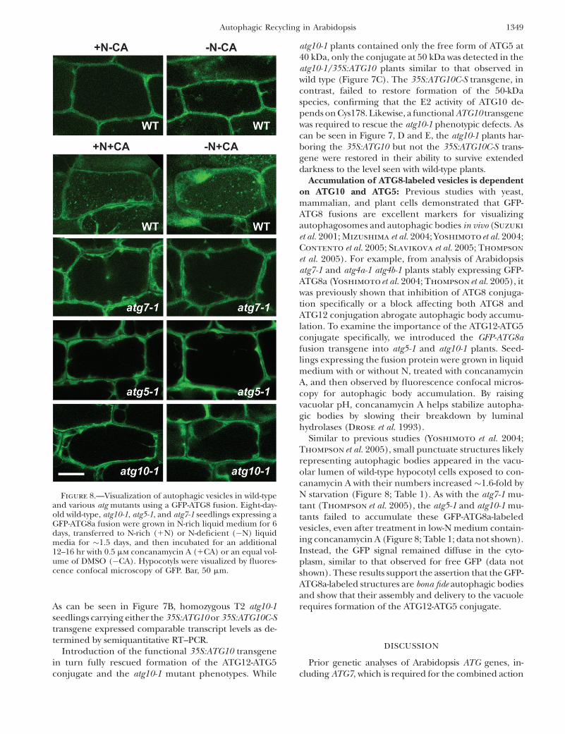
Accumulation of ATG8-labeled vesicles is dependenton ATG10 and ATG5: Previous studies with yeast,mammalian, and plant cells demonstrated that GFP-ATG8 fusions are excellent markers for visualizingautophagosomes and autophagic bodies in vivo (Suzuki
et al. 2001; Mizushima et al. 2004; Yoshimotoet al. 2004;Contento et al. 2005; Slavikova et al. 2005; Thompson
et al. 2005). For example, from analysis of Arabidopsisatg7-1 and atg4a-1 atg4b-1 plants stably expressing GFP-ATG8a (Yoshimoto et al. 2004; Thompson et al. 2005), itwas previously shown that inhibition of ATG8 conjuga-tion specifically or a block affecting both ATG8 andATG12 conjugation abrogate autophagic body accumu-lation. To examine the importance of the ATG12-ATG5conjugate specifically, we introduced the GFP-ATG8afusion transgene into atg5-1 and atg10-1 plants. Seed-lings expressing the fusion protein were grown in liquidmedium with or without N, treated with concanamycinA, and then observed by fluorescence confocal micros-copy for autophagic body accumulation. By raisingvacuolar pH, concanamycin A helps stabilize autopha-gic bodies by slowing their breakdown by luminalhydrolases (Drose et al. 1993).
Similar to previous studies (Yoshimoto et al. 2004;Thompson et al. 2005), small punctuate structures likelyrepresenting autophagic bodies appeared in the vacu-olar lumen of wild-type hypocotyl cells exposed to con-canamycin A with their numbers increased �1.6-fold byN starvation (Figure 8; Table 1). As with the atg7-1 mu-tant (Thompson et al. 2005), the atg5-1 and atg10-1 mu-tants failed to accumulate these GFP-ATG8a-labeledvesicles, even after treatment in low-N medium contain-ing concanamycin A (Figure 8; Table 1; data not shown).Instead, the GFP signal remained diffuse in the cyto-plasm, similar to that observed for free GFP (data notshown). These results support the assertion that the GFP-ATG8a-labeled structures are bona fide autophagic bodiesand show that their assembly and delivery to the vacuolerequires formation of the ATG12-ATG5 conjugate.
DISCUSSION
Prior genetic analyses of Arabidopsis ATG genes, in-cluding ATG7, which is required for the combined action
Figure 8.—Visualization of autophagic vesicles in wild-typeand various atg mutants using a GFP-ATG8 fusion. Eight-day-old wild-type, atg10-1, atg5-1, and atg7-1 seedlings expressing aGFP-ATG8a fusion were grown in N-rich liquid medium for 6days, transferred to N-rich (1N) or N-deficient (�N) liquidmedia for �1.5 days, and then incubated for an additional12–16 hr with 0.5 mm concanamycin A (1CA) or an equal vol-ume of DMSO (�CA). Hypocotyls were visualized by fluores-cence confocal microscopy of GFP. Bar, 50 mm.
Autophagic Recycling in Arabidopsis 1349

of ATG8 and ATG12, have revealed several importantroles for autophagy in plants. These include assisting inthe remobilization of nutrients under N starvation andfixed C-limiting conditions and during senescence, re-moving oxidized proteins from the cytoplasm, andlimiting the spread of necrosis during HR (Doelling
et al. 2002; Hanaoka et al. 2002; Liu et al. 2005; Thompson
et al. 2005; Xiong et al. 2007). In addition, cytological andmolecular studies support the involvement of autophagyin PCD and HR (Liu et al. 2005; van Doorn andWoltering 2005; Bozhkov and Jansson 2007).
Here, we demonstrate the importance of the ATG12conjugation pathway specifically through the reversegenetic analysis of the ATG10 gene encoding the E2responsible for the ligation of this polypeptide tag. AT-DNA insertion mutant preventing ATG10 accumula-tion fails to form the ATG12-ATG5 conjugate, demonstrat-ing that this E2 directs ATG12 ligation. Phenotypically,the atg10-1 mutant plants resemble atg5-1 and atg7-1plants previously characterized (Doelling et al. 2002;Thompson et al. 2005). Under standard growth condi-tions, the atg10-1 plants germinate and develop nor-mally, but in SD they grow slower, flower later, senesceearlier, and produce less seed. More importantly, theydisplay enhanced chlorosis and die more rapidly duringN starvation and when exposed to extended darknessthat substantially reduces fixed C availability. These phe-notypic defects can be rescued by transgenic expressionof wild-type ATG10 but not by transgenic expression ofan active-site mutant (Cys178-Ser), demonstrating thatthe enzymatic activity of ATG10 is required. The pheno-typic similarity between the atg5-1 and atg10-1 mutantscoupled with the detection of a single conjugate in wild-type plant extracts using either anti-ATG12 (Suzuki et al.2005) or anti-ATG5 (this report) antibodies strongly sug-gests that ATG5 is the main, if not, sole target of ATG12in plants.
The phenotypic similarity of atg10-1 and atg5-1 plantsalso supports expectations based on data from yeasts(Ohsumi 2001) that ATG5 functions optimally whenconjugated to ATG12. However, for both N- and C-
limiting growth conditions, we reproducibly observed aslight decrease in sensitivity for the atg10-1 plants ascompared to atg7-1 and atg5-1. Since all three mutantsappear to represent null alleles (Doelling et al. 2002:Thompsonet al. 2005; this report), mutant strength is anunlikely explanation for this small difference. Otherpossibilities include (i) the action of free ATG5 by itself,(ii) the noncovalent association of ATG12 with ATG5,(iii) the direct transfer of activated ATG12 from ATG7 toATG5 without an E2 intermediate, and/or (iv) theformation of the ATG12-ATG5 conjugate by anothermechanism (e.g., using the E2 ATG3). However, theabsence of an immunodetectable ATG12-ATG5 conju-gate in the atg10-1 plants may preclude the last twoscenarios (Figure 2C). We also note that almost all ATG5in wild-type plants is present in the conjugated formregardless of the age of the plants or nutritional status.This constitutive conjugation combined with only smallchanges in ATG12a/b transcript abundance during Climitation imply that the formation of the ATG12-ATG5conjugate, while necessary for autophagy, is not thetrigger for this recycling pathway during senescence orunder nutrient-limiting conditions. The ATG8-PE con-jugate could be the trigger, given the substantial up-regulation of various ATG8 mRNAs and proteins duringC starvation of wild-type plants. The abundance of var-ious ATG8 isoforms increases further in several atg mu-tant backgrounds (Thompson et al. 2005; this report).Since both the atg5-1 and atg10-1 mutations increase thesteady-state levels of ATG8 transcripts, part of thisincrease likely reflects increased protein synthesis. How-ever, it is also possible that atg defects indirectly raise thelevels of ATG8 proteins by decreasing their breakdownduring autophagy.
In yeast, formation of the autophagosomes andautophagic bodies requires both the ATG8-PE andATG12-ATG5 conjugates (Suzuki et al. 2004, 2007). Asimilar scenario likely exists in plants. Prior studies usingArabidopsis mutants blocked in ATG8 processing(Yoshimoto et al. 2004) or expression of the ATG7 E1(Thompson et al. 2005) together confirmed the impor-
TABLE 1
Autophagic body accumulation in the vacuoles of wild-type and atg mutant seedlings
1N�CA �N�CA 1N1CA �N1CA
Wild-type Col-0a 0.09 6 0.01 0.05 6 0.01 2.46 6 0.07 3.88 6 0.33atg10-1b 0.08 6 0.13 0.11 6 0.14 0.03 6 0.08 0.11 6 0.18atg5-1b 0.08 6 0.13 0.08 6 0.15 0.03 6 0.08 0.08 6 0.12atg7-1b 0.07 6 0.14 0.10 6 0.13 0.13 6 0.13 0.11 6 0.13
Each line expressed GFP-ATG8 under the control of the CaMV 35S promoter. Seedlings were grown in N-deficient liquid medium for 2 days and then treated with 0.5 mm concanamycin A (CA) for 12–16 hr prior toconfocal fluorescence microscopy of the central vacuole.
a Values are the average number of vacuolar vesicles per 100 mm2 of 9–32 cells each for three independentexperiments (6SE).
b Values are the average of number of vacuolar vesicles per 100 mm2 of 12–36 cells each for one experiment(6SD).
1350 A. R. Phillips, A. Suttangkakul and R. D. Vierstra

tance of ATG8 conjugation. Here, we extend theseobservations to the ATG12-ATG5 conjugate specifically.Like atg7-1 plants (Thompson et al. 2005), atg5-1 andatg10-1 plants fail to accumulate GFP-ATG8-labeledvesicles in the vacuolar lumen upon treatment withconcanamycin A under either N-rich or N-deficientconditions. The results with atg5-1 and atg10-1 plants inparticular further support the notion that while forma-tion of ATG8-PE conjugates may not be blocked byremoval of the ATG12-ATG5 conjugate, their incorpora-tion into autophagosomes/autophagic bodies is inhibited.However, we cannot discount the remote possibility thatautophagic vesicles can be assembled without the ATG8decoration.
One phenotypic conundrum for the collection of atgmutants is that, compared to wild-type plants, theysenesce earlier and die more rapidly under N- and C-limiting environments despite their inability to directautophagic breakdown. One probable scenario is thatdefects in autophagy under starvation conditions irre-versibly trigger other stress-activated PCD pathways thatthen compromise cell viability more quickly than nor-mal. Possibilities include apoptosis ½although true apo-ptosis involving phagocytosis does not occur in plants(van Doorn and Woltering 2005)�, nonlysosomalPCD, necrosis, and/or an ATG-independent autophagicsystem. In support, we detected several hallmarks ofvarious cell death pathways when atg mutants were ex-posed to darkness, including the fragmentation of ge-nomic DNA into HMW and LMW species, loss of turgorin leaf tissue, and death of the shoot meristem. This wasaccompanied by more rapid leaf chlorosis and a fasterloss of specific proteins from most, if not, all cellularcompartments, including chloroplasts, mitochondria,and cytoplasm, which suggests a severe disruption ofcellular homeostasis (Thompson et al. 2005; this report).
Prior to their more rapid death, the atg mutant plantsdramatically increase mRNA abundance for a suite ofgenes often associated with senescence-induced PCD,including SEN1, SAG12, PED1, GPX2, and NYE1, whichimplies that one or more PCD pathways are activatedby extended darkness that in turn accelerate cell death(Swidzinski et al. 2002). This upregulation could reflect adirect connection between autophagy and stress pathwaysor an indirect result of atg mutants attempting to copewith acute stress. However, not all factors associated withPCD and senescence were increased by autophagicdefects. Most notably, levels of the VPEg protein werenot retained despite the proposed role of this caspase-likeprotease in activating hydrolyases needed during senes-cence and HR (Hatsugai et al. 2004).
With respect to the mechanism(s) of accelerateddeath, the absence of large-scale patches of dead cellsjust after the atg plants exited darkness would precludenecrotic mechanisms. However, it remains possiblethat necrosis occurs only after returning the plants tofull light when oxidative damage induced by light
would become challenging. The subsequent failure toupregulate CSD1 and CAT3 expression, both of whichhelp scavenge oxidative species, could then accentuatethe problem. One likely contributor to the enhancedchlorosis is premature activation of an autophagy-independent chlorophyll catabolic pathway involvingNYE1, which is dramatically upregulated when atg plantsencounter prolonged darkness. Coupled with the morerapid loss of RUBISCO, we propose that nonautophagicchloroplast breakdown represents an important sourceof nutrients when autophagy is compromised.
It remains unclear what signaling pathway(s) triggerPCD in the atg mutants and what are the molecularconsequences of this upregulation. One likely effectorcould be reactive oxygen species (ROS). Xiong et al.(2007) reported that autophagy is enhanced when plantsencounter severe oxidative stress, which then works toeliminate oxidized proteins and potentially dampen cyto-solic ROS accumulation. In addition, Liu et al. (2005)endorsed a role for autophagy in protecting cells fromdamage by reactive oxygen intermediates during HR.One direct target of ROS could be ATG4. Recent studieswith the human autophagy system showed that this pro-tease is activated by starvation-induced oxidation, thusproviding a mechanism to directly regulate ATG8 avail-ability (Scherz-Shouval et al. 2007). Considering thatROS activate PCD, it is possible that levels of ROS areabnormally high in the absence of autophagy and arefurther increased during dark-induced senescence toaccelerate PCD. Clearly, a more complete picture of genesaffected by the inhibition of autophagy, coupled withgenetic and biochemical analyses of ROS accumulationin various atg backgrounds, is now needed to test thisconnection.
We thank Archie Portis, Natasha Raikhel, and Sara Patterson for thesupply of the RUBISCO, VPEg, and SAG2 antibodies, respectively, andJoseph Walker, Taijoon Chung, and Scott Saracco for technical assistanceand helpful discussions. This project was supported by a grant from theNational Research Initiative of the U. S. Department of AgricultureCooperative State Research, Education and Extension Service (2005-35301-15768) to R.D.V.; a Thailand Predoctoral Fellowship to A.S.; andWisconsin Alumni Research Foundation and Louis and Elsa ThomsenWisconsin Distinguished Predoctoral Fellowships to A.R.P.
LITERATURE CITED
Alonso, J. M., A. N. Stepanova, T. J. Leisse, C. J. Kim, H. M. Chen
et al., 2003 Genome-wide insertional mutagenesis of Arabidopsisthaliana. Science 301: 653–657.
Balk, J., and C. J. Leaver, 2001 The PET1-CMS mitochondrial mu-tation in sunflower is associated with premature programmedcell death and cytochrome c release. Plant Cell 13: 1803–1818.
Bassham, D. C., 2007 Plant autophagy: more than a starvation re-sponse. Curr. Opin. Plant Biol. 10: 587–593.
Bozhkov, P., and C. Jansson, 2007 Autophagy and cell-death pro-teases in plants: two wheels of a funeral cart. Autophagy 3:136–138.
Brown, D. G., X. M. Sun and G. M. Cohen, 1993 Dexamethasone-induced apoptosis involves cleavage of DNA to large fragments priorto internucleosomal fragmentation. J. Biol. Chem. 268: 3037–3039.
Bursch, W., 2001 The autophagosomal-lysosomal compartment inprogrammed cell death. Cell Death Differ. 8: 569–581.
Autophagic Recycling in Arabidopsis 1351

Bursch, W., A. Ellinger, C. Gerner and R. Schulte-Hermann,2004 Autophagocytosis and programmed cell death, pp. 287–303 in Autophagy, edited by D. J. Klionsky. Eurekah.com/LandesBioscience, Georgetown, TX.
Clough, S. J., and A. F. Bent, 1998 Floral dip: a simplified methodfor Agrobacterium-mediated transformation of Arabidopsis thaliana.Plant J. 16: 735–743.
Contento, A. L., S. J. Kim and D. C. Bassham, 2004 Transcriptomeprofiling of the response of Arabidopsis suspension culture cells toSuc starvation. Plant Physiol. 135: 2330–2347.
Contento, A. L., Y. Xiong and D. C. Bassham, 2005 Visualization ofautophagy in Arabidopsis using the fluorescent dye monodansylca-daverine and a GFP-AtATG8e fusion protein. Plant J. 42: 598–608.
Doelling, J. H., J. M. Walker, E. M. Friedman, A. R. Thompson andR. D. Vierstra, 2002 The APG8/12-activating enzyme APG7 isrequired for proper nutrient recycling and senescence in Arabi-dopsis thaliana. J. Biol. Chem. 277: 33105–33114.
Drose, S., K. U. Bindseil, E. J. Bowmama, A. Siebers, A. Zeeck et al.,1993 Inhibitory effect of modified bafilomycins and concana-mycins and P- and V-type adenosinetriphosphatases. Biochemis-try 32: 3902–3906.
Earley, K. W., J. R. Haag, O. Pontes, K. Opper, T. Juehne et al.,2006 Gateway-compatible vectors for plant functional genomicsand proteomics. Plant J. 45: 616–629.
Fujiki, Y., K. Yoshimoto and Y. Ohsumi, 2007 An Arabidopsis homo-log of yeast ATG6/VPS30 is essential for pollen germination.Plant Physiol. 143: 1132–1139.
Giuliano, G., N. E. Hoffman, K. Ko, P. A. Scolnik and A. R.Cashmore, 1988 A light-entrained circadian clock controlstranscription of several plant genes. EMBO J. 7: 3635–3642.
Grbic, V., 2003 SAG2 and SAG12 protein expression in senescingArabidopsis plants. Physiol. Plant. 119: 263–269.
Greenberg, J. T., 1996 Programmed cell death: a way of life forplants. Proc. Natl. Acad. Sci. USA 93: 12094–12097.
Hanaoka, H., T. Noda, Y. Shirano, T. Kato, H. Hayashi et al.,2002 Leaf senescence and starvation-induced chlorosis are ac-celerated by the disruption of an Arabidopsis autophagy gene.Plant Physiol. 129: 1181–1193.
Harding, T. M., K. A. Morano, S. V. Scott and D. J. Klionsky,1995 Isolation and characterization of yeast mutants in the cy-toplasm to vacuole protein targeting pathway. J. Cell Biol. 131:591–602.
Hatsugai, N., M. Kuroyanagi, K. Yamada, T. Meshi, S. Tsuda et al.,2004 A plant vacuolar protease, VPE, mediates virus-inducedhypersensitive cell death. Science 305: 855–858.
Hayashi, M., K. Toriyama, M. Kondo and M. Nishimura, 1998 2,4-Dichlorophenoxybutyric acid-resistant mutants of Arabidopsishave defects in glyoxysomal fatty acid beta-oxidation. Plant Cell10: 183–195.
Ichimura, Y., T. Kirisako, T. Takao, Y. Satomi, Y. Shimonishi et al.,2000 A ubiquitin-like system mediates protein lipidation. Na-ture 408: 488–492.
Juhasz, G., B. Erdi, M. Sass, and T. P. Neufeld, 2007 Atg7-dependent autophagy promotes neuronal health, stress toler-ance, and longevity but is dispensable for metamorphosis inDrosophila. Genes Dev. 21: 3061–3066.
Kinoshita, T., K. Yamada, N. Hiraiwa, M. Kondo, M. Nishimura
et al., 1999 Vacuolar processing enzyme is up-regulated in thelytic vacuoles of vegetative tissues during senescence and undervarious stressed conditions. Plant J. 19: 43–53.
Kirisako, T., M. Baba, N. Ishihara, K. Miyazawa, M. Ohsumi et al.,1999 Formation process of autophagosome is traced withApg8/Aut7p in yeast. J. Cell Biol. 147: 435–446.
Kirisako, T., Y. Ichimura, H. Okada, Y. Kabeya, N. Mizushima et al.,2000 The reversible modification regulates the membrane-binding state of Apg8/Aut7 essential for autophagy and the cyto-plasm to vacuole targeting pathway. J. Cell Biol. 151: 263–276.
Klionsky, D. J., 2007 Autophagy: from phenomenology to molecu-lar understanding in less than a decade. Nat. Rev. Mol. Cell Biol.11: 931–937.
Kuma, A., N. Mizushima, N. Ishihara and Y. Ohsumi,2002 Formation of the approximately 350-kDa Apg12-Ap-g5.Apg16 multimeric complex, mediated by Apg16 oligomeriza-tion, is essential for autophagy in yeast. J. Biol. Chem. 277:18619–18625.
Levine, B., and D. J. Klionsky, 2004 Development by self-digestion:molecular mechanisms and biological functions of autophagy.Dev. Cell 6: 463–477.
Liu, Y., M. Schiff, K. Czymmek, Z. Talloczy, B. Levine et al.,2005 Autophagy regulates programmed cell death during theplant innate immune response. Cell 121: 567–577.
Mizushima, N., T. Noda, T. Yoshimori, Y. Tanaka, T. Ishii et al.,1998 A protein conjugation system essential for autophagy. Na-ture 395: 395–398.
Mizushima, N., T. Noda and Y. Ohsumi, 1999 Apg16p is requiredfor the function of the Apg12p-Apg5p conjugate in the yeast au-tophagy pathway. EMBO J. 18: 3888–3896.
Mizushima, N., T. Yoshimori and Y. Ohsumi, 2002 Mouse Apg10as an Apg12-conjugating enzyme: analysis by the conjugation-mediated yeast two-hybrid method. FEBS Lett. 532: 450–454.
Mizushima, N., A. Yamamoto, M. Matsui, T. Yoshimori and Y.Ohsumi, 2004 In vivo analysis of autophagy in response to nu-trient starvation using transgenic mice expressing a fluorescentautophagosome marker. Mol. Biol. Cell 15: 1101–1111.
Mullineaux, P., L. Ball, C. Escobar, B. Karpinska, G. Creissen
et al., 2000 Are diverse signalling pathways integrated in the reg-ulation of Arabidopsis antioxidant defence gene expression in re-sponse to excess excitation energy? Philos. Trans. R. Soc. Lond. BBiol. Sci. 355: 1531–1540.
Nemoto, T., I. Tanida, E. Tanida-Miyake, N. Minematsu-Ikeguchi,M. Yokota et al., 2003 The mouse APG10 homologue, an E2-like enzyme for Apg12p conjugation, facilitates MAP-LC3 modi-fication. J. Biol. Chem. 278: 39517–39526.
Oh, S. A., S. Y. Lee, I. K. Chung, C. H. Lee and H. G. Nam, 1996 Asenescence-associated gene of Arabidopsis thaliana is distinctivelyregulated during natural and artificially induced leaf senescence.Plant Mol. Biol. 30: 739–754.
Ohsumi, Y., 2001 Molecular dissection of autophagy: two ubiquitin-like systems. Nat. Rev. Mol. Cell Biol. 2: 211–216.
Osuna, D., B. Usadel, R. Morcuende, Y. Gibon, O. E. Blasing et al.,2007 Temporal responses of transcripts, enzyme activities andmetabolites after adding sucrose to carbon-deprived Arabidopsisseedlings. Plant J. 49: 463–491.
Pennell, R. I., and C. Lamb, 1997 Programmed cell death in plants.Plant Cell 9: 1157–1168.
Qin, G., Z. Ma, L. Zhang, S. Xing, X. Hou et al., 2007 ArabidopsisAtBECLIN 1/AtAtg6/AtVps30 is essential for pollen germinationand plant development. Cell Res. 17: 249–263.
Rate, D. N., J. V. Cuenca, G. R. Bowman, D. S. Guttman and J. T.Greenberg, 1999 The gain-of-function Arabidopsis acd6 mutantreveals novel regulation and function of the salicylic acid signal-ing pathway in controlling cell death, defenses, and cell growth.Plant Cell 11: 1695–1708.
Ren, G., K. An, Y. Liao, X. Zhou, Y. Cao et al., 2007 Identification ofa novel chloroplast protein AtNYE1 regulating chlorophyll deg-radation during leaf senescence in Arabidopsis. Plant Physiol. 144:1429–1441.
Rojo, E., J. Zouhar, C. Carter, V. Kovaleva and N. V. Raikhel,2003 A unique mechanism for protein processing and degradationin Arabidopsis thaliana. Proc. Natl. Acad. Sci. USA 100: 7389–7394.
Rose, T. L., L. Bonneau, C. Der, D. Marty-Mazars and F. Marty,2006 Starvation-induced expression of autophagy-related genesin Arabidopsis. Biol. Cell. 98: 53–67.
Scherz-Shouval, R., E. Shvets, E. Fass, H. Shorer, L. Gil et al.,2007 Reactive oxygen species are essential for autophagy andspecifically regulate the activity of Atg4. EMBO J. 26: 1749–1760.
Seay, M., S. Patel and S. P. Dinesh-Kumar, 2006 Autophagy andplant innate immunity. Cell. Microbiol. 8: 899–906.
Shintani, T., N. Mizushima, Y. Ogawa, A. Matsuura, T. Noda et al.,1999 Apg10p, a novel protein-conjugating enzyme essential forautophagy in yeast. EMBO J. 18: 5234–5241.
Slavikova, S., G. Shy, Y. Yao, R. Glozman, H. Levanony et al.,2005 The autophagy-associated Atg8 gene family operates bothunder favourable growth conditions and under starvationstresses in Arabidopsis plants. J. Exp. Bot. 56: 2839–2849.
Smalle, J., and R. D. Vierstra, 2004 The ubiquitin 26S proteasomeproteolytic pathway. Annu. Rev. Plant Biol. 55: 555–590.
Smalle, J., J. Kurepa, P. Yang, E. Babiychuk, S. Kushnir et al.,2002 Cytokinin growth responses in Arabidopsis involve the26S proteasome subunit RPN12. Plant Cell 14: 17–32.
1352 A. R. Phillips, A. Suttangkakul and R. D. Vierstra
Surpin, M., H. Zheng, M. T. Morita, C. Saito, E. Avila et al.,2003 The VTI family of SNARE proteins is necessary for plantviability and mediates different protein transport pathways. PlantCell 15: 2885–2899.
Suzuki, K., T. Kirisako, Y. Kamada, N. Mizushima, T. Noda et al.,2001 The pre-autophagosomal structure organized by con-certed functions of APG genes is essential for autophagosome for-mation. EMBO J. 20: 5971–5981.
Suzuki, K., T. Noda and Y. Ohsumi, 2004 Interrelationships amongATG proteins during autophagy in Saccharomyces cerevisiae. Yeast21: 1057–1065.
Suzuki, K., Y. Kubota, T. Sekito and Y. Ohsumi, 2007 Hierarchy ofATG proteins in pre-autophagosomal structure organization.Genes Cells 12: 209–218.
Suzuki, N. N., K. Yoshimoto, Y. Fujioka, Y. Ohsumi and F. Inagaki,2005 The crystal structure of plant ATG12 and its biological im-plication in autophagy. Autophagy 1: 119–126.
Swidzinski, J. A., L. J. Sweetlove and C. J. Leaver, 2002 A custommicroarray analysis of gene expression during programmed celldeath in Arabidopsis thaliana. Plant J. 30: 431–446.
Thompson, A. R., and R. D. Vierstra, 2005 Autophagic recycling:lessons from yeast help define the process in plants. Curr. Opin.Plant Biol. 8: 165–173.
Thompson, A. R., J. H. Doelling, A. Suttangkakul and R. D.Vierstra, 2005 Autophagic nutrient recycling in Arabidopsisdirected by the ATG8 and ATG12 conjugation pathways. PlantPhysiol. 138: 2097–2110.
Thumm, M., R. Egner, B. Koch, M. Schlumpberger, M. Straub et al.,1994 Isolation of autophagocytosis mutants of Saccharomyces cer-evisiae. FEBS Lett. 349: 275–280.
Tsukada, M., and Y. Ohsumi, 1993 Isolation and characterization ofautophagy-defective mutants of Saccharomyces cerevisiae. FEBSLett. 333: 169–174.
Ueno, T., I. Tanida and E. Kominami, 2004 Autophagy and neuro-muscular diseases, pp. 264–286 in Autophagy, edited by D. J.Klionsky. Eurekah.com/Landes Bioscience, Georgetown, TX.
van der Graaff, E., R. Schwacke, A. Schneider, M. Desimone, U. I.Flugge et al., 2006 Transcription analysis of Arabidopsis mem-brane transporters and hormone pathways during developmen-tal and induced leaf senescence. Plant Physiol. 141: 776–792.
van Doorn, W. G., and E. J. Woltering, 2005 Many ways to exit?Cell death categories in plants. Trends Plant Sci. 10: 117–122.
Weaver, L. M., and R. M. Amasino, 2001 Senescence is induced inindividually darkened Arabidopsis leaves, but inhibited in wholedarkened plants. Plant Physiol. 127: 876–886.
Weaver, L. M., S. Gan, B. Quirino and R. M. Amasino, 1998 A com-parisonof the expressionpatternsof several senescence-associatedgenes in response to stress and hormone treatment. Plant Mol.Biol. 37: 455–469.
Xiong, Y., A. L. Contento and D. C. Bassham, 2005 AtATG18a isrequired for the formation of autophagosomes during nutrientstress and senescence in Arabidopsis thaliana. Plant J. 42: 535–546.
Xiong, Y., A. L. Contento, P. Q. Nguyen and D. C. Bassham,2007 Degradation of oxidized proteins by autophagy during ox-idative stress in Arabidopsis. Plant Physiol. 143: 291–299.
Yoshimoto, K., H. Hanaoka, S. Sato, T. Kato, S. Tabata et al.,2004 Processing of ATG8s, ubiquitin-like proteins, and their de-conjugation by ATG4s are essential for plant autophagy. PlantCell 16: 2967–2983.
Zhang, X., Y. Chen, Z. Y. Wang, Z. Chen, H. Gu et al., 2007 Con-stitutive expression of CIR1 (RVE2) affects several circadian-regulated processes and seed germination in Arabidopsis. Plant J.51: 512–525.
Communicating editor: B. Bartel
Autophagic Recycling in Arabidopsis 1353