the anti-inflammatory effects of oral-formulated … immunosuppressive anti-inflammatory effects of...
TRANSCRIPT
© 2017 The Korean Academy of Medical Sciences.This is an Open Access article distributed under the terms of the Creative Commons Attribution Non-Commercial License (http://creativecommons.org/licenses/by-nc/4.0) which permits unrestricted non-commercial use, distribution, and reproduction in any medium, provided the original work is properly cited.
pISSN 1011-8934eISSN 1598-6357
The Anti-Inflammatory Effects of Oral-Formulated Tacrolimus in Mice with Experimental Autoimmune Encephalomyelitis
Multiple sclerosis (MS) is a T-lymphocyte-mediated autoimmune disease that is characterized by inflammation in the central nervous system (CNS). Although many disease-modifying therapies (DMTs) are presumed effective in patients with MS, studies on the efficacy and safety of DMTs for preventing MS relapse are limited. Therefore, we tested the immunosuppressive anti-inflammatory effects of oral-formulated tacrolimus (FK506) on MS in a mouse model of experimental autoimmune encephalomyelitis (EAE). The mice were randomly divided into 3 experimental groups: an untreated EAE group, a low-dose tacrolimus-treated EAE group, and a high-dose tacrolimus-treated EAE group. After autoimmunization of the EAE mice with myelin oligodendrocyte glycoprotein, symptom severity scores, immunohistochemistry of the myelination of the spinal cord, and western blotting were used to evaluate the EAE mice. After the autoimmunization, the symptom scores of each EAE group significantly differed at times. The group treated with the larger tacrolimus dose had the lowest symptom scores. The tacrolimus-treated EAE groups exhibited less demyelination and inflammation and weak immunoreactivity for all of the immunization biomarkers. Our results revealed that oral-formulated tacrolimus inhibited the autoimmunization in MS pathogenesis by inactivating inflammatory cells.
Keywords: Multiple Sclerosis; Neuromyelitis Optica; Experimental Autoimmune Encephalomyelitis; EAE; Tacrolimus; FK506
Myung-Jin Kim,1 Jung-Joon Sung,2 Seung Hyun Kim,3 Jeong-Min Kim,1 Gye Sun Jeon,2 Seog-Kyun Mun,4 and Suk-Won Ahn1
1Department of Neurology, Chung-Ang University Hospital, Chung-Ang University College of Medicine, Seoul, Korea; 2Department of Neurology, Seoul National University Hospital, Seoul National University College of Medicine, Seoul, Korea; 3Department of Neurology, Hanyang University Hospital, Hanyang University College of Medicine, Seoul, Korea; 4Department of Otorhinolaryngology, Head and Neck Surgery, Chung-Ang University College of Medicine, Seoul, Korea
Received: 22 December 2016Accepted: 28 May 2017
Address for Correspondence:Suk-Won Ahn, MD, PhDDepartment of Neurology, Chung-Ang University Hospital, Chung-Ang University College of Medicine, 102 Heukseok-ro, Dongjak-gu, Seoul 06973, KoreaE-mail: [email protected]
Funding: This research was supported by the Chung-Ang University Graduate Research Scholarship (2016), and Basic Science Research Program through the National Research Foundation of Korea (NRF) funded by the Ministry of Education (2016R1D1A1B03936287).
https://doi.org/10.3346/jkms.2017.32.9.1502 • J Korean Med Sci 2017; 32: 1502-1507
INTRODUCTION
Multiple sclerosis (MS) is a T-lymphocyte-mediated autoim-mune disease that is characterized by relapsing and remitting inflammatory demyelination in the central nervous system (CNS) (1-3). Patients with MS exhibit long-term disabilities, such as weakness, sensory symptoms, decreased visual acuity, urina-tion difficulties, and cognitive deficits (1-4). Previous studies on MS have concentrated on the mechanisms that are hypothesized to underlie the CNS inflammation, includ-ing T-cell autoimmunization, environmental factors, altered genetic influences, impaired vitamin D deficiency, and combi-nations of autoimmune diseases (1-4). However, the underly-ing mechanisms and treatment of MS remain uncertain. Recently, considerable progress has been made toward un-derstanding the basic pathogenesis of MS and the effectiveness of disease-modifying therapies (DMTs) in preventing the re-lapse of patients with MS (5-8). Although many DMTs are pre-sumed effective in patients with MS, studies on the efficacy of
DMTs for preventing the relapse of patients with MS are limit-ed. In addition, DMTs can induce serious adverse effects (9,10). We tested the anti-inflammatory effects of an oral-formulat-ed immunosuppressant with a better side effect profile in a mouse model of experimental autoimmune encephalomyelitis (EAE) (11). Tacrolimus (FK506; macrolide lactone immunosup-pressant) acts as a calcineurin inhibitor that blocks interleu-kin-2 production, which results in decreased T-cell proliferation (12,13). Tacrolimus is therefore used in the treatment of T-cell-mediated autoimmune diseases and prevention of organ trans-plant rejection (12,13). The EAE animal model is used most in studies of MS in laboratory animals. Both MS and EAE are char-acterized by perivascular inflammation and demyelination in the spinal cord and brain. EAE is a CD4+ T-cell-mediated auto-immune disease in which CNS inflammation is induced after the animals are immunized against a myelin-specific antigen that induces the migration of activated autoreactive T-cells across the blood-brain barrier and into the CNS (14,15). In this study, we used C57BL/6 mice that had been immu-
ORIGINAL ARTICLENeuroscience
1 / 1CROSSMARK_logo_3_Test
2017-03-16https://crossmark-cdn.crossref.org/widget/v2.0/logos/CROSSMARK_Color_square.svg
Kim M-J, et al. • Tacrolimus in EAE Mice
http://jkms.org 1503https://doi.org/10.3346/jkms.2017.32.9.1502
nized against the myelin oligodendrocyte glycoprotein (MOG35–55) peptide, which is a method that is widely applied to induce EAE in animals. Because of the promising beneficial effects and safe-ty of tacrolimus, the present study aimed to assess the therapeu-tic effects of oral tacrolimus in MS.
MATERIALS AND METHODS
Induction of EAE and animal careAll of the procedures were performed in accordance with the Institutional Animal Care and Use Committee (IACUC) guide-lines for the care and use. Animals exhibiting paralysis were kept on soft bed of each cage and fed and watered through animal feeding tube. All surgery was performed under sodium pento-barbital anesthesia, and all efforts were made to minimize suf-fering. If any mouse came to the moribund stage, it was decapi-tated after anesthesia with sodium pentobarbital. Sixty-four mice that were approximately 80 days old were randomly divided into 3 experimental groups: an untreated EAE group, a 5-mg/kg tacrolimus-treated EAE group, and a 10-mg/kg tacrolimus-treated EAE group. During the week before the start of and during the experiments, the animals were housed in individual cages with an ambient temperature of 23°C ± 3°C, stable air humidity, and a natural day-and-night cycle. The adop-tive transfer of EAE was performed as described previously. Brief-ly, 11-week-old female C57BL/6 mice (Central Lab. Animal Inc., Seoul, Korea) were immunized against the MOG35-55 (MEVG-WYRSPFSRVVHLYRNGK) peptide in incomplete Freund’s ad-juvant supplemented with Mycobacterium tuberculosis (EAE induction Hooke kitsTM, Catalog No. EK-2110; Hooke Laborato-ries, Inc., Lawrence, MA, USA) (16-18). Each mouse was immu-nized subcutaneously with MOG (200 μg), and pertussis toxin (200 ng) was intraperitoneally injected once. The animals were sacrificed 14 days after the autoimmunization (19,20). During the 14 days before the sacrifice of the EAE mice, regular doses (5 or 10 mg/kg) of oral tacrolimus (Prograf®; Astellas Pharma Inc., Tokyo, Japan) were administered orally once per day beginning after the autoimmunization of the tacrolimus-treated EAE groups. The untreated EAE group was used for comparison.
Clinical characteristics of EAEAll of the animals were checked daily after tacrolimus treatment, and their symptoms were recorded. EAE mice show flaccid pa-ralysis, which is characterized by decreased muscle tone that progresses from the tail upward through the body (16,19). A 6- point scale (0–5) was used to rate the severity of the symptoms, with a score of 1 denoting tail paralysis, a score of 4 indicating quadriplegia, and a score of 5 signifying death. An investigator who was blind to the groups rated the experimental animals as follows: 0, no clinical disease; 0.5, piloerection; 1, tail weakness; 1.5, tail paralysis; 2, hind limb weakness; 3, hind limb paralysis;
3.5, forelimb weakness; 4, forelimb paralysis; or 5, moribund or death. The symptom scores of the mice are expressed in mean ± standard error. The data were analyzed with analysis of vari-ance tests and Dunnett’s post-hoc tests, if necessary (SPSS, ver-sion 19.0; IBM Corp., Armonk, NY, USA). P values less than 0.05 were considered statistically significant.
Immunoblotting and antibodiesFourteen days after immunization, each mouse was perfused with phosphate-buffered saline (PBS) while they were deeply anesthetized, and the CNS was removed. The spinal cords were homogenized in ice-cold radio immunoprecipitation assay buffer containing protease inhibitor and phosphatase inhibitor cocktails (Catalog No. 01906845001 and 11697498001; Roche Diagnostics Corporation, Indianapolis, IN, USA). The samples were centrifuged at 14,000 rpm for 25 minutes at 4°C, and the supernatants were collected. The protein concentrations of the samples were measured with Thermo Scientific Pierce Micro BCA Protein Assay Kits (Thermo Fisher Scientific Inc., Waltham, MA, USA). The samples were denatured in loading buffer for 10 minutes at 95°C, loaded into 15% gel for separation by sodium dodecyl sulfate-polyacrylamide gel electrophoresis, and then blotted onto nitrocellulose membranes with a Trans-Blot Cell system (Bio-Rad Laboratories, Inc., Hercules, CA, USA). After the transfer, the membranes were blocked for 1 hour at room temperature with 5% skim milk (Duchefa Biochemie B.V., Haar-lem, The Netherlands) in Tris-buffered saline (20 mM Tris, 150 mM NaCl [pH 7.5]) with 0.1% Tween 20 (TBST). The membranes were then incubated overnight at 4°C with one of the following primary antibodies: MOG (1:1,000, Catalog No. ab109746; Ab-cam plc, Cambridge, UK), myelin basic protein (1:1,000, Cata-log No. ab40390; Abcam plc), glial fibrillary acidic protein (GFAP, 1:4,000, Catalog No. ab7260; Abcam plc), ionized calcium bind-ing adaptor 1 (Iba1, 1:250, Catalog No. ab48004; Abcam plc), or CD4 (1:200, Catalog No. sc-1140; Santa Cruz Biotechnology, Inc., Dallas, TX, USA). The membranes were then washed in TBST buffer and incubated for 1 hour with a horseradish per-oxidase (HRP)-conjugated goat anti-rabbit immunoglobulin G (IgG) antibody (1:5,000, Catalog No. ADI-SAB-300; Enzo Life Sciences, Inc., Farmingdale, NY, USA) or HRP-conjugated don-key anti-goat IgG (1:2,000, Catalog No. A50-101P; Bethyl Labo-ratories, Inc., Montgomery, TX, USA) that was diluted in 5% (w/v) skim milk in TBST buffer. The membranes were then washed in TBST and developed with a Chemiluminescent Sensitive Plus HRP Microwell and/or Membrane Substrate (Surmodics, Inc., Eden Prairie, MN, USA). The membranes were stripped with Restore Western Blot Stripping Buffer (Thermo Fisher Sci-entific Inc.) at room temperature for 20 minutes and then incu-bated with an antibody to β-actin (1:1,000, Catalog No. 4970; Cell Signaling Technology, Inc., Danvers, MA, USA), which was used as a loading control.

Kim M-J, et al. • Tacrolimus in EAE Mice
1504 http://jkms.org https://doi.org/10.3346/jkms.2017.32.9.1502
ImmunohistochemistryAfter the mice were killed and the spinal cord removed, the spi-nal cord was fixed with 4% (w/v) paraformaldehyde in PBS buf-fer overnight at 4°C. The spinal cords were dehydrated in 30% sucrose at 4°C until the tissue sank. The tissue was then embed-ded in O.C.T. compound (Sakura Finetek USA, Inc., Torrance, CA, USA) and frozen with dry ice (21,22). The samples were cut into 10-μm coronal sections from the cervical spine to thoracic spine. The sections were stained with Luxol fast blue (LFB) stain or hematoxylin & eosin (22). For the LFB stain, which was per-formed to stain myelin, the sections were incubated overnight at 60°C in 0.1% LFB (Solvent Blue 38; Sigma-Aldrich Co., LLC, St. Louis, MO, USA). The sections were rinsed in distilled water (DW) twice and then differentiated by dipping them in 0.01% lithium carbonate (Cat#L0224; Tokyo Chemical Industry Co., Ltd., Tokyo, Japan). The sections were rinse twice in DW and then Nissl stained by incubating them in 0.1% cresyl violet (Sig-ma-Aldrich Co., LLC) for 5 minutes (22,23). The sections were then rinsed quickly in fresh DW. The sections were dehydrated by dipping them in 3 changes of absolute ethanol (Merck Chem-icals GmbH, Darmstadt, Germany). For the hematoxylin & eo-sin, which was performed to analyze inflammation, the tissue sections were hydrated in DW for 10 minutes, stained with May-er’s Hematoxylin solution (Sigma-Aldrich Co., LLC) for 10 min-utes, washed in DW for 5 minutes, and then stained with Eosin Y solution, alcoholic (Sigma-Aldrich Co., LLC) for 3 minutes (22,23). The sections were rinsed quickly in 70% Ethanol and dehydrated in 3 changes of absolute ethanol (Merck Chemicals GmbH). All of the sections were mounted on slides and cover slipped with Permount (Thermo Fisher Scientific Inc.). Light microscopy was used to examine the slides.
Ethics statementAll of the procedures were performed in accordance with the Institutional Animal Care and Use Committee (IACUC) guide-lines and were approved by IACUC (Permit No. 2015-00026) of the Chung-Ang University for the care and use.
RESULTS
In this study, we evaluated the clinical EAE symptom scores in each EAE group every day during the 14-day study in order to examine the functional loss and alterations of the mice in this EAE model. In addition, we examined inflammatory cell infil-tration and demyelination in the spinal cord specimens with immunohistochemistry to determine if the severity of the spi-nal cord histopathology correlated with tacrolimus administra-tion. Furthermore, we investigated autoimmunization and CNS inflammation by evaluating the presence of activated microgli-al cells, interleukin-2, macrophages, and T-cell activation in the EAE animals.
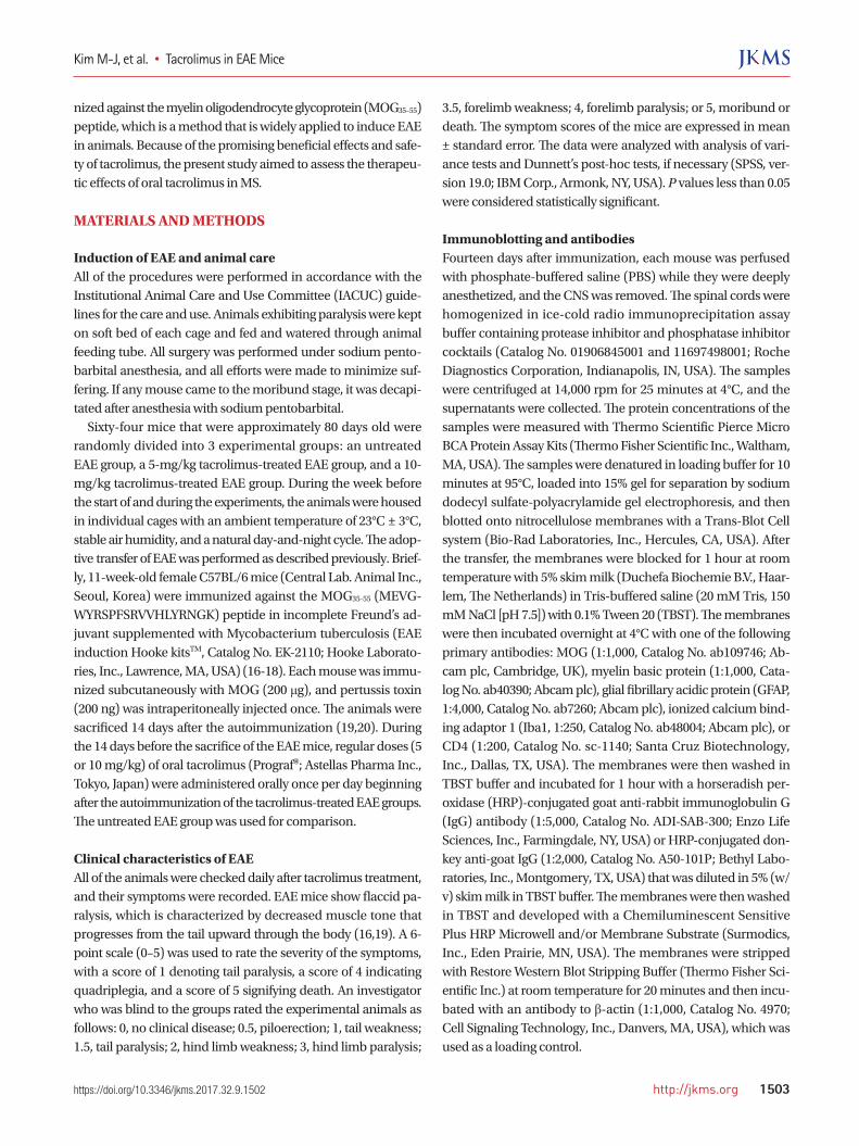
After autoimmunization with MOG35–55, the EAE symptom scores increased gradually in each group over time, and signifi-cant differences were found among the groups beginning the 10th day after immunization (Fig. 1; P < 0.05). The tacrolimus-treated mice had lower symptom scores compared with the untreated mice. The 10-mg/kg tacrolimus-treated group had lower EAE symptom scores compared with the 5-mg/kg tacro-limus-treated group. These results demonstrated that the oral form of tacrolimus improved the clinical symptoms and disease progress of the EAE mice model. We stained the spinal cord sections with LFB to visualize my-elin and the demyelination zones in the white matter (Fig. 2). Tacrolimus treatment remarkably maintained the levels of my-elination in the spinal cords of the EAE mice at levels similar to those in control mice. The untreated EAE mice showed marked demyelination in the white matter of the spinal cord and inflam-matory cell infiltration in the perivascular area. The tacrolimus-treated EAE groups exhibited higher myelination levels and de-creased inflammation compared with the control EAE mice. In addition, compared with the 5-mg/kg tacrolimus-treated EAE mice, the 10-mg/kg tacrolimus-treated EAE mice had higher levels of myelination and less inflammatory infiltration, and cuffed vessels were observed in the spinal cord. Tacrolimus treatment markedly inhibited autoimmunization and the infil-tration of inflammatory cells into the spinal cords of the EAE mice. These results demonstrated that tacrolimus treatment in-hibited the immune cascade abnormal infiltration of inflam-matory cells, and spinal cord demyelination of the EAE mice. In order to determine the clinical significance of the tacroli-mus-mediated decreases in CD4 T-cell activity, we investigated
Fig. 1. Behavioral tests of EAE mice. After autoimmunization with MOG35–55, the symp-tom scores of each EAE group gradually increased over time, and significant differ-ences were found among the 3 groups (P < 0.05) beginning on the 10th day. The 10-mg/kg tacrolimus-treated EAE mice exhibited lower EAE scores, which suggested that they had less clinical symptoms, compared with the untreated EAE mice and 5-mg/kg tacrolimus-treated EAE mice.EAE = experimental autoimmune encephalomyelitis, MOG = myelin oligodendrocyte glycoprotein.
EAE
scor
e
Day
8 9 10 11 12 13 14
2.5
2.0
1.5
1.0
0.5
0
*
*
*
*
**
Control5 mg/kg10 mg/kg
Kim M-J, et al. • Tacrolimus in EAE Mice
http://jkms.org 1505https://doi.org/10.3346/jkms.2017.32.9.1502
Fig. 2. Histopathology of EAE mice. The spinal cord specimens of the untreated EAE mice exhibited marked demyelination (A) and inflammatory cell infiltration in the perivascu-lar area (D). However, the tacrolimus-treated EAE mice exhibited preserved myelination and decreased inflammation compared with the untreated group. Additionally, the 10-mg/kg tacrolimus-treated EAE mice exhibited more myelination (C) and decreased inflammation (F) compared with the 5-mg/kg tacrolimus-treated EAE mice (B, E).EAE = experimental autoimmune encephalomyelitis.
Control EAE mice 5 mg/kg Tacrolimus EAE mice 10 mg/kg Tacrolimus EAE mice
A B C
D E F
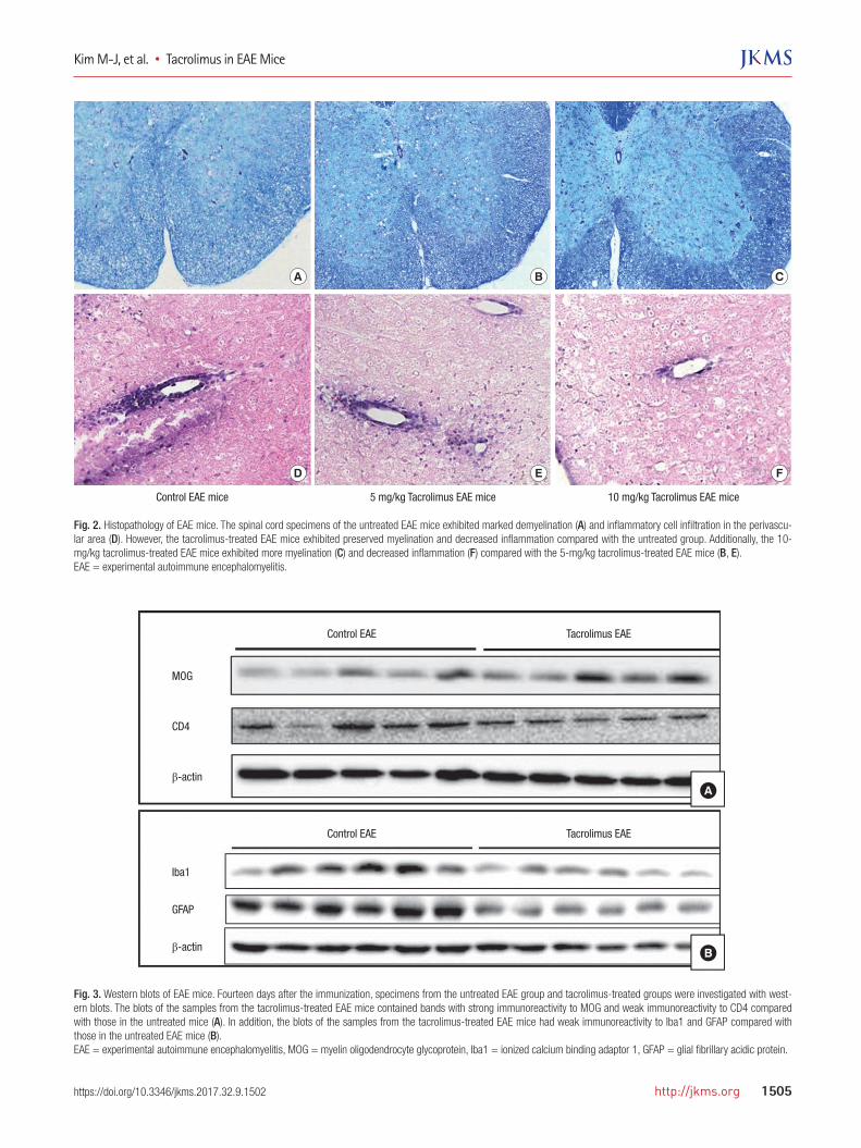
Fig. 3. Western blots of EAE mice. Fourteen days after the immunization, specimens from the untreated EAE group and tacrolimus-treated groups were investigated with west-ern blots. The blots of the samples from the tacrolimus-treated EAE mice contained bands with strong immunoreactivity to MOG and weak immunoreactivity to CD4 compared with those in the untreated mice (A). In addition, the blots of the samples from the tacrolimus-treated EAE mice had weak immunoreactivity to Iba1 and GFAP compared with those in the untreated EAE mice (B).EAE = experimental autoimmune encephalomyelitis, MOG = myelin oligodendrocyte glycoprotein, Iba1 = ionized calcium binding adaptor 1, GFAP = glial fibrillary acidic protein.
MOG
CD4
β-actin
Control EAE
Control EAE
Tacrolimus EAE
Tacrolimus EAE
Iba1
GFAP
β-actin
A
B
Kim M-J, et al. • Tacrolimus in EAE Mice
1506 http://jkms.org https://doi.org/10.3346/jkms.2017.32.9.1502
whether tacrolimus prevented the clinical symptoms of EAE in the mice. During tacrolimus treatment, the in vivo functions of the CD4 cells were blocked in the EAE mice. Taken together, these results suggested that CD4 T-cell activity plays an impor-tant role in the tacrolimus-mediated decrease in inflammatory reaction by autoimmunity in EAE model. Western blotting was used to compare the levels of immuni-zation biomarkers in the untreated and tacrolimus-treated EAE groups (Fig. 3). Compared with the untreated EAE mice, the ta-crolimus-treated EAE mice exhibited stronger immunoreactiv-ity to MOG and weaker immunoreactivity to CD4, Iba1, and GFAP. Collectively, these results suggested that tacrolimus sup-pressed the EAE in the mice by inhibiting T-cell activation.
DISCUSSION
In this study, we evaluated the anti-inflammatory effects of ta-crolimus in an EAE mouse model and found that tacrolimus decreased the severity of the clinical symptoms of EAE and in-hibited autoimmunization and inflammation in the EAE mod-el. Thus, these results suggested that tacrolimus might be effec-tive in the treatment of MS. EAE in mice and MS are thought to have similar causes: the infiltration of autoreactive T-cells and associated inflammatory cells into the CNS, which results in the immune-associated CNS demyelination that is observed in pa-tients with MS and EAE animals (24,25). MS is a devastating demyelinating disease of the human CNS, and expensive DMTs are currently used to treat patients with MS (26). However, therapies that effectively prevent the relapse of MS are not yet available. In particular, the limited efficacy and severe side effects, as well as treatment costs, of these ther-apies often limit their availability. The side effects include flu-like symptoms, menstrual disorders in women, decreased neu-trophil and white blood cell counts, progressive multifocal en-cephalopathy, cardiac problems, increased aspartate transami-nase and alanine transaminase levels, and the development of neutralizing antibodies (9,10,27,28). Therefore, it is necessary to investigate safe, effective, and less expensive therapeutic op-tions for patients with MS. The results of the present study suggested that tacrolimus might be useful for treating patients with MS because of its neu-roprotective properties, safety, and reasonable cost. The oral form of tacrolimus, which has been clinically approved for oth-er diseases (12,13), has recently been shown to have significant immunosuppressive properties that result in the inhibition of CNS demyelination and axonal injury in EAE mice and patients with MS. Furthermore, these results suggest that the anti-inflam-matory properties of immunosuppression are responsible for the decreased clinical symptoms in the EAE mice, and these properties might be critical for the effective inhibition of relapse in patients with MS.
Relatively few studies have been conducted on the use of ta-crolimus as a treatment for immune-associated CNS diseases, and, to the best of our knowledge, no studies have investigated the use of the approved oral form of tacrolimus in EAE model. Of course, a few studies revealed therapeutic pathomechanisms of tacrolimus on EAE model by the peritoneal injections of FK506, however our study identified therapeutic effectiveness of oral formulated tacrolimus which would be much feasible as strate-gic therapeutics compared with injection (29,30). Also, a clini-cal study on the combination therapy of tacrolimus and inter-feron beta was reported, however it did not identify pure thera-peutic effects of oral formulated tacrolimus in MS because in-terferon therapy has been established DMT for MS (31). However, the results of our study provide evidence that the oral administration of tacrolimus to mice suppressed the dis-ease process underlying EAE, inhibited the invasion of mono-nuclear cells into the spinal cord, and restored myelination in the CNS. Additionally, we did not observe any side effects in any of the mice treated with tacrolimus. Therefore, this study would be the cornerstone for further clinical researches on MS with using tacrolimus. In summary, these results revealed that tacrolimus was ther-apeutic by inhibiting autoimmunization in EAE mice. These therapeutic effects of tacrolimus might result in the inactivation of the CD4 T-cell immune pathway and decreased inflammato-ry cells. In conclusion, the results of the present study suggested that the oral administration of tacrolimus might be an ideal al-ternative DMT for patients with MS because of its safety, anti-inflammatory effects, and affordability. Further studies are re-quired to identify the therapeutic dose of tacrolimus in patients with MS.
DISCLOSURE
The authors have no potential conflicts of interest to disclose.
AUTHOR CONTRIBUTION
Conceptualization: Kim MJ, Sung JJ, Kim SH, Ahn SW. Data cu-ration: Kim MJ, Kim JM, Mun SK, Ahn SW. Investigation: Kim MJ, Kim JM, Mun SK, Ahn SW. Writing - original draft: Kim MJ, Jeon GS, Mun SK, Ahn SW.
ORCID
Myung-Jin Kim https://orcid.org/0000-0001-9199-1724Jung-Joon Sung https://orcid.org/0000-0001-7525-5313Seung Hyun Kim https://orcid.org/0000-0001-9644-9598Jeong-Min Kim https://orcid.org/0000-0001-7213-5527Gye Sun Jeon https://orcid.org/0000-0001-5090-4292Seog-Kyun Mun https://orcid.org/0000-0001-8624-2964

Kim M-J, et al. • Tacrolimus in EAE Mice
http://jkms.org 1507https://doi.org/10.3346/jkms.2017.32.9.1502
Suk-Won Ahn https://orcid.org/0000-0002-9979-4589
REFERENCES
1. Compston A, Coles A. Multiple sclerosis. Lancet 2008; 372: 1502-17.
2. Bar-Or A. Immunology of multiple sclerosis. Neurol Clin 2005; 23: 149-
75.
3. Frohman EM, Racke MK, Raine CS. Multiple sclerosis--the plaque and its
pathogenesis. N Engl J Med 2006; 354: 942-55.
4. Weinshenker BG, Bass B, Rice GP, Noseworthy J, Carriere W, Baskerville J,
Ebers GC. The natural history of multiple sclerosis: a geographically based
study. I. Clinical course and disability. Brain 1989; 112: 133-46.
5. Brinkmann V, Billich A, Baumruker T, Heining P, Schmouder R, Francis G,
Aradhye S, Burtin P. Fingolimod (FTY720): discovery and development
of an oral drug to treat multiple sclerosis. Nat Rev Drug Discov 2010; 9:
883-97.
6. Kappos L, Polman CH, Freedman MS, Edan G, Hartung HP, Miller DH,
Montalban X, Barkhof F, Bauer L, Jakobs P, et al. Treatment with interfer-
on beta-1b delays conversion to clinically definite and McDonald MS in
patients with clinically isolated syndromes. Neurology 2006; 67: 1242-9.
7. Neuhaus O, Farina C, Wekerle H, Hohlfeld R. Mechanisms of action of
glatiramer acetate in multiple sclerosis. Neurology 2001; 56: 702-8.
8. Mikol DD, Barkhof F, Chang P, Coyle PK, Jeffery DR, Schwid SR, Stubinski
B, Uitdehaag B; REGARD study group. Comparison of subcutaneous in-
terferon beta-1a with glatiramer acetate in patients with relapsing multi-
ple sclerosis (the REbif vs Glatiramer Acetate in Relapsing MS Disease
[REGARD] study): a multicentre, randomised, parallel, open-label trial.
Lancet Neurol 2008; 7: 903-14.
9. Walther EU, Hohlfeld R. Multiple sclerosis: side effects of interferon beta
therapy and their management. Neurology 1999; 53: 1622-7.
10. Herndon RM, Rudick RA, Munschauer FE 3rd, Mass MK, Salazar AM,
Coats ME, Labutta R, Richert JR, Cohan SL, Genain C, et al. Eight-year
immunogenicity and safety of interferon beta-1a-Avonex treatment in
patients with multiple sclerosis. Mult Scler 2005; 11: 409-19.
11. Pahan K. Neuroimmune pharmacological control of EAE. J Neuroimmune
Pharmacol 2010; 5: 165-7.
12. Furukawa Y, Yoshikawa H, Iwasa K, Yamada M. Clinical efficacy and cy-
tokine network-modulating effects of tacrolimus in myasthenia gravis. J
Neuroimmunol 2008; 195: 108-15.
13. Friedrich RB, Coradini K, Fonseca FN, Guterres SS, Beck RC, Pohlmann
AR. Lipid-core nanocapsules improved antiedematogenic activity of ta-
crolimus in adjuvant-induced arthritis model. J Nanosci Nanotechnol
2016; 16: 1265-74.
14. Mondal S, Roy A, Pahan K. Functional blocking monoclonal antibodies
against IL-12p40 homodimer inhibit adoptive transfer of experimental
allergic encephalomyelitis. J Immunol 2009; 182: 5013-23.
15. Kuerten S, Lehmann PV. The immune pathogenesis of experimental au-
toimmune encephalomyelitis: lessons learned for multiple sclerosis? J
Interferon Cytokine Res 2011; 31: 907-16.
16. Miller SD, Karpus WJ, Davidson TS. Experimental autoimmune encepha-
lomyelitis in the mouse. Curr Protoc Immunol 2010; Chapter 15: Unit 15.1.
17. Mendel I, Kerlero de Rosbo N, Ben-Nun A. A myelin oligodendrocyte gly-
coprotein peptide induces typical chronic experimental autoimmune
encephalomyelitis in H-2b mice: fine specificity and T cell receptor V beta
expression of encephalitogenic T cells. Eur J Immunol 1995; 25: 1951-9.
18. Pachner AR. Experimental models of multiple sclerosis. Curr Opin Neu-
rol 2011; 24: 291-9.
19. van der Star BJ, Vogel DY, Kipp M, Puentes F, Baker D, Amor S. In vitro
and in vivo models of multiple sclerosis. CNS Neurol Disord Drug Tar-
gets 2012; 11: 570-88.
20. Hofstetter HH, Shive CL, Forsthuber TG. Pertussis toxin modulates the
immune response to neuroantigens injected in incomplete Freund’s ad-
juvant: induction of Th1 cells and experimental autoimmune encephalo-
myelitis in the presence of high frequencies of Th2 cells. J Immunol 2002;
169: 117-25.
21. Cinar O, Semiz O, Can A. A microscopic survey on the efficiency of well-
known routine chemical fixatives on cryosections. Acta Histochem 2006;
108: 487-96.
22. Lamberts R, Goldsmith PC. Fixation, fine structure, and immunostaining
for neuropeptides: perfusion versus immersion of the neuroendocrine
hypothalamus. J Histochem Cytochem 1986; 34: 389-98.
23. Mondal S, Pahan K. Cinnamon ameliorates experimental allergic enceph-
alomyelitis in mice via regulatory T cells: implications for multiple sclero-
sis therapy. PLoS One 2015; 10: e0116566.
24. Viglietta V, Baecher-Allan C, Weiner HL, Hafler DA. Loss of functional
suppression by CD4+CD25+ regulatory T cells in patients with multiple
sclerosis. J Exp Med 2004; 199: 971-9.
25. Trapp BD, Peterson J, Ransohoff RM, Rudick R, Mörk S, Bö L. Axonal tran-
section in the lesions of multiple sclerosis. N Engl J Med 1998; 338: 278-
85.
26. Kobelt G, Berg J, Atherly D, Hadjimichael O. Costs and quality of life in
multiple sclerosis: a cross-sectional study in the United States. Neurology
2006; 66: 1696-702.
27. Petersen B, Bendtzen K, Koch-Henriksen N, Ravnborg M, Ross C, Sorensen
PS; Danish Multiple Sclerosis Group. Persistence of neutralizing antibod-
ies after discontinuation of IFNbeta therapy in patients with relapsing-re-
mitting multiple sclerosis. Mult Scler 2006; 12: 247-52.
28. Frohman EM, Brannon K, Alexander S, Sims D, Phillips JT, O’Leary S, Hawk-
er K, Racke MK. Disease modifying agent related skin reactions in multi-
ple sclerosis: prevention, assessment, and management. Mult Scler 2004;
10: 302-7.
29. Gold BG, Voda J, Yu X, McKeon G, Bourdette DN. FK506 and a nonimmu-
nosuppressant derivative reduce axonal and myelin damage in experimen-
tal autoimmune encephalomyelitis: neuroimmunophilin ligand-mediat-
ed neuroprotection in a model of multiple sclerosis. J Neurosci Res 2004;
77: 367-77.
30. Kang Y, Sun Y, Zhang J, Gao W, Kang J, Wang Y, Wang B, Xia G. Treg cell
resistance to apoptosis in DNA vaccination for experimental autoimmune
encephalomyelitis treatment. PLoS One 2012; 7: e49994.
31. Jacques F, Gaboury I, Christie S, Grand’maison F. Combination therapy of
interferon Beta-1b and tacrolimus: a pilot safety study. Mult Scler Int 2012;
2012: 935921.