the alzheimer's brain: finding out what's broken tells us how to fix it
TRANSCRIPT
Rous-Whipple Award LectureThe Alzheimer’s Brain
Finding Out What’s Broken Tells Us How to Fix It
John Q. Trojanowski and Virginia M.-Y. LeeFrom the Department of Pathology and Laboratory Medicine and
Institute on Aging, Center for Neurodegenerative Disease
Research, The University of Pennsylvania School of Medicine,
Philadelphia, Pennsylvania
The unusual form of dementia described �100 years agoby Alois Alzheimer, now known as Alzheimer’s disease(AD), has become a scourge in the 21st century due tothe unprecedented increase in human life expectancysince 1900, when AD was an uncommon cause of de-mentia. Indeed, because the risk of developing AD in-creases exponentially beyond the seventh decade of life,the prevalence of this neurodegenerative disorder willcontinue to rise inexorably in the coming decades unlesseffective interventions can delay its onset or retard itsprogression. The widespread international recognition ofthe urgency of this problem has accelerated efforts totranslate the remarkable discoveries throughout the past20 years from research on AD into meaningful therapiesfor this disorder. For example, advances in understand-ing mechanisms underlying the conversion of normalsoluble tau and A� into insoluble fibrils that form neuro-fibrillary tangles (NFTs) and senile plaques (SPs), re-spectively, are beginning to clarify how these hallmarklesions of AD contribute to the dysfunction and degener-ation of selectively vulnerable neurons in the AD brain.These new insights have led to the identification of mul-tiple novel targets for AD drug discovery to block orreverse A� amyloidosis and, more recently, to developtau-focused therapeutic interventions for the treatment ofAD and related tauopathies.
AD and many other aging-related neurodegenerativedisorders appear to share common disease mechanismsbecause they are known to associate with the patholog-ical aggregation of proteins that misfold and accumulateas fibrillar amyloid deposits in selectively vulnerable re-gions of the central nervous system.1–3 For example,NFTs and SPs, the two diagnostic hallmark lesions of ADthat were first described by Alois Alzheimer at the begin-ning of the 20th century, are formed by intraneuronalaccumulations of abnormal tau filaments and extracellu-lar deposits of A� fibrils, respectively.1–3 These lesions
have long been thought to compromise the function aswell as the viability of neurons, and a growing body ofevidence supports this, including the discovery of muta-tions in the genes encoding tau, A� precursor proteins(APP), and presenilins that are pathogenic for familialneurodegenerative disorders. Nonetheless, the exactmechanisms whereby brain degeneration results fromNFTs and SPs remain incompletely understood.1–3 How-ever, although most AD drug discovery efforts have fo-cused on ameliorating A� pathologies, a growing body ofevidence supporting the hypothesis that abnormal tau iscausally linked to AD neurodegeneration has stimulatedincreasing interest in mechanisms leading to the forma-tion of tau pathology as tractable targets for developingnovel AD therapies.
As reviewed in more detail elsewhere,2,3 the A�-cen-tric focus of most current AD drug discovery efforts re-flects remarkable progress in understanding the fibrilliza-tion and aggregation of A� to form SPs as well as howthese events contribute to the pathogenesis of AD. Themost direct evidence implicating A� in the pathogenesisof AD has come from studies of gene mutations thatcause autosomal dominantly inherited forms of familial
Supported by grants from the National Institutes of Health (AG10124,AG14382, AG17586), the Marian S. Ware Alzheimer Program, and Angio-tech Pharmaceuticals, Inc. (microtubule stabilizing interventions for Alz-heimer’s disease have been licensed to Angiotech from the University ofPennsylvania).
Accepted for publication August 2, 2005.
V.M.Y.L. is the John H. Ware Third Professor for Alzheimer’s DiseaseResearch and J.Q.T. is the William Maul Measy-Truman G. Schnabel Jr.M.D. Professor of Geriatric Medicine and Gerontology.
The Rous-Whipple Award was established by the American Society forInvestigative Pathology to recognize a career of outstanding scientificcontribution. John Q. Trojanowski, the 2005 recipient of the Rous-WhippleAward, delivered a lecture entitled, “The Alzheimer Brain: Finding outWhat’s Broken Tells Us How to Fix It” after accepting the award on April4, 2005 at the annual meeting of the American Society for InvestigativePathology in San Diego, California.
Address reprint requests to John Q. Trojanowski, M.D., Ph.D., Depart-ment of Pathology and Laboratory Medicine, University of PennsylvaniaSchool of Medicine, Maloney 3, HUP, 3600 Spruce St., Philadelphia, PA19104-4283. E-mail: [email protected].
American Journal of Pathology, Vol. 167, No. 5, November 2005
Copyright © American Society for Investigative Pathology
1183
AD. Most but not all of these mutations lead to increasedproduction and accumulation of specific A� species(A�42), either through effects on APP itself, or througheffects on presenilin 1 or 2, which form part of �-secre-tase, one of the proteolytic complexes that cleaves APPto generate A�.2,3 These and many other observationssupport the A� amyloid cascade hypothesis of AD thatwas proposed �10 years ago and predict that increasedproduction, aggregation, and accumulation of A� ini-tiates a cascade of events leading to neurotoxicity andeventually to clinical symptoms of AD.2,3 Accordingly,there has been great interest in developing therapies anddiagnostic tools aimed at A�, and most drugs currently indevelopment or clinical trials for AD have been designedto target one or more mechanisms of A� amyloidosissuch as by inhibiting or reducing the production of amy-loidogenic A� peptides or by promoting the clearance ofSPs and A� fibrils.2,3 Thus, inhibitors of �- and �-secre-tases, passive and active A� vaccines, metal-bindingdrugs such as clioquinol, statins, nonsteroidal anti-inflam-matory drugs such as flurbiprofen, and glycosaminogly-can mimetics are among the compounds that are invarious stages of preclinical or clinical testing.2,3
The tau hypothesis of AD neurodegeneration emergedfrom studies of the pathobiology of NFTs as well as fromresearch into the normal biology and functions of tau.1,2
Six soluble tau isoforms are expressed in the normal adulthuman brain and are generated from a single gene byalternative splicing. It was known that normal tau bindsand stabilizes microtubules (MTs) thereby maintainingthe network of MTs that are essential for axonal transportin neurons.1,2 Moreover, the phosphorylation of tau wasknown to negatively regulate the binding of all six tauisoforms to MTs. Thus, when we identified abnormallyphosphorylated tau at the amino acid sequence level asthe building blocks or subunit proteins of the pairedhelical filaments (PHFs) that form AD NFTs,4 we quicklyappreciated that the conversion of normal tau into PHFs(known as PHFtau) could result in a loss of tau function.5,6
Specifically, unlike normal tau, PHFtau no longer had theability to bind MTs. Based on the wealth of information onthe normal biology of tau and MTs available at the time,we inferred that the pathological conversion of tau intoPHFtau in AD could lead to the depolymerization of MTs,thereby impairing axonal transport followed by neurode-generation, and we therefore proposed a hypotheticalmodel of PHFtau-mediated neurodegeneration in AD.7
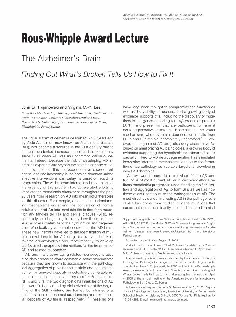
The major predictions of this tau hypothesis of AD neu-rodegeneration were that by converting normal tau intofunctionally impaired PHFtau, MTs would be unstableand depolymerize, thereby disrupting MT-based axonaltransport and that the attendant failure of affected neu-rons to export proteins from the cell body to distal pro-cesses and to retrieve substances (eg, trophic factors)internalized at axon terminals would compromise thefunction and viability of these neurons. Thus, as schemat-ically illustrated in Figure 1, we proposed that theseevents would culminate in neuronal dysfunction and de-generation, leading to the onset/progression of AD.
Since the most significant predictions of this tau hy-pothesis of AD neurodegeneration were that tau pathol-
ogies would cause brain degeneration by impairing in-traneuronal transport thereby compromising the functionand viability of affected cells, we subsequently proposedthat MT-stabilizing compounds such as the Food andDrug Administration-approved anti-cancer drug pacli-taxel could be used for the treatment of AD by offsettingthe loss of tau function after its conversion into PHFtau.8
However, the tau hypothesis of AD neurodegenerationdid not garner broad support until recently, and a signif-icant catalyst that led to a reassessment of this hypoth-esis came from a series of remarkable discoveries be-ginning in 1998. These studies showed that mutations inthe gene encoding tau were pathogenic for hereditaryfrontotemporal dementia (FTD) with parkinsonism linkedto chromosome 17 (FTDP-17) syndromes, all of which arecharacterized by prominent tau pathologies in the ab-sence of significant amounts of other disease-specificamyloid lesions.9–12 Thus, just like the discoveries ofgenetic mutations pathogenic for familial AD, these find-ings on the genetic basis of FTDP-17 provided compel-ling evidence that tau abnormalities were sufficient tocause neurodegenerative disease. Indeed, this notionwas supported further by evidence from follow-up studiesshowing that a number of tau gene mutations pathogenicfor FTDP-17 resulted in loss of tau functions or gain ofpotentially toxic properties, such as an increased amy-loidogenic propensity for mutant tau isoforms.13 Othermore recent catalysts for dispelling doubt about the crit-ical importance of tau pathologies as mediators of neu-rodegeneration in AD and other neurodegenerative dis-orders have been the successful development of worm,fly, and mouse models of tau pathologies that show closesimilarity to their authentic human counterparts.14 Forthese reasons, sporadic FTDs characterized by promi-nent tau lesions, familial FTDP-17 syndromes, and thepathological significance of tau abnormalities for mech-anisms of brain degeneration have become increasinglyintense focuses of basic and clinical research.
Thus, there is a growing body of evidence from diverselines of research indicating that brain degeneration in ADis mediated by tau pathologies, and we propose that thiscould be a major consequence of impaired intraneuronaltransport resulting from loss of function defects in tauand/or from toxic gain of functions by pathologically al-tered tau proteins, including their propensity to misfold,fibrillize, and form AD NFTs in neurons. Indeed, axonaltransport failure may be an underlying mechanism of anumber of other neurodegenerative disorders, some ofwhich are caused by pathogenic mutations in genesencoding proteins that serve as molecular motors foraxonal transport.15
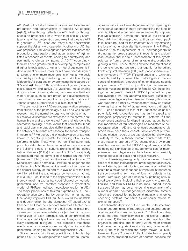
A schematic depiction of the currently understood mo-lecular underpinnings of retrograde and anterograde ax-onal transport is shown in Figure 2.15,16 This figure illus-trates the three major elements of the axonal transportmachinery: 1) the transported cargo (ie, vesicles, otherorganelles, proteins, and so forth); 2) the motor proteinsthat move the cargo (ie, kinesins, dynein, dynactin, etc);and 3) the rails on which the cargo moves (ie, MTs).However, Figure 2 does not fully illustrate the complexityof the axonal transport system of neurons because this
1184 Trojanowski and LeeAJP November 2005, Vol. 167, No. 5
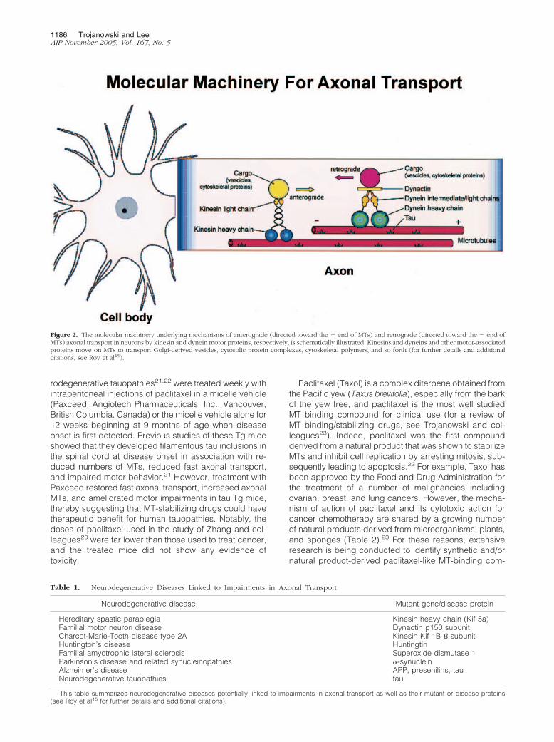
system also includes numerous other components suchas the many adaptor/linker and regulatory proteins thatare involved in the translocation of cargo away from(anterograde) or toward (retrograde) the cell body of aneuron.15,16 Because nearly all protein synthesis occursin the perikarya of neurons, defects in any one of thethree major components of the axonal transport systemdescribed above could impair axonal transport and resultin the dysfunction and death of neurons. Thus, axonaltransport has been hypothesized to be implicated inmechanisms of several neurodegenerative diseases assummarized in Table 1. Moreover, increasing support forthis hypothesis has come from the discovery of mutationsin genes encoding motor proteins, contributing to humanneurodegenerative diseases, as well as from emergingevidence for axonal transport defects in cell culture andanimal models of neurodegenerative disorders, despitecontroversies on the role of some neurodegenerative dis-ease proteins in modulating axonal transport.15,17–19 Ac-cordingly, because tau is a well characterized molecularcomponent of the axonal transport machinery that hasbeen shown to play a critical role in maintaining the MTnetwork required for axonal transport, it is highly plausi-ble that the tau abnormalities associated with AD andother neurodegenerative tauopathies, including heredi-
tary FTDP-17 syndromes caused by tau gene mutations,could disrupt axonal transport. Thus, by analogy with arailway transportation system, tau functions like the crossties on railroad tracks (MTs) on which trains (molecularmotors) convey cargo (vesicles, other organelles, pro-teins) to and from nodes on the railway network (destina-tions in neuronal perikarya and their processes) so thatthe loss of a critical number of trestle cross ties (theconsequence of converting tau into PHFtau) would resultin the buckling or splaying of the railroad tracks leadingto derailment of trains and the failure to deliver cargo toassigned destinations (impaired axonal transport) withdeleterious consequences for the railway network andthe communities it serves (the dysfunction and death ofaffected neurons).
To test the hypothesis that MT-stabilizing drugs couldhave therapeutic benefit in AD and related humantauopathies by offsetting the loss of normal tau functionsresulting from its hyperphosphorylation and sequestra-tion into tangles, we investigated this possibility using ananimal model of filamentous tau pathology.20 Signifi-cantly, these studies provided the first proof of conceptdata validating the use of MT-stabilizing drugs to treat ADand related tauopathies.20 Briefly, transgenic (Tg) mice(PrPT44) that model the neuropathology of human neu-
Figure 1. The misfolding, fibrillization, and sequestration of tau into filamentous inclusions is schematically depicted and is described in greater detail in the text.Proceeding from the normal neuron on the left, this tau hypothesis of AD neurodegeneration predicts that the cascade of events depicted schematically willcompromise the function and viability of neurons (shown to the right) through the pathological conversion of normal tau into PHFtau, which forms NFTs anddystrophic tau neurites thereby depleting levels of functional MT-binding/stabilizing tau below a critical point that results in the depolymerization of MTs and adisruption of axonal transport. As shown on the far right, the dissolution of degenerating tangle and dystrophic neurite-bearing neurons in the AD brain releasesabnormal tau into the extracellular space resulting in elevated levels of cerebrospinal fluid (CSF) tau, which is one of the most robust biomarkers of AD in livingpatients.
Tau-Focused AD Therapies 1185AJP November 2005, Vol. 167, No. 5
rodegenerative tauopathies21,22 were treated weekly withintraperitoneal injections of paclitaxel in a micelle vehicle(Paxceed; Angiotech Pharmaceuticals, Inc., Vancouver,British Columbia, Canada) or the micelle vehicle alone for12 weeks beginning at 9 months of age when diseaseonset is first detected. Previous studies of these Tg miceshowed that they developed filamentous tau inclusions inthe spinal cord at disease onset in association with re-duced numbers of MTs, reduced fast axonal transport,and impaired motor behavior.21 However, treatment withPaxceed restored fast axonal transport, increased axonalMTs, and ameliorated motor impairments in tau Tg mice,thereby suggesting that MT-stabilizing drugs could havetherapeutic benefit for human tauopathies. Notably, thedoses of paclitaxel used in the study of Zhang and col-leagues20 were far lower than those used to treat cancer,and the treated mice did not show any evidence oftoxicity.
Paclitaxel (Taxol) is a complex diterpene obtained fromthe Pacific yew (Taxus brevifolia), especially from the barkof the yew tree, and paclitaxel is the most well studiedMT binding compound for clinical use (for a review ofMT binding/stabilizing drugs, see Trojanowski and col-leagues23). Indeed, paclitaxel was the first compoundderived from a natural product that was shown to stabilizeMTs and inhibit cell replication by arresting mitosis, sub-sequently leading to apoptosis.23 For example, Taxol hasbeen approved by the Food and Drug Administration forthe treatment of a number of malignancies includingovarian, breast, and lung cancers. However, the mecha-nism of action of paclitaxel and its cytotoxic action forcancer chemotherapy are shared by a growing numberof natural products derived from microorganisms, plants,and sponges (Table 2).23 For these reasons, extensiveresearch is being conducted to identify synthetic and/ornatural product-derived paclitaxel-like MT-binding com-
Figure 2. The molecular machinery underlying mechanisms of anterograde (directed toward the � end of MTs) and retrograde (directed toward the � end ofMTs) axonal transport in neurons by kinesin and dynein motor proteins, respectively, is schematically illustrated. Kinesins and dyneins and other motor-associatedproteins move on MTs to transport Golgi-derived vesicles, cytosolic protein complexes, cytoskeletal polymers, and so forth (for further details and additionalcitations, see Roy et al15).
Table 1. Neurodegenerative Diseases Linked to Impairments in Axonal Transport
Neurodegenerative disease Mutant gene/disease protein
Hereditary spastic paraplegia Kinesin heavy chain (Kif 5a)Familial motor neuron disease Dynactin p150 subunitCharcot-Marie-Tooth disease type 2A Kinesin Kif 1B � subunitHuntington’s disease HuntingtinFamilial amyotrophic lateral sclerosis Superoxide dismutase 1Parkinson’s disease and related synucleinopathies �-synucleinAlzheimer’s disease APP, presenilins, tauNeurodegenerative tauopathies tau
This table summarizes neurodegenerative diseases potentially linked to impairments in axonal transport as well as their mutant or disease proteins(see Roy et al15 for further details and additional citations).
1186 Trojanowski and LeeAJP November 2005, Vol. 167, No. 5
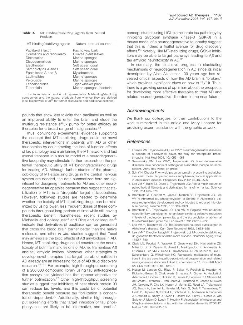
pounds that show less toxicity than paclitaxel as well asan improved ability to enter the brain and elude themultidrug resistance efflux pump for better efficacy astherapies for a broad range of malignancies.23
Thus, convincing experimental evidence supportingthe concept that MT-stabilizing drugs could be noveltherapeutic interventions in patients with AD or othertauopathies by counteracting the loss of function effectsof tau pathology and maintaining the MT network and fastaxonal transport in a mouse model of a neurodegenera-tive tauopathy may stimulate further research on the po-tential therapeutic utility of MT binding/stabilizing drugsfor treating AD. Although further studies of the pharma-cobiology of MT-stabilizing drugs in the central nervoussystem are needed, the data summarized here are sig-nificant for designing treatments for AD and other neuro-degenerative tauopathies because they suggest that sta-bilization of MTs is a “drugable” target in tauopathies.However, follow-up studies are needed to determinewhether the toxicity of MT-stabilizing drugs can be mini-mized by using lower, less frequent doses of these com-pounds throughout long periods of time while still havingtherapeutic benefit. Nonetheless, recent studies byMichaelis and colleagues24 and Rice and colleagues25
indicate that derivatives of paclitaxel can be generatedthat cross the blood brain barrier better than the nativemolecule, and other in vitro studies suggest that Taxolmay ameliorate the toxic effects of A� amyloidosis in AD.Hence, MT-stabilizing drugs could counteract the neuro-toxicity of both hallmark lesions of AD, ie, filamentous A�and tau amyloid lesions. Moreover, other strategies todevelop novel therapies that target tau abnormalities inAD already are an increasing focus of AD drug discoveryresearch.26–30 For example, high-throughput screeningof a 200,000 compound library using tau anti-aggrega-tion assays has yielded hits that appear attractive forfurther optimization.27 Other high-throughput screeningstudies suggest that inhibitors of heat shock protein 90can reduce tau levels, and this could be of potentialtherapeutic benefit because tau fibrillization is concen-tration-dependent.30 Additionally, similar high-through-put screening efforts that target inhibition of tau phos-phorylation are likely to be informative, and proof-of-
concept studies using LiCl to ameliorate tau pathology byinhibiting glycogen synthase kinase-3 (GSK-3) in amouse model of a neurodegenerative tauopathy suggestthat this is indeed a fruitful avenue for drug discoveryefforts.26 Notably, like MT-stabilizing drugs, GSK-3 inhib-itors may be able to target pathways leading to A� andtau amyloid neurotoxicity in AD.31
In summary, the extensive progress in elucidatingmechanisms of neurodegeneration in AD since its initialdescription by Alois Alzheimer 100 years ago has re-vealed critical aspects of how the AD brain is “broken,”which provides significant clues on how to “fix” it. Thus,there is a growing sense of optimism about the prospectsfor developing more effective therapies to treat AD andrelated neurodegenerative disorders in the near future.
Acknowledgments
We thank our colleagues for their contributions to thework summarized in this article and Mary Leonard forproviding expert assistance with the graphic artwork.
References
1. Forman MS, Trojanowski JQ, Lee VM-Y: Neurodegenerative diseases:a decade of discoveries paves the way for therapeutic break-throughs. Nat Med 2004, 10:1055–1063
2. Skovronsky DM, Lee VM-Y, Trojanowski JQ: Neurodegenerativediseases: new concepts of pathogenesis and their therapeutic impli-cations. Annu Rev Pathol (in press)
3. Suh Y-H, Checler F: Amyloid precursor protein, presenilins and alpha-synuclein: molecular pathogenesis and pharmacological applicationsin Alzheimer’s disease. Pharmacol Rev 2002, 54:469–525
4. Lee VM-Y, Balin BJ, Otvos L, Trojanowski JQ: A68: a major subunit ofpaired helical filaments and derivatized forms of normal tau. Science1991, 251:675–678
5. Bramblett GT, Goedert M, Jakes R, Merrick SE, Trojanowski JQ, LeeVM-Y: Abnormal tau phosphorylation at Ser396 in Alzheimer’s dis-ease recapitulates development and contributes to reduced microtu-bule binding. Neuron 1993, 10:1089–1099
6. Bramblett GT, Trojanowski JQ, Lee VM-Y: Regions with abundantneurofibrillary pathology in human brain exhibit a selective reductionin levels of binding-competent tau and the accumulation of abnormaltau-isoforms (A68 proteins). Lab Invest 1992, 66:212–222
7. Lee VM-Y, Trojanowski JQ: The disordered neuronal cytoskeleton inAlzheimer’s disease. Curr Opin Neurobiol 1992, 2:653–656
8. Lee VM-Y, Daughenbaugh R, Trojanowski JQ: Microtubule stabilizingdrugs for the treatment of Alzheimer’s disease. Neurobiol Aging 1994,15:S87–S89
9. Clark LN, Poorkaj P, Wszolek Z, Geschwind DH, Nasreddine ZS,Miller B, Li D, Payami H, Awert F, Markopoulou K, Andreadis A,D’Souza I, Lee VM-Y, Reed L, Trojanowski JQ, Zhukareva V, Bird T,Schellenberg G, Wilhelmsen KC: Pathogenic implications of muta-tions in the tau gene in pallido-ponto-nigral degeneration and relatedneurogenerative disorders linked to chromosome 17. Proc Natl AcadSci USA 1998, 95:13103–13107
10. Hutton M, Lendon CL, Rizzu P, Baker M, Froelich S, Houlden H,Pickering-Brown S, Chakraverty S, Isaacs A, Grover A, Hackett J,Adamson J, Lincoln S, Dickson D, Davies P, Petersen RC, Stevens M,de Graaff E, Wauters E, van Baren J, Hillebrand M, Joosse M, KwonJM, Nowotny P, Che LK, Norton J, Morris JC, Reed LA, TrojanowskiJQ, Basun H, Lannfelt L, Neystat M, Fahn S, Dark F, Tannenberg T,Dodd P, Hayward N, Kwok JBJ, Schofield PR, Andreadis A, SnowdenJ, Craufurd D, Neary D, Owen F, Oostra BA, Hardy J, Goate A, vanSwieten J, Mann D, Lynch T, Heutink P: Association of missense and5�-splice-site-mutations in tau with the inherited dementia FTDP-17.Nature 1998, 393:702–705
Table 2. MT Binding/Stabilizing Agents from NaturalProducts
MT binding/stabilizng agents Natural product source
Paclitaxel (Taxol) Pacific yew barkCoumarins and dicoumarol Diverse plant leavesDictostatins Marine spongesDiscodermolides Marine spongesEleutherobin Soft ocean coralSarcodictyins A and B Soft ocean coralEpothilones A and B MyxobacteriaLaulimalides Marine spongesPeloruside Marine spongesTaccalonolides Tiger whisker plantTubercidin Marine sponges, bacteria
This table lists a number of representative MT-binding/stabilizingcompounds and the natural products from whence they are derived(see Trojanowski et al23 for further discussion and additional citations).
Tau-Focused AD Therapies 1187AJP November 2005, Vol. 167, No. 5
11. Poorkaj P, Bird TE, Wijsman E, Nemens E, Garruto RM, Anderson L,Andreadis A, Wiederholt WC, Raskind M, Schellenberg GD: Tau is acandidate gene for chromosome 17 frontotemporal dementia. AnnNeurol 1998, 43:815–825
12. Spillantini MG, Murrell TR, Goedert M, Farlow MR, Klug A, Ghetti B:Mutation in the tau gene in familial multiple system tauopathy withpresenile dementia. Proc Natl Acad Sci USA 1998, 95:7737–7741
13. Hong M, Zhukareva V, Vogelsberg-Ragaglia V, Wszolek Z, Reed L,Miller BI, Geschwind DH, Bird TD, McKeel D, Goate A, Morris JC,Wilhelmsen KC, Schellenberg GD, Trojanowski JQ, Lee VM-Y: Muta-tion-specific functional impairments in distinct tau isoforms of hered-itary FTDP-17. Science 1998, 282:1914–1917
14. Lee VM-Y, Kenyon TK, Trojanowski JQ: Transgenic animal models oftauopathies. Biochem Biophys Acta 2005, 1739:251–259
15. Roy S, Zhang B, Lee VM-Y, Trojanowski JQ: Axonal transport defects:a common theme in neurodegenerative diseases. Acta Neuropathol2005, 109:5–13
16. Brown A: Axonal transport of membranous and nonmembranouscargoes: a unified perspective. J Cell Biol 2003, 160:817–821
17. Gunawardena S, Goldstein LS: Cargo-carrying motor vehicles on theneuronal highway: transport pathways and neurodegenerative dis-ease. J Neurobiol 2004, 58:258–271
18. Lazarov O, Morfini GA, Lee EB, Farah MH, Szodorai A, DeBoer SR,Koliatsos VE, Kins S, Lee VM-Y, Wong PC, Price DL, Brady ST,Sisodia SS: Axonal transport, amyloid precursor protein, kinesin-1,and the processing apparatus: revisited. J Neurosci 2005,25:2386–2395
19. Mandelkow EM, Stamer K, Vogel R, Thies E, Mandelkow E: Cloggingof axons by tau, inhibition of axonal traffic and starvation of synapses.Neurobiol Aging 2003, 24:1079–1085
20. Zhang B, Maiti A, Shively S, Lakhani F, McDonald-Jones G, Bruce J,Lee EB, Xie SX, Joyce S, Li C, Toleikis PM, Lee VM-Y, Trojanowski JQ:Microtubule binding drugs offset tau sequestration by stabilizingmicrotubules and reversing fast axonal transport deficits in a murineneurodegenerative tauopathy model. Proc Natl Acad Sci USA 2005,102:227–231
21. Ishihara T, Hong M, Zhang B, Nakagawa Y, Lee MK, Trojanowski JQ,Lee VM-Y: Age-dependent emergence and progression of a tauopa-
thy in transgenic mice engineered to overexpress the shortest humantau isoform. Neuron 1999, 24:751–762
22. Ishihara T, Zhang B, Higuchi M, Yoshiyama Y, Trojanowski JQ, LeeVM-Y: Age dependent induction of congophilic neurofibrillary tauinclusions in tau transgenic mice. Am J Pathol 2001, 158:555–561
23. Trojanowski JQ, Smith III AB, Huryn D, Lee VM-Y: Microtubule-stabi-lising drugs for therapy of Alzheimer’s disease and other neurode-generative disorders with axonal transport impairments. Exp OpinPharmacother 2005, 6:683–686
24. Michaelis ML, Ansar S, Chen Y, Reiff ER, Seyb KI, Himes RH, AudusKL, Georg GI: Beta-amyloid-induced neurodegeneration and protec-tion by structurally diverse microtubule-stabilizing agents. J Pharma-col Exp Ther 2005, 312:659–668
25. Rice A, Liu Y, Michaelis ML, Himes RH, Georg GI, Audus KL: Chem-ical modification of paclitaxel (Taxol) reduces P-glycoprotein interac-tions and increases permeation across the blood-brain barrier in vitroand in situ. J Med Chem 2005, 48:832–838
26. Noble W, Planel E, Zehr C, Olm V, Meyerson J, Suleman F, Gaynor K,Wang L, Lafrancois J, Feinstein B, Burns M, Krishnamurthy P, Wen Y,Bhat R, Lewis J, Dickson D, Duff K: Inhibition of glycogen synthasekinase-3 by lithium correlates with reduced tauopathy and degener-ation in vivo. Proc Natl Acad Sci USA 2005, 102:6990–6995
27. Pickhardt M, Gazova Z, von Bergen M, Khlistunova I, Wang Y, Has-cher A, Mandelkow EM, Biernat J, Mandelkow E: Anthraquinonesinhibit tau aggregation and dissolve Alzheimer’s paired helical fila-ments in vitro and in cells. J Biol Chem 2005, 280:3628–3635
28. Fillit HM, Refolo LM: Advancing drug discovery for Alzheimer’s dis-ease. Curr Alzheimer Res 2005, 2:105–109
29. Pickhardt M, von Bergen M, Gazova Z, Hascher A, Biernat J, Man-delkow EM, Mandelkow E: Screening for inhibitors of tau polymeriza-tion. Curr Alzheimer Res 2005, 2:219–226
30. Dickey CA, Eriksen J, Kamal A, Burrows F, Kasibhatla S, Eckman CB,Hutton M, Petrucelli L: Development of a high throughput drugscreening assay for the detection of changes in tau levels: proof ofconcept with HSP90 inhibitors. Curr Alzheimer Res 2005, 2:231–239
31. Phiel CJ, Wilson CA, Lee VM-Y, Klein PS: GSK-3 alpha regulatesproduction of Alzheimer’s disease amyloid-beta peptides. Nature2003, 423:435–439
1188 Trojanowski and LeeAJP November 2005, Vol. 167, No. 5