tf 06928151958 chapter9 skin hair
TRANSCRIPT
������������
117
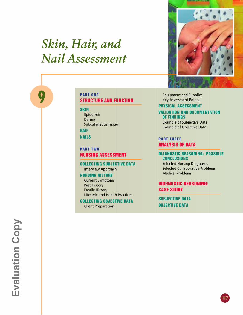
Skin, Hair, and Nail Assessment
9 P A R T O N E
STRUCTURE AND FUNCTION
SKINEpidermisDermisSubcutaneous Tissue
HAIR
NAILS
P A R T T W O
NURSING ASSESSMENT
COLLECTING SUBJECTIVE DATAInterview Approach
NURSING HISTORYCurrent SymptomsPast HistoryFamily HistoryLifestyle and Health Practices
COLLECTING OBJECTIVE DATAClient Preparation
Equipment and SuppliesKey Assessment Points
PHYSICAL ASSESSMENT
VALIDATION AND DOCUMENTATION OF FINDINGSExample of Subjective DataExample of Objective Data
P A R T T H R E E
ANALYSIS OF DATA
DIAGNOSTIC REASONING: POSSIBLECONCLUSIONSSelected Nursing DiagnosesSelected Collaborative ProblemsMedical Problems
DIOGNOSTIC REASONING: CASE STUDY
SUBJECTIVE DATA
OBJECTIVE DATA
������������
Structure and Function
118
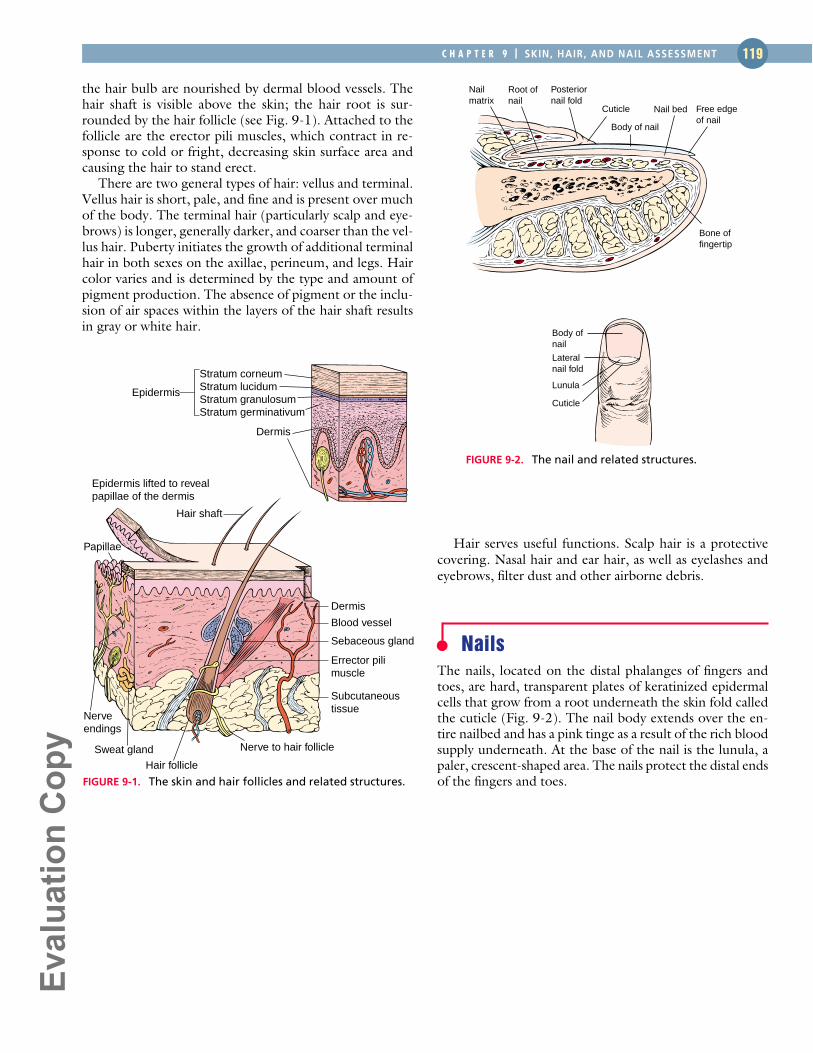
The skin, hair, and nails are external structures that serve avariety of specialized functions. The sebaceous and sweatglands originating within the skin also have many vital func-tions. Each of these structures and their function is de-scribed separately.
SkinThe skin is composed of three layers, the epidermis, dermis,and subcutaneous tissue (Fig. 9-1). The skin is thicker onthe palms of the hands and soles of the feet and is continu-ous with the mucous membranes at the orifices of the body.Subcutaneous tissue, which contains varying amounts offat, connects the skin to underlying structures.
The skin is a physical barrier that protects the under-lying tissues and structures from microorganisms, physicaltrauma, ultraviolet radiation, and dehydration. It plays avital role in temperature maintenance, fluid and electrolytebalance, absorption, excretion, sensation, immunity, andvitamin D synthesis. The skin also provides an individualidentity to a person’s appearance.
EPIDERMIS
The epidermis, the outer layer of skin, is composed of fourdistinct layers (see Fig. 9-1): the stratum corneum, stratumlucidum, stratum granulosum, and stratum germinativum.The outermost layer consists of dead, keratinized cells thatrender the skin waterproof. (Keratin is a scleroprotein thatis insoluble in water. The epidermis, hair, nails, dentalenamel, and horny tissues are composed of keratin.) Theepidermal layer is almost completely replaced every 3 to 4 weeks. The innermost layer of the epidermis (stratum ger-minativum) is the only layer that undergoes cell division andcontains melanin (brown pigment) and keratin-forming cells.Skin color depends on the amount of melanin and carotene(yellow pigment) contained in the skin and the volume ofblood containing hemoglobin, the oxygen-binding pigmentthat circulates in the dermis.
DERMIS
The inner layer of skin is the dermis (see Fig. 9-1). It is con-nected to the epidermis by means of papillae. These papil-lae form the base for the visible swirls or friction ridges thatprovide the unique pattern of fingerprints with which we
are familiar. Ridges also appear on the palms of the hands,the toes, and the soles of the feet. The dermis is a well-vascularized connective tissue layer containing collagen andelastic fibers, nerve endings, and lymph vessels. It is also theorigin of hair follicles, sebaceous glands, and sweat glands.
Sebaceous Glands
The sebaceous glands (see Fig. 9-1) develop from hair fol-licles and, therefore, are present over most of the body, ex-cluding the soles and palms. They secrete an oily substancecalled sebum that lubricates hair and skin and reduces waterloss through the skin. Sebum also has some fungicidal andbactericidal effects.
Sweat Glands
Sweat glands (see Fig. 9-1) are of two types, eccrine andapocrine. The eccrine glands are located over the entire skinsurface and secrete an odorless, colorless fluid, the evapora-tion of which is vital to the regulation of body temperature.The apocrine glands are concentrated in the axillae, per-ineum, and areolae of the breast and usually open througha hair follicle. They secrete a milky sweat. The interaction ofsweat with skin bacteria produces a characteristic body odor.Apocrine glands are dormant until puberty, at which timethey become active. In women, apocrine secretions arelinked with the menstrual cycle.
SUBCUTANEOUS TISSUE
Merging with the dermis is the subcutaneous tissue, whichis a loose connective tissue containing fat cells, blood ves-sels, nerves, and the remaining portions of sweat glands andhair follicles (see Fig. 9-1). The subcutaneous tissue assistswith heat regulation and contains the vascular pathways forthe supply of nutrients and removal of waste products fromthe skin.
HairHair consists of layers of keratinized cells found over muchof the body except for the lips, nipples, soles of the feet,palms of the hands, labia minora, and penis. Hair developswithin a sheath of epidermal cells called the hair follicle.Hair growth occurs at the base of the follicle, where cells in
P A R TO N E
������������
Hair serves useful functions. Scalp hair is a protectivecovering. Nasal hair and ear hair, as well as eyelashes andeyebrows, filter dust and other airborne debris.
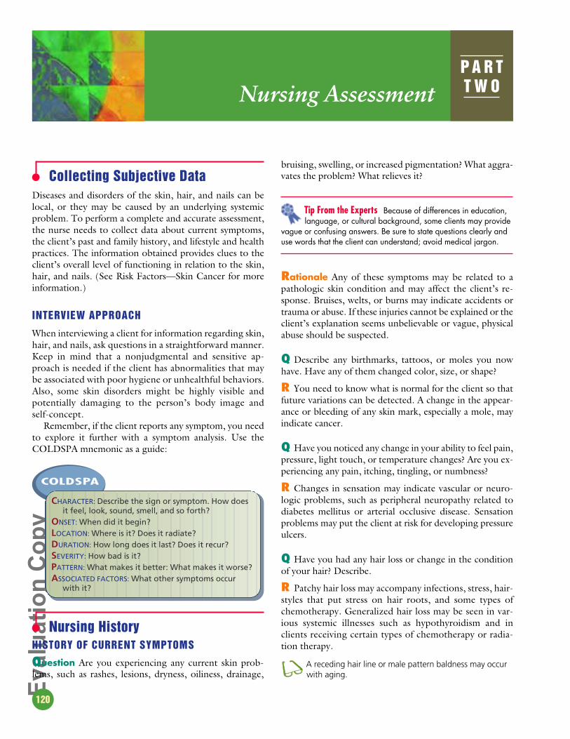
NailsThe nails, located on the distal phalanges of fingers andtoes, are hard, transparent plates of keratinized epidermalcells that grow from a root underneath the skin fold calledthe cuticle (Fig. 9-2). The nail body extends over the en-tire nailbed and has a pink tinge as a result of the rich bloodsupply underneath. At the base of the nail is the lunula, apaler, crescent-shaped area. The nails protect the distal endsof the fingers and toes.
the hair bulb are nourished by dermal blood vessels. Thehair shaft is visible above the skin; the hair root is sur-rounded by the hair follicle (see Fig. 9-1). Attached to thefollicle are the erector pili muscles, which contract in re-sponse to cold or fright, decreasing skin surface area andcausing the hair to stand erect.
There are two general types of hair: vellus and terminal.Vellus hair is short, pale, and fine and is present over muchof the body. The terminal hair (particularly scalp and eye-brows) is longer, generally darker, and coarser than the vel-lus hair. Puberty initiates the growth of additional terminalhair in both sexes on the axillae, perineum, and legs. Haircolor varies and is determined by the type and amount ofpigment production. The absence of pigment or the inclu-sion of air spaces within the layers of the hair shaft resultsin gray or white hair.
C H A P T E R 9 | SKIN, HAIR, AND NAIL ASSESSMENT 119
Stratum corneumStratum lucidumStratum granulosumStratum germinativum
Epidermis
Epidermis lifted to revealpapillae of the dermis
Hair shaft
Dermis
Papillae
Nerveendings
Sweat gland Nerve to hair follicle
Hair follicle
Dermis
Errector pili muscle
Blood vessel
Sebaceous gland
Subcutaneous tissue
FIGURE 9-1. The skin and hair follicles and related structures.
Nailmatrix
Body of nail
Cuticle
Root ofnail
Posteriornail fold
Bone of fingertip
Free edgeof nail
Nail bed
FIGURE 9-2. The nail and related structures.
Body ofnail
Lateralnail fold
Lunula
Cuticle
������������
Nursing Assessment
120
Collecting Subjective DataDiseases and disorders of the skin, hair, and nails can belocal, or they may be caused by an underlying systemicproblem. To perform a complete and accurate assessment,the nurse needs to collect data about current symptoms,the client’s past and family history, and lifestyle and healthpractices. The information obtained provides clues to theclient’s overall level of functioning in relation to the skin,hair, and nails. (See Risk Factors—Skin Cancer for moreinformation.)
INTERVIEW APPROACH
When interviewing a client for information regarding skin,hair, and nails, ask questions in a straightforward manner.Keep in mind that a nonjudgmental and sensitive ap-proach is needed if the client has abnormalities that maybe associated with poor hygiene or unhealthful behaviors.Also, some skin disorders might be highly visible and potentially damaging to the person’s body image and self-concept.
Remember, if the client reports any symptom, you needto explore it further with a symptom analysis. Use theCOLDSPA mnemonic as a guide:
bruising, swelling, or increased pigmentation? What aggra-vates the problem? What relieves it?
Tip From the Experts Because of differences in education,language, or cultural background, some clients may provide
vague or confusing answers. Be sure to state questions clearly anduse words that the client can understand; avoid medical jargon.
Rationale Any of these symptoms may be related to apathologic skin condition and may affect the client’s re-sponse. Bruises, welts, or burns may indicate accidents ortrauma or abuse. If these injuries cannot be explained or theclient’s explanation seems unbelievable or vague, physicalabuse should be suspected.
Q Describe any birthmarks, tattoos, or moles you nowhave. Have any of them changed color, size, or shape?
R You need to know what is normal for the client so thatfuture variations can be detected. A change in the appear-ance or bleeding of any skin mark, especially a mole, mayindicate cancer.
Q Have you noticed any change in your ability to feel pain,pressure, light touch, or temperature changes? Are you ex-periencing any pain, itching, tingling, or numbness?
R Changes in sensation may indicate vascular or neuro-logic problems, such as peripheral neuropathy related todiabetes mellitus or arterial occlusive disease. Sensationproblems may put the client at risk for developing pressureulcers.
Q Have you had any hair loss or change in the conditionof your hair? Describe.
R Patchy hair loss may accompany infections, stress, hair-styles that put stress on hair roots, and some types ofchemotherapy. Generalized hair loss may be seen in var-ious systemic illnesses such as hypothyroidism and inclients receiving certain types of chemotherapy or radia-tion therapy.
A receding hair line or male pattern baldness may occurwith aging.
P A R TT W O
CHARACTER: Describe the sign or symptom. How doesit feel, look, sound, smell, and so forth?
ONSET: When did it begin?LOCATION: Where is it? Does it radiate?DURATION: How long does it last? Does it recur?SEVERITY: How bad is it?PATTERN: What makes it better: What makes it worse?ASSOCIATED FACTORS: What other symptoms occur
with it?
Nursing HistoryHISTORY OF CURRENT SYMPTOMS
Question Are you experiencing any current skin prob-lems, such as rashes, lesions, dryness, oiliness, drainage,
������������
RISK FACTORSSkin Cancer
OVERVIEW
Skin cancer is the most common of cancers. It occurs in three types: melanoma, basal cell carcinoma(BCC), and squamous cell carcinoma (SCC). BCC and SCC are nonmelanomas. It was estimated that92,000 new melanoma cases and 2.75 million nonmelanocyte cases occur worldwide each year(Armstrong & Dricker, 1995). The American Cancer Society predicted that there would be 1,900deaths from nonmelanoma skin cancers and 7,700 deaths from melanoma during the year 2000.
The incidence of melanoma (new cases per 100,000 population) increased from 5.7 to 13.8 be-tween 1973 and 2000. Melanoma accounts for 4% of skin cancers but 79% of skin cancer deaths.Nonmelanocyte skin cancers are the most common and are also increasing in populations that areheavily exposed to sunlight, especially in areas of ozone depletion. Davidowitz, Belafsky, andAmedee (1999) state that melanoma will develop in 1 in 70 white Americans.
Intermittent exposure to the sun or ultraviolet radiation is associated with greatest risk formelanoma and for BCC, but overall amount of exposure is thought to be associated with SCC. SCC ismost common on body sites with very heavy sun exposure, whereas BCC is most common on siteswith moderate exposure (ie, upper trunk or women’s lower legs).
Precursor lesions occur for some melanomas (benign or dysplastic nevi) and for invasive SCC(actinic keratoses or SCC in situ), but there are no precursor lesions for BCC (Gloster & Brodland, 1995).
R ISK FACTORS
• Sun exposure, especially intermittent patternwith sunburn; risk increases if excessive sunexposure began in childhood
• Nonsolar sources of ultraviolet radiation(tanning booth, sunlamps)
• Medical therapies, such as PUVA and ionizingradiation
• Family history and genetic susceptibility (espe-cially for malignant melanoma)
• Moles, especially atypical lesions
• Pigmentation irregularities (albinism, burn scars)
• Fair skin that burns and freckles easily; light hair
• Immunosuppression
• Age; risk increases with increasing age
• Male gender (for nonmelanoma cancers)
• Chemical exposure (arsenic, tar, coal, paraffin,some oils for nonmelanoma cancers)
• Human papillomavirus (nonmelanoma cancers)
• Xeroderma pigmentosum (rare, inheritedcondition)
• Long-term skin inflammation or injury(nonmelanoma)
RISK REDUCT ION TEACHING T IPS
• Reduce sun exposure.
• Always use sunscreen (SPF 15 or higher) whensun exposure is anticipated.
• Wear long-sleeved shirts and wide-brimmedhats.
• Avoid sunburns.
• Avoid intermittent tanning.
• Understand the link between sun exposure andskin cancer and the accumulating effects of sunexposure on developing cancers.
• Examine the skin for suspect lesions. If there isanything unusual, seek professional advice assoon as possible.
CULTURAL CONSIDERAT IONS
The darkness of skin pigmentation affects the incidence of all skin cancers, with the lowest rates occur-ring in Asians and the highest rates in white Australians. The most susceptible are people with palewhite, freckled skin and red hair. Australians have mounted an intense campaign that emphasizeswearing sunscreen, long sleeves, and hats any time they are in the sun. Although less susceptible to skincancer, African Americans have two additional risk factors beside the ones listed previously. These arehigher incidences of chronic inflammatory skin diseases and chronic discoid lupus erythematosus (Halder& Bridgeman-Shah, 1995). Teaching about skin cancer prevention and diagnosis should be provided toAfrican Americans and Asians, even though they have a lower incidence than whites (Hall & Rogers, 1999).
C H A P T E R 9 | SKIN, HAIR, AND NAIL ASSESSMENT 121
������������
HEALTH ASSESSMENT IN NURSING 122
Q Have you had any change in the condition or appear-ance of your nails? Describe.
R Nail changes may be seen in systemic disorders such asmalnutrition or with local irritation (eg, nail biting).
Q Do you have trouble controlling body odor? How muchdo you perspire?
R Uncontrolled body odor or excessive or insufficient per-spiration may indicate an abnormality with the sweat glandsor an endocrine problem such as hypothyroidism or hyper-thyroidism. Poor hygiene practices may account for bodyodor, and health education may be indicated.
Perspiration decreases with aging because sweat glandactivity decreases.
Because of decreased sweat production, most Asians andNative Americans have mild to no body odor, whereas
Caucasians and African Americans tend to have a strong bodyodor (Andrews & Boyle, 1999), unless they use antiperspirant ordeodorant products. Any strong body odor may indicate anabnormality.
PAST HISTORY
Q Describe any previous problems with skin, hair, or nails,including any treatment or surgery and its effectiveness.
R Current problems may be a recurrence of previous ones.Visible scars may be explained by previous problems.
Q Have you ever had any allergic skin reactions to food,medications, plants, or other environmental substances?
R Various types of allergens can precipitate a variety of skineruptions.
Q Have you had a fever, nausea, vomiting, gastrointestinal(GI), or respiratory problems?
R Some skin rashes or lesions may be related to viruses orbacteria.
FAMILY HISTORY
Q Has anyone in your family had a recent illness, rash, orother skin problem or allergy? Describe.
R Acne and atopic dermatitis tend to be familial. Viruses(eg, chickenpox, measles) can be highly contagious. Someallergies may be identified from family history.
Q Has anyone in your family had skin cancer?
R A genetic component is associated with skin cancer,especially malignant melanoma.
LIFESTYLE AND HEALTH PRACTICES
Q Do you sunbathe? How much sun or tanning-booth ex-posure do you get? What type of protection do you use?
R Sun exposure can cause premature aging of skin and in-crease the risk of cancer. Hair can also be damaged by toomuch sun.
Q In your daily activities, are you regularly exposed tochemicals that may harm the skin (eg, paint, bleach, clean-ing products, weed killers, insect repellents, petroleum)?
R Any of these substances have the potential to irritate ordamage the skin, hair, or nails.
Q Do you spend long periods of time sitting or lying inone position?
R Older, disabled, or immobile clients who spend long pe-riods of time in one position are at risk for pressure ulcers.
Q Have you had any exposure to extreme temperatures?
R Temperature extremes affect the blood supply to theskin and can damage the skin layers. Examples includefrostbite and burns.
Q What is your daily routine for skin, hair, and nail care?What products do you use (eg, soaps, lotions, oils, cosmetics,self-tanning products, razor type, hair spray, shampoo,coloring, nail enamel)? How do you cut your nails?
R Regular habits provide information on hygiene andlifestyle. The products used may also be a cause of an ab-normality. Improper nail-cutting technique can lead to in-grown nails or infection.
Decreased flexibility and mobility may impair the abilityof some elderly clients to maintain proper hygiene prac-
tices, such as nail cutting, bathing, and hair care.
Q What kinds of foods do you consume in a typical day?How much fluid do you drink each day?
R A balanced diet is necessary for healthy skin, hair, andnails. Adequate fluid intake is required to maintain skinelasticity.
Q For female clients: Are you pregnant? Are your menstrualperiods regular?
R Some skin and hair conditions can result from hormonalimbalance.
Q Do skin problems limit any of your normal activities?
R Certain activities such as hiking, camping, and gardeningmay expose the client to allergens such as poison ivy.Moreover, exposure to the sun can aggravate conditionssuch as scleroderma. In addition, general home maintenance(eg, cleaning, car washing) may expose the client to certaincleaning products to which he or she is sensitive or allergic.
������������
Q Describe any skin disorder that prevents you from en-joying your relationships.
R Skin, hair, or nail problems, especially if visible, may im-pair the client’s ability to interact comfortably with othersbecause of embarrassment or rejection by others.
C H A P T E R 9 | SKIN, HAIR, AND NAIL ASSESSMENT 123
Q How much stress do you have in your life? Describe.R Stress can cause or exacerbate skin abnormalities.
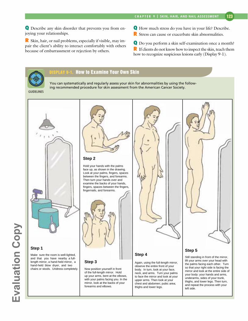
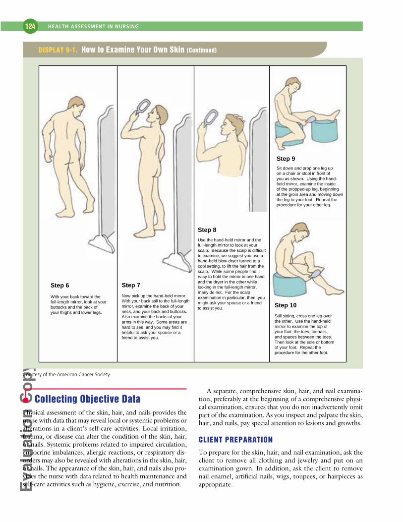
Q Do you perform a skin self-examination once a month?R If clients do not know how to inspect the skin, teach themhow to recognize suspicious lesions early (Display 9-1).
DISPLAY 9-1. How to Examine Your Own Skin
You can systematically and regularly assess your skin for abnormalities by using the follow-ing recommended procedure for skin assessment from the American Cancer Society.
Step 1
Step 2
Step 3
Step 4Make sure the room is well-lighted,and that you have nearby a full-length mirror, a hand-held mirror, a hand-held blow dryer, and two chairs or stools. Undress completely.
Hold your hands with the palmsface up, as shown in the drawing.Look at your palms, fingers, spacesbetween the fingers, and forearms.Then turn your hands over andexamine the backs of your hands,fingers, spaces between the fingers,fingernails, and forearms.
Now position yourself in frontof the full-length mirror. Holdup your arms, bent at the elbows,with your palms facing you. In themirror, look at the backs of your forearms and elbows.
Again, using the full-length mirror,observe the entire front of your body. In turn, look at your face, neck, and arms. Turn your palms to face the mirror and look at your upper arms. Then look at your chest and abdomen; pubic area; thighs and lower legs.
Step 5
Still standing in front of the mirror,lift your arms over your head withthe palms facing each other. Turnso that your right side is facing the mirror and look at the entire side ofyour body: your hands and arms,underarms, sides of your trunk, thighs, and lower legs. Then turn,and repeat the process with your left side.
GUIDELINES
������������
Collecting Objective DataPhysical assessment of the skin, hair, and nails provides thenurse with data that may reveal local or systemic problems oralterations in a client’s self-care activities. Local irritation,trauma, or disease can alter the condition of the skin, hair, or nails. Systemic problems related to impaired circulation,endocrine imbalances, allergic reactions, or respiratory dis-orders may also be revealed with alterations in the skin, hair,or nails. The appearance of the skin, hair, and nails also pro-vides the nurse with data related to health maintenance andself-care activities such as hygiene, exercise, and nutrition.
A separate, comprehensive skin, hair, and nail examina-tion, preferably at the beginning of a comprehensive physi-cal examination, ensures that you do not inadvertently omitpart of the examination. As you inspect and palpate the skin,hair, and nails, pay special attention to lesions and growths.
CLIENT PREPARATION
To prepare for the skin, hair, and nail examination, ask theclient to remove all clothing and jewelry and put on anexamination gown. In addition, ask the client to removenail enamel, artificial nails, wigs, toupees, or hairpieces asappropriate.
HEALTH ASSESSMENT IN NURSING 124
Courtesy of the American Cancer Society.
Step 6 Step 7
Step 9
Step 10With your back toward thefull-length mirror, look at yourbuttocks and the back ofyour thighs and lower legs.
Now pick up the hand-held mirror.With your back still to the full-lengthmirror, examine the back of your neck, and your back and buttocks.Also examine the backs of your arms in this way. Some areas are hard to see, and you may find ithelpful to ask your spouse or afriend to assist you.
Sit down and prop one leg upon a chair or stool in front ofyou as shown. Using the hand-held mirror, examine the inside of the propped-up leg, beginningat the groin area and moving downthe leg to your foot. Repeat theprocedure for your other leg.
Still sitting, cross one leg overthe other. Use the hand-held mirror to examine the top of your foot. the toes, toenails,and spaces between the toes.Then look at the sole or bottomof your foot. Repeat the procedure for the other foot.
Step 8
Use the hand-held mirror and thefull-length mirror to look at yourscalp. Because the scalp is difficult to examine, we suggest you use ahand-held blow dryer turned to acool setting, to lift the hair from thescalp. While some people find iteasy to hold the mirror in one hand and the dryer in the other whilelooking in the full-length mirror,many do not. For the scalp examination in particular, then, youmight ask your spouse or a friendto assist you.
DISPLAY 9-1. How to Examine Your Own Skin (Continued)
������������
(continued )
PHYSICAL ASSESSMENTASSESSMENT PROCEDURE NORMAL FINDINGS ABNORMAL FINDINGS
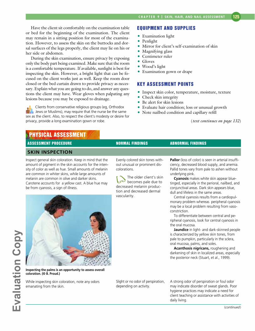
Inspect general skin coloration. Keep in mind that theamount of pigment in the skin accounts for the inten-sity of color as well as hue. Small amounts of melaninare common in whiter skins, while large amounts ofmelanin are common in olive and darker skins.Carotene accounts for a yellow cast. A blue hue maybe from cyanosis, a sign of illness.
Evenly colored skin tones with-out unusual or prominent dis-colorations.
The older client’s skinbecomes pale due to
decreased melanin produc-tion and decreased dermalvascularity.
Pallor (loss of color) is seen in arterial insuffi-ciency, decreased blood supply, and anemia.Pallid tones vary from pale to ashen withoutunderlying pink.
Cyanosis makes white skin appear blue-tinged, especially in the perioral, nailbed, andconjunctival areas. Dark skin appears blue,dull and lifeless in the same areas.
Central cyanosis results from a cardiopul-monary problem whereas peripheral cyanosismay be a local problem resulting from vaso-constriction.
To differentiate between central and pe-ripheral cyanosis, look for central cyanosis inthe oral mucosa.
Jaundice in light- and dark-skinned peopleis characterized by yellow skin tones, frompale to pumpkin, particularly in the sclera,oral mucosa, palms, and soles.
Acanthosis nigricans, roughening anddarkening of skin in localized areas, especiallythe posterior neck (Stuart, et al., 1999).
SKIN INSPECTION
Inspecting the palms is an opportunity to assess overallcoloration. (© B. Proud.)
Have the client sit comfortably on the examination tableor bed for the beginning of the examination. The clientmay remain in a sitting position for most of the examina-tion. However, to assess the skin on the buttocks and dor-sal surfaces of the legs properly, the client may lie on his orher side or abdomen.
During the skin examination, ensure privacy by exposingonly the body part being examined. Make sure that the roomis a comfortable temperature. If available, sunlight is best forinspecting the skin. However, a bright light that can be fo-cused on the client works just as well. Keep the room doorclosed or the bed curtain drawn to provide privacy as neces-sary. Explain what you are going to do, and answer any ques-tions the client may have. Wear gloves when palpating anylesions because you may be exposed to drainage.
Clients from conservative religious groups (eg, OrthodoxJews or Muslims), may require that the nurse be the same
sex as the client. Also, to respect the client’s modesty or desire forprivacy, provide a long examination gown or robe.
EQUIPMENT AND SUPPLIES
• Examination light• Penlight• Mirror for client’s self-examination of skin• Magnifying glass• Centimeter ruler• Gloves• Wood’s light• Examination gown or drape
KEY ASSESSMENT POINTS
• Inspect skin color, temperature, moisture, texture• Check skin integrity• Be alert for skin lesions• Evaluate hair condition; loss or unusual growth• Note nailbed condition and capillary refill
C H A P T E R 9 | SKIN, HAIR, AND NAIL ASSESSMENT 125
(text continues on page 132)
While inspecting skin coloration, note any odors emanating from the skin.
Slight or no odor of perspiration,depending on activity.
A strong odor of perspiration or foul odormay indicate disorder of sweat glands. Poorhygiene practices may indicate a need forclient teaching or assistance with activities ofdaily living.
������������
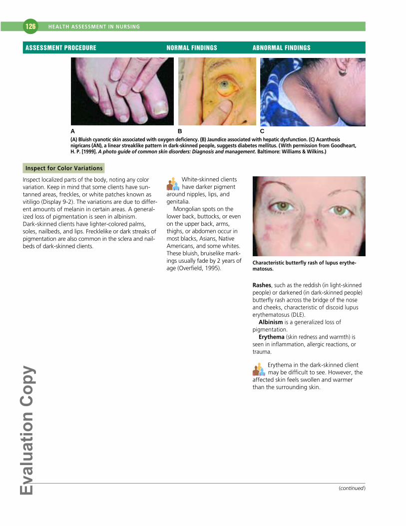
Characteristic butterfly rash of lupus erythe-matosus.
Erythema in the dark-skinned clientmay be difficult to see. However, the
affected skin feels swollen and warmerthan the surrounding skin.
HEALTH ASSESSMENT IN NURSING 126
(continued )
Inspect for Color Variations
Inspect localized parts of the body, noting any colorvariation. Keep in mind that some clients have sun-tanned areas, freckles, or white patches known asvitiligo (Display 9-2). The variations are due to differ-ent amounts of melanin in certain areas. A general-ized loss of pigmentation is seen in albinism.Dark-skinned clients have lighter-colored palms,soles, nailbeds, and lips. Frecklelike or dark streaks ofpigmentation are also common in the sclera and nail-beds of dark-skinned clients.
ASSESSMENT PROCEDURE NORMAL FINDINGS ABNORMAL FINDINGS
White-skinned clientshave darker pigment
around nipples, lips, and genitalia.
Mongolian spots on thelower back, buttocks, or evenon the upper back, arms,thighs, or abdomen occur inmost blacks, Asians, NativeAmericans, and some whites.These bluish, bruiselike mark-ings usually fade by 2 years ofage (Overfield, 1995).
A B C(A) Bluish cyanotic skin associated with oxygen deficiency. (B) Jaundice associated with hepatic dysfunction. (C) Acanthosis nigricans (AN), a linear streaklike pattern in dark-skinned people, suggests diabetes mellitus. (With permission from Goodheart,H. P. [1999]. A photo guide of common skin disorders: Diagnosis and management. Baltimore: Williams & Wilkins.)
Rashes, such as the reddish (in light-skinnedpeople) or darkened (in dark-skinned people)butterfly rash across the bridge of the noseand cheeks, characteristic of discoid lupuserythematosus (DLE).
Albinism is a generalized loss of pigmentation.
Erythema (skin redness and warmth) isseen in inflammation, allergic reactions, ortrauma.
������������
C H A P T E R 9 | SKIN, HAIR, AND NAIL ASSESSMENT 127
(continued)
Inspect for Skin Integrity
Check skin integrity, especially carefully in pressurepoint areas (eg, sacrum, hips, elbows). If any skin break-down is noted, use a scale to document the degree ofskin breakdown.
Inspect for Lesions
Observe the skin surface to detect abnormalities.
If you suspect a fungus, shine a Wood’s light (an ultra-violet light filtered through a special glass) on the lesion.
If you observe a lesion, note its location, distribution,and configuration.
Palpate Skin to Assess Texture
Use the palmar surface of your three middle fingers topalpate skin texture.
Palpate to Assess Thickness
If lesions are noted when assessing skin thickness, putgloves on and palpate the lesion between the thumband finger. Observe for drainage or other characteris-tics. Measure the lesion with a centimeter ruler.
ASSESSMENT PROCEDURE NORMAL FINDINGS ABNORMAL FINDINGS
Skin is intact, and there are noreddened areas.
Smooth, without lesions.Stretch marks (striae), healedscars, freckles, moles, or birth-marks are common findings (see Display 9-2).
Older clients may haveskin lesions because of
aging. Some examples areseborrheic or senile keratoses,senile lentigines, cherry an-giomas, purpura, and cuta-neous tags and horns.
Lesion does not fluoresce.
Normal lesions may be moles,freckles, birthmarks, and thelike. They may be scattered overthe skin in no particular pattern.
Skin is smooth and even.
Skin is normally thin, but cal-luses (rough, thick sections ofepidermis) are common onareas of the body that are ex-posed to constant pressure.
Skin breakdown is initially noted as a reddenedarea on the skin that may progress to seriousand painful pressure ulcers (Display 9-3).Depending on the color of the client’s skin,reddened areas may not be prominent, al-though the skin may feel warmer in the areaof breakdown than elsewhere.
Lesions may indicate local or systemic prob-lems. Primary lesions (Display 9-4) arise fromnormal skin due to irritation or disease.Secondary lesions (Display 9-5) arise fromchanges in primary lesions. Vascular lesions(Display 9-6), reddish-bluish lesions, are seenwith bleeding, venous pressure, aging, liverdisease, or pregnancy. Skin cancer lesions canbe either primary or secondary lesions and areclassified as squamous cell carcinoma, basalcell carcinoma, or malignant melanoma(Display 9-7).
Blue-green fluorescence indicates fungalinfection.
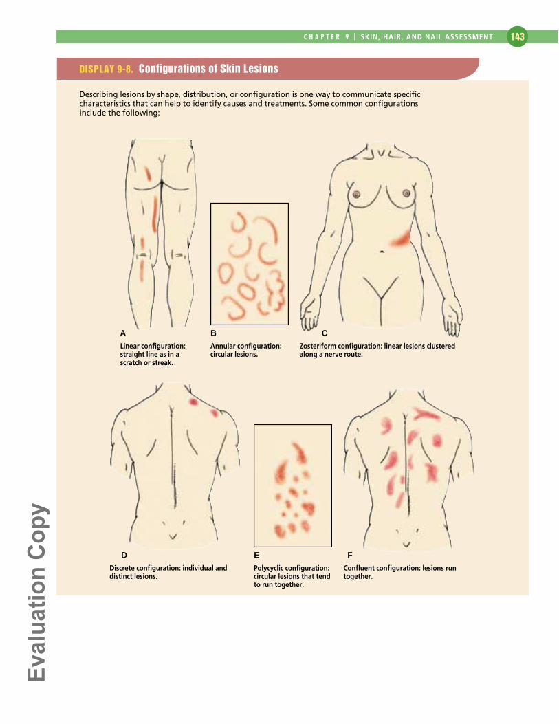
In abnormal findings, distribution may bediffuse (scattered all over), localized to onearea, or in sun-exposed areas. Configurationmay be discrete (separate and distinct),grouped (clustered), confluent (merged), linear (in a line), annular and arciform (circu-lar or arcing), or zosteriform (linear along anerve route; Display 9-8).
Rough, flaky, dry skin is seen in hypothyroidism.
Very thin skin may be seen in clients with arterial insufficiency or in those on steroidtherapy.
Tip From the Experts When examining femaleor obese clients, lift the breasts (or ask the
client to lift them) and skin folds to inspect all areas forlesions. Note color, shape, and size of lesion. Forvery small lesions, use a magnifying glass to notethese characteristics.
������������
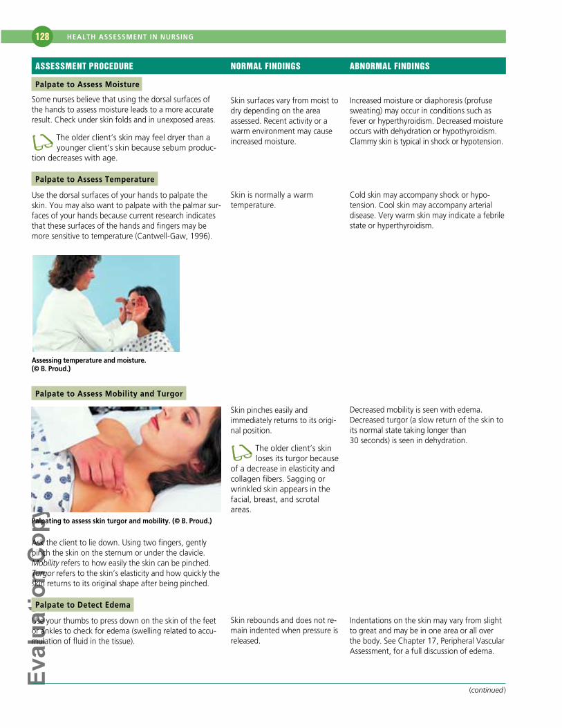
Palpate to Assess Mobility and Turgor
Ask the client to lie down. Using two fingers, gentlypinch the skin on the sternum or under the clavicle.Mobility refers to how easily the skin can be pinched.Turgor refers to the skin’s elasticity and how quickly theskin returns to its original shape after being pinched.
Palpate to Detect Edema
Use your thumbs to press down on the skin of the feetor ankles to check for edema (swelling related to accu-mulation of fluid in the tissue).
Skin pinches easily andimmediately returns to its origi-nal position.
The older client’s skinloses its turgor because
of a decrease in elasticity andcollagen fibers. Sagging orwrinkled skin appears in thefacial, breast, and scrotalareas.
Skin rebounds and does not re-main indented when pressure isreleased.
Decreased mobility is seen with edema.Decreased turgor (a slow return of the skin toits normal state taking longer than 30 seconds) is seen in dehydration.
Indentations on the skin may vary from slightto great and may be in one area or all overthe body. See Chapter 17, Peripheral VascularAssessment, for a full discussion of edema.
HEALTH ASSESSMENT IN NURSING 128
(continued )
Palpate to Assess Moisture
Some nurses believe that using the dorsal surfaces ofthe hands to assess moisture leads to a more accurateresult. Check under skin folds and in unexposed areas.
The older client’s skin may feel dryer than ayounger client’s skin because sebum produc-
tion decreases with age.
Palpate to Assess Temperature
Use the dorsal surfaces of your hands to palpate theskin. You may also want to palpate with the palmar sur-faces of your hands because current research indicatesthat these surfaces of the hands and fingers may bemore sensitive to temperature (Cantwell-Gaw, 1996).
ASSESSMENT PROCEDURE NORMAL FINDINGS ABNORMAL FINDINGS
Skin surfaces vary from moist todry depending on the areaassessed. Recent activity or awarm environment may causeincreased moisture.
Skin is normally a warm temperature.
Increased moisture or diaphoresis (profusesweating) may occur in conditions such asfever or hyperthyroidism. Decreased moistureoccurs with dehydration or hypothyroidism.Clammy skin is typical in shock or hypotension.
Cold skin may accompany shock or hypo-tension. Cool skin may accompany arterialdisease. Very warm skin may indicate a febrilestate or hyperthyroidism.
Assessing temperature and moisture. (© B. Proud.)
Palpating to assess skin turgor and mobility. (© B. Proud.)

������������
C H A P T E R 9 | SKIN, HAIR, AND NAIL ASSESSMENT 129
At 1-inch intervals, separate the hair from the scalp andinspect and palpate the hair and scalp for cleanliness,dryness or oiliness, parasites, and lesions.
Scalp is clean and dry. Sparsedandruff may be visible. Hair issmooth and firm, somewhat elas-tic. However, as people age,hair feels coarser and drier.
Individuals of blackAfrican descent often
have very dry scalps and dry,fragile hair, which the clientmay condition with oil or apetroleum jelly like product.(This kind of hair is of geneticorigin and not related to thy-roid disorders or nutrition.Such hair needs to be handledvery gently.)
Excessive scaliness may indicate dermatitis.Raised lesions may indicate infections ortumor growth. Dull, dry hair may be seenwith hypothyroidism and malnutrition. Poorhygiene may indicate a need for client teach-ing or assistance with activities of daily living.
(continued )
ASSESSMENT PROCEDURE NORMAL FINDINGS ABNORMAL FINDINGS
Have the client remove any hair clips, hair pins, or wigs.Wear gloves if lesions are suspected or if hygiene ispoor. Then inspect the scalp and hair.
Inspect amount and distribution of scalp, body, axillae,and pubic hair. Look for unusual growth elsewhere onthe body.
Natural hair color, as opposedto chemically colored hair, variesamong clients from pale blondto black to gray or white. Thecolor is determined by theamount of melanin present.
Varying amounts of terminal haircover the scalp, axillary, body,and pubic areas according tonormal gender distribution. Finevellus hair covers the entire bodyexcept for the soles, palms, lips,and nipples. Normal male patternbalding is symmetric.
Older clients have thin-ner hair because of a
decrease in hair follicles. Pubic,axillary, and body hair also de-crease with aging. Alopecia isseen, especially in men. Hair lossoccurs from the periphery of thescalp and moves to the center.
Elderly women may have ter-minal hair growth on the chinowing to hormonal changes.
Excessive generalized hair loss may occurwith infection, nutritional deficiencies, hormonal disorders, thyroid or liver disease,drug toxicity, hepatic or renal failure(Sabbagh,1999). It may also result fromchemotherapy or radiation therapy.
Nutritional deficiencies may causepatchy gray hair in some clients.
Severe malnutrition in African-Americanchildren may cause a copper-red hair color(Andrews & Boyle, 1999).
Hirsutism (facial hair on females) is a charac-teristic of Cushing’s disease and results froman imbalance of adrenal hormones, or it maybe a side effect of steroids.
SCALP AND HAIR: CONDITION AND TEXTURE
Patchy hair loss may result from infections of thescalp, discoid or systemic lupus erythematosus,and some types of chemotherapy. (CourtesyNeutrogena Skin Care Institute.)
Male pattern balding.
Inspecting the scalp and hair. (© B. Proud.)
������������
HEALTH ASSESSMENT IN NURSING 130
ASSESSMENT PROCEDURE NORMAL FINDINGS ABNORMAL FINDINGS
Inspect nail grooming and cleanliness.
Inspect nail color and markings.
Inspect shape of nails.
Palpate nail to assess texture.
Palpate to assess texture and consistency, notingwhether nailplate is attached to nailbed.
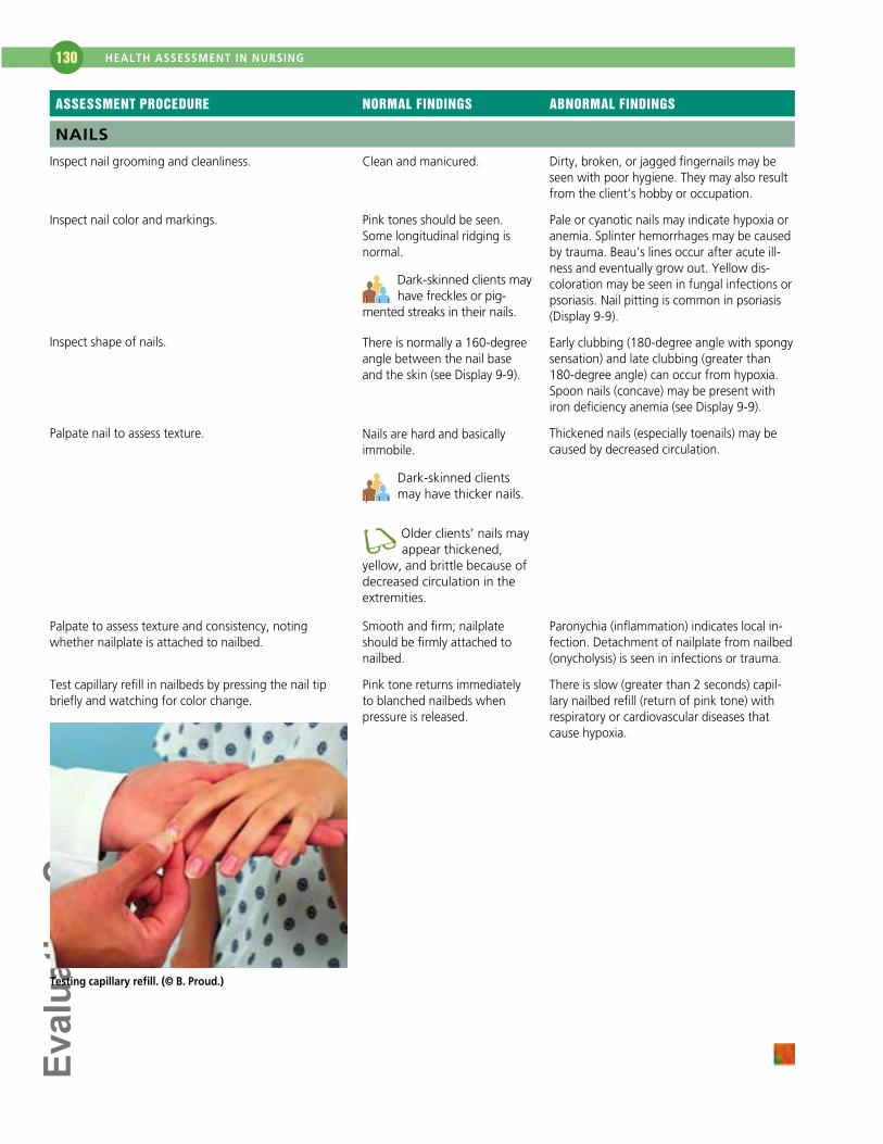
Test capillary refill in nailbeds by pressing the nail tipbriefly and watching for color change.
Clean and manicured.
Pink tones should be seen.Some longitudinal ridging isnormal.
Dark-skinned clients mayhave freckles or pig-
mented streaks in their nails.
There is normally a 160-degreeangle between the nail baseand the skin (see Display 9-9).
Nails are hard and basically immobile.
Dark-skinned clientsmay have thicker nails.
Older clients’ nails mayappear thickened,
yellow, and brittle because ofdecreased circulation in theextremities.
Smooth and firm; nailplateshould be firmly attached tonailbed.
Pink tone returns immediatelyto blanched nailbeds whenpressure is released.
Dirty, broken, or jagged fingernails may beseen with poor hygiene. They may also resultfrom the client’s hobby or occupation.
Pale or cyanotic nails may indicate hypoxia oranemia. Splinter hemorrhages may be causedby trauma. Beau’s lines occur after acute ill-ness and eventually grow out. Yellow dis-coloration may be seen in fungal infections orpsoriasis. Nail pitting is common in psoriasis(Display 9-9).
Early clubbing (180-degree angle with spongysensation) and late clubbing (greater than 180-degree angle) can occur from hypoxia.Spoon nails (concave) may be present with iron deficiency anemia (see Display 9-9).
Thickened nails (especially toenails) may becaused by decreased circulation.
Paronychia (inflammation) indicates local in-fection. Detachment of nailplate from nailbed(onycholysis) is seen in infections or trauma.
There is slow (greater than 2 seconds) capil-lary nailbed refill (return of pink tone) withrespiratory or cardiovascular diseases thatcause hypoxia.
NAILS
Testing capillary refill. (© B. Proud.)
������������
C H A P T E R 9 | SKIN, HAIR, AND NAIL ASSESSMENT 131
DISPLAY 9-2. Common Skin Variations
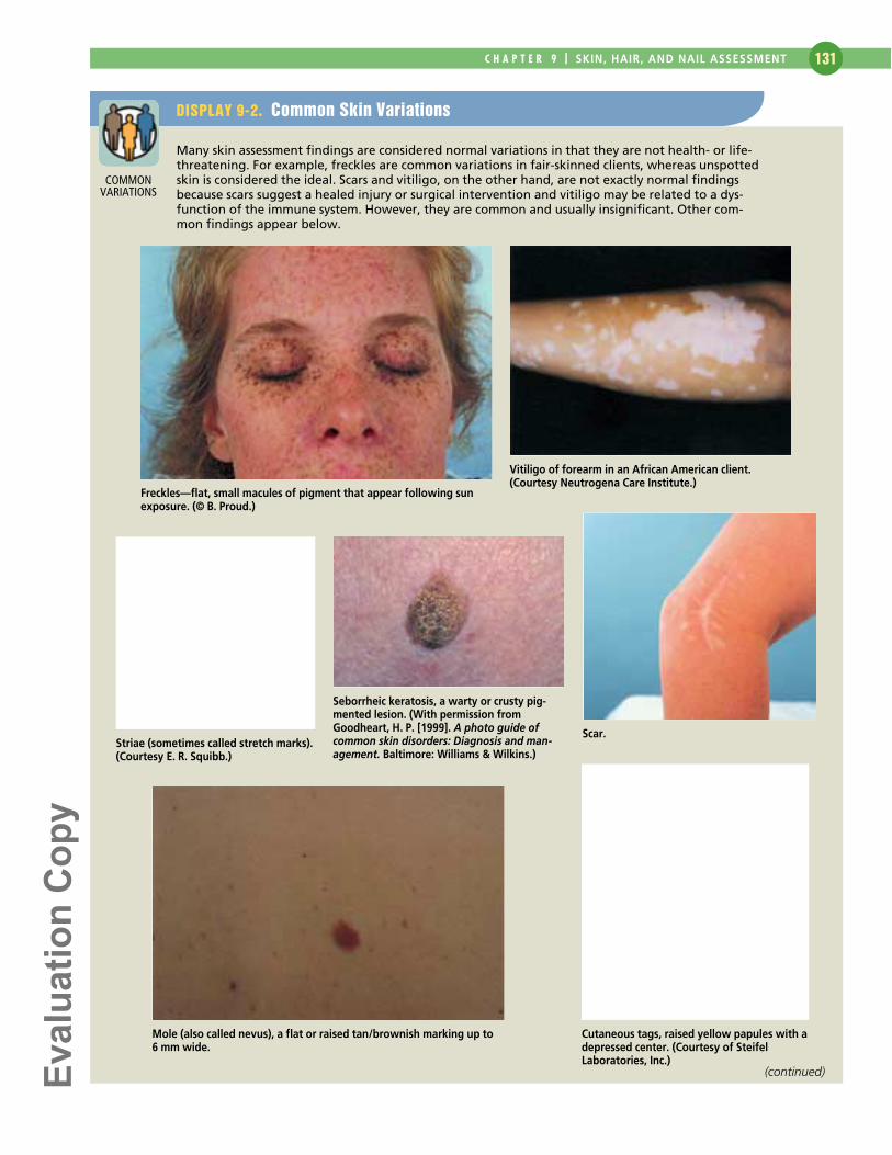
Many skin assessment findings are considered normal variations in that they are not health- or life-threatening. For example, freckles are common variations in fair-skinned clients, whereas unspottedskin is considered the ideal. Scars and vitiligo, on the other hand, are not exactly normal findingsbecause scars suggest a healed injury or surgical intervention and vitiligo may be related to a dys-function of the immune system. However, they are common and usually insignificant. Other com-mon findings appear below.
Freckles—flat, small macules of pigment that appear following sunexposure. (© B. Proud.)
Vitiligo of forearm in an African American client.(Courtesy Neutrogena Care Institute.)
Striae (sometimes called stretch marks).(Courtesy E. R. Squibb.)
Seborrheic keratosis, a warty or crusty pig-mented lesion. (With permission fromGoodheart, H. P. [1999]. A photo guide ofcommon skin disorders: Diagnosis and man-agement. Baltimore: Williams & Wilkins.)
Scar.
Mole (also called nevus), a flat or raised tan/brownish marking up to 6 mm wide.
Cutaneous tags, raised yellow papules with adepressed center. (Courtesy of SteifelLaboratories, Inc.)
COMMONVARIATIONS
(continued)

������������
HEALTH ASSESSMENT IN NURSING 132
Cutaneous horn. Cherry angiomas, small raised spots (1–5 mm wide) typically seen with aging.
DISPLAY 9-2. Common Skin Variations (Continued)
Validation and Documentation of Findings
Validate your normal and abnormal findings with theclient, other health care workers, or your instructors. Next,document the skin, hair, and nail assessment data thatyou have collected on the appropriate form your school oragency uses. The following is a summary of areas of cover-age and findings that are considered normal in a skin, hair,and nail assessment. Of course, abnormal findings would becarefully documented too. Normal findings can act as abaseline for findings that may change later.
EXAMPLE OF SUBJECTIVE DATA
Thirty-five-year-old woman with no history of skin lesions,excessive hair loss, or nail disorders. Reports one episode offine, raised, reddened rash on trunk after taking ampicillin forear infection. Rash cleared within 3 days after discontinuationof ampicillin and administration of antihistamine. Showers inAM and bathes in PM with deodorant soap. Shampoos withbaby shampoo each AM. Applies moisturizer to skin after eachcleansing; conditions hair after shampoo. Uses antiperspiranttwice daily. Shaves legs and axillae with electric razor twiceweekly. Weekly, trims toenails and fingernails and applies
nail enamel to fingernails. Denies exposure to chemicals,abrasives, or excessive sunlight.
EXAMPLE OF OBJECTIVE DATA
Skin pink, warm, dry, and elastic. No lesions or excoriationsnoted. Old appendectomy scar right lower abdomen, 4 inches long, thin, and white. Sprinkling of freckles notedacross nose and cheeks. Hair brown, shoulder-length, clean,shiny. Normal distribution of hair on scalp and perineum.Hair has been removed from legs, axillae. Nails form 160-degree angle at base; are hard, smooth, and immobile.Nailbeds pink without clubbing. Cuticles smooth; no de-tachment of nail plate. Fingernails well manicured with clearenamel. Toenails clean and well trimmed.
After you have collected your assessment data, analyze thedata, using diagnostic reasoning skills. You can review the key steps in diagnostic reasoning in Chapter 7. Then, readon to “Diagnostic Reasoning: Possible Conclusions.” Thisdiscussion features an overview of common conclusions thatyou may reach from raw data after a skin, hair, and nail as-sessment. Then, “Diagnostic Reasoning: Case Study” showsyou how to analyze skin, hair, and nail assessment data for aspecific client. Finally, you will have an opportunity to ana-lyze data in your laboratory manual/study guide in the“Critical Thinking Exercise.”
������������
C H A P T E R 9 | SKIN, HAIR, AND NAIL ASSESSMENT 133
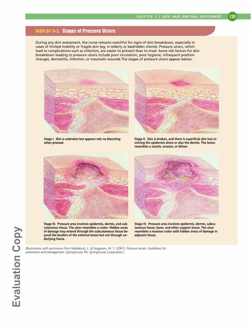
DISPLAY 9-3. Stages of Pressure Ulcers
During any skin assessment, the nurse remains watchful for signs of skin breakdown, especially incases of limited mobility or fragile skin (eg, in elderly or bedridden clients). Pressure ulcers, whichlead to complications such as infection, are easier to prevent than to treat. Some risk factors for skinbreakdown leading to pressure ulcers include poor circulation, poor hygiene, infrequent positionchanges, dermatitis, infection, or traumatic wounds.The stages of pressure ulcers appear below.
(Illustrations with permission from Maklebust, J., & Sieggreen, M. Y. [2001]. Pressure ulcers: Guidelines forprevention and management. Springhouse, PA: Springhouse Corporation.)
Stage I. Skin is unbroken but appears red; no blanchingwhen pressed.
Stage II. Skin is broken, and there is superficial skin loss in-volving the epidermis alone or also the dermis. The lesionresembles a vesicle, erosion, or blister.
Stage III. Pressure area involves epidermis, dermis, and sub-cutaneous tissue. The ulcer resembles a crater. Hidden areasof damage may extend through the subcutaneous tissue be-yond the borders of the external lesion but not through un-derlying fascia.
Stage IV. Pressure area involves epidermis, dermis, subcu-taneous tissue, bone, and other support tissue. The ulcerresembles a massive crater with hidden areas of damage inadjacent tissue.
������������
HEALTH ASSESSMENT IN NURSING 134
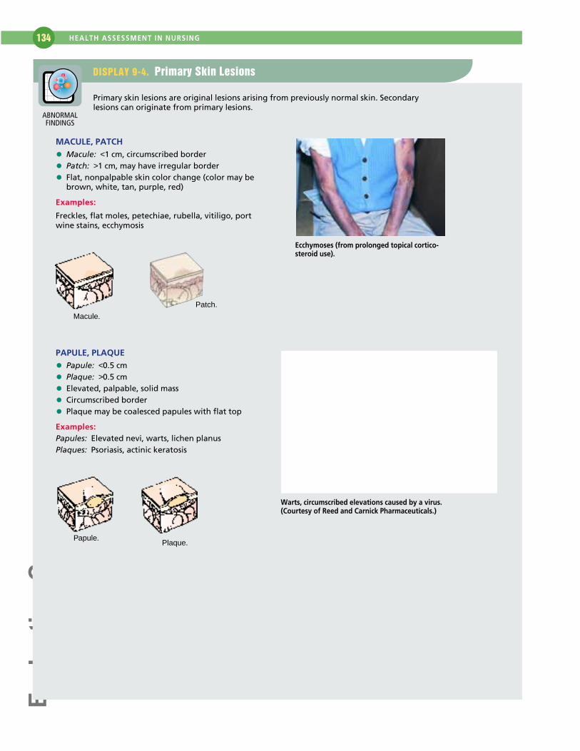
DISPLAY 9-4. Primary Skin Lesions
Primary skin lesions are original lesions arising from previously normal skin. Secondarylesions can originate from primary lesions.
MACULE, PATCH
• Macule: <1 cm, circumscribed border
• Patch: >1 cm, may have irregular border
• Flat, nonpalpable skin color change (color may bebrown, white, tan, purple, red)
Examples:
Freckles, flat moles, petechiae, rubella, vitiligo, portwine stains, ecchymosis
PAPULE, PLAQUE
• Papule: <0.5 cm
• Plaque: >0.5 cm
• Elevated, palpable, solid mass
• Circumscribed border
• Plaque may be coalesced papules with flat top
Examples:Papules: Elevated nevi, warts, lichen planusPlaques: Psoriasis, actinic keratosis
Macule.
Patch.
Ecchymoses (from prolonged topical cortico-steroid use).
Papule.Plaque.
Warts, circumscribed elevations caused by a virus. (Courtesy of Reed and Carnick Pharmaceuticals.)
ABNORMALFINDINGS
������������
C H A P T E R 9 | SKIN, HAIR, AND NAIL ASSESSMENT 135
DISPLAY 9-4. Primary Skin Lesions (Continued)
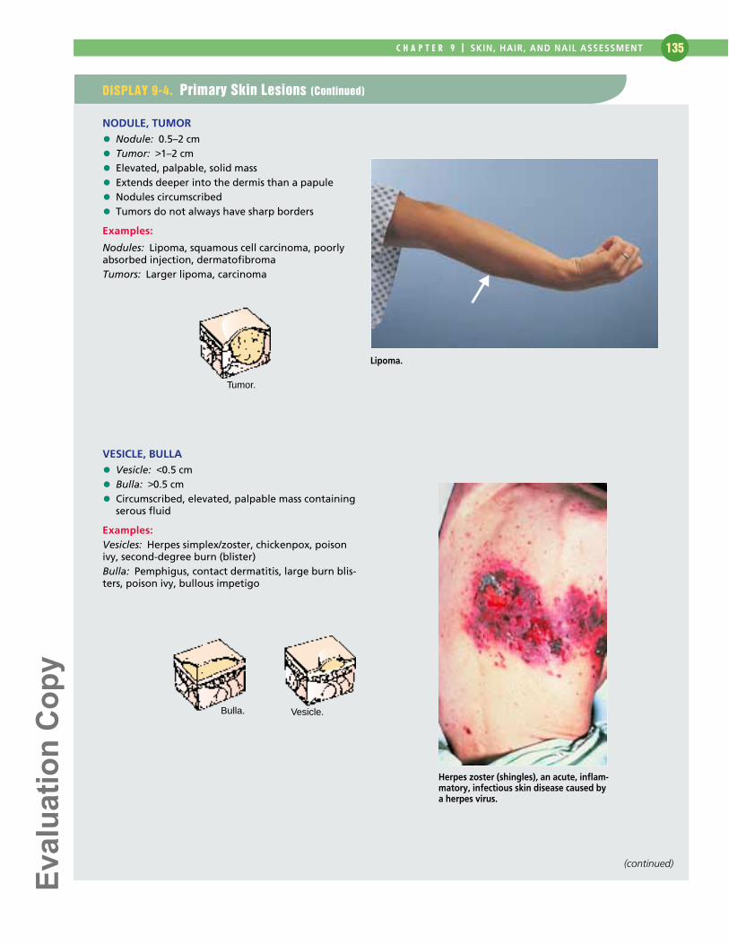
NODULE, TUMOR
• Nodule: 0.5–2 cm
• Tumor: >1–2 cm
• Elevated, palpable, solid mass
• Extends deeper into the dermis than a papule
• Nodules circumscribed
• Tumors do not always have sharp borders
Examples:
Nodules: Lipoma, squamous cell carcinoma, poorlyabsorbed injection, dermatofibromaTumors: Larger lipoma, carcinoma
VESICLE, BULLA
• Vesicle: <0.5 cm
• Bulla: >0.5 cm
• Circumscribed, elevated, palpable mass containingserous fluid
Examples:Vesicles: Herpes simplex/zoster, chickenpox, poisonivy, second-degree burn (blister)Bulla: Pemphigus, contact dermatitis, large burn blis-ters, poison ivy, bullous impetigo
Tumor.
Bulla. Vesicle.
Herpes zoster (shingles), an acute, inflam-matory, infectious skin disease caused bya herpes virus.
Lipoma.
(continued)
������������
HEALTH ASSESSMENT IN NURSING 136
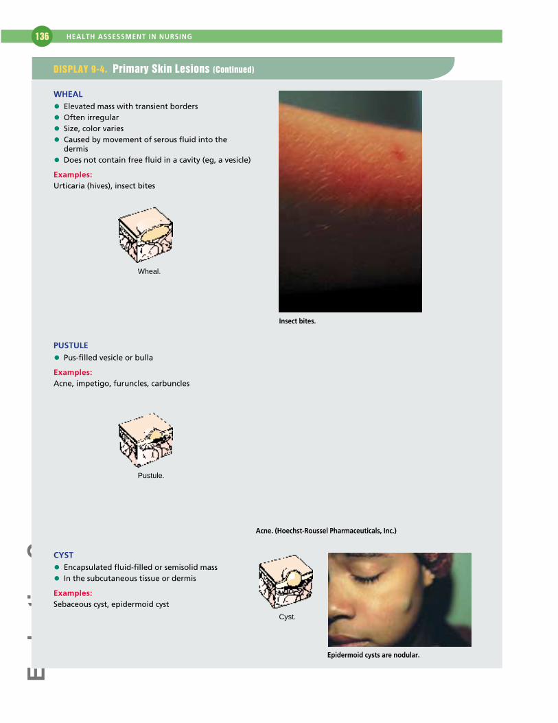
WHEAL
• Elevated mass with transient borders
• Often irregular
• Size, color varies
• Caused by movement of serous fluid into thedermis
• Does not contain free fluid in a cavity (eg, a vesicle)
Examples:Urticaria (hives), insect bites
PUSTULE
• Pus-filled vesicle or bulla
Examples:Acne, impetigo, furuncles, carbuncles
CYST
• Encapsulated fluid-filled or semisolid mass
• In the subcutaneous tissue or dermis
Examples:Sebaceous cyst, epidermoid cyst
Wheal.
Insect bites.
Pustule.
Acne. (Hoechst-Roussel Pharmaceuticals, Inc.)
Cyst.
Epidermoid cysts are nodular.
DISPLAY 9-4. Primary Skin Lesions (Continued)
������������
C H A P T E R 9 | SKIN, HAIR, AND NAIL ASSESSMENT 137
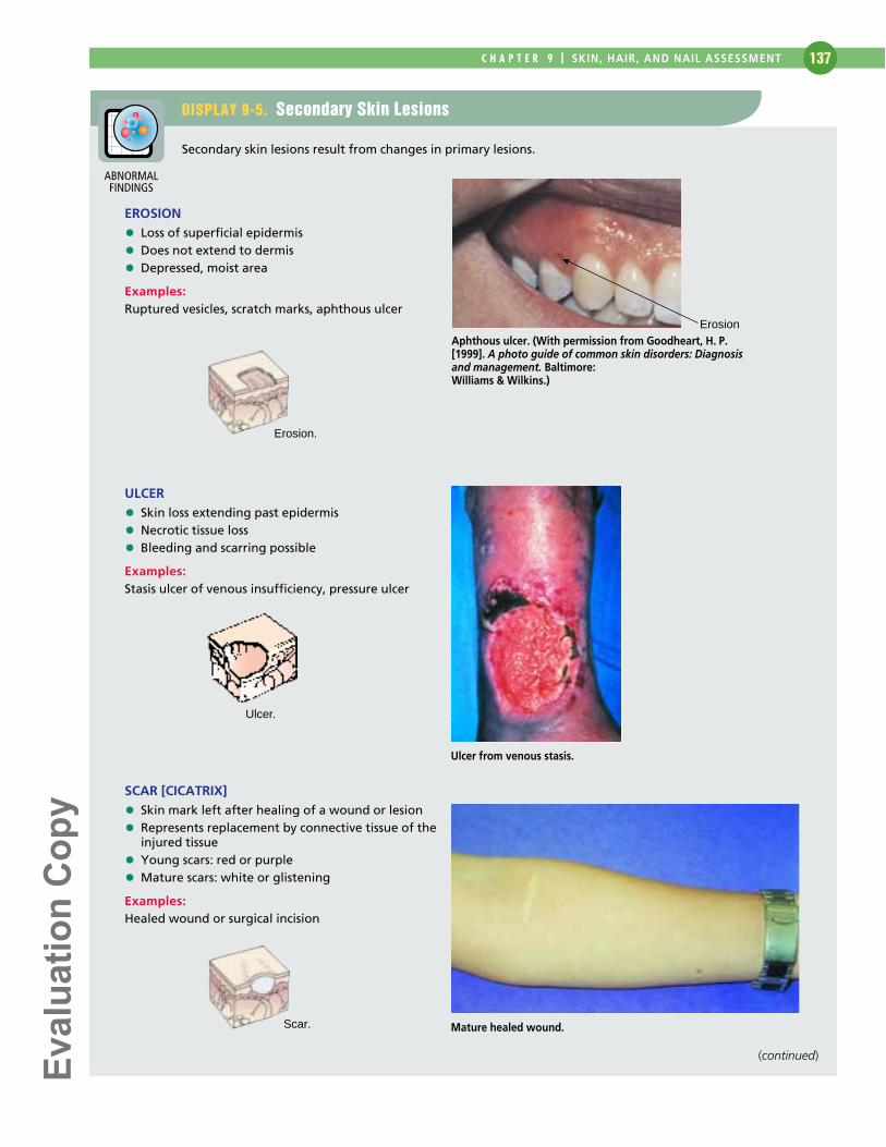
DISPLAY 9-5. Secondary Skin Lesions
Secondary skin lesions result from changes in primary lesions.
EROSION
• Loss of superficial epidermis
• Does not extend to dermis
• Depressed, moist area
Examples:Ruptured vesicles, scratch marks, aphthous ulcer
ULCER
• Skin loss extending past epidermis
• Necrotic tissue loss
• Bleeding and scarring possible
Examples:Stasis ulcer of venous insufficiency, pressure ulcer
SCAR [CICATRIX]
• Skin mark left after healing of a wound or lesion
• Represents replacement by connective tissue of theinjured tissue
• Young scars: red or purple
• Mature scars: white or glistening
Examples:Healed wound or surgical incision
ErosionAphthous ulcer. (With permission from Goodheart, H. P.[1999]. A photo guide of common skin disorders: Diagnosisand management. Baltimore: Williams & Wilkins.)
Erosion.
Ulcer.
Ulcer from venous stasis.
Scar. Mature healed wound.
ABNORMALFINDINGS
(continued)
������������
HEALTH ASSESSMENT IN NURSING 138
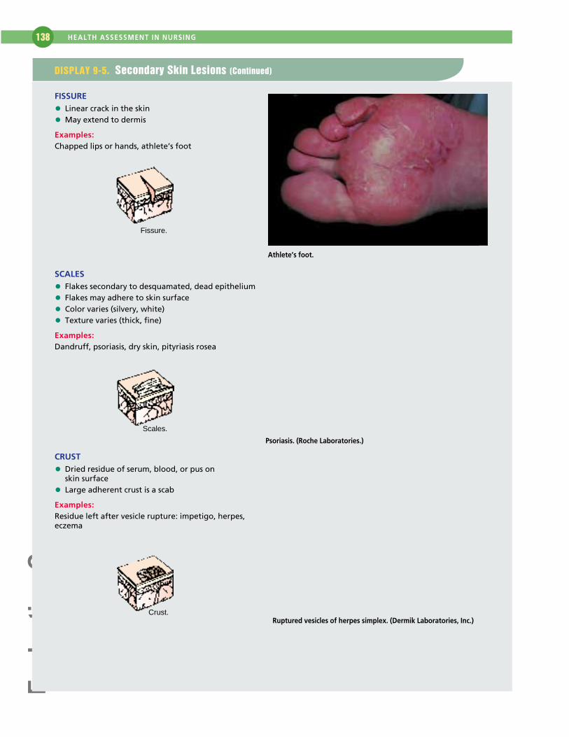
FISSURE
• Linear crack in the skin
• May extend to dermis
Examples:Chapped lips or hands, athlete’s foot
SCALES
• Flakes secondary to desquamated, dead epithelium
• Flakes may adhere to skin surface
• Color varies (silvery, white)
• Texture varies (thick, fine)
Examples:Dandruff, psoriasis, dry skin, pityriasis rosea
CRUST
• Dried residue of serum, blood, or pus on skin surface
• Large adherent crust is a scab
Examples:Residue left after vesicle rupture: impetigo, herpes, eczema
Fissure.
Athlete’s foot.
Scales.
Psoriasis. (Roche Laboratories.)
Crust.Ruptured vesicles of herpes simplex. (Dermik Laboratories, Inc.)
DISPLAY 9-5. Secondary Skin Lesions (Continued)
������������
C H A P T E R 9 | SKIN, HAIR, AND NAIL ASSESSMENT 139
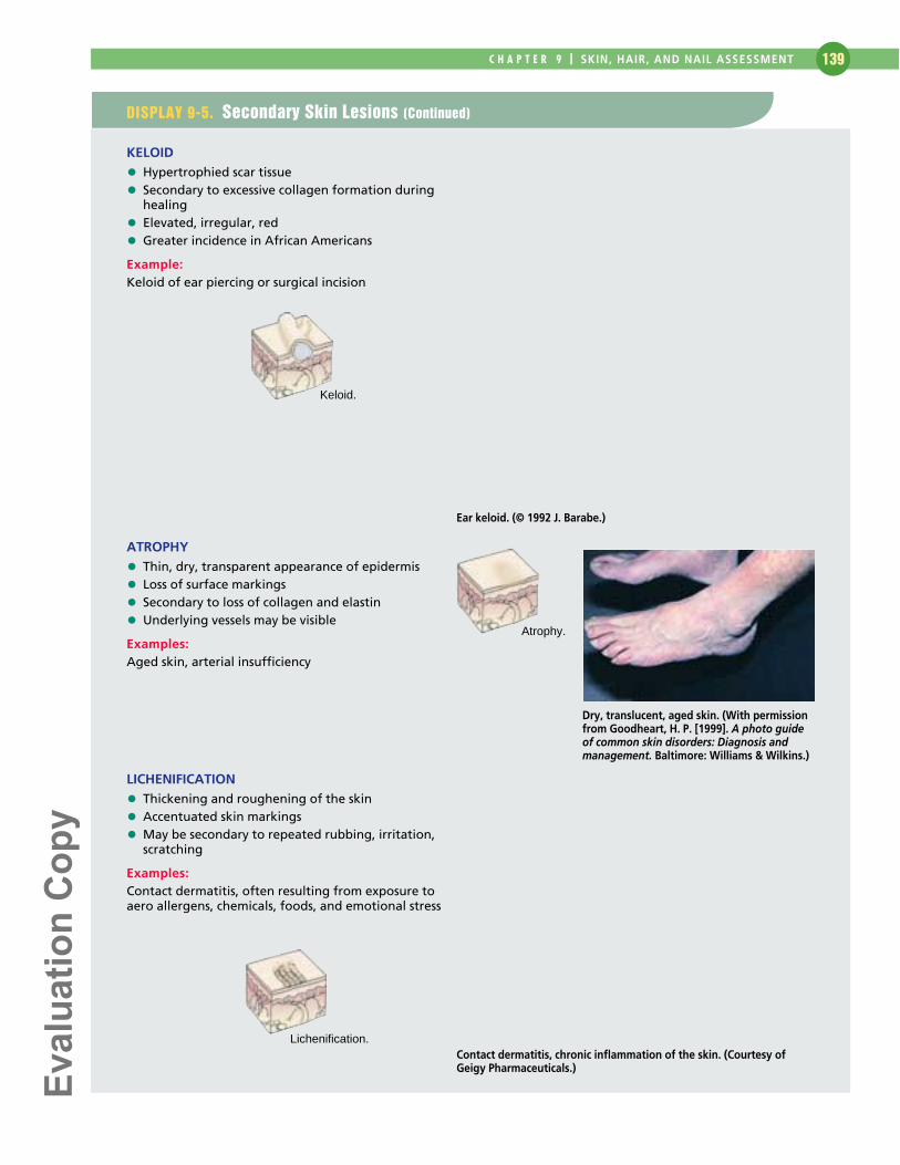
KELOID
• Hypertrophied scar tissue
• Secondary to excessive collagen formation duringhealing
• Elevated, irregular, red
• Greater incidence in African Americans
Example:Keloid of ear piercing or surgical incision
ATROPHY
• Thin, dry, transparent appearance of epidermis
• Loss of surface markings
• Secondary to loss of collagen and elastin
• Underlying vessels may be visible
Examples:Aged skin, arterial insufficiency
LICHENIFICATION
• Thickening and roughening of the skin
• Accentuated skin markings
• May be secondary to repeated rubbing, irritation,scratching
Examples:Contact dermatitis, often resulting from exposure toaero allergens, chemicals, foods, and emotional stress
Keloid.
Ear keloid. (© 1992 J. Barabe.)
Atrophy.
Dry, translucent, aged skin. (With permissionfrom Goodheart, H. P. [1999]. A photo guideof common skin disorders: Diagnosis andmanagement. Baltimore: Williams & Wilkins.)
Lichenification.Contact dermatitis, chronic inflammation of the skin. (Courtesy ofGeigy Pharmaceuticals.)
DISPLAY 9-5. Secondary Skin Lesions (Continued)
������������
HEALTH ASSESSMENT IN NURSING 140
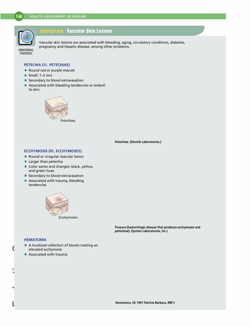
DISPLAY 9-6. Vascular Skin Lesions
Vascular skin lesions are associated with bleeding, aging, circulatory conditions, diabetes,pregnancy and hepatic disease, among other problems.
PETECHIA (PL. PETECHIAE)
• Round red or purple macule
• Small: 1–2 mm
• Secondary to blood extravasation
• Associated with bleeding tendencies or emboli to skin
ECCHYMOSIS (PL. ECCHYMOSES)
• Round or irregular macular lesion
• Larger than petechia
• Color varies and changes: black, yellow, and green hues
• Secondary to blood extravasation
• Associated with trauma, bleeding tendencies
HEMATOMA
• A localized collection of blood creating an elevated ecchymosis
• Associated with trauma
Petechiae.
Petechiae. (Dermik Laboratories.)
Ecchymoses.
Purpura (hemorrhagic disease that produces ecchymoses andpetechiae). (Syntex Laboratories, Inc.)
Hematoma. (© 1991 Patricia Barbara, RBP.)
ABNORMALFINDINGS
������������
C H A P T E R 9 | SKIN, HAIR, AND NAIL ASSESSMENT 141
DISPLAY 9-6. Vascular Skin Lesions (Continued)
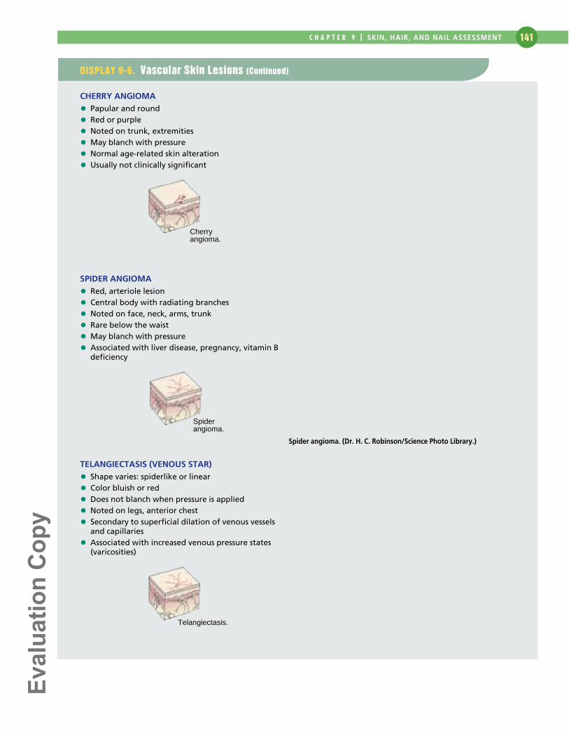
CHERRY ANGIOMA
• Papular and round
• Red or purple
• Noted on trunk, extremities
• May blanch with pressure
• Normal age-related skin alteration
• Usually not clinically significant
SPIDER ANGIOMA
• Red, arteriole lesion
• Central body with radiating branches
• Noted on face, neck, arms, trunk
• Rare below the waist
• May blanch with pressure
• Associated with liver disease, pregnancy, vitamin Bdeficiency
TELANGIECTASIS (VENOUS STAR)
• Shape varies: spiderlike or linear
• Color bluish or red
• Does not blanch when pressure is applied
• Noted on legs, anterior chest
• Secondary to superficial dilation of venous vesselsand capillaries
• Associated with increased venous pressure states(varicosities)
Cherryangioma.
Spiderangioma.
Spider angioma. (Dr. H. C. Robinson/Science Photo Library.)
Telangiectasis.
������������
HEALTH ASSESSMENT IN NURSING 142
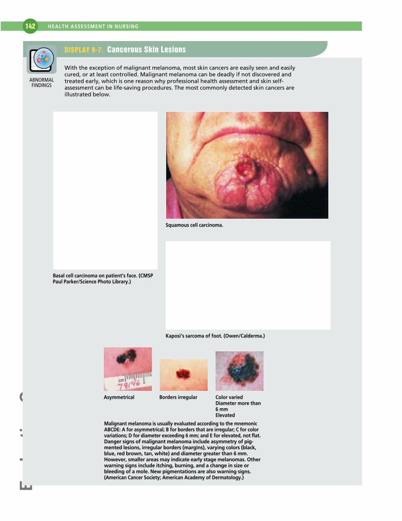
DISPLAY 9-7. Cancerous Skin Lesions
With the exception of malignant melanoma, most skin cancers are easily seen and easilycured, or at least controlled. Malignant melanoma can be deadly if not discovered andtreated early, which is one reason why professional health assessment and skin self-assessment can be life-saving procedures. The most commonly detected skin cancers areillustrated below.
Basal cell carcinoma on patient’s face. (CMSPPaul Parker/Science Photo Library.)
Squamous cell carcinoma.
Kaposi’s sarcoma of foot. (Owen/Calderma.)
Asymmetrical Borders irregular Color variedDiameter more than6 mm Elevated
Malignant melanoma is usually evaluated according to the mnemonicABCDE: A for asymmetrical; B for borders that are irregular; C for colorvariations; D for diameter exceeding 6 mm; and E for elevated, not flat.Danger signs of malignant melanoma include asymmetry of pig-mented lesions, irregular borders (margins), varying colors (black,blue, red brown, tan, white) and diameter greater than 6 mm.However, smaller areas may indicate early stage melanomas. Otherwarning signs include itching, burning, and a change in size orbleeding of a mole. New pigmentations are also warning signs.(American Cancer Society; American Academy of Dermatology.)
ABNORMALFINDINGS
������������
C H A P T E R 9 | SKIN, HAIR, AND NAIL ASSESSMENT 143
DISPLAY 9-8. Configurations of Skin Lesions
Describing lesions by shape, distribution, or configuration is one way to communicate specificcharacteristics that can help to identify causes and treatments. Some common configurations include the following:
Linear configuration:straight line as in ascratch or streak.
Annular configuration:circular lesions.
Zosteriform configuration: linear lesions clusteredalong a nerve route.
Discrete configuration: individual anddistinct lesions.
Polycyclic configuration:circular lesions that tendto run together.
Confluent configuration: lesions runtogether.
A B C
D E F
������������
HEALTH ASSESSMENT IN NURSING 144
DISPLAY 9-9. Common Nail Disorders
Many clients have nails with lines, ridges, spots, and uncommon shapes that suggest anunderlying disorder. Some examples follow:
180°
>180°
A Beau’s lines (acute illness)
B Spoon nails (iron deficiencyanemia)
C Early clubbing (oxygen deficiency)
D Late clubbing (oxygen deficiency)
E Pitting (psoriasis)
F Paronychia (local infection)
ABNORMALFINDINGS
������������
145
Analysis of Data
Diagnostic Reasoning:Possible Conclusions
Listed below are some possible conclusions that the nursemay make after assessing a client’s skin, hair, and nails.
SELECTED NURSING DIAGNOSES
After compiling subjective and objective data related to theclient’s skin, hair, and nails, you will need to identify ab-normalities and cluster the data to reveal any significant pat-terns or abnormalities. These data may then be used tomake clinical judgments (nursing diagnoses: wellness, risk,or actual) about the status of the client’s skin, hair, andnails. The following is a list of selected nursing diagnosesthat may be identified when analyzing data from a skin,hair, and nail assessment.
Nursing Diagnoses (Wellness)
• Opportunity to enhance skin, hair, and nail integrity re-lated to healthy hygiene and skin care practices, avoid-ance of overexposure to sun
• Health-Seeking Behavior: Requests information on skinreactions and effects of using a sun-tanning booth
Nursing Diagnoses (Risk)
• Risk for Impaired Skin Integrity related to excessive ex-posure to cleaning solutions and chemicals
• Risk for Impaired Skin Integrity related to prolongedsun exposure
• Risk for Impaired Skin Integrity related to immobility,decreased production of natural oils, and thinning skin
• Risk for Impaired Skin Integrity of toes related to thick-ened, dried toenails
• Risk for Altered Body Temperature related to severediaphoresis
• Risk for Infection related to scratching of rash• Risk for Impaired Nail Integrity related to prolonged use
of nail polish
• Risk for Altered Nutrition: Less Than Body Require-ments related to increased vitamin and protein require-ments necessary for healing of a wound
Nursing Diagnoses (Actual)
• Altered Health Maintenance related to lack of hygieniccare of the skin, hair, and nails
• Impaired Skin Integrity related to immobility and de-creased circulation
• Impaired Skin Integrity related to poor nutritional in-take and bowel/bladder incontinence
• Body Image Disturbance related to scarring, rash, orother skin condition that alters skin appearance
• Sleep Pattern Disturbance related to persistent itching ofthe skin
• Fluid Volume Deficit related to excessive diaphoresissecondary to excessive exercise and high environmentaltemperatures
SELECTED COLLABORATIVE PROBLEMS
After grouping the data, certain collaborative problems maybecome apparent. Remember that collaborative problemsdiffer from nursing diagnoses in that they cannot be pre-vented or managed with independent nursing interventions.However, these physiologic complications of medical con-ditions can be detected and monitored by the nurse. In ad-dition, the nurse can use physician- and nurse-prescribedinterventions to minimize the complications of these prob-lems. The nurse may also have to refer the client in suchsituations for further treatment of the problem. The follow-ing is a list of collaborative problems that may be identifiedwhen assessing the skin, hair, and nails. These problems areworded as Potential Complications (or PC), followed by theproblem.
• PC: Allergic reaction• PC: Skin rash• PC: Insect/animal bite• PC: Septicemia• PC: Hypovolemic shock• PC: Skin infection
P A R TT H R E E

������������
HEALTH ASSESSMENT IN NURSING 146
Diagnostic Reasoning: Case Study
The case study presents assessment data for a specific client.It is followed by an analysis of the data, by following the seven key steps found in Chapter 7, to arrive at specific conclusions.
Mary Michaelson, a 29-year-old divorced woman, works as anoffice manager for a large, prestigious law firm. She reportsshe recently went to see a doctor because “my hair was fallingout in chunks, and I have a red rash on my face and chest. Itlooks like a bad case of acne.” After doing some blood work,her physician diagnosed her condition as discoid lupuserythematosus (DLE). She says she has come to see you, theoccupational health nurse, because she feels “so ugly,” andshe is concerned that she may lose her job because of how she looks.
During the interview, she tells you that she is a surfer and isout in the sun all day nearly every weekend. She shares that sheuses sunscreen but forgets to put it on at regular intervalsduring the day.
Your physical examination reveals an attractive, tanned,thin, anxious-appearing young woman. You note confluentand nonconfluent maculopapular lesions on her neck, chestabove the nipple line, and over the shoulders and upper backto about the level of the T5 vertebra. Many of the lesions
appear as red, scaling plaques with depressed, pale centers. A few of the lesions on her forehead and cheeks appear blis-tered. Patchy alopecia is also present. Her vital signs are withinnormal limits, and no other abnormalities are apparent at this time.
1 Identify abnormal data and strengths (in both subjectiveand objective data).
SUBJECTIVE DATA
• “Hair falling out in chunks”• Red rash on face and chest—“looks like a bad case of acne”• “So ugly”• Concerned that she may lose her job because of how
she looks• Surfer—out in the sun all day on weekends—minimal use
of sunscreen• Sought out occupational health nurse
OBJECTIVE DATA
• Anxious appearing• Diagnosed with discoid lupus erythematosus• Red, raised plaques on face, neck, shoulders, back,
and chest• Patchy alopecia
Possible Nursing DefiningCue Clusters Inferences Diagnoses Characteristics Confirm or Rule Out
A• Diagnosed with DLE
• Discoid lesions appar-ent on face, neck, chest,back—raised red patcheswith some blistering
B• Rash on face, neck,chest, and back
• Patchy alopecia
• “So ugly”
2 3 4 5 6
Textbook picture for DLEas diagnosed by physicianMonitor for collaborativeproblems
Changes in physical appearance are affectingself-perception
Body Image Disturbancerelated to changes inphysical appearance
Ineffective IndividualCoping related tochanges in physical appearance and newlydiagnosed disease
Major: Verbal negativeresponse to actualchange in structure
Minor: Negative feelingsabout body
Major: None
Minor: None
Accept diagnosis because it meets defin-ing characteristics and is validated by client.
Rule out diagnosis be-cause it has none of thedefining characteristics;however, more datashould be collected regarding her support systems and coping behaviors.
• PC: Skin lesion• PC: Ischemic skin ulcers• PC: Graft rejection• PC: Hemorrhage• PC: Burns
MEDICAL PROBLEMS
After grouping the data, it may become apparent that theclient has signs and symptoms that require medical diag-nosis and treatment. Referral to a primary care provider isnecessary.
������������
C H A P T E R 9 | SKIN, HAIR, AND NAIL ASSESSMENT 147
Possible Nursing DefiningCue Clusters Inferences Diagnoses Characteristics Confirm or Rule Out
• Surfer, out in sunlightweekly
• Inadequate applicationsof sunscreen
C• Sought out occupa-tional health nurse
D• Anxious appearing
• Concerned that shemay lose her job becauseof how she looks
• Office manager inlarge, prestigious law firm
2 3 4 5 6
Excessive sun exposurecan worsen lesions caus-ing blistering, weeping,and scarring
Does not seem to knowabout these effects
Possibly seeking informa-tion about managing herillness
Perceives current positiondepends on attractiveappearance
Risk for Altered HealthMaintenance related toknowledge deficit of effects of sunlight onskin lesions
Health-Seeking Behavior
Anxiety related to possi-ble loss of work positionsecondary to perceivedunattractiveness
Major: Reports unhealthypractice (not using sun-screen effectively)
Minor: None
Major: Sought out occu-pational health nurse
Minor: None
Major: Physical appear-ance (unspecified anxiety)and self-deprecation(about physical appear-ance)
Minor: None
Confirm diagnosis because it meets majordefining characteristicsand is validated by client.
This is ambiguous. Theclient sought out the occupational healthnurse for more informa-tion, but after she wasalready diagnosed, ratherthan for health promo-tion before the fact.Collect more data beforeaccepting this diagnosis.
Accept diagnosis becauseit meets defining charac-teristics, but collect moredata to confirm this diag-nosis. Fear may be moreappropriate, but dataneeded for that as well.
Collaborative problems related to the medical diagnosis couldinclude:
• PC: Skin Infection/Scarring• PC: Ischemic Ulcers• PC: Systemic Lupus Erythematosus (SLE) and all related
complications (1 in 20 people diagnosed with discoid lupus erythematosus [DLE] progress to systemic lupuserythematosus [SLE])
7 Document conclusions.
The following nursing diagnoses are appropriate for Ms. Michaelson at this time:
• Body Image Disturbance related to changes in physicalappearance
• Risk for Altered Health Maintenance related to knowledgedeficit of effects of sunlight on skin lesions
• Anxiety related to possible loss of work position secondary to perceived unattractiveness
REFERENCES AND SELECTED READINGSAndrews, M., & Boyle, J. (1999). Transcultural concepts in nursing
care (3rd ed.). Philadelphia: Lippincott Williams & Wilkins.Conditions of the skin. (1999). American Family Physician, 60(4),
1258–1261.Correale, C. E., & Walker, C. (1999). Atopic dermatitis: A review of
diagnosis and treatment. American Family Physician, 60(4), 1191–1198.Guttman, C. (2000). Nail exam should be routine in elderly patients.
Dermatology Times, 21(4), 40.———. (1999). Practical approach provides key clues to hair disorder
diagnosis. Dermatology Times, 19(10), 14.Kuznar, W. (1997). Skin holds clues to many systemic disorders.
Dermatology Times, 18(4), 11.McMichael, A. J. (1999). A review of cutaneous disease in African-
American patients. Dermatology Nursing, 11(1), 35–36, 41–47.
Muirhead, G. (1999). Common dermatologic problems in people ofcolor. Patient Care, 33(20), 97.
National Institutes of Health. (1992). The NIH Consensus Devel-opment Panel on Melanoma: Diagnosis and treatment of early melanoma.Journal of the American Medical Association, 268, 1314–1319.
Noronha, P., & Zubkov, B. (1997). Nails and nail disorders in childrenand adults. American Family Physician, 55(6), 21–29.
Overfield, T. (1995). Biologic variation in health and illness: Race, age,and sex differences (2nd ed.). Boca Raton, FL: CRC Press.
Parhizgar, B. (2000). Skin signs of systemic disease. Cortlandt Forum,13(1), 169.
Passchier, J. (1998). Quality of life issues in male pattern hair loss.Dermatology, 197(3), 217–218.
Rudikoff, D., & Lebwohl, M. (1998). Atopic dermatitis. Lancet, 351(9117), 1715.
������������
HEALTH ASSESSMENT IN NURSING 148
Sabbagh, L. (1999). Hair loss in women should be taken seriously.Dermatology Times, 20(12), 24–25.
Sinclair, R. (1998). Male pattern androgenetic alopecia. BritishMedical Journal, 317(7162), 865.
Smoker, A. (1999). Skin care in old age. Nursing Standard, 13(48),47–54.
Stuart, C., Driscoll, M., Lundquist, K., Gilkison, C., Shaheb, S., &Smith, M. (1999). Acanthosis nigricans. Journal of Basic Clinical Physiologyand Pharmacology, 9(2–4), 407–418.
Wysocki, A. B. (1999). Skin anatomy, physiology, and pathophysiol-ogy. Nursing Clinics of North America, 34(4), 777–797.
Risk Factors–Skin CancerAmerican Cancer Society (ACS). (2000). Cancer Resource Center.
Available: www.cancer.org.Armstrong, B., & Dricker, A. (1995). Skin cancer. Dermatology
Clinics, 13, 583–594.
Davidowitz, S., Belafsky, P., & Amedee, R. (1999). The epidemiologyof malignant melanoma in Louisiana and beyond. Journal of the LouisianaState Medical Society, 15(10), 493–499.
Gloster, H., & Brodland, D. (1995). The epidemiology of skin cancer.Dermatology Surgery, 22, 217–226.
Goodheart, H. P. (1999). A photoguide of common skin disorders:Diagnosis and management. Baltimore, MD: Williams & Wilkins.
Halder, R., & Bridgeman-Shah, S. (1995). Skin cancer in AfricanAmericans. Cancer, 75(Suppl. 2), 667–673.
Hall, H., & Rogers, J. (1999). Sun protection behaviors among AfricanAmericans. Ethnic Diseases, 9(1), 126–131.
Nicol, N., & Penske, N. (1993). Photodamage: Cause, clinical mani-festations, and prevention. Dermatology Nursing, 5, 263–277, 326.
For additional information on this book, be sure tovisit http://connection.lww.com.