tertiary contacts of helix v in the lactose permease determined by site-directed chemical...
TRANSCRIPT
Tertiary Contacts of Helix V in the Lactose Permease Determined by Site-DirectedChemical Cross-Linking in Situ†
Jianhua Wu,‡ Dorothy Hardy,§ and H. Ronald Kaback*,§
Department of Physiology and Howard Hughes Medical Institute, Departments of Physiology and Microbiology & MolecularGenetics, Molecular Biology Institute, UniVersity of California, Los Angeles, California 90095-1662
ReceiVed September 23, 1998; ReVised Manuscript ReceiVed NoVember 16, 1998
ABSTRACT: The six N-terminal transmembrane helices (N6) and the six C-terminal transmembrane helices(C6) in lactose permease, each containing a single Cys residue, were coexpressed, and cross-linking wasstudied. The proximity of paired Cys residues in helices V and VII, VIII, or X was studied by thiol-specific chemical cross-linking. The results demonstrate that Cys residues in the periplasmic half of helixV cross-link with Cys residues in the periplasmic half of helix VII. In contrast, no cross-linking is evidentwith paired Cys residues in the cytoplasmic halves of helices V and VII. Moreover, Cys residues on oneentire face of helix V cross-link with Cys residues on one face of helix VIII. Finally, paired Cys residuesat the cytoplasmic ends of helices V and X cross-link, but no cross-linking is observed when paired Cysresidues are placed at the periplasmic ends of the two helices. Taken together, the results indicate that theperiplasmic halves of helices V and VII are in close proximity and that the two helices tilt away from oneanother toward the cytoplasmic side of the membrane. Furthermore, helices V and VIII are in closeproximity throughout their lengths and do not tilt appreciably with respect to one another, and helices Vand X are in close proximity at the cytoplasmic but not at the periplasmic face of the membrane.
The lactose (lac)1 permease ofEscherichia coliis typicalof secondary transport proteins that transduce free energystored in an electrochemical ion gradient into a concentrationgradient (reviewed in refs1-3). This hydrophobic, polytopicmembrane protein which catalyzes the coupled stoichiometrictranslocation ofâ-galactosides and H+ has been solubilized,purified to homogeneity, reconstituted into proteoliposomes,and shown to be solely responsible forâ-galactoside transport(reviewed in ref4) as a monomer (5). All available evidence(reviewed in refs6-8) indicates that the permease iscomposed of 12R-helical rods that traverse the membranein zigzag fashion connected by relatively hydrophilic loopswith the N and C termini on the cytoplasmic face (see Figure1).
Site-directed and Cys-scanning mutagenesis have dem-onstrated that only six amino acid side chains in this 417-residue polypeptide are irreplaceable with respect to themechanism of active transport: Glu126 (helix IV) andArg144 (helix V) which are indispensable for substratebinding and Glu269 (helix VIII), Arg302 (helix IX), His322,and Glu325 (helix) which are involved in H+ translocation
and coupling (reviewed in ref9). However, structural anddynamic information at high resolution is required forunderstanding the precise role of these residues in sugar andH+ translocation, as well as the coupling between the twoprocesses. Since hydrophobic membrane proteins are notori-ously difficult to crystallize, a high-resolution structure oflac permease is not available, and development of alternativemethods for obtaining structural information is essential (7-9). By the combined use of multifaceted site-directedapproaches which include excimer fluorescence, chemicalcleavage, engineered divalent metal binding sites, electronparamagnetic resonance spectroscopy, thiol-specific cross-linking, and identification of discontinuous mAb epitopes,a general helix packing model of lac permease has beenformulated (Figure 2). Although many of the spatial relation-ships between transmembrane helices have been establishedby more than one experimental approach (reviewed in ref8), determination of helix tilting is essential to placing theessential residues and to delineating the substrate and H+
translocation pathways. In this regard, tilting of helices I,II, IV, VII, and XI has been studied by site-directed cross-linking with a functional permease construct expressed astwo discontinuous, nonoverlapping fragments [N6/C6 per-mease (10)] containing single Cys residues in each half ofthe protein (11-13). The results indicate that the periplasmichalves of helices VII and I, II, or IV are in close proximity,while the cytoplasmic halves of helices XI and II or IV areclose to one another.
In this report, tertiary contacts between helices V and VII,VIII, or X are examined in situ using a general cross-linkingapproach developed recently (12, 14). Paired Cys residueswere placed in two helices in N6/C6 permease, and proximity
† This work was supported in part by NIH Grant DK51131.* To whom correspondence should be addressed: HHMI/UCLA,
5-748 MacDonald Research Laboratories, Box 951662, Los Angeles,CA 90095-1662. Telephone: (310) 206-5053. Fax: (310) 206-8623.
‡ Department of Physiology.§ Howard Hughes Medical Institute.1 Abbreviations: lac, lactose; C-less permease, functional lac per-
mease devoid of Cys residues; N6, six N-terminal transmembranehelices; C6, six C-terminal transmembrane helices; TDG,â-D-galac-topyranosyl 1-thio-â-D-galactopyranoside; IPTG, isopropyl 1-thio-â-D-galactopyranoside;o-PDM, N,N′-o-phenylenedimaleimide;p-PDM,N,N′-p-phenylenedimaleimide; NaDodSO4, sodium dodecyl sulfate;PAGE, polyacrylamide gel electrophoresis; NEM,N-ethylmaleimide.
2320 Biochemistry1999,38, 2320-2325
10.1021/bi982288z CCC: $18.00 © 1999 American Chemical SocietyPublished on Web 02/02/1999
was assessed by thiol-specific chemical cross-linking in thenative membrane. The results demonstrate that the periplas-mic halves of helices V and VII are in close proximity,helices V and VIII are in close proximity throughout theirlengths, and the cytoplasmic ends of helices V and X are inclose proximity.
EXPERIMENTAL PROCEDURES
Materials. Protein A-conjugated horseradish peroxidase(PA-HRP) and enhanced chemiluminescence (ECL) detec-tion kits were obtained from Amersham (Arlington Heights,IL). Avidin-conjugated horseradish peroxidase (avidin-HRP)was purchased from Pierce (Rockford, IL).N,N′-o-Phen-ylenedimaleimide (o-PDM) andN,N′-p-phenylenedimaleim-ide (p-PDM) were from Sigma (St. Louis, MO).
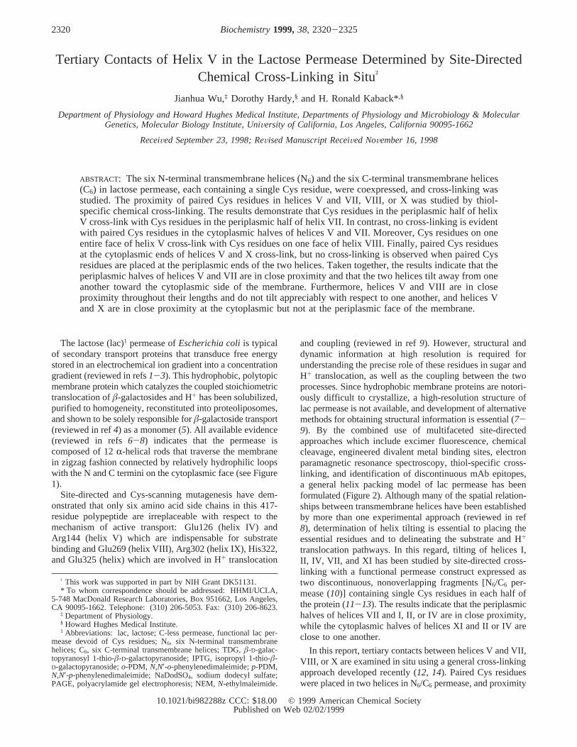
Construction of Single-Cys N6 and C6 Permease.Con-struction of permease mutants containing single-Cys replace-ments in helices V, VII, VIII, and X has been described(15-18). To each mutant with a single-Cys replacement atposition 140 to 143, 145, 147 to 149, 151, 152, 155, 158, or159 in helix V, the biotin acceptor domain from theKlebsiella pneumoniaoxaloacetate decarboxylase was in-serted into the middle cytoplasmic loop as described previ-ously (19). The 3′ half of the lacY gene in each constructwas then deleted byAflII digestion followed by intra-molecular ligation resulting in plasmid pN6, which encodesthe six N-terminal transmembrane helices (N6) with a singleCys residue at a given position and the biotin acceptordomain at the C terminus (Figure 1). Construction of plasmidpC6 encoding the six C-terminal transmembrane helices (C6)has been described (14, 20). To introduce a single Cysresidue into helix VII (position 227, 229, 231, 232, 234, 235,238, 239, 242, 243, 245, or 246), VIII (position 261, 265,272, 275, 278, or 279), or X (position 315 or 333) in C6, theBstXI-HindIII fragment of pC6 was replaced by the corre-sponding DNA fragment from an indicated single-Cyspermease mutant (Figure 1). Each Cys replacement mutantin N6 or C6 was verified by using the dideoxynucleotidetermination method (21).
Expression of Split Permease and Membrane Preparations.E. coli HB101 cells (lacY-Z+) were transformed with bothpN6 and pC6 encoding N6 and C6, respectively, each with agiven single Cys residue. Cultures (50 mL) were grown at37 °C in LB medium containing 100µg/mL ampicillin and20 µg/mL chloramphenicol to an OD600 of 1.0 and inducedwith 1 mM isopropyl 1-thio-â-D-galactopyranoside for 2 h.Cells were harvested by centrifugation, washed once with20 mM Tris-HCl (pH 7.4)/2.0 mM EDTA, and suspendedin the same buffer followed by incubation with 100µg/mLlysozyme for 10 min on ice. Membranes were prepared bysonification and suspended in 20 mM Tris-HCl (pH 7.4).
Chemical Cross-Linking and Analysis.All cross-linkingexperiments were carried out at room temperature for 30 minby adding a given thiol-specific chemical linker to a finalconcentration of 0.5 mM to membrane preparations at aprotein concentration of 2 mg/mL. Reactions were terminatedby adding sodium dodecyl sulfate (NaDodSO4) sample buffercontaining 5% (v/v) â-mercaptoethanol. Samples weresubjected to electrophoresis in NaDodSO4-12% polyacryl-amide gels (PAGE). C6 was detected by immunoblotting withrabbit polyclonal antibodies against a C-terminal dodecopep-tide corresponding to the C terminus of lac permease. N6
which contains the biotin acceptor domain at the C terminuswas detected with avidin-conjugated horseradish peroxidase(avidin-HRP). Cross-linked N6/C6 reacts with both anti-C-terminal antibody and avidin-HRP.
Protein Determinations.Protein was assayed by using aMicro BCA protein determination kit (Pierce).
RESULTS
Cross-Linking N6/C6 Permease with Paired Cys Residuesin Helices V and VII.Transmembrane helix V in the lacpermease was shown initially to be in close proximity tohelices VII and VIII by site-directed chemical cleavage (22),a finding confirmed subsequently by electron paramagneticresonance (EPR) with double nitroxide spin labeling andcross-linking studies with the 5 Å homobifunctional cross-linking agent dibromobimane (23). To further examine
FIGURE 1: Secondary structure model of N6/C6 split permease. The lac permease is shown as the six N-terminal transmembrane helices(N6) and the six C-terminal transmembrane helices (C6). N6 has a biotin acceptor domain (BD) at the C terminus. Single-Cys replacementsin helices V (position 140, 141, 142, 143, 145, 147, 148, 149, 151, 152, 155, 158, or 159), VII (position 227, 229, 231, 232, 234, 235, 238,239, 242, 243, 245, or 246), VIII (position 261, 265, 272, 275, 278, or 279), and X (position 315 or 333) are numbered and highlighted.
Tertiary Contacts of Helix V in Lac Permease Biochemistry, Vol. 38, No. 8, 19992321
proximity and tilting between helices V and VII, site-directedchemical cross-linking was carried out with N6/C6 permeasecontaining paired Cys residues at various positions in thetwo helices. As shown in Figures 1 and 2, a Cys residuewas placed at given positions along the length of helix V inN6 and along the length of helix VII in C6. To assess theproximity between given paired Cys residues, N6 and C6
fragments were coexpressed and cross-linking was carriedout in situ with homobifunctional chemical linkers.o-PDMand p-PDM which are thiol-specific reagents containingmaleimido groups coupled to benzene rings in the ortho andpara positions at fixed distances of about 6 and 10 Å,respectively, were chosen because of their relatively shortlength, rigidity, and hydrophobicity. Hydrophobicity ispresumably important because the Cys residues are thoughtto be in a hydrophobic environment within the membrane.
When membranes containing N6/C6 permease with pairedCys residues at positions in the periplasmic halves of helicesV (position 151, 152, 155, 158, or 159) and VII (position238, 239, 242, 243, 245, or 246) are treated witho-PDM orp-PDM, cross-linking of the N6 and C6 fragments is clearlyevident (Figures 3 and 6). C6 which reacts with anti-C-
terminal antibody migrates at anMr of about 20 kDa (Figure3A); N6 with the biotin acceptor domain which reacts withavidin-HRP migrates with anMr of about 35 kDa (Figure3B), and cross-linked N6/C6 which reacts with both anti-C-terminal antibody and avidin-HRP migrates with anMr ofabout 52 kDa (Figure 3A,B).o-PDM with 6 Å separatingthe maleimido groups readily cross-links paired Cys residues159 and 246, 158 and 246, 155 and 243, 155 and 242, 152and 239, 151 and 239, and 151 and 238 (Figures 3 and 6).Similar results are obtained by using the 10 Å cross-linkingagentp-PDM (data not shown). In marked contrast, whenpaired Cys residues are placed at positions in the cytoplasmichalves of helix V (position 140, 141, 142, 143, 145, 147,148, or 149) and helix VII (position 227, 229, 231, 232, 234,or 235), no cross-linking is observed (Figure 6). The resultsindicate that helices V and VII are in close proximity withinthe periplasmic side of the membrane and tilt away fromeach other toward the cytoplasmic side.
Cross-Linking N6/C6 Permease with Paired Cys Residuesin Helices V and VIII.To examine proximity and tiltingbetween helix V and helix VIII, N6 containing a Cys residueat given positions in helix V was coexpressed with C6 with
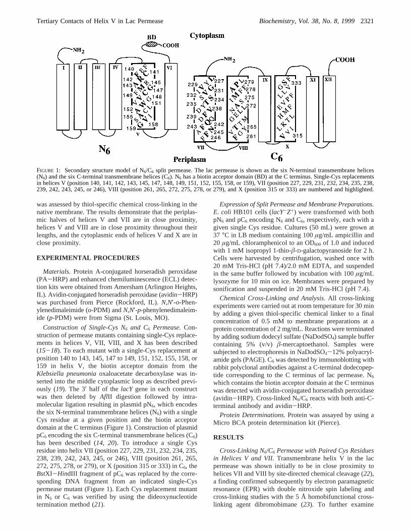
FIGURE 2: Helix packing in the lac permease viewed from the cytoplasmic face. The six essential residues [Glu126 (helix IV), Arg144(helix V), Glu269 (helix VIII), Arg302 (helix IX), and His322 and Glu325 (helix X)] and two interacting pairs of Asp and Lys residues[Asp237 (helix VII) and Lys358 (helix XI) and Asp240 (helix VII) and Lys319 (helix X)] are highlighted. Positions of NEM-sensitive Cysreplacements are indicated with a small black dot. Cys replacement mutants in helices V, VII, VIII, and X tested for cross-linking in thisstudy are highlighted and numbered.
2322 Biochemistry, Vol. 38, No. 8, 1999 Wu et al.
a Cys residue at given positions in helix VIII (Figures 1 and2). When membranes containing N6/C6 with paired Cysresidues 140 and 279, 140 and 278, 143 and 278, 143 and275, 147 and 275, 147 and 272, 158 and 265, or 158 and261 are treated witho-PDM (Figures 4 and 6) orp-PDM(not shown), cross-linking is clearly observed. The resultsindicate that helices V and VIII are in close proximitythroughout the entire transmembrane domain. In addition,since no cross-linking is evident in N6/C6 containing paired
Cys residues 141 and 278, 141 and 279, 142 and 278, 142and 279, 145 and 275, or 149 and 272 (Figure 6), it is likelythat the face of helix V with Phe140, Ala143, Gly147, andVal158 is close to the face of helix VIII with Pro279, Ala278,Met275, Ala272, Thr265, and Phe261 (Figures 2 and 7).
Cross-Linking N6/C6 Permease with Paired Cys Residuesin Helices V and X.To examine the proximity and tiltingbetween helices V and X, cross-linking of N6/C6 permease
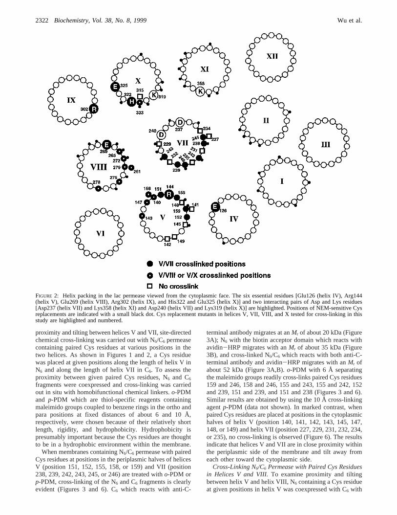
FIGURE 3: Chemical cross-linking of split N6/C6 permease contain-ing paired Cys residues in helices V and VII. Membranes (2 mg ofprotein/mL) were prepared from cells expressing N6 and C6fragments, with each containing a single Cys residue at givenpositions as indicated. Chemical cross-linking was carried out atroom temperature for 30 min by addingo-PDM to a finalconcentration of 0.5 mM. Reactions were terminated by addingNaDodSO4 sample buffer containing 5% (v/v)â-mercaptoethanol.Samples containing approximately 20µg of membrane protein weresubjected to NaDodSO4-PAGE and electroblotted. The immuno-blot was probed with anti-C-terminal antibody or avidin-HRP. N6and C6 fragments and the N6/C6 cross-linked product are denotedby arrows: (A) cross-linking of paired Cys residues 159 and 246probed with anti-C-terminal antibody, (B) cross-linking of pairedCys residues 159 and 246 probed with avidin-HRP, (C) cross-linking of paired Cys residues 158 and 246 and 155 and 243 probedwith avidin-HRP, and (D) cross-linking of paired Cys residues151 and 239 and 151 and 238 probed with avidin-HRP.
(A) (B)
FIGURE 4: Chemical cross-linking of N6/C6 permease containingpaired Cys residues in helices V and VIII. Membranes wereprepared from cells expressing N6 and the C6 fragments, each witha Cys residue at given positions as indicated. Chemical cross-linkingwas carried out at room temperature by incubating membranes (2mg of protein/mL) with 0.5 mMo-PDM for 30 min. Reactionswere terminated by adding NaDodSO4 sample buffer containing5% (v/v) â-mercaptoethanol. Samples were analyzed as describedin the legend of Figure 3 with avidin-HRP: (A) paired Cysresidues 140 and 279 and 140 and 278 and (B) paired Cys residues143 and 275, 147 and 272, and 158 and 265.
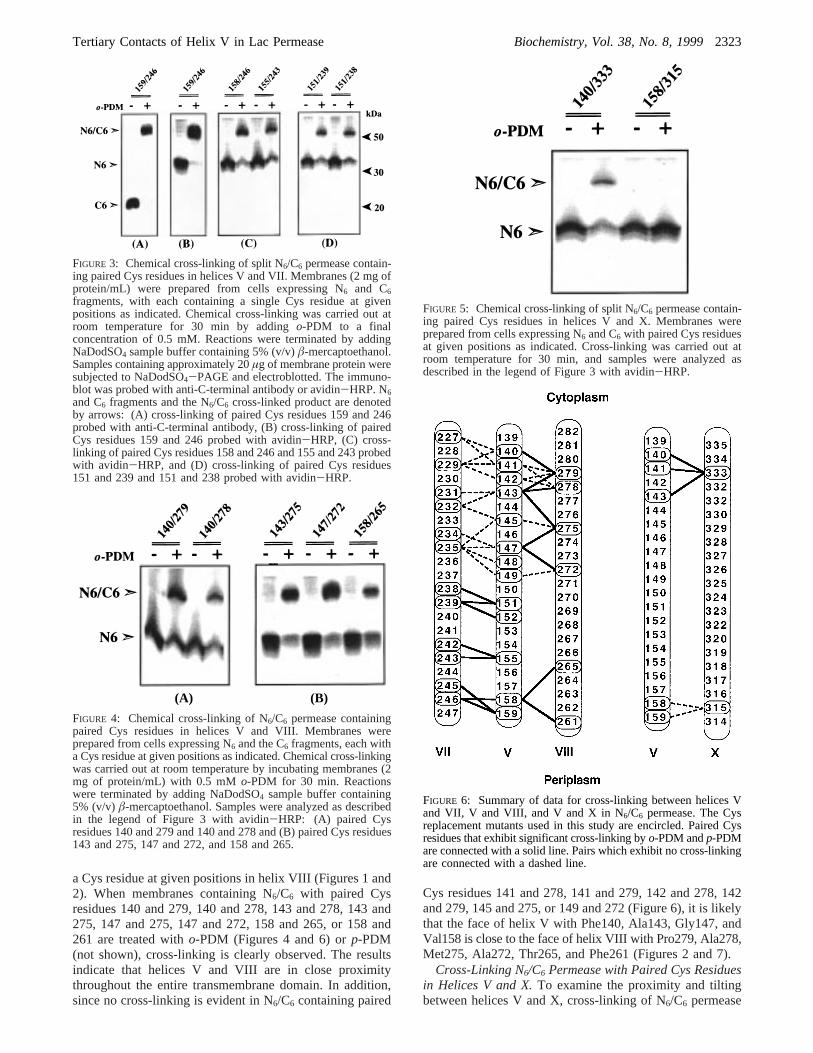
FIGURE 5: Chemical cross-linking of split N6/C6 permease contain-ing paired Cys residues in helices V and X. Membranes wereprepared from cells expressing N6 and C6 with paired Cys residuesat given positions as indicated. Cross-linking was carried out atroom temperature for 30 min, and samples were analyzed asdescribed in the legend of Figure 3 with avidin-HRP.
FIGURE 6: Summary of data for cross-linking between helices Vand VII, V and VIII, and V and X in N6/C6 permease. The Cysreplacement mutants used in this study are encircled. Paired Cysresidues that exhibit significant cross-linking byo-PDM andp-PDMare connected with a solid line. Pairs which exhibit no cross-linkingare connected with a dashed line.
Tertiary Contacts of Helix V in Lac Permease Biochemistry, Vol. 38, No. 8, 19992323
containing paired Cys residues in the two helices at positionsin the cytoplasmic or periplasmic ends was carried out. Asshown (Figures 5 and 6), paired Cys residues at positions333 (helix X) and 140, 141, or 143 (helix V) are cross-linkedby o-PDM (Figures 5 and 6) orp-PDM (not shown),indicating that the two helices are in close proximity in thecytoplasmic side of the membrane. In contrast, when pairedCys residues are placed at the periplasmic ends of the twohelices at positions 315 (helix X) and 158 or 159 (helix V),no cross-linking is evident by eithero-PDM (Figures 5 and6) or p-PDM (not shown), suggesting that the two helicestilt away from each other toward the periplasmic side of themembrane (Figure 7).
DISCUSSION
On the basis of previous studies in which site-directedchemical cleavage (22), site-directed spin labeling, and cross-linking between Cys residues (23) were used, helix V wasplaced in the vicinity of helices VII and VIII (Figure 2). Tofurther investigate tertiary contacts between helix V and theC-terminal half of lac permease, site-directed thiol-specificcross-linking was carried out with N6/C6 split permeasecontaining paired Cys residues at various positions in helicesV (N6) and VII, VIII, or X (C6). As shown previously(11-14), cross-linking of paired Cys residues in N6/C6 split
permease has provided proximity and tilting information forhelices I, II, IV, VII, XI, and XII in the lac permease, andthe technique has also provided important informationregarding other other membrane proteins (24-30).
With paired Cys residues in helices V and VII, cross-linking is observed only when the two Cys residues are inthe periplasmic halves of the helices (positions 151-159 inhelix V and 238-246 in helix VII), while no cross-linkingwhatsoever between helices V and VII is evident with pairedCys residues in the cytoplasmic halves of these helices. Theresults indicate that the two helices are in close proximitywithin the periplasmic side of the membrane and tilt awayfrom each other toward the cytoplasmic side of the mem-brane. In addition, since no cross-linking is observed withpaired Cys residues at positions in the cytoplasmic halvesof helices VII and I, II, or IV (12, 13), it is likely that thecytoplasmic end of helix VII tilts away from the helices inthe N-terminal half of the permease (Figure 7).
Since cross-linking is evident with paired Cys residues atpositions extending over the entire length of the transmem-brane domains of helices V and VIII, it is likely that thesetwo helices are in close proximity as they traverse themembrane without major tilting with respect to one another.Furthermore, no cross-linking is evident between helices Vand VIII when Cys residues are placed at positions 141, 142,145, or 149 in helix V and positions 279, 278, 275, or 272in helix VII. Thus, it is likely that the face of helix V withPhe140, Ala143, Gly147, and Val158 is directed toward theface of helix VIII with Pro279, Ala278, Met275, Ala272,Thr265, and Phe261 (Figures 2 and 7).
On the basis of previous studies (8, 12, 13) and the findingspresented here, a tertiary helix packing model is presented(Figure 7). This model is consistent with the followingobservations. (i) Close proximity between the periplasmichalves of helices VII and I, II, IV, or V has been documentedby thiol cross-linking (11-14). (ii) Helix VII tilts away fromthe N-terminal half of the lac permease toward the cyto-plasmic face of the membrane, since no cross-linking isobserved with paired Cys residues at various positions inthe cytoplasmic halves of helices VII and I, II, IV, or V(11-14). (iii) Tilting of the cytoplasmic end of helix XItoward helices II and IV is consistent with cross-linkingbetween paired Cys residues at positions in the cytoplasmichalves of helices XI and II or IV, but not the periplasmichalves (12). (iv) Helices V and VIII are in close proximitywithout major tilting with respect to one another, since cross-linking is observed with paired Cys residues on one face ofeach of these helices throughout their lengths. (v) Thecytoplasmic end of helix X tilts toward helix V, since pairedCys residues at the cytoplasmic ends of the two helices cross-link effectively. (vi) The periplasmic end of helix I tiltstoward helix VII (13). (vii) Close proximity between helicesIV and V has been documented recently by site-directedcross-linking of pairs of Cys residues placed in the twohelices (C. Wolin and H. R. Kaback, unpublished results).
Electron microscopic studies (31-35) reveal a notch orcleft in the permease that is likely to result from helical tiltsand may be related to the substrate translocation pathway.As shown in Figure 7, the tilting of helices I, II, IV, V, VII,VIII, X, and XI results in a cleft in the tertiary structurewhich is composed of one face of helices II, V, and VIII,the cytoplasmic halves of helices IV, X, and XI, and the
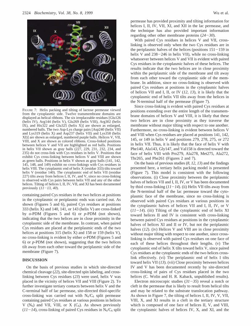
FIGURE 7: Helix packing and tilting of lactose permease viewedfrom the cytoplasmic side. Twelve transmembrane domains aredisplayed as helical ribbons. The six irreplaceable residues [Glu126(helix IV), Arg144 (helix V), Glu269 (helix VIII), Arg302 (helixIX), and His322 and Glu325 (helix X)] are shown as enlarged,numbered balls. The two Asp-Lys charge pairs [Asp240 (helix VII)and Lys319 (helix X) and Asp237 (helix VII) and Lys358 (helixXI)] are shown as enlarged, numbered purple balls. Helices V, VII,VIII, and X are shown as colored ribbons. Cross-linked positionsbetween helices V and VII are highlighted as red balls. Positionsin helix VII shown as gray balls (227, 229, 231, 232, 234, and235) do not cross-link with Cys residues in helix V. Positions thatexhibit Cys cross-linking between helices V and VIII are shownas green balls. Positions in helix V shown as gray balls (141, 142,145, 148, and 149) exhibit no cross-linkings with Cys residues inhelix VIII. The cytoplasmic end of helix X (residue 333) tilts towardhelix V (residue 140). The cytoplasmic end of helix VII (residue227) tilts away from helices I, II, IV, and V, since no cross-linkingis observed with Cys pairs between helix VII and any of the otherhelices. Tilting of helices I, II, IV, VII, and XI has been documentedpreviously (11-13, 40).
2324 Biochemistry, Vol. 38, No. 8, 1999 Wu et al.
periplasmic halves of helices I and VII. Consistent with thenotion that the cleft may be related to the sugar translocationpathway, residues thought to be directly involved in substratebinding [Glu126 (helix IV) and Arg144, Cys148, and Met145(helix V)] (36-38) are clearly accessible within the cleft.Moreover, substrate protectable single-Cys replacements atpositions 264, 268, and 272 (39) on one face of helix VIIIlie in the vicinity of helix V and line the cleft. Finally, NEM-sensitive single-Cys replacements in helices I, II, V, VII,VIII, X, and XI cluster on helical faces that also line thecleft (8, 9), and Cys residues at these positions are accessibleto solvent (P. Venkatesan and H. R. Kaback, manuscript inpreparation).
ACKNOWLEDGMENT
We thank K. C. Zen for providing plasmids containingsingle-Cys replacements in helix V with the biotin acceptordomain in the middle cytoplasmic loop.
REFERENCES
1. Kaback, H. R. (1983)J. Membr. Biol. 76, 95-112.2. Kaback, H. R. (1989)HarVey Lect. 83, 77-103.3. Poolman, B., and Konings, W. N. (1993)Biochim. Biophys.
Acta 1183, 5-39.4. Viitanen, P., Newman, M. J., Foster, D. L., Wilson, T. H.,
and Kaback, H. R. (1986)Methods Enzymol. 125, 429-452.5. Sahin-To´th, M., Lawrence, M. C., and Kaback, H. R. (1994)
Proc. Natl. Acad. Sci. U.S.A. 91, 5421-5425.6. Kaback, H. R. (1996) inHandbook of Biological Physics:
Transport Processes in Eukaryotic and Prokaryotic Organisms(Konings, W. N., Kaback, H. R., and Lolkema, J. S., Eds.) pp203-227, Elsevier, Amsterdam.
7. Kaback, H. R., Voss, J., and Wu, J. (1997)Curr. Opin. Struct.Biol. 7, 537-542.
8. Kaback, H. R., and Wu, J. (1997)Q. ReV. Biophys. 30, 333-364.
9. Frillingos, S., Sahin-To´th, M., Wu, J., and Kaback, H. R.(1998)FASEB J. 12, 1281-1299.
10. Bibi, E., and Kaback, H. R. (1990)Proc. Natl. Acad. Sci.U.S.A. 87, 4325-4329.
11. Wu, J., and Kaback, H. R. (1997)J. Mol. Biol. 270, 285-293.
12. Wu, J., Hardy, D., and Kaback, H. R. (1998)J. Mol. Biol.282, 959-967.
13. Wu, J., Hardy, D., and Kaback, H. R. (1998)Biochemistry37, 15785-15790.
14. Wu, J., and Kaback, H. R. (1996)Proc. Natl. Acad. Sci. U.S.A.93, 14498-14502.
15. Weitzman, C., and Kaback, H. R. (1995)Biochemistry 34,2310-2318.
16. Frillingos, S., Sahin-Toth, M., Persson, B., and Kaback, H.R. (1994)Biochemistry 33, 8074-8081.
17. Frillingos, S., Ujwal, M. L., Sun, J., and Kaback, H. R. (1997)Protein Sci. 6, 431-437.
18. Sahin-To´th, M., and Kaback, H. R. (1993)Protein Sci. 2,1024-1033.
19. Consler, T. G., Persson, B. L., Jung, H., Zen, K. H., Jung, K.,Prive, G. G., Verner, G. E., and Kaback, H. R. (1993)Proc.Natl. Acad. Sci. U.S.A. 90, 6934-6938.
20. Wu, J., Sun, J., and Kaback, H. R. (1996)Biochemistry 35,5213-5219.
21. Sanger, F., Nicklen, S., and Coulsen, A. R. (1977)Proc. Natl.Acad. Sci. U.S.A. 74, 5463-5467.
22. Wu, J., Perrin, D., Sigman, D., and Kaback, H. (1995)Proc.Natl. Acad. Sci. U.S.A. 92, 9186-9190.
23. Wu, J., Voss, J., Hubbell, W. L., and Kaback, H. R. (1996)Proc. Natl. Acad. Sci. U.S.A. 93, 10123-10127.
24. Falke, J. J., and Koshland, D. E., Jr. (1987)Science 237, 1596-1600.
25. Pakula, A., and Simon, M. (1992)Proc. Natl. Acad. Sci. U.S.A.89, 4144-4148.
26. Yu, H., Kono, M., McKee, T. D., and Oprian, D. D. (1995)Biochemistry 34, 14963-14969.
27. Chervitz, S. A., and Falke, J. J. (1996)Proc. Natl. Acad. Sci.U.S.A. 93, 2545-2550.
28. Hughson, A. G., and Hazelbauer, G. L. (1996)Proc. Natl.Acad. Sci. U.S.A. 93, 11546-11551.
29. Loo, T. W., and Clarke, D. M. (1996)J. Biol. Chem. 271,27482-27487.
30. Rice, W. J., Green, N. M., and MacLennan, D. H. (1997)J.Biol. Chem. 272, 31412-31429.
31. Costello, M. J., Viitanen, P., Carrasco, N., Foster, D. L., andKaback, H. R. (1984)J. Biol. Chem. 259, 15579-15586.
32. Costello, M. J., Escaig, J., Matsushita, K., Viitanen, P. V.,Menick, D. R., and Kaback, H. R. (1987)J. Biol. Chem. 262,17072-17082.
33. Li, J., and Tooth, P. (1987)Biochemistry 26, 4816-4823.34. Li, J., and Tooth, P. (1988)Prog. Clin. Biol. Res. 273, 93-
98.35. Zhuang, J., Prive´, G. G., Verner, G. E., Ringler, P., Kaback,
H. R., and Engel, A. (1999)J. Struct. Biol.(in press).36. Jung, H., Jung, K., and Kaback, H. R. (1994)Biochemistry
33, 12160-12165.37. Wu, J., and Kaback, H. R. (1994)Biochemistry 33, 12166-
12171.38. Venkatesan, P., and Kaback, H. R. (1998)Proc. Natl. Acad.
Sci. U.S.A. 95, 9802-9807.39. Frillingos, S., and Kaback, H. R. (1997)Protein Sci. 6, 438-
443.40. Wu, J., Hardy, D., and Kaback, H. R. (1999)Biochemistry
(in press).
BI982288Z
Tertiary Contacts of Helix V in Lac Permease Biochemistry, Vol. 38, No. 8, 19992325