terrible triad fracture-dislocations of the elbow -...
TRANSCRIPT

198
*UpperWo
†DepartHar
Addres021
Terrible Triad Fracture-Dislocationsof the ElbowGeorge Dyer, MD,* and David Ring, MD, PhD†
1048-6666/12/doi:http://dx.do
Extremity Divimen’s Hospitalment of Orthvard Medical Ss reprint reque15. E-mail: gdy
Management of terrible triad injuries of the elbow has improved in the last 2 decades based onan understanding of injury patterns, fracture morphology, and management pitfalls. But someintimidation with these injuries remains, in part because some of the surgical techniques arechallenging and because there is still debate about certain steps in surgery and choices inmanagement. This article highlights important aspects of the care of terrible triad injuries toclarify the anatomy and patho-physiology, to highlight pitfalls of treatment, and to point outareas that might benefit from innovation.Oper Tech Orthop 23:198-204 C 2013 Elsevier Inc. All rights reserved.
KEYWORDS terrible triad, management, controversy, surgery, radial head, coronoid, stability,collateral ligament
Introduction
The combination of an elbow dislocation, a coronoidfracture, and a fracture of the radial head has been termed
the “terrible triad” of the elbow because it is associated withrecurrent or persistent subluxation anddislocation.1 In the past2 decades, the terrible triad has been managed somewhatbased on an understanding of injury patterns, fracturemorphology, and management pitfalls.2 However, difficultiesremain. There is trepidation and intimidation with theseinjuries, in part because some of the surgical techniques arestill challenging (eg, repair of a fracture of the coronoid or radialhead) and in part from debate and about certain steps insurgery and choices in management. This article aims tohighlight important aspects of the care of terrible triad injurieselucidated in recent years, to clarify the anatomy and patho-physiology, and to highlight pitfalls of treatment, as well asareas of debate and areas that might benefit from innovation.
ExaminationNeurovascular injury and compartment syndrome are morelikely in the setting of high-energy trauma.3 The combination
$-see front matter & 2013 Elsevier Inc. All rights reserved.i.org/10.1053/j.oto.2013.08.006
sion, Department of Orthopaedic Surgery, Brigham and, Harvard Medical School, Boston, MA.opaedic Surgery, Massachusetts General Hospital,chool, Boston, MA.sts to George S.M. Dyer, MD, 75 Francis St, Boston,[email protected]
of unstable elbow and wrist injuries is at particular risk forcompartment syndrome.4 A baseline neurologic examinationis important because approximately 20% of patients woulddevelop ulnar neuropathywithin thefirst 2 years after a terribletriad injury.5 Patients should also be aware of the risk ofdeveloping heterotopic ossification as well.6
Initial ManagementA dislocated elbow should be reduced in the EmergencyDepartment, but many of these injuries are unstable withoutsurgery and reduction can be difficult to maintain (Fig. 1). Inthese cases, repeat reduction is not helpful, and the elbow canbe left dislocated while surgery is planned.
ImagingA computed tomography (CT) scan, particularly a 3-dimien-sional (3D) reconstruction if available, can be helpful inplanning the management of the coronoid and radial headfractures. Three-dimensional reconstructions can be madeusing software in radiology departments or using freeware(eg, Osirix). The unfractured bones can be digitally subtractedfrom the images to improve fracture visualization. In ouropinion, a 3D view is more intuitive, easier to interpret, andreflective of what the surgeon would find in the operatingroom, thereby reducing surprises.7
Are CT scans necessary for effective treatment? An experi-enced surgeon would anticipate that the coronoid fracture
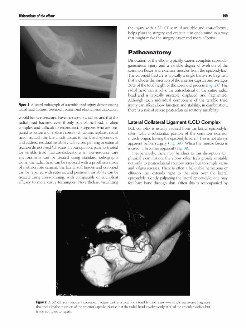
Figure 1 A lateral radiograph of a terrible triad injury demonstratingradial head fracture, coronoid fracture, and ulnohumeral dislocation.
Dislocations of the elbow 199
would be transverse and have the capsule attached and that theradial head fracture, even if only part of the head, is oftencomplex and difficult to reconstruct. Surgeons who are pre-pared to suture and replace a coronoid fracture, replace a radialhead, reattach the lateral soft tissues to the lateral epicondyle,and address residual instability with cross-pinning or externalfixation do not need CT scans. In our opinion, patients treatedfor terrible triad fracture-dislocations in low-resource careenvironments can be treated using standard radiographsalone, the radial head can be replaced with a prosthesis madeof methacrylate cement, the lateral soft tissues and coronoidcan be repaired with sutures, and persistent instability can betreated using cross-pinning, with comparable or equivalentefficacy to more costly techniques. Nevertheless, visualizing
Figure 2 A 3D CT scan shows a coronoid fracture that is typicathat includes the insertion of the anterior capsule. Notice that theis too complex to repair.
the injury with a 3D CT scan, if available and cost-effective,helps plan the surgery and execute it in one’s mind in a waythat might make the surgery easier and more effective.
PathoanatomyDislocation of the elbow typically causes complete capsuloli-gamentous injury and a variable degree of avulsion of thecommon flexor and extensor muscles from the epicondyles.1
The coronoid fracture is typically a single transverse fragmentthat includes the insertion of the anterior capsule and averages30% of the total height of the coronoid process (Fig. 2).8 Theradial head can involve the anterolateral or the entire radialhead and is typically unstable, displaced, and fragmented.Although each individual component of the terrible triadinjury can affect elbow function and stability, in combination,there is a risk of severe posterolateral rotatory instability.
Lateral Collateral Ligament (LCL) ComplexLCL complex is usually avulsed from the lateral epicondyle,often with a substantial portion of the common extensormuscle origin, leaving the epicondyle bare.9 This is not alwaysapparent before surgery (Fig. 3A). When the muscle fascia isincised, it becomes apparent (Fig. 3B).Preoperatively, there may be clues to this disruption. On
physical examination, the elbow often feels grossly unstablenot only to posterolateral rotatory stress but to simple varusand valgus stresses. There is often a ballotable hematoma oreffusion that extends right to the skin over the lateralepicondyle. Gently palpating the lateral epicondyle, one mayfeel bare bone through skin. Often this is accompanied by
l for a terrible triad injury—a single transverse fragmentradial head involves only 40% of the articular surface but

Figure 3 (A) After elevation of a lateral flap, there is no clear rent in thefascia. (B) When the fascia is incised, it becomes apparent that theorigin of the lateral collateral ligament and common extensors isavulsed from the lateral epicondyle. (Color version of figure isavailable online.)
Figure 4 A lateral radiograph 3 months after injury in a patient with aterrible triad injury treated nonoperatively.
G. Dyer and D. Ring200
bruising that begins at the epicondyle and streaks distally alongthe line of the deep rents in the extensor muscle fascia
Coronoid FractureUsing 3D CT images, Doornberg et al8 found that the averageheight of the coronoid fracture fragment in a terrible triad injurywas 39% of the total height, ranging from 19%-59%, numbersthat do not include the cartilage tip of the fragment. In otherwords, these fragments, although small, are larger than onemight think. It is not clear whether smaller coronoid fracturescontribute to instability via loss of bonybuttress (biomechanicalstudies would suggest not),10 loss of anterior capsule, or both,or whether the coronoid fracture is a marker for a more severesoft tissue injury than typical of other fracture-dislocations. Thefragment always includes the anterior capsular insertion, whichis 4-5 mm distal to the tip of the olecranon.11,12
TreatmentThe goal of treatment is to keep the ulnohumeral jointconcentrically reduced for the 3-4 weeks that the collateral
ligaments need to heal, while limiting injury (eg, restriction offorearm rotation from radial head malunion) and treatment-related complications.2
Nonoperative TreatmentA small subset of terrible triad injuries,with small coronoid andradial head fractures that remain concentrically reduced inpatients who can avoid shoulder abduction and flex theirelbow actively with confidence, can be treated effectivelywithout surgery (Fig. 4). The patients we have treated non-operatively were seen a few days or a week after injury andwere either out of the splint moving well or were verymotivated to avoid surgery.
Operative TreatmentThe general operative strategy is to work primarily on thelateral side of the elbow to expose and define the injury,component by component, chronologically from the “outside”to the “inside”—LCL or common extensors, then radial headfracture, and then coronoid fracture—and then stabilize thesecomponents sequentially in the reverse order from “inside” tothe “outside.” First, consideration is given to repairing thefractured coronoid; then the radial head is replaced or repaired;and finally, the origins of the LCL and common extensormusculature are reattached to the lateral epicondyle.
PositioningWe usually position the patient with the arm on a hand table.The surgeon can rotate the shoulder and work on alternatesides of the hand table to expose the lateral and medial aspectsof the elbow. This provides easy access for a small imageintensifier with the arc oriented parallel with the floor. Weprefer to use a sterile tourniquet.

Dislocations of the elbow 201
ExposureA direct posterior skin incision with a lateral skin flap raised toprovide access to themuscle interval wheremost of thework isdone also provides access posteriorly to pass sutures for thecoronoid. A medial flap can be elevated if the ulnar nerve ormedial collateral ligament complex is addressed. Starting withamidline posterior incision has fewdrawbacks13 and leaves themost flexibility for subsequent surgery if needed. Alternatively,separate lateral, posterior, and medial skin incisions can beused for each interval, perhaps limiting hematoma or fluidcollection and overall dissection.Several muscle intervals are described for access to the radial
head, the capitellum, the lateral collateral ligaments, and thecommon extensor origin, as well as to the lateral aspect ofthe coronoid. We find that an “available interval approach” isthe most common and practical way to expose this injury. Thedamage to ligaments, muscle, and tendon associated withfracture-dislocations of the elbow often present the surgeonwith a convenientwindowof disrupted structures. After raisingskin flaps, we look for a hole in the fascia with joint fluid andblood leaking out. Sometimes this takes a little poking withsmall blunt-tip scissors in areas that look injured. This fascialrent is typically roughly in the interval between the extensorcarpi radialis brevis and the extensor digitorum. If there is norent and this interval is not apparent, one can start by elevatingthe origin of the extensor carpi radialis longus (ECRL) from thesupracondylar ridge of the distal humerus—a distinct andreliable anatomical landmark. Working distally down theanterior part of the distal humerus, one eventually encountersthe capitellum. The common extensor muscles are split at the50:50 anterior-posterior divide point of the capitellum. As theLCL origin and the common extensor muscles are oftenavulsed from the lateral epicondyle, there is typically verygood exposure once the ECRL origin has been elevated and thecommon extensor muscles split. Sometimes it can help to splitthe supinator a bit distally.
The Radial Head FractureThe fracture of the radial head provides exposure to thecoronoid. Even a partial articular fracture usually provides
Figure 5 Oversizing the radial headmay “stabilize” the elbowby pthis is an error. (A) Anteroposterior radiograph showing excessivtranslation due to overstuffing of the radial head prosthesis. (B)that is too large. (C) A sketch showing the appropriate anatomica
enough exposure. The fragments are usually displaced andunstable with little or no soft tissue attachment and can bemoved out of theway or extracted and set aside even if the planis to attempt internal fixation.Howhard should youwork to repair it rather than replacing
it with a prosthesis? One consideration is how easily recon-structible it is. Although the surgeon’s natural bias is to preservea patient’s own anatomy when possible, a prosthesis canreadily restore elbow stability and avoids some of the problemsassociated with internal fixation such as early failure, nonun-ion, and restriction of forearm rotation.One- or 2-part partial articular fractures and fractures that
separate the entire head in 1 piece with a clean break aregenerally amenable to repair. We prefer to replace the headwhen we judge that struggling to reconstruct the radial headwould either greatly extend the length of the operation, whenthe resulting construct would be too delicate towithstand earlymotion, or when the complexity and deformity portend a poorprognosis for union or forearm rotation.14 In series of limitedsize, some authors have found short-term results to be similarwhether the radial head is replaced or repaired.15 Whenplacing a plate on the radial head and neck, it is importantto avoid placing implants where they will interfere with theproximal radioulnar joint.16
Though long-term results of radial head replacement aredifficult to interpret, we have come to conceptualize theseimplants in a very different way thanwe think of implants usedfor total joint arthroplasty.17 When replacing the radial head,we prefer a simple monoblock implant with an intentionallyloose smooth stem. Other designs include a fixed monoblockor bipolar prostheses, some of which attempt to be moreanatomical. However, we believe that in the setting of fracture-dislocation, the function of the prosthesis is best seen as aspacer that improves stability of the elbow in the short termrather than a long-term anatomical replacement of the radialhead. When resources are limited, a prosthesis can be madeout of methacrylate cement and a K-wire or screw. Otherswould argue that a loose prosthesis can cause pain (whichpresent data contradict18,19) and that a fixed anatomicalprosthesis helps protect the ulnohumeral joint (a concept thatwould be difficult to prove).
lacing the entire lateral soft tissue complex on tension, bute lateral ulnohumeral joint space andmedial ulnohumeralThe anatomical relationships with a radial head prosthesisl relationships. (Color version of figure is available online.)

Figure 6 It is difficult to predict whether coronoid repair is necessary tostabilize each given fracture. It is therefore our practice to repair everyone. (A) Lateral radiograph after radial head prosthetic replacementand repair of the coronoid fracture. (B) Lateral radiograph afterfixation of the radial head and reattachment of the lateral collateralligament to the lateral epicondyle. The surgeon thought the coronoidwas too small to repair.
G. Dyer and D. Ring202
It is important not to overstuff the joint with an overly longradial head implant. This can cause subluxation of the elbow,capitellar erosions, and limited flexion (Fig. 5).20 A usefulguide is that the reconstructed articular surface should end upno more than 1 mm proud of the corner of the lesser sigmoidnotch or that the center of dish of the radial head is even withthis point.21
Figure 7 Test elbow stability in full gravity extension. (Color version offigure is available online.)
The Coronoid FractureThere is some debate about the importance of repairing verysmall coronoid fractures. Some biomechanical studies havesuggested that when the coronoid fragment is small enough,then functional stability can be restored with radial headreplacement and LCL repair alone.22 We advocate routinerepair of the coronoid fracture, particularly if a surgeon doesnot frequently operate on complex elbow trauma. We areconcerned that human nature is to try to do less, particularly inan unfamiliar situation. The bad consequences of leaving anelbow unstable after surgery are clear, repairing the coronoidprovides noticeable increases in stability, and we cannotpredict which elbows would be unstable without repairingthe coronoid (Fig. 6). Given these factors and because weworkfrom inside out and would need to take down the LCL andradial head repairs to address the coronoid if the elbowremained unstable, we routinely fix it.The transverse tip fractures of the coronoid that are typically
associated with a terrible triad injury can be addressed throughthe lateral exposure. Exposure to the coronoid is improved bydisplacing or removing fragments of the fractured radial head
andby releasing the origin of the ECRL from the supracondylarridge of the distal humerus. Once this exposure has beenperformed, it is safe to place retractors anterior to the distalhumerus, but placement anterior to the radial head and neckshould be avoided.We rely on suture repair for the vast majority of the small
transverse coronoid fractures associated with elbow fracture-dislocations. Sutures passed through drill holes in theproximal ulnar metaphysis and coronoid base are passedthrough the anterior capsular attachments to the coronoid. Adrill guide, such as the guide used to drill the tibial tunnel inanterior cruciate ligament reconstruction, can facilitate accu-rate placement of the tunnels for suture passage. For relativelylarge coronoid fracture fragments, the sutures can be passedthrough drill holes in the coronoid fracture fragment.Confirm reduction of the fracture with tensioning of thesuture. The alignment of the coronoid does not have to beperfect as long as it renders the elbow stable. The anatomicalattachment point for the anterior capsule is on the anteriorsurface of the coronoid, not at the very tip. Crossing thetunnels through the proximal ulna and coronoid base canmake it easier to keep from skiving off the angled bone of theproximal ulna diaphyseal-metaphyseal junction and make iteasier to stay within the bone. If possible, place the drill holesslightly off the very ridge of the ulna to avoid prominence ofthe suture knot. The suture ends are retrieved dorsally andtied after all of the injuries have been addressed.
The LCL complexThe LCL usually avulses from its attachment point on thelateral epicondyle at the isometric point of rotation at thecapitellum, distal and anterior to the nubbin of the apophysison the epicondyle. The LCLmay be repaired eitherwith a boneanchor or through transosseous drill holes, tying the sutureposterior to the lateral ridge of humerus.Finally, reduce the ulnohumeral and radiocapitellar joints
and tension and tie the coronoid and LCL sutures. Cycle theelbow through an arc of flexion and extension with gentletension on the sutures to remove slackness before tying them.One of us likes to leave the tails long enough to pass under

Figure 8 Slight “sagging” of the joint in the early postoperative periodas shown in this lateral radiograph is analogous to pseudosubluxationof the shoulder. It usually resolves with active flexion exercises and theavoidance of shoulder abduction.
Dislocations of the elbow 203
some flexor carpi ulnaris fascia to bury the knot in an attemptto make it less irritating.
What if the Elbow Still Dislocates?With repair of the coronoid, repair or replacement of the radialhead, and reattachment of the origin of the lateral collateralligament complex to the lateral epicondyle, the elbow is usuallystable and repair of the medial collateral ligament is generallynot necessary.5 But if the elbow dislocates or markedlysubluxates even after these structures are repaired, medialinstability may be the cause.23 Test the final stability by placingthe elbow in full gravity extension with the forearm in neutraland checking a lateral image (Fig. 7). If a clunk or reductionfrom this position occurs or if there is subluxation ordislocation seen on the image, additional treatments can beconsidered. Options include reattaching the origin of themedial collateral ligament and the common flexor muscles tothe medial epicondyle or stabilizing the elbow with a tempo-rary external fixator (hinged or static) or by temporarily cross-pinning the elbow joint.Slight subluxation or sagging of the joint in the early
postoperative period is analogous to pseudosubluxation ofthe shoulder and can usually be addressed with active flexionexercises and the avoidance of shoulder abduction (Fig. 8).
RecoveryIt takes a year to recover from this injury. Initially, patients areencouraged to make a tight fist repeatedly and to use theirhand for light functional tasks to avoid stiffness and resolveedema and ecchymosis.When patients are ready (could be thenext day, but within a week or 2 is fine), they are instructed in
active, self-assisted stretches of the forearm and the elbow.Readiness can be judged by confidence and success doingfinger exercises.Patients often ask, “Will I need therapy?” In fact, this can
occur at the first office visit and before surgery. This oftenreflects a passive attitude toward recovery (ie, who is going tofix my arm). Patients may feel detached from a painful limb.They may hold the arm as if they carried it in, as if it is nottheirs. They need to understand that recovery is their job—they need to take an active role.We instruct patients who ask ifthey would need therapy that they would need to do lots ofexercises to stretch their elbow and regain motion. A therapistcan be a good coach and companion, but a therapist can alsoreinforce passivity, vulnerability (by pushing on the patient),and catastrophic thinking (by saying things like “work to painbut not beyond… pain creates inflammation”).We let patientsdecide how much coaching they desire. Given the choice andclear instruction on the exercises we find that most patients arehappy to work on their own.Most patients regain full finger motion within a week and
full forearm rotationwithin 2months (if there is no heterotopicbone or implants in the way), but elbow flexion and extensioncan improve for more than a year. Patients who are slow toregain motion should be evaluated for ulnar neuropathy,heterotopic ossification, subluxation, malunion, and errantimplants. If the stiffness is entirely due to capsular contracture,then their slow progress is usually owing to overprotection.Stretching can be counterintuitive when one is in pain andrecovering from injury. It is natural to feel protective andprepare for the worst when in pain. It may feel like the healingor repair would come undone. It can be difficult to understandthat these are healthy stretches andworking out the tightness isnecessary to an optimal recovery. This is not something onecan talk another into. Patience is important because patientsusually figure out for themselves, usually as they begin to dothings with the arm that are meaningful to them, realize thatthe arm would be useful, and begin to get past the sense thatthe arm would never be dependable.Patients can start handling increasing amounts of weight
1 month after surgery. By 3 months, there is sufficient healingfor all activities. We never use braces. A sling for comfort isdiscarded as comfort allows.
References1. Mezera K, Hotchkiss R: Fractures and dislocations of the elbow. In:
Bucholz R, Heckman J (eds): Rockwood and Green’s Fractures In Adults,ed 5 Philadelphia, Lippincott Williams and Wilkins, 921-953, 2001
2. Pugh DM, Wild LM, Schemitsch EH, et al: Standard surgical protocol totreat elbow dislocations with radial head and coronoid fractures. J BoneJoint Surg Am 86-A:1122-1130, 2004
3. Pugh DM, McKee MD: The “terrible triad” of the elbow. Tech Hand UpExtrem Surg 6:21-29, 2002
4. Hwang RW, de Witte PB, Ring D: Compartment syndrome associatedwith distal radial fracture and ipsilateral elbow injury. J Bone Joint SurgAm 91:642-645, 2009
5. Forthman C, Henket M, Ring DC: Elbow dislocation with intra-articularfracture: The results of operative treatment without repair of the medialcollateral ligament. J Hand Surg Am 32:1200-1209, 2007

G. Dyer and D. Ring204
6. Bauer AS, Lawson BK, Bliss RL, et al: Risk factors for posttraumaticheterotopic ossification of the elbow: Case-control study. J Hand Surg Am37:1422-9. e1-6, 2012.
7. Lindenhovius A, Karanicolas PJ, BhandariM, et al: Interobserver reliabilityof coronoid fracture classification: Two-dimensional versus three-dimensional computed tomography. J Hand Surg Am 34:1640-1646,2009
8. Doornberg JN, van Duijn J, Ring D: Coronoid fracture height in terrible-triad injuries. J Hand Surg Am 31:794-797, 2006
9. McKee MD, Pugh DM,Wild LM, et al: Standard surgical protocol to treatelbow dislocations with radial head and coronoid fractures. Surgicaltechnique. J Bone Joint Surg Am 87:22-32, 2005
10. Schneeberger AG, Sadowski MM, Jacob HA: Coronoid process and radialhead as posterolateral rotatory stabilizers of the elbow. J Bone Joint SurgAm 86-A:975-982, 2004
11. Doornberg JN, Ring D: Coronoid fracture patterns. J Hand Surg Am31:45-52, 2006
12. CageDJ, Abrams RA, Callahan JJ, et al: Soft tissue attachments of the ulnarcoronoid process. An anatomic study with radiographic correlation. ClinOrthop Relat Res 320:154-158, 1995
13. Dowdy PA, Bain GI, King GJ, et al: The midline posterior elbow incision.An anatomical appraisal. J Bone Joint Surg Br 77:696-699, 1995
14. Ring D: Open reduction and internal fixation of fractures of the radialhead. Hand Clin 20:415-427, 2004; vi
15. LeighWB, Ball CM: Radial head reconstruction versus replacement in thetreatment of terrible triad injuries of the elbow. J Shoulder Elbow Surg21:1336-1341, 2012.
16. Smith GR, Hotchkiss RN: Radial head and neck fractures: Anatomicguidelines for proper placement of internal fixation. J Shoulder ElbowSurg 5:113-117, 1996
17. Ring D, King G: Radial head arthroplasty with a modular metal spacer totreat acute traumatic elbow instability Surgical technique. J Bone JointSurg Am 90:63-73, 2008
18. Doornberg J, Elsner A, Kloen P, et al: Apparently isolated partial articularfractures of the radial head: Prevalence and reliability of radiographicallydiagnosed displacement. J Shoulder Elbow Surg 16:603-608, 2007
19. Pollock JW, Pichora J, Brownhill J, et al: The influence of type II coronoidfractures, collateral ligament injuries, and surgical repair on the kinematicsand stability of the elbow: An in vitro biomechanical study. J ShoulderElbow Surg 18:408-417, 2009
20. Birkedal JP, Deal DN, Ruch DS: Loss of flexion after radial headreplacement. J Shoulder Elbow Surg 13:208-213, 2004
21. Doornberg JN, Linzel DS, Zurakowski D, et al: Reference points for radialhead prosthesis size. J Hand Surg Am 31:53-57, 2006
22. Pollock JW, Brownhill J, Ferreira L, et al: The effect of anteromedial facetfractures of the coronoid and lateral collateral ligament injury on elbowstability and kinematics. J Bone Joint Surg Am 91:1448-1458, 2009
23. Sotereanos DG, Darlis NA, Wright TW, et al: Unstable fracture-dislocations of the elbow. Instr Course Lect 56:369-376, 2007