temporomandibular disorders/orofacial pain.doc.doc
TRANSCRIPT
OROFACIAL PAIN / TEMPOROMANDIBULAR DISORDERSDIAGNOSIS AND MANAGEMENT
Peter M Bertrand
I. DEFINITION
A. Temporomandibular Disorders (TMD):
-TMD is a collective term embracing "a number of clinical problems that involve the masticatory musculature, the TMJ and associated structures."
McNeil: Temporomandibular Disorders: Guidelines for Classification, Assessment, and Management (ed 2). Chicago, Quintessence Pub Co, Inc, 1993, p 19-22.
The American Academy of Orofacial Pain.Okeson: Orofacial Pain: Guidelines for Assessment, Diagnosis, and Management.
Chicago, Quintessence Pub Co, Inc, 1996. The American Academy of Orofacial Pain.
-Synonymous with the term Craniomandibular Disorders (CMD). The brain is the most important “associated structure”.
B. The Problem -Dentistry inherited TMD because of the anatomical location of symptoms
-Costen 1936-No consensus exists about the etiology and diagnosis of TMD
-NIH TMD Consensus Conference 1996 -TMD is a Trigeminal Nerve problem: must know the cranial nerves.
Trigeminal Function: MOSTLY A SENSORY NERVESensory -all potential pain sources (nociception) from all cranial nerves and cranial nerves C1 to C4 feed into the Trigeminal Nerve. In some patients as low as T2 may feed into the Trigeminal Spinal Nucleus. This is Trigeminal Convergence.-proprioception from throughout the head and neck feeds into the trigeminal nerveMotor-muscles of mastication except the posterior digastric which is controlled by the
Facial NerveElevators: Masseter, Temporalis, Medial PterygoidDepressors: Lateral Pterygoid, Digastric, Hyoids
-Tensor Tympani inserts into the malleus bone in the ear drum-Tensor Veli Palatini controls the patency of the Eusthacian tube
The Emotional Motor System: Olsson 1996-the cortex communicates with the Trigeminal Nerve-thoughts and emotions have direct trigeminal motor impact-muscles always react
The Trigeminal System (as are all cranial nerves) is acutely sensitive to changing levels of
1
norepinerphrine, serotonin and acetylcholine produced in the brain.
The Masseter Reflex (jaw closing) is the most norepinephrine facilitated reflex. It is heavily studied by neuroscientists to study behavioral responses to stressors.
C. Differential Diagnoses for TMD in 1996 now should include:
•undiagnosed odontogenic pain •disequilibrium
•hyperventilation •abuse type issues
•neuropathies / neuralgia •headache
•otalgia or sinus pain •co-contraction
•cervical myofascial pain •classic masticatory muscle pain
•fatigue - peripheral or central •intracapsular diagnoses
Traditional dental focus:Extracapsular -disc displacementsvs. -disc adhesionsIntracapsular -arthritis: osteo or rheumatoid
-arthrosis-synovitis or capsulitis-occlusion
Confusion about TMD etiology compelled a group from U. of Toronto to offer this TMD classification.
Idiopathic TMD (iTMD) - signs and symptoms arise for no apparent reasonsPosttraumatic TMD (pTMD) - signs and symptoms seem to be associated with trauma
like a motor vehicle accident or blow to the head or neck.
Both groups have decreased: 1. Reaction times2. Concentration timesThese deficits are greater with the pTMD patients.
Goldberg et.al. J Orof Pain 1996Tendon and ligament insertion damage (Sharpey Fibers) inhibits normal range of motion necessary for orienting head for sensory input that the brain uses for generating appropriate responses.
Remember:Fatigue (metabolic changes) and nociception (neuronal signals generated by tissue damage) are conveyed to the CNS by the same peripheral neurons. Fatigue and nociception both induce immediate early gene expression (cfos and cjun as examples) that marks nerve cell activity and may mediate the production of compounds associated with neurogenic inflammation, increased pain perception and anxiety. Neurogenic inflammation was recently proclaimed to be the neurobiologic basis for TMD pharmacotherapy. Denucchi et. al. JADA 1996 *However, this article failed to discuss the full spectrum of stimuli that can result in neurogenic inflammation. *Brain generated muscle activity (functional or parafunctional) may be the stimuli that result in fatigue that produce the symptoms of TMD.
2
D. Treatment → Management1. No magic bullet and no cure.2. Management more appropriate term than treatment
a. Chronic pain
3
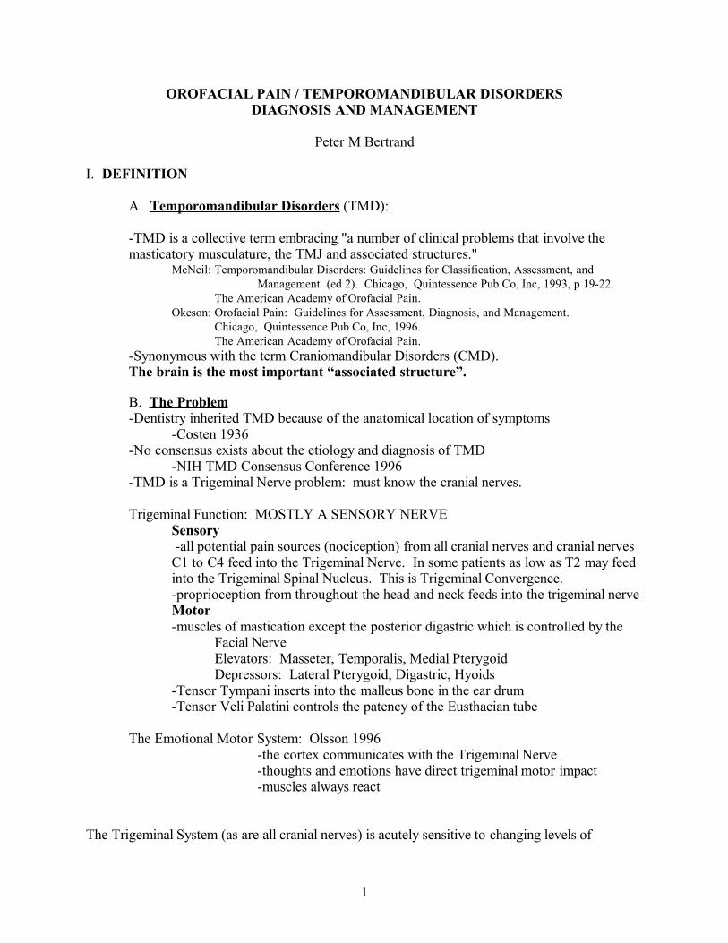
Fatigue NociceptionProprioception
Brainstem(trigeminal)(nuclei 1st)
T-C-BG circuitsunder limbic influence
Pain,AnxietyBrainstem
Trigeminal Pain ModelBertrand & Carlson ‘98
12
3
45
C2C3
12
34
5
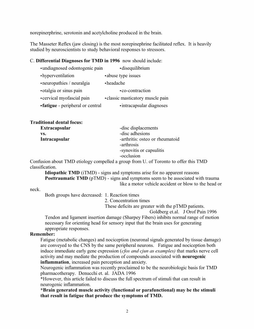
Pain
IntNACHNE5HT
Thalamus
T-C-BG
snC
Nociceptive / Fatigue Barrages Neurogenic Inflammation
second orderneuron
MSN
C2
C3
C4
thirdorderneuron

b. Psychological componentc. Irreparable joint damage
3. Management takes TIME and an understanding of head and neck physiology and stress behavior at a brain level.
4. Pain is a physiologic disturbance that something is wrong.a. Diagnose and manage the physiologic disturbances.b. Anatomy is the platform on which physiology performs behavior.
E. TMD: THE MEDICAL ORPHAN: Who owns it? "An evolutionary transgressor strategically suspended in limbo between medicine and
dentistry, the TMJ, when pathologic, masquerades as multiple other medical maladies. Misaligned by nature, maligned by medicine, misused by man, and misunderstood by most, this synovial magna cum laude has not yet joined the other articulations in orthopedics, has been turned a deaf ear by otolaryngology, and neglected by neurology." Howard
1. Shortage of scientific data/research.2. Failure of teflon proplast implants.3. TM disorders are distinct but interrelated entities with complex etiology that involves the full extent of the Trigeminal Nervous System.
II. EPIDEMIOLOGY (from AAOP Guidelines)A. 75% have at least one sign of joint dysfunction
(joint noise, tenderness, attrition, condylar remodeling etc).B. 33% have at least one symptom (face pain, joint pain, etc).C. Age range 15-45 years (mean 32.9 yrs).D. Female-to-male ratio 3:1 to 9:1.E. One parafunctional habit 60%, this number may be greater!F. Only about 5% need of treatment.
The above symptomology is a dental perspective. The list fails to address the entire extent of the Trigeminal System. Such a narrow focus will omit most of the vital differential diagnoses absolutely essential to helping patients with TMD like problems.
4
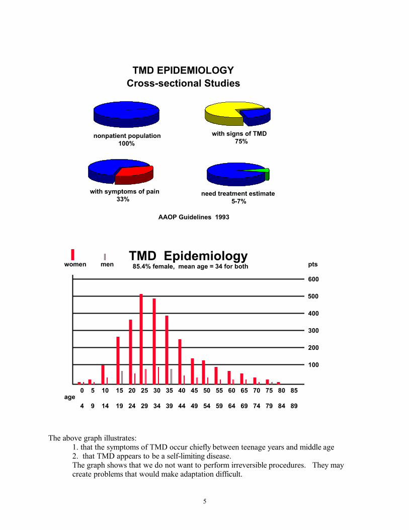
Cross-sectional Studies
nonpatient population 100%
with signs of TMD 75%
with symptoms of pain 33%
need treatment estimate 5-7%
AAOP Guidelines 1993
TMD EPIDEMIOLOGY
TMD Epidemiology
600
500
400
300
200
100
0 5 10 15 20 25 30 35 40 45 50 55 60 65 70 75 80 85 4 9 14 19 24 29 34 39 44 49 54 59 64 69 74 79 84 89
age
women men pts85.4% female, mean age = 34 for both
The above graph illustrates:1. that the symptoms of TMD occur chiefly between teenage years and middle age2. that TMD appears to be a self-limiting disease. The graph shows that we do not want to perform irreversible procedures. They may create problems that would make adaptation difficult.
5
*One problem with the data that created this graph was that neck palpation pain was considered irrelevant to TMD symptoms. In other words, there was physiologic disregard about the full extent of the Trigeminal System.
III. History and Diagnosis
80% of the data needed to make TMD diagnoses can be derived from the patient's history. History should be operator guided to identify specific problems but let patients tell their story.
A. Identify the problem, the chief complaint(s).B. Duration of pain/dysfunction. When did it start?C. Review the medical history
1. Be alert for findings that relate to TMD and other pain sources. 2. Question about pain sources below the shoulders. 3. Establish the chronology and location of all pain sources4. Pre-existing Spinal Thalamic pain can drive Trigeminal Symptoms.
D. HospitalizationsE. Arthritides and infections like Lyme's F. Symptoms of cervical, neural, otic, optic and other cranial nerve disorders.G. Sleep and stress disorders (depression, anxiety, bruxism, hyperventilation)
1. Greater than 50% of chronic TMD patients may be abuse victimsCurran et.al J Orof Pain 1996
2. About 25% of chronic TMD patients have Post Traumatic Stress SyndromeSherman et.al submitted abstract
H. Alcohol/tobaccoI. Diet (caffeine, MSG, changes in diet)J. Medications and AllergiesK. Be alert for mimickers of TM disorders.L. The patient's description may virtually make many diagnoses. (Listen to the patient)
IV. DENTAL HISTORY
A. Recent dental exam, any perceived odontogenic pain?B. Any gross changes in perceived occlusion, how teeth fit? C. Iatrogenic factors ( Restorative, Orthodontics, Orthognathic, Equilibrations)D. Is the patient aware of parafunction?
Bruxism, tongue protrusion, hyperventilation, head tilt….. Quantify patient’s perception of daily tooth contact to provide a starting point for recognizing oral parafunction.
How do you recognize brain controlled behaviors?Pain patients estimate of total tooth contact: Bertrand @ Nicoll ‘98
Range: 12 minutes to 24 hoursBruxism unaware patients: 9.2 hours Bruxism aware patients: 7.6 hoursNo statistical difference between groups.
V. PSYCHO-SOCIAL - avoid being judgmental A. What is patient's level of activity, anxiety, depression, life stressors.
6
B. Evaluate presence of factors suspected to precipitate muscle and joint symptoms: -posture
-coping mechanisms-trauma, both gross anatomic like MVA and abuse related-litigation
C. Life stressors alter brainstem and upper brain neurochemistry that facilitates increased Trigeminal and cranial nerve activity.
D. Parker's Dynamic Model of Etiology in TMD: Trauma Life Stressors
Nutrition/Health Sleep DisordersCoping Pain/Depression
Structure OcclusionGender Posture
Adaptability Hyperfunction
Orthofunction → Pathofunction
VI. CLINICAL EXAMINATION AND EVALUATION
A. Use a systematic clinical examBy exam try to rule out the differential diagnoses developed from history, do not try to reinforce to differential. 1. chief complaint (patient's own words) and other pain sources2. duration of pains (acute vs chronic) 3. pain character: sharp, dull, burning, tight, throbbing, pulsing4. diagram pain/referred pain
B. Use repeated Pain level scales - Visual Analog or NumericPatients can accurately describe the cyclic nature of their pain
0 being no discomfort, 10 being worst pain conceivable1. Present pain 0_________________102. Worst day 0_________________103. Best day 0_________________104. Average day 0_________________10
7
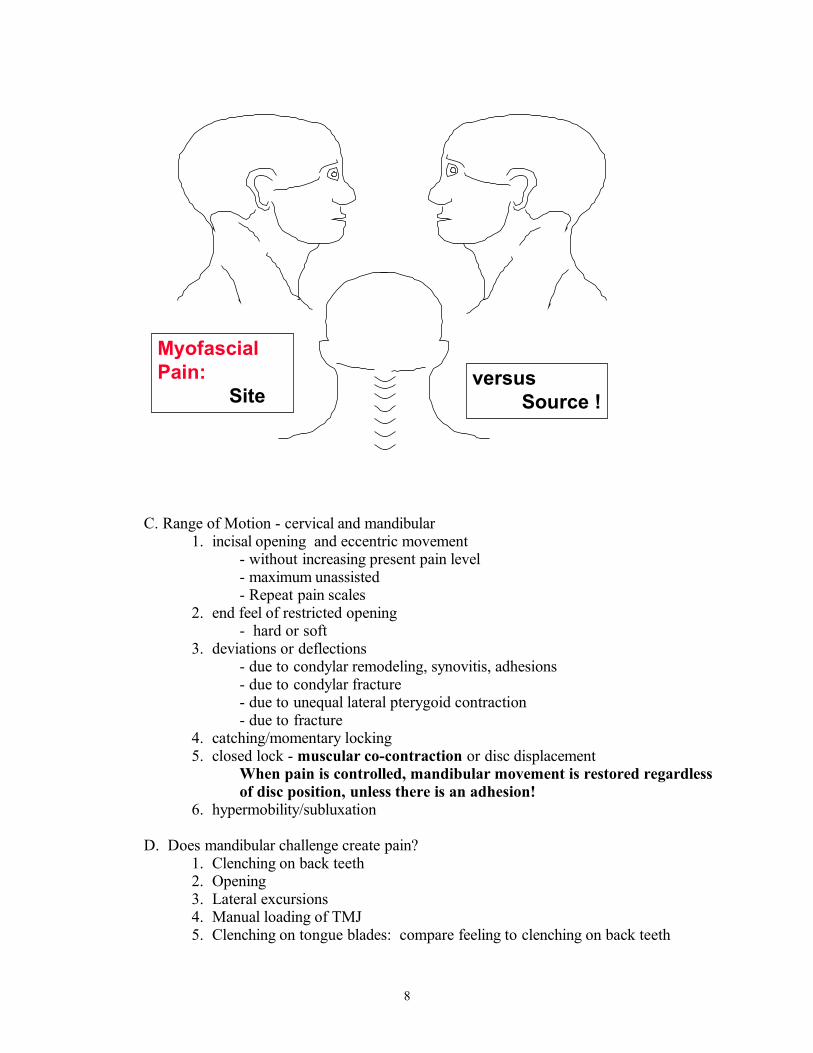
Myofascial Pain: Site
versus Source !
C. Range of Motion - cervical and mandibular 1. incisal opening and eccentric movement
- without increasing present pain level- maximum unassisted- Repeat pain scales
2. end feel of restricted opening - hard or soft
3. deviations or deflections- due to condylar remodeling, synovitis, adhesions- due to condylar fracture- due to unequal lateral pterygoid contraction- due to fracture
4. catching/momentary locking5. closed lock - muscular co-contraction or disc displacement
When pain is controlled, mandibular movement is restored regardless of disc position, unless there is an adhesion!
6. hypermobility/subluxation
D. Does mandibular challenge create pain? 1. Clenching on back teeth2. Opening3. Lateral excursions4. Manual loading of TMJ5. Clenching on tongue blades: compare feeling to clenching on back teeth
8
Is it Better Worse Same as clenching on teeth?a. bilaterallyb. right c. left
E. Rule out neurologic involvement1. Any vision, hearing, taste, balance problems2. Any paresthesias3. Cranial nerve exam
F. Vital Signs1. Blood pressure, pulse2. Repeated respiratory rates and form, best done by assistant3. Pain patients are hypocapnic and overuse their neck muscles to breathe
G. Palpation Exam - palpate the full range of the Trigeminal Nerve for myofascial and TMJ pain - head and neck muscles are evaluated by palpation and functional testing for
tenderness, hypertrophy, atrophy and referred pain/trigger points. Bilateral and sequential evaluation of the following muscles:
1. cervical musculature- Trapezius- Sternocleidomastoid- Paracervicals (splenius captius and splenius cervicus)- Occipital insertions- Hyoidsa. Any referral patternsb. Repeat pain scale 0_______________10c. Repeat incisal opening measurements
a. may identify cervical induced co-contraction
2. facial musculature- Masseter
a. superficialb. deep
- Temporalis (ant., middle, post.)- Frontalis
3. TMJs before and with movement- lateral pole- through EAM- vertical loading in CR
4. Resisted protrusion for Lateral Pterygoid function- cannot palpate the lateral pterygoid- has 2 bellies
- superior is the smaller, inserts primarily on the disc and contracts during closing to maintain disc position on the head of the
condyle- inferior is about 85% of the muscle, attachs on the condyle, and
9
contracts during opening and eccentric movements5. Intraoral
- Temporalis tendon- medial pterygoid - digastric
1. Any referral patterns2. Repeat pain scale 0_______________103. Repeat incisal opening measurements
a. may identify masticatory induced co-contractionH. Auscultation/Joint Sounds
1. crepitus (degenerative arthritis, perforation)2. popping, clicking (displaced disc) (Mahan's classification)
reciprocal click - the condyle slips with a click or pop from the posterior part of disc into the thin intermediate zone
on opening, and on closing, clicks back onto the posterior zone of the medially-anteriorly displaced disc
4. know joint anatomy and synovial function5. synovial lubrication only occurs during non-loading passive movement
Levick, Microculation 19956. Does protrusion eliminate reciprocal click?
I. Radiographs - establish need by examination1. Panorex for screening2. Transcranial - so what if the condyle is remodeled3. Tomogram - little application4. Cat scan - definite with neoplasm suspicion or with intracapsular pain and
severe translation changes that may be related to osteoarthritic remodeling
5. MRI - so what if the disc is displacedGross anatomic changes suggested by imaging may only correlate with pain when there is acute injury. With chronic pain the gross anatomic changes may represent adaptation to the same forces that creating pain.
H. TMD Diagnosis: Extracapsular vs IntracapusularIntracapsular
1. Capsulitis or synovitis2. Displaced disc, with or without reduction3. Discal adhesions4. Arthritis
- osteo- rheumatoid (Mycoplasma infection)- infection related such as Lyme's or venereal
5. Arthrosis - condylar remodeling but no pain
Extracapsular 1. Muscle findings and diagnoses according to AAOP guidelines
10
-Myofascial pain-Myositis-Spasm-Protective splinting or co-contraction-Contracture-Neoplasia
2. Cervical myofascial pain or arthritis- rotation/side bending (tilt)
- flexion/extension 3. Occlusion
- Angle's classification- anterior overjet/overbite- dental midline- chin scar/trauma- CRC to MI slide- excursive interferences- wear facets
- history of bruxing or clenching- lock and key facets that provoke symptoms
4. Undiagnosed dental pain5. Life stressors - chronic pain/depression
>50% of chronic pain patients have abusive histories6. Unconscious parafunction and habits
- morning or evening jaw or tooth pain- all thoughts and emotions elicit cranial nerve mediated muscle
activity- peripheral parethesias
7. Sleep disorder8. Headache - migraine vs tension-type
VII. CONDITIONS MIMICKING TM JOINT DISORDERS
Many conditions result in perceived TMJ pain. Preauricular or TMJ pain can have multiple sources: neck muscles, posterior teeth, parotid gland, major blood vessels, maxillary sinus, and multiple nerve pathways from cranial nerves 5, 7, 9, and 10. Referred pain must always be ruled out.
A. Ear pain - very common complaint. middle ear infection (otitis media)1. Otomandibular syndrome2. Anterior malleolar ligament3. Myofascial referral4. Fullness or fluid sensation associated Tensor Tympani tightening ear drum
and/or Tensor Veli Palatini narrowing the Eustachian tube letting air pressure into the middle ear but not allowing air release.
B. Infection of dental origin C. Maxillary sinusitis D. Salivary glands
1. parotid gland2. parotid-masseter hypertrophy-traumatic occlusion syndrome
11
3. submandibular/sublingual glandsE. Eagle's syndrome F. Ernst syndrome G. Hypertrophied coronoid process H. Rheumatoid arthritis and other arthritides
1. Sjogren's2. Lymes3. Venereal
I. Collagen vascular diseases - psoriasis, SLE, sclerodermaJ. Infections of the TM joint - (local or systemic)K. Trauma - MVA, sports, etc.L. Surgical trauma
1. auriculotemporal syndrome (Frey's)2. repeated TMJ surgeries without comprehensive conservative management
M. Gout N. Vascular changes
1. migraine headaches2. cluster headaches3. temporal arteritis4. carotodynia
O. Mucosal contact - headache/atypical facial painP. Neural origin
1. trigeminal neuralgia2. glossopharyngeal neuralgia3. neuropathy/neuritis4. intracranial nerve lesions5. odontogenic pain
Q. Synovial Chondromatosis - "joint mice" R. Tumors
1. Osteochondroma2. metastatic Adenocarcinoma3. osteochondroma
VIII. MANAGEMENT STRATEGIES FOR TM DISORDERS CONSERVATIVE - AVOID OVERTREATMENT
A. SYMPTOMATIC TREATMENT for acute TMD, before a definitive diagnosis is made, is loaded with potential hazards. Initial conservative therapy should include the following:1. education to avoid parafunction and minimize anxiety- assurance
a. See enclosed Physical Self-Regulation (PSR)sheetb. A recently completed controlled study at Bethesda shows that PSR by itself is at least as effective as traditional dental splints, soft diet and medications for chronic pain patients who have been told they have TMD.
12
2. Rest - avoid painful activity3. A diet that does not increase pain4. Moist heat or ice pack on painful area5. Analgesics, Muscle Relaxers - load and hold; not prn
Example: Motrin and/ or Flexeril Motrin or Ibuprofen- 800mg TID or 600mg qid by the clock, never PRN- many different NSAIs- do not take pain medications and continue parafunctionFlexeril - 10mg tid or 1 hour before bed
*Medications given without explaining the instructive nature of pain (pain is a physiologic disturbance that indicates something is wrong) may create a chronic pain situation if parafunctions are not addressed.
6. Tincture of time - cyclic nature of stressors often results in a cycling of symptoms. 7. Placebo - any treatment confidently prescribed will probably be enhanced, placebo
may be related to the endogenous opioid system
C. DIAGNOSIS-RELATED THERAPY: Management should always be aimed towards diagnoses: muscle and/or joint and
specific life stressors. Therapy should be physiological, functional/behavioral, with adjunctive pharmacological support.
The goal of diagnosis-related treatment may be RESTORATION TO NORMAL or ACCOMMODATION. In TMD patients ACCOMMODATION or adaptation is often a legitimate and viable treatment objective.
D. TREATMENT FOR MUSCLE DISORDERS1. Recgonition of parafunction: Physical Self-Regulation (PSR)
- The foundation- Bruxism, Postural Fatigue, Hyperventilation
2. Psychology referral - Abuse or phobias
3. Physiotherapya. spray and stretchb. moist heat/icec. massage; manual manipulationd. stretching and relaxation exercisese. ultrasoundf. transcutaneous electrical neural stimulation (TENS)g. ultra low frequency tens
4. Functional/Behavioral Therapya. anterior deprogrammerb. stabilization splint - the workhorsec. biofeedback
(1) repeat timer(2) clinical psychology
13

5. Pharmacotherapy
a. Local anesthesia - for diagnosis of pain sources and pain relief1. for trigger points and co-contraction relief2. for auriculo-temporal nerve blocks3. traditional dental injections
b. Muscle relaxants - work on CNS, not muscles1. Flexeril (Cyclobenzaprine)
10 mg/tid, max. 60 mg/day2. Parafon forte
c. Analgesics - load and hold1. mild - moderate pain: Aspirin or tylenol2. mod - severe: Percocet or other narcotic by the clock 3. acute pain - NSAIs
d. Tricyclic Antidepressants - chronic pain/sleep disorders/atpical odontolgia - analgesic at low dose- Therapeutic Window - 10-75 mg for sleep and chronic pain
- Amitriptyline (Elavil)- Doxepin (Sinequan)- Imipramine (Tofranil)- Pamelor (nortriopyline)
-The induction of sleep, Stage 3/4, may be there greatest benefit-Inhibit the synaptic reuptake of norepinephrine and serotonin, and are
anticholinergic-Enhance endogenous opioid functione. Anxiolytics
1. Valium (diazepam) - with anxiety, mediocre muscle relaxant; 2, 5, 10mg
2. Benzodiazepams facilitate stage 2 sleep but may inhibit stage 3/4 sleep3. May enhance immediate early gene expression in the amygdala and
locus coeruleus while decreasing brain reactivity elsewhere.
E. TREATMENT FOR JOINT PATHOLOGY: Inflammation, derangement, adhesion, degeneration:
Bethesda operates on hypotheses: • Muscle parafunction with or without muscle pain exceeds the TMJ's
synovial function that creates the changes that produce intracapsular pain and condylar remodeling.
• Management aimed at maximizing synovial capacity.• Synovial diffusion into any joint depends on non-loading movement.
1. Pharmacotherapy - similar to muscle.
14
a. Local anesthesia - Auriculotemporal Nerve Block - rule out muscle vs joint - Marcaine, then avoid all jaw function until anesthesia wears off
- Lidocaine 2% without epib. Muscle relaxants - brain inhibition of muscles that may overload synovial
capacityc. Analgesics
- narcotics and Tylenol may be preferred to NSAIs- want to control pain associated with inflammation but not inhibit healing
that inflammation mediates.d. Tricyclic Antidepressants
2. Physiotherapya. Iontophoresis - steroids?b. Capsaicin
3. Functional/Behavioral Measuresa. Rest to avoid signs and symptomsb. change in diet
- avoid pain inducing diet- maximize hydration, minimize caffeine, nicotine
c. stabilization splint (MRS)d. anterior repositioning splint (ARS)
4. Physiological Measures
a. manage/eliminate associated muscle componentb. manipulation to recapture disc prior to surgical procedure. ARSc. Surgical procedures - only if auriculo-temporal nerve block profoundly
eliminates perception of TMJ pain(1) Arthrocentesis - lavage & lyse adhesions(2) Arthroscopy - diagnostic, lavage & lyse(3) Arthrotomy
(a) plication and disc repair only for acute situations(b) discectomy
I. STRESSOR RELATED THERAPY: Must address stimuli that cause disorders. themselves. Stressors are the source of brain neurochemical activity that causes overwork of the Trigeminal system.
We may not be able to change a person’s stressors but we can teach them to recognize brain controlled muscle reactions to those stressors. Such reactions invovle all cranial nerves and head and neck muslces.
This concept is the basis for Physical Self-Regulation. See PSR form.
A. Recognize Parafunction1. awareness, patient education: PSR2. interocclusal devices, that modify behavior
15
3. biofeedback (repeat timer, clinical psychology)4. relaxation training (diaphragmatic breathing)
B. Life stressors1. professional counseling2. relaxation therapy3. environmental modification4. anxiolytics - rarely and for short durations5. psychiatric intervention, when medically indicated
C. Sleep disorder leading to nocturnal bruxism1. reducing life stressors2. nocturnal splint - empirically effective3. low dose tricyclic antidepressant4. flexeril
D. Pain must be treated aggressively to preclude chronicity. Treat the cause of pain to stop neurogenic inflammation. 1. anesthetic injection2. analgesics (as previously discussed)3. TENS 4. physical therapy - neuroprobe
E. Occlusion1. awareness (clenching/bruxing/nonfunctional tooth contact), patient education2. deprogrammer - occlusal disengagement3. stabilization splint - temporary correction of disharmony4. equilibration - only rarely after muscles and joints are asymptomatic
F. Adverse Posture1. awareness/education2. tongue, neck3. occupational changes in posture4. physical therapy – exercise without fatigue or pain5. chiropractic therapy
G. Adverse Coping Behavior: chronic pain, somatizing, secondary gain, defensive reaction
1. education awareness2. professional (non-dentist) counseling3. behavioral medicine4. chronic pain clinic
H. Health/Nutrition1. balanced diet2. caffeine reduction, nicotine elimination 3. adequate hydrationrest and sleep4. AEROBIC symmetrical exercise without fatigue
16
improve mitochondria capacity to extract O2 and glucose from blood
THE PATIENT MUST BE A CO-THERAPIST.Their pain is their problem. It is not your problem. Your obligations as a dentist are:• develop a superb history, may take multiple appointments• diagnose at a physiologic level• devise a management plan based on physiology that makes sense to the patient• help the patient execute the plan • always reassess and modify• never be afraid to admit not knowing• know the full extent of the Trigeminal System
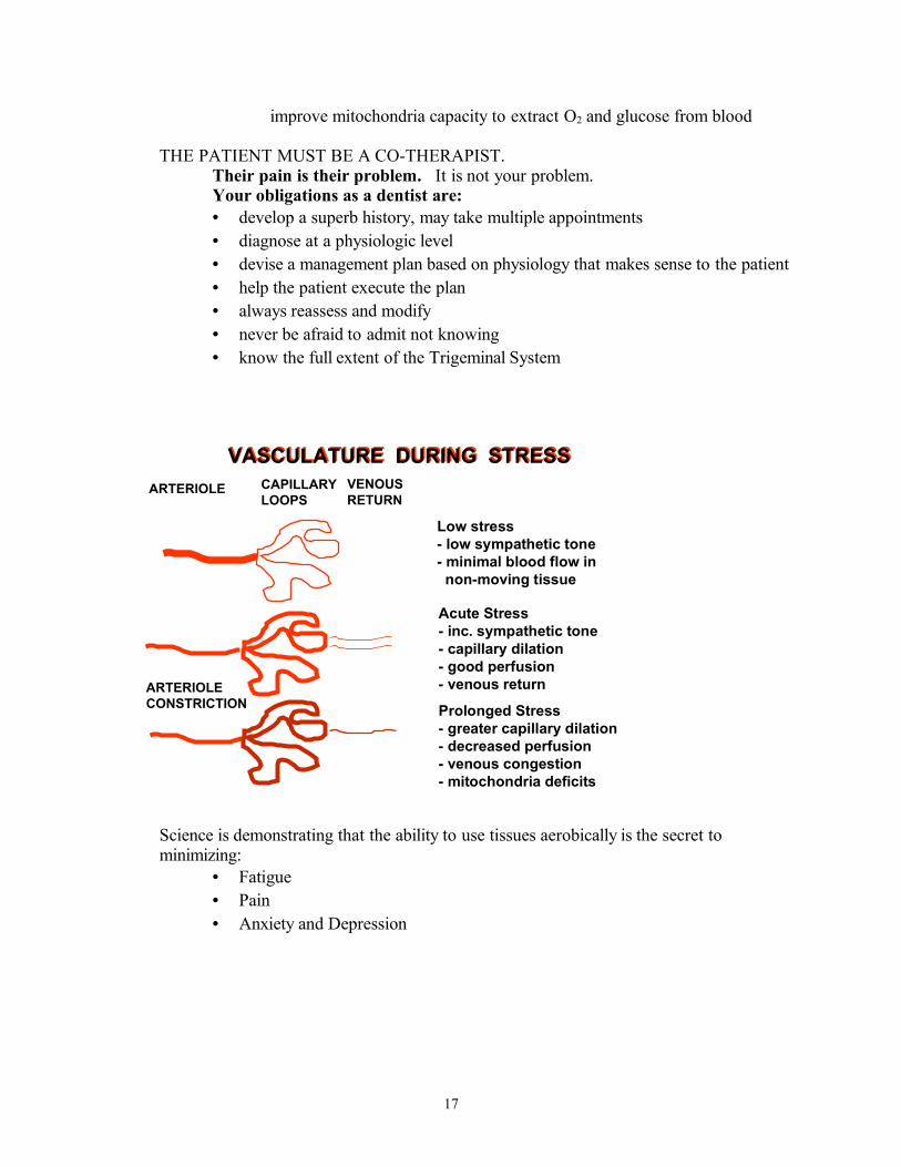
Science is demonstrating that the ability to use tissues aerobically is the secret to minimizing:
• Fatigue• Pain• Anxiety and Depression
17
ARTERIOLE CAPILLARYLOOPS
VASCULATURE DURING STRESSVASCULATURE DURING STRESS
ARTERIOLECONSTRICTION
Acute Stress- inc. sympathetic tone- capillary dilation- good perfusion- venous return
Prolonged Stress- greater capillary dilation- decreased perfusion- venous congestion- mitochondria deficits
Low stress- low sympathetic tone- minimal blood flow in non-moving tissue
VENOUSRETURN