targeted cross-linking of the human β-globin gene in living cells mediated by a triple helix...
TRANSCRIPT
Targeted Cross-linking of the Humanâ-Globin Gene in Living Cells Mediated by aTriple Helix Forming Oligonucleotide†
Kazi Abdus Shahid, Alokes Majumdar,‡ Rowshon Alam,‡ Su-Ting Liu,‡ Jean Y. Kuan,# Xuifen Sui,‡
Bernard Cuenoud,§ Peter M. Glazer,# Paul S. Miller,| and Michael M. Seidman*,‡
Laboratory of Molecular Gerontology, National Institute on Aging, National Institutes of Health, Baltimore, Maryland 21224,NoVartis Pharmaceuticals Ltd, 4002 Basel, Switzerland, Department of Biochemistry and Molecular Biology, Bloomberg School
of Public Health, Johns Hopkins UniVersity, Baltimore, Maryland 21205, and Departments of Therapeutic Radiology andGenetics, Yale UniVersity School of Medicine, P.O. Box 208040, New HaVen, Connecticut 06520-8040
ReceiVed October 14, 2005; ReVised Manuscript ReceiVed NoVember 29, 2005
ABSTRACT: Triple helix forming oligonucleotides (TFOs) may have utility as gene targeting reagents for“in situ” gene therapy of genetic disorders. Triplex formation is challenged by negative charge repulsionbetween third strand and duplex phosphates, and destabilizing positive charge repulsion between adjacentprotonated cytosines within pyrimidine motif third strands. Here we describe the synthesis of TFOs designedto target a site in the humanâ-globin gene, which is the locus for mutations that underlie theâ-globinopathies, including sickle cell anemia. The target is an uninterrupted polypurine:polypyrimidinesequence, containing four adjacent cytosines, next to a psoralen cross-link site. Pyrimidine motif TFOsthat contained four adjacent cytosines or 5-methylcytosines did not form stable triplexes at physiologicalpH, despite the introduction of otherwise stabilizing base and sugar analogues. We synthesized a seriesof pso-TFOs containing 2′-O-methyl (OMe) and 2′-O-aminoethoxy substitutions (AE), as well as 8-oxo-adenine (A8) and 2′-O-methylpseudoisocytidine (P) as neutral cytosine replacements. Thermal stabilitymeasurements indicated that TFOs with A8 did not meet criteria established in previous work. However,TFOs with P did form triplexes with appropriateTm andkON values. A pso-TFO with AE and P residueswas sufficiently active to permit the determination of targeting in living cells by direct measurement ofcross-link formation at the target site. Our results validate the modification format described in our previousstudies and indicate that P substitutions are an effective solution to the problem of targeting genomicsequences containing adjacent cytosines.
An efficient protocol for site specific genomic manipula-tion in living mammalian cells would have many researchand practical applications. The strategy would require a genetargeting reagent that can find and bind a specific sequencewith high affinity and specificity. Perturbation of thestructural, or chemical, integrity of the target sequence wouldengage cellular functions that would eventually yield thedesired change mutagenesis, gene conversion, gene knockin, etc. Such technology might eventually permit “in situ”gene therapy in which a disabling mutation in a critical genewould be restored to the wild type sequence in a clinicallyrelevant cell population. This would avoid the complicationsassociated with the random integration of an exogenous copyof a wild type gene under potentially disruptive control (1).
One approach to gene targeting is based on the DNA triplehelix which can form when a third strand of nucleic acid
lies in the major groove of an intact duplex (2). The moststable complexes are formed on polypurine:polypyrimidineelements, which are relatively abundant in mammaliangenomes (3). The structure is sequence specific (4) andstabilized by two Hoogsteen hydrogen bonds between thebases in the third strand and the purine strand in the duplex.When the third strand consists of pyrimidines (pyrimidinemotif), the triplets are T‚A:T and C+‚G:C. The third strandcytosine must be protonated at N3 in order for a secondHoogsteen hydrogen bond to form.
Although triplex formation is relatively straightforwardinVitro, in a cellular environment there are many impediments.Some are the result of the chromatin structure of mammaliangenomes, which may preclude access to target sequences (5,6). We have shown that manipulations of the cell biologycan (partially) overcome this restriction (7). We found thattarget access was greatest in S phase and very low in G0.
Similarly, transcriptional activation also renders targets moreaccessible to triple helix forming oligonucleotides (TFOs)1
(8).There are also many issues related to the chemistry and
biochemistry of TFOs, particularly those in the pyrimidine
† The research described here was supported by the IntramuralResearch Program of the National Institute on Aging, National Institutesof Health.
* Corresponding author. Address: LMG/NIA/NIH, 5600 NathanShock Dr., Baltimore, MD 21224. Telephone: 410 558 8565. Fax: 410558 8157. E-mail: [email protected].
‡ National Institutes of Health.§ Novartis Pharmaceuticals Ltd.| Johns Hopkins University.# Yale University School of Medicine.
1 Abbreviations: TFO, triple helix forming oligonucleotide; Pso,psoralen; 5MeC, 5-methylcytosine; P, pseudoisocytidine; A8, 8-oxo-adenine; AE, 2′-aminoethoxy.
1970 Biochemistry2006,45, 1970-1978
10.1021/bi0520986 CCC: $33.50 © 2006 American Chemical SocietyPublished on Web 01/24/2006
motif. Most of these have been addressed with some degreeof success by introduction of base and sugar analogues intoTFOs. Triplex formation by deoxyribose third strandsimposes conformational restrictions on the TFO and somedistortion of the underlying duplex. Typically triplexesformed by deoxyribose third strands are less stable than theunderlying duplex. RNA analogue sugars can “preorganize”third strands, greatly reducing the entropic barriers to triplexformation and requiring relatively little distortion of theunderlying duplex (9). Pyrimidine motif triplexes are gener-ally unstable at physiological pH because of the requirementfor cytosine protonation (cytosine pKa ) 4.5). This can bealleviated, at least in part, by the use of 5-methylcytosine(5MeC) (10). The stabilization by the methyl substitutionresults from stacking interactions and hydrophobic effects(11). This is effective for triplex sequences with isolatedcytosines, and isolated cytosines make a considerablecontribution to triplex stability. The positive charge partiallyreduces the negative charge repulsion between the thirdstrand and the duplex (12), which is also ameliorated bymagnesium ions (13). We have described pyrimidine motifTFOs containing 5MeC and 2′-O-methylribose that formtriplexes at neutral pH that are as stable as the underlyingduplex (14). On the other hand, targets with adjacentcytosines are more problematic. Triplex stability is greatlycompromised by runs of cytosines, thought to be due torepulsion between the positive charges resulting from theN3 protonation (10, 15). Alternatively there may be acompetition for protons by the adjacent cytosines (16). Ithas been shown that at physiological pH cytosine protonationis reduced in runs of cytosines relative to isolated cytosines(11). Several base analogues have been proposed as solutionsto this problem. These are purine or pyrimidine analoguesthat are protonated at neutral pH such that two Hoogsteenhydrogen bonds with guanine are possible. They include8-oxoadenine (17), 2′-O-methylpseudoisocytidine (18, 19),and 2-amino-5-(2′-deoxy-â-D-ribofuranosyl)pyridine (2-ami-nopyridine) (20-22) (see also refs23and24). Although theresults of biochemical analyses of TFOs with these deriva-tives are encouraging, their efficacy in biological assays hasnot been described.
Negative charge repulsion between the TFO and the duplexcan be suppressed by the incorporation of 2′-aminoethoxy(AE) substitutions which are protonated at neutral pH (25,26). This modification stabilizes the C3′-endo conformationof the ribose and also forms a bridge with thei - 1 phosphatein the purine strand of the duplex (26). We have describedthe construction and characterization of TFOs containing acluster of AE residues that are bioactive in a gene knockout assay in living mammalian cells (7, 27, 28). The TFOswere designed to target a sequence in the endogenousHPRT(hypoxanthine phosphoribosyl transferase) gene, which iscommonly used as a mutation reporter gene. The targetsequence was a 17 base polypurine:polypyrimidine elementwith isolated cytosines, terminating in an ideal site for cross-linking by psoralen. Psoralen linked TFOs containing dif-ferent modifications were tested for activity in an assay thatmeasured targeted mutagenesis of theHPRTgene (14, 27,28). TheHPRTmutation assay is straightforward and reportsmutational events over a range of 5 orders of magnitude.However, most genes of interest for gene targeting do notlend themselves to simple selection based assays that report
the desired event. Consequently, the TFO must be sufficientlyactive to allow detection of target binding with relativelyinsensitive biochemical methods. In this report we describethe development of a biologically active psoralen linked TFOdesigned to target a site containing adjacent cytosines in thehumanâ-globin gene. The site is of interest because of thechallenge to triplex formation presented by its sequence, andthe manyâ-globinopathies in the human population resultingfrom mutations in the gene.
MATERIALS AND METHODS
Reagents.Reagent grade chemicals were used unlessotherwise noted. HPLC grade acetonitrile was dried overcalcium hydride. Anhydrous pyridine, dimethyl formamide,benzene, tetrahydrofuran, and methylene chloride were fromAldrich Chemical Co. Inc. 5-â-D-Ribofuranosyluracil (pseudo-uridine) was the generous gift of Dr. Kris Pankiewicz. The5′-O-(4,4′-dimethoxytrityl)-5-methyluridine-2′-O-methyl-3′-O-(â-cyanoethyl-N,N-diisopropyl) phosphoramidite and the5′-O-(4,4′-dimethoxytrityl)-5-methyluridine-2′-O-methyl-3′-O-succinamido-N6-hexanamidoN3-propyl-controlled poreglass support were purchased from Chemgenes, Ashland,MA. Protected deoxyribonucleoside phosphoramidites, theN4-acetyl-5′-O-(4,4′-dimethoxytrityl)-5-methylcytidine-2′-O-methyl-3′-O-(â-cyanoethyl-N,N-diisopropyl) phosphoramid-ite, 6-[4′-(hydroxymethyl)-4,5′,8-trimethylpsoralen] hexyl-1-O-(â-cyanoethyl-N,N-diisopropyl) phosphoramidite, and8-hydroxy-5′-dimethoxytrityl-N6-benzoyl-deoxyadenosine-3′-[(2-cyanoethyl)(N,N-diisopropyl)] phosphoramidite were pur-chased from Glen Research, Inc., Sterling, VA. The modifiednucleosides 5′-O-(4,4′-dimethoxytrityl)-5-methyluridine-2′-O-(2-aminoethyl)-3′-O-(â-cyanoethyl-N,N-diisopropyl) phos-phoramidite andN4-(N-methylpyrrolidineamidine)-5′-O-(4,4′-dimethoxytrityl)-5-methylcytidine-2′-O-(2-aminoethyl)-3′-O-(â-cyanoethyl-N,N-diisopropyl) phosphoramidite were synthe-sized as described (25). All the reagents used for oligonucleo-tide synthesis were standard and were obtained from Chem-genes, Ashland, MA. [[γ]-32P]ATP was purchased fromAmersham Inc., and T4 polynucleotide kinase was purchasedfrom United States Biochemical Corp. Thin-layer chroma-tography (TLC) was performed on silica gel 60F254 plates(0.2 mm), and flash chromatography was carried out usingEM Science Kieselgel 60 (230-400 mesh). Proton NMRspectra were recorded on a JEOL 400 MHz spectrometerwith tetramethylsilane as reference for chemical shift.Electrophoresis was carried out in 12% polyacrylamide gelswith or without 7 M urea. The TAE running buffer contained40 mM Tris, 10 mM MgAc2, and 5 mM NaAc buffered atpH 7.0. Reversed phase HPLC was carried out using aSymmetry 300 C18 column from Waters on a ShimadzuHPLC system (LC-10ADvp) with a duel wavelength detector(SPD-10AVvp) and an autoinjector (SIL-10AVvp).
Synthesis of Pseudoisocytidine Phosphoramidite.Thesynthesis of 2-[[(dimethylamino)methylene]amino]-5-[2-O-methyl-5-O-(dimethoxytrityl)-â-D-ribofuranosyl]-4(1H)-py-rimidinone-N2-[(dimethylamino)methylene]-2′-O-methyl-5′-O-(dimethoxytrityl)-pseudoisocytidine-3′-O-(â-cyanoethyl-N,N-diisopropyl) phosphoramidite was done using the routereported previously (19). This involved conversion ofpseudouridine to 1,3-dimethyl-3′-5′-O-(tetraisopropyldisi-loxanyl)pseudouridine followed by methylation of the 2′-hydroxy group. Then deprotection of the silyl group,
â-Globin Gene Targeting by a Triplex Forming Oligonucleotide Biochemistry, Vol. 45, No. 6, 20061971
reprotection of 5′-hydroxy group with dimethoxytrityl,guanidinylation, and finally the protection of the amino groupby (N,N-dimethylamino)methylene gave 2-[[(dimethylami-no)methylene]amino]-5-[2-O-methyl-5-O-(dimethoxytrityl)-â-D-ribofuranosyl]-4(1H)-pyrimidinone-N2-[(dimethylamino)-methylene]-2′-O-methyl-5′-O-(dimethoxytrityl)pseudoisocy-tidine. Phosphitylation of the 3′-hydroxy group gave theexpected pseudoisocytidine phosphoramidite, which wasconfirmed by NMR and MS analysis.
Oligonucleotide Syntheses.The oligonucleotides weresynthesized on CPG supports using an Expedite model 8909DNA/RNA synthesizer. All protected nucleoside phosphora-mides were dissolved in anhydrous acetonitrile at a concen-tration of 0.05 M. The nucleoside pseudoisocytidine phos-phoramidite solution was stored for 2 h over molecular sievesprior to use. Standard coupling times were employed forgeneral nucleosides, except for pseudoisocytidine phosphora-midite and for the psoralen phosphoramidites, for which 360and 600 s, respectively, were used. The synthesizer wasprogramed to carry out a capping step, an oxidation step,and then another capping step after each coupling step andfinally to remove the last 5′-terminal dimethoxytrityl groupfrom the protected oligomer. The psoralen-derivatized oli-gomers were prepared on the controlled pore glass supportusing 2-[4′-(hydroxymethyl)-4,5′,8-trimethylpsoralen]hexyl-1-O-[(2-cyanoethyl)-(N,N-diisopropyl)] phosphoramidite inthe final coupling reaction.
Deprotection and Purification of Oligonucleotides.Thenon-psoralen oligonucleotides were deprotected by treatmentwith a solution of 28-30% ammonium hydroxide at 55°Cfor 5 h. Oligonucleotides containing 8-oxoadenine andpseudoisocytidine were deprotected by treating the support-bound oligomer with a solution of ethylenediamine in 95%ethanol (1:1 v/v) at room temperature for 16 h. Psoralenlinked oligonucleotides were incubated in a 1:1 mixture of28% ammonium hydroxide and 40% aqueous methylaminesolution, at room temperature for 90 min. The deprotectedoligomers were taken up in distilled water and purified byanalytical and semipreparative anion exchange (IE) HPLCusing a DIONEX DNAPac column on a Shimadzu HPLCsystem (LC-10ADvp) with a dual wavelength detector (SPD-10ADvp) and an autoinjector (SIL-10AVvp). The columnwas eluted using a linear gradient of 0-50% acetonitrile in100 mM Tris-HCl buffer (pH 7.8) at a flow rate of 1.5 mL/min and monitored at 254 and 315 nm. The oligomers weredesalted on a SEP PAK C18 cartridge following standardprocedures. The purified oligomers migrated as single bandson 12% polyacrylamide gels and were characterized bymatrix-assisted laser desorption-ionization time-of-flight(MALDI TOF) mass spectrometry.
Thermal Stability Measurements.The constituent strandsof the target duplexes (1µM) were dissolved in buffercontaining 100 mM NaCl, 2 mM MgCl2, and 10 mM Na-cacodylate, pH 7.0. The solutions were heated at 80°C for3 min and allowed to come to room temperature. The TFOs(1 µM) were added to the duplex solution and incubated atroom temperature overnight. The thermal denaturationexperiments were carried out using a Cary 3E UV-visspectrophotometer fitted with a thermostated sample holderand temperature controller. Triplexes were heated from 10to 85 °C at a rate of 0.4°C/min, and the absorbance at 260nm was recorded as a function of the temperature. All
analyses were performed at least two times with an error ofno more than 0.5°C.
TFO Association Analysis by Absorbance Decay. The TFO+ duplexf triplex transition is accompanied by a decreasein UV absorbance. This decay curve has been used toestimate the association rates of a TFO with the target duplex(29). For our experiments, the duplex strand was formed inkinetics buffer I (1 mM MgCl2, 10 mM sodium cacodylate(pH 7.2), 150 mM KCl) to give a final concentration of 1µM. 1 mL of the duplex stock solution was monitored byUV in a cuvette at 25°C and gave a horizontal line as afunction of time. The analyses were done at 25°C (using aPeltier temperature controller). An aliquot of the TFO stocksolution in kinetics buffer I was added with vigorous mixingto 1 mL of duplex stock solution. The mixing process tookless than 10 s. The experiments were run on a Cary dualbeam spectrophotometer. The decay curves were fit usingsecond order kinetics with the software supplied with theinstrument. Rate constants were reported as an average ofthree or more experiments.
Band Shift Analysis of Triplex Formation.The pyrimidinestrand of the duplex was labeled with32P, the duplex formed,and then the triplex formed by incubation of the duplex (∼1nM) and the third strand (2µM) overnight in triplexformation buffer. The samples were then electrophoresed on12% neutral polyacrylamide gels, in 10 mM Tris acetate (pH7.0), 5 mM MgCl2.
Psoralen Integrity Assay. After triplex formation on a32Plabeled duplex, the samples were exposed to UVA (365 nM)for 10 min and then denatured by heating in loading buffer(containing 90% formamide) and then electrophoresed in a12% denaturing polyacrylamide gel in 10 mM Tris borate,pH 8.0, 7 M urea.
Cross-linking of the Triplex Target in the Humanâ-GlobinGene in Cultured Cells. Human erythroleukemia K562 cellswere suspended in a 100µL solution with TFO-22 at 4µMand then electroporated (Amaxa). The cells were suspendedin medium and incubated for 3 h atroom temperature. Theywere then exposed to UVA (365 nM) for 3 min in a Rayonetchamber at 1.8 J/cm2. Genomic DNA was then extractedfrom the cells, purified, and then digested withEcoRIrestriction enzyme (see ref7 for details on the extractionand purification procedure). The digested samples weredenatured by heating in 90% formamide and then electro-phoresed in a neutral 1.5% agarose gel. The gel was blottedonto a nylon filter which was hybridized with a32P labeledprobe against the humanâ-globin gene in a 5 kbrestrictionfragment.
RESULTS
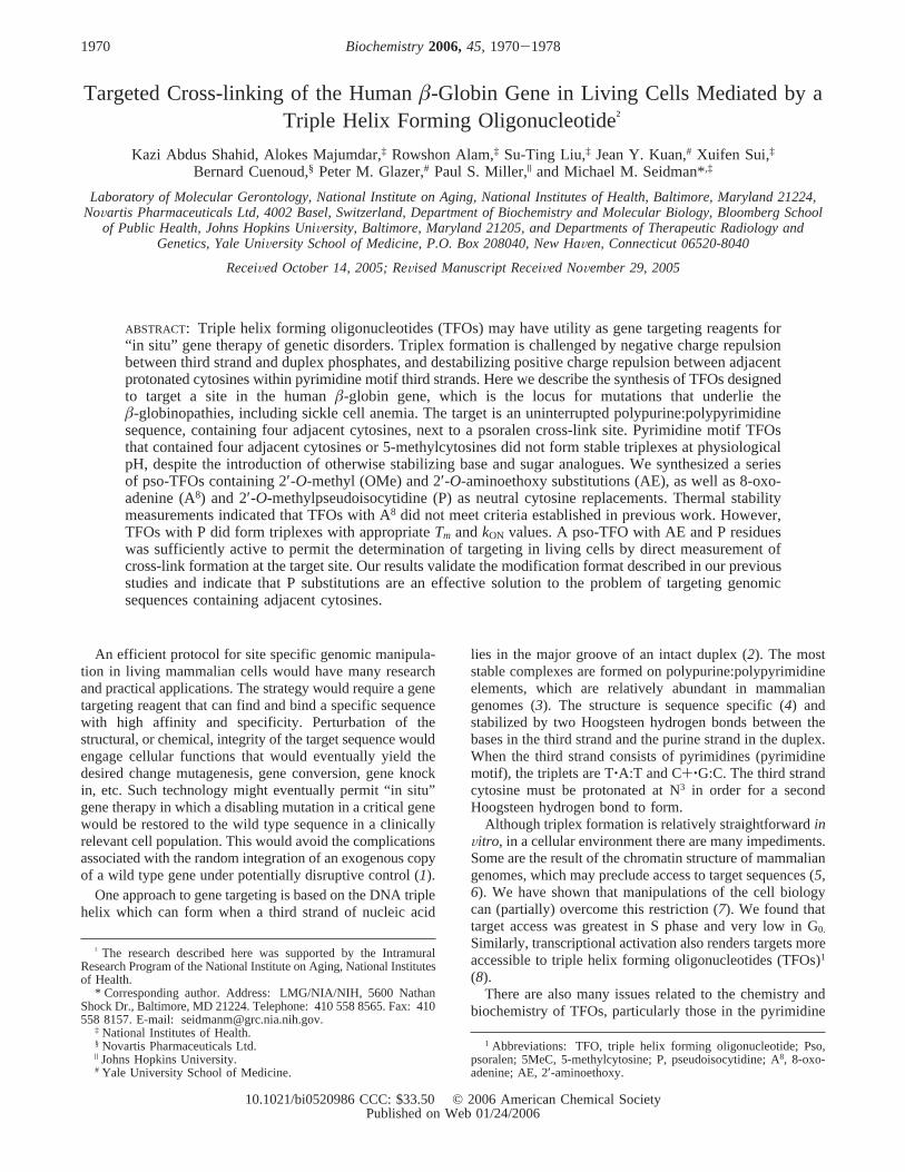
Theâ-Globin IVS2 Target Sequence.The humanâ-globingene consists of three exons and two introns. Inside thesecond intron is a 21 base polypurine:polypyrimidine elementterminated by a 5′ TA step, which is a favored site forpsoralen cross-linking (Figure 1). The triplex target sequencecontains four adjacent cytosines. We began our studies bycharacterizing a series of pyrimidine and purine TFOs,containing adjacent cytosines or 5 methylcytosines and withdifferent base, sugar, and backbone modifications, all shownto enhance triplex stability. The base modification was 5-(1-propynyl)-2′-deoxyuridine (pdU) (30). The sugar modifica-
1972 Biochemistry, Vol. 45, No. 6, 2006 Shahid et al.
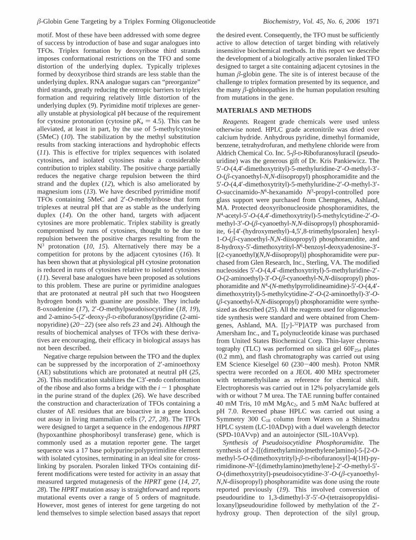
tions were 2′-guanidoethyl (2′-OGE) (31), 2′-O,4′-C-methylene ribose (BNA/LNA) (32, 33), 2′-O-(2-methoxyethyl)(2′-OME) (34), 2′-O-(N-(methyl)acetamido) (35), morpholino(36), 2′-O-methyl (2′-OMe), and 2′-aminoethoxy (2′-AE)(25). The backbone modification was the nonbridgingphosphate derivative diethylethylenediamine (DEED) (37).Target binding by the TFOs shown in Table 1 was analyzedin a band shift assay at pH 5.6 and 7.2. While a number ofthe TFOs were able to form stable triplexes at pH 5.6 (arepresentative example is shown in Figure 2), they eithershowed weak binding (the DEED TFO,Kd ) 10-6) or nobinding (all other TFOs) at pH 7.2. Of particular interestwere the TFOs containing 2′ AE residues in the cytosinepatch. The AE moiety is positively charged at neutral pHand has been shown to stabilize triplexes (25). The band shiftassays with all the oligonucleotides, including the AE TFOs,indicated that the inhibitory effect of the cytosine runovercame substitutions that otherwise stabilize triplexes atneutral pH.
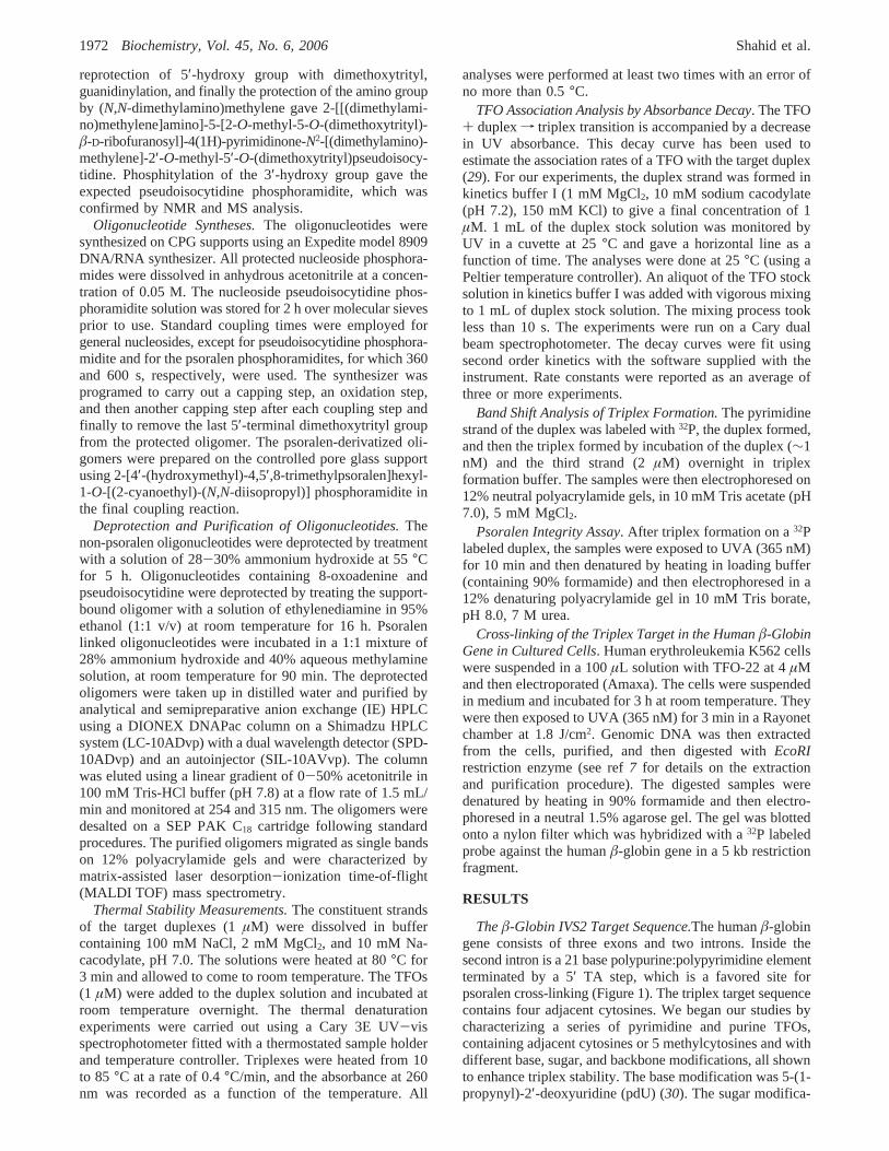
All of the pyrimidine TFOs contained cytosine or 5MeC,both isolated and adjacent. The failure of these constructionsprompted us to consider the effect of substitutions of cytosinereplacements within the cytosine patch. C+‚G:C or 5-MeC+‚G:C triplets are stabilized by hydrogen bonding, and alsothe positive charge, which appears to make a major contribu-tion (12). However, cytosine replacements, such as 8-oxoad-enine (A8) or 2′-O-methylpseudoisocytidine (P) (Figure 3A),used in the following experiments, lack the positive charge.
Table 1: Sequence and Composition of TFOs Containing Adjacent C or 5MeC (Pyrimidine Motif) or G (Purine Motif) Residuesa
TFO base sugar
TCTTTTCTTCCCCTTTCTTTT 5MeC deoxyAAAAGAAAGGGGAAGAAAAGA A, G deoxyUCUUUUCUUCCCCUUUCUUUU 5MeC/pdU deoxyU*CU*U*U*U*CTTCCCCTTTCTTTU* 5MeC/T/U* 2 ′-OGE*, 2′-OMeTCTTTTCTTCCCCTTTCTTTU∧ 5MeC 2′-OMOE, 3′-OMOE∧TCTTTTCTTCAECAECAECAETTTCTTTT 5MeC 2′-AE, 2′-OMeTCTTTTCTTCCAECAECTTTCTTTT 5MeC 2′-AE, 2′-OMeTGTTTTGTTGGGGTTTGTTTT G, T deoxyTLCTLTTLTCLTTLCCLCCLTTLTCLTTLT C, T LNA (alt)(UCUUUUCUUCCCCUUUCUUU)MA U 5MC/5MeU 2′-OMATCTTTTCTTCCCCTTTCTTTT-(MORPH) C, T morpholinoA+G+A+A+A+G+G+G+G+A+A+G+A+A+A+A+G+A+ A, G DEED
a AE, 2′-aminoethoxy; pdU, 5-(1-propynyl)-2′-deoxyuridine; OGE, 2′-O-(2-guanidoethyl)-5 methyl-U); OMOE, methoxyethyl; LNA, 2′-O,4′-C-methylene linked locked nucleic acid; OMA, 2′-O-(N-(methyl)acetamido)-5-methyl; MORPH, morpholino; DEED, diethylethylenediamine. TheTFO containing the OGE derivative has the OGE sugar modification linked to 5 methyl-U.
FIGURE 1: Organization of the humanâ-globin gene and thesequence of the triplex target in Intron 2.
FIGURE 2: Band shift analysis of the deoxyribose TFO with adjacent5-methylcytosines in the cytosine run: (A) pH 5.6; (B) pH 7.2.The arrow marks the position of the triplex. The lane marked Dhad only the duplex. The concentration of the TFO in eachincubation is indicated at the top of the lane. These results weretypical of the binding assays performed with the TFOs in Table 1.
FIGURE 3: (A) Sequence of the humanâ-globin triplex target, andvariants. The four clustered G:C pairs are separated from theremainder of the sequence, and the psoralen cross-link site isindicated in larger font. In duplex 2 the adjacent G:C pairs werereplaced with A:T. In duplex 3 G:C and A:T pairs were alternated,while in duplex 4 the G:C flanked adjacent A:T pairs. (B) Structureof (a) C+‚G:C, (b) T‚A:T, (c) 8-oxo-A (A8)‚G:C, and (d) 2′-O-methylpseudoisocytidine (P)‚G:C triplets.
â-Globin Gene Targeting by a Triplex Forming Oligonucleotide Biochemistry, Vol. 45, No. 6, 20061973
To assess the consequences for triplex stability of the lossof the protonated cytosine charge, we synthesized duplextargets for binding studies in which all (duplex 2) or two ofthe four cytosines (duplexes 3 and 4) were replaced with T(Figure 3B). These targets provided the opportunity toexamine the stability of complexes in which the T‚A:Ttriplets would provide a reference for triplets formed by theuncharged cytosine replacements.
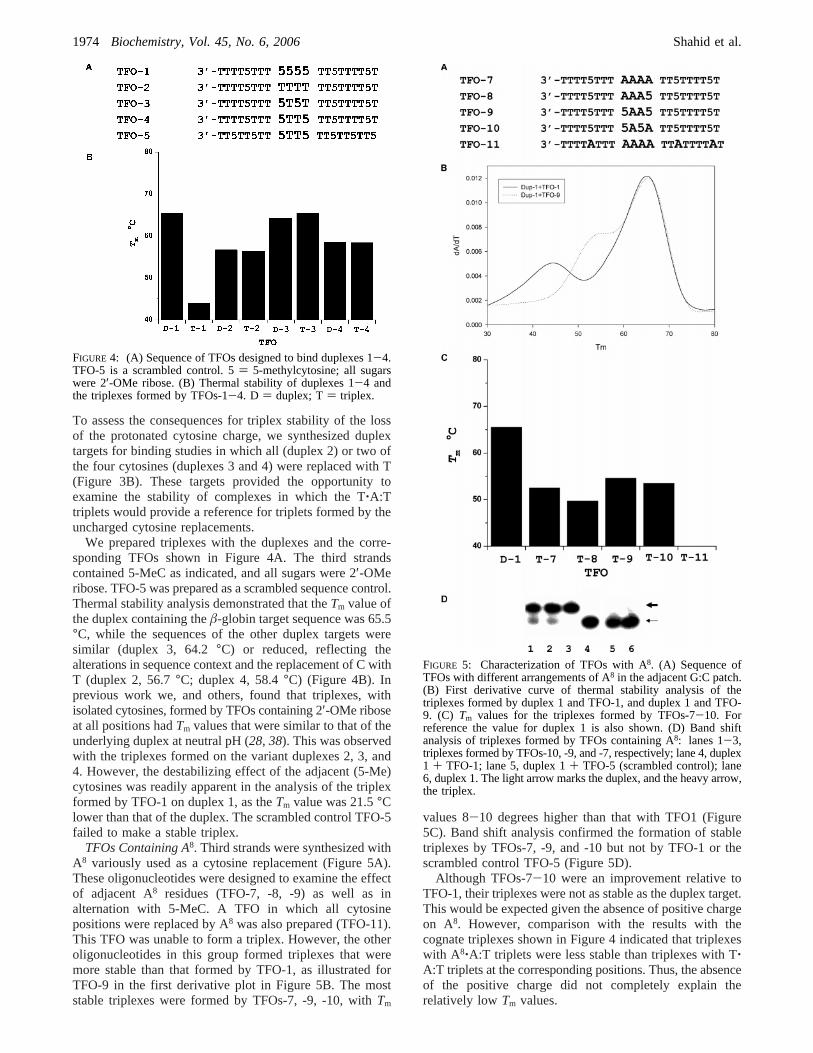
We prepared triplexes with the duplexes and the corre-sponding TFOs shown in Figure 4A. The third strandscontained 5-MeC as indicated, and all sugars were 2′-OMeribose. TFO-5 was prepared as a scrambled sequence control.Thermal stability analysis demonstrated that theTm value ofthe duplex containing theâ-globin target sequence was 65.5°C, while the sequences of the other duplex targets weresimilar (duplex 3, 64.2°C) or reduced, reflecting thealterations in sequence context and the replacement of C withT (duplex 2, 56.7°C; duplex 4, 58.4°C) (Figure 4B). Inprevious work we, and others, found that triplexes, withisolated cytosines, formed by TFOs containing 2′-OMe riboseat all positions hadTm values that were similar to that of theunderlying duplex at neutral pH (28, 38). This was observedwith the triplexes formed on the variant duplexes 2, 3, and4. However, the destabilizing effect of the adjacent (5-Me)cytosines was readily apparent in the analysis of the triplexformed by TFO-1 on duplex 1, as theTm value was 21.5°Clower than that of the duplex. The scrambled control TFO-5failed to make a stable triplex.
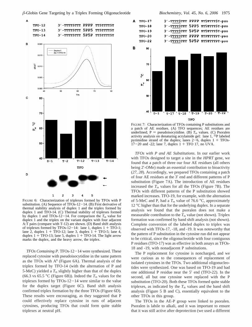
TFOs Containing A8. Third strands were synthesized withA8 variously used as a cytosine replacement (Figure 5A).These oligonucleotides were designed to examine the effectof adjacent A8 residues (TFO-7, -8, -9) as well as inalternation with 5-MeC. A TFO in which all cytosinepositions were replaced by A8 was also prepared (TFO-11).This TFO was unable to form a triplex. However, the otheroligonucleotides in this group formed triplexes that weremore stable than that formed by TFO-1, as illustrated forTFO-9 in the first derivative plot in Figure 5B. The moststable triplexes were formed by TFOs-7, -9, -10, withTm
values 8-10 degrees higher than that with TFO1 (Figure5C). Band shift analysis confirmed the formation of stabletriplexes by TFOs-7, -9, and -10 but not by TFO-1 or thescrambled control TFO-5 (Figure 5D).
Although TFOs-7-10 were an improvement relative toTFO-1, their triplexes were not as stable as the duplex target.This would be expected given the absence of positive chargeon A8. However, comparison with the results with thecognate triplexes shown in Figure 4 indicated that triplexeswith A8‚A:T triplets were less stable than triplexes with T‚A:T triplets at the corresponding positions. Thus, the absenceof the positive charge did not completely explain therelatively low Tm values.
FIGURE 4: (A) Sequence of TFOs designed to bind duplexes 1-4.TFO-5 is a scrambled control. 5) 5-methylcytosine; all sugarswere 2′-OMe ribose. (B) Thermal stability of duplexes 1-4 andthe triplexes formed by TFOs-1-4. D ) duplex; T) triplex.
FIGURE 5: Characterization of TFOs with A8. (A) Sequence ofTFOs with different arrangements of A8 in the adjacent G:C patch.(B) First derivative curve of thermal stability analysis of thetriplexes formed by duplex 1 and TFO-1, and duplex 1 and TFO-9. (C) Tm values for the triplexes formed by TFOs-7-10. Forreference the value for duplex 1 is also shown. (D) Band shiftanalysis of triplexes formed by TFOs containing A8: lanes 1-3,triplexes formed by TFOs-10, -9, and -7, respectively; lane 4, duplex1 + TFO-1; lane 5, duplex 1+ TFO-5 (scrambled control); lane6, duplex 1. The light arrow marks the duplex, and the heavy arrow,the triplex.
1974 Biochemistry, Vol. 45, No. 6, 2006 Shahid et al.
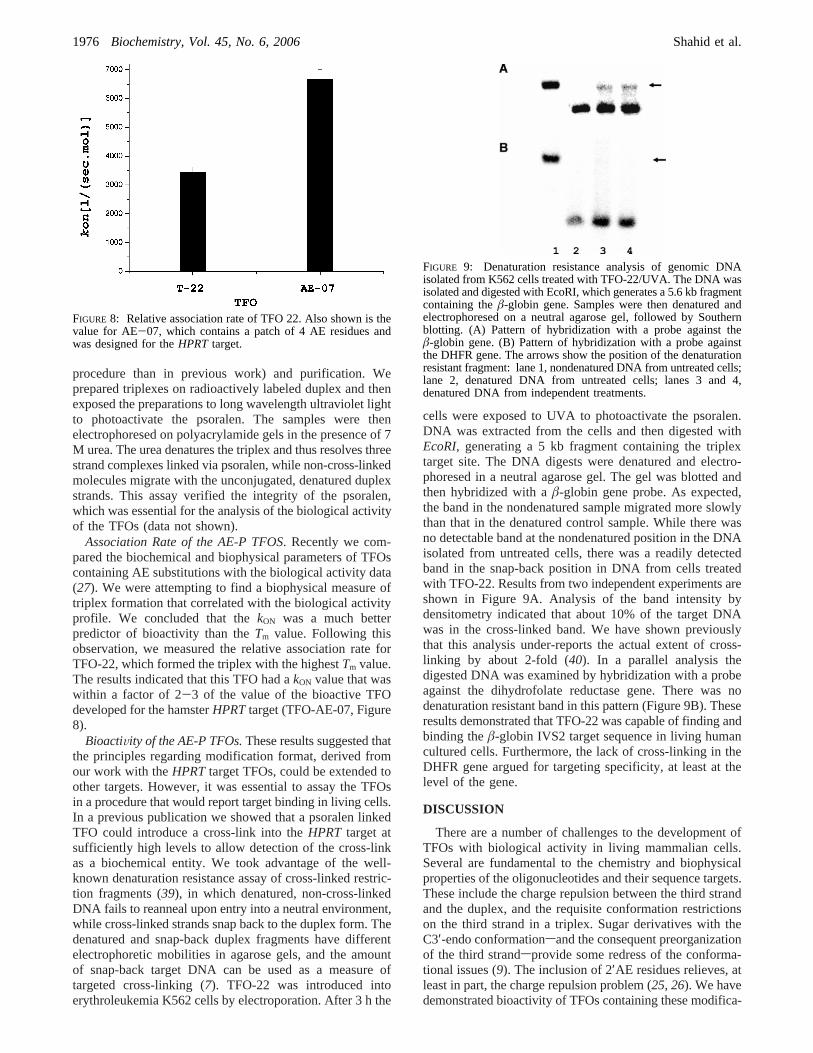
TFOs Containing P. TFOs-12-14 were synthesized. Thesereplaced cytosine with pseudoisocytidine in the same patternas the TFOs with A8 (Figure 6A). Thermal analysis of thetriplex formed by TFO-14 (with the alternation of P and5-MeC) yielded aTm slightly higher than that of the duplex(66.3 vs 65.5°C (Figure 6B)). Indeed theTm values for thetriplexes formed by TFOs-12-14 were similar to the valuefor the duplex target (Figure 6C). Band shift analysisconfirmed triplex formation by the three TFOs (Figure 6D).These results were encouraging, as they suggested that Pcould effectively replace cytosine in runs of adjacentcytosines, producing TFOs that could form quite stabletriplexes at neutral pH.
TFOs with P and AE Substitutions. In our earlier workwith TFOs designed to target a site in theHPRTgene, wefound that a patch of three our four AE residues (all othersbeing 2′-OMe) made an essential contribution to bioactivity(27, 28). Accordingly, we prepared TFOs containing a patchof four AE residues at the 3′ end and different patterns of Psubstitution (Figure 7A). The introduction of AE residuesincreased theTm values for all the TFOs (Figure 7B). TheTFOs with different patterns of the P substitution showedmarked increases. TFO-19, for example, with the alternationof 5-MeC and P, had aTm value of 76.6°C, approximately12°C higher than that for the underlying duplex. In a separateanalysis we found that the psoralen does not make ameasurable contribution to theTm value (not shown). Triplexformation was confirmed by band shift analysis (not shown).Complete conversion of the labeled duplex to triplex wasobserved with TFOs-17, -18, and -19. It was noteworthy thatthe pattern of P substitution in the cytosine run did not appearto be critical, since the oligonucleotide with four contiguousP residues (TFO-17) was as effective in both assays as TFOs-18 and -19, with nonadjacent P substitutions.
The P replacement for cytosine is noncharged, and wewere curious as to the consequences of replacement ofisolated cytosines in the TFOs. Two additional oligonucleo-tides were synthesized. One was based on TFO-19 and hadone additional P residue near the 5′ end (TFO-22). In thesecond, all but one cytosine were replaced with the Psubstitution (TFO-20). Both these TFOs formed quite stabletriplexes, as indicated by theTm values and the band shiftanalysis (Figure 5 B and C), essentially equivalent to theother TFOs in this group.
The TFOs in the AE-P group were linked to psoralen.Psoralen is labile to alkali, and it was important to ensurethat it was still active after deprotection (we used a different
FIGURE 6: Characterization of triplexes formed by TFOs with Psubstitution. (A) Sequence of TFOs-12-14. (B) First derivative ofthermal stability analysis of duplex 1 and the triplex formed byduplex 1 and TFO-14. (C) Thermal stability of triplexes formedby duplex 1 and TFOs-12-14. For comparison theTm value forduplex 1 and the triplex on the variant duplex with four adjacentA:T pairs (compare with T-12) are shown. (D) Band shift analysisof triplexes formed by TFOs-12-14: lane 1, duplex 1+ TFO-1;lane 2, duplex 1+ TFO-12; lane 3, duplex 1+ TFO-5; lane 4,duplex 1+ TFO-13; lane 5, duplex 1+ TFO-14. The light arrowmarks the duplex, and the heavy arrow, the triplex.
FIGURE 7: Characterization of TFOs containing P substitutions anda patch of AE residues. (A) TFO sequences; AE residues areunderlined; P) pseudoisocytidine. (B)Tm values. (C) Psoralenactivity analysis on denaturing acrylamide gel: lane 1,32P labeledpyrimidine strand of the duplex; lanes 2-6, duplex 1+ TFOs-17-20 and -22; lane 7, duplex 1+ TFO 17, no UVA.
â-Globin Gene Targeting by a Triplex Forming Oligonucleotide Biochemistry, Vol. 45, No. 6, 20061975
procedure than in previous work) and purification. Weprepared triplexes on radioactively labeled duplex and thenexposed the preparations to long wavelength ultraviolet lightto photoactivate the psoralen. The samples were thenelectrophoresed on polyacrylamide gels in the presence of 7M urea. The urea denatures the triplex and thus resolves threestrand complexes linked via psoralen, while non-cross-linkedmolecules migrate with the unconjugated, denatured duplexstrands. This assay verified the integrity of the psoralen,which was essential for the analysis of the biological activityof the TFOs (data not shown).
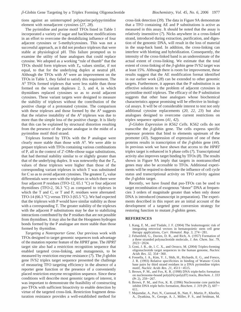
Association Rate of the AE-P TFOS. Recently we com-pared the biochemical and biophysical parameters of TFOscontaining AE substitutions with the biological activity data(27). We were attempting to find a biophysical measure oftriplex formation that correlated with the biological activityprofile. We concluded that thekON was a much betterpredictor of bioactivity than theTm value. Following thisobservation, we measured the relative association rate forTFO-22, which formed the triplex with the highestTm value.The results indicated that this TFO had akON value that waswithin a factor of 2-3 of the value of the bioactive TFOdeveloped for the hamsterHPRTtarget (TFO-AE-07, Figure8).
BioactiVity of the AE-P TFOs.These results suggested thatthe principles regarding modification format, derived fromour work with theHPRTtarget TFOs, could be extended toother targets. However, it was essential to assay the TFOsin a procedure that would report target binding in living cells.In a previous publication we showed that a psoralen linkedTFO could introduce a cross-link into theHPRT target atsufficiently high levels to allow detection of the cross-linkas a biochemical entity. We took advantage of the well-known denaturation resistance assay of cross-linked restric-tion fragments (39), in which denatured, non-cross-linkedDNA fails to reanneal upon entry into a neutral environment,while cross-linked strands snap back to the duplex form. Thedenatured and snap-back duplex fragments have differentelectrophoretic mobilities in agarose gels, and the amountof snap-back target DNA can be used as a measure oftargeted cross-linking (7). TFO-22 was introduced intoerythroleukemia K562 cells by electroporation. After 3 h the
cells were exposed to UVA to photoactivate the psoralen.DNA was extracted from the cells and then digested withEcoRI, generating a 5 kb fragment containing the triplextarget site. The DNA digests were denatured and electro-phoresed in a neutral agarose gel. The gel was blotted andthen hybridized with aâ-globin gene probe. As expected,the band in the nondenatured sample migrated more slowlythan that in the denatured control sample. While there wasno detectable band at the nondenatured position in the DNAisolated from untreated cells, there was a readily detectedband in the snap-back position in DNA from cells treatedwith TFO-22. Results from two independent experiments areshown in Figure 9A. Analysis of the band intensity bydensitometry indicated that about 10% of the target DNAwas in the cross-linked band. We have shown previouslythat this analysis under-reports the actual extent of cross-linking by about 2-fold (40). In a parallel analysis thedigested DNA was examined by hybridization with a probeagainst the dihydrofolate reductase gene. There was nodenaturation resistant band in this pattern (Figure 9B). Theseresults demonstrated that TFO-22 was capable of finding andbinding theâ-globin IVS2 target sequence in living humancultured cells. Furthermore, the lack of cross-linking in theDHFR gene argued for targeting specificity, at least at thelevel of the gene.
DISCUSSION
There are a number of challenges to the development ofTFOs with biological activity in living mammalian cells.Several are fundamental to the chemistry and biophysicalproperties of the oligonucleotides and their sequence targets.These include the charge repulsion between the third strandand the duplex, and the requisite conformation restrictionson the third strand in a triplex. Sugar derivatives with theC3′-endo conformationsand the consequent preorganizationof the third strandsprovide some redress of the conforma-tional issues (9). The inclusion of 2′AE residues relieves, atleast in part, the charge repulsion problem (25, 26). We havedemonstrated bioactivity of TFOs containing these modifica-
FIGURE 8: Relative association rate of TFO 22. Also shown is thevalue for AE-07, which contains a patch of 4 AE residues andwas designed for theHPRT target.
FIGURE 9: Denaturation resistance analysis of genomic DNAisolated from K562 cells treated with TFO-22/UVA. The DNA wasisolated and digested with EcoRI, which generates a 5.6 kb fragmentcontaining theâ-globin gene. Samples were then denatured andelectrophoresed on a neutral agarose gel, followed by Southernblotting. (A) Pattern of hybridization with a probe against theâ-globin gene. (B) Pattern of hybridization with a probe againstthe DHFR gene. The arrows show the position of the denaturationresistant fragment: lane 1, nondenatured DNA from untreated cells;lane 2, denatured DNA from untreated cells; lanes 3 and 4,denatured DNA from independent treatments.
1976 Biochemistry, Vol. 45, No. 6, 2006 Shahid et al.
tions against an uninterrupted polypurine:polypyrimidineelement with nonadjacent cytosines (27, 28).
The pyrimidine and purine motif TFOs listed in Table 1incorporated a variety of sugar and backbone modificationsin an effort to overcome the destabilizing influence of fouradjacent cytosines or 5-methylcytosines. This was not asuccessful approach, as it did not produce triplexes that werestable at physiological pH. This failure prompted us toexamine the utility of base analogues that could replacecytosine. We adopted as a working “rule of thumb” that theTFOs should form triplexes withTm values similar, if notequal, to that for the underlying duplex at neutral pH.Although the TFOs with A8 were an improvement on theTFOs in Table 1, they failed to satisfy this requirement. TheA8 TFOs formed triplexes that were less stable than thoseformed on the variant duplexes 2, 3, and 4, in whichthymidines replaced cytosines so as to avoid adjacentcytosines. These variant targets were designed to measurethe stability of triplexes without the contribution of thepositive charge of a protonated cytosine. The comparisonwith these triplexes and those formed by the A8 suggeststhat the relative instability of the A8 triplexes was due tomore than the simple loss of the positive charge. It is likelythat this can be explained by structural distortion resultingfrom the presence of the purine analogue in the midst of apyrimidine motif third strand.
Triplexes formed by TFOs with the P analogue wereclearly more stable than those with A8. We were able toprepare triplexes with TFOs containing various combinationsof P, all in the context of 2′-OMe-ribose sugar substitutions,that had thermal stability similar to or slightly greater thanthat of the underlying duplex. It was noteworthy that theTm
values of these triplexes were higher than those of thecorresponding variant triplexes in which T was substitutedfor C so as to avoid adjacent cytosines. The greatestTm valuedifferentials were seen with the triplexes in which there werefour adjacent P residues (TFO-12, 65.1°C) or four adjacentthymidines (TFO-2, 56.3°C) as compared to triplexes inwhich the T and C, or T and P, residues were alternated:TFO-14 (66.3°C) versus TFO-3 (65.5°C). We had expectedthat the triplexes with P would have similar stability as thosewith a corresponding T. The greater stability of the triplexeswith the adjacent P substitutions may be due to stabilizinginteractions contributed by the P residues that are not possiblefrom thymidines. It may also be that the Hoogsteen hydrogenbonds formed by the P analogue are more stable than thoseformed by thymidine.
Targeting a Nonreporter Gene.Our previous work withTFOs designed to target genomic sequences took advantageof the mutation reporter feature of theHPRTgene. TheHPRTtarget site also had a restriction recognition sequence thatenabled targeted cross-linking, and mutagenesis, to bemeasured by restriction enzyme resistance (7). Theâ-globingene IVS2 triplex target sequence presented the challengeof measuring TFO targeting efficiency in the absence of areporter gene function or the presence of a convenientlyplaced restriction enzyme recognition sequence. Since theseconditions will describe most genomic targets of interest, itwas important to demonstrate the feasibility of constructingpso-TFOs with sufficient bioactivity to enable detection byvirtue of the targeted cross-link. Restriction fragment dena-turation resistance provides a well-established method for
cross-link detection (39). The data in Figure 9A demonstratethat a TFO containing AE and P substitutions is active asmeasured in this assay. It should be noted that the assay isrelatively insensitive (7). Nicks anywhere in a cross-linkedstrand, introduced during extraction, purification, and diges-tion of the genomic DNA, will result in the loss of materialin the snap-back band. In addition, the cross-linking caninterfere with blotting and hybridization. Consequently, theintensity of the cross-linked band is an underestimate of theactual extent of cross-linking. We estimate that the totalextent of cross-linking of theâ-globin gene IVS2 target wasat least 15%. Although there is room for improvement, theseresults suggest that the AE modification format identifiedin our earlier work (28) can be extended to other genomictargets. Furthermore, it appears that the P substitution is aneffective solution to the problem of adjacent cytosines inpyrimidine motif triplexes. The efficacy of the P substitutionsuggests that other base analogues whose biochemicalcharacteristics appear promising will be effective in biologi-cal assays. It will be of considerable interest to test not onlyadditional cytosine replacements (20, 22, 23) but alsoanalogues designed to overcome current restrictions ontriplex sequence options (41, 42).
â-Globin Expression in K562 Cells.K562 cells do nottranscribe theâ-globin gene. The cells express specificrepressor proteins that bind to elements upstream of thepromoter (43). Suppression of expression of one of theseproteins results in transcription of theâ-globin gene (44).In previous work we have shown that access to theHPRTtriplex target is enhanced in S phase cells (7). Transcriptionalactivity also improves target binding by TFOs (8). The resultsshown in Figure 9A imply that targets in nontranscribedgenes may also be accessible to TFOs. Additional experi-ments will be required to determine the influence of cell cyclestatus and transcriptional activity on TFO activity againstthe â-globin target.
Recently, we have found that pso-TFOs can be used totarget recombination of exogenous “donor” DNA at frequen-cies 3 orders of magnitude greater than when only donorDNA is introduced (manuscript in preparation). The experi-ments described in this report are an initial account of thedevelopment of a targeted gene conversion strategy forrestoring function to mutantâ-globin genes.
REFERENCES
1. Kang, E. M., and Tisdale, J. F. (2004) The leukemogenic risk ofintegrating retroviral vectors in hematopoietic stem cell genetherapy applications,Curr. Hematol. Rep. 3, 274-281.
2. Felsenfeld, G., Davies, D. R., and Rich, A. (1957) Formation ofa three stranded polynucleotide molecule,J. Am. Chem. Soc. 79,2023-2024.
3. Goni, J. R., de, l. C. X., and Orozco, M. (2004) Triplex-formingoligonucleotide target sequences in the human genome,NucleicAcids Res. 32, 354-360.
4. Fossella, J. A., Kim, Y. J., Shih, H., Richards, E. G., and Fresco,J. R. (1993) Relative specificities in binding of Watson-Crickbase pairs by third strand residues in a DNA pyrimidine triplexmotif, Nucleic Acids Res. 21, 4511-4515.
5. Brown, P. M., and Fox, K. R. (1998) DNA triple-helix formationon nucleosome-bound poly(dA)‚poly(dT) tracts,Biochem. J. 333(Pt 2), 259-267.
6. Brown, P. M., and Fox, K. R. (1996) Nucleosome core particlesinhibit DNA triple helix formation,Biochem. J. 319(Pt 2), 607-611.
7. Majumdar, A., Puri, N., Cuenoud, B., Natt, F., Martin, P., Khorlin,A., Dyatkina, N., George, A. J., Miller, P. S., and Seidman, M.
â-Globin Gene Targeting by a Triplex Forming Oligonucleotide Biochemistry, Vol. 45, No. 6, 20061977
M. (2003) Cell Cycle Modulation of Gene Targeting by a TripleHelix-forming Oligonucleotide,J. Biol. Chem. 278, 11072-11077.
8. Macris, M. A., and Glazer, P. M. (2002) Transcription dependenceof chromosomal gene targeting by triplex-forming oligonucle-otides,J. Biol. Chem. 278, 3357-3362.
9. Asensio, J. L., Carr, R., Brown, T., and Lane, A. N. (1999)Conformational and thermodynamic properties of parallel in-tramolecular triple helixes containing a DNA, RNA, or 2′-OMeDNA third strand,J. Am. Chem. Soc. 121, 11063-11070.
10. Lee, J. S., Woodsworth, M. L., Latimer, L. J., and Morgan, A. R.(1984) Poly(pyrimidine). poly(purine) synthetic DNAs containing5- methylcytosine form stable triplexes at neutral pH,NucleicAcids Res. 12, 6603-6614.
11. Leitner, D., Schroder, W., and Weisz, K. (2000) Influence ofsequence-dependent cytosine protonation and methylation on DNAtriplex stability,Biochemistry 39, 5886-5892.
12. Asensio, J. L., Lane, A. N., Dhesi, J., Bergqvist, S., and Brown,T. (1998) The contribution of cytosine protonation to the stabilityof parallel DNA triple helices,J. Mol. Biol. 275, 811-822.
13. Coman, D., and Russu, I. M. (2004) Site-resolved stabilization ofa DNA triple helix by magnesium ions,Nucleic Acids Res. 32,878-883.
14. Puri, N., Majumdar, A., Cuenoud, B., Natt, F., Martin, P., Boyd,A., Miller, P. S., and Seidman, M. M. (2001) Targeted geneknockout by 2′-O-aminoethyl modified triplex forming oligo-nucleotides,J. Biol. Chem. 276, 28991-28998.
15. Volker, J., and Klump, H. H. (1994) Electrostatic effects in DNAtriple helices,Biochemistry 33, 13502-13508.
16. Sugimoto, N., Wu, P., Hara, H., and Kawamoto, Y. (2001) pHand cation effects on the properties of parallel pyrimidine motifDNA triplexes,Biochemistry 40, 9396-9405.
17. Miller, P. S., Bi, G., Kipp, S. A., Fok, V., and DeLong, R. K.(1996) Triplex formation by a psoralen-conjugated oligodeoxyri-bonucleotide containing the base analog 8-oxo-adenine,NucleicAcids Res. 24, 730-736.
18. Ono, A., Ts’o, P. O., and Kan, L. (1991) Triple helix formationof oligonucleotides containing 2′-O-methylpseudoisocytidine insubstitition for 2′-deoxycytidine,J. Am. Chem. Soc. 113, 4032-4033.
19. Ono, A., Ts’o, P. O., and Kan, L. (1992) Triplex formation of anoligonucleotide containing 2′-O-methylpseudoisocytidine with aDNA duplex at neutral pH,J. Org. Chem. 57, 3225-3230.
20. Hildebrandt, S., Blaser, A., Parel, S. P., and Leumann, C. J. (1997)5-Substituted 2-aminopyridine C-nucleosides as protonated cyti-dine equivalents: increasing efficiency and selectivity in DNAtriple helix formation,J. Am. Chem. Soc. 119, 5499-5511.
21. Bates, P. J., Laughton, C. A., Jenkins, T. C., Capaldi, D. C., Roselt,P. D., Reese, C. B., and Neidle, S. (1996) Efficient triple helixformation by oligodeoxyribonucleotides containing alpha- or beta-2-amino-5-(2-deoxy-D-ribofuranosyl) pyridine residues,NucleicAcids Res. 24, 4176-4184.
22. Cassidy, S. A., Slickers, P., Trent, J. O., Capaldi, D. C., Roselt,P. D., Reese, C. B., Neidle, S., and Fox, K. R. (1997) Recognitionof GC base pairs by triplex forming oligonucleotides containingnucleosides derived from 2-aminopyridine,Nucleic Acids Res. 25,4891-4898.
23. Xiang, G., Bogacki, R., and McLaughlin, L. W. (1996) Use of apyrimidine nucleoside that functions as a bidentate hydrogen bonddonor for the recognition of isolated or contiguous G-C basepairs by oligonucleotide-directed triplex formation,Nucleic AcidsRes. 24, 1963-1970.
24. Mayer, A., Haberli, A., and Leumann, C. J. (2005) Synthesis andtriplex forming properties of pyrrolidino pseudoisocytidine con-taining oligodeoxynucleotides,Org. Biomol. Chem 3, 1653-1658.
25. Cuenoud, B., Casset, F., Husken, D., Natt, F., Wolf, R. M.,Altmann, K. H., Martin, P., and Moser, H. E. (1998) Dualrecognition of double stranded DNA by 2′-aminoethoxy-modifiedoligonucleotides,Angew. Chem., Intl. Ed. 37, 1288-1291.
26. Blommers, M. J., Natt, F., Jahnke, W., and Cuenoud, B. (1998)Dual recognition of double-stranded DNA by 2′-aminoethoxy-modified oligonucleotides: the solution structure of an intramo-lecular triplex obtained by NMR spectroscopy,Biochemistry 37,17714-17725.
27. Puri, N., Majumdar, A., Cuenoud, B., Miller, P. S., and Seidman,M. M. (2004) Importance of Clustered 2′-O-(2-Aminoethyl)Residues for the Gene Targeting Activity of Triple Helix-FormingOligonucleotides,Biochemistry 43, 1343-1351.
28. Puri, N., Majumdar, A., Cuenoud, B., Natt, F., Martin, P., Boyd,A., Miller, P. S., and Seidman, M. M. (2002) Minimum Numberof 2′-O-(2-Aminoethyl) Residues Required for Gene KnockoutActivity by Triple Helix Forming Oligonucleotides,Biochemistry41, 7716-7724.
29. Arya, D. P., Coffee, R. L., Jr., Willis, B., and Abramovitch, A. I.(2001) Aminoglycoside-nucleic acid interactions: remarkablestabilization of DNA and RNA triple helices by neomycin,J. Am.ChemSoc.123, 5385-5395.
30. Lacroix, L., Lacoste, J., Reddoch, J. F., Mergny, J. L., Levy, D.D., Seidman, M. M., Matteucci, M. D., and Glazer, P. M. (1999)Triplex formation by oligonucleotides containing 5-(1-propynyl)-2′-deoxyuridine: decreased magnesium dependence and improvedintracellular gene targeting,Biochemistry 38, 1893-1901.
31. Prakash, T. P., Puschl, A., Lesnik, E., Mohan, V., Tereshko, V.,Egli, M., and Manoharan, M. (2004) 2′-O-[2-(guanidinium)ethyl]-modified oligonucleotides: stabilizing effect on duplex and triplexstructures,Org. Lett. 6, 1971-1974.
32. Obika, S., Uneda, T., Sugimoto, T., Nanbu, D., Minami, T., Doi,T., and Imanishi, T. (2001) 2′-O,4′-C-Methylene bridged nucleicacid (2′, 4′-BNA): synthesis and triplex-forming properties,Bioorg. Med. Chem 9, 1001-1011.
33. Jepsen, J. S., Sorensen, M. D., and Wengel, J. (2004) Lockednucleic acid: a potent nucleic acid analog in therapeutics andbiotechnology,Oligonucleotides 14, 130-146.
34. Lind, K. E., Mohan, V., Manoharan, M., and Ferguson, D. M.(1998) Structural characteristics of 2′-O-(2-methoxyethyl)-modi-fied nucleic acids from molecular dynamics simulations,NucleicAcids Res. 26, 3694-3799.
35. Pattanayek, R., Sethaphong, L., Pan, C., Prhavc, M., Prakash, T.P., Manoharan, M., and Egli, M. (2004) Structural rationalizationof a large difference in RNA affinity despite a small differencein chemistry between two 2′-O-modified nucleic acid analogues,J. Am. Chem Soc. 126, 15006-15007.
36. Summerton, J., and Weller, D. (1997) Morpholino antisenseoligomers: design, preparation, and properties,Antisense NucleicAcid Drug DeV. 7, 187-195.
37. Dagle, J. M., and Weeks, D. L. (1996) Positively chargedoligonucleotides overcome potassium-mediated inhibition oftriplex DNA formation,Nucleic Acids Res. 24, 2143-2149.
38. Shimizu, M., Konishi, A., Shimada, Y., Inoue, H., and Ohtsuka,E. (1992) Oligo(2′-O-methyl)ribonucleotides. Effective probes forduplex DNA,FEBS Lett. 302, 155-158.
39. Hartley, J. A., Souhami, R. L., and Berardini, M. D. (1993)Electrophoretic and chromatographic separation methods used toreveal interstrand crosslinking of nucleic acids,J. Chromatogr.618, 277-288.
40. Majumdar, A., Puri, N., McCollum, N., Richards, S., Cuenoud,B., Miller, P., and Seidman, M. M. (2003) Gene Targeting byTriple Helix-Forming Oligonucleotides,Ann. N. Y. Acad. Sci.1002, 141-153.
41. Rusling, D. A., Powers, V. E., Ranasinghe, R. T., Wang, Y.,Osborne, S. D., Brown, T., and Fox, K. R. (2005) Four baserecognition by triplex-forming oligonucleotides at physiologicalpH, Nucleic Acids Res. 33, 3025-3032.
42. Gowers, D. M., and Fox, K. R. (1999) Towards mixed sequencerecognition by triple helix formation,Nucleic Acids Res. 27,1569-1577.
43. Ebb, D., Tang, D. C., Drew, L., Chin, K., Berg, P. E., and Rodgers,G. P. (1998) Identification of upstream regulatory elements thatrepress expression of adult beta-like globin genes in a primitiveerythroid environment,Blood Cells, Mol. Dis. 24, 356-369.
44. Zoueva, O. P., and Rodgers, G. P. (2004) Inhibition of beta protein1 expression enhances beta-globin promoter activity and beta-globin mRNA levels in the human erythroleukemia (K562) cellline, Exp. Hematol. 32, 700-708.
BI0520986
1978 Biochemistry, Vol. 45, No. 6, 2006 Shahid et al.