takayasu arteritis in children
TRANSCRIPT
BioMed CentralPediatric Rheumatology
ss
Open AcceCase ReportTakayasu arteritis in childrenSafia Al abrawi1, Marine Fouillet-Desjonqueres1, Louis David1, Xavier Barral2, Pierre Cochat1 and Rolando Cimaz*1Address: 1Département de pédiatrie, Hôpital Edouard-Herriot and Université Claude-Bernard Lyon1, Lyon, France and 2Service de chirurgie vasculaire, CHU Saint-Etienne, France
Email: Safia Al abrawi - [email protected]; Marine Fouillet-Desjonqueres - [email protected]; Louis David - [email protected]; Xavier Barral - [email protected]; Pierre Cochat - [email protected]; Rolando Cimaz* - [email protected]
* Corresponding author
AbstractTakayasu arteritis (TA) is a large vessel vasculitis that usually affects young female patients duringthe second and third decades of life, but has been reported in children as young as 24 months ofage. Aim of this report was to describe four children (two girls) with TA, as well as summarizingmain published studies. The mean age at presentation of our cases was 11 years (range 8–15).Three patients were Caucasians and one Asian. Arterial hypertension was the commonest modeof presentation followed by systemic symptoms. Other related symptoms were due to ischemiaand consisted of abdomen, chest, and limb pain. An abdominal bruit was noted in only one patient.Inflammation markers were always abnormal. Angiography was performed in all cases; leftsubclavian artery and common carotid artery were more frequently involved. Renal artery stenosiswas observed in two patients. One boy was diagnosed as having an associated immune deficiency(Wiskott-Aldrich syndrome). Treatment modalities included prednisone (n = 4), methotrexate (n= 3), and mycophenolate mofetil (MMF) (n = 1). Surgery was required in two patients. Follow-upranged from 3 to 10 years since diagnosis. In three cases antihypertensive drugs and methotrexatewere stopped, and prednisone was reduced to 7.5 mg/day.
IntroductionTakayasu arteritis (TA) is a large vessel vasculitis affectingmainly the aorta and its major branches. TA occurs mostcommonly in female patients in the second and third dec-ades of life, but has also been reported in children asyoung as 24 months of age [1]. The disease is more fre-quent in Asian populations, but has been reported inpatients of all ethnical background. Descriptions of TA inthe pediatric age are scanty. Moreover, treatment optionshave been limited so far, with few reports focusing onimmunosuppressive treatment (2 methotrexate, 1 cyclo-phosphamide) in pediatric TA [2-4].
The aim of the present report was to describe four cases ofpediatric TA seen in a French tertiary pediatric rheumatol-ogy center, with an emphasis on both medical and surgi-cal management, as well as providing a recent review ofthe literature. A representative clinical history is detailedbelow, while Table 1 summarizes the characteristics of thepatients.
Case presentationPH, male, with unremarkable family or past medical his-tory, at the age of 11 years was admitted for the occurrenceof arterial hypertension (180/100 mm Hg) with the pres-
Published: 28 September 2008
Pediatric Rheumatology 2008, 6:17 doi:10.1186/1546-0096-6-17
Received: 13 September 2007Accepted: 28 September 2008
This article is available from: http://www.ped-rheum.com/content/6/1/17
© 2008 Al abrawi et al; licensee BioMed Central Ltd. This is an Open Access article distributed under the terms of the Creative Commons Attribution License (http://creativecommons.org/licenses/by/2.0), which permits unrestricted use, distribution, and reproduction in any medium, provided the original work is properly cited.
Page 1 of 5(page number not for citation purposes)
Pediatric Rheumatology 2008, 6:17 http://www.ped-rheum.com/content/6/1/17
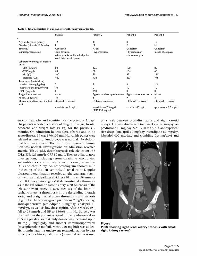
ence of headache and vomiting for the previous 2 days.His parents reported a history of fatigue, myalgia, frontalheadache and weight loss (9 kg) for the previous 2months. On admission he was alert, afebrile and in noacute distress. BP was 170/105 mm Hg. All his pulses werefelt and symmetric. Fundoscopy was normal. No abdom-inal bruit was present. The rest of his physical examina-tion was normal. Investigations on admission revealedanemia (Hb 79 g/L), thrombocytosis (platelet count 738G/L), ESR 125 mm/h, CRP 80 mg/L. The rest of laboratoryinvestigations, including serum creatinine, electrolytes,autoantibodies, and urinalysis, were normal, as well asECG and chest X-ray. An echocardiogram showed mildthickening of the left ventricle. A renal color Dopplerultrasound examination revealed a right renal artery sten-osis with a small ipsilateral kidney (70 mm vs 104 mm forthe left kidney). An angio-MRI demonstrated a thrombo-sis in the left common carotid artery, a 70% stenosis of theleft subclavian artery, a 80% stenosis of the brachio-cephalic artery, a thrombosis in the descending thoracicaorta, and a right renal artery thrombosis and stenosis(Figure 1). The boy was given prednisone 2 mg/kg per day,antihypertensives (amlodipine 5 mg/day, enalapril 10mg/day), as well as low-dose aspirin. After 2 weeks, ESRfell to 26 mm/h and BP to 156/68 mm Hg. Surgery wasplanned, but the patient relapsed at the prednisone doseof 5 mg per day, so that daily dosage was increased up to40 mg (1 mg/kg/d), and another immunosuppressive(mycophenolate mofetil, MMF, 250 mg bid) was added.Six months later he underwent revascularization bypasssurgery of brachiocephalic trunk (a femoral vein was used
as a graft between ascending aorta and right carotidartery). He was discharged two weeks after surgery onprednisone 10 mg/day, MMF 250 mg bid, 4 antihyperten-sive drugs (enalapril 10 mg/day, nicardipine 60 mg/day,labetalol 400 mg/day, and clonidine 0.3 mg/day) and
Table 1: Characteristics of our patients with Takayasu arteritis.
Patient 1 Patient 2 Patient 3 Patient 4
Age at diagnosis (years) 12 11 8 15Gender (M, male; F, female) F M F MEthnicity Caucasian Asian Caucasian CaucasianClinical presentation -pain left arm -hypertension - hypertension -acute chest pain
-absent radial and brachial pulse, weak left carotid pulse
-abdominal pain
Laboratory findings at disease onset:
-ESR (mm/hr) 80 125 100 80-CRP (mg/l) 60 80 134 60-Hb (g/l) 100 79 92 110-platelets (G/l) 400 738 487 745
Treatment (initial dose):-prednisone (mg/kg/day) 2 2 2 1.5-methotrexate (mg/m2/wk) 10 0 10 10-MMF (mg bid) 0 250 0 0Surgical intervention none Bypass brachiocephalic trunk Bypass abdominal aorta NoneFollow up (years) 10 3 9 3Outcome and treatment at last visit
-Clinical remission - Clinical remission - Clinical remission - Clinical remission
-prednisone 5 mg/d -prednisone 7.5 mg/dMMF 750 mg bid
-aspirin 100 mg/d -prednisone 7.5 mg/d
MRA showing right renal artery stenosis with small right kid-ney (arrow)Figure 1MRA showing right renal artery stenosis with small right kidney (arrow).
Page 2 of 5(page number not for citation purposes)
Pediatric Rheumatology 2008, 6:17 http://www.ped-rheum.com/content/6/1/17
low-dose aspirin. At last visit at 14 years of age, BP wascontrolled (135/65 mmHg) by three medications; he wastaking prednisone 7.5 mg per day, MMF (750 mg bid),and low-dose aspirin. His last investigation showed ESR28 mm/h, Hb 115 g/dL, platelet count 557 G/L, and a nor-mal renal function. His last MRI angiogram showed nor-mal visualization of the brachiocephalic trunk and rightinternal carotid artery. Neurological examination wasnormal, but periodic headaches were still present.
DiscussionTakayasu arteritis has been rarely reported in childhood.A common clinical mode of disease presentation in ourpatients was arterial hypertension (2/4 cases), togetherwith nonspecific symptoms (headache, fatigue, myalgia,weight loss). Symptoms due to ischemia, which are fre-quent in adults, have been seldom reported in children.However, although true claudication (upon effort) waspresent only in one patient, we have observed ischemicfindings (chest, limb, and abdominal pain) in three of ourfour patients. The disease is also called 'pulseless disease',since peripheral pulses are often absent due to vascularobstruction; however, this feature was present only in oneof our patients. In addition, an abdominal bruit can oftenhelp in the diagnosis, but this was noted in only 1/4 of ourcases.
The largest series of TA in children has been reported byHong et al., who described 70 cases [5]; the male to femaleratio was 1:4.4 and the youngest patient was 3 years old.Arterial hypertension was seen in 65/70 patients (93%).In another report of 31 children with TA from SouthAfrica [6], arterial hypertension was the most commonpresenting feature, followed by cardiac failure, bruits, andabsent pulses. Jain et al [7] from India reported 24 chil-dren with TA; again, arterial hypertension was the com-monest mode of presentation, seen in 83% of patients;the male to female ratio was 1:5. In a Turkish multicenterserie, TA represented 1.5% of pediatric vasculitides [8].Among these patients, 71.4% described constitutionalsymptoms. Hypertension was the leading feature, andrenal involvement was present in 86% of cases. Half of thepatients had involvement of both thoracic and abdominal
aorta. Table 2 summarizes clinical findings of main pub-lished series, as well as our own data.
The diagnosis of TA is based on characteristic findings ofdiseased aorta and its major branches seen on angiogra-phy. This is demonstrated by luminal abnormalities suchas stenosis or aneurysmal dilatation of the aorta, its majorbranches, and the pulmonary arteries. With regard toimaging studies, traditionally the angiographic patternshave been divided in: type I, affecting the aortic arch; typeII, the thoracic and abdominal aorta; type III, the aortaboth above and below the diaphragm; and type IV, theaorta and the pulmonary arteries. In our cases type I wasthe predominant pattern, whereas in two series type II wasthe predominant one [7,9].
Ultrasonography and positron emission tomography arenew, promising techniques to assess large-vessel vascu-litides. Color-coded Doppler sonography can facilitate anaccurate diagnosis of Takayasu arteritis by the characteris-tic appearance. Homogeneous circumferential intima-media thickening of the common carotid arteries is a spe-cific ultrasonographic finding in patients with Takayasuarteritis [10-12]. More recently MRI has been used toestablish the diagnosis of TA in children, to monitor dis-ease activity and to guide treatment. Early in the disease ofTA, smooth muscle thickened vessel walls, which may bethe only manifestation of vascular inflammation, may benot detected by conventional angiography but MRI canvisualize the thickened vessel wall directly, and in addi-tion it can show other signs of active inflammation suchas mural edema with T2-weighted imaging and increasedwall vascularity with enhanced imaging [13,14]. For onepatient in our series MRI was used to establish the diagno-sis of TA and for follow-up. Table 3 summarizes the evo-lution of vascular involvement in our patients, as studiedby echodoppler, MRI and/or angiography.
With regard to treatment, in our series corticosteroidshave been used in all cases, with the adjunction of meth-otrexate in three cases and MMF in one. Although thenumbers are small and the retrospective nature of thisreport does not allow to draw firm conclusions, our
Table 2: Summary of main clinical features in our series and in published reports of pediatric Takayasu arteritis.
Gender F/M Hypertension Elevated inflammatory parameters
Claudication Renal artery involvement
Abdominal aortic involvement
Thoracic aortic involvement
Present series 2/2 50% 100% 25% 25% 25% 25%Ozen et al. [8] 7/7 86% 100% NA NA 57.1% 28.5%Hong et al. [5] 57/13 93% NA NA NA NA NAHahn et al. [6] 13/18 84% 74% 13% 71% 42% 16%Jain et al. [7] 4/20 83% 42% NA 75% 71% 21%
NA, data not available.
Page 3 of 5(page number not for citation purposes)
Pediatric Rheumatology 2008, 6:17 http://www.ped-rheum.com/content/6/1/17
patients have obtained clinical and laboratory remissionwith this regimen of early immunosuppression. Ourpatients were followed from 2 to 10 years since diagnosis(mean, 6 years) and medications at the last visit includedonly low-dose prednisone in three cases (one of whomstill needed MMF and anti-hypertensives), while thefourth patient is now only on low-dose aspirin. Bypasssurgery was required in two patients because of severe vas-cular occlusion, with excellent results as well. One bypasswas carried out between the ascending aorta and the rightcarotid artery, the other one between the lower thoracicaorta and infra-renal aorta; in the latter a reimplantationof left renal artery and auto transplantation of right renalartery to common iliac was also performed.
Due to the rarity of the disease, there are no controlledstudies of medical treatment of children with TA. An inter-esting new possibility is represented by the use of sildena-fil, that has been recently reported in a 8-year old girl [15].We have been able to find in the literature only a previousFrench case, a 6 year-old girl who was treated with meth-otrexate [4]. Methotrexate has been used both in adultsand children with good results [16,17]. Mycophenolatemofetil (MMF) has also recently been introduced in thetreatment of adult patients with TA [18].
TA has been associated with other autoimmune diseasessuch as systemic lupus erythematosus, juvenile idiopathic
arthritis, anterior uveitis, sarcoidosis, seronegative spond-yloarthropathy, Crohn's disease, Wegener's granulomato-sis, and Sweet syndrome [19-26]. One boy in our series,who suffered from eczema and thrombocytopenia sinceinfancy, was diagnosed as having Wiskott-Aldrich syn-drome (WAS) at the age of 16 years; this was subsequentlyconfirmed by genetic study. The peculiar association of TAand WAS has already been mentioned in another previousreport [27].
TA is a disease with severe prognosis, mortality rate beingreported in children from 35 to 40% by five years [28]. Itis therefore important to have a high index of suspicionand in doubtful cases a low threshold for diagnostic eval-uation. We underline the possibility of TA in any youngpatient with unexplained arterial hypertension.
We conclude that TA is not such a rare disease in a pediat-ric rheumatology setting, and also that it has to be consid-ered in cases of unexplained arterial hypertension orunexplained inflammatory syndromes without signs oflocalization. A thorough physical examination can lead tothe correct diagnosis if pulses cannot be felt or if anabdominal bruit is heard, even if these are not constantfindings. Since the disease can be progressive and life-threatening, an early recognition is vital in order to startimmunosuppression, which proved to be very successfulin our patients.
Table 3: Vascular involvement during the disease course of our patients as investigated by imaging studies.
At diagnosis On treatment (2–5 years after diagnosis)
During a disease flare
At last visit
Patient 1 Echodoppler:thrombosis left subclavian artery, left carotid thickening
Echodoppler:stability of previous findings
Echodoppler:After 6 years thickening of left carotid artery
Echodoppler:stability of left carotid thickening
Angiography-reduction left subclavian artery diameter without thrombosis, hypoplastic left carotid without parietal lesions
Patient 2 angioMRI: thrombosis left common carotid artery, stenosis left subclavian artery, stenosis of the brachiocephalic artery, thrombosis descending thoracic aorta, right renal artery thrombosis and stenosis.
Echodoppler: (after surgery):patency of left carotid and carotid trunk
No flares angioMRI:normal visualization of the brachiocephalic trunk and right internal carotid artery
Echodoppler:left carotid thrombosis, left subclavian stenosis, tronc brachiocephalic trunk stenosis
Patient 3 Angiography:left subclavian stenosis, abdominal aortic stenosis, superior mesenteric artery stenosis, bilateral renal artery stenosis
Echodoppler:stability subclavian lesion, improvement aortic wall involvement
Echodoppler:stability of subclavian and aortic lesions
Angiography:stenosis abdominal aorta
Patient 4 Echodoppler:left subclavian thrombosis
Page 4 of 5(page number not for citation purposes)
Pediatric Rheumatology 2008, 6:17 http://www.ped-rheum.com/content/6/1/17
Publish with BioMed Central and every scientist can read your work free of charge
"BioMed Central will be the most significant development for disseminating the results of biomedical research in our lifetime."
Sir Paul Nurse, Cancer Research UK
Your research papers will be:
available free of charge to the entire biomedical community
peer reviewed and published immediately upon acceptance
cited in PubMed and archived on PubMed Central
yours — you keep the copyright
Submit your manuscript here:http://www.biomedcentral.com/info/publishing_adv.asp
BioMedcentral
Competing interestsThe authors declare that they have no competing interests.
Authors' contributionsSAA and RC wrote the manuscript, MFD and LD retrieveddata from charts, and XB and PC conceived and super-vised the study. All authors read and approved the finalmanuscript.
AcknowledgementsWe thank the patients or their parents, who gave permission to publish their clinical history.
References1. Ladhani S, Tulloh R, Anderson D: Takayasu disease masquerad-
ing as interruption aorta in a 2 years old child. Cardiol Young2001, 11:244-246.
2. Shetty AK, Stopa AR, Gedalia A: Low dose methotrexate as asteroid-sparing agent in a child with Takayasu arteritis. ClinExp Rheumatol 1998, 16:335-336.
3. Brunette MG, Bonny Y, Spigelbatt L, Barrette G: Long term immu-nosuppressive treatment of a child with Takayasu arteritisand high IgE immunoglobulins. Pediatr Nephrol 1996, 10:67-69.
4. Besson-Léaud L, Grenier N, Besson-Léaud M, Boniface C, Guillard JM:Maladie de Takayasu: intérêt du traitement par méthotrex-ate. Arch Pediatr 2001, 8:724-727.
5. Hong CY, Yung YS, Choi JY, Sul JH, Lee KS, Cha SH, et al.: Takayasuarteritis in Korean children clinical report of seventy cases.Heart Vessels 1992, 7:91-96.
6. Hahn D, Thomson PD, Kala U, Beale PG, Levin SE: A review ofTakayasu arteritis in children in Gauteng, South Africa. Pedi-atr Nephrol 1998, 12:668-675.
7. Jain S, Sharma N, Singh S, Bali HK, Kumar L, Sharma BK: Takayasuarteritis in children and young Indians. Int J Cardiol 2000,75:53-57.
8. Ozen S, Bakkaloglu A, Dusunsel R, Soylemezoglu O, Ozaltin F, Poyra-zoglu H, Kasapcopur O, Ozkaya O, Yalcinkaya F, Balat A, Kural N,Donmez O, Alpay H, Anarat A, Mir S, Gur-Guven A, Sonmez F, GokF, Turkish Pediatric Vasculitis Study Group: Childhood vasculitidesin Turkey: a nationwide survey. Clin Rheumatol 2007,26(2):196-200. 2006; Apr 4;
9. Muranjan MN, Bavdekar SB, More V, Deshmukh H, Tripathi M, Vas-wani R: Study of Takayasu arteritis in children: clinical profileand management. J Postgrad Med 2000, 46:3-8.
10. Schmidt WA, Blackmans D: Use of ultrasonography and positionemission tomography in the diagnosis and assessment oflarge-vessel vasculitis. Curr Opin Rheumatol 2005, 17:9-15.
11. Schmidt WA, Nerenheim A, Seipelt E, Poehls C, Gromnica-Ihle E:Diagnosis of early Takayasu artertis with sonography. Rheu-matology (Oxford) 2002, 41:496-502.
12. Chaubal N, Dighe M, Shah M: Sonographic and color Dopplerfinding in aortoarteritis Takayasu arteritis. J Ultrasound Med2004, 23:937-944.
13. Aluquin VP, Albano SA, Chan F, Sandborg C, Pitlick PT: Magneticresonance imaging in the diagnosis and follow up of Taka-yasu artertis in children. Ann Rheum Dis 2002, 61:526-529.
14. Choe YH, Han BK, Koh EM, Kim DK, Do YS, Lee WR: Takayasuarteritis: assessment of disease activity with contrast-enhanced MR imaging. AJR Am J Roentgenol 2000, 175(2):505-511.
15. Uthman IW, Chaaban H: The use of sildenafil in pediatric Taka-yasu arteritis. Clin Rheumatol 2006, 25:550.
16. Hoffman GS, Leavitt RY, Kerr GS, Rotten M, Sneller MC, Fauci AS:Treatment of glucocorticoid-resistant or relapsing Takayasuarteritis with methotrexate. Arthritis Rheum 1994, 37:578-582.
17. Sato EL, Lima DN, Espirito Santo B, Hata F: Takayasu arteritis,treatment and prognosis in a university center in Brazil. IntJ Cardiol 2000, 75:163-166.
18. Diana E, Schieppati A, Remuzzi G: Mycophenolate Mofetil for thetreatment of Takayasu arteritis: report of three cases. AnnIntern Med 1999, 130(5):422-426.
19. Hall S, Nelson AM: Takayasu arteritis and juvenile rheumatoidarthritis. J Rheumatol 1986, 13:431-432.
20. Mc Donald MA, Ojaimi E, Favilla I: Anterior uveitis in a child withTakayasu arteritis. Clin Exp Ophthalmol 2004, 32:336-339.
21. Opastirakul S, Chartapisak W, Sirivanichai C: A girl with Takayasuarteritis with possible systemic lupus erythomatosus. PediatrNephrol 2004, 19:463-466.
22. Acar B, Yalcinkaya F, Ozturk B, Yuksel S, Ozcakar ZB, Fitoz S, et al.:Seronegative spondyloarthropathy associated with Taka-yasu arteritis in a child. Clin Exp Rheumatol 2005, 23:278-279.
23. Van Elburg RM, Henar EL, Bijleveld CM, Prins TR, Heymans HS: Vas-cular compromise prior to intestinal manifestations ofCrohn disease in a 14 years old girl. J Pediatr Gastroenterol Nutr1992, 14:97-100.
24. Rose CD, Eichenfield AH, Goldsmith DP, Athreya BH: Early onsetsarcoidosis with aortitis juvenile systemic granulomatosis. JRheumatol 1990, 17:102-106.
25. Mejia-Hermandez C, Alvarez-Mendoza A, Deleon-Bojorge B: Taka-yasu arteritis coexisting with Wegener granulomatosis in ateenager with renal insufficiency. Pediatr Dev Pathol 1999,2:385-388.
26. Campos LMA, Castellanos ALZ, Afiune JY, Kiss MHB, Silva CAA:Takayasu arteritis with aortic aneurysm associated withSweet syndrome in childhood. Ann Rheum Dis 2005, 64:168-169.
27. Lau YL, Wong SN, Lawton WM: Takayasu arteritis associatedwith Wiskott-Aldrich syndrome. J Paediatr Child Health 1992,28:407-409.
28. Morale E, Pineda C, Martinez-Lavin M: Takayasu arteritis chil-dren. J Rheumatol 1991, 18:1081-1084.
Page 5 of 5(page number not for citation purposes)