systemic blood pressure in glaucoma patients
TRANSCRIPT
Clinical &vestigations Graefc's Archive for Elinical and Experimental
Ophthalmology © Springer-Verlag 1993
Graefe's Arch Clin Exp Ophthalmol (1993) 231:677-680
Systemic blood pressure in glaucoma patients Hedwig J. Kaiser, Josef Flammer, Therese Graf, Daniela Stiimpfig
Universitfits-Augenklinik, Mittlere Strasse 91, CH~4056 Basel, Switzerland
Received: 19 January 1993 / Accepted: 7 July 1993
Abstract. Blood pressure was monitored for 24 h in 32 control patients, 38 open-angle glaucoma patients re- ferred because of decompensated IOP despite maximum treatment, 40 patients with open-angle glaucoma re- ferred because of progressive damage despite controlled lOP, and 39 normal-tension glaucoma patients. In the control group a physiological drop in blood pressure during the night was observed. The patients referred with uncontrolled IOP had blood pressure very similar to that of the control group during both day and night. How- ever, the open-angle glaucoma patients with progression despite well-controlled IOP and also the patients with normal-tension glaucoma had markedly, and statistically significantly, lower systolic blood pressure during both day and night. The difference in diastolic blood pressure was smaller. Thus, blood pressure should be considered in diagnosis.
Introduction
Glaucomatous damage is characterized by excavation of the optic nerve head and by visual field defects. The main cause of such damage is an elevated intraoperative pres- sure (IOP). The existence of normal-tension glaucoma and the weak correlation between progression and the level of IOP [4] indicate that other factors must also be involved in the pathogenesis of glaucomatous damage.
Besides age and demographic and genetic factors, vascular and rheological factors are the main ones that have been reported in the literature. The prevalence of peripheral vasospasm, as quantified with nailfold capil- laroscopy, is increased in normal-tension glaucoma [12]. The prevalence of cardiac rhythm and conduction abnor- malities is increased in glaucoma patients [5, 19]. Several authors have described an association of low systemic blood pressure and glaucomatous damage [8, 9, 18, 24]. The tolerance to elevated IOP has been reported to be
Correspondence to: J. Flammer
decreased in patients with systemic hypotension [10, 14, 15, 21]. Some authors have reported lower blood pres- sure in normal-tension than in high-tension glaucoma patients [2, 6, 7, 13, 16]; others, however, have found no difference in blood pressure between glaucoma patients and normal controls [17, 23, 25].
The present study was designed to compare systemic blood pressure, monitored for 24 h, among clinically different groups of patients.
Materials and methods
Included were 149 patients hospitalized in 1991 at the University Eye Clinic in Basel. Patients with diabetes mellitus or other systemic diseases that might influence blood pressure were excluded. Each patient included in the study belonged to one of the following four groups:
Group A: Control patients (n=32) hospitalized for either reti- nal or cataract surgery, in whom the IOP was less than 19 mmHg (range 10-19 mmHg).
Group B: Open-angle glaucoma patients (n = 38) with definite and progressing visual-field defects, referred for filtering procedure or laser treatment because of increased IOP despite maximum therapy. The IOP of the progressing eyes was higher than 24 mmHg in all patients (range 24-46 mmHg).
Group C: Open-angle glaucoma patients (n = 40), referred because of progression in spite of medically or surgically con- trolled IOP. Diurnal tension curve over 2 days did not show IOP of 21 mmHg or more in either eye (range 14-21 mmHg).
Group D: Patients with normal-tension glaucoma (n=39), in whom untreated IOP over a 2-day diurnal tension curve did not exceed 21 mmHg (range 11-20 mmHg).
Demographic data are given in Table 1. When operative treat- ment was performed the blood pressure was monitored at least 3 days postoperatively to avoid distortion of the results by a direct influence of the surgical procedure or of the local anesthesia. Nei- ther a history of blood pressure nor one of treatment of hyperten- sion or hypotension was a criterion for inclusion or exclusion. The technician in charge of blood pressure monitoring did not know to which group any patient belonged. The patients were told to take the same local and systemic medications during the test period as
678
Table 1. Demographic data
Group A B C D
n(m/f) 32 (17/15) 38 (20/18) 40 (18/22) 39 (15/24)
Mean age (+ SD) m 58 (15) 65 (11) 63 (12) 55 (15) f 58 (24) 63 (11) 60 (15) 57 (15) Mean IOP ( i SD) m 14.5 (2.7) 28 (5.2) 17.4 (2.3) 15.5 (2.0) f 14.8 (2.8) 29.6 (6.2) 17.2 (1.9) 14.3 (1.9)
Group A, control patients hospitalized for retinal or cataract sur- gery, with intraocular pressure (IOP)< 19 mm Hg; group B, pa- tients with open-angle glaucoma involving definite and progressive visual-field defects and IOP>24mmHg; group C, open-angle glaucoma patients with progression despite medically or surgically controlled IOP (14-21 mmHg); group D, patients with normal- tension glaucoma (untreated IOP not over 21 mmHg); m, male; f, female
moan of systolic blood-pressure values 1 4 0
mmHg
1 3 0
1 2 0
1 1 0
- ~ - POAG I
t00 - - - - POAG II
Normal - tens ion glauoomas
9 0 ~ ~ , ~ , , ~ , , , - - 8 12 16 2 0 24 4 8h
time of day
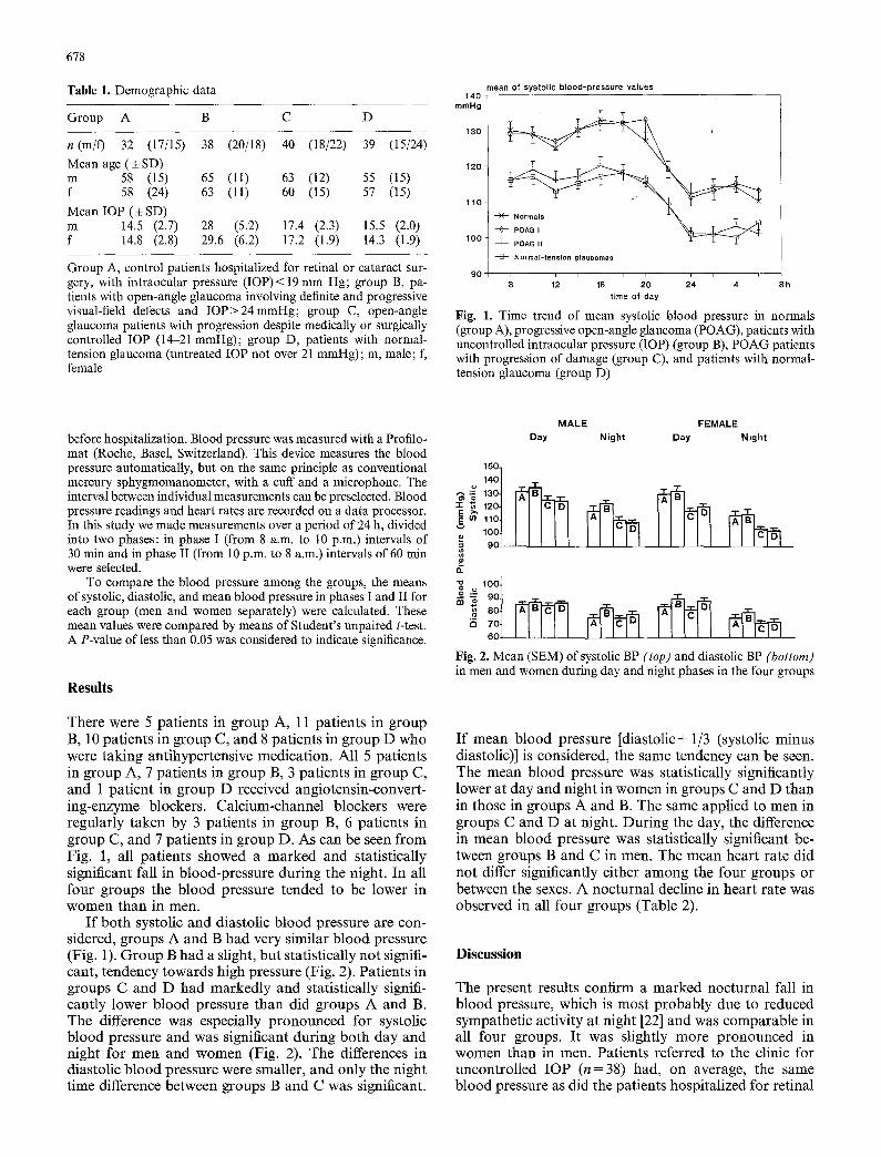
Fig. 1. Time trend of mean systolic blood pressure in normals (group A), progressive open-angle glaucoma (POAG), patients with uncontrolled intraocular pressure (IOP) (group B), POAG patients with progression of damage (group C), and patients with normal- tension glaucoma (group D)
before hospitalization. Blood pressure was measured with a Profilo- mat (Roche, Basel, Switzerland). This device measures the blood pressure automatically, but on the same principle as conventional mercury sphygmomanometer, with a cuff and a microphone. The interval between individual measurements can be preselected. Blood pressure readings and heart rates are recorded on a data processor. In this study we made measurements over a period of 24 h, divided into two phases: in phase I (from 8 a.m. to 10 p.m.) intervals of 30 min and in phase II (from 10 p.m. to 8 a.m.) intervals of 60 rain were selected.
To compare the blood pressure among the groups, the means of systolic, diastolic, and mean blood pressure in phases I and II for each group (men and women separately) were calculated. These mean values were compared by means of Student's unpaired t-test. A P-value of less than 0.05 was considered to indicate significance.
Results
There were 5 patients in group A, 11 patients in group B, 10 patients in group C, and 8 patients in group D who were taking antihypertensive medication. All 5 patients in group A, 7 patients in group B, 3 patients in group C, and 1 patient in group D received angiotensin-convert- ing-enzyme blockers. Calcium-channel blockers were regularly taken by 3 patients in group B, 6 patients in group C, and 7 patients in group D. As can be seen f rom Fig. 1, all patients showed a marked and statistically significant fall in blood-pressure during the night. In all four groups the blood pressure tended to be lower in women than in men.
I f both systolic and diastolic blood pressure are con- sidered, groups A and B had very similar blood pressure (Fig. 1). Group B had a slight, but statistically not signifi- cant, tendency towards high pressure (Fig. 2). Patients in groups C and D had markedly and statistically signifi- cantly lower blood pressure than did groups A and B. The difference was especially pronounced for systolic blood pressure and was significant during bo th day and night for men and women (Fig. 2). The differences in diastolic blood pressure were smaller, and only the night time difference between groups B and C was significant.
M A L E F E M A L E
Day Night Day N i g h t
150~
u 1 4 0 t
1- "~ 12OJ
Io ;o'°t 9 0
O.
! ._ 1°°t •
60
Fig. 2. Mean (SEM) of systolic BP (top) and diastolic BP (bottom) in men and women during day and night phases in the four groups
I f mean blood pressure [diastolic+ 1/3 (systolic minus diastolic)] is considered, the same tendency can be seen. The mean blood pressure was statistically significantly lower at day and night in women in groups C and D than in those in groups A and B. The same applied to men in groups C and D at night. During the day, the difference in mean blood pressure was statistically significant be- tween groups B and C in men. The mean heart rate did not differ significantly either among the four groups or between the sexes. A nocturnal decline in heart rate was observed in all four groups (Table 2).
Discussion
The present results confirm a marked nocturnal fall in blood pressure, which is most probably due to reduced sympathetic activity at night [22] and was comparable in all four groups. I t was slightly more pronounced in women than in men. Patients referred to the clinic for uncontrolled IOP (n=38) had, on average, the same blood pressure as did the patients hospitalized for retinal
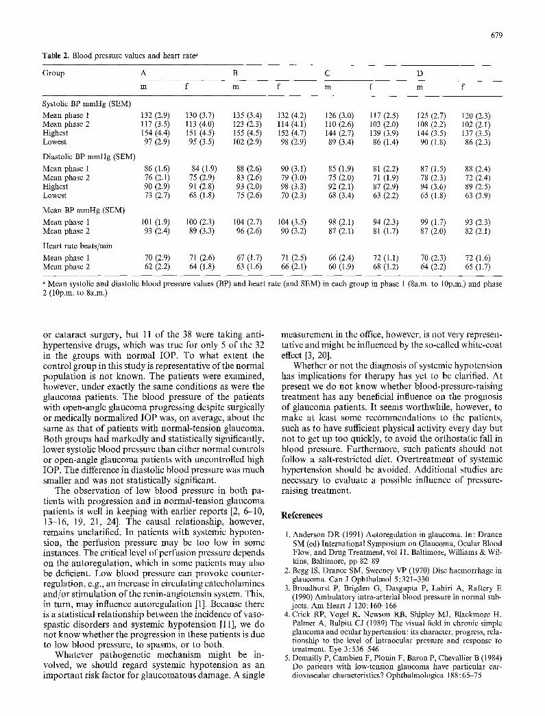
Table 2. Blood pressure values and heart rate"
679
Group A B C
m f m f m D
f m f
Systolic BP mmHg (SEM)
Mean phase 1 132 (2.9) 130 (3.7) 135 (3.4) 132 (4.2) 126 (3.0) 117 (2.5) 125 (2.7) 120 (2.3) Mean phase 2 117 (3.5) 113 (4.0) 123 (2.3) 114 (4.1) 110 (2.6) 103 (2.0) 108 (2.2) 102 (2.1) Highest 154 (4.4) 151 (4.5) 155 (4.5) 152 (4.7) 144 (2.7) 139 (3.9) 144 (3.5) 137 (3,5) Lowest 97 (2.9) 95 (3.5) 102 (2.9) 98 (2.9) 89 (3.4) 86 (1.4) 90 (1.8) 86 (2.3)
Diastolic BP mmHg (SEM)
Mean phase 1 86 (1.6) 84 (1.9) 88 (2.6) 90 (3.1) 85 (1.9) 81 (2.2) 87 (1.5) 88 (2.4) Mean phase 2 76 (2.1) 75 (2.9) 83 (2.6) 79 (3.0) 75 (2.0) 71 (1.9) 78 (2.3) 72 (2.4) Highest 90 (2.9) 91 (2.8) 93 (2.0) 98 (3.3) 92 (2.1) 87 (2.9) 94 (3.6) 89 (2.5) Lowest 73 (2.7) 68 (1.8) 75 (2.6) 70 (2.3) 68 (3.4) 63 (2.2) 65 (1.8) 63 (3.9)
Mean BP mmHg (SEM)
Mean phase 1 101 (1.9) 100 (2.3) 104 (2.7) 104 (3.5) 98 (2.1) 94 (2.3) 99 (1.7) 93 (2.3) Mean phase 2 93 (2.4) 89 (3.3) 96 (2.6) 90 (3.2) 87 (2.1) 81 (1.7) 87 (2.0) 82 (2.1)
Heart rate beats/rain
Mean phase 1 70 (2.9) 71 (2.6) 67 (1.7) 71 (2.5) 66 (2.4) 72 (1.1) 70 (2.3) 72 (1.6) Mean phase 2 62 (2,2) 64 (1.8) 63 (1.6) 66 (2.1) 60 (1.9) 68 (1.2) 64 (2.2) 65 (1.7)
" Mean systolic and diastolic blood pressure values (BP) and heart rate (and SEM) in each group in phase 1 (8a.m. to 10p.m.) and phase 2 (10p.m. to 8a.m.)
or cataract surgery, but 11 of the 38 were taking anti- hypertensive drugs, which was true for only 5 of the 32 in the groups with normal IOP. To what extent the control group in this study is representative of the normal population is not known. The patients were examined, however, under exactly the same conditions as were the glaucoma patients. The blood pressure of the patients with open-angle glaucoma progressing despite surgically or medically normalized IOP was, on average, about the same as that of patients with normal-tension glaucoma. Both groups had markedly and statistically significantly, lower systolic blood pressure than either normal controls or open-angle glaucoma patients with uncontrolled high IOP. The difference in diastolic blood pressure was much smaller and was not statistically significant.
The observation of low blood pressure in both pa- tients with progression and in normal-tension glaucoma patients is well in keeping with earlier reports [2, 6-10, 13-16, 19, 21, 24]. The causal relationship, however, remains unclarified. In patients with systemic hypoten- sion, the perfusion pressure may be too low in some instances. The critical level of perfusion pressure depends on the autoregulation, which in some patients may also be deficient. Low blood pressure can provoke counter- regulation, e.g., an increase in circulating catecholamines and/or stimulation of the renin-angiotensin system. This, in turn, may influence autoregulation [1]. Because there is a statistical relationship between the incidence of vaso- spastic disorders and systemic hypotension [11], we do not know whether the progression in these patients is due to low blood pressure, to spasms, or to both.
Whatever pathogenetic mechanism might be in- volved, we should regard systemic hypotension as an important risk factor for glaucomatous damage. A single
measurement in the office, however, is not very represen- tative and might be influenced by the so-called white-coat effect [3, 20].
Whether or not the diagnosis of systemic hypotension has implications for therapy has yet to be clarified. At present we do not know whether blood-pressure-raising treatment has any beneficial influence on the prognosis of glaucoma patients. It seems worthwhile, however, to make at least some recommendations to the patients, such as to have sufficient physical activity every day but not to get up too quickly, to avoid the orthostatic fall in blood pressure. Furthermore, such patients should not follow a salt-restricted diet. Overtreatment of systemic hypertension should be avoided. Additional studies are necessary to evaluate a possible influence of pressure- raising treatment.
References
1. Anderson DR (1991) Autoregulation in glaucoma. In: Drance SM (ed) International Symposium on Glaucoma, Ocular Blood Flow, and Drug Treatment, vol 11. Baltimore, Williams & Wil- kins, Baltimore, pp 82-89
2. Begg IS, Drance SM, Sweeney VP (1970) Disc haemorrhage in glaucoma. Can J Ophthalmol 5:321-330
3. Broadhurst P, Brigden G, Dasgupta P, Lahiri A, Raftery E (1990) Ambulatory intra-arterial blood pressure in normal sub- jects. Am Heart J 120:160-166
4. Crick RP, Vogel R, Newson RB, Shipley M J, Blackmore H, Palmer A, Bulpitt CJ (1989) The visual field in chronic simple glaucoma and ocular hypertension: its character, progress, rela- tionship to the level of intraocular pressure and response to treatment. Eye 3 : 536-546
5. Demailly P, Cambien F, Plouin F, Baron P, Chevallier B (1984) Do patients with low-tension glaucoma have particular car- diovascular characteristics? Ophthalmologica 188:65 75
680
6. Drance SM (1972) Some factors in the production of low- tension glaucoma. Br J Ophthalmol 56:229-242
7. Drance SM, Sweeney VP, Morgan RW, Feldman F (1973) Studies of factors involved in the production of low-tension glaucoma. Arch Ophthalmol 89:457-465
8. Freyler H, Menapace R (1988) Ist die Erblindung am Glaukom vermeidbar? Spektrum Augenheilkd 2:121-127
9. Friihauf A (1971) Untersuchungen fiber den Einfluss des ar- teriellen Blutdrucks auf den Vertauf des Glaucoma simplex. Dtsch Gesundheitswesen 26:273-275
10. Gafner F, Goldmann H (1955) Experimentelle Untersuchungen fiber den Zusammenhang von Augendrucksteigerung und Gesichtsfeldsch/idigung. Ophthalmologica 130: 357-377
11. Gasser P (1991) Clinical syndromes with vasoconstrictor re- sponse. Wien Klin Wochenschr 103 : 217-221
12. Gasser P, Flammer J (1991) Blood-cell velocity in the nailfold capillaries of patients with normal-tension or high-tension glaucoma and of healthy controls. Am J Ophthalmol 111 : 585-588
13. Gramer E, Leydhecker W (1985) Glaukom ohne Hochdruck: eine klinische Studie. Klin Monatsbl Augenheilkd 186:262-267
14. Harrington D (1959) The pathogenesis of the glaucoma field. Am J Ophthalmol 47:177-185
15. Heilmann K (1972) Augendruck, Blutdruck und Glaukom- schaden. (Bficherei des Augenarztes, vol 61) Enke, Stuttgart, pp 14-56
16. Kaiser H J, Flammer J (1991) Systemic hypotension: a risk factor for glaucomatous damage. Ophthalmologica 203:105-108
17. Levene RZ (1980) Low-tension glaucoma: a critical review and new material. Surv Ophthalmol 24:621-664
18. Per~isalo R, Raitta C (1990) Low blood pressure: a risk factor for nerve fibre loss in institutionalized geriatric glaucoma pa- tients. Acta Ophthalmol [Suppl] 68:65-67
19. Per~isalo R, Per/isalo J, Raitta C (1992) Electrocardiographic changes in institutionalized geriatric glaucoma patients. Graefe's Arch Clin Exp Ophthalmol 230:213-217
20. Pickering TG, James GD, Boddie C, Harshfield GA, Blank S, Laragh JH (1988) How common is white-coat hypertension? JAMA 259: 225-228
21. Reese AB, McGavic JS (1942) Relation of field contraction to blood pressure in chronic primary glaucoma. Arch Ophthalmol 27:845-850
22. Richards AM, Nicholls MG, Espiner EA, Ikram H, Cullens M, Hinton D (1986) Diurnal patterns of blood pressure, heart rate, and vasoactive hormones in normal man. Clin Exp Theory Pract 2:153-166
23. Simonett B (1959) Der Brachialisdruck und seine Beziehung zum Verlauf des Glaucoma simplex. Klin Monatsbl Augen- heilkd 135: 196-205
24. Sobanski J (1936) Der Augendruck und sein Einfluss auf den Blutkreislauf der Netzhaut. Graefe's Arch Ophthalmol I35:383-400
25. Sunde O (1951) On the relationship between simple glaucoma and general vascular diseases. Acta Ophthalmol 29:213-226