synergistic signaling of kras and thyroid hormone …synergistic signaling of kras and thyroid...
TRANSCRIPT
www.neoplasia.com
Volume 16 Number 9 September 2014 pp. 757–769 757
Synergistic Signaling of KRAS andThyroid Hormone Receptor βMutantsPromotes Undifferentiated ThyroidCancer through MYC Up-Regulation1,2
Xuguang Zhu, Li Zhao, Jeong Won Park,Mark C. Willingham and Sheue-yann Cheng
Laboratory of Molecular Biology, Center for CancerResearch, National Cancer Institute, National Institutes ofHealth, Bethesda, MD 20892, US
AbstractUndifferentiated thyroid carcinoma is one of the most aggressive human cancers with frequent RAS mutations.Howmutations of theRAS gene contribute to undifferentiated thyroid cancer remains largely unknown.Mice harboringa potent dominant negative mutant thyroid hormone receptor β, TRβPV (ThrbPV/PV), spontaneously develop well-differentiated follicular thyroid cancer similar to human cancer.Wegenetically targeted theKrasG12Dmutation to thyroidepithelial cells of ThrbPV/PV mice to understand how KrasG12D mutation could induce undifferentiated thyroid cancer inThrbPV/PVKrasG12D mice. ThrbPV/PVKrasG12D mice exhibited poorer survival due to more aggressive thyroid tumors withcapsular invasion, vascular invasion, and distant metastases to the lung occurring at an earlier age and at a higherfrequency than ThrbPV/PV mice did. Importantly, ThrbPV/PVKrasG12D mice developed frequent anaplastic foci withcomplete loss of normal thyroid follicular morphology. Within the anaplastic foci, the thyroid-specific transcriptionfactor paired box gene 8 (PAX8) expression was virtually lost and the loss of PAX8 expression was inversely correlatedwith elevated MYC expression. Consistently, co-expression of KRASG12D with TRβPV upregulated MYC levels in ratthyroid pccl3 cells, andMYCacted to enhance the TRβPV-mediated repression of thePax8promoter activity of a distantupstream enhancer, critical for thyroid-specific Pax8 expression. Our findings indicated that synergistic signaling ofKRASG12D and TRβPV led to increased MYC expression. Upregulated MYC contributes to the initiationof undifferentiated thyroid cancer, in part, through enhancing TRβPV-mediated repression of the Pax8 expression.Thus, MYC might serve as a potential target for therapeutic intervention.
Neoplasia (2014) 16, 757–769
Address all correspondence to: Dr. Sheue-yann Cheng, Laboratory of Molecular Biology,National Cancer Institute, 37 Convent Dr, Room5128, Bethesda,MD20892-4264, US.E-mail: [email protected] research was supported by the Intramural Research Program of the Center forCancer Research, National Cancer Institute, National Institutes of Health. Disclosureof potential conflicts of interest: The authors declare no conflicts of interest.2This article refers to supplementary materials, which are designated by Figures S1 andS2 and are available online at www.neoplasia.com.Received 17 June 2014; Revised 8 August 2014; Accepted 12 August 2014
Published by Elsevier Inc. on behalf of Neoplasia Press, Inc. This is an open access articleunder the CC BY-NC-ND license (http://creativecommons.org/licenses/by-nc-nd/3.0/).1476-5586/14http://dx.doi.org/10.1016/j.neo.2014.08.003
IntroductionThyroid cancer is themost commonmalignancy of the endocrine organs.The follicular cell–derived cancers are classified into well-differentiatedpapillary and follicular carcinomas, poorly differentiated carcinoma, andundifferentiated carcinoma. Undifferentiated thyroid carcinoma is one ofthe most aggressive malignancies. It spreads quickly to other organs anddoes not respond well to radioiodine therapy. So far, no effective targettreatments are available. Ten-year survival rate is less than 10% [1].Among prevalent genetic alterations found in undifferentiated thyroid
cancer are point mutations of the RAS, TP53, and CTNNB1 genes.Pathway analysis shows that these mutations lead to activated mitogen-activated protein kinases (MAPK) and phosphatidylinositol 3-kinase(PI3K)–protein kinase B (AKT) signaling pathways critical for thedevelopment of thyroid cancer. While mutations in the TP53 andCTNNB1 genes are found only in undifferentiated thyroid cancers [1],mutations in the RAS gene are frequently found in well-differentiatedthyroid cancer. These RAS mutations could represent an early event inthyroid carcinogenesis. It is unclear, however, how RAS mutations could
initiate undifferentiated thyroid carcinoma, especially in view of thefindings that theRasmutations alone in the thyroid failed to induce thyroidcancer in mice [2,3].
Previously, we demonstrated that mice with a mutant thyroidhormone receptor β, TRβPV (ThrbPV/PV), spontaneously develop
758 MYC in undifferentiated thyroid cancer Zhu et al. Neoplasia Vol. 16, No. 9, 2014
well-differentiated follicular thyroid cancer with similar pathologicprogression and frequency of metastasis as in human thyroidcancer [4,5]. The PV mutation was originally identified in apatient with resistance to thyroid hormone [6]. The PV mutationhas completely lost T3 binding activity and transcription capacity.It acts to abnormally regulate the expression of the T3 target genethrough dominant negative activity. Detailed pathway analysis inthe thyroid tumors of ThrbPV/PV mice indicated that the PI3K-AKT signaling pathway, which is frequently activated inundifferentiated thyroid carcinoma [7], is aberrantly overactivatedin ThrbPV/PV mice [8]. CTNNB1 signaling is also increased inthese mice [9,10], which was proposed to initiate tumordedifferentiation in the late stage of tumorigenesis [1]. However,the RAS mutant–activated MAPK pathway, critical for undiffer-entiated thyroid carcinoma, is apparently not altered in thethyroid of ThrbPV/PV mice. We hypothesized that activation of theMAPK pathway driven by RAS mutation in the thyroid ofThrbPV/PV mice might phenotypically mimic the altered signalingobserved in human thyroid cancer, thereby initiating undifferen-tiated thyroid cancer.
To investigate this question, we genetically introduced theKrasG12D mutation to express specifically in the thyroids of theThrbPV/PV mice. Our aim was to learn whether the mice withThrbPV and KrasG12D double mutations would begin developingundifferentiated thyroid carcinoma. Indeed, we found theoccurrence of anaplastic foci with a high frequency in the thyroidof ThrbPV/PVKrasG12D mice. These anaplastic foci had lost normalthyroid follicular morphology and the expression of transcriptionfactor paired box gene 8 (PAX8). We demonstrated that synergisticsignaling of TRβPV and KRASG12D mutants led to an elevatedlevel of MYC protein to suppress the Pax8 expression through aPax8 upstream enhancer. Thus, our study established a mousemodel of undifferentiated thyroid cancer that could further beused to understand altered signaling pathways of undifferentiatedthyroid cancer.
Materials and Methods
Experimental AnimalsAll animal experiments were performed according to the protocols
approved by the Animal Care and Use Committee at the NationalCancer Institute. The ThrbPV/+, KrasLSL-G12D/+, TPO-Cre (Cre) micewere previously described [4,11,12]. Mice were in a mixed C57BL/6and 129Svj genetic background. Thyroids and other tissues wereharvested from the mice and wild-type (WT) littermates for weighing,histologic analysis, and biochemical studies.
Generation of Rat pccl3 Cell Lines Stably Expressing TRβ,TRβPV, or MYC
Rat thyroid pccl3 cells were cultured in Ham's F-12 mediumsupplemented with 10% FBS and containing six hormones (1 mU/mlbovine thyroid stimulating hormone (TSH), 10 μg/ml insulin, 5 μg/ml transferrin, 10 ng/ml glycyl-L-histidyl-L-lysine, 10 ng/mlsomatostatin, and 0.36 ng/ml hydrocortisone; 6H medium). Thepccl3 cells were transfected with an expression plasmid containingcDNA encoding THRB, THRBPV, MYC, or the control emptyvectors and selected with G418, puromycin (Invitrogen, Carlsbad,CA), or blasticidin for 2 weeks. The expression of TRβ, TRβPV, orMYC protein was verified by Western blot analysis using monoclonalanti-TRβ/anti-TRβPV antibody (J53) or anti-MYC antibody.
Adenovirus Infection of Rat pccl3 Cells Expressing TRβ,TRβPV, or KRASG12D
Rat thyroid pccl3 cells were cultured in Ham's F-12 mediumsupplemented with 10% FBS and containing six hormones. Beforeaddition of adenovirus, the cells were cultured inOpti-MEM Imedium(Life Science, Grand Island, NY). The pccl3 cells were infected withadenovirus at a 5:1 ratio of adenovirus to pccl3 cells. After 5 hours, themediumwas changed toHam's F-12medium supplemented with 10%thyroid hormone deficient serum (Td) and containing six hormones inthe absence or presence of 100 nM T3. After 18 hours, the cells werecollected for the preparation of total RNA or to prepare cell lysates forWestern blot analysis.
Western Blot AnalysisThe Western blot analysis was carried out as described by
Furumoto et al. [13]. Primary antibodies for phosphorylatedextracellular signal regulated kinase (ERK) (p-ERK; #4376S), totalERK (#9102), and glyceraldehyde-3-phosphate dehydrogenase(GAPDH; #2118) were purchased from Cell Signaling Technology(Danvers, MA). anti-TTF1 (sc-13040) antibody was purchased fromSanta Cruz Biotechnology (Santa Cruz, CA). anti-PAX8 antibody(10336-1-AP) was purchased from Proteintech Group, Inc. (Chicago,IL). Cyclin D1 (RB-9041-P0) was purchased from Neomarkers(Fremont, CA). Antibodies were used at a concentration recommendedby the manufacturers. For control of protein loading, the blot wasprobed with the antibody against GAPDH.
Electrophoretic Mobility Gel Shift AssaysElectrophoretic mobility gel shift assay was conducted similarly as
described in [14]. Briefly, the [α-32P]-dCTP–labeled probes wereincubated with the in vitro synthesized TRβ1, TRβ1PV, RXRβ, orMYC and the reaction mixture was analyzed by 5% polyacrylamidegel electrophoresis. The gel was dried and autoradiographed.
Histologic Analysis and ImmunohistochemistryThyroid glands, heart, and lung were dissected and embedded in
paraffin. Five-micrometer-thick sections were prepared and stainedwithhematoxylin and eosin (H&E). For eachmouse, single random sectionsthrough the thyroid, lung, and heart were examined. Immunohisto-chemistry was performed with paraffin sections by standard methods.Dewaxed sections were treated with 0.05% citraconic anhydride buffer(pH 7.4) at 98°C for 45 minutes to expose the antigen epitopes. Aprimary antibody against TTF1 (sc-13040, Santa Cruz Biotechnology),PAX8 (10336-1-AP; Proteintech), or β-catenin (#9562 s; Cell SignalingTechnology) was incubated with tissue section overnight at 4°C.Peroxidase activity from the secondary antibody was detected by addingsubstrate 3,3′-diaminobenzidine, and the sections were counterstainedwith hematoxylin. Bromodeoxyuridine (BrdU) incorporation assay wasperformed similarly as described in Zhao et al. [15].
Hormone AssaysThe serum levels of total T4 (TT4) and T3 (TT3) were determined
by using a Gamma Coat T4 and T3 assay RIA kit. TSH levels inserum were measured as described [15].
RNA Extraction and Real-Time ReverseTranscription–Polymerase Chain Reaction
Total RNA from thyroids was isolated using TRIzol (Invitrogen),as indicated by the protocol of the manufacturer. Real-time reversetranscription–polymerase chain reaction (RT-PCR) was performed
Neoplasia Vol. 16, No. 9, 2014 MYC in undifferentiated thyroid cancer Zhu et al. 759
using a QuantiTect SYBR green RT-PCR kit from Qiagen (Valencia,CA), following the instructions of the manufacturer. Primers were asfollows: for mouse Pax8, forward, 5′-cacccttcaatgcctttcc-3′; reverse,5′-aatacggggtgtggctgtag-3′; for the endogenous control gene mouseGapdh, forward, 5′-cgtcccgtagacaaaatggt-3′; reverse, 5′-gaatttgccgtgagtggagt-3′.
Luciferase Reporter AssayThe Pax8 upstream enhancer element cloned into pGL3b
(CNS87-pGL3b) was generously provided by Dr R. Di Lauro [16].Established rat thyroid pccl3 cells stably expressing TRβ, TRβPV, orMYC were seeded at a density of 5 × 10⁵ in six-well culture plates andpreincubated for 24 hours with Td medium. Cells were transfectedusing Lipofectamine 2000 (Invitrogen). Cells were lysed 24 hourslater with 1× cell lysis buffer (Promega, Madison, WI), and luciferaseactivity was measured using Victor 3 (PerkinElmer Life and AnalyticalSciences, Waltham, MA). Luciferase values were standardized to theratio of β-galactosidase activity and protein concentration.
Statistical AnalysisAll data are expressed as means ± standard errors. Statistical analysis
was performed and P b .05 was considered significant. All statisticaltests were two-sided. GraphPad Prism version 5.0 for Mac OS X wasused to perform Kaplan-Meier cumulative survival analysis, Student'st test, Chi-square test, and analysis of variances (ANOVAs).
Results
ThrbPV/PVKrasG12D Mice Manifest Poor Survival withMarkedly Enlarged ThyroidsTo investigate whether ThrbPV/PVKrasG12D mice develop undifferen-
tiated thyroid cancer, we targeted the KrasG12D mutation to the thyroidepithelial cells ofThrbPV/PVmice throughTPO-Cre–mediated expressionof the KrasG12D gene after removal of the STOP cassette. We crossedthree lines of ThrbPV/+, KrasLSL-G12D/+, and TPO-Cre mice to generatethe mice with four different genotypes: Thrb+/+Kras+/+Cre−,Thrb+/+KrasLSL-G12D/+Cre+, ThrbPV/PVKras+/+Cre−, and ThrbPV/PV
KrasLSL-G12D/+Cre+, and we designate them in the following descriptionas WT, KrasG12D, ThrbPV/PV, and ThrbPV/PVKrasG12D mice, respectively.To examine whether the expression of the KrasG12D and the Cre
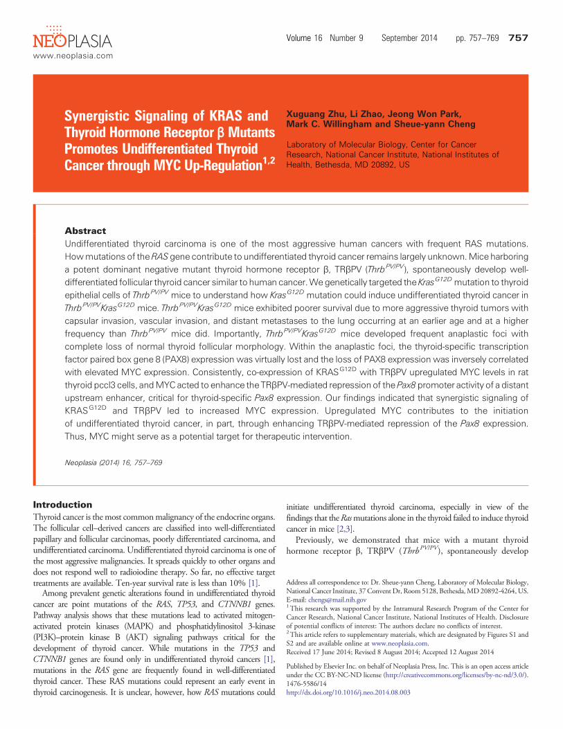
genes in the thyroids of ThrbPV/PVKrasG12D mice led to functionalactivation of the MAPK signaling, we evaluated the phosphorylationstatus of the downstream effector, ERK. p-ERK, a signature ofdownstream Ras signaling, was assessed by Western blot analysis(Figure 1A, I-a). GAPDH was used as the loading control (Figure 1A,I-e). The protein levels of p-ERK were increased by ~5- and 14-foldin KrasG12D and ThrbPV/PVKrasG12D mice, respectively (comparelanes 3 and 4 with 1 and 2, and lanes 7 and 8 with 5 and 6; Figure 1A,I-a), indicating that the KrasG12D mutant was functionally expressedin the thyroids of ThrbPV/PVKrasG12D mice. There was also a higherp-ERK activation in ThrbPV/PVKrasG12D mice than in KrasG12D mice(compare lanes 7 and 8 with lanes 3 and 4). In addition, we also foundthat AKT was more activated in thyroid tumors of ThrbPV/PVKrasG12D
mice (lanes 7 and 8) than in ThrbPV/PV mice (lanes 5 and 6) andKrasG12D mice (lanes 3 and 4; Figure 1A, I-c and I-d). Quantitativeanalysis of p-ERK, total ERK, p-AKT, and total AKT band intensitiesindicated that the p-ERK/total ERK ratio and p-AKT/total AKT was1.5-fold and 1.9-fold higher in ThrbPV/PVKrasG12D mice than inKrasG12D mice, respectively (Figure 1A, II and III). Previously, we haveshown that AKTwas activated in thyroid tumors ofThrbPV/PVmice [8].
These results suggest the contribution of TRβPV in the furtheractivating of KRASG12D and AKT signaling.
Analysis of Kaplan-Meier cumulative survival curves was conductedfor WT, KrasG12D, ThrbPV/PV, and ThrbPV/PVKrasG12D mice over aperiod of 10.5 months (Figure 1B). No WT mice or KrasG12D micedied during that period, but about 30% of ThrbPV/PV mice died. Bycontrast, only 50% of ThrbPV/PVKrasG12D mice lived to the age of 4.8months, and none survived beyond 10.5 months. The differencesbetween the survival rates of the ThrbPV/PV and ThrbPV/PVKrasG12D
mice were highly significant (P b .01). These results indicate thatsynergistic effects of TRβPV and KRASG12D mutants led to poorsurvival of ThrbPV/PVKrasG12D mice.
The thyroid weights of KrasG12D mice (3.6 ± 0.6 mg, n = 13) weresimilar to those of WT mice (2.7 ± 0.3 mg, n = 13; Figure 1C, bar 2vs 1). This observation is consistent with a previous report thatKrasG12D mutation alone is not sufficient to increase the thyroidweight [2]. The thyroid of ThrbPV/PV mice was markedly enlargedwith an average weight of 84 mg (84.47 ± 11.11 mg, n = 29). Amongmice with four genotypes, the double mutant mice had the largestthyroid (216.7 ± 18.52 mg, n = 54). The increase in thyroid weightof ThrbPV/PVKrasG12D mice was 80-, 60-, and 2.6-fold greater thanin WT, KrasG12D, and ThrbPV/PV mice, respectively (bar 4 vs 1, bar 4vs 2, bar 4 vs 3; Figure 1C).
Increased Thyroid Growth in ThrbPV/PVKrasG12D Mice Is NotMediated by Elevated TSH Levels
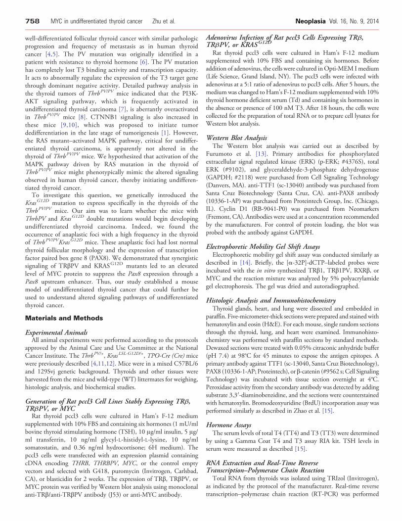
TSH is the major stimulator of thyrocyte proliferation, and its levelsare regulated by the thyroid hormones (T4 and T3) through a negativefeedback loop [17]. To evaluate whether TSH could contribute to themarkedly increased thyroid growth in ThrbPV/PVKrasG12D mice, wecompared serumTSH, serum total T4, and total T3 betweenThrbPV/PV
and ThrbPV/PVKrasG12D mice (Figure 2). There were no significantdifferences between ThrbPV/PV and ThrbPV/PVKrasG12D mice in serumlevels of TSH (WT: 38.3 ± 7.4 ng/ml,n = 18;KrasG12D: 14.8 ± 4.2 ng/ml,n = 10; ThrbPV/PV: 28420 ± 6969 ng/ml, N = 9; ThrbPV/PVKrasG12D:39990 ± 5331 ng/ml, n = 11; Figure 2A), total T4 (WT: 2.6 ± 0.2 μg/ml,n = 10;KrasG12D: 4.0 ± 0.5 μg/ml,n = 9;ThrbPV/PV: 27.7 ± 1.3 μg/ml,n =14;ThrbPV/PVKrasG12D: 19.6 ± 2.7 μg/ml, n = 9; Figure 2B), and total T3(WT: 1.0 ± 0.1 ng/ml,N = 6;KrasG12D: 1.0 ± 0.2 ng/ml,n = 9;ThrbPV/PV:7.2 ± 0.7 ng/ml, n = 9; ThrbPV/PVKrasG12D: 4.9 ± 0.9 ng/ml, n = 10;Figure 2C). These data indicated that KrasG12D activation in thyroidsdid not further affect the hypothalamus-pituitary-thyroid axis inThrbPV/PVKrasG12D mice. Thus, the increased thyroid weight inThrbPV/PVKrasG12Dmice was not due to an elevatedTSH level (Figure 2A).
Increased Proliferation of Thyroid Tumor Cells inThrbPV/PVKrasG12D Mice
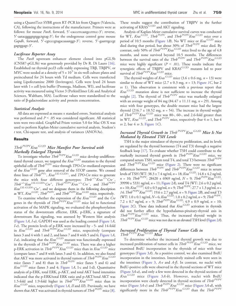
To determine whether the increased thyroid growth was due toincreased proliferation of tumor cells in ThrbPV/PVKrasG12D mice, weexamined BrdU incorporation in the thyroids of mice with fourgenotypes (Figure 3A). As a positive control, we also examined BrdUincorporation in the intestines. Intensively stained cells were seen inthe intestines (Figure 3, A-e and A-f). In contrast, no nuclei withBrdU-positive cells were observed in the thyroid sections of WT mice(Figure 3A-a), and only a few were detected in the thyroid sections ofKrasG12D mice (Figure 3A-b). However, nuclei with BrdUincorporation were clearly detected in thyroid sections of ThrbPV/PV
mice (Figure 3A-c) and ThrbPV/PVKrasG12D mice (Figure 3A-d), withsignificantly more in the ThrbPV/PVKrasG12D than the ThrbPV/PV
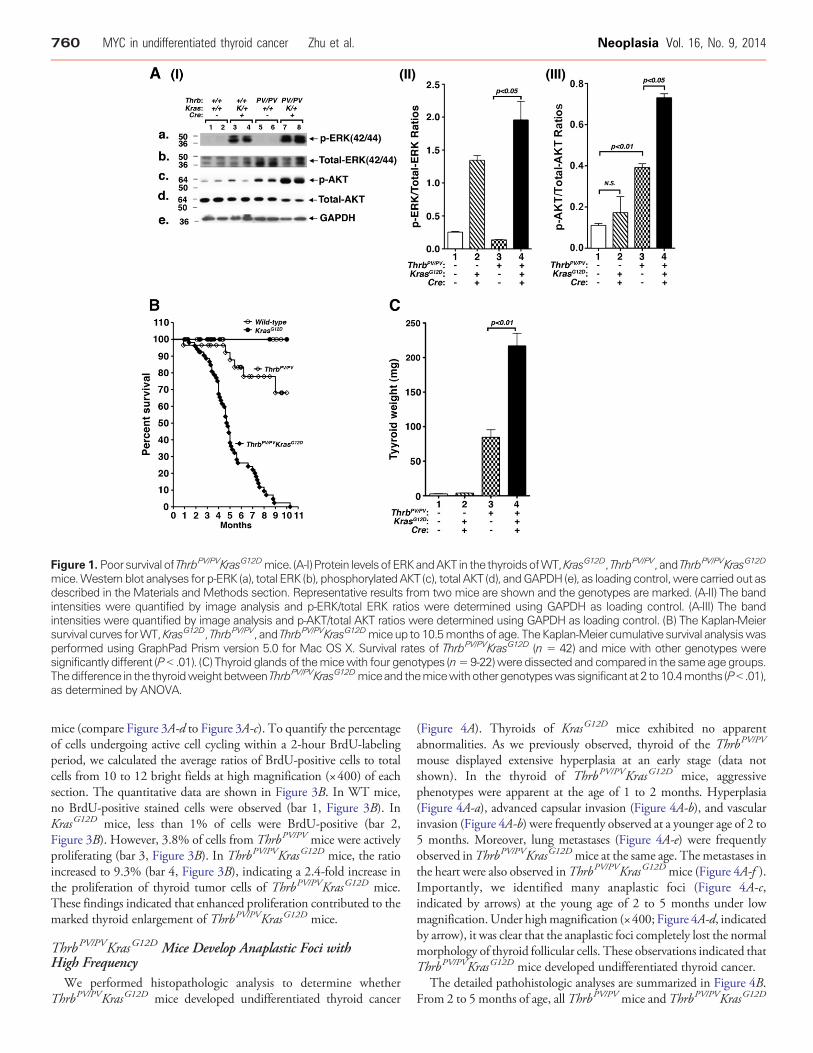
Figure 1. Poorsurvival of ThrbPV/PVKrasG12Dmice. (A-I) Protein levelsofERKandAKT in the thyroidsofWT,KrasG12D, ThrbPV/PV, and ThrbPV/PVKrasG12D
mice.Western blot analyses for p-ERK (a), total ERK (b), phosphorylatedAKT (c), total AKT (d), andGAPDH (e), as loading control,were carried out asdescribed in the Materials and Methods section. Representative results from two mice are shown and the genotypes are marked. (A-II) The bandintensities were quantified by image analysis and p-ERK/total ERK ratios were determined using GAPDH as loading control. (A-III) The bandintensities were quantified by image analysis and p-AKT/total AKT ratios were determined using GAPDH as loading control. (B) The Kaplan-Meiersurvival curves forWT,KrasG12D, ThrbPV/PV, and ThrbPV/PVKrasG12Dmiceup to 10.5monthsof age. TheKaplan-Meier cumulative survival analysiswasperformed using GraphPad Prism version 5.0 for Mac OS X. Survival rates of ThrbPV/PVKrasG12D (n = 42) and mice with other genotypes weresignificantly different (P b .01). (C) Thyroid glands of themicewith four genotypes (n=9-22)were dissected and compared in the same age groups.Thedifference in the thyroidweightbetweenThrbPV/PVKrasG12Dmiceand themicewithothergenotypeswassignificant at 2 to10.4months (Pb .01),as determined by ANOVA.
760 MYC in undifferentiated thyroid cancer Zhu et al. Neoplasia Vol. 16, No. 9, 2014
mice (compare Figure 3A-d to Figure 3A-c). To quantify the percentageof cells undergoing active cell cycling within a 2-hour BrdU-labelingperiod, we calculated the average ratios of BrdU-positive cells to totalcells from 10 to 12 bright fields at high magnification (×400) of eachsection. The quantitative data are shown in Figure 3B. In WT mice,no BrdU-positive stained cells were observed (bar 1, Figure 3B). InKrasG12D mice, less than 1% of cells were BrdU-positive (bar 2,Figure 3B). However, 3.8% of cells from ThrbPV/PV mice were activelyproliferating (bar 3, Figure 3B). In ThrbPV/PVKrasG12D mice, the ratioincreased to 9.3% (bar 4, Figure 3B), indicating a 2.4-fold increase inthe proliferation of thyroid tumor cells of ThrbPV/PVKrasG12D mice.These findings indicated that enhanced proliferation contributed to themarked thyroid enlargement of ThrbPV/PVKrasG12D mice.
ThrbPV/PVKrasG12D Mice Develop Anaplastic Foci withHigh Frequency
We performed histopathologic analysis to determine whetherThrbPV/PVKrasG12D mice developed undifferentiated thyroid cancer
(Figure 4A). Thyroids of KrasG12D mice exhibited no apparentabnormalities. As we previously observed, thyroid of the ThrbPV/PV
mouse displayed extensive hyperplasia at an early stage (data notshown). In the thyroid of ThrbPV/PVKrasG12D mice, aggressivephenotypes were apparent at the age of 1 to 2 months. Hyperplasia(Figure 4A-a), advanced capsular invasion (Figure 4A-b), and vascularinvasion (Figure 4A-b) were frequently observed at a younger age of 2 to5 months. Moreover, lung metastases (Figure 4A-e) were frequentlyobserved inThrbPV/PVKrasG12Dmice at the same age. Themetastases inthe heart were also observed inThrbPV/PVKrasG12Dmice (Figure 4A-f ).Importantly, we identified many anaplastic foci (Figure 4A-c,indicated by arrows) at the young age of 2 to 5 months under lowmagnification.Under highmagnification (×400; Figure 4A-d, indicatedby arrow), it was clear that the anaplastic foci completely lost the normalmorphology of thyroid follicular cells. These observations indicated thatThrbPV/PVKrasG12D mice developed undifferentiated thyroid cancer.
The detailed pathohistologic analyses are summarized in Figure 4B.From 2 to 5 months of age, all ThrbPV/PV mice and ThrbPV/PVKrasG12D
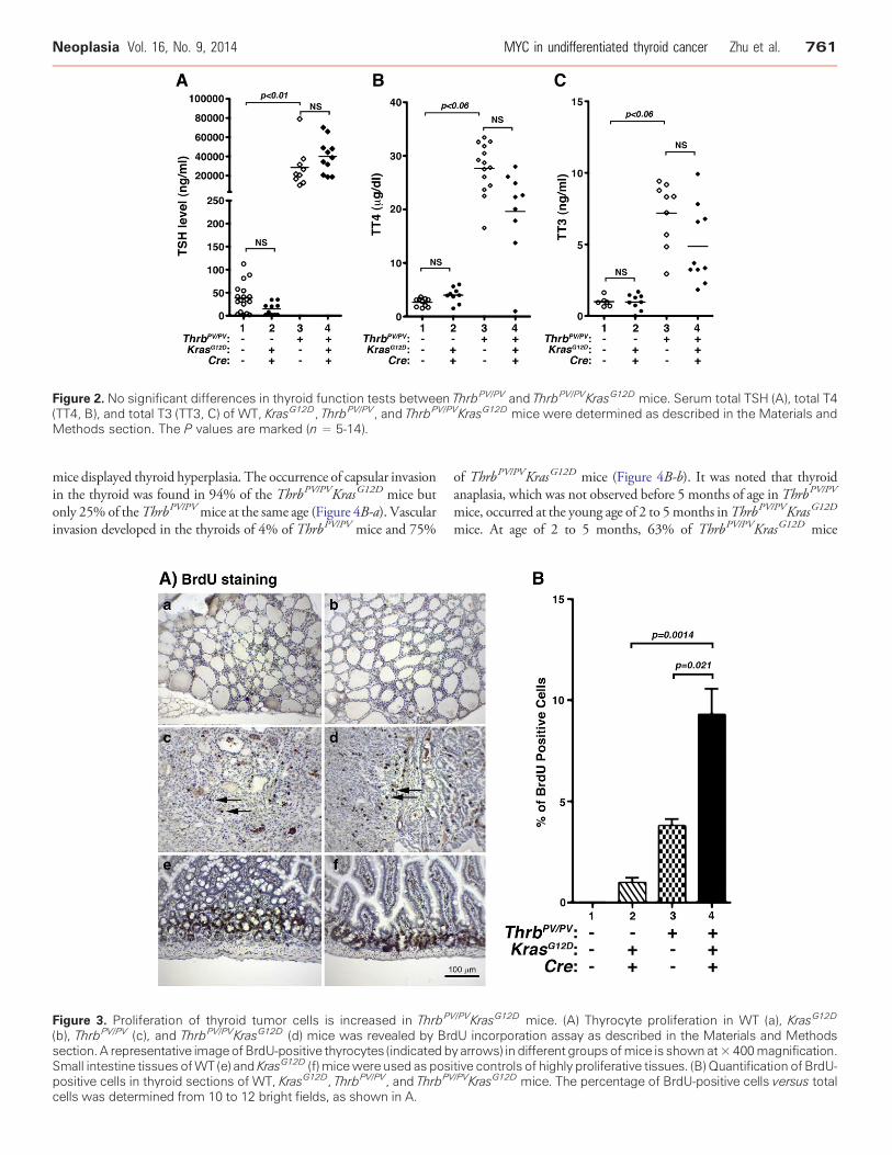
Figure 2. No significant differences in thyroid function tests between ThrbPV/PV and ThrbPV/PVKrasG12D mice. Serum total TSH (A), total T4(TT4, B), and total T3 (TT3, C) of WT, KrasG12D, ThrbPV/PV, and ThrbPV/PVKrasG12D mice were determined as described in the Materials andMethods section. The P values are marked (n = 5-14).
Neoplasia Vol. 16, No. 9, 2014 MYC in undifferentiated thyroid cancer Zhu et al. 761
mice displayed thyroid hyperplasia. The occurrence of capsular invasionin the thyroid was found in 94% of the ThrbPV/PVKrasG12D mice butonly 25% of theThrbPV/PVmice at the same age (Figure 4B-a). Vascularinvasion developed in the thyroids of 4% of ThrbPV/PV mice and 75%
Figure 3. Proliferation of thyroid tumor cells is increased in ThrbP
(b), ThrbPV/PV (c), and ThrbPV/PVKrasG12D (d) mice was revealed by Brsection. A representative imageofBrdU-positive thyrocytes (indicated bSmall intestine tissues ofWT (e) and KrasG12D (f) micewere used as pospositive cells in thyroid sections of WT, KrasG12D, ThrbPV/PV, and ThrbPV
cells was determined from 10 to 12 bright fields, as shown in A.
of ThrbPV/PVKrasG12D mice (Figure 4B-b). It was noted that thyroidanaplasia, which was not observed before 5 months of age in ThrbPV/PV
mice, occurred at the young age of 2 to 5months inThrbPV/PVKrasG12D
mice. At age of 2 to 5 months, 63% of ThrbPV/PVKrasG12D mice
V/PVKrasG12D mice. (A) Thyrocyte proliferation in WT (a), KrasG12D
dU incorporation assay as described in the Materials and Methodsy arrows) in different groupsofmice is shown at×400magnification.itive controls of highly proliferative tissues. (B) Quantification of BrdU-/PVKrasG12D mice. The percentage of BrdU-positive cells versus total
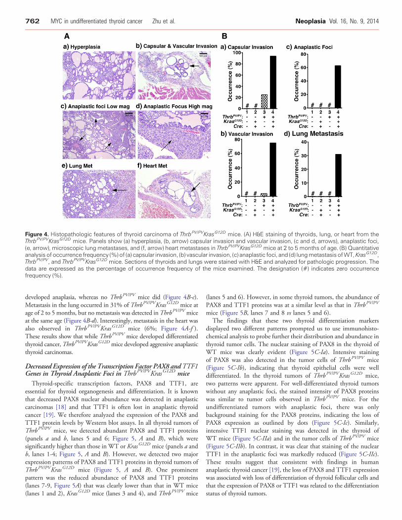
Figure 4. Histopathologic features of thyroid carcinoma of ThrbPV/PVKrasG12D mice. (A) H&E staining of thyroids, lung, or heart from theThrbPV/PVKrasG12D mice. Panels show (a) hyperplasia, (b, arrow) capsular invasion and vascular invasion, (c and d, arrows), anaplastic foci,(e, arrow), microscopic lung metastases, and (f, arrow) heart metastases in ThrbPV/PVKrasG12D mice at 2 to 5 months of age. (B) Quantitativeanalysis of occurrence frequency (%) of (a) capsular invasion, (b) vascular invasion, (c) anaplastic foci, and (d) lungmetastasis ofWT, KrasG12D,ThrbPV/PV, and ThrbPV/PVKrasG12D mice. Sections of thyroids and lungs were stained with H&E and analyzed for pathologic progression. Thedata are expressed as the percentage of occurrence frequency of the mice examined. The designation (#) indicates zero occurrencefrequency (%).
762 MYC in undifferentiated thyroid cancer Zhu et al. Neoplasia Vol. 16, No. 9, 2014
developed anaplasia, whereas no ThrbPV/PV mice did (Figure 4B-c).Metastasis in the lung occurred in 31% of ThrbPV/PVKrasG12D mice atage of 2 to 5 months, but no metastasis was detected inThrbPV/PV miceat the same age (Figure 4B-d). Interestingly, metastasis in the heart wasalso observed in ThrbPV/PVKrasG12D mice (6%; Figure 4A-f ).These results show that while ThrbPV/PV mice developed differentiatedthyroid cancer,ThrbPV/PVKrasG12D mice developed aggressive anaplasticthyroid carcinomas.
Decreased Expression of the Transcription Factor PAX8 andTTF1Genes in Thyroid Anaplastic Foci in ThrbPV/PVKrasG12D mice
Thyroid-specific transcription factors, PAX8 and TTF1, areessential for thyroid organogenesis and differentiation. It is knownthat decreased PAX8 nuclear abundance was detected in anaplasticcarcinomas [18] and that TTF1 is often lost in anaplastic thyroidcancer [19]. We therefore analyzed the expression of the PAX8 andTTF1 protein levels by Western blot assays. In all thyroid tumors ofThrbPV/PV mice, we detected abundant PAX8 and TTF1 proteins(panels a and b, lanes 5 and 6; Figure 5, A and B), which weresignificantly higher than those in WT or KrasG12D mice (panels a andb, lanes 1-4; Figure 5, A and B). However, we detected two majorexpression patterns of PAX8 and TTF1 proteins in thyroid tumors ofThrbPV/PVKrasG12D mice (Figure 5, A and B). One prominentpattern was the reduced abundance of PAX8 and TTF1 proteins(lanes 7-9, Figure 5A) that was clearly lower than that in WT mice(lanes 1 and 2), KrasG12D mice (lanes 3 and 4), and ThrbPV/PV mice
(lanes 5 and 6). However, in some thyroid tumors, the abundance ofPAX8 and TTF1 proteins was at a similar level as that in ThrbPV/PV
mice (Figure 5B, lanes 7 and 8 vs lanes 5 and 6).The findings that these two thyroid differentiation markers
displayed two different patterns prompted us to use immunohisto-chemical analysis to probe further their distribution and abundance inthyroid tumor cells. The nuclear staining of PAX8 in the thyroid ofWT mice was clearly evident (Figure 5C-Ia). Intensive stainingof PAX8 was also detected in the tumor cells of ThrbPV/PV mice(Figure 5C-Ib), indicating that thyroid epithelial cells were welldifferentiated. In the thyroid tumors of ThrbPV/PVKrasG12D mice,two patterns were apparent. For well-differentiated thyroid tumorswithout any anaplastic foci, the stained intensity of PAX8 proteinswas similar to tumor cells observed in ThrbPV/PV mice. For theundifferentiated tumors with anaplastic foci, there was onlybackground staining for the PAX8 proteins, indicating the loss ofPAX8 expression as outlined by dots (Figure 5C-Ic). Similarly,intensive TTF1 nuclear staining was detected in the thyroid ofWT mice (Figure 5C-IIa) and in the tumor cells of ThrbPV/PV mice(Figure 5C-IIb). In contrast, it was clear that staining of the nuclearTTF1 in the anaplastic foci was markedly reduced (Figure 5C-IIc).These results suggest that consistent with findings in humananaplastic thyroid cancer [19], the loss of PAX8 and TTF1 expressionwas associated with loss of differentiation of thyroid follicular cells andthat the expression of PAX8 or TTF1 was related to the differentiationstatus of thyroid tumors.
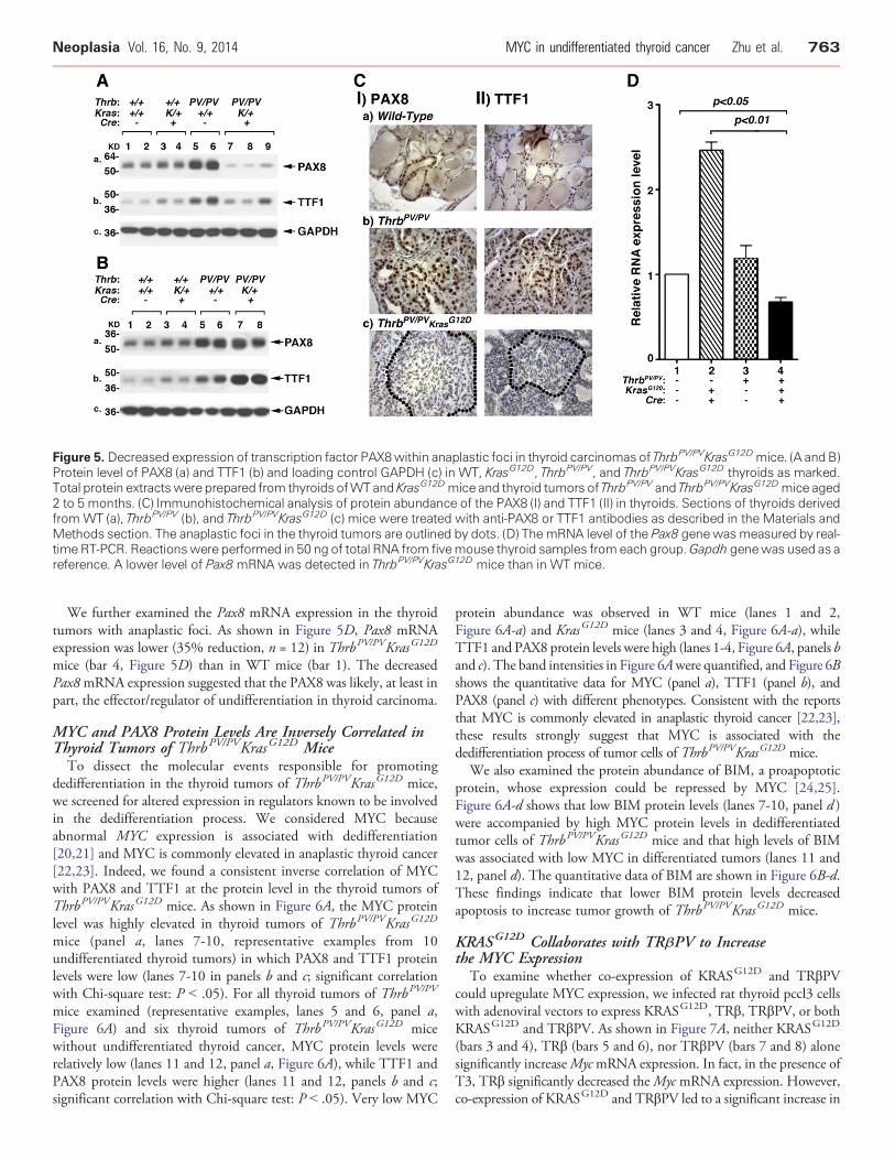
Figure 5. Decreased expression of transcription factor PAX8within anaplastic foci in thyroid carcinomas of ThrbPV/PVKrasG12Dmice. (A and B)Protein level of PAX8 (a) and TTF1 (b) and loading control GAPDH (c) in WT, KrasG12D, ThrbPV/PV, and ThrbPV/PVKrasG12D thyroids as marked.Total protein extractswere prepared from thyroids ofWTand KrasG12Dmice and thyroid tumors of ThrbPV/PV and ThrbPV/PVKrasG12Dmice aged2 to 5 months. (C) Immunohistochemical analysis of protein abundance of the PAX8 (I) and TTF1 (II) in thyroids. Sections of thyroids derivedfrom WT (a), ThrbPV/PV (b), and ThrbPV/PVKrasG12D (c) mice were treated with anti-PAX8 or TTF1 antibodies as described in the Materials andMethods section. The anaplastic foci in the thyroid tumors are outlined by dots. (D) The mRNA level of the Pax8 genewasmeasured by real-time RT-PCR. Reactionswere performed in 50 ng of total RNA from fivemouse thyroid samples fromeach group.Gapdh genewas used as areference. A lower level of Pax8 mRNA was detected in ThrbPV/PVKrasG12D mice than in WT mice.
Neoplasia Vol. 16, No. 9, 2014 MYC in undifferentiated thyroid cancer Zhu et al. 763
We further examined the Pax8 mRNA expression in the thyroidtumors with anaplastic foci. As shown in Figure 5D, Pax8 mRNAexpression was lower (35% reduction, n = 12) in ThrbPV/PVKrasG12D
mice (bar 4, Figure 5D) than in WT mice (bar 1). The decreasedPax8mRNA expression suggested that the PAX8 was likely, at least inpart, the effector/regulator of undifferentiation in thyroid carcinoma.
MYC and PAX8 Protein Levels Are Inversely Correlated inThyroid Tumors of ThrbPV/PVKrasG12D MiceTo dissect the molecular events responsible for promoting
dedifferentiation in the thyroid tumors of ThrbPV/PVKrasG12D mice,we screened for altered expression in regulators known to be involvedin the dedifferentiation process. We considered MYC becauseabnormal MYC expression is associated with dedifferentiation[20,21] and MYC is commonly elevated in anaplastic thyroid cancer[22,23]. Indeed, we found a consistent inverse correlation of MYCwith PAX8 and TTF1 at the protein level in the thyroid tumors ofThrbPV/PVKrasG12D mice. As shown in Figure 6A, the MYC proteinlevel was highly elevated in thyroid tumors of ThrbPV/PVKrasG12D
mice (panel a, lanes 7-10, representative examples from 10undifferentiated thyroid tumors) in which PAX8 and TTF1 proteinlevels were low (lanes 7-10 in panels b and c; significant correlationwith Chi-square test: P b .05). For all thyroid tumors of ThrbPV/PV
mice examined (representative examples, lanes 5 and 6, panel a,Figure 6A) and six thyroid tumors of ThrbPV/PVKrasG12D micewithout undifferentiated thyroid cancer, MYC protein levels wererelatively low (lanes 11 and 12, panel a, Figure 6A), while TTF1 andPAX8 protein levels were higher (lanes 11 and 12, panels b and c;significant correlation with Chi-square test: P b .05). Very low MYC
protein abundance was observed in WT mice (lanes 1 and 2,Figure 6A-a) and KrasG12D mice (lanes 3 and 4, Figure 6A-a), whileTTF1 and PAX8 protein levels were high (lanes 1-4, Figure 6A, panels band c). The band intensities in Figure 6Awere quantified, and Figure 6Bshows the quantitative data for MYC (panel a), TTF1 (panel b), andPAX8 (panel c) with different phenotypes. Consistent with the reportsthat MYC is commonly elevated in anaplastic thyroid cancer [22,23],these results strongly suggest that MYC is associated with thededifferentiation process of tumor cells of ThrbPV/PVKrasG12D mice.
We also examined the protein abundance of BIM, a proapoptoticprotein, whose expression could be repressed by MYC [24,25].Figure 6A-d shows that low BIM protein levels (lanes 7-10, panel d )were accompanied by high MYC protein levels in dedifferentiatedtumor cells of ThrbPV/PVKrasG12D mice and that high levels of BIMwas associated with low MYC in differentiated tumors (lanes 11 and12, panel d). The quantitative data of BIM are shown in Figure 6B-d.These findings indicate that lower BIM protein levels decreasedapoptosis to increase tumor growth of ThrbPV/PVKrasG12D mice.
KRASG12D Collaborates with TRβPV to Increasethe MYC Expression
To examine whether co-expression of KRASG12D and TRβPVcould upregulate MYC expression, we infected rat thyroid pccl3 cellswith adenoviral vectors to express KRASG12D, TRβ, TRβPV, or bothKRASG12D and TRβPV. As shown in Figure 7A, neither KRASG12D
(bars 3 and 4), TRβ (bars 5 and 6), nor TRβPV (bars 7 and 8) alonesignificantly increaseMycmRNA expression. In fact, in the presence ofT3, TRβ significantly decreased theMyc mRNA expression. However,co-expression of KRASG12D and TRβPV led to a significant increase in
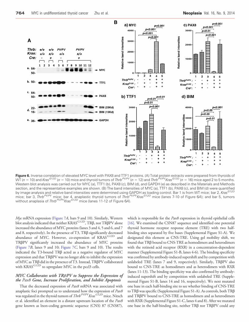
Figure 6. Inverse correlation of elevatedMYC level with PAX8 and TTF1 proteins. (A) Total protein extracts were prepared from thyroids ofWT (n= 10) and KrasG12D (n= 10) mice and thyroid tumors of ThrbPV/PV (n= 12) and ThrbPV/PVKrasG12D (n= 16) mice aged 2 to 5 months.Western blot analysis was carried out for MYC (a), TTF1 (b), PAX8 (c), BIM (d), and GAPDH (e) as described in the Materials and Methodssection, and the representative examples are shown. (B) The band intensities of MYC (a), TTF1 (b), PAX8 (c), and BIM (d) were quantifiedby image analysis and relative band intensities were determined using GAPDH as loading control. Bar 1 is fromWT mice; bar 2, KrasG12D
mice; bar 3, ThrbPV/PV mice; bar 4, anaplastic thyroid tumors of ThrbPV/PVKrasG12D mice (lanes 7-10 of Figure 6A); and bar 5, tumorswithout anaplasia of ThrbPV/PVKrasG12D mice (lanes 11-12 of Figure 6A).
764 MYC in undifferentiated thyroid cancer Zhu et al. Neoplasia Vol. 16, No. 9, 2014
Myc mRNA expression (Figure 7A, bars 9 and 10). Similarly, Westernblot analysis indicated that neither KRASG12D, TRβ, nor TRβPV aloneincreased the abundance ofMYCproteins (lanes 3 and 4, 5 and 6, and 7and 8, respectively). In the presence of T3, TRβ significantly decreasedabundance of MYC. However, co-expression of KRASG12D andTRβPV significantly increased the abundance of MYC proteins(Figure 7B, lanes 9 and 10; Figure 7C, bars 9 and 10). The resultsindicated the T3-bound TRβ acted as a negative regulator of MYCexpression and that TRβPVwas no longer able to inhibit the expressionofMYC asTRβ did in the presence of T3. Instead, TRβPV collaboratedwith KRASG12D to upregulate MYC in the pccl3 cells.
MYC Collaborates with TRβPV to Suppress the Expression ofthe Pax8 Gene, Increase Proliferation, and Inhibit Apoptosis
That the decreased expression of Pax8 mRNA was associated withanaplastic foci prompted us to understand how the expression of Pax8was regulated in the thyroid tumors ofThrbPV/PVKrasG12Dmice. Nitschet al. identified an element in a distant upstream location of the Pax8gene known as lnon-coding genomic sequence (CNS) 87 (CNS87),
which is responsible for the Pax8 expression in thyroid epithelial cells[16]. We examined the CSN87 sequence and identified one potentialthyroid hormone receptor response element (TRE) with two half-binding sites separated by five bases (Supplemental Figure S1-A). Wedesignated this element as CNS-TRE. Using gel mobility shift, wefound that TRβ bound to CNS-TRE as homodimers and heterodimerswith the retinoid acid receptor (RXR) in a concentration-dependentmanner (Supplemental Figure S1-B, lanes 4-6). The binding specificitywas confirmed by antibody-induced supershift and by competition withunlabeled TRE (lanes 7 and 9, respectively). Similarly, TRβPV alsobound to CNS-TRE as homodimers and as heterodimers with RXR(lanes 11-13). The binding specificity was also confirmed by antibody-induced supershift and by competition with unlabeled TRE (Supple-mental Figure S1-B, lanes 14 and 16, respectively). We next mutatedone base in each half-binding site to see whether binding of CNS-TREtoTRβwas specific (Supplemental Figure S1-A). As controls, both TRβand TRβPV bound to CNS-TRE as homodimers and as heterodimerswith RXR (Supplemental Figure S1-C, lanes 4 and 8). After wemutatedone base in the half-binding site, neither TRβ nor TRβPV could any
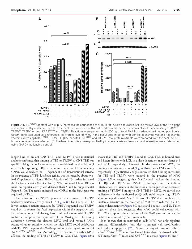
Figure 7. KRASG12D together with TRβPV increases the abundance of MYC in rat thyroid pccl3 cells. (A) The mRNA level of theMyc genewas measured by real-time RT-PCR in the pccl3 cells infected with control adenoviral vector or adenoviral vectors expressing KRASG12D,TRβWT, TRβPV, or both KRASG12D and TRβPV. Reactions were performed in 200 ng of total RNA from adenovirus-infected pccl3 cells.Gapdh gene was used as a reference. (B) Protein level of MYC in the pccl3 cells infected with control adenoviral vector or adenoviralvectors expressing KRASG12D, TRβWT, TRβPV, or both KRASG12D and TRβPV. Total protein extracts were prepared from the pccl3 cells 18hours after adenovirus infection. (C) The band intensities were quantified by image analysis and relative band intensities were determinedusing GAPDH as loading control.
Neoplasia Vol. 16, No. 9, 2014 MYC in undifferentiated thyroid cancer Zhu et al. 765
longer bind to mutant CNS-TRE (lanes 12-19). These mutationalanalyses confirmed that binding of TRβ or TRβPV to CNS-TRE wasspecific. Using the luciferase reporter in established rat thyroid pccl3cells stably expressing TRβ, we examined whether TRE-containingCNS87 could mediate the T3-dependent TRβ transcriptional activity.In the presence of TRβ, luciferase activity was increased by about two-fold (Supplemental Figure S1-D). Addition of T3 further increasedthe luciferase activity (bar 4 vs bar 3). When mutated CNS-TRE wasused, no reporter activity was detected (bars 5 and 6; SupplementalFigure S1-D). The results indicated that CNS87 in the Pax8 gene wasregulated by TRβ.Comparison of the CNS87 reporter activities shows that TRβPV
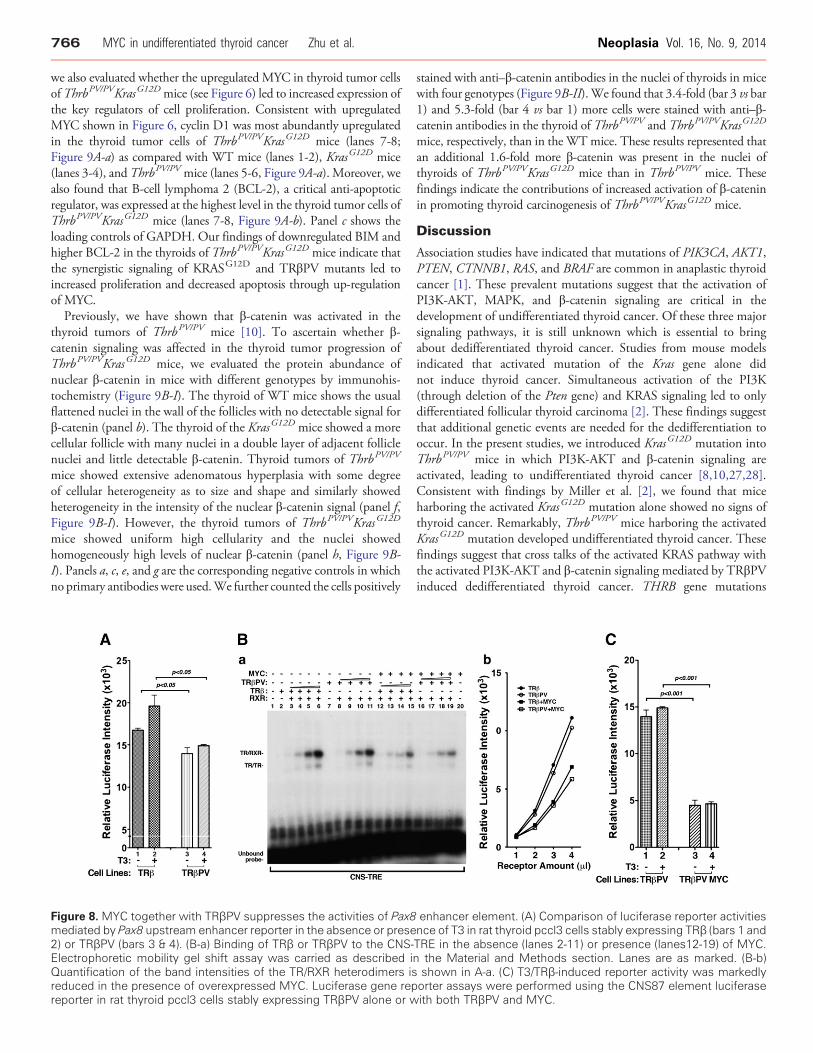
had lower luciferase activity than TRβ (Figure 8A, bar 4 vs bar 2). Thelower luciferase activity mediated by TRβPV suggested that TRβPVcould act to repress the Pax8 expression within the anaplastic foci.Furthermore, other cellular regulator could collaborate with TRβPVto further suppress the expression of the Pax8 gene. The strongassociation between the elevated MYC level and reduced PAX8prompted us to examine whether the high MYC could collaboratewith TRβPV to repress the Pax8 expression in the thyroid tumors ofThrbPV/PVKrasG12D mice. Accordingly, we examined whether MYCaffected the binding of TRβ or TRβPV to CNS-TRE. Figure 8B-a
shows that TRβ and TRβPV bound to CNS-TRE as homodimersand heterodimers with RXR in a dose-dependent manner (lanes 3-6and 8-11, respectively). However, in the presence of MYC, thebinding intensity was reduced (Figure 8B-a, lanes 12-15 and 16-19,respectively). Quantitative analysis indicated that binding intensitiesfor TRβ and TRβPV were reduced in the presence of MYC(Figure 8B-b), suggesting that MYC could weaken the bindingof TRβ and TRβPV to CNS-TRE through direct or indirectinterference. To ascertain the functional consequence of decreasedbinding of TRβPV binding to CNS-TRE by MYC, we carried outluciferase activities in thyroid pccl3 cells stably expressing TRβPValone or together with MYC. Because TRβPV does not bind T3,luciferase activities in the presence of MYC were reduced in a T3-independent manner (Figure 8C, bars 3 and 4 vs bars 1 and 2). Takentogether, these data suggested that MYC could collaborate withTRβPV to suppress the expression of the Pax8 gene and induce thededifferentiation of thyroid tumor cells.
It is known that in mammalian cells MYC not only regulatesterminal differentiation but also stimulates cell proliferationand induces apoptosis [26]. Since the thyroid tumor cells ofThrbPV/PVKrasG12D mice proliferated faster than the thyroid cells ofWT mice, KrasG12D mice, and ThrbPV/PV mice (see Figures 1C and 3),
766 MYC in undifferentiated thyroid cancer Zhu et al. Neoplasia Vol. 16, No. 9, 2014
we also evaluated whether the upregulated MYC in thyroid tumor cellsofThrbPV/PVKrasG12Dmice (see Figure 6) led to increased expression ofthe key regulators of cell proliferation. Consistent with upregulatedMYC shown in Figure 6, cyclin D1 was most abundantly upregulatedin the thyroid tumor cells of ThrbPV/PVKrasG12D mice (lanes 7-8;Figure 9A-a) as compared with WT mice (lanes 1-2), KrasG12D mice(lanes 3-4), and ThrbPV/PV mice (lanes 5-6, Figure 9A-a). Moreover, wealso found that B-cell lymphoma 2 (BCL-2), a critical anti-apoptoticregulator, was expressed at the highest level in the thyroid tumor cells ofThrbPV/PVKrasG12D mice (lanes 7-8, Figure 9A-b). Panel c shows theloading controls of GAPDH. Our findings of downregulated BIM andhigher BCL-2 in the thyroids of ThrbPV/PVKrasG12D mice indicate thatthe synergistic signaling of KRASG12D and TRβPV mutants led toincreased proliferation and decreased apoptosis through up-regulationof MYC.
Previously, we have shown that β-catenin was activated in thethyroid tumors of ThrbPV/PV mice [10]. To ascertain whether β-catenin signaling was affected in the thyroid tumor progression ofThrbPV/PVKrasG12D mice, we evaluated the protein abundance ofnuclear β-catenin in mice with different genotypes by immunohis-tochemistry (Figure 9B-I). The thyroid of WT mice shows the usualflattened nuclei in the wall of the follicles with no detectable signal forβ-catenin (panel b). The thyroid of the KrasG12D mice showed a morecellular follicle with many nuclei in a double layer of adjacent folliclenuclei and little detectable β-catenin. Thyroid tumors of ThrbPV/PV
mice showed extensive adenomatous hyperplasia with some degreeof cellular heterogeneity as to size and shape and similarly showedheterogeneity in the intensity of the nuclear β-catenin signal (panel f,Figure 9B-I). However, the thyroid tumors of ThrbPV/PVKrasG12D
mice showed uniform high cellularity and the nuclei showedhomogeneously high levels of nuclear β-catenin (panel h, Figure 9B-I). Panels a, c, e, and g are the corresponding negative controls in whichno primary antibodies were used.We further counted the cells positively
Figure 8. MYC together with TRβPV suppresses the activities of Pax8mediated by Pax8 upstream enhancer reporter in the absence or prese2) or TRβPV (bars 3 & 4). (B-a) Binding of TRβ or TRβPV to the CNS-Electrophoretic mobility gel shift assay was carried as described iQuantification of the band intensities of the TR/RXR heterodimers isreduced in the presence of overexpressed MYC. Luciferase gene repreporter in rat thyroid pccl3 cells stably expressing TRβPV alone or w
stained with anti–β-catenin antibodies in the nuclei of thyroids in micewith four genotypes (Figure 9B-II).We found that 3.4-fold (bar 3 vs bar1) and 5.3-fold (bar 4 vs bar 1) more cells were stained with anti–β-catenin antibodies in the thyroid of ThrbPV/PV and ThrbPV/PVKrasG12D
mice, respectively, than in the WTmice. These results represented thatan additional 1.6-fold more β-catenin was present in the nuclei ofthyroids of ThrbPV/PVKrasG12D mice than in ThrbPV/PV mice. Thesefindings indicate the contributions of increased activation of β-cateninin promoting thyroid carcinogenesis of ThrbPV/PVKrasG12D mice.
Discussion
Association studies have indicated that mutations of PIK3CA, AKT1,PTEN, CTNNB1, RAS, and BRAF are common in anaplastic thyroidcancer [1]. These prevalent mutations suggest that the activation ofPI3K-AKT, MAPK, and β-catenin signaling are critical in thedevelopment of undifferentiated thyroid cancer. Of these three majorsignaling pathways, it is still unknown which is essential to bringabout dedifferentiated thyroid cancer. Studies from mouse modelsindicated that activated mutation of the Kras gene alone didnot induce thyroid cancer. Simultaneous activation of the PI3K(through deletion of the Pten gene) and KRAS signaling led to onlydifferentiated follicular thyroid carcinoma [2]. These findings suggestthat additional genetic events are needed for the dedifferentiation tooccur. In the present studies, we introduced KrasG12D mutation intoThrbPV/PV mice in which PI3K-AKT and β-catenin signaling areactivated, leading to undifferentiated thyroid cancer [8,10,27,28].Consistent with findings by Miller et al. [2], we found that miceharboring the activated KrasG12D mutation alone showed no signs ofthyroid cancer. Remarkably, ThrbPV/PV mice harboring the activatedKrasG12D mutation developed undifferentiated thyroid cancer. Thesefindings suggest that cross talks of the activated KRAS pathway withthe activated PI3K-AKT and β-catenin signaling mediated by TRβPVinduced dedifferentiated thyroid cancer. THRB gene mutations
enhancer element. (A) Comparison of luciferase reporter activitiesnce of T3 in rat thyroid pccl3 cells stably expressing TRβ (bars 1 andTRE in the absence (lanes 2-11) or presence (lanes12-19) of MYC.n the Material and Methods section. Lanes are as marked. (B-b)shown in A-a. (C) T3/TRβ-induced reporter activity was markedlyorter assays were performed using the CNS87 element luciferaseith both TRβPV and MYC.
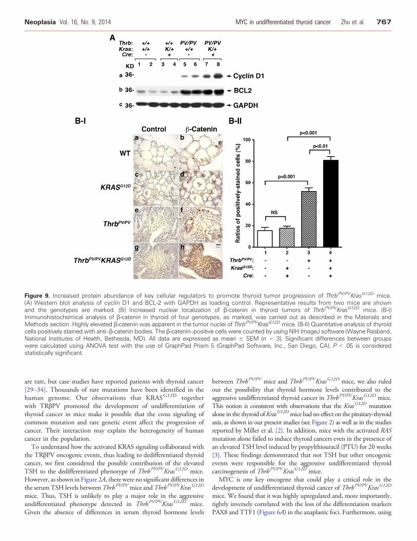
Figure 9. Increased protein abundance of key cellular regulators to promote thyroid tumor progression of ThrbPV/PVKrasG12D mice.(A) Western blot analysis of cyclin D1 and BCL-2 with GAPDH as loading control. Representative results from two mice are shownand the genotypes are marked. (B) Increased nuclear localization of β-catenin in thyroid tumors of ThrbPV/PVKrasG12D mice. (B-I)Immunohistochemical analysis of β-catenin in thyroid of four genotypes, as marked, was carried out as described in the Materials andMethods section. Highly elevated β-catenin was apparent in the tumor nuclei of ThrbPV/PVKrasG12D mice. (B-II) Quantitative analysis of thyroidcells positively stained with anti–β-catenin bodies. The β-catenin–positive cells were counted by using NIH ImageJ software (Wayne Rasband,National Institutes of Health, Bethesda, MD). All data are expressed as mean ± SEM (n = 3). Significant differences between groupswere calculated using ANOVA test with the use of GraphPad Prism 5 (GraphPad Software, Inc., San Diego, CA). P b .05 is consideredstatistically significant.
Neoplasia Vol. 16, No. 9, 2014 MYC in undifferentiated thyroid cancer Zhu et al. 767
are rare, but case studies have reported patients with thyroid cancer[29–34]. Thousands of rare mutations have been identified in thehuman genome. Our observations that KRASG12D togetherwith TRβPV promoted the development of undifferentiation ofthyroid cancer in mice make it possible that the cross signaling ofcommon mutation and rare genetic event affect the progression ofcancer. Their interaction may explain the heterogeneity of humancancer in the population.To understand how the activated KRAS signaling collaborated with
the TRβPV oncogenic events, thus leading to dedifferentiated thyroidcancer, we first considered the possible contribution of the elevatedTSH to the dedifferentiated phenotype of ThrbPV/PVKrasG12D mice.However, as shown in Figure 2A, there were no significant differences inthe serumTSH levels betweenThrbPV/PVmice andThrbPV/PVKrasG12D
mice. Thus, TSH is unlikely to play a major role in the aggressiveundifferentiated phenotype detected in ThrbPV/PVKrasG12D mice.Given the absence of differences in serum thyroid hormone levels
between ThrbPV/PV mice and ThrbPV/PVKrasG12D mice, we also ruledout the possibility that thyroid hormone levels contributed to theaggressive undifferentiated thyroid cancer in ThrbPV/PVKrasG12D mice.This notion is consistent with observations that the KrasG12D mutationalone in the thyroid ofKrasG12Dmice had no effect on the pituitary-thyroidaxis, as shown in our present studies (see Figure 2) as well as in the studiesreported by Miller et al. [2]. In addition, mice with the activated RASmutation alone failed to induce thyroid cancers even in the presence ofan elevated TSH level induced by propylthiouracil (PTU) for 20 weeks[3]. These findings demonstrated that not TSH but other oncogenicevents were responsible for the aggressive undifferentiated thyroidcarcinogenesis of ThrbPV/PVKrasG12D mice.
MYC is one key oncogene that could play a critical role in thedevelopment of undifferentiated thyroid cancer of ThrbPV/PVKrasG12D
mice. We found that it was highly upregulated and, more importantly,tightly inversely correlated with the loss of the differentiation markersPAX8 and TTF1 (Figure 6A) in the anaplastic foci. Furthermore, using
768 MYC in undifferentiated thyroid cancer Zhu et al. Neoplasia Vol. 16, No. 9, 2014
rat thyroid pccl3 cells, we found that co-expression of KRASG12D andTRβPV upregulated the levels of MYC at both the mRNA and proteinlevels (Figure 7). Although it is not yet clear howMYCwas upregulatedin the anaplastic tumors of ThrbPV/PVKrasG12D mice, up-regulation ofMYC by KRASG12D/TRβPV in the rat thyroid pccl3 cell line and thetight inverse association with PAX8 and TTF1 lead us to argue for thecritical involvement of MYC in the induction of undifferentiatedthyroid tumors inThrbPV/PVKrasG12Dmice. This postulate is consistentwith other studies that support MYC’s key role in the differentiationprocess. MYC has been recognized as one of the most highly amplifiedoncogenes in human cancers [35]. Cell differentiation leads to down-regulation of the MYC expression, and overexpression of MYC resultsin the dedifferentiation phenotype [36]. Overexpressed MYC is alsoknown to inhibit Ras-mediated differentiation by blocking c-Jun up-regulation [37] (Supplemental Figure S2). MYC also plays a key role inreprogramming human somatic cells to pluripotent stem cells [38,39].In the above studies, however, the mechanisms by whichMYC inducesdedifferentiation were not clearly elucidated. In the present studies, weuncovered one mechanism by which MYC could act to participate inthe induction of dedifferentiated thyroid cancer of ThrbPV/PVKrasG12D
mice through the repression of one of the differentiation transcriptionfactors. Molecular analyses showed that TRβPV, which does not bindT3, repressed the Pax8 gene expression. This repression was furtheraugmented by MYC, leading to added repression in the Pax8transcription. At present, we could not rule out the possibility thatMYC could repress the expression of the Pax8 gene through othermechanisms independent of PV. However, our data could explain the invivo findings that the Pax8mRNA level was less than that in WT mice,KrasG12D mice, and ThrbPV/PV mice (Figure 5D). While mutations ofthe THRβ gene are rare in human thyroid cancer [29–34,40], thecollaboration of MYC with TRβ mutants to suppress the expression ofthe Pax8 gene has yet to be uncovered. However, our findingsexemplified howMYC could collaborate with other transcription factorsand oncogenes to suppress the expression of the Pax8 gene and therebycould participate in the induction of the dedifferentiation process. In linewith our findings, others have shown that MYC expressed in transgenicmice triggers aggressive mammary tumorigenesis by collaborating with aKRAS mutation [41].
The up-regulation of MYC in the dedifferentiated thyroid cancerof ThrbPV/PVKrasG12D mice provides insight into a potentialtherapeutic intervention. MYC is known to be essential in themaintenance of established tumors [42,43]. In vitro knockdown ofMYC in established cancer cell lines reduces cell proliferation and, insome instances, induces apoptosis [44,45]. In transgenic mousemodels with inducible MYC expression, established tumors regressupon withdrawal of ectopic expressed MYC. These observationssuggest that MYC plays a role in tumor maintenance, and onceestablished, these tumors are addicted to MYC for maintaining tumorphenotypes [46]. Studies using mice harboring an activated KrasG12D
mutation showed that the blockade of MYC functions throughsystematic induction of a dominant-negative MYC allele resulted inthe regression of lung carcinomas and pancreatic carcinomas [42].Investigations have further shown that targeting MYC transcriptionfunctions by disruption of chromatin-dependent signal transductioncould be effective as a therapeutic strategy. Indeed, a potent, selectivesmall-molecule inhibitor of BET bromodomains, JQ1, was developed[47]. The efficacy of JQ1 was demonstrated in producing potentantiproliferative effects and cellular senescence in murine models ofmultiple myeloma [48,49]. Therefore, inhibiting MYC functions
by small inhibitors could be a novel potential therapeutic strategyin the treatment of undifferentiated thyroid carcinoma. Thus, theThrbPV/PVKrasG12D mouse offers an opportunity not only to furtherelucidate the role of the KRAS in the initiation of undifferentiatedthyroid cancer but also to further test novel therapeutic targets as anintervention for anaplastic thyroid cancer.
Supplementary data to this article can be found online at http://dx.doi.org/10.1016/j.neo.2014.08.003.
References
[1] Nikiforov YE and Nikiforova MN (2011). Molecular genetics and diagnosis ofthyroid cancer. Nat Rev Endocrinol 7(10), 569–580.
[2] Miller KA, Yeager N, Baker K, Liao XH, Refetoff S, and Di Cristofano A(2009). Oncogenic Kras requires simultaneous PI3K signaling to induce ERKactivation and transform thyroid epithelial cells in vivo. Cancer Res 69(8),3689–3694.
[3] Franco AT, Malaguarnera R, and Refetoff S, et al (2011). Thyrotrophin receptorsignaling dependence of Braf-induced thyroid tumor initiation in mice. Proc NatlAcad Sci U S A 108(4), 1615–1620.
[4] Kaneshige M, Kaneshige K, and Zhu X, et al (2000). Mice with a targetedmutation in the thyroid hormone beta receptor gene exhibit impaired growth andresistance to thyroid hormone. Proc Natl Acad Sci U S A 97(24), 13209–13214.
[5] Suzuki H, Willingham MC, and Cheng SY (2002). Mice with a mutation in thethyroid hormone receptor β gene spontaneously develop thyroid carcinoma: amouse model of thyroid carcinogenesis. Thyroid 12(11), 963–969.
[6] Parrilla R, Mixson AJ, McPherson JA, McClaskey JH, and Weintraub BD (1991).Characterization of seven novel mutations of the c-erbA beta gene in unrelatedkindreds with generalized thyroid hormone resistance. Evidence for two “hot spot”regions of the ligand binding domain. J Clin Invest 88(6), 2123–2130.
[7] Hou P, Liu D, and Shan Y, et al (2007). Genetic alterations and their relationshipin the phosphatidylinositol 3-kinase/Akt pathway in thyroid cancer. Clin CancerRes 13(4), 1161–1170.
[8] Furuya F, Hanover JA, and Cheng SY (2006). Activation of phosphatidylinositol3-kinase signaling by a mutant thyroid hormone β receptor. Proc Natl Acad SciU S A 103(6), 1780–1785.
[9] Lu C, Zhao L, Ying H, Willingham MC, and Cheng SY (2010).Growth activation alone is not sufficient to cause metastatic thyroid cancerin a mouse model of follicular thyroid carcinoma. Endocrinology 151(4),1929–1939.
[10] Guigon CJ, Zhao L, Lu C, Willingham MC, and Cheng SY (2008). Regulationof β-catenin by a novel nongenomic action of thyroid hormone beta receptor.Mol Cell Biol 28(14), 4598–4608.
[11] Jackson EL, Willis N, Mercer K, Bronson RT, Crowley D, Montoya R,Jacks T, and Tuveson DA (2001). Analysis of lung tumor initiation andprogression using conditional expression of oncogenic K-ras. Genes Dev 15(24), 3243–3248.
[12] Kusakabe T, Kawaguchi A, Kawaguchi R, Feigenbaum L, and Kimura S (2004).Thyrocyte-specific expression of Cre recombinase in transgenic mice. Genesis 39(3),212–216.
[13] Furumoto H, Ying H, Chandramouli GV, Zhao L, Walker RL, Meltzer PS,Willingham MC, and Cheng SY (2005). An unliganded thyroid hormone βreceptor activates the cyclin D1/cyclin-dependent kinase/retinoblastoma/E2Fpathway and induces pituitary tumorigenesis. Mol Cell Biol 25(1), 124–135.
[14] Zhu XG, McPhie P, and Cheng SY (1997). Differential sensitivity of thyroidhormone receptor isoform homodimers and mutant heterodimers to hormone-induced dissociation from deoxyribonucleic acid: its role in dominant negativeaction. Endocrinology 138(4), 1456–1463.
[15] Zhao L, Zhu X, Won Park J, Fozzatti L, Willingham M, and Cheng SY (2012).Role of TSH in the spontaneous development of asymmetrical thyroid carcinomain mice with a targeted mutation in a single allele of the thyroid hormone-βreceptor. Endocrinology 153(10), 5090–5100.
[16] Nitsch R, Di Dato V, di Gennaro A, de Cristofaro T, Abbondante S, De FeliceM, Zannini M, and Di Lauro R (2010). Comparative genomics reveals afunctional thyroid-specific element in the far upstream region of the PAX8 gene.BMC Genomics 11, 306.
[17] Rivas M and Santisteban P (2003). TSH-activated signaling pathways in thyroidtumorigenesis. Mol Cell Endocrinol 213(1), 31–45.
Neoplasia Vol. 16, No. 9, 2014 MYC in undifferentiated thyroid cancer Zhu et al. 769
[18] Nonaka D, Tang Y, Chiriboga L, Rivera M, and Ghossein R (2008). Diagnosticutility of thyroid transcription factors Pax8 and TTF-2 (FoxE1) in thyroidepithelial neoplasms. Mod Pathol 21(2), 192–200.
[19] Fabbro D, Di Loreto C, Beltrami CA, Belfiore A, Di Lauro R, and Damante G(1994). Expression of thyroid-specific transcription factors TTF-1 and PAX-8 inhuman thyroid neoplasms. Cancer Res 54(17), 4744–4749.
[20] Shachaf CM, Kopelman AM, and Arvanitis C, et al (2004). MYC inactivationuncovers pluripotent differentiation and tumour dormancy in hepatocellularcancer. Nature 431(7012), 1112–1117.
[21] Henriksson M and Luscher B (1996). Proteins of the Myc network: essentialregulators of cell growth and differentiation. Adv Cancer Res 68, 109–182.
[22] Kurihara T, Ikeda S, and Ishizaki Y, et al (2004). Immunohistochemical andsequencing analyses of the Wnt signaling components in Japanese anaplasticthyroid cancers. Thyroid 14(12), 1020–1029.
[23] Smallridge RC, Marlow LA, and Copland JA (2009). Anaplastic thyroid cancer:molecular pathogenesis and emerging therapies. Endocr Relat Cancer 16(1), 17–44.
[24] Reynolds C, Roderick JE, and Labelle JL, et al (2014). Repression of BIMmediates survival signaling by MYC and AKT in high-risk T-cell acutelymphoblastic leukemia. Leukemia (advance online publication 25 March 2014).
[25] Salmanidis M, Brumatti G, and Narayan N, et al (2013). Hoxb8 regulatesexpression of microRNAs to control cell death and differentiation. Cell DeathDiffer 20(10), 1370–1380.
[26] Grandori C, Cowley SM, James LP, and Eisenman RN (2000). The Myc/Max/Mad network and the transcriptional control of cell behavior. Annu Rev Cell DevBiol 16, 653–699.
[27] Furuya F, Lu C, Willingham MC, and Cheng SY (2007). Inhibition ofphosphatidylinositol 3-kinase delays tumor progression and blocks metastaticspread in a mouse model of thyroid cancer. Carcinogenesis 28(12), 2451–2458.
[28] Guigon CJ, Zhao L, Willingham MC, and Cheng SY (2009). PTEN deficiencyaccelerates tumour progression in a mouse model of thyroid cancer. Oncogene 28(4), 509–517.
[29] Kim HK, Kim D, and Yoo EH, et al (2010). A case of resistance to thyroidhormone with thyroid cancer. J Korean Med Sci 25(9), 1368–1371.
[30] Ramos-Prol A, Antonia Pérez-LázaroM, Isabel del Olmo-GarcíaM, León-de Zayas B,Moreno-Macián F, Navas-de Solis S, and Merino-Torres JF (2013). Differentiatedthyroid carcinoma in a girl with resistance to thyroid hormone management withtriiodothyroacetic acid. J Pediatr Endocrinol Metab 26(1–2), 133–136.
[31] Xifra G, Mauri S, Gironès J, Rodriguez Hermosa JI, Oriola J, Ricart W, andFernández-Real JM (2013). Multiple Hürthle cell adenomas in a patient withthyroid hormone resistance. Endocrinol Diabetes Metab Case Rep 2013, 130032.
[32] Paragliola RM, Lovicu RM, Locantore P, Senes P, Concolino P, Capoluongo E,Pontecorvi A, and Corsello SM (2011). Differentiated thyroid cancer in twopatients with resistance to thyroid hormone. Thyroid 21(7), 793–797.
[33] Ünlütürk U, Sriphrapradang C, Erdoğan MF, Emral R, Güldiken S, Refetoff S,and Güllü S (2013). Management of differentiated thyroid cancer in the presence
of resistance to thyroid hormone and TSH-secreting adenomas: a report of fourcases and review of the literature. J Clin Endocrinol Metab 98(6), 2210–2217.
[34] Weinert LS, Ceolin L, Romitti M, Camargo EG, and Maia AL (2012). Is there arole for inherited TRβ mutation in human carcinogenesis? [corrected]. Arq BrasEndocrinol Metabol 56(1), 67–71.
[35] Beroukhim R, Mermel CH, and Porter D, et al (2010). The landscape of somaticcopy-number alteration across human cancers. Nature 463(7283), 899–905.
[36] Demeterco C, Itkin-Ansari P, Tyrberg B, Ford LP, Jarvis RA, and Levine F(2002). c-Myc controls proliferation versus differentiation in human pancreaticendocrine cells. J Clin Endocrinol Metab 87(7), 3475–3485.
[37] Vaqué JP, Fernández-García B, and Garcia-Sanz P, et al (2008). c-Myc inhibitsRas-mediated differentiation of pheochromocytoma cells by blocking c-Jun up-regulation. Mol Cancer Res 6(2), 325–339.
[38] Park IH, Zhao R, and West JA, et al (2008). Reprogramming of human somaticcells to pluripotency with defined factors. Nature 451(7175), 141–146.
[39] Takahashi K and Yamanaka S (2006). Induction of pluripotent stem cells frommouse embryonic and adult fibroblast cultures by defined factors. Cell 126(4),663–676.
[40] Joseph B, Ji M, Liu D, Hou P, and Xing M (2007). Lack of mutations in thethyroid hormone receptor (TR) α and β genes but frequent hypermethylation ofthe TRβ gene in differentiated thyroid tumors. J Clin Endocrinol Metab 92(12),4766–4770.
[41] D'Cruz CM, Gunther EJ, and Boxer RB, et al (2001). c-MYC induces mammarytumorigenesis by means of a preferred pathway involving spontaneous Kras2mutations. Nat Med 7(2), 235–239.
[42] Soucek L, Whitfield J, and Martins CP, et al (2008). Modelling Myc inhibitionas a cancer therapy. Nature 455(7213), 679–683.
[43] Soucek L, Whitfield JR, Sodir NM, Massó-Vallés D, Serrano E, Karnezis AN,Swigart LB, and Evan GI (2013). Inhibition of Myc family proteins eradicatesKRas-driven lung cancer in mice. Genes Dev 27(5), 504–513.
[44] Koh CM, Gurel B, and Sutcliffe S, et al (2011). Alterations in nucleolar structureand gene expression programs in prostatic neoplasia are driven by the MYConcogene. Am J Pathol 178(4), 1824–1834.
[45] Wang H, Mannava S, Grachtchouk V, Zhuang D, Soengas MS, Gudkov AV,Prochownik EV, and Nikiforov MA (2008). c-Myc depletion inhibitsproliferation of human tumor cells at various stages of the cell cycle. Oncogene27(13), 1905–1915.
[46] Arvanitis C and Felsher DW (2006). Conditional transgenic models define howMYC initiates and maintains tumorigenesis. Semin Cancer Biol 16(4), 313–317.
[47] Filippakopoulos P, Qi J, and Picaud S, et al (2010). Selective inhibition of BETbromodomains. Nature 468(7327), 1067–1073.
[48] Delmore JE, Issa GC, and Lemieux ME, et al (2011). BET bromodomaininhibition as a therapeutic strategy to target c-Myc. Cell 146(6), 904–917.
[49] Loven J, Hoke HA, and Lin CY, et al (2013). Selective inhibition of tumoroncogenes by disruption of super-enhancers. Cell 153(2), 320–334.