swiss soft days 7 epfl, lausanne...
TRANSCRIPT

SWISS SOFT DAYS 7TH
EPFL, LAUSANNE
13/2/2012
LOCAL ORGANIZERS:
Giuseppe Foffi
Sylvia Jeney
Suliana Manley

SWISS SOFT DAYS 7th WORKSHOP, Lausanne 13.02.2012
2
DIRECTIONS FOR SSD7 FROM LAUSANNE STATION
- Take the metro M2 (in front of the main entrance of the station) in the direction Croisettes (going
up)
- Get off at Lausanne-Flon (one stop)
- Take the metro M1 (it is upstair with respect to M2) . This is is the terminus and you can go in only
one direction (Renens-Gare)
- Get off at UNIL-Sorge
- In front of the Metro stop you will find the Cupotron (see map)
- SSD7 will take place at the 2nd Floor in the room BSP 231. We will set up signs.

SWISS SOFT DAYS 7th WORKSHOP, Lausanne 13.02.2012
3
10.00-10.05 Welcome
Theme 1: Materials & Assemblies
10.05-10.45 Langevin
(U d’Orsay)
Physical Chemistry of Foams
10.45-11.00 I.Ziemecka
(U Delft)
All-aqueous single and double emulsions by
microfluidics 11.00-11.15 Gour
(U Geneva)
Self-Assembling Peptide-Nucleotide Hybrids
11.15-11.30 Vallooran (ETHZ) Macroscopic Alignment of Lyotropic Liquid Crystals
Using Magnetic Nanoparticles
Theme 2: Methods
11.30-11.45 Isa
(ETHZ) Unravelling adsorption and alignment of amyloid fibrils
at interfaces by probe particle tracking
11.45-12.00 Croccolo
(U Fribourg)
CUDA/C++ implementation of the Differential
Dynamic algorithm
12.00-13.30 Lunch/Posters/Discussion
Theme 2: Colloids & Polymers
13.30-14.00 Felderhof
(RWTH Aachen)
Brownian motion and hydrodynamics near a wall
14.00-14.15 Farage
(U Fribourg)
Three-dimensional flow of colloidal glasses
14.15-14.30 Zahn
(ETHZ)
Redox-active polyelectrolyte multilayers: all about
ion-exchange and entropy of water
14.30-14.45 Nigro
(EPFL)
Tunneling conductivity in composites of attractive
colloids
14.45-15.00 Kowal
(U Basel)
Solid-supported planar polymer membranes
15.00-15.15 Griffete
(U Fribourg)
Synthesis of polymer-coated magnetic nanoparticles
using the diazonium salt chemistry
15.15-16.00 Coffee/Posters/Discussion
Theme 3: Biophysics
16.00-16.15 Assenza
(EPFL)
A simple model for amyloid fibrils twisting structure
16.15-16.30 Benke
(EPFL)
Direct live-cell super-resolution imaging of cellular
DNA
16.30-16.45 Ambühl
(EPFL)
Exploring the cell lamellipodium dynamics using image
segmentation
16.45-17.00 Langowska
(U Basel)
In situ synthesis of antibiotics inside nanoreactors
17.00-17.15 Cremmel
(ETHZ)
Orthogonal particle-height and density gradient for
biomedical applications
SWISS SOFT DAYS 7th WORKSHOP, Lausanne 13.02.2012
4
Abstracts Talks
Theme 1: Materials & Assembly 10.05 – 11.30
Physical chemistry of Foams
Dominique Langevin
Laboratoire de Physique des Solides, Université d’Orsay
Foams are dispersions of bubbles in liquids (or solids). We will discuss the case of aqueous foams, usually
stabilized by surfactants. Among applications, let us mention : detergency, food industry, petroleum industry
(injection of foams for oil recovery). Foams are also used to obtain insulation materials (glass and polymer
foams), and for car industry (metallic foams).
Despite the large amount of work devoted to the understanding of foaming and foam stability, many
questions still remain unclear. Improving this knowledge is very important for the understanding and
improvement of different technological processes. We will describe the different destabilisation mechanisms
: gravity drainage, coarsening (gas diffusion from small to large bubbles) and coalescence. We will discuss
the relevance of the surface viscoelasticity concept in the case of foams stabilised by surfactants and
proteins. We will also discuss the case of particles (micro and nano) which can be also used as stabilisers and
are extremely efficient (as in Pickering emulsions).
This key-note lecture has been sponsored by the NCCR on Chemical Biology
All-aqueous single and double emulsions by microfluidics
I.Ziemecka1, V.van Steijn
1, G.J.M..Koper
1, M.T. Kreutzer
1, J.van Esch
1
1 Delft University of Technology, Department of Chemical Engineering, Delft, The Netherlands
INTRODUCTION: Aqueous two-phase systems (ATPS) are a biocompatible soft-matter in which all
constituent phases share water as the common solvent but differ in their dissolved components. Because of
the very low interfacial tension, these systems are attractive for partitioning and separation of biomolecules
as ATPS provide the mild conditions that do not denature or destabilize these biomaterials. This low
interfacial tension, however, prevents the facile formation of droplets. To overcome this problem, we
developed a method to form monodisperse microspheres in a surfactant-free, organic-solvent free, all-
aqueous environment. We show how to use it to form hydrogel microspheres as well as to produce droplets
with internal structure (core-shell).
METHODS: Microfluidic devices such as the one shown in Fig. 1 were use to produce single and double
emulsions. We used the well-known ATPS composed of dextran and polyethylene glycol (PEG). Streams

SWISS SOFT DAYS 7th WORKSHOP, Lausanne 13.02.2012
5
were injected into the device through separate inlets. Key to the method is the forcing we apply to one of the
streams. Without this forcing, the immiscible streams flow in parallel without the formation of drops.
Actuation of a piezo electric bending disc embedded in our devices (Fig. 1) induces perturbations, which
leads to the periodic formation of monodisperse water-in-water droplets. After formation, these droplets were
polymerized to yield microspheres. We also present an extension of this method to produce droplets with an
internal structure.
RESULTS: We were able to form monodisperse hydrogel microspheres from water-in-water droplets.
Drop formation was forced by destabilizing the interface between dextran and PEG using low frequency
mechanical oscillations
Fig. 1: Monodisperse hydrogel microspheres produced by forced jet breakup in ATPS.
(20-50Hz) induced by the piezo electric bending disc. The diameter of the droplets could be easily controlled
in the range between 30 to 60µm using the flow rates as well as the frequency of the AC voltage used to
actuate the piezo disc. Subsequent on-chip polymerization of the droplets yielded stable hydrogel particles.
[1]
Fig. 2: All-aqueous core-shell emulsion droplets flowing through a microfluidic channel
We used a similar approach to also create droplets-inside-droplets (double emulsions). This is achieved by
encapsulating a mixture of PEG and dextran inside droplets that are carried in a solution of PEG. Phase
separation inside the droplets readily occurs and yields core-shell droplets as shown in Fig. 2. Also here, the
frequency together with the flow rates can be used to tune the diameter of the core and the thickness of the
shell. [2]
DISCUSSION & CONCLUSIONS: We have demonstrated the continuous generation of single and
double emulsion microdroplets using aqueous polymer solutions. Our methods allow the efficient
encapsulation of biomaterial in a biofriendly environment, free of organic solvents. This work forms an
excellent and versatile starting point for the fabrication of stable all-aqueous microstuctured systems, for
instance by on-line crosslinking of the shell of the double emulsion droplets. Without a physical barrier in
the form of a lipid bilayer, we foresee that the core-shell structures offer the potential to impact on synthetic
cell studies, as biomaterial can readily transfer from the core to the surroundings in these all-aqueous
structured fluids. We expect the approach we presented to be useful in various fields such as in food
engineering, tissue engineering and drug delivery, where microspheres with and without internal structure
often need to be biocompatible and monodisperse.
REFERENCES: 1
I.Ziemecka et al.; 2011; Lab on a Chip; 4: 620-
624. 2
I.Ziemecka et al.; 2011; Soft Matter; 21: 9878-9880.

SWISS SOFT DAYS 7th WORKSHOP, Lausanne 13.02.2012
6
Self-Assembling Peptide-Nucleotide Hybrids
Nidhi Gour, Dawid Kedracki, Ilyès Safir, Kien Xuan Ngo, and Corinne Vebert-Nardin
Department of inorganic, analytical and applied chemistry, University of Geneva-Sciences II;
30,quai Ernest-Ansermet, CH-1211 Geneva 4, Switzerland.
Aside from being the universal carrier of the genetic information, DNA and short nucleotide sequences
thereof are involved in a plethora of vital biological mechanisms like gene silencing for instance. However,
the main obstacles of using nucleotide sequences as such is their limited plasma half-life as well as cellular
penetrability and uptake. To overcome these issues, their conjugation with biocompatible ligands has thus
increasingly become a topic of intense research. One may also envisage to design nucleic acid decorated
structures or supramolecular assemblies which may serve as delivery cargo molecules. Peptides due to their
inherent property of self-assembling in solution may be used as one of such ligand. We demonstrate in here
for the very first time that conjugation of peptide ligand with a nucleotide sequence results in a peptide-
nucleotide hybrid which self-assembles to give rise to vesicular structures which was assessed by various
techniques i.e., imaging of the vesicles using fluorescence microscopy, atomic force microscopy (AFM).
scanning electron microscopy (SEM) and transmission electron microscopy (TEM). Vesicular structures
efficiently encapsulate a hydrophilic dye and pH triggered release reveals their potential for application as
carriers for drug delivery for instance. The design of this fully biocompatible self‐assembling material paves
the way to many possible future uses e.g., as targeted drug delivery vehicle for sustained drug release and/
gene therapy just to cite few examples.
Macroscopic Alignment of Lyotropic Liquid Crystals Using
Magnetic Nanoparticles
J.J.Vallooran, S.Bolisetty, R.Mezzenga Laboratory of Food and Soft Materials, ETH Zürich, Switzerland.
INTRODUCTION: Liquid crystalline (LC) mesophases such as the columnar hexagonal phase and lamellar
phase can be manipulated to acquire desirable long-range orientational order typical of anisotropic species.
However, bulk LC samples are usually not uniformly aligned and possess a polydomain structure, which
smears out the long-range anisotropic order. Controlled alignment of nanostructured domains in these self-
assembled soft materials presents a significant challenge, especially in bulk. By doping lyotropic liquid
crystal systems with magnetic nanoparticles, macroscopic alignment of the mesophase can in principle be
achieved in low magnetic field [1], although this remains a challenging task due to the constraint in the
choice of nanoparticle size, which shall remain smaller than lamellar periodicity, and the pronounced
tendency of the nanoparticles to aggregation. METHODS: Spherical magnetite nanoparticles were synthesized by co-precipitation method. The system
was studied with small angle x-rays and neutron scattering (SAXS, SANS), and optical microscopy.
RESULTS: In the presence of a magnetic field, when the sample is cooled from the isotropic micellar phase
to the lamellar phase it is observed that the dark structures were aligned along the field (Figure 1). This is
interpreted as the result of the alignment of magnetite nanoparticles in presence of the applied field.
SWISS SOFT DAYS 7th WORKSHOP, Lausanne 13.02.2012
7
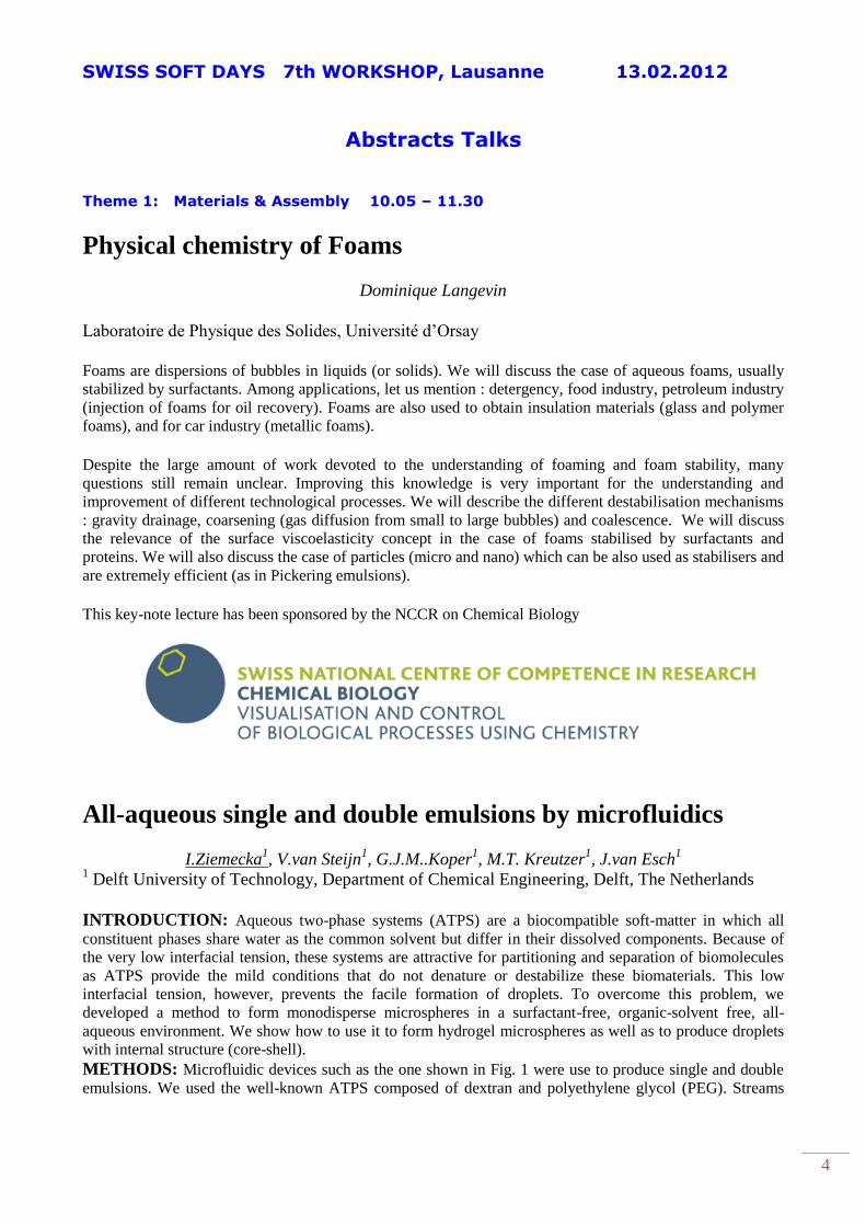
Fig. 1: Bright field optical micrographs of magnetic nanoparticles-doped lamellar phase: in the absence of
magnetic field (left) in the presence of 1.1T magnetic field as indicated (right).
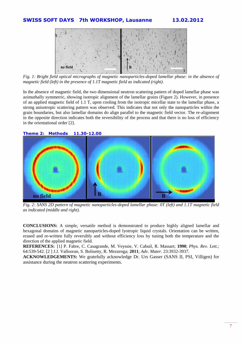
In the absence of magnetic field, the two dimensional neutron scattering pattern of doped lamellar phase was
azimuthally symmetric, showing isotropic alignment of the lamellar grains (Figure 2). However, in presence
of an applied magnetic field of 1.1 T, upon cooling from the isotropic micellar state to the lamellar phase, a
strong anisotropic scattering pattern was observed. This indicates that not only the nanoparticles within the
grain boundaries, but also lamellar domains do align parallel to the magnetic field vector. The re-alignment
in the opposite direction indicates both the reversibility of the process and that there is no loss of efficiency
in the orientational order [2].
Theme 2: Methods 11.30-12.00
Fig. 2: SANS 2D pattern of magnetic nanoparticles-doped lamellar phase: 0T (left) and 1.1T magnetic field
as indicated (middle and right).
CONCLUSIONS: A simple, versatile method is demonstrated to produce highly aligned lamellar and
hexagonal domains of magnetic nanoparticles-doped lyotropic liquid crystals. Orientation can be written,
erased and re-written fully reversibly and without efficiency loss by tuning both the temperature and the
direction of the applied magnetic field. REFERENCES: [1] P. Fabre, C. Casagrande, M. Veyssie, V. Cabuil, R. Massart; 1990; Phys. Rev. Lett.;
64:539-542. [2 ] J.J. Vallooran, S. Bolisetty, R. Mezzenga; 2011; Adv. Mater. 23:3932-3937.
ACKNOWLEDGEMENTS: We gratefully acknowledge Dr. Urs Gasser (SANS II, PSI, Villigen) for
assistance during the neutron scattering experiments.

SWISS SOFT DAYS 7th WORKSHOP, Lausanne 13.02.2012
8
Theme 2: Methods 11.30-12.00
Unravelling adsorption and alignment of amyloid fibrils at
interfaces by probe particle tracking
Lucio Isa,1
Jin-Mi Jung2 and Raffaele Mezzenga
3
[1] ETH Zürich, Laboratory for Surface Science and Technology E-mail: [email protected]
[2] Department of Physics, University of Fribourg E-mail: [email protected]
[3]ETH Zurich, Food and Soft Materials Science, E-mail: [email protected]
We report the first direct, non-invasive experimental
evidence of a 2D isotropic–nematic transition for
highly anisotropic nanoparticles at liquid–liquid
interfaces by using passive fluorescent particle
tracking. In order to illustrate the potential of this
approach on systems of high real practical and
biological relevance, we select as a model
anisotropic nanoparticles, β-lactoglobulin amyloid
fibrils of varying aspect ratios. Upon nanoparticle
adsorption at the interface, we follow, in real time
and as a function of fibril length and bulk
concentration, the development of a 2D nematic
phase by studying the anisotropy in probe particle
traces. We furthermore demonstrate the long-range
nature of the nematic phase by calculating order
parameters for the traces as high as 0.8 over 102 to
103 mm2 areas. The presented route is independent
of the system investigated, and thus these findings
open a new, general strategy for the experimental
assessment of 2D structural changes at anisotropic
fluid–fluid interfaces1.
Fig. 1 AFM images of long (a) and short (b) β-
lactoglobulin fibrils. (c) Schematics of the
microscopy cell set-up used for interfacial
particle tracking.
REFERENCES: [1]L. Isa, J-M Jung and R Mezzenga “Unravelling
adsorption and alignment of amyloid fibrils at
interfaces by probe particle tracking”, 2011, Soft
Matter 7, pp. 8127 – 8134.
CUDA/C++ implementation of the Differential Dynamic
algorithm
G. Cerchiari, F. Croccolo, F. Cardinaux and F. Scheffold
Department of Physics / University of Fribourg
The Differential Dynamic Algorithm (DDA) is a powerful tool to extract the correlation function from a
signal fluctuating in time. While it was first proposed to evaluate the temporal correlation function out of a
pressure detector [1], it has been successfully applied to a variety of optical techniques of the Near Field
Scattering family, like Shadowgraph [2,3], Schlieren [4], Near Field Scattering [5], Differential Dynamic
SWISS SOFT DAYS 7th WORKSHOP, Lausanne 13.02.2012
9
Microscopy [6], and X-ray near Field Scattering [7]. In all these cases the aim is that of obtaining the
temporal correlation function for distinct spatial frequencies which requires the execution of Fast Fourier
Transforms (FFTs) of the acquired images. This turns out to be rather demanding from the computational
point of view, so that, for example, the analysis of a typical set of 2,000 images of 2kx2k pixels requires
about 50 hours of calculus. Fortunately the algorithm is perfectly suited for strong parallelization and
therefore is worth the effort to implement it in the CUDA/C++ language which takes advantage of the large
number of parallel processors of a modern graphic card. Here we show how the code has been implemented
in connection with a dedicated strategy for the optimization of the dataflow between the PC Rapid Access
Memory (RAM) and the graphic device memory (GRAM) resulting in an increase of the program speed up
to 30-fold. An accurate analysis of the timings needed for single operations provides useful hints for further
developments of the existing hardware.
(1) E.O. Schulz-DuBois and I. Rehberg, Appl. Phys. 24, 323-329 (1981)
(2) F. Croccolo et al., Ann. N. Y. Acad. Sci. 1077, 365-379 (2006)
(3) F. Croccolo et al., Phys. Rev. E 76, 41112-1-9 (2007)
(4) F. Croccolo et al., App. Opt. 45, 2166-2173 (2006)
(5) D. Magatti et al., App. Phys. Lett. 92, 241101-1-3 (2008)
(6) R. Cerbino, and V. Trappe, Phys. Rev. Lett. 100, 188102-1-4 (2008)
(7) R. Cerbino et al., Nat. Phys. 4, 238-243 (2008)
SWISS SOFT DAYS 7th WORKSHOP, Lausanne 13.02.2012
10
Theme 3: Colloids & Polymers 13.30-15.30
Brownian motion and hydrodynamics near a wall
B. Ubbo Felderhof
RWTH Aachen
Brownian motion of a particle immersed in a viscous fluid may be used as a tool for the analysis of the
properties of the fluid and the environment. In experiment it is convenient to keep the particle near a specific
position by means of an optical trap. Analysis of the velocity autocorrelation function of a particle in bulk
fluid may allow determination of the viscoelasticity of the fluid. Alternatively one may use the position
correlation function. If the particle is located near a planar wall, the hydrodynamic environment is modified,
and this must be taken into account. One may determine the properties of the wall, for example its elasticity
or its slip coefficient, from the correlation function. The theoretical prediction of the correlation function is
calculated from linear hydrodynamics.
Three-dimensional flow of colloidal glasses
T.F.F. Farage1, J.M. Brader
1
University of Fribourg, Fribourg, Switzerland
Recent experiments performed on a variety of soft glassy materials have demonstrated that any imposed
shear flow serves to simultaneously fluidize these systems in all spatial directions [Ovarlez et al. (2010)].
When probed with a second shear flow, the viscous response of the experimental system is determined by the
rate of the primary, fluidizing flow. Motivated by these findings, we employ a recently developed schematic
mode-coupling theory [Brader et al. (2009)] to investigate the three dimensional flow of a colloidal glass,
subject to a combination of simple shear and uniaxial compression. Despite differences in the specific choice
of superposed flow, the flow curves obtained show good qualitative agreement with the experimental
findings and recover the observed power law describing the decay of the scaled viscosity as a function of the
dominant rate. We then proceed to perform a more formal analysis of our constitutive equation for different
kind of 'mixed' flows consisting of a dominant primary flow subject to a weaker perturbing flow. Our study
provides further evidence that the theory of Brader et al. (2009) reliably describes the dynamic arrest and
mechanical fluidization of dense particulate suspensions.
REFERENCE: T.F.F. Farage and J.M. Brader, J. Rheol. 56(2), 259-278 (2012)
G. Ovarlez, Q. Barral, and P. Coussot, Nature Materials, 9, 115-119 (2010) J.M. Brader, T.
Voigtmann, M. Fuchs, R.G. Larson, and M.E. Cates, Proceedings of the National Academy of
Science, 106, 15186-15191 (2009)
Redox-active polyelectrolyte multilayers: All about ion-
exchange and entropy of water
R. Zahn1, K. Bickel
2, R. Schuster
2, F. Boulmedais
3, J. Vörös
1, P. Schaaf
3 and T. Zambelli
1
1Laboratory of Biosensors and Bioelectronics, Institute for Biomedical Engineering, ETHZ
2 Institute of
Physical Chemistry, Condensed Matter Division, Karlsruhe Institute of Technology (KIT), Germany. 3
Institut Charles Sadron, CNRS, Strasbourg, France.
SWISS SOFT DAYS 7th WORKSHOP, Lausanne 13.02.2012
11
INTRODUCTION: Polyelectrolyte Multilayers (PEMs) are formed by alternating layer-by-layer (LBL)
deposition of polyanions and polycations, and have been widely studied during the last decade. The
properties of these layers can be tuned by varying the PEM composition and current research is aimed at
applications of LBL films in areas like drug delivery, ion filtration and anti-flammable coatings. However,
the internal properties of PEMs are still not very well understood. This makes it hard to understand and
predict the characteristics of custom-designed films. In particular, the ion and the water content of PEMs are
known to have a crucial effect on the properties of LBL films.
Here we present a PEM consisting of alternating layers of Poly-L-Glutamic Acid (PGA) and Poly-
(Allylamine Hydrochloride) (PAH) containing Ferrocyanide (FC) ions as the electrochemically active
species. We found that oxidation of the incorporated FC ions caused an expansion of the PEM films. This
behavior was attributed to charge compensation by counter ions. Upon oxidation of the FC, anions diffuse
into the PEM and replace the missing negative charge. These ions, and their accompanying hydration shell,
cause an increase in the osmotic pressure within the layer, which leads to the observed swelling behavior.
RESULTS & DISCUSSION: Using electrochemical quartz crystal microbalance with dissipation
monitoring (EC-QCM-D) it is possible to determine the number of ion and water molecules exchanged
during the electrochemical swelling. Measurements involving different counter ions showed a strong
dependency on the anion species, charge, and molarity.
We performed electrochemical microcalorimetry experiments to obtain additional thermodynamical
information about the exchange processes during electrochemical swelling, and found that the entropy of the
water molecules taken up by the film is lower than the entropy of bulk water. This indicates a reduced
mobility of the molecules in the PEMs. A series of microcalorimetry experiments with different counter
anions at several concentrations enabled us to determine the entropy change of 1 mole of H2O molecules that
is taken up by the multilayer to be 0.9 ± 0.2 J/mol∙K
Tunneling conductivity in composites of attractive colloids
Biagio Nigro
Ecole Polytechnique Federale de Lausanne
In conductor-insulator nanocomposites in which conducting fillers are dispersed in an insulating matrix the
electrical connectedness is established by interparticle tunneling or hopping processes.
These systems are intrinsically non-percolative and a coherent description of the functional dependence of
the conductivity σ on the filler properties, and in particular of the conductor-insulator transition, requires to
go beyond the usual continuum percolation approach by relaxing the constraint of a fixed connectivity
distance.
In our work we consider dispersions of conducting spherical particles which are connected to all others by
tunneling conductances and which are subjected to an effective attractive square well potential. We show
that the conductor-insulator transition at low contents φ of the conducting fillers does not determine the
behavior of σ at larger concentrations, in striking contrast to what is predicted by percolation theory. In
particular, we find that at low φ the conductivity is governed almost entirely by the stickiness of the
attraction, while at larger φ values σ depends mainly on the depth of the potential well. As a consequence, by
varying the range and depth of the potential by keeping the stickiness fixed, composites with similar
conductor-insulator transitions may display conductivity variations of several orders of magnitude at
intermediate and large φ values. By using a recently developed effective medium theory and the critical path
approximation we explain this behavior in terms of dominant tunneling processes which involve interparticle
distances spanning different regions of the square-well fluid structure as φ is varied. Our results could be
tested in experiments by changing the potential profile with different depletants in polymer nanocomposites.
Solid-supported planar polymer membranes
SWISS SOFT DAYS 7th WORKSHOP, Lausanne 13.02.2012
12
J. Kowal1 and W. Meier
1
1Department of Chemistry, University of Basel, Switzerland
Solid-supported polymer membranes are of great interest because they can act as synthetic models in
investigations of processes taking place within cell membranes. In contrast to natural phospholipid
membranes, polymer membranes are characterized by higher mechanical stability, greater thickness and
rigidity, while remaining fluid enough to incorporate proteins. Such systems may find application, for
example, in sensor development. We present the preparation and characterization of planar polymer bilayers
composed of poly(dimethylsiloxane)-block-poly(2-methyloxazoline) amphiphilic diblock copolymers
covalently attached to an amino-silane modified silicon surface. We produced well organized membranes
through Langmuir-Blodgett and Langmuir-Schaefer transfers from a water-air interface. We showed, by
means of surface analysis techniques (AFM, ellipsometry), that the obtained structures were homogenous,
with thickness of approximately 14 nm.
Synthesis of polymer-coated magnetic nanoparticles using the
diazonium salt chemistry
N. Griffete1, A. Lamouri
1, H. Dietsch
2, F. Scheffold
2 and C. Mangeney
1
1 ITODYS, Université Paris Diderot (UMR 7086), 15 rue Jean de Baïf, 75013 Paris, France
University of Fribourg, Switzerland 2
INTRODUCTION: The grafting of polymer brushes from metal oxide nanoparticles via surface-initiated
controlled radical polymerization has been the subject of intense research these last years1. One of the key
steps for obtaining stable hybrid metal oxide cores/polymer shells nanostructures relies on the grafting of an
appropriate coupling agent between the nanoparticle surface and the polymer coating. Although many
coupling agents have been explored to this end, the anchoring groups of the chelate type often fail on metal
oxide surfaces in aqueous or protic media due to the hydrolytic instability of the surface attachment and/or
the dynamic nature of the interaction. Therefore, the development of versatile and efficient surface
modification strategies for obtaining strong and stable linkages in aqueous media, between the metal oxide
NP surface and the polymer coating still remains challenging.
DISCUSSION & RESULTS: In this talk, we address this issue by proposing a novel and facile
methodology for the in-situ surface functionalization of Fe3O4 nanoparticles, based on the use of diazonium
salts chemistry as coupling agents. Such unique chemistry has never been extended to the spontaneous
functionalization of metal oxyde nanoparticles, probably due to the poor reducing character of this type of
material inhibiting the generation of aryl radicals able to bind to the NP surface. We circumvented this
difficulty by taking advantage of the transformation of diazonium species to diazoates in basic media which
dediazonize spontaneously to give aryl radicals able to bind to the iron oxide surface through highly stable
covalent linkages. These aryl grafted groups were then used as macro-initiators for the growing of pH-
sensitive poly(methacrylic acid) polymer chains (PMAA) from the NPs surface, by the iniferter method. This
strategy provided individually dispersed, highly soluble and pH-sensitive PMAA-coated magnetic
nanoparticles. IR spectra after each treatment steps were made: a very intense band is seen at ca. 1715 cm-1
due to the C=O stretching mode. This feature, characteristic of poly(methacrylic acid), indicates

SWISS SOFT DAYS 7th WORKSHOP, Lausanne 13.02.2012
13
Fig. 1: (LEFT) Surface functionalization strategy of Fe3O4 NPs; (RIGHT) Numerical photographs
of aqueous dispersions of NP-PMAA particles.
that the polymerization step has been effective. XPS confirmed the efficiency of the grafting procedure as
well as the living character of the polymerization process. The magnetic properties of the functionalized
Fe3O4 nanoparticles were then examined. The saturation magnetization of surface functionalized Fe3O4
nanoparticles appeared significantly reduced compared to pure Fe3O4 nanoparticles. The stability of the NP-
PMAA colloids in water was checked qualitatively. Fig. b reveals that the pH strongly influences the
dispersion with flocculated NPs in deionized water (pH 5.5) while they appear highly stable in basic medium
(pH 8).
CONCLUSIONS: Our approach offers several advantages over conventional methods: (i) ease and rapidity
of the diazonium-based coupling agent grafting at the NP surface; (ii) presence of a covalent bonding
between the polymer and the NP surface; (iii) opportunities offered by the living character of the
polymerization process. We believe this synthetic approach will not only pave an additional way for the
preparation of water-soluble and pH-sensitive PMAA-coated Fe3O4 nanocrystals but also provide a new
general functionalization strategy for magnetic nanoparticles
REFERENCES: 1R. Barbey and al. 2009; Chem. Rev .2N. Griffete and al. 2011; J. Am. Chem. Soc.; 133:
1646–1649
SWISS SOFT DAYS 7th WORKSHOP, Lausanne 13.02.2012
14
Theme 4: Biophysics 15.30 – 17.15
A simple model for amyloid fibrils twisting structure
Salvatore Assenza[1], Paolo De Los Rios [1] and Giovanni Dietler [2]
1Laboratoire de Biophysique Statistique, EPFL, 2 Laboratoire de Physique de la Matière Vivante,
EPFL
Under abnormal circumstances, normally soluble proteins may aggregate and form highly stable filamentous
structures, known as amyloid fibrils. Such structures play a central role in neurodegenerative disorders such
as Alzheimer’s, Parkinson’s and Huntington’s diseases, to cite but a few examples. Because of the
heterogeneity in sequence of the several proteins able to undergo a disorder-leading fibrillation process, it
has been proposed that such ability is a general property of polypeptides, and systematic in vitro experiments
have shown that even disease-unrelated proteins form fibrils and probed their structure features. Some
recent experiments focusing on b-lactoglobuline fibrils showed that they are made of several inter-twisting
filaments organized in ribbon-like structures, thus showing a pitch, i.e. a period in length, which depends on
the width of the ribbon and on the concentration of salt added in the solution. Here, we propose a simple
model where the filaments are considered as chains made of springs and charged beads, whose interaction is
implemented by means of the Poisson-Boltzmann equation. In spite of its simplicity, the model turns out not
only to capture the physics of the system, but also to quantitatively describe the experimental data.
Direct live-cell super-resolution imaging of cellular DNA
Alexander Benke and Suliana Manley
Laboratory of experimental Biophysics, EPFL
Direct stochastic optical reconstruction microscopy (dSTORM) is an imaging method that relies on the
stochastic photoswitching of single fluorophores and enables resolution of structures down to tens of
nanometers in biological samples. Recently, several proteins have been visualized with dSTORM in live
cells by using genetically encoded tags labeled with chemical dyes, including DNA-associating proteins.
However, despite its importance in cellular processes, live-cell super-resolution imaging of DNA structure
itself has never been demonstrated.
We present the imaging of DNA with dSTORM based on direct DNA labeling. We optimized buffer
conditions to achieve the reversible photoswitching required for dSTORM in living cells and used it to
resolve nuclear and mitochondrial DNA structures. Furthermore, due to the excellent preservation of the
dyes, we were able to perform time-lapse super-resolution imaging. This illustrates that is possible to
monitor the sub-diffraction limited organization of DNA in individual cells over time.
This protocol in combination with protein super-resolution imaging provides an advantageous tool to study
processes related to DNA dynamic structural rearrangements such as those occurring during cell division or
in response to cell stress.

SWISS SOFT DAYS 7th WORKSHOP, Lausanne 13.02.2012
15
Figure: Wide-field (left) and dSTORM (left) images of cellular nucleus. Scale bar 2.5μm
Exploring the cell lamellipodium dynamics using image
segmentation
M.Ambühl1, A.Verkhovsky
1, I.Sbalzarini
2,
1 Laboratory of Cell Biophysics, EPF Lausanne,
2 Institute of Theoretical Computer Science and
Swiss Institute of Bioinformatics, ETH Zürich
INTRODUCTION: Accurate extraction of cell outlines from microscopy images is essential for analyzing
the dynamics of migrating cells. Phase-contrast microscopy is one of the most common and convenient
imaging modalities for observing cell motility since it does not require exogenous labelling and uses only
moderate light levels with generally negligible phototoxicity effects. Automatic extraction and tracking of
cell outlines from phase-contrast images, however, is difficult due to complex and nonuniform edge
intensity. We present a novel image-processing method based on refined level-set segmentation for accurate
extraction of cell outlines from phase-contrast images [1]. METHODS: The present segmentation algorithm relies on a two-step procedure to extract accurate and
smooth representations of cell outlines from phase-contrast microscopy images. The first step aims at
roughly separating the image area containing the cell of interest (i.e. the foreground) from the background, as
well as from areas containing other cells possibly present in the same image. This is done by near-optimal
thresholding. The second step then determines the accurate outline shape of the cell of interest to sub-pixel resolution by
evolving a geometric active contour [2,3] according to an adapted level-set equation with an energy term
suitably chosen for phase-contrast images.
RESULTS: First we benchmarked the accuracy and reliability of the algorithm on artificially generated
images with known ground truth corrupted with appropriate noise, leading to a proportion of correctly
classified pixels above 98% for a Signal-to-Noise Ratio greater than 6.
It is then applied to time-lapse sequences showing polarizing and migrating keratocyte cells. For each frame,
the algorithm returns a function φ, the zero-level set of which is the contour segmenting the cell from the
background. This contour is represented as a set of (x, y) coordinates, resampled such that the contour
contains the same number of points in each frame, evenly spaced along the cell outline. The first point (x0,
y0) is always placed in the same direction from the cell’s centroid. This representation allows us to
characterize each point on the cell contour by its curvilinear coordinate s along the outline, which enables
SWISS SOFT DAYS 7th WORKSHOP, Lausanne 13.02.2012
16
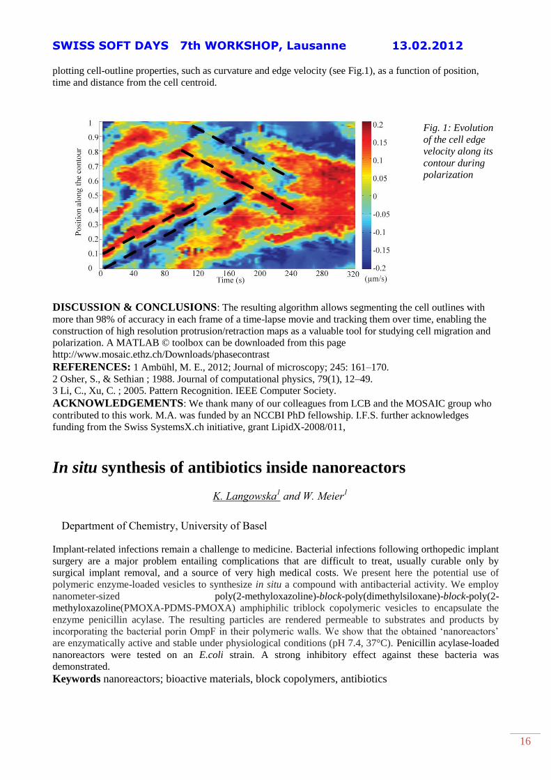
plotting cell-outline properties, such as curvature and edge velocity (see Fig.1), as a function of position,
time and distance from the cell centroid.
Fig. 1: Evolution
of the cell edge
velocity along its
contour during
polarization
DISCUSSION & CONCLUSIONS: The resulting algorithm allows segmenting the cell outlines with
more than 98% of accuracy in each frame of a time-lapse movie and tracking them over time, enabling the
construction of high resolution protrusion/retraction maps as a valuable tool for studying cell migration and
polarization. A MATLAB © toolbox can be downloaded from this page
http://www.mosaic.ethz.ch/Downloads/phasecontrast
REFERENCES: 1 Ambühl, M. E., 2012; Journal of microscopy; 245: 161–170.
2 Osher, S., & Sethian ; 1988. Journal of computational physics, 79(1), 12–49.
3 Li, C., Xu, C. ; 2005. Pattern Recognition. IEEE Computer Society.
ACKNOWLEDGEMENTS: We thank many of our colleagues from LCB and the MOSAIC group who
contributed to this work. M.A. was funded by an NCCBI PhD fellowship. I.F.S. further acknowledges
funding from the Swiss SystemsX.ch initiative, grant LipidX-2008/011,
In situ synthesis of antibiotics inside nanoreactors
K. Langowska1 and W. Meier
1
Department of Chemistry, University of Basel
Implant-related infections remain a challenge to medicine. Bacterial infections following orthopedic implant
surgery are a major problem entailing complications that are difficult to treat, usually curable only by
surgical implant removal, and a source of very high medical costs. We present here the potential use of
polymeric enzyme-loaded vesicles to synthesize in situ a compound with antibacterial activity. We employ
nanometer-sized poly(2-methyloxazoline)-block-poly(dimethylsiloxane)-block-poly(2-
methyloxazoline(PMOXA-PDMS-PMOXA) amphiphilic triblock copolymeric vesicles to encapsulate the
enzyme penicillin acylase. The resulting particles are rendered permeable to substrates and products by
incorporating the bacterial porin OmpF in their polymeric walls. We show that the obtained ‘nanoreactors’
are enzymatically active and stable under physiological conditions (pH 7.4, 37°C). Penicillin acylase-loaded
nanoreactors were tested on an E.coli strain. A strong inhibitory effect against these bacteria was
demonstrated.
Keywords nanoreactors; bioactive materials, block copolymers, antibiotics
SWISS SOFT DAYS 7th WORKSHOP, Lausanne 13.02.2012
17
Orthogonal Particle-Height and Density Gradients for
Biomedical Applications
Clément Cremmel, Christian Zink, Lucy Y. Clasohm, Doris M. Spori, Lucio Isa & Nicholas D.
Spencer
ETH – Zurich, Laboratory for Surface Science and Technology, Zürich (Switzerland)
The osseointegration of titanium implants is known to be highly influenced by their surface roughness. This
surface property has a significant influence on cell behaviour such as adhesion, proliferation and
differentiation. Roughness gradients offer an ideal high-throughput solution in order to investigate the effect
of roughness on cell attachment1.
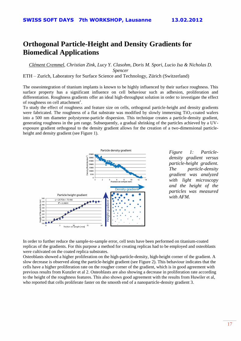
To study the effect of roughness and feature size on cells, orthogonal particle-height and density gradients
were fabricated. The roughness of a flat substrate was modified by slowly immersing TiO2-coated wafers
into a 500 nm diameter polystyrene-particle dispersion. This technique creates a particle-density gradient,
generating roughness in the µm range. Subsequently, a gradual shrinking of the particles achieved by a UV-
exposure gradient orthogonal to the density gradient allows for the creation of a two-dimensional particle-
height and density gradient (see Figure 1).
Figure 1: Particle-
density gradient versus
particle-height gradient.
The particle-density
gradient was analyzed
with light microscopy
and the height of the
particles was measured
with AFM.
In order to further reduce the sample-to-sample error, cell tests have been performed on titanium-coated
replicas of the gradients. For this purpose a method for creating replicas had to be employed and osteoblasts
were cultivated on the coated replica substrates.
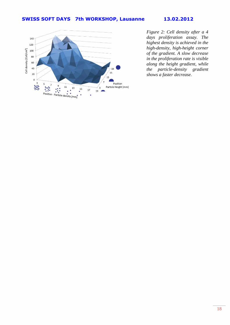
Osteoblasts showed a higher proliferation on the high-particle-density, high-height corner of the gradient. A
slow decrease is observed along the particle-height gradient (see Figure 2). This behaviour indicates that the
cells have a higher proliferation rate on the rougher corner of the gradient, which is in good agreement with
previous results from Kunzler et al 2. Osteoblasts are also showing a decrease in proliferation rate according
to the height of the roughness features. This also shows good agreement with the results from Huwiler et al,
who reported that cells proliferate faster on the smooth end of a nanoparticle-density gradient 3.
SWISS SOFT DAYS 7th WORKSHOP, Lausanne 13.02.2012
18
Figure 2: Cell density after a 4
days proliferation assay. The
highest density is achieved in the
high-density, high-height corner
of the gradient. A slow decrease
in the proliferation rate is visible
along the height gradient, while
the particle-density gradient
shows a faster decrease.