sustained attentional states require distinct temporal ... · omission rates (passetti et al.,...
TRANSCRIPT
ORIGINAL RESEARCHpublished: 31 August 2016
doi: 10.3389/fncir.2016.00070
Frontiers in Neural Circuits | www.frontiersin.org 1 August 2016 | Volume 10 | Article 70
Edited by:
Michael M. Halassa,
New York University, USA
Reviewed by:
Torfi Sigurdsson,
Goethe University Frankfurt, Germany
Jason Tucciarone,
Stony Brook Medicine, USA
*Correspondence:
Huibert D. Mansvelder
†These authors have contributed
equally to this work.‡Shared senior authorship.
Received: 14 June 2016
Accepted: 12 August 2016
Published: 31 August 2016
Citation:
Luchicchi A, Mnie-Filali O, Terra H,
Bruinsma B, de Kloet SF,
Obermayer J, Heistek TS, de Haan R,
de Kock CPJ, Deisseroth K, Pattij T
and Mansvelder HD (2016) Sustained
Attentional States Require Distinct
Temporal Involvement of the Dorsal
and Ventral Medial Prefrontal Cortex.
Front. Neural Circuits 10:70.
doi: 10.3389/fncir.2016.00070
Sustained Attentional States RequireDistinct Temporal Involvement of theDorsal and Ventral Medial PrefrontalCortexAntonio Luchicchi 1, Ouissame Mnie-Filali 1 †, Huub Terra 1†, Bastiaan Bruinsma 1 †,
Sybren F. de Kloet 1 †, Joshua Obermayer 1, Tim S. Heistek 1, Roel de Haan 1,
Christiaan P. J. de Kock 1, Karl Deisseroth 2, Tommy Pattij 3‡ and Huibert D. Mansvelder 1*‡
1Department of Integrative Neurophysiology, Center for Neurogenomics and Cognitive Research, VU University Amsterdam,
Amsterdam, Netherlands, 2Department of Bioengineering, Stanford University, Stanford, CA, USA, 3Department of Anatomy
and Neurosciences, VU University Medical Center, Amsterdam, Netherlands
Attending the sensory environment for cue detection is a cognitive operation that occurs
on a time scale of seconds. The dorsal and ventral medial prefrontal cortex (mPFC)
contribute to separate aspects of attentional processing. Pyramidal neurons in different
parts of the mPFC are active during cognitive behavior, yet whether this activity is causally
underlying attentional processing is not known. We aimed to determine the precise
temporal requirements for activation of the mPFC subregions during the seconds prior
to cue detection. To test this, we used optogenetic silencing of dorsal or ventral mPFC
pyramidal neurons at defined time windows during a sustained attentional state. We find
that the requirement of ventral mPFC pyramidal neuron activity is strictly time-locked to
stimulus detection. Inhibiting the ventral mPFC 2 s before or during cue presentation
reduces response accuracy and hampers behavioral inhibition. The requirement for
dorsal mPFC activity on the other hand is temporally more loosely related to a preparatory
attentional state, and short lapses in pyramidal neuron activity in dorsal mPFC do not
affect performance. This only occurs when the dorsal mPFC is inhibited during the entire
preparatory period. Together, our results reveal that a dissociable temporal recruitment
of ventral and dorsal mPFC is required during attentional processing.
Keywords: attention, dorsomedial prefrontal cortex, ventromedial prefrontal cortex, optogenetics, pyramidal
neurons
INTRODUCTION
The medial prefrontal cortex (mPFC) plays a crucial role in several cognitive functions, amongwhich attentional processes (Dalley et al., 2004). Pharmacological and lesion studies in rodentsperforming in different visual attention probing paradigms, including the 5-choice serial reactiontime task (5-CSRTT) (Olton et al., 1988;Muir et al., 1996; Granon et al., 1998; Broersen andUylings,1999; Robbins, 2002; Kahn et al., 2012), have shown that deactivation of the mPFC impairs rodentperformance (Muir et al., 1996). Furthermore, more detailed investigations have pointed towarda functional diversity in the management of various visuospatial attention-related functions bydifferent mPFC areas (Passetti et al., 2002; Dalley et al., 2004). Along the dorsomedial-ventromedialaxis of the PFC, the most dorsal subregions (including anterior cingulated cortex, ACg) might

Luchicchi et al. Medial Prefrontal Cortex in Attention
more prominently participate in sustained attentional states,controlling accuracy of responding to light cues as well asomission rates (Passetti et al., 2002; Dalley et al., 2004), whereasthe ventral stations (prelimbic and infralimbic cortices) mightbe more involved in executive functions such as inhibition ofinappropriate responses and behavioral flexibility (Chudasamaand Muir, 2001; Passetti et al., 2002; Chudasama et al., 2003).
Pharmacological interventions and lesions of brain regionsinterfere with brain function on a time scale of hours to weeks,thereby exceeding the time scale of attentional processing. Whenan organism pays attention to its sensory environment foraccurate detection of sensory cues in demanding tasks, attention-related neuronal activity typically occurs on a time scale ofseconds (Totah et al., 2009, 2013; Donnelly et al., 2015; Kimet al., 2016). During these seconds of changed neuronal activity,both the ACg and the ventral regions of the mPFC processinformation to prepare the organism to respond to a stimulus(Totah et al., 2009). It was shown recently that activity of fast-spiking parvalbumin-containing interneurons in the mPFC isrequired for attentional processing, since optogenetic inhibitionof these neurons on a seconds time-scale increases errors inperformance (Kim et al., 2016). In addition, it has been reportedthat mPFC GABA interneurons might be crucially involved inthe modulation of executive functions (Cho et al., 2015). Despitethis, it is unknown how activity of pyramidal neurons in specificsubcompartments of the mPFC is causally related to attentionalprocessing in the seconds that precede the cue presentation aswell as in the actual period of instrumental action, when rodentshave to produce an adaptive response to the stimulus.
Pyramidal neurons represent 80–90% of cells in the mPFC(Riga et al., 2014) and their laminar organization renders theirrole in complex cognitive functions difficult to disentangle.For example, it has been shown that while superficial layerpyramidal neurons send their projections mainly intracortically,deep layer cells (among which those residing in layer V-VI) sendefferent connection to subcortical and limbic structures (Douglasand Martin, 2004). Notably, layer V-VI cells in the mPFC arealso strongly interconnected with the mediodorsal thalamus(Gabbott et al., 2005), a crucial region for the modulation ofcognitive flexibility (Parnaudeau et al., 2015) and attention-related functions (Chudasama and Muir, 2001).
Due to the importance of pyramidal neurons in attentionalprocessing, we addressed here the temporal requirements foractivation of pyramidal neurons in the dorsomedial PFC(DmPFC, encompassing the ACg and the dorsal portion ofthe PL) and ventromedial PFC (VmPFC, centered in theborder between the ventral part of PL and the dorsal IL) inrats performing in the 5-CSRTT. Since attention is a multi-dimensional construct, this task assesses aspects of a sustainedvisuospatial attentive state by testing the ability to monitor 5different spatial locations over an extensive amount of trials. Inaddition, the task also provides information on other behavioralfunctions such as motivation, motor behavior, inhibitory control,decision-making strategies and timing (see for review Robbins,2002). Using the 5-CRSTT, we tested whether the involvementof DmPFC and VmPFC excitatory cells was required duringspecific phases of preparatory attentional states, or whether these
two subcompartments modulate this function at different time-scales and epochs. By optogenetic silencing of either DmPFCor VmPFC pyramidal neurons (Yizhar et al., 2011) at definedtime windows of a few seconds prior and during cue detection,we find that pyramidal neuron activity in DmPFC and VmPFCshows distinct temporal requirements during early and latephases of preparatory sustained attentional states, and duringcue detection/instrumental action. These findings help to betterdisentangle the intricate network activity of the mPFC duringcomplex cognitive tasks, providing a temporal view on mPFCactivity requirements for adaptive and maladaptive behaviors.
MATERIALS AND METHODS
AnimalsAll experimental procedures were in accordance with Europeanand Dutch law and approved by the animal ethical carecommittee of the VU University and VU University MedicalCenter. Male Long Evans wild-type rats (Janvier Labs, France;8–10 weeks old at the start of the experiments) were usedfor all the experiments. Rats were individually housed on a12 h light/dark reversed cycle (lights OFF: 7 a.m.). Only whenassigned to behavioral experiments rats were food deprived. Foodrestriction began 1 week before the initiation of operant trainingin order to achieve and maintain about 85–90% of the free-feeding body weight. Water was provided ad libitum. In total 31rats were included in this study (29 for behavioral testing and 2for structural imaging).
Opsin Virus Delivery and Implantation ofOptic FibersCaMKIIα promoter-driven opsin pAAV-enhancedhalorhodopsin (eNPHR3.0)::eYFP, pAAV-enhancedarchaerhodopsin (eARCH3.0)::eYFP and pAAV::eYFP werepackaged as AAV serotype 2 virus (titer 1.0–6.0 × 10¹²). Ratswere anesthetized with isoflurane (2.5%) and then mountedin a stereotaxic frame (Kopf instruments, Tujunga, USA).The skin of the scalp was retracted and 2 holes were drilledat the level of the medial prefrontal cortex (mPFC). Stainlesssteel micro-needles connected to a syringe (Hamilton, USA)were inserted at the desired coordinates to deliver the virusin the brain. For the DmPFC group, injections were made atAP+2.76mm; ML ±1.49mm; DV −2.94 and −2.84mm frombregma (infusion angle 10◦), while for the VmPFC group atAP+2.76mm; ML ±1.45mm; DV −4.87 and −4.77mm fromskull (10◦ infusion angle) (Paxinos and Watson, 2007). Onemicroliter virus was injected per hemisphere in two steps of 500nL at an infusion rate of 6 µL/h. A total of 8 rats were injectedwith AAV2-eNPhR3.0::EYFP, 13 with AAV2-eARCH3.0::EYFPand 8 with AAV2::EYFP. 14 rats in total were injected in theDmPFC and 15 rats were injected in the VmPFC (includingcontrol rats).
Then, 2 guide screws and 2 chronic implantable glass fibers(200 µm diameter, 0.20 numerical aperture, ThorLabs, Newton,NJ, USA) mounted in a sleeve (1.25mm diameter; ThorLabs,Newton, NJ, USA) were placed in the rat brain. The fibers wereimplanted right on top of the viral injection location (200–300
Frontiers in Neural Circuits | www.frontiersin.org 2 August 2016 | Volume 10 | Article 70

Luchicchi et al. Medial Prefrontal Cortex in Attention
µm on average). Finally, a double component dental cement(Pulpdent©, Watertown, USA) mixed with black carbon powder(Sigma Aldrich, USA) was used in order to secure the opticfibers. All the surgical manipulations were performed before thebehavioral training and testing.
Behavioral ProceduresAfter 1 week of recovery from surgery and 1 week of habituationin the reverted light/dark cycle, rats started training in the 5-CSRTT in operant cages (Med Associates Inc., St. Albans, VT,USA). Training consisted of a period during which rats learnedto respond to a brief visual cue that was randomly lit in one outof the five apertures of the operant cage (Bari et al., 2008). Toassociate cue with the delivery of reward rats were first trainedwith all the apertures illuminated (all holes on, Figure 3B)in order to learn that a nose-poke returns a food pellet andsubsequently with only one aperture constantly illuminated (onehole on, Figure 3B) to learn responding into this illuminatedaperture is associated with reward delivery. After the learningphase, titration of shortening the stimulus duration was basedon individual performance of each rat, and was reduced from 16to 1 s. Criteria to move to a shortened stimulus duration werethe percentage of accuracy (>80%) and omitted trials (<20%).Finally, when rats met the criteria at 1 s stimulus duration theywere moved to the pretesting phase. In the pretesting phase, agreen custom-made LED replaced the normal house-light of theoperant cages, (<1mW intensity) to mask reflections by the laserlight used for the experiments. The LED house-light did not affectperformance when compared to normal house-light.
After three consecutive sessions during which rats performedaccording to the aforementioned criteria with the LED on,additional baseline sessions were conducted (3 consecutivesessions). During these sessions subjects were connected to thepatch-cable (Doric Lenses, Quebec city, Canada) used to deliverthe light into the brain. In this condition, accuracy was typicallyabove 80%. However, they often did not show less than 20%omissions. This was most likely due to the fact that the animalswere connected to the optic fiber patch cable and therefore lessfree to move in combination with the short time window for theanimal to respond (i.e., within 2 s after the cue light went off).This parameter makes the paradigmmore demanding than otherversions of the 5-CSRTT in which response time is usually set to5 s (Passetti et al., 2002). Therefore, the omission criterion wasincreased to less than 40% omissions.
After acquisition of baseline rats were assigned to the testingphase where the task comprised 100 consecutive trials with arandom assignment to the condition of laser ONor laser OFF (seebelow). In the whole text we refer to completed trials (correct,incorrect, omissions) while in the 100 trials premature responsesare left apart from the count.
To light-activate the opsins in vivo, we used a diode-pumped laser (532 nm, Shanghai Laser and Optics CenturyCo, China) directly connected to the rat optic glass fiberimplant. Light was delivered at 9–12mW for experimentsperformed with eNPhR3.0 and at 7–8mW for experimentscarried out with eARCH3.0. These stimulation regimensare able to produce a theoretical irradiance which ranges
between 9.76 and 13.01 mW/mm2 500 µm from the fiber tipfor the eNPhR3.0 experiments (corresponding to the centerof the viral transfection) and ranging between 7.59 and8.68mW/mm2 for eARCH3.0 experiments (http://web.stanford.edu/group/dlab/cgi-bin/graph/chart.php).
Light was delivered according to scheduled epochs by astimulator (master 9, AMPI Jerusalem, Israel) connected to thecomputer interface.
For the testing phase, the following parameters have beenacquired and analyzed through a box-computer interface (Med-PC, USA) and custom written MATLAB scripts (Mathworks):accuracy on responding to cues (ratio between the number ofcorrect responses per session over the sum between correct andincorrect hits, expressed as percentage); absolute and percentageof correct, incorrect responses and errors of omission; corrector incorrect response latency; latency to collect reward; numberof premature and perseverative responses. Percent of correct,incorrect and omissions were calculated based on the number ofstarted trials (Semenova et al., 2007).
In line with previous studies (Pinto et al., 2013), nodifferences were found in behavioral effects of eARCH3.0 andeNPhR3.0 injected animals (data not shown). Therefore, datafrom eARCH3.0 and eNPhR3.0 injected animals were pooled.
Optical Inhibition ProtocolsRats were randomly assigned to different stimulation protocolsand received different optical inhibition epochs. Opticalinhibition sessions were done 2–3 times a week with a baselinesession in between to control for potential carry-over effects.Rats were tested according to the following optical inhibitionprotocols: (a) 3 s at the trial onset, (b) 2 s at the end of thepreparatory period of a sustained attentional state, (c) 5 sthroughout the whole preparatory period, (d) 1 s during light cuepresentation. During a session, animals received only one lightstimulation protocol. We chose these light regimens to makea clear distinction between prestimulus period and stimuluspresentation/instrumental response period (Totah et al., 2013)(protocol a, b, and c vs. protocol d) and to differentiate betweenthe whole pre-cue period and the period which consists in theactual orienting activity of the rat toward the task ports (Totahet al., 2009, 2013; Donnelly et al., 2015) (protocol c vs. protocolb). Light-ON and light-OFF trials were assigned semi-randomlywith approximately 50% ON trials and 50% OFF trials. Themajority of animals (28 out of 29) completed 100 trials within thefirst 20–25min. One animal did not complete 100 trials beforethe time cut off of 60min.Whereas animals were tested in all fourdifferent optical inhibition protocols, in some rats due to fiberloss not all protocols could be completed. Moreover, reporteddata for the majority of rats refer to the first optical inhibitionsession after establishment of stable baseline performance. Insome cases, as described below, rats were retested in the sameoptical inhibition session.
Exclusion CriteriaSingle sessions were excluded from analysis when technicalproblems (i.e., patch-cables disconnected during the task) madethe results unreliable. In all these cases, we repeated the same
Frontiers in Neural Circuits | www.frontiersin.org 3 August 2016 | Volume 10 | Article 70
Luchicchi et al. Medial Prefrontal Cortex in Attention
protocol after re-acquisition of baseline criteria and used datafrom these sessions.
Histological VerificationAfter behavioral testing, brains were checked for fiber placementand viral expression. For this, rats were anesthetized withisoflurane and a mix of ketamine (200mg/kg i.p.) and dormitol(100mg/kg i.p.) and then transcardially perfused (50–100mLNaCl and 200–400 mL PFA 4%). Brains were removed andmaintained in 4% PFA for at least 24 h. After that, brains weresliced with a vibratome (Leica Biosystem, Germany) into 50–100µm coronal sections and mPFC slices were mounted on glassslides covered by 2% Mowiol and anti-fading mounting covers.Images were taken with a confocal microscope (LSM 510 Meta;Zeiss, Germany) with excitation wavelength of 514 nm bandpassfiltered between 530 and 600 nm, and further analyzed usingImageJ (NIH, USA).
In vitro Physiological RecordingsFollowing behavioral testing, five rats (by that time 8–10 monthsold) were used for electrophysiological recordings. Animals wereanesthetized with 5% isoflurane and an i.p. injection of 0.1ml/gPentobarbital and subsequently perfused with 35ml of ice-cold N-Methyl-D-glucamin solution (NMDG solution; in mM:NMDG 93, KCl 2.5, NaH2PO4 1.2, NaHCO3 30, HEPES 20,Glucose 25, NAC 12, Sodium ascorbate 5, Sodium pyruvate 3,MgSO410, CaCl2 0.5, at pH 7.4 adjusted with 10M HCl). Afterdecapitation the brain was removed and incubated for 10 minin ice-cold NMDG solution. Coronal mPFC slices (350 µm)were made in ice-cold NMDG solution and incubated afterwardsfor 3 min in 34◦C NMDG solution. Slices were maintained inan incubation chamber for at least 1 h before recordings wereconducted at room temperature in oxygenated holding solutioncontaining the following (Holding solution; in mM): NaCl 92,KCl 2.5, NaH2PO4 1.2, NaHCO3 30, HEPES 20, Glucose 25,NAC 1, Sodium ascorbate 5, Sodium pyruvate 3, MgSO4 0.5,CaCl2 1M.
Whole-cell recordings from pyramidal neurons were made at32◦C in oxygenated artificial cerebrospinal fluid (ACSF; in mM:NaCl 125, KCl 3, NaH2PO4 1.25,MgSO4 1, CaCl2 2, NaHCO3 26,Glucose 10). For recordings a potassium-based internal solutionwas used (in mM: K-gluconate 135, NaCl 4, Hepes 10, Mg-ATP2,K2Phos 10, GTP 0.3, EGTA 0.2) with patch-pipettes that had aresistance of 3–6 M�. Recorded neurons were kept at a holdingpotential close to−70mV.
For recordings Multiclamp 700/B amplifiers (MolecularDevices) were used and data was collected with a sampling rateof 10 kHz and low-pass filtering at 3 kHz (Axon Digidata 1440Aand pClamp 10 software; Molecular Devices).
Optogenetic Slice StimulationTo optically activate opsins, green light (530 nm) was appliedto the slices. Light pulses were evoked by using a DC4100 4-channel LED-driver (Thorlabs, Newton, NJ) or a Fluorescencelamp (X-Cite Series 120q, Lumen Dynamics). During recordingsfifty sweeps, each 10 s apart were applied. One sweep consists of asingle light pulse with a duration of 1 or 5 s. These pulse regimes
represent the shortest and the longest stimulation protocol usedfor behavioral experiments, respectively. The intensity of the lightsource was adjusted to 1.7, 3, 7, or 17mW. For recording thein/output curves 1 s light pulse with all different stimulationintensities were applied for five sweeps with an interval of 10 s.
Statistical Analyses for BehavioralExperimentsTo evaluate the main behavioral data between the opsingroup and eYFP control group, two-way ANOVAs for repeatedmeasures were performed. Corrected values for multiplecomparison with Sidak’s test were used when interaction betweenlight and virus was significant. In all cases, the ANOVAs werepreceded by the Kolmogorov-Smirnov (KS) test for normaldistribution. In cases when the KS p-value was >0.05, factorialanalysis was performed on the raw data per parameter. In theother cases, raw data were first transformed with square-root orarcsin transformation.
Data were analyzed by MATLAB 2014a (Mathworks),Microsoft Excel (Office) and graphs were plotted by GraphPadPrism. In all cases the significance level was p < 0.05.
RESULTS
To express inhibitory opsins in excitatory pyramidal neuronsof either DmPFC or VmPFC, we used an AAV2 plasmidcontaining the CamkIIα promoter driving expression of eitherarchaerhodopsin (eARCH3.0) or halorhodopsin (eNPHR3.0)and eYFP (Yizhar et al., 2011). For the control group we injectedthe same virus with eYFP only (Figure 1A). Injections in theDmPFC targeted the border between the ventral part of thepregenual anterior cingulated cortex (ACg) and the dorsal partof the prelimbic cortex (PL), whereas VmPFC viral infusionstransfected neurons in the ventral PL and the dorsal infralimbiccortex (IL) (Figures 1B,C). In both cases AAV2 injectionsprimarily targeted the deep layers (layer V-VI) of the mPFC(Figure 1D). Same pattern was revealed in rats dissected after 5-CSRTT experiments (Figures 1E,F), where also fiber placementin both the Dm- and the VmPFC was mainly located in thearea ranging from layer V to layer VI (Figure 1G). Whole-cellpatch clamp recordings performed in rats that previously weretested in the 5-CSRTT, confirmed the correct expression of theinhibitory opsins eNPHr3.0 or eARCH3.0 in pyramidal cells.Brief light pulses of similar length as used for the behavioralexperiments (1 or 5 s; 530 nm) triggered after 50 consecutiverepetitions a marked hyperpolarization response in the recordedcells (Figures 2A–D). Hyperpolarization remained stable acrossthe different trials (Figures 2C,D), with a slight reduction (about20%) when light was consecutively delivered at the durationof 5 s (Figure 2D). In addition, input/output curves confirmedthat: (a) light manipulation of pyramidal neurons was intensity-dependent, with stronger hyperpolarization following higherlight intensity and that (b) also the lowest light intensity (1.3mW)produced a sustained hyperpolarization of the cells (Figure 2E).We did not observe rebound action potentials following light-induced inhibition.
Frontiers in Neural Circuits | www.frontiersin.org 4 August 2016 | Volume 10 | Article 70
Luchicchi et al. Medial Prefrontal Cortex in Attention
FIGURE 1 | Viral expression in rats injected with AAV2-eYFP, AAV2-eNPHR3.0, and AAV2-eARCH3.0 and optical fiber location to achieve selective
illumination of either Dm- or VmPFC. (A) Schematic representation of the viruses used to achieve expression of inhibitory opsins and eYFP in either DmPFC or
VmPFC. (B) Graphic representation of the injections made in either the DmPFC or the VmPFC to test the spread of transfection of the virus in both regions (C)
Overview (zoom 10×) of injection location in both the DmPFC (left panel) and the VmPFC (right panel). In this figure animals were injected with AAV2-eYFP::CamkIIα.
Scale bar is 1 mm for both pictures. (D) Magnified (zoom 40×) confocal picture reporting an example of the transfected neurons by using the same viral plasmid used
for the behavioral experiments. White dotted lines illustrate the empirical differentiation between the different mPFC layers, indicating that the majority of the
transfected cells were in the deep-layers with a reduced amount in the upper-layers. Scale bar is 200 µm. Also in this example viral infusions were made using
AAV2-eYFP::CamkIIα. (E) Visual identification of the virus spread in a sample of rats previously used to perform behavioral experiments and injected with either
AAV2-eNPHR3.0-eYFP::CamkIIα or AAV2-eARCH3.0-eYFP::CamkIIα. Dark green wider circles represent the maximal expression achieved, while light green small
shapes report the smallest expression detected (n = 10 in total). Confocal pictures of exemplificative images in this batch are reported in (F) (scale bar is 500 µm for
both images). In this examples rats were injected with AAV2-eNPHR3.0-eYFP::CamkIIα. (G) Visual identification of fiber placement in a sample of rats previously used
for 5-CSRTT experiments and injected with either AAV2-eNPHR3.0-eYFP:: CamkIIα or AAV2-eARCH3.0-eYFP::CamkIIα. Inset reports an example of the fiber location
in the mPFC (scale bar is 500 µm) in a rat injected with AAV2-eARCH3.0-eYFP::CamkIIα. Blue asterisks are referred to optic fibers located to achieve regional
inhibition in the VmPFC, while red asterisks report the same fiber placement in the DmPFC (n = 12 in total).
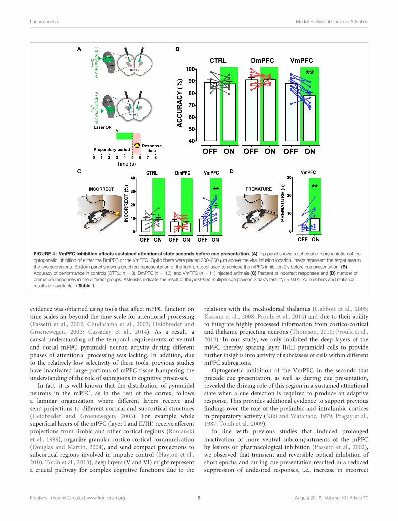
Transient mPFC Inhibition Immediatelybefore and during Cue PresentationTo address whether a reversible inactivation of pyramidal neuronactivity in either Dm or VmPFC affects rodent performance atspecific time points during a preparatory attentional state, wetrained rats in the 5-CSRTT (Figure 3A) and tested the effectof subregion-specific deactivation during precise time-windowsin the task (see methods). Neither training [two-way ANOVA,effect of interaction group x protocol: F(12, 156) = 0.992; p =
0.452; effect of group: F(2, 26) = 0.684; p = 0.513; Figure 3B],nor baseline performance differed between groups [Accuracy:one-way ANOVA: F(2, 28) = 1.607; p = 0.220; omissions: one-way ANOVA: F(2, 28) = 0.117; p = 0.893; Figure 3C]. Duringthe preparatory period, when the animal is actively attendingthe cue-holes, single-units in the ACg and PL area show atransient pre-cue increase in firing rate (Totah et al., 2009).However, it is not known whether this activity causally drives
a sustained attentional state. To test whether increased activityduring this period in either DmPFC or VmPFC is requiredfor proper performance, pyramidal neurons in either of thesesubregions were inhibited by light for 2 s prior to cue presentation(Figure 4A), during the time window that represents the actualperiod when the rat orients and actively awaits the upcomingstimulus, before it is required to produce a response to thecue (Totah et al., 2013). Only inhibition of VmPFC pyramidalneurons resulted in a reduction of accuracy of responding [two-way repeatedmeasures ANOVA: effect of light x virus interaction:F(2, 26) = 5.984; p = 0.007; effect of virus: F(2, 26) = 6.154;p = 0.006; effect of light: F(1, 26) = 4.175; p = 0.051; Sidak’smultiple comparison test OFF vs. ON: CTRL: p= 0.965; DmPFC:p = 0.854; VmPFC: p = 0.001; Figure 4B]. This effect wasprimarily due to an increase in the percentage of incorrectresponses [two-way repeated measures ANOVA: effect of lightx virus interaction: F(2, 26) = 4.115; p = 0.028; Sidak’s multiple
Frontiers in Neural Circuits | www.frontiersin.org 5 August 2016 | Volume 10 | Article 70
Luchicchi et al. Medial Prefrontal Cortex in Attention
FIGURE 2 | Correct incorporation of inhibitory opsins in pyramidal cells. (A) Trace showing a typical eARCH3.0-mediated voltage waveform in a layer V
pyramidal neuron in response to green light (530 nm, 1 s, 7 mW). (B) Schematic representation of recording configuration in mPFC coronal slices of a rat. White dotted
lines represent the borders of the mPFC. Scale bar is 200 µm. (C) Top panel shows characteristic voltage waveforms monitored in response to one green light pulse
(1 s duration: n = 14) in a layer VI pyramidal neuron transfected with the AAV2-eARCH3.0::eYFP. Bottom panel graph reports the normalized hyperpolarization
amplitude of each trial (50 trials, 1 s light pulse, repeated each 10 s, 7 mW light intensity). All responses were normalized to the maximal amplitude of the first response
(graph report values as mean ± S.E.M.). (D) top and bottom panels report the same example and analysis showed in (C) with a longer light pulse (5 s; n = 13). (E)
Example traces show that pyramidal neurons responded to light pulses in an intensity-dependent fashion, with more pronounced hyperpolarization following higher
light intensities (top panel). Bottom panel shows an input/output curve for different light intensities (n = 11 neurons, data are reported as mean ± S.E.M.). Percentage
of hyperpolarization: 1.7 mW = 49.28 ± 4.09%; 3 mW = 63.39 ± 4.377%; 7 mW = 80.11 ± 3.812%, Data are normalized in each cell to the maximal response
(evoked by a 17 mW light pulse). Average amplitude at 17 mW light pulses is −23.464 ± 3.361mV (n = 22; data are reported as mean ± S.E.M.).
comparison test OFF vs. ON: CTRL: p = 0.952; DmPFC: p =
0.999; VmPFC: p = 0.002; Figure 4C], and accompanied by anincrease in premature responses (Wilcoxonmatched-pairs signedrank test; p= 0.008; Figure 4D). Inhibition of pyramidal neuronsin the DmPFC 2 s prior to cue presentation did not affect anyparameter of performance in the 5-CSRTT (Figure 4B, Table 1).These results suggest that a reduction in accurate respondingmight be due to the reduced ability to control inappropriateresponses when VmPFC activity is inhibited for 2 s before cuepresentation.
We next tested whether pyramidal neuron activity of theVmPFC or DmPFC is necessary during cue presentation fora proper sustained attentional state. Inhibition of VmPFCpyramidal neurons during cue presentation resulted in areduction of the accuracy of responding [two-way repeatedmeasures ANOVA: effect of light x virus interaction: F(2, 14) =
4.393; p= 0.033; effect of virus: F(2, 14) = 1.864; p= 0.192; effectof light: F(1, 14) = 6.273; p = 0.025; Sidak’s multiple comparisontest OFF vs. ON: CTRL: p = 0.270; DmPFC: p = 0.826; VmPFC:p = 0.014; Figures 5A,B]. This effect was due to an increase of
incorrect responses and a decrease in correct responses [two-way repeated measures ANOVA: effect of interaction light x viruscorrect: F(2, 14) = 5.535; p = 0.017; Sidak’s multiple comparisontest OFF vs. ON: CTRL: p = 0.494; DmPFC: p = 0.524; VmPFC:p = 0.013; incorrect: effect of interaction light x virus: F(2, 14) =3.809; p = 0.048; Sidak’s multiple comparison test OFF vs. ON:CTRL: p = 0.304; DmPFC: p = 0.714; VmPFC: p = 0.044;Figures 5C,D]. Also in this case, inhibition of DmPFC pyramidalneurons during cue presentation did not affect any parameterof performance (Figure 5B, Table 1). Thus, pyramidal neuronactivity in the VmPFC is required during the preparatory phase,2 s before cue presentation as well as during cue presentationitself, when rats are requested to prepare cue detection and totranslate this into an instrumental response.
Sustained Inhibition of mPFC during aPreparatory Sustained Attentional StateIs the DmPFC causally involved in a sustained attentional stateat these second time scales (Chudasama et al., 2003; Dalley
Frontiers in Neural Circuits | www.frontiersin.org 6 August 2016 | Volume 10 | Article 70

Luchicchi et al. Medial Prefrontal Cortex in Attention
FIGURE 3 | 5-CSRTT: protocols, training and baseline performance. (A)
After stable baseline performance (BAS) for three consecutive sessions rats
were assigned to the testing phase. Colored squares in the top-right panel
represent the different light epochs of stimulation used. Numbers represent the
length of the stimulation per session. White squares in between the stimulation
days represent a baseline session when no light was delivered in the brain.
Bottom-right panel represents a schematic picture of a single trial of the task.
The first 5 s reported in the x axis shows the preparatory period of sustained
attentional state, the light brown period (5th to 6th s in the x axis) refers to the
presentation of the cue, and the last 2 s represent the limited hold period.
Colored dots represent the possible responses that were recorded during the
session. Responses before cue presentation were considered as premature
and punished with a 5 s time-out period. Correct responses were rewarded
with a food pellet, whereas incorrect pokes were punished with a time-out
period. If a response did not occur within the limited hold period, an omitted
trial was recorded. Green lines represent the different light epochs (see
methods). Left panel reports a representative illustration of a rat performing in
the 5-CSRTT. Rats are bilaterally connected via patch cables to a laser, which
delivers (ON) or does not deliver (OFF) light in the desired epoch. The
percentage of trials with light ON and OFF was approximately fifty for both
options. (B) Illustration of the number of sessions within each training phase
and stimulus duration of the task for the three different groups of rats included
in the study (CTRL: n = 8; DmPFC: n = 10; VmPFC: n = 11; data are
expressed as mean ± S.E.M.). (C) Graphs illustrating the averaged baseline
with cables in accuracy and omissions for the 3 groups. Results are expressed
as mean ± S.E.M.
et al., 2004; Totah et al., 2009)? To test whether activity of theVmPFC or DmPFC is required earlier in the task to guide asustained attentional state, we inhibited pyramidal neurons ineither the dorsal or the ventral mPFC for 3 s starting 5 s before cuepresentation during the early phases of the preparatory sustainedattentional state (Figure 6A). Optogenetic inhibition of VmPFCor DmPFC pyramidal neurons during this period did not affectany of the behavioral parameters in the task [two-way repeatedmeasures ANOVA; effect of light x virus interaction: F(2, 14) =
0.827; p = 0.457; effect of virus: F(2, 14) = 0.514; p = 0.609;effect of light: F(1, 14) = 1.238; p = 0.285, Figure 6B, Table 1].
In contrast, a sustained inhibition of the DmPFC for 5 s duringthe entire preparatory sustained attentional state (Figure 7A)did significantly affect the rodent accuracy of responding inthe 5-CSRTT [two-way repeated measures ANOVA: effect oflight x virus interaction F(2, 22) = 11.760; p = 0.0003; effectof virus: F(2, 22) = 0.849; p = 0.441; effect of light: F(1, 22) =0.856; p= 0.365; Sidak’s multiple comparison test OFF vs. ON:CTRL: p= 0.194; DmPFC: p= 0.005; Figure 7B]. This effect wasexplained by a reduction in the percentage of correct responses, aswell as an increase in the percentage of incorrect responses [two-way repeated measures ANOVA correct: effect of interactionF(2, 22) = 14.790; p = 0.0001; Sidak’s multiple comparison testOFF vs. ON: CTRL: p = 0.991; DmPFC: p = 0.0001; incorrect:F(2, 22) = 9.199; p = 0.001; Sidak’s multiple comparison testOFF vs. ON: CTRL: p = 0.268; DmPFC: p = 0.021; Figure 7C].In addition, the response latencies for incorrect responses wassignificantly longer during ON trials, when compared to OFFtrials (OFF vs. ON= 1.30± 0.16 s vs. 1.51± 0.18 s; paired t-test:p = 0.021) suggesting that prolonged inhibition of the DmPFCmay interfere with responding to a cue.
Optical inhibition of the VmPFC during the entire 5 s ofpreparatory phase did not reduce control over a sustainedattentional state, but to our surprise, slightly improved accurateresponding, by decreasing the percentage of incorrect responses(Sidak’s multiple comparison test OFF vs. ON accuracy: p =
0.037; % incorrect: p = 0.045; Figures 7B,C) while not affectingreaction latencies for both correct and incorrect responses(Correct response latency, OFF vs. ON: 0.62 ± 0.04 vs. 0.61± 0.04; paired t-test: p = 0.749; incorrect response latency,OFF vs. ON: 1.11 ± 0.18 vs. 1.14 ± 0.10; paired t-test: p= 0.863). Nevertheless, taken together, these results show thatthe requirements for neuronal activity in the DmPFC andVmPFC during a sustained attentional state are temporallydissociated.
DISCUSSION
In this study we found that pyramidal neurons in theDmPFC and VmPFC require distinct temporal activationprofiles during a preparatory sustained attentional state. Inparticular, we found that the VmPFC plays an important rolein the seconds that immediately precede and coincide withcue presentation. Transient inhibition of VmPFC pyramidalneurons during these seconds impairs visuospatial sustainedattentional states as measured in the 5-CSRTT task and affectsvarious parameters, including premature responses. In contrast,the visuospatial sustained attentional state is less sensitive toshort inactivation of the DmPFC. Only when the DmPFCis inhibited for the entire preparatory phase before stimuluspresentation and cue detection, a reduction in the sustainedattentional state was observed. Since response latencies anderrors of omission were not altered by optogenetic silencing,the observed findings were not secondary to changes in motorperformance.
Even though a functional distinct role of different mPFC areasin cognitive functions has been previously shown, most of this
Frontiers in Neural Circuits | www.frontiersin.org 7 August 2016 | Volume 10 | Article 70
Luchicchi et al. Medial Prefrontal Cortex in Attention
FIGURE 4 | VmPFC inhibition affects sustained attentional state seconds before cue presentation. (A) Top panel shows a schematic representation of the
optogenetic inhibition of either the DmPFC or the VmPFC. Optic fibers were placed 200–300 µm above the viral infusion location. Insets represent the target area in
the two subregions. Bottom panel shows a graphical representation of the light protocol used to achieve the mPFC inhibition 2 s before cue presentation. (B)
Accuracy of performance in controls (CTRL; n = 8), DmPFC (n = 10), and VmPFC (n = 11) injected animals (C) Percent of incorrect responses and (D) number of
premature responses in the different groups. Asterisks indicate the result of the post-hoc multiple comparison Sidak’s test. **p < 0.01. All numbers and statistical
results are available in Table 1.
evidence was obtained using tools that affect mPFC function ontime scales far beyond the time scale for attentional processing(Passetti et al., 2002; Chudasama et al., 2003; Heidbreder andGroenewegen, 2003; Cassaday et al., 2014). As a result, acausal understanding of the temporal requirements of ventraland dorsal mPFC pyramidal neuron activity during differentphases of attentional processing was lacking. In addition, dueto the relatively low selectivity of these tools, previous studieshave inactivated large portions of mPFC tissue hampering theunderstanding of the role of subregions in cognitive processes.
In fact, it is well known that the distribution of pyramidalneurons in the mPFC, as in the rest of the cortex, followsa laminar organization where different layers receive andsend projections to different cortical and subcortical structures(Heidbreder and Groenewegen, 2003). For example whilesuperficial layers of the mPFC (layer I and II/III) receive afferentprojections from limbic and other cortical regions (Romanskiet al., 1999), organize granular cortico-cortical communication(Douglas and Martin, 2004), and send compact projections tosubcortical regions involved in impulse control (Hayton et al.,2010; Totah et al., 2013), deep layers (V and VI) might representa crucial pathway for complex cognitive functions due to the
relations with the mediodorsal thalamus (Gabbott et al., 2005;Kassam et al., 2008; Proulx et al., 2014) and due to their abilityto integrate highly processed information from cortico-corticaland thalamic projecting neurons (Thomson, 2010; Proulx et al.,2014). In our study, we only inhibited the deep layers of themPFC thereby sparing layer II/III pyramidal cells to providefurther insights into activity of subclasses of cells within differentmPFC subregions.
Optogenetic inhibition of the VmPFC in the seconds thatprecede cue presentation, as well as during cue presentation,revealed the driving role of this region in a sustained attentionalstate when a cue detection is required to produce an adaptiveresponse. This provides additional evidence to support previousfindings over the role of the prelimbic and infralimbic corticesin preparatory activity (Niki and Watanabe, 1979; Pragay et al.,1987; Totah et al., 2009).
In line with previous studies that induced prolongedinactivation of more ventral subcompartments of the mPFCby lesions or pharmacological inhibition (Passetti et al., 2002),we observed that transient and reversible optical inhibition ofshort epochs and during cue presentation resulted in a reducedsuppression of undesired responses, i.e., increase in incorrect
Frontiers in Neural Circuits | www.frontiersin.org 8 August 2016 | Volume 10 | Article 70

Luchicchi et al. Medial Prefrontal Cortex in Attention
TABLE 1 | Complete overview of the different parameters analyzed in the 5CSRTT under the four different light epochs.
CTRL DmPFC VmPFC
ACCURACY (%) OFF ON OFF ON OFF ON
2 s before cue
1 s during cue
First 3 s of the trial
5 s before cue
88.78 ± 2.21
87.54 ± 3.79
86.88 ± 3.02
85.39 ± 3.04
87.61 ± 1.83
82.21 ± 5.14
84.24 ± 4.79
88.58 ± 3.81
90.69 ± 1.91
86.41 ± 2.04
88.55 ± 2.19
93.21 ± 1.65
91.18 ± 1.00
90.19 ± 2.39
89.38 ± 2.12
88.26 ± 2.41*
87.6 ± 2.11
87.99 ± 2.10
89.68 ± 1.64
85.76 ± 1.19
77.93 ± 2.44*
79.09 ± 3.85*
85.23 ± 3.43
90.01 ± 1.38*
OMISSIONS (%)
2 s before cue
1 s during cue
First 3 s of the trial
5 s before cue
22.07 ± 2.98
15.69 ± 2.19
14.19 ± 1.04
19.65 ± 4.99
23.45 ± 5.55
15.13 ± 2.86
12.37 ± 1.7
22.56 ± 6.43
21.37 ± 2.91
18.4 ± 3.66
13.3 ± 3.15
24.67 ± 3.95
27.35 ± 5.14
16.7 ± 7.45
19.72 ± 5.44
29.45 ± 4.82
21.32 ± 2.7
13.2 ± 2.73
11.75 ± 1.1
15.84 ± 2.79
19.85 ± 3.91
15.5 ± 4.61
14.54 ± 1.02
16.35 ± 3.27
CORRECT (%)
2 s before cue
1 s during cue
First 3 s of the trial
5 s before cue
69.18 ± 3.16
73.50 ± 3.87
74.63 ± 3.33
68.54 ± 4.91
67.34 ± 5.54
69.74 ± 4.49
73.74 ± 4.19
68.08 ± 5.86
71.07 ± 2.37
76.53 ± 2.40
76.34 ± 2.51
69.93 ± 3.36
66.23 ± 4.55
71.92 ± 5.22
72.13 ± 5.22
61.79 ± 3.58*
69.00 ± 3.16
76.29 ± 2.53
79.21 ± 2.28
72.05 ± 2.00
62.52 ± 3.94
66.76 ± 4.55*
72.99 ± 3.72
75.27 ± 2.32
INCORRECT (%)
2 s before cue
1 s during cue
First 3 s of the trial
5 s before cue
8.74 ± 1.79
10.4 ± 3.04
11.18 ± 2.5
11.81 ± 2.73
9.21 ± 1.47
15.21 ± 4.38
13.83 ± 4.12
9.37 ± 3.16
7.55 ± 1.65
10.16 ± 2.19
10.11 ± 2.16
5.39 ± 1.38
6.62 ± 1.00
8.36 ± 1.63
8.39 ± 1.64
8.95 ± 2.29*
9.66 ± 1.76
10.51 ± 1.93
9.04 ± 1.32
12.11 ± 1.28
17.62 ± 2.1*
17.73 ± 3.59*
12.46 ± 2.78
8.63 ± 1.41*
PREMATURE (n)
2 s before cue
1 s during cue
First 3 s of the trial
5 s before cue
3.37 ± 1.12
6.6 ± 2.2
4.75 ± 2.01
4.57 ± 1.7
5.62 ± 1.67
7 ± 2.53
3.5 ± 1.94
4.28 ± 1.64
2.2 ± 0.42
3.67 ± 0.67
2.57 ± 0.89
3.4 ± 1.27
2.8 ± 0.63
4.67 ± 1.93
2.00 ± 0.95
3.6 ± 1.45
3.09 ± 0.94
5.16 ± 3.00
4.17 ± 1.35
3.37 ± 0.96
7.18 ± 1.89*
5.67 ± 1.43
4.5 ± 0.92
5.12 ± 1.27
RESPONSE TIME CORRECT (sec)
2 s before cue
1 s during cue
First 3 s of the trial
5 s before cue
0.68 ± 0.04
0.62 ± 0.03
0.63 ± 0.04
0.66 ± 0.05
0.69 ± 0.04
0.66 ± 0.01
0.65 ± 0.07
0.67 ± 0.05
0.71 ± 0.05
0.66 ± 0.04
0.64 ± 0.04
0.69 ± 0.04
0.69 ± 0.05
0.64 ± 0.04
0.64 ± 0.02
0.72 ± 0.06
0.78 ± 0.07
0.62 ± 0.03
0.61 ± 0.06
0.62 ± 0.04
0.87 ± 0.15
0.66 ± 0.05
0.61 ± 0.05
0.61 ± 0.04
RESPONSE TIME INCORRECT (sec)
2 s before cue
1 s during cue
First 3 s of the trial
5 s before cue
1.03 ± 0.18
0.81 ± 0.13
1.12 ± 0.09
1.00 ± 0.24
1.13 ± 0.12
1.04 ± 0.1
1.44 ± 0.29
0.96 ± 0.14
1.38 ± 0.18
0.91 ± 0.20
1.2 ± 0.22
1.30 ± 0.16
1.57 ± 0.23
1.08 ± 0.28
0.97 ± 0.19
1.51 ± 0.18*
1.09 ± 0.15
0.78 ± 0.18
1.35 ± 0.25
1.11 ± 0.18
1.15 ± 0.1
1.14 ± 0.17
1.18 ± 0.26
1.14 ± 0.1
MAGAZINE LATENCY (s)
2 s before cue
1 s during cue
First 3 s of the trial
5 s before cue
2.02 ± 0.4
1.68 ± 0.31
1.27 ± 0.22
2.05 ± 0.22
2.07 ± 0.35
1.94 ± 0.41
1.25 ± 0.2
1.94 ± 0.28
1.98 ± 0.14
1.79 ± 0.13
1.91 ± 0.22
2.63 ± 0.57
1.98 ± 0.12
1.81 ± 0.16
2.16 ± 0.29
2.05 ± 0.29
1.85 ± 0.16
2.37 ± 0.38
2.58 ± 0.58
2.03 ± 0.17
1.92 ± 0.25
2.46 ± 0.55
2.12 ± 0.45
1.87 ± 0.18
PERSEVERATIVE RESPONSES ON TARGET (%)
2 s before cue
1 s during cue
First 3 s of the trial
5 s before cue
0.07 ± 0.02
0.01 ± 0.01
0.06 ± 0.01
0.08 ± 0.03
0.05 ± 0.01
0.04 ± 0.04
0.02 ± 0.01
0.01 ± 0.01
0.05 ± 0.02
0.04 ± 0.02
0.03 ± 0.01
0.05 ± 0.02
0.07 ± 0.02
0.05 ± 0.03
0.05 ± 0.02
0.06 ± 0.02
0.1 ± 0.03
0.06 ± 0.02
0.02 ± 0.004
0.07 ± 0.01
0.07 ± 0.02
0.08 ± 0.04
0.05 ± 0.02
0.04 ± 0.01
PERSEVERATIVE RESP OFF TARGET (%)
2 s before cue
1 s during cue
First 3 s of the trial
5 s before cue
0.02 ± 0.01
0.08 ± 0.06
0.02 ± 0.01
0.08 ± 0.04
0.03 ± 0.02
0.08 ± 0.07
0.02 ± 0.01
0.04 ± 0.03
0.03 ± 0.01
0.004 ± 0.004
0.01 ± 0.01
0.01 ± 0.01
0.02 ± 0.01
0.02 ± 0.02
0.03 ± 0.01
0.02 ± 0.01
0.01 ± 0.01
0.03 ± 0.02
0.04 ± 0.02
0.04 ± 0.02
0.004 ± 0.004
0.004 ± 0.004
0.03 ± 0.01
0.01 ± 0.01
Data are expressed as mean ± S.E.M. and asterisks represent significant differences between the light OFF vs. light ON condition in the same protocol.
responding and increase in premature responding. Other studieshave also shown that selective lesions of the PL/ILmantle, sparingACg, are able to impair the preparatory processes in the conditionmovements triggered by the stimulus, affecting both the rate ofcorrect responses and premature responses in a reaction time task
(Risterucci et al., 2003), suggesting that VmPFC inhibition mightalso influence the instrumental response per se. Interestingly, weobserved that the effect on undesired responses was primarilypresent when the manipulation immediately preceded stimuluspresentation, and not observed when inhibitions were prolonged
Frontiers in Neural Circuits | www.frontiersin.org 9 August 2016 | Volume 10 | Article 70

Luchicchi et al. Medial Prefrontal Cortex in Attention
FIGURE 5 | VmPFC inhibition affects sustained attentional state during cue presentation: (A) Graphical representation of the protocol used to optically inhibit
mPFC neurons during cue presentation (CTRL: n = 5; DmPFC: n = 6; VmPFC: n = 6). (B) Accuracy of performance in DmPFC and VmPFC injected animals in light
ON and light OFF trials. (C,D) Graphs showing the effect of the VmPFC inactivation on percent of correct and incorrect responses. Bar graphs are expressed as mean
± S.E.M.; lines report the performance per subject in the 2 different light conditions (ON vs. OFF). Asterisks indicate the result of the post-hoc multiple comparison
Sidak’s test. *p < 0.05.
during the whole preparatory period, suggesting that pyramidalneuron-dependent withholding of non-desired responses mightbe a process that occurs late in the inter-trial interval. This isalso in line with studies performed in the rodent PFC duringvisual and cross-modal attention tasks and auditory stimulusselection task that showed that this region might enhance neuralrepresentation of the target stimulus suppressing representationof other distractor stimuli (Miller and Cohen, 2001; Mooreet al., 2003; Rodgers and Deweese, 2014; Wimmer et al., 2015).In particular, optogenetic perturbation of the PFC in miceperforming a visual/auditory cognitive task reported impairmentin the ability to select between conflicting sensory cues (Wimmeret al., 2015). As a consequence, it is then possible that our findingsin the VmPFC might also be due to alterations in top-downcontrol of a sustained attentional state that this subregion mightexert on sensory regions before stimulus presentation.
We found that only short lapses of inhibition of ventromedialsubregions affect performance in the 5-CSRTT. This may beexplained by the fact that PL/IL have been regarded as pivotalplayers in representing the association between cue and response(Totah et al., 2009) and that IL cortex has been shown tobe crucial in the modulation of habitual behaviors (Killcrossand Coutureau, 2003; Smith et al., 2012). Thus, inhibition ofthe VmPFC in the seconds around stimulus presentation mayprimarily affect the planning of entering the illuminated port,also impairing the pattern of habitual responses which may be
present in well-trained rodents (Totah et al., 2009), leading tomore inappropriate response (e.g., too early as in the case ofpremature responses, or in a poorly adequate manner as in thecase of incorrect nose-pokes).
It was previously found that rats with vast lesions of the PL/ILcortices or pharmacological inhibition of the mPFC showedincreases in perseverative responses (Chudasama andMuir, 2001;Passetti et al., 2002; Murphy et al., 2012; Feja and Koch, 2014).We did not observe an increase in perseverative responding inour study, which may be explained by various reasons. First, thetime-scale of our inhibition protocols was much smaller thanthe time scales from hours to week achieved with lesions orpharmacological agents. To increase perseveration may requirelonger mPFC inhibition for a behaviorally manifestation thereof.Second, since in our experiments opsins were expressed in thedeep layers of the mPFC, it is possible that cognitive modules thatsuppress perseveration reside in upper layers rather than deeperlayers of the mPFC. This is in line with evidence on a compactlayer II/III projection to impulse-related subcortical regions, suchas the core of the nucleus accumbens Pyramidal neurons indeep layers have been reported to exert a pivotal function inmodulating (Hayton et al., 2010; Totah et al., 2013). Therefore,since we did not inhibit layers II/III of the VmPFC, this mightexplain the difference in findings on perseverative responding.Finally, the earlier studies inactivated the PL and IL cortices intheir entirety, whereas in our study only the ventral part of the PL
Frontiers in Neural Circuits | www.frontiersin.org 10 August 2016 | Volume 10 | Article 70

Luchicchi et al. Medial Prefrontal Cortex in Attention
FIGURE 6 | mPFC inhibition during the first 3 s from trial onset does
not affect sustained attentional state. (A) Schematic representation of the
protocol used to inhibit either DmPFC or VmPFC pyramidal cells in the first 3 s
of the trial. (CTRL: n = 4; DmPFC: n = 7; VmPFC: n = 6); (B) Performance is
not affected by the optogenetic manipulation of the mPFC in either Dm or
VmPFC rats during the first 3 s of the trial, suggesting that optical inhibition in
this epoch does not suffice to influence sustained attentional state.
cortex and the dorsal part of the IL cortex were affected by opticalmanipulation. As a consequence, our protocols of inhibition maynot have been targeted to a sufficiently large area to exert asustained effect on perseveration in our animals. Future studieswill have to clarify the specific temporal requirements and exactmPFC regions that control impulsive and compulsive responses.
Deactivation of the DmPFC during the entire preparatoryperiod reduced the sustained attentional state, whereas transientinhibition of the DmPFC for only 3 s at the start of thepreparatory phase or immediately preceding cue presentationand during cue presentation, had no effect on the sustainedattentional state. This suggests that the ACg and dorsal PLhave an active role in preparatory processing, but the timing ofDmPFC activity is not strictly time-locked to the cue. As longas the DmPFC was not inhibited during the entire preparatoryphase, 5-CSRTT performance was unaffected. Neuronal activityin the ACg is increased during a preparatory sustained attentionalstate (Totah et al., 2009), and relatively long-lasting chemogeneticinhibition of this area reduced attention-related performancein mice (Koike et al., 2015). The DmPFC is interconnectedwith a number of cortical and subcortical regions among
FIGURE 7 | Inhibition of DmPFC during the entire preparatory period
reduces sustained attentional state. (A) Graphical representation of the
light protocol used, indicating that the laser was ON for half of the trials for 5 s
before cue presentation. (B) Accuracy of performance in controls, DmPFC (n
= 10), and VmPFC (n = 8) injected animals in light ON and light OFF trials. (C)
Percentage of correct responses and incorrect responses that were
significantly altered in the light ON condition. *p < 0.05, **p < 0.01.
which the sensorimotor areas (Sesack et al., 1989) and thevisual cortex (Sesack et al., 1989; Zhang et al., 2014; Zingget al., 2014; Koike et al., 2015) and recent electrophysiologicalobservations have shown that afferents from the mediodorsalthalamus promote feed-forward inhibition of ACg pyramidalcells via recruitment of parvalbumin-containing interneuronsmodulating the network activity that is crucial to maintainadaptive behaviors (Delevich et al., 2015). Therefore, it islikely that long-lasting inhibition might have hampered thecommunication between DmPFC and other brain regionsthat hold and manipulate the sensory representation of theimminent cue, and/or might have dysregulated the delicateexcitation/inhibition balance that is maintained functional byinhibitory parvalbumin-positive interneurons. This may suggestthat the DmPFC plays a role in cognitive and sensory flexible
Frontiers in Neural Circuits | www.frontiersin.org 11 August 2016 | Volume 10 | Article 70

Luchicchi et al. Medial Prefrontal Cortex in Attention
FIGURE 8 | Diagram summarizing the main findings of this study.
During the 5CSRTT performance, temporally segregated manipulation of
pyramidal neuron activity in either the Dm- or the VmPFC exert differential
effect. VmPFC activity is necessary in the seconds that precede and coincide
with the stimulus presentation (yellow star) where it might play a role in
withholding the unwanted responses and process the information of the
stimulus. DmPFC is required throughout the whole preparatory period to likely
integrate the temporal and visuospatial aspect related to the task.
representation of the rule to respond into the illuminatedport.
Other studies have indeed shown that the ACg/DmPFCis involved in representing the task-rules in a set-shiftingperformance task (Park et al., 2016), may be sequencingtemporally ordered behaviors in a go/no-go task (Delatour andGisquet-Verrier, 2001), and is able to maintain the task-ruleacross delay periods before a response in a win-shift radial armmaze task (Gisquet-Verrier and Delatour, 2006).
Notably, the mPFC is also involved in a number of otherbehavioral functions that may be interrelated with attentionalprocessing. For example, it has been shown that PL and ILcortices exert opposing roles in the expression and extinctionof fear responses (Gourley and Taylor, 2016) and that silencingof IL projections to the basomedial amygdala causes increasein anxiety (Adhikari et al., 2015). Moreover, whereas the ILseems more crucial for habitual behaviors, the PL compartmentmight be more influential in developing goal-directed behaviors(Gourley and Taylor, 2016). Future work is warranted to unravelas to what extent these other behavioral functions relate to thecurrent findings.
Surprisingly, we also observed that sustained inhibition ofthe VmPFC during the entire preparatory phase of a sustainedattentional state slightly improved accuracy of responding, incontrast to the short inhibition protocols. It is at this point notclear how the 5 s inhibition of deep layers of the VmPFC ledto improvement of performance. Possibly, the inhibition of thedeep layers was compensated for by activation of other PFCregions, since PFC subregions are anatomically and functionallyinterconnected (Gabbott et al., 2005; van Aerde et al., 2008; Totahet al., 2009; Murphy et al., 2012; Pezze et al., 2014). Alternatively,the 5-s long inhibition of the VmPFC may have resulted incircuit re-modulation and change in functionality. Recordingsof unit activity within the medial PFC during a visuospatialtask showed that neurons can change their activity in opposite
directions, either increasing or decreasing their activity (Totahet al., 2009, 2013). Optogenetic inhibition of pyramidal neuronactivity as we did here may favor neurons that reduce theiractivity during the preparatory period of a sustained attentionalstate. How this translates into behavioral performance is notunderstood.
Our findings reveal that pyramidal neurons in the VmPFCand DmPFC require distinct temporal activation profiles duringa sustained attentional state. Albeit effect sizes on performancewere in the order of 5–10% (from baseline levels of approximately85%) and as such may seem modest, they were very consistentacross rats. Given the strong connectivity that the mPFC has withother cortical and subcortical structures, and the relative quickoptical manipulations we used it is also possible that changes weobserved in some of our parameters may result at least in partfrom propagated network activity in afferent/efferent structuresrather than a direct engagement of pyramidal cells.
Activity in the VmPFC is strictly time-locked to cue onsetand is required shortly before and during cue presentation,whereas activity of DmPFC is temporally more loosely associatedwith cue onset, but is required during the preparatory phaseof sustained attentional states. Thus, our results show thata dissociable temporal recruitment of VmPFC and DmPFCin cognitive functions exists during sustained attentionalstates as measured by the 5-CSRTT. During the preparatorysustained attentional state, the VmPFC controls behavior bywithholding inappropriate responses and by processing theimminent stimulus presentation (Passetti et al., 2002; Paineet al., 2011; Murphy et al., 2012), whereas the DmPFCmay integrate temporal and visuospatial information (Sesacket al., 1989) to temporally organize task-related responding(e.g., rule to enter the illuminated port) (Figure 8). It isinteresting to note that studies employing prefrontocorticalelectrophysiological recordings during selective attention tasksin macaque, and other non-human primates also underscoreda functional dissociation between the activity of the ACgand the VmPFC. In this regard, it has been observed thatwhile confined clusters of neurons in the macaque VmPFCtransfer stimulus information values during task performance,ACg neurons predict the stimulus location to allow shiftsin attentive state (Kaping et al., 2011). Moreover, whereasventrolateral regions of the PFC might maintain internalstimulus representations, more dorsal PFC regions mightmanipulate this information for task-relevant aspects (Petrides,2000).
To conclude, our interventions may reveal the timingrequirements to modulate cortical and subcortical areas to setup control over attentional processing in the context of rewardexpectation (Gruber et al., 2009; Totah et al., 2009) and preparethe organism to integrate cognitive and sensory inputs to produceadaptive responses to achieve a goal.
AUTHOR CONTRIBUTIONS
HM obtained funding for this study. HM, TP, and AL designedthe study. KD provided viral tools. AL and OM performed
Frontiers in Neural Circuits | www.frontiersin.org 12 August 2016 | Volume 10 | Article 70

Luchicchi et al. Medial Prefrontal Cortex in Attention
surgeries, behavior, perfusions, and anatomy experiments. HT,BB, and SD assisted in the training, behavior and anatomyexperiments. RD and CD provided analysis tools and MATLABscripts. AL, HM, and TP analyzed the behavioral data. JOassisted with transcardial perfusions, and together with THperformed ex vivo electrophysiology experiments. JO, TH,and HM analyzed the electrophysiological data. AL, HM,and TP wrote the manuscript with input from all otherauthors.
FUNDING
HM received funding for this work from the NetherlandsOrganization for Scientific Research (NWO; 917.76.360,912.06.148, and a VICI grant), ERC StG “BrainSignals,” theDutch Fund for Economic Structure Reinforcement (FES, 0908“NeuroBasic PharmaPhenomics project”), EU 7th FrameworkProgramme (HEALTH-F2-2009-242167 “SynSys” and agreementno. 604102 “Human Brain Project”).
REFERENCES
Adhikari, A., Lerner, T. N., Finkelstein, J., Pak, S., Jennings, J. H., Davidson, T. J.,et al. (2015). Basomedial amygdala mediates top-down control of anxiety andfear. Nature 527, 179–185. doi: 10.1038/nature15698
Bari, A., Dalley, J. W., and Robbins, T. W. (2008). The application of the 5-choiceserial reaction time task for the assessment of visual attentional processes andimpulse control in rats. Nat. Protoc. 3, 759–767. doi: 10.1038/nprot.2008.41
Broersen, L. M., and Uylings, H. B. (1999). Visual attention task performancein Wistar and Lister hooded rats: response inhibition deficits after medialprefrontal cortex lesions. Neuroscience 94, 47–57. doi: 10.1016/S0306-4522(99)00312-7
Cassaday, H. J., Nelson, A. J. D., and Pezze, M. A. (2014). From attentionto memory along the dorsal-ventral axis of the medial prefrontal cortex:some methodological considerations. Front. Syst. Neurosci. 8:160. doi:10.3389/fnsys.2014.00160
Cho, K. K. A., Hoch, R., Lee, A. T., Patel, T., Rubenstein, J. L. R., andSohal, V. S. (2015). Gamma rhythms link prefrontal interneuron dysfunctionwith cognitive inflexibility in Dlx5/6(+/-) mice. Neuron 85, 1332–1343. doi:10.1016/j.neuron.2015.02.019
Chudasama, Y., and Muir, J. L. (2001). Visual attention in the rat: a role forthe prelimbic cortex and thalamic nuclei? Behav. Neurosci. 115, 417–428. doi:10.1037/0735-7044.115.2.417
Chudasama, Y., Passetti, F., Rhodes, S. E. V., Lopian, D., Desai, A., and Robbins, T.W. (2003). Dissociable aspects of performance on the 5-choice serial reactiontime task following lesions of the dorsal anterior cingulate, infralimbic andorbitofrontal cortex in the rat: differential effects on selectivity, impulsivity andcompulsivity. Behav. Brain Res. 146, 105–119. doi: 10.1016/j.bbr.2003.09.020
Dalley, J. W., Cardinal, R. N., and Robbins, T. W. (2004). Prefrontal executive andcognitive functions in rodents: neural and neurochemical substrates. Neurosci.Biobehav. Rev. 28, 771–784. doi: 10.1016/j.neubiorev.2004.09.006
Delatour, B., and Gisquet-Verrier, P. (2001). Involvement of the dorsal anteriorcingulate cortex in temporal behavioral sequencing: subregional analysis ofthe medial prefrontal cortex in rat. Behav. Brain Res. 126, 105–114. doi:10.1016/S0166-4328(01)00251-0
Delevich, K., Tucciarone, J., Huang, Z. J., and Li, B. (2015). The mediodorsalthalamus drives feedforward inhibition in the anterior cingulatecortex via parvalbumin interneurons. J. Neurosci. 35, 5743–5753. doi:10.1523/JNEUROSCI.4565-14.2015
Donnelly, N. A., Paulsen, O., Robbins, T. W., and Dalley, J. W. (2015). Rampingsingle unit activity in the medial prefrontal cortex and ventral striatum reflectsthe onset of waiting but not imminent impulsive actions. Eur. J. Neurosci. 41,1524–1537. doi: 10.1111/ejn.12895
Douglas, R. J., and Martin, K. C. (2004). Neuronal circuits of the neocortex. Annu.Rev. Neurosci. 27, 419–451. doi: 10.1146/annurev.neuro.27.070203.144152
Feja, M., and Koch, M. (2014). Ventral medial prefrontal cortex inactivationimpairs impulse control but does not affect delay-discounting in rats. Behav.Brain Res. 264, 230–239. doi: 10.1016/j.bbr.2014.02.013
Gabbott, P. L. A., Warner, T. A., Jays, P. R. L., Salway, P., and Busby, S. J. (2005).Prefrontal cortex in the rat: projections to subcortical autonomic, motor, andlimbic centers. J. Comp. Neurol. 492, 145–177. doi: 10.1002/cne.20738
Gisquet-Verrier, P., and Delatour, B. (2006). The role of the ratprelimbic/infralimbic cortex in working memory: not involved in the
short-term maintenance but in monitoring and processing functions.Neuroscience 141, 585–596. doi: 10.1016/j.neuroscience.2006.04.009
Gourley, S. L., and Taylor, J. R. (2016). Going and stopping: dichotomies inbehavioral control by the prefrontal cortex. Nat. Neurosci. 19, 656–664. doi:10.1038/nn.4275
Granon, S., Hardouin, J., Courtièr, A., and Poucet, B. (1998). Evidence for theinvolvement of the rat prefrontal cortex in sustained attention. Q. J. Exp.Psychol. B 51, 219–233.
Gruber, A. J., Hussain, R. J., and O’donnell, P. (2009). The nucleusaccumbens: a switchboard for goal-directed behaviors. PLoS ONE 4:e5062. doi:10.1371/journal.pone.0005062
Hayton, S. J., Lovett-Barron, M., Dumont, E. C., and Olmstead, M. C. (2010).Target-specific encoding of response inhibition: increased contribution ofAMPA to NMDA receptors at excitatory synapses in the prefrontal cortex. J.Neurosci. 30, 11493–11500. doi: 10.1523/JNEUROSCI.1550-10.2010
Heidbreder, C. A., and Groenewegen, H. J. (2003). The medial prefrontal cortexin the rat: evidence for a dorso-ventral distinction based upon functionaland anatomical characteristics. Neurosci. Biobehav. Rev. 27, 555–579. doi:10.1016/j.neubiorev.2003.09.003
Kahn, J. B., Ward, R. D., Kahn, L. W., Rudy, N. M., Kandel, E. R., Balsam, P.D., et al. (2012). Medial prefrontal lesions in mice impair sustained attentionbut spare maintenance of information in working memory. Learn. Mem. (Cold
Spring Harbor, N.Y.) 19, 513–517. doi: 10.1101/lm.026302.112Kaping, D., Vinck, M., Hutchison, R. M., Everling, S., and Womelsdorf, T. (2011).
Specific contributions of ventromedial, anterior cingulate, and lateral prefrontalcortex for attentional selection and stimulus valuation. PLoS Biol. 9:e1001224.doi: 10.1371/journal.pbio.1001224
Kassam, S. M., Herman, P. M., Goodfellow, N. M., Alves, N. C., andLambe, E. K. (2008). Developmental excitation of corticothalamic neuronsby nicotinic acetylcholine receptors. J. Neurosci. 28, 8756–8764. doi:10.1523/JNEUROSCI.2645-08.2008
Killcross, S., and Coutureau, E. (2003). Coordination of actions and habitsin the medial prefrontal cortex of rats. Cereb. Cortex 13, 400–408. doi:10.1093/cercor/13.4.400
Kim, H., Ährlund-Richter, S., Wang, X., Deisseroth, K., and Carlén, M. (2016).Prefrontal parvalbumin neurons in control of attention. Cell 164, 208–218. doi:10.1016/j.cell.2015.11.038
Koike, H., Demars, M. P., Short, J. A., Nabel, E. M., Akbarian, S., Baxter, M. G.,et al. (2015). Chemogenetic inactivation of dorsal anterior cingulate cortexneurons disrupts attentional behavior in mouse. Neuropsychopharmacology 41,1014–1023. doi: 10.1038/npp.2015.229
Miller, E. K., and Cohen, J. D. (2001). An integrative theory ofprefrontal cortex function. Annu. Rev. Neurosci. 24, 167–202. doi:10.1146/annurev.neuro.24.1.167
Moore, T., Armstrong, K. M., and Fallah, M. (2003). Visuomotor origins of covertspatial attention. Neuron 40, 671–683. doi: 10.1016/S0896-6273(03)00716-5
Muir, J. L., Everitt, B. J., and Robbins, T. W. (1996). The cerebral cortex of the ratand visual attentional function: dissociable effects of mediofrontal, cingulate,anterior dorsolateral, and parietal cortex lesions on a five-choice serial reactiontime task. Cereb. Cortex 6, 470–481. doi: 10.1093/cercor/6.3.470
Murphy, E. R., Fernando, A. B. P., Urcelay, G. P., Robinson, E. S. J., Mar, A. C.,Theobald, D. E. H., et al. (2012). Impulsive behaviour induced by both NMDAreceptor antagonism and GABAA receptor activation in rat ventromedial
Frontiers in Neural Circuits | www.frontiersin.org 13 August 2016 | Volume 10 | Article 70

Luchicchi et al. Medial Prefrontal Cortex in Attention
prefrontal cortex. Psychopharmacology 219, 401–410. doi: 10.1007/s00213-011-2572-1
Niki, H., and Watanabe, M. (1979). Prefrontal and cingulate unit activity duringtiming behavior in the monkey. Brain Res. 171, 213–224. doi: 10.1016/0006-8993(79)90328-7
Olton, D. S., Wenk, G. L., Church, R. M., and Meck, W. H. (1988). Attentionand the frontal cortex as examined by simultaneous temporal processing.Neuropsychologia 26, 307–318. doi: 10.1016/0028-3932(88)90083-8
Paine, T. A., Slipp, L. E., and Carlezon, W. A. Jr. (2011). Schizophrenia-likeattentional deficits following blockade of prefrontal cortex GABAA receptors.Neuropsychopharmacology 36, 1703–1713. doi: 10.1038/npp.2011.51
Park, J., Wood, J., Bondi, C., Del Arco, A., and Moghaddam, B. (2016).Anxiety evokes hypofrontality and disrupts rule-relevant encoding bydorsomedial prefrontal cortex neurons. J. Neurosci. 36, 3322–3335. doi:10.1523/JNEUROSCI.4250-15.2016
Parnaudeau, S., Taylor, K., Bolkan, S. S., Ward, R. D., Balsam, P. D., andKellendonk, C. (2015). Mediodorsal thalamus hypofunction impairsflexible goal-directed behavior. Biol. Psychiatry 77, 445–453. doi:10.1016/j.biopsych.2014.03.020
Passetti, F., Chudasama, Y., and Robbins, T. W. (2002). The frontal cortexof the rat and visual attentional performance: dissociable functions ofdistinct medial prefrontal subregions. Cereb. Cortex 12, 1254–1268. doi:10.1093/cercor/12.12.1254
Paxinos, G., and Watson, C. (2007). The Rat Brain in Stereotaxic Coordinates.London: Elsevier Academic Press.
Petrides, M. (2000). Dissociable roles of mid-dorsolateral prefrontal and anteriorinferotemporal cortex in visual working memory. J. Neurosci. 20, 7496–7503.
Pezze, M., McGarrity, S., Mason, R., Fone, K. C., and Bast, T. (2014). Too little andtoo much: hypoactivation and disinhibition of medial prefrontal cortex causeattentional deficits. J. Neurosci. 34, 7931–7946. doi: 10.1523/JNEUROSCI.3450-13.2014
Pinto, L., Goard, M. J., Estandian, D., Xu, M., Kwan, A. C., Lee, S.-H., et al. (2013).Fast modulation of visual perception by basal forebrain cholinergic neurons.Nat. Neurosci. 16, 1857–1863. doi: 10.1038/nn.3552
Pragay, E. B., Mirsky, A. F., and Nakamura, R. K. (1987). Attention-related unit activity in the frontal association cortex during a go/no-govisual discrimination task. Exp. Neurol. 96, 481–500. doi: 10.1016/0014-4886(87)90213-5
Proulx, E., Piva,M., Tian,M. K., Bailey, C. D. C., and Lambe, E. K. (2014). Nicotinicacetylcholine receptors in attention circuitry: the role of layer VI neurons ofprefrontal cortex. Cell. Mol. Life Sci. 71, 1225–1244. doi: 10.1007/s00018-013-1481-3
Riga, D., Matos, M. R., Glas, A., Smit, A. B., Spijker, S., and Van Den Oever, M. C.(2014). Optogenetic dissection of medial prefrontal cortex circuitry. Front. Syst.Neurosci. 8:230. doi: 10.3389/fnsys.2014.00230
Risterucci, C., Terramorsi, D., Nieoullon, A., and Amalric, M. (2003). Excitotoxiclesions of the prelimbic-infralimbic areas of the rodent prefrontal cortexdisrupt motor preparatory processes. Eur. J. Neurosci. 17, 1498–1508. doi:10.1046/j.1460-9568.2003.02541.x
Robbins, T. W. (2002). The 5-choice serial reaction time task: behaviouralpharmacology and functional neurochemistry. Psychopharmacology 163,362–380. doi: 10.1007/s00213-002-1154-7
Rodgers, C. C., and Deweese, M. R. (2014). Neural correlates of task switching inprefrontal cortex and primary auditory cortex in a novel stimulus selection taskfor rodents. Neuron 82, 1157–1170. doi: 10.1016/j.neuron.2014.04.031
Romanski, L. M., Tian, B., Fritz, J., Mishkin, M., Goldman-Rakic, P. S., andRauschecker, J. P. (1999). Dual streams of auditory afferents target multipledomains in the primate prefrontal cortex. Nat. Neurosci. 2, 1131–1136. doi:10.1038/16056
Semenova, S., Stolerman, I. P., and Markou, A. (2007). Chronic nicotineadministration improves attention while nicotine withdrawal inducesperformance deficits in the 5-choice serial reaction time task in rats. Pharmacol.
Biochem. Behav. 87, 360–368. doi: 10.1016/j.pbb.2007.05.009Sesack, S. R., Deutch, A. Y., Roth, R. H., and Bunney, B. S. (1989). Topographical
organization of the efferent projections of the medial prefrontal cortexin the rat: an anterograde tract-tracing study with Phaseolus vulgarisleucoagglutinin. J. Comp. Neurol. 290, 213–242. doi: 10.1002/cne.902900205
Smith, K. S., Virkud, A., Deisseroth, K., and Graybiel, A. M. (2012). Reversibleonline control of habitual behavior by optogenetic perturbation of medialprefrontal cortex. Proc. Natl. Acad. Sci. U.S.A. 109, 18932–18937. doi:10.1073/pnas.1216264109
Thomson, A. M. (2010). Neocortical layer 6, a review. Front. Neuroanat. 4:13. doi:10.3389/fnana.2010.00013
Totah, N. K. B., Jackson, M. E., and Moghaddam, B. (2013). Preparatoryattention relies on dynamic interactions between prelimbic cortex andanterior cingulate cortex. Cereb. Cortex 23, 729–738. doi: 10.1093/cercor/bhs057
Totah, N. K. B., Kim, Y. B., Homayoun, H., and Moghaddam, B. (2009).Anterior cingulate neurons represent errors and preparatory attentionwithin the same behavioral sequence. J. Neurosci. 29, 6418–6426. doi:10.1523/JNEUROSCI.1142-09.2009
van Aerde, K. I., Heistek, T. S., and Mansvelder, H. D. (2008). Prelimbic andinfralimbic prefrontal cortex interact during fast network oscillations. PLoSONE 3:e2725. doi: 10.1371/journal.pone.0002725
Wimmer, R. D., Schmitt, L. I., Davidson, T. J., Nakajima, M., Deisseroth, K.,and Halassa, M. M. (2015). Thalamic control of sensory selection in dividedattention. Nature 526, 705–709. doi: 10.1038/nature15398
Yizhar, O., Fenno, L. E., Davidson, T. J., Mogri, M., and Deisseroth,K. (2011). Optogenetics in neural systems. Neuron 71, 9–34. doi:10.1016/j.neuron.2011.06.004
Zhang, S., Xu, M., Kamigaki, T., Hoang Do, J. P., Chang, W.-C., Jenvay,S., et al. (2014). Selective attention. Long-range and local circuits for top-down modulation of visual cortex processing. Science 345, 660–665. doi:10.1126/science.1254126
Zingg, B., Hintiryan, H., Gou, L., Song, M. Y., Bay, M., Bienkowski, M. S., et al.(2014). Neural networks of the mouse neocortex. Cell 156, 1096–1111. doi:10.1016/j.cell.2014.02.023
Conflict of Interest Statement: The authors declare that the research wasconducted in the absence of any commercial or financial relationships that couldbe construed as a potential conflict of interest.
Copyright © 2016 Luchicchi, Mnie-Filali, Terra, Bruinsma, de Kloet, Obermayer,
Heistek, de Haan, de Kock, Deisseroth, Pattij and Mansvelder. This is an open-access
article distributed under the terms of the Creative Commons Attribution License (CC
BY). The use, distribution or reproduction in other forums is permitted, provided the
original author(s) or licensor are credited and that the original publication in this
journal is cited, in accordance with accepted academic practice. No use, distribution
or reproduction is permitted which does not comply with these terms.
Frontiers in Neural Circuits | www.frontiersin.org 14 August 2016 | Volume 10 | Article 70