surgical removal of an impacted supernumerary tooth ... · confirmed the presence of the...
TRANSCRIPT
Remedy Publications LLC.
Journal of Dentistry and Oral Biology
2017 | Volume 2 | Issue 2 | Article 10271
IntroductionSupernumerary tooth is a relatively frequent disorder of odontogenesis characterized by the
presence of a tooth in addition to the normal series. The most common type of supernumerary tooth as indicated by Alberti is mesiodens. The term mesiodens was coined by Balk in 1917 to denote a supernumerary tooth located mesial to both central incisor appearing as peg-shaped crown in normal or inverted position. A mesiodens has an overall prevalence of 0.15–1.9% [1-4]. Mesiodens account for 80% of all supernumerary teeth [5]. It can occur individually or as multiples (mesiodentes), may appear unilaterally or bilaterally, and often remain unerupted. It is most frequently found in males than females in the proportion of 2:1 [6]. Although the exact aetiology remains unclear, various theories were proposed which include atavism (evolutionary throw back), hyperactivity of the dental lamina, dichotomy of the tooth germ and other genetic factors. More recently, a multifactorial aetiology has been suggested [7].
In general mesiodens remain impacted and asymptomatic and are commonly discovered during routine radiographic examination. The most common complications of mesiodentes are the delay or prevention of eruption (26-52%) and displacement/rotation of maxillary permanent incisors (28-60%). Relatively less common complications include crowding, diastema, dilaceration of permanent teeth, cyst formation and eruption into the nasal cavity [8]. They may also be associated with syndromes like Cleidocranial dysplasia, Gardner’s syndrome, Hallermann-Streiff’s syndrome, oro-facial digital syndromes, cleft lip and palate, etc [9].
When any of the above complication occurs or is anticipated, surgical removal of the supernumerary tooth is indicated. This is a case report describing the presence of an impacted, inverted, and obliquely placed and dilacerated mesiodens which was detected during routine radiographic examination and its surgical removal.
Case PresentationAn 8 year old male patient reported to the Pediatric dental OPD in Meenakshi Ammal Dental
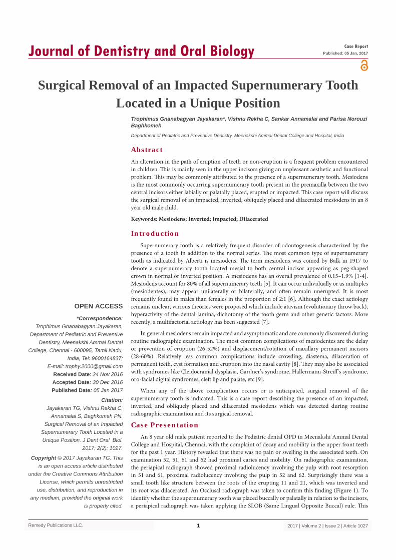
College and Hospital, Chennai, with the complaint of decay and mobility in the upper front teeth for the past 1 year. History revealed that there was no pain or swelling in the associated teeth. On examination 52, 51, 61 and 62 had proximal caries and mobility. On radiographic examination, the periapical radiograph showed proximal radiolucency involving the pulp with root resorption in 51 and 61, proximal radiolucency involving the pulp in 52 and 62. Surprisingly there was a small tooth like structure between the roots of the erupting 11 and 21, which was inverted and its root was dilacerated. An Occlusal radiograph was taken to confirm this finding (Figure 1). To identify whether the supernumerary tooth was placed buccally or palatally in relation to the incisors, a periapical radiograph was taken applying the SLOB (Same Lingual Opposite Buccal) rule. This
Surgical Removal of an Impacted Supernumerary Tooth Located in a Unique Position
OPEN ACCESS
*Correspondence:Trophimus Gnanabagyan Jayakaran,
Department of Pediatric and Preventive Dentistry, Meenakshi Ammal Dental
College, Chennai - 600095, Tamil Nadu, India, Tel: 9600164837;
E-mail: [email protected] Date: 24 Nov 2016Accepted Date: 30 Dec 2016Published Date: 05 Jan 2017
Citation: Jayakaran TG, Vishnu Rekha C,
Annamalai S, Baghkomeh PN. Surgical Removal of an Impacted
Supernumerary Tooth Located in a Unique Position. J Dent Oral Biol.
2017; 2(2): 1027.
Copyright © 2017 Jayakaran TG. This is an open access article distributed
under the Creative Commons Attribution License, which permits unrestricted
use, distribution, and reproduction in any medium, provided the original work
is properly cited.
Case ReportPublished: 05 Jan, 2017
AbstractAn alteration in the path of eruption of teeth or non-eruption is a frequent problem encountered in children. This is mainly seen in the upper incisors giving an unpleasant aesthetic and functional problem. This may be commonly attributed to the presence of a supernumerary tooth. Mesiodens is the most commonly occurring supernumerary tooth present in the premaxilla between the two central incisors either labially or palatally placed, erupted or impacted. This case report will discuss the surgical removal of an impacted, inverted, obliquely placed and dilacerated mesiodens in an 8 year old male child.
Keywords: Mesiodens; Inverted; Impacted; Dilacerated
Trophimus Gnanabagyan Jayakaran*, Vishnu Rekha C, Sankar Annamalai and Parisa Norouzi Baghkomeh
Department of Pediatric and Preventive Dentistry, Meenakshi Ammal Dental College and Hospital, India
Trophimus Gnanabagyan Jayakaran, et al., Journal of Dentistry and Oral Biology
Remedy Publications LLC. 2017 | Volume 2 | Issue 2 | Article 10272
confirmed the presence of the supernumaray tooth on the palatal side. A diagnosis of an inverted mesiodens was made and surgical removal of the mesiodens using palatal approach was planned.
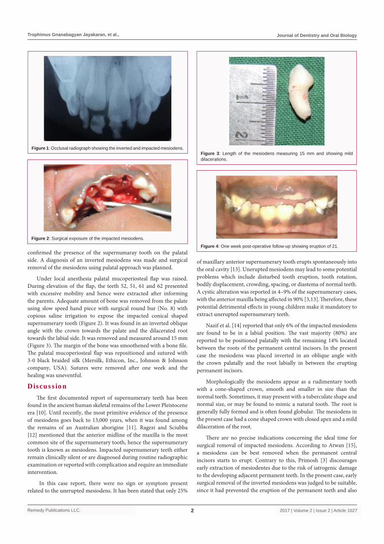
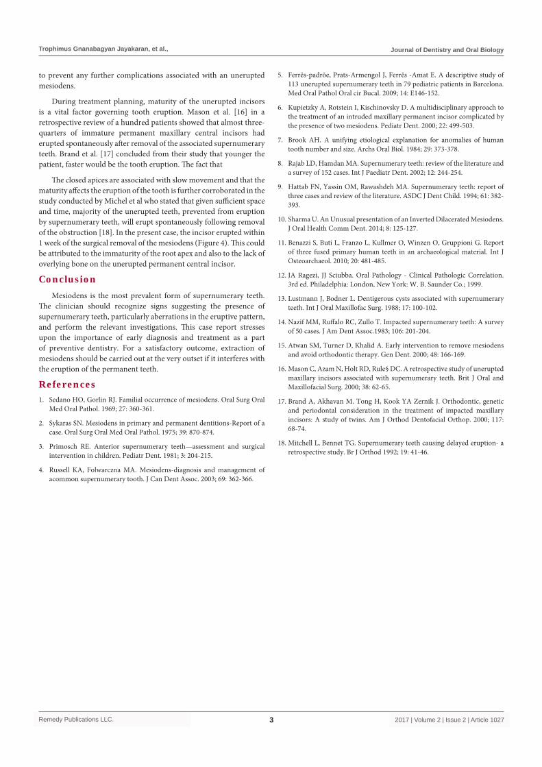
Under local anesthesia palatal mucoperiosteal flap was raised. During elevation of the flap, the teeth 52, 51, 61 and 62 presented with excessive mobility and hence were extracted after informing the parents. Adequate amount of bone was removed from the palate using slow speed hand piece with surgical round bur (No. 8) with copious saline irrigation to expose the impacted conical shaped supernumerary tooth (Figure 2). It was found in an inverted oblique angle with the crown towards the palate and the dilacerated root towards the labial side. It was removed and measured around 15 mm (Figure 3). The margin of the bone was smoothened with a bone file. The palatal mucoperiosteal flap was repositioned and sutured with 3-0 black braided silk (Mersilk, Ethicon, Inc., Johnson & Johnson company, USA). Sutures were removed after one week and the healing was uneventful.
DiscussionThe first documented report of supernumerary teeth has been
found in the ancient human skeletal remains of the Lower Pleistocene era [10]. Until recently, the most primitive evidence of the presence of mesiodens goes back to 13,000 years, when it was found among the remains of an Australian aborigine [11]. Ragezi and Sciubba [12] mentioned that the anterior midline of the maxilla is the most common site of the supernumerary tooth, hence the supernumerary tooth is known as mesiodens. Impacted supernumerary teeth either remain clinically silent or are diagnosed during routine radiographic examination or reported with complication and require an immediate intervention.
In this case report, there were no sign or symptom present related to the unerupted mesiodens. It has been stated that only 25%
of maxillary anterior supernumerary tooth erupts spontaneously into the oral cavity [13]. Unerupted mesiodens may lead to some potential problems which include disturbed tooth eruption, tooth rotation, bodily displacement, crowding, spacing, or diastema of normal teeth. A cystic alteration was reported in 4–9% of the supernumerary cases, with the anterior maxilla being affected in 90% [3,13]. Therefore, these potential detrimental effects in young children make it mandatory to extract unerupted supernumerary teeth.
Nazif et al. [14] reported that only 6% of the impacted mesiodens are found to be in a labial position. The vast majority (80%) are reported to be positioned palatally with the remaining 14% located between the roots of the permanent central incisors. In the present case the mesiodens was placed inverted in an oblique angle with the crown palatally and the root labially in between the erupting permanent incisors.
Morphologically the mesiodens appear as a rudimentary tooth with a cone-shaped crown, smooth and smaller in size than the normal teeth. Sometimes, it may present with a tuberculate shape and normal size, or may be found to mimic a natural tooth. The root is generally fully formed and is often found globular. The mesiodens in the present case had a cone shaped crown with closed apex and a mild dilaceration of the root.
There are no precise indications concerning the ideal time for surgical removal of impacted mesiodens. According to Atwan [15], a mesiodens can be best removed when the permanent central incisors starts to erupt. Contrary to this, Primosh [3] discourages early extraction of mesiodentes due to the risk of iatrogenic damage to the developing adjacent permanent teeth. In the present case, early surgical removal of the inverted mesiodens was judged to be suitable, since it had prevented the eruption of the permanent teeth and also
Figure 1: Occlusal radiograph showing the inverted and impacted mesiodens.
Figure 2: Surgical exposure of the impacted mesiodens.
Figure 3: Length of the mesiodens measuring 15 mm and showing mild dilacerations.
Figure 4: One week post-operative follow-up showing eruption of 21.
Trophimus Gnanabagyan Jayakaran, et al., Journal of Dentistry and Oral Biology
Remedy Publications LLC. 2017 | Volume 2 | Issue 2 | Article 10273
to prevent any further complications associated with an unerupted mesiodens.
During treatment planning, maturity of the unerupted incisors is a vital factor governing tooth eruption. Mason et al. [16] in a retrospective review of a hundred patients showed that almost three-quarters of immature permanent maxillary central incisors had erupted spontaneously after removal of the associated supernumerary teeth. Brand et al. [17] concluded from their study that younger the patient, faster would be the tooth eruption. The fact that
The closed apices are associated with slow movement and that the maturity affects the eruption of the tooth is further corroborated in the study conducted by Michel et al who stated that given sufficient space and time, majority of the unerupted teeth, prevented from eruption by supernumerary teeth, will erupt spontaneously following removal of the obstruction [18]. In the present case, the incisor erupted within 1 week of the surgical removal of the mesiodens (Figure 4). This could be attributed to the immaturity of the root apex and also to the lack of overlying bone on the unerupted permanent central incisor.
ConclusionMesiodens is the most prevalent form of supernumerary teeth.
The clinician should recognize signs suggesting the presence of supernumerary teeth, particularly aberrations in the eruptive pattern, and perform the relevant investigations. This case report stresses upon the importance of early diagnosis and treatment as a part of preventive dentistry. For a satisfactory outcome, extraction of mesiodens should be carried out at the very outset if it interferes with the eruption of the permanent teeth.
References1. Sedano HO, Gorlin RJ. Familial occurrence of mesiodens. Oral Surg Oral
Med Oral Pathol. 1969; 27: 360-361.
2. Sykaras SN. Mesiodens in primary and permanent dentitions-Report of a case. Oral Surg Oral Med Oral Pathol. 1975; 39: 870-874.
3. Primosch RE. Anterior supernumerary teeth—assessment and surgical intervention in children. Pediatr Dent. 1981; 3: 204-215.
4. Russell KA, Folwarczna MA. Mesiodens-diagnosis and management of acommon supernumerary tooth. J Can Dent Assoc. 2003; 69: 362-366.
5. Ferrĕs-padrŏe, Prats-Armengol J, Ferrĕs -Amat E. A descriptive study of 113 unerupted supernumerary teeth in 79 pediatric patients in Barcelona. Med Oral Pathol Oral cir Bucal. 2009; 14: E146-152.
6. Kupietzky A, Rotstein I, Kischinovsky D. A multidisciplinary approach to the treatment of an intruded maxillary permanent incisor complicated by the presence of two mesiodens. Pediatr Dent. 2000; 22: 499-503.
7. Brook AH. A unifying etiological explanation for anomalies of human tooth number and size. Archs Oral Biol. 1984; 29: 373-378.
8. Rajab LD, Hamdan MA. Supernumerary teeth: review of the literature and a survey of 152 cases. Int J Paediatr Dent. 2002; 12: 244-254.
9. Hattab FN, Yassin OM, Rawashdeh MA. Supernumerary teeth: report of three cases and review of the literature. ASDC J Dent Child. 1994; 61: 382-393.
10. Sharma U. An Unusual presentation of an Inverted Dilacerated Mesiodens. J Oral Health Comm Dent. 2014; 8: 125-127.
11. Benazzi S, Buti L, Franzo L, Kullmer O, Winzen O, Gruppioni G. Report of three fused primary human teeth in an archaeological material. Int J Osteoarchaeol. 2010; 20: 481-485.
12. JA Ragezi, JJ Sciubba. Oral Pathology - Clinical Pathologic Correlation. 3rd ed. Philadelphia: London, New York: W. B. Saunder Co.; 1999.
13. Lustmann J, Bodner L. Dentigerous cysts associated with supernumerary teeth. Int J Oral Maxillofac Surg. 1988; 17: 100-102.
14. Nazif MM, Ruffalo RC, Zullo T. Impacted supernumerary teeth: A survey of 50 cases. J Am Dent Assoc.1983; 106: 201-204.
15. Atwan SM, Turner D, Khalid A. Early intervention to remove mesiodens and avoid orthodontic therapy. Gen Dent. 2000; 48: 166-169.
16. Mason C, Azam N, Holt RD, Rule§ DC. A retrospective study of unerupted maxillary incisors associated with supernumerary teeth. Brit J Oral and Maxillofacial Surg. 2000; 38: 62-65.
17. Brand A, Akhavan M. Tong H, Kook YA Zernik J. Orthodontic, genetic and periodontal consideration in the treatment of impacted maxillary incisors: A study of twins. Am J Orthod Dentofacial Orthop. 2000; 117: 68-74.
18. Mitchell L, Bennet TG. Supernumerary teeth causing delayed eruption- a retrospective study. Br J Orthod 1992; 19: 41-46.