supplementary material molecular discrimination of ... · supplementary material molecular...
TRANSCRIPT
1
Supplementary Material
Molecular discrimination of structurally equivalent Lys63-linked and linear polyubiquitin chains
David Komander1,2*, Francisca Reyes-Turcu3#, Julien D. F. Licchesi1, Peter
Odenwaelder2$, Keith D. Wilkinson3 and David Barford2
1 MRC Laboratory of Molecular Biology, Hills Road, Cambridge CB2 0QH, UK
2 Section of Structural Biology, The Institute of Cancer Research, 237 Fulham
Road, London SW3 6JB, UK
3 Department of Biochemistry, Emory University School of Medicine, Atlanta,
Georgia 303222, USA
# Current address: Laboratory of Biochemistry and Molecular Biology, NCI,
National Institutes of Health, Bethesda, Maryland 20892, USA
$ Current address: Max-Planck-Institute for Biophysical Chemistry,
Goettingen, Germany.
* Correspondence should be addressed to:
David Komander, MRC Laboratory of Molecular Biology, Protein and Nucleic
Acid Chemistry Division, Hills Road, Cambridge, CB2 0QH, UK.
Email: [email protected], Tel: +441223402300, Fax: +441223412178
2
Supplementary Text
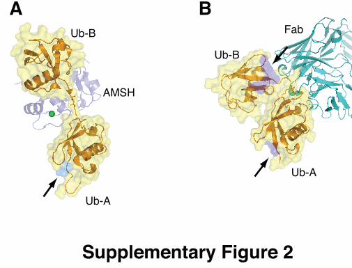
Comparison with K63-linked diUb complexes Two recent co-crystal structures of K63-linked diUb bound to an antibody
fragment (Newton et al, 2008) and the AMSH-LP DUB (Sato et al, 2008) have
revealed further features of K63-chain conformational flexibility. The structure
of AMSH-LP, a JAMM (JAB1/MPN/Mov34 metalloprotease) family DUB
specific for K63-linked chains, has revealed the molecular basis for its
specificity (Supp. Fig. 2A and see main text). In contrast, the structure of a
K63-specific antibody in complex with K63-diUb reveals that the diUb
molecule is bent along its linker, with the antibody contacting the linkage
residues (Supp. Fig. 2B). This conformation of K63-linked chains highlights
their flexibility, although such antibody-based UBD does not occur in vivo.
Comparison with K48-linked polyubiquitin
The structures of linear and K63-linked polyUb presented here are in marked
contrast to the previously reported structures of K48-linked Ub dimers and Ub
tetramers (Eddins et al, 2007) (Fig. 2D). K48 resides on an opposite surface
of Ub relative to K63, and K48-linked Ub dimers adopt a compact structure
with multiple hydrophobic contacts between their Ile44 patches. Longer
polymers form from stacked Ub dimers (Fig. 2D). However, even the compact
K48-linked chains display dynamic features (Trempe et al, 2005). Overall,
K48-linked chains have a more compact polymeric structure compared to
K63- and linear Ub chains (Fig. 2D,E).

3
Supplementary Material and Methods Protein production and purification
Ubiquitin (Ub) was expressed and purified according to (Pickart & Raasi,
2005), Ubc13 and Uev1a were expressed and purified according to (Zhang et
al, 2005), E1 enzyme was expressed and purified from Sf9 cells according to
(Trempe et al, 2005). K63- and K48-linked Ub-chains were generated as
described in (Komander et al, 2008), and the expression vector and
purification protocol for linear tetraUb was described in (Reyes-Turcu et al,
2008). A20, AMSH and TRABID, as well as GST-tagged Ub binding domains
of TRABID, NEMO, TAB2, cIAP1, ABIN2 and MUD1 were expressed in
bacteria and purified using standard protocols or according to published
procedures (Komander & Barford, 2008; McCullough et al, 2006; Tran et al,
2008; Trempe et al, 2005). CYLD was expressed in Sf9 cells and purified
according to (Komander et al, 2008). UCH-L2, UCH-L3, IsoT, USP2, and
USP15 were obtained from BIOMOL (Exeter, UK).
Data collection, phasing and refinement
Diffraction data from diUb crystals was collected at the ESRF (Grenoble,
France), beamline ID23. The crystals belong to the cubic space group P4332
with one diUb per asymmetric unit (Supplementary Table I). The structure
was solved by molecular replacement using Phaser (McCoy et al, 2005) and
monoUb (1ubq) as the search model. The initial phases of K63-linked diUb
were interpreted with ARP/wARP (Perrakis et al, 1999) which built >95 % of
the model. This model was subsequently refined with Refmac5 (Murshudov et
al, 1997), incorporating TLS parameters, resulting in the final statistics in
Supplementary Table I. The linear diUb crystals were solved by individual
rigid body refinement of both Ub monomers, and subsequent rounds of
simulated annealing in Phenix (Adams et al, 2002), while final refinement runs
were performed in Refmac5.
Mammalian expression and DUB assay
HEK293 cells were seeded in 6-wells plates 18 hrs prior to transfection. One
µg of either empty HA vector or HA-TRABID was transfected per well (2 wells
per condition) using Lipofectamine 2000. Twenty-four hours following

4
transfection, proteins were extracted using NP-40 buffer (150 mM NaCl, 50
mM Tris [pH 8.0], 1% NP-40, Complete EDTA-Free protease inhibitor tablets),
and pre-blocked using 25 µl of 50% protein G sepharose bead slurry (Zymed).
700 µg of protein were immunoprecipitated using a Rat anti-HA antibody
(Roche, 4 µg/ml final concentration) for 2 hrs at 4ºC. Immune complexes were
captured with 50 µl of 50% protein G sepharose bead slurry for 1 hr at 4ºC.
Beads were washed 3 times in NP-40 buffer, resuspended in 150 µl of NP-40
buffer and incubated for 1 hr at 37ºC with either 100 ng of K48, K63 or linear
chains. 4X sample buffer was added to each reaction and samples loaded
onto SDS PAGE. As an input control 50 ng of each linkage were loaded.
Following transfer, the PVDF membrane was blocked in 5% milk (PBS, 0.1%
Tween 20) and incubated with a rabbit anti-ubiquitin antibody (Millipore 07-
375; 1/2000) and detected with a goat anti rabbit IgG-HRP antibody. 15 µg of
input protein was loaded onto a SDS PAGE gel and detected using a rat anti-
HA (Roche: 1/2000) followed by goat anti-rat IgG-HRP antibody
Supplemental references
Adams PD, Grosse-Kunstleve RW, Hung LW, Ioerger TR, McCoy AJ, Moriarty NW, Read RJ, Sacchettini JC, Sauter NK, Terwilliger TC (2002) PHENIX: building new software for automated crystallographic structure determination. Acta Crystallogr D Biol Crystallogr 58(Pt 11): 1948-1954 Eddins MJ, Varadan R, Fushman D, Pickart CM, Wolberger C (2007) Crystal structure and solution NMR studies of Lys48-linked tetraubiquitin at neutral pH. J Mol Biol 367(1): 204-211 Komander D, Barford D (2008) Structure of the A20 OTU domain and mechanistic insights into deubiquitination. Biochem J 409(1): 77-85 Komander D, Lord CJ, Scheel H, Swift S, Hofmann K, Ashworth A, Barford D (2008) The structure of the CYLD USP domain explains its specificity for Lys63-linked polyubiquitin and reveals a B box module. Mol Cell 29(4): 451-464 McCoy AJ, Grosse-Kunstleve RW, Storoni LC, Read RJ (2005) Likelihood-enhanced fast translation functions. Acta Crystallogr D Biol Crystallogr 61(Pt 4): 458-464
5
McCullough J, Row PE, Lorenzo O, Doherty M, Beynon R, Clague MJ, Urbe S (2006) Activation of the endosome-associated ubiquitin isopeptidase AMSH by STAM, a component of the multivesicular body-sorting machinery. Curr Biol 16(2): 160-165 Murshudov GN, Vagin AA, Dodson EJ (1997) Refinement of macromolecular structures by the maximum- likelihood method. Acta Crystallographica Section D-Biological Crystallography 53: 240-255 Newton K, Matsumoto ML, Wertz IE, Kirkpatrick DS, Lill JR, Tan J, Dugger D, Gordon N, Sidhu SS, Fellouse FA, Komuves L, French DM, Ferrando RE, Lam C, Compaan D, Yu C, Bosanac I, Hymowitz SG, Kelley RF, Dixit VM (2008) Ubiquitin chain editing revealed by polyubiquitin linkage-specific antibodies. Cell 134(4): 668-678 Perrakis A, Morris R, Lamzin VS (1999) Automated protein model building combined with iterative structure refinement. Nat Struct Biol 6(5): 458-463 Pickart CM, Raasi S (2005) Controlled synthesis of polyubiquitin chains. Methods Enzymol 399: 21-36 Reyes-Turcu FE, Shanks JR, Komander D, Wilkinson KD (2008) Recognition of polyubiquitin isoforms by the multiple ubiquitin binding modules of isopeptidase T. J Biol Chem 283(28): 19581-19592 Sato Y, Yoshikawa A, Yamagata A, Mimura H, Yamashita M, Ookata K, Nureki O, Iwai K, Komada M, Fukai S (2008) Structural basis for specific cleavage of Lys 63-linked polyubiquitin chains. Nature 455(7211): 358-362 Tran H, Hamada F, Schwarz-Romond T, Bienz M (2008) Trabid, a new positive regulator of Wnt-induced transcription with preference for binding and cleaving K63-linked ubiquitin chains. Genes Dev 22(4): 528-542 Trempe JF, Brown NR, Lowe ED, Gordon C, Campbell ID, Noble ME, Endicott JA (2005) Mechanism of Lys48-linked polyubiquitin chain recognition by the Mud1 UBA domain. EMBO J 24(18): 3178-3189 Zhang M, Windheim M, Roe SM, Peggie M, Cohen P, Prodromou C, Pearl LH (2005) Chaperoned ubiquitylation--crystal structures of the CHIP U box E3 ubiquitin ligase and a CHIP-Ubc13-Uev1a complex. Mol Cell 20(4): 525-538
6
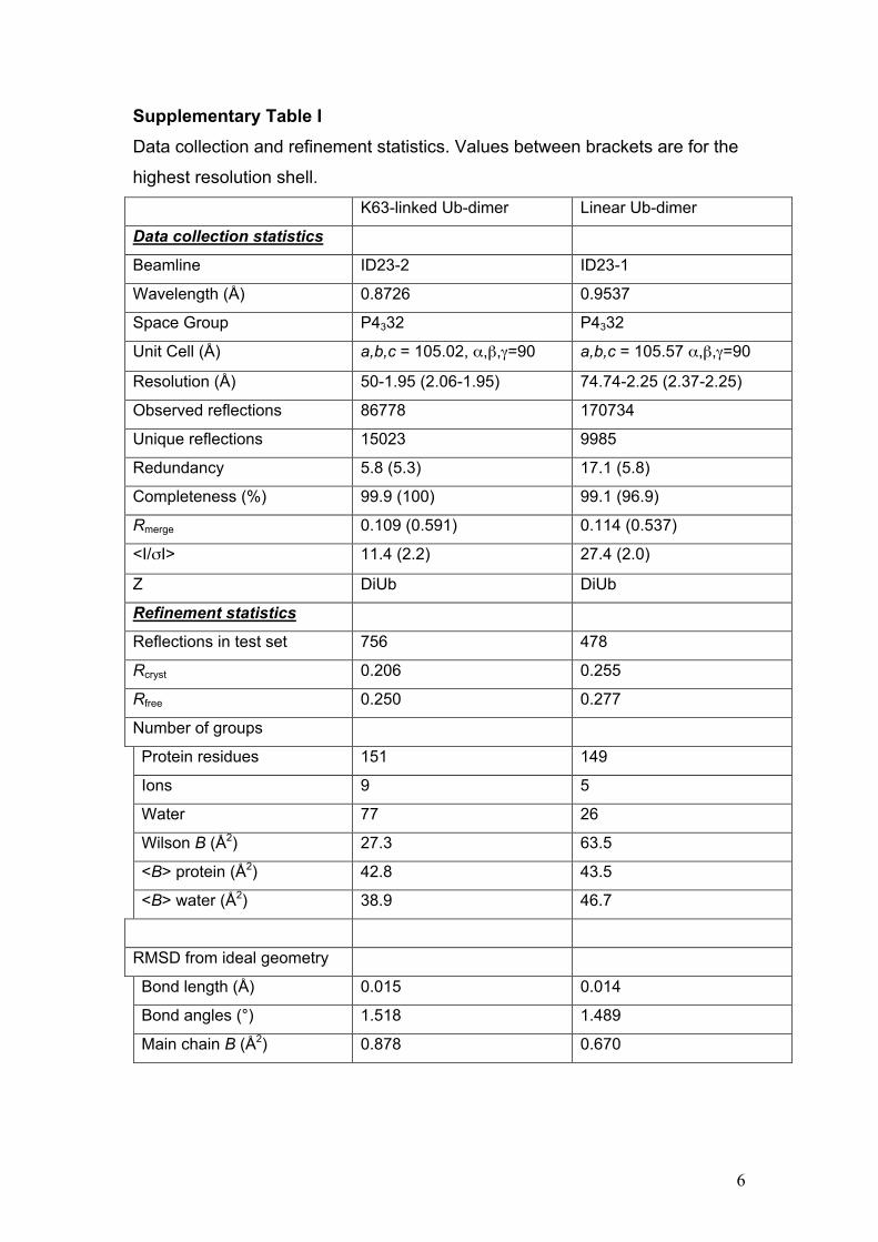
Supplementary Table I Data collection and refinement statistics. Values between brackets are for the
highest resolution shell.
K63-linked Ub-dimer Linear Ub-dimer
Data collection statistics
Beamline ID23-2 ID23-1
Wavelength (Å) 0.8726 0.9537
Space Group P4332 P4332
Unit Cell (Å) a,b,c = 105.02, α,β,γ=90 a,b,c = 105.57 α,β,γ=90
Resolution (Å) 50-1.95 (2.06-1.95) 74.74-2.25 (2.37-2.25)
Observed reflections 86778 170734
Unique reflections 15023 9985
Redundancy 5.8 (5.3) 17.1 (5.8)
Completeness (%) 99.9 (100) 99.1 (96.9)
Rmerge 0.109 (0.591) 0.114 (0.537)
<I/σI> 11.4 (2.2) 27.4 (2.0)
Z DiUb DiUb
Refinement statistics
Reflections in test set 756 478
Rcryst 0.206 0.255
Rfree 0.250 0.277
Number of groups
Protein residues 151 149
Ions 9 5
Water 77 26
Wilson B (Å2) 27.3 63.5
<B> protein (Å2) 42.8 43.5
<B> water (Å2) 38.9 46.7
RMSD from ideal geometry
Bond length (Å) 0.015 0.014
Bond angles (°) 1.518 1.489
Main chain B (Å2) 0.878 0.670

7
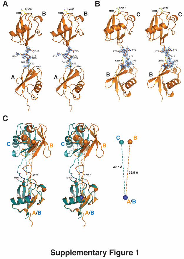
Supplementary Figure legends Supplementary Figure 1. A) Crystal structure of K63-linked diUb in stereo representation. A 2|Fo|-|Fc|
electron density map contoured at 1 σ is shown for the linking residues. B)
Crystal structure of linear diUb as in A. C) Superposition of K63-linked
(orange) and linear (green) diUb as observed in mol A - mol B and mol B –
mol C, respectively, aligned on the proximal molecule. Spheres represent the
centre-of-mass for each ubiquitin moiety. While mol B and mol C are oriented
slightly differently with respect to the proximal molecule through the flexible
linker, importantly, the distance between individual Ubs is equivalent (39.5 Å
vs 39.7 Å).
Supplementary Figure 2. A semitransparent surface covers the Ub molecules in cartoon representation,
and the position of the hydrophobic surface patch formed by Ile44-Val70-Leu8
is coloured in blue on the surface, indicated by arrows. The diUb molecules
are aligned on the proximal Ub moiety. A) Structure of AMSH-LP bound to the
extended K63 diUb (pdb id 2znv, (Sato et al, 2008). B) Structure of K63-diUb
recognised by a K63-specific Fab fragment (pdb-id 3dvg, (Newton et al,
2008).
Supplementary Figure 3. Membranes from the pull-down experiment stained in Ponceau Red after
blotting. Analysed proteins are labelled by arrows. The small amount of
tetraUb used in the experiment is undetectable by Ponceau Red.
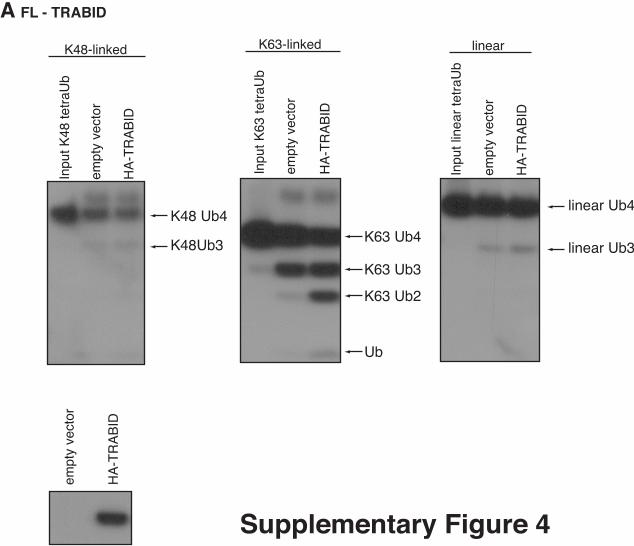
Supplementary Figure 4. Analysis of full-length TRABID. Empty HA vector and HA-tagged full-length
TRABID were immunoprecipitated from HEK293 cells and the precipitates
were incubated with tetraUb chains for 1 hr. Western blotting analysis with α-
Ub and α-HA shows TRABID-mediated cleavage of K63 chains only.
A
A A
B B
B B
C C
B
R72
R74 L73G75
G76K63
R72
R74 L73G75
G76K63
Lys63 Lys63
R74L73
G75
G76
M1
Met1
R74L73
G75
G76
M1
Met1Lys63 Lys63
Met1 Met1
Lys63 Lys63
Met1 Met1
Supplementary Figure 1
C
BCBC
A/B
Met1Lys63
A/B
Lys63Met1
A/B
BC
39.5 Å
39.7 Å
Ub-A
Ub-B
AMSH
A
Ub-B
Ub-A
BFab
Supplementary Figure 2

K48-linked
10%
inpu
tG
ST-
UB
DG
ST
Supplementary Figure 3
K63-linked linear
10%
inpu
tG
ST-
UB
DG
ST
10%
inpu
tG
ST-
UB
DG
ST
Mar
ker
GST
GST-TAB2-C
28
38
49
GST
GST-NEMO UBD
28
38
49
GST-MUD1-UBAGST28
38
49
GST
GST-TRABID-N
28
38
49
28
38
49
62GST-ABIN2 (FL)BSA (in Ub4 input)
GST
GST-ABIN2 (degradation products)
GST
GST-cIAP1-C
28
38
49
A FL - TRABID
empt
y ve
ctor
HA
-TR
AB
ID
K63 Ub4
K63-linked
Inpu
t K63
tetr
aUb
empt
y ve
ctor
HA
-TR
AB
ID
K63 Ub3
K63 Ub2
Ub
linear Ub4
linear
Inpu
t lin
ear
tetr
aUb
empt
y ve
ctor
HA
-TR
AB
ID
linear Ub3
Inpu
t K48
tetr
aUb
empt
y ve
ctor
HA
-TR
AB
ID
K48-linked
K48 Ub4
K48Ub3
Supplementary Figure 4