successful chemotherapy management of disseminated
TRANSCRIPT

CASE REPORT Open Access
Successful chemotherapy management ofdisseminated intravascular coagulationpresenting with metastatic clear cell renalcarcinoma: a case report and review of theliteratureHuy Le Trinh, Vuong Thi Nguyen, Ngan Kim Mai, Bach Trung Tran and Quynh Nga Pham*
Abstract
Background: Disseminated intravascular coagulation is a critical complication of advanced clear cell renal cellcarcinoma, despite the rarity of the occurrence of disseminated intravascular coagulation in such tumors. Thediagnosis of cancer-related disseminated intravascular coagulation is mostly based on clinical bleeding andlaboratory test; available data suggest that treating the primary cancer also treats the disseminated intravascularcoagulation. Among three reported cases of renal cell carcinoma-related disseminated intravascular coagulation inthe literature, this is the first patient whose disseminated intravascular coagulation was successfully treated, inparticular, with chemotherapy without any anti-disseminated intravascular coagulation therapies.
Case presentation: This case is a 66-year-old Vietnamese man who presented disseminated intravascularcoagulation 2 weeks after his admission for severe back pain. At admission, his initial laboratory work-up revealedonly a mild thrombocytopenia with a platelet count of 93 × 109/L (normal range, 150–450 × 109/L) without clinicalbleeding. His past medical history and family history were unremarkable. An open-biopsy was performed and thedefinitive diagnosis was bone metastatic clear cell renal cell carcinoma based on immunohistochemistry. Twoweeks after admission, the diagnosis of disseminated intravascular coagulation was confirmed according to theInternational Society on Thrombosis and Haemostasis. Immediately, he was treated with a paclitaxel pluscarboplatin regimen and disseminated intravascular coagulation completely disappeared after one cycle of systemicchemotherapy. Until recently, 11 months subsequent to the diagnosis of disseminated intravascular coagulation, hehad been being undergoing maintenance therapy for metastatic clear cell renal cell carcinoma.
Conclusions: First, an early detection of overt disseminated intravascular coagulation is essential, althoughdisseminated intravascular coagulation in cancer presents as a chronic or even subclinical process with uniquethrombocytopenia. Second, making a decision of systemic chemotherapy without delay at the time ofdisseminated intravascular coagulation diagnosis is the key to successful cancer-related disseminated intravascularcoagulation treatment.
Keywords: Disseminated intravascular coagulation, Clear cell renal cell carcinoma, Chemotherapy
© The Author(s). 2020 Open Access This article is licensed under a Creative Commons Attribution 4.0 International License,which permits use, sharing, adaptation, distribution and reproduction in any medium or format, as long as you giveappropriate credit to the original author(s) and the source, provide a link to the Creative Commons licence, and indicate ifchanges were made. The images or other third party material in this article are included in the article's Creative Commonslicence, unless indicated otherwise in a credit line to the material. If material is not included in the article's Creative Commonslicence and your intended use is not permitted by statutory regulation or exceeds the permitted use, you will need to obtainpermission directly from the copyright holder. To view a copy of this licence, visit http://creativecommons.org/licenses/by/4.0/.The Creative Commons Public Domain Dedication waiver (http://creativecommons.org/publicdomain/zero/1.0/) applies to thedata made available in this article, unless otherwise stated in a credit line to the data.
* Correspondence: [email protected] of Oncology, Hanoi Medical University Hospital, Hanoi, Vietnam
Trinh et al. Journal of Medical Case Reports (2020) 14:52 https://doi.org/10.1186/s13256-020-02369-x

BackgroundClear cell renal carcinoma (CCRCC) is the most com-mon histologic pattern of renal cell carcinomas (RCCs),accounting for approximately 75 to 85% of such tumors[1]. RCCs have a silent natural history for the multistepdevelopment of tumors; therefore, most patients are di-agnosed with advanced disease. Disseminated intravas-cular coagulation (DIC) is a very rare presentingsyndrome in solid tumors, particularly in metastaticRCCs, which is characterized by thrombosis, bleeding, orboth and signs of activation of clotting and fibrinolyticsystem in a laboratory [2]. Only two cases of RCCs withDIC have been reported previously in the literature andthe two cases died due to DIC [3, 4].Based on current data, chemotherapy has no role in
the management of advanced CCRCC due to the theoryof resistance to cytotoxic agents of these cells and thedevelopment of immunotherapy and molecularly tar-geted therapy. Until recently, no data of methods in thetreatment of DIC in CCRCC have been reported. Thus,we report a case of metastatic CCRCC presenting withchronic DIC, which was successfully managed with ini-tial chemotherapy without any anti-DIC therapies.
Case presentationThis is a case of a 66-year-old Vietnamese man who wasadmitted to the Oncology Department at Hanoi MedicalUniversity Hospital on 3 December 2018 for severelower back pain. He had no past medical history or fam-ily history. On examination, an initial lumbosacral spinemagnetic resonance imaging (MRI) revealed enlargedlytic lesions of sacral segment 1 with ilium, soft tissueinvasion, and compression of the nerve roots from L5 toS2 (Fig. 1).
At admission, initial laboratory results exhibited only amild thrombocytopenia with a platelet count of 93 ×109/L (normal range, 150–450 × 109/L); his liver and kid-ney function were normal. Subsequent chest and ab-dominal computed tomography (CT) images showed nomass or abnormalities except a lytic lesion (Fig. 2). Onthe ninth day of admission, a surgical decompressionwas performed but the tumor could not be totally re-moved. Pathology results suggested two distinct diseasesincluding CCRCC and parathyroid carcinoma metastasis(Fig. 3). Therefore, an immunohistochemistry (IHC) testwas undertaken to acquire a confirmed diagnosis. Dur-ing a period of days, while waiting for the IHC results, 2weeks after admission, our patient complained of mod-erate fatigue and his laboratory data indicatedthrombocytopenia with a platelet count of 78 × 109/L(from 93 × 109/L at baseline) and anemia with a severehemoglobin level of 69 g/L (from 142 g/L at baseline).The calculated DIC score by the International Societyon Thrombosis and Haemostasis (ISTH) was 4 [2], thenon-overt DIC with D-dimer level strongly increased to4.94 μg/mL (normal range, < 0.5 μg/mL), fibrinogen levelwas 2.16 g/L, and prolonged prothrombin time was 1.5seconds. Therefore, a bone marrow biopsy was per-formed to rule out bone marrow involvement and theresult was negative. Four days later, after 20 days of ad-mission, our patient presented with moderate subcuta-neous hemorrhage and platelet count of 51 × 109/L.According to ISTH criteria, the diagnosis of DIC wasconfirmed with score of 5. The laboratory results associ-ated with DIC are summarized in Table 1. Following thedefinitive diagnosis of DIC, he received immediate initialchemotherapy despite lack of the IHC results. Paclitaxel(175 mg/m2 over 3 hours) plus carboplatin (AUC = 6)regimen was indicated and repeated every 3 weeks
Fig. 1 Lumbosacral spine magnetic resonance imaging revealed enlarged lytic lesions of S1 with ilium, soft tissue invasion, and compression ofthe nerve roots from L5 to S2 (December 2018)
Trinh et al. Journal of Medical Case Reports (2020) 14:52 Page 2 of 6

without a transfusion of platelets or other anti-DIC ther-apies. One day following the first course of treatment,IHC results exhibited that tumor cells were positive forPAX, RCC, and vimentin and negative for CD10, S100,and chromogranin (Fig. 4), which supported the diagno-sis of bone metastatic CCRCC.After the first cycle of systemic chemotherapy, the plate-
let cell count of our patient recovered to the normal rangewith 168 × 109/L and his subcutaneous bleeding com-pletely disappeared. He tolerated treatment well withoutsevere adverse events during the period of chemotherapy.After four cycles of paclitaxel plus carboplatin, he wasevaluated with an abdominal CT scan and he had achieveda stable disease response at that time and 3 months later,according to the Response Evaluation Criteria in Solid Tu-mors (RECIST) 1.1 system (Fig. 5, 6). He has been receiv-ing maintenance therapy with sorafenib (400mg twicedaily) up to now. Recently, 11months subsequent to thediagnosis of DIC, he was still alive and had a good per-formance status with sorafenib treatment.
Discussion and conclusionDIC is a common complication of a number of illnessesincluding sepsis, trauma, malignancy, and liver disease[5]. A systemic activation of coagulation is an essential
capability of DIC, which leads to contribution to fibrinclots and consumption of platelets and coagulation fac-tors. Therefore, the diagnosis of DIC should be based onclinical bleeding, thrombosis, and laboratory information[2]. In a cohort study of 217 consecutive patients in in-tensive care units (ICUs) who had a clinical suspicionfor DIC, 70 patients (32%) were diagnosed as havingDIC by the ISTH scoring system. Of these patients, onlyfour cases of DIC (5.7%) had a fibrinogen level under 1g/L. Thus, platelet count, prolonged prothrombin time,and D-dimer level were mostly encountered in the ISTHDIC score [6]. These findings reflected the contributionsto the DIC score of the present patient. In addition, datafrom the study showed that the proportion of 28-daymortality in patients with DIC and without DIC were45% and 25%, respectively [6].In contrast to such acute disease, in which DIC ap-
pears as a life-threatening emergency, DIC in cancermight present as a chronic or even subclinical processwith only aberrant laboratory results [7]. Some patientshave unique thrombocytopenia which is a feature in upto 98% of cases of DIC [2]. The prevalence of DIC oc-currence in solid tumors was approximately 7%; in par-ticular, there is a predilection to DIC in elderly patients,male patients, patients with advanced tumors, patients
Fig. 2 An abdominal computed tomography scan revealed enlarged lytic lesions of S1 with ilium, soft tissue invasion, and compression of thenerve roots from L5 to S2; it did not detect tumor in bilateral kidney (25 December 2018)
Fig. 3 Hematoxylin-eosin staining of tumor
Trinh et al. Journal of Medical Case Reports (2020) 14:52 Page 3 of 6

with breast cancer, and with the presence of necrosis inthe tumor. Among 7% of cancer-related cases of DIC,RCC was only in 5%, which means that DIC occurrencein RCCs is extremely rare [8]. Hence, only two caseshave been reported previously in the literature. Accord-ing to a study of 1117 patients with solid tumors, casesof overt DIC with advanced tumors had significantlylower survival than cases of non-overt DIC (9 versus 14months, p = 0.005). This finding demonstrated the im-pact of a serious complication, including DIC, on theoutcome of patients with cancer [8].The basis of the treatment of DIC is to manage the
underlying disease [9]. In a cohort of 1117 patients withsolid tumors, among 76 cases who were diagnosed as hav-ing DIC, one third of these patients achieved response toDIC treatment including replacement therapy (fresh fro-zen plasma, platelet transfusion, and red blood cell trans-fusion), heparin, antithrombin III concentrates, andmanagement of the underlying malignancy. In addition,the study indicated that the median survival of the patientswith advanced tumors-related DIC was 9 months [8].Based on our knowledge, we indicated a systemic chemo-therapy for our patient to resolve cancer-related DIC priorto a confirmed diagnosis of IHC results.
As mentioned previously, our patient is the first casewith successfully managed RCC-related DIC. Of the tworeported cases, one case was diagnosed as having sub-acute DIC in liver metastatic RCC and the other was anautopsy case of pulmonary metastasis of RCC with DIC.In the first reported case, the calculated DIC score bythe ISTH was 6 and he died 4 weeks later with onlyanticoagulation treatment after admission [3]. Currently,RCC is considered a chemotherapy-resistant cancer, inparticular in clear cell, which is based on a review of 72cytotoxic chemotherapy agents in 3502 patients with ad-vanced RCC. The study showed that only 197 cases (6%)had a complete or partial response [10]. Until recently,the mechanisms of chemotherapeutical resistance inRCC remained unclear. Some cytotoxic regimens re-vealed modest activity, which suggests that chemother-apy might benefit subsets of patients with RCC, such asa rapid tumor growth, to withdraw the development ofneoplasm or progress of tumors on cytokines or targetedagents [11]. In the current case, paclitaxel plus carbopla-tin regimen completely removed DIC; however, heachieved only a stable disease for the evaluation oftumor response after four cycles of chemotherapy. Theoutcome of our patient might be interpreted as follows:
Fig. 4 Immunohistochemistry results of tumor: a PAX8 positive; b RCC positive; c vimentin positive; d CD10 negative; e S100 negative;f chromogranin negative
Table 1 Laboratory findings associated with disseminated intravascular coagulation
Platelet count (109/l) D-dimer (μg/mL) Prolonged PT (seconds) Fibrinogen level (g/l) DIC Score
At admission (D0) 93 (1p) Unmeasured 1.6 (0p) 3.18 (0p)
At operation (D9) 91 (1p) 1.83 (2p) 2.7 (0p) 2.49 (0p) 3
D15 78 (1p) 4.94 (3p) 1.5 (0p) 2.16 (0p) 4
D20 51 (1p) 4.48 (3p) 3.1 (1p) 3.23 (0p) 5
D day, DIC disseminated intravascular coagulation, DIC score based on the International Society on Thrombosis and Haemostasis criteria, p point,PT prothrombin time
Trinh et al. Journal of Medical Case Reports (2020) 14:52 Page 4 of 6
Tissue factor (TF), which is expressed by either theendothelial cells of the vessels or neoplastic cells, bindscirculating factor VII(a). This complex enables the acti-vated factors IX and X to trigger systemic activation ofcoagulation [7]. According to the proportion and num-ber of tumor cells containing TF, a non-overt or overtDIC develops [7]. Interestingly, in the second reportedcase, the autopsy of a pulmonary metastatic RCC withDIC revealed that most of the vascular endothelial cellswere alternated by metastatic carcinoma cells [4]. Thus,it might result in sustaining thrombin generation andconsumption of fibrinogen and platelet cells [7]. Alterna-tively, metastatic dissemination is the last stage in a var-iety of steps of tumor progression and there is a geneticevolution occurring in the neoplastic cells during theirdevelopment. Therefore, a primary tumor is probablydifferent from the one contributed by metastatic carcin-oma cells in distant organs. Accordingly, the characteris-tic of chemoresistant incipient clear cell RCC might be
alternated in disseminated cells. However, the limitationsof this case include the suboptimal regimen of manage-ment in RCC and the lack of total laboratory assessmentfor DIC following the first cycle of chemotherapy. Asmentioned previously, the optimal treatment for RCCrecently is immunotherapy and molecularly targetedtherapy. Due to financial issues and the availability ofdrugs, this patient could not be treated with the optimaltherapy. Also, based on the current data, paclitaxel pluscarboplatin is not the optimal regimen in metastaticRCC management, in terms of systemic chemotherapy.The combination of gemcitabine and a fluoropyrimidine(infusion 5-FU or capecitabine) produced the greatest ef-ficacy compared to other chemotherapy regimens, andpaclitaxel, which is an anti-microtubule, failed to dem-onstrate an efficacy in RCC treatment [12]. The rationalfor the use of paclitaxel-carboplatin regimen in thepresent case derives from the confirmed diagnosis at thetime.
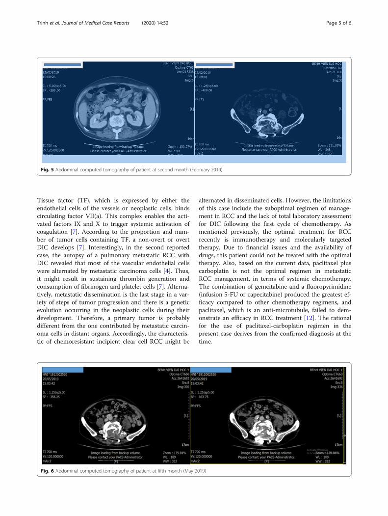
Fig. 5 Abdominal computed tomography of patient at second month (February 2019)
Fig. 6 Abdominal computed tomography of patient at fifth month (May 2019)
Trinh et al. Journal of Medical Case Reports (2020) 14:52 Page 5 of 6

From this patient, cytotoxic chemotherapy still has acrucial role in CCRCC-related DIC management, despitethe era of immunotherapy and molecularly targetedtherapy in the treatment of metastatic CCRCC and thechemotherapy-resistant feature of these neoplastic cells.An early detection of overt DIC and the indication ofsystemic chemotherapy without delay are the key to suc-cessful cancer-related DIC treatment.
AcknowledgementsNot applicable.
Authors’ contributionsAll authors analyzed the patient data regarding the disease and conductedpatient care. QNP collected patient data and described it in the case reportwith literature review. HT, NM, VN, and BT performed literature review andmade significant contributions to the writing of the manuscript. All authorsread and approved the final manuscript.
FundingNo funding available.
Availability of data and materialsAll data generated or analyzed during this study are included in thispublished article.
Ethics approval and consent to participateNot applicable.
Consent for publicationWritten informed consent was obtained from the patient for publication ofthis case report and any accompanying images. A copy of the writtenconsent is available for review by the Editor-in-Chief of this journal.
Competing interestsThe authors declare that they have no competing interests.
Received: 18 November 2019 Accepted: 16 March 2020
References1. Setlik DE, McCluskey KM, McDavit JA. Best cases from the AFIP: renal cell
carcinoma manifesting as a solitary bone metastasis. Radiographics. 2009;29(7):2184–9.
2. Levi M, Toh CH, Thachil J, Watson HG. Guidelines for the diagnosis andmanagement of disseminated intravascular coagulation. British Committeefor Standards in Haematology. Br J Haematol. 2009;145(1):24–33.
3. van der Wekken LC, Loffeld R. Subacute Disseminated IntravascularCoagulation in a Patient with Liver Metastases of a Renal Cell Carcinoma.Case Rep Oncol Med. 2017;2017:1023538.
4. Kobayashi H, Tamashima S, Shigeyama J, Shimizu S, Suchi T. Vascular intimalcarcinomatosis: an autopsy case of unusual form of pulmonary metastasisof transitional cell carcinoma. Pathol Int. 1997;47(9):655–7.
5. Levi M, Ten Cate H. Disseminated intravascular coagulation. N Engl J Med.1999;341(8):586–92.
6. Bakhtiari K, Meijers JC, de Jonge E, Levi M. Prospective validation of theInternational Society of Thrombosis and Haemostasis scoring system fordisseminated intravascular coagulation. Crit Care Med. 2004;32(12):2416–21.
7. Levi M. Cancer-related coagulopathies. Thromb Res. 2014;133(Suppl 2):S70–5.8. Sallah S, Wan JY, Nguyen NP, Hanrahan LR, Sigounas G. Disseminated
intravascular coagulation in solid tumors: clinical and pathologic study.Thromb Haemost. 2001;86(3):828–33.
9. Levi M. Clinical characteristics of disseminated intravascular coagulation inpatients with solid and hematological cancers. Thromb Res. 2018;164(Suppl1):S77–s81.
10. Yagoda A, Petrylak D, Thompson S. Cytotoxic chemotherapy for advancedrenal cell carcinoma. Urol Clin North Am. 1993;20(2):303–21.
11. Nanus DM, Garino A, Milowsky MI, Larkin M, Dutcher JP. Activechemotherapy for sarcomatoid and rapidly progressing renal cell carcinoma.Cancer. 2004;101(7):1545–51.
12. Diamond E, Molina AM, Carbonaro M, Akhtar NH, Giannakakou P, TagawaST, et al. Cytotoxic chemotherapy in the treatment of advanced renal cellcarcinoma in the era of targeted therapy. Crit Rev Oncol Hematol. 2015;96(3):518–26.
Publisher’s NoteSpringer Nature remains neutral with regard to jurisdictional claims inpublished maps and institutional affiliations.
Trinh et al. Journal of Medical Case Reports (2020) 14:52 Page 6 of 6