structure of the traf4 traf domain with a coiled-coil...
TRANSCRIPT
research papers
2 doi:10.1107/S139900471302333X Acta Cryst. (2014). D70, 2–10
Acta Crystallographica Section D
BiologicalCrystallography
ISSN 1399-0047
Structure of the TRAF4 TRAF domain with acoiled-coil domain and its implications for theTRAF4 signalling pathway
Jong Hwan Yoon,a Young-jin
Chob and Hyun Ho Parka*
aSchool of Biotechnology and Graduate School
of Biochemistry, Yeungnam University,
Gyeongsan, Republic of Korea, and bNew
Drug Development Center, Daegu-Gyeongbuk
Medical Innovation Foundation, Daegu,
Republic of Korea
Correspondence e-mail: [email protected]
# 2014 International Union of Crystallography
The TNF receptor-associated factor (TRAF) proteins are
structurally similar scaffold proteins that mediate between
members of the TNF receptor (TNFR) family and down-
stream effector molecules such as kinases in the immune
signalling pathway. Seven TRAFs have been identified,
including TRAF4, which is a unique member that participates
in many ontogenic processes, including nerve-system devel-
opment. TRAFs commonly contain the TRAF domain, which
mediates interaction with target receptors and effectors. As a
first step towards elucidating the molecular mechanisms of the
TRAF4-mediated signalling pathway, the first crystal structure
of the human TRAF4 TRAF domain with a coiled-coil
domain is reported at 2.3 A resolution.
Received 17 June 2013
Accepted 19 August 2013
PDB Reference: human
TRAF4 TRAF domain, 4k8u
1. Introduction
The tumour necrosis factor (TNF) receptor-associated factor
(TRAF) proteins are important cellular signalling molecules
in the tumour necrosis factor receptor (TNFR) and Toll-like
receptor (TLR) family signalling pathways that play a critical
role in the regulation of inflammation, apoptosis and antiviral
response (Inoue et al., 2000; Chung et al., 2002; Bradley &
Pober, 2001). TRAFs are also involved in the proper function
of many immune cells, including CD40-expressing antigen-
presenting cells such as B lymphocytes, macrophages and
dendritic cells (Rothe et al., 1995; Bishop, 2004). TRAFs
function as scaffold proteins that link receptors to downstream
signalling molecules, which are primarily protein kinases and
include interleukin-1 receptor-associated kinases (IRAKs),
receptor-interacting protein 1 (RIP1), RIP2, transforming
growth factor �-activated kinase 1 (TAK1), mitogen-activated
protein kinase kinase kinase 1 (MEKK1) and apoptosis signal-
regulating kinase 1 (ASK1) (Arch et al., 1998; Hsu et al., 1996;
Song et al., 1997; Hoeflich et al., 1999). Several ubiquitin
ligases are also recruited to membrane receptors via direct
interaction with TRAFs (Song et al., 1996). Antagonistic
functions of TRAFs in the TNFR and TLR signalling path-
ways have also been reported by showing that several TRAFs
were not able to recruit any downstream effectors (Ye, Mehlen
et al., 1999; Zapata et al., 2001). Owing to their involvement
in many human diseases, including cancer, autoimmunity and
inflammatory diseases, TRAFs have been suggested to be
suitable targets for therapeutic intervention (Zapata et al.,
2007).
Seven TRAF proteins have been identified in mammals
(TRAF1–TRAF7; Chung et al., 2002; Xie, 2013), all of which
contain the TRAF domain with the exception of TRAF7,
sequence analysis of which indicated that it is unlikely to
contain a TRAF domain. The TRAF domain is an eight-
stranded antiparallel �-sandwich structure composed of
around 180 amino acids (Rothe et al., 1994). The TRAF
domain mediates self-association and upstream interactions
with receptors and other signalling molecules. Despite the
structural similarity of the TRAF domain, each TRAF protein
exhibits specific biological functions, possessing specificity in
its interactions with upstream receptors and downstream
effector molecules. For instance, TRAF6 is known as a key
adaptor molecule in the receptor activator of the NF-�B
(RANK)/RANK ligand (RANKL) signalling pathway, which
is critical to regulation of bone modelling and lymph-node
organogenesis (Darnay et al., 1999).
Even though TRAF4 is one of the canonical TRAF
proteins, it contains unique domain boundaries and has unique
functions in the cell (Kedinger & Rio, 2007). Only TRAF4
contains the unique N-terminal RING-finger motif and a
nuclear localization signal (NLS). In addition, the TRAF4
TRAF domain contains the shortest coiled-coil domain among
the TRAFs (Xie, 2013). A structure-based sequence-alignment
study showed that the receptor-interaction hot spot in TRAF3
is conserved in TRAF2 and TRAF5 but is not conserved in
TRAF4 and TRAF6. This may indicate that TRAF4 can
accommodate novel receptors or binding partners, leading to
a novel signalling pathway (Ely & Li, 2002). Although the
cellular functions of TRAF4 are not well understood, several
studies, including a mouse knockout study, have indicated that
it is involved in gross tracheal, neural tube and skeletal
formation (Kedinger & Rio, 2007; Shiels et al., 2000; Regnier et
al., 2002). While most TRAF-knockout mice showed altera-
tion of the immune system, TRAF4-deficient and TRAF6-
deficient mice exhibited nervous alterations. The most recent
studies have also shown that TRAF4 specifically inhibits the
activation of NF-�B by interacting with NOD-like receptors
(Marinis et al., 2011) and is particularly involved in IL-17
signalling and Th17-mediated disease (Zepp et al., 2012).
Previously reported crystal structures of the TRAF domains
of TRAF2 (Park et al., 1999), TRAF3 (Zhang et al., 2012),
TRAF5 (Zhang et al., 2012) and TRAF6 (Ye et al., 2002) have
shown that TRAF domains are composed of an antiparallel
7–8 �-sheet fold followed by a coiled-coil region. TRAF
domains usually form mushroom-like trimeric structures in
solution. Despite the emerging roles of TRAF4 in human
disease states, limited structural information is available
(Izban et al., 2000; Aston et al., 2004).
In this study, we report the first crystal structure of the
TRAF4 TRAF domain with an extra coiled-coil domain
comprising amino acids 290–470 at 2.3 A resolution. Although
the TRAF4 TRAF domain has the typical TRAF-domain fold,
the high-resolution structure reveals both similarities and
differences between this structure and those of other TRAF
family members, which may be functionally relevant. Further
structure and sequence analysis revealed that the conserved
surface residues of the TRAF4 TRAF domain might be
critical for interaction with signalling molecules.
2. Materials and methods
2.1. Protein expression and purification
The expression and purification methods used in this study
have been described in detail elsewhere (Yoon & Park, 2013).
In summary, the human TRAF4 TRAF domain with a coiled-
coil domain comprising amino acids 290–470 was cloned into
pET24a vector and expressed in Escherichia coli BL21 (DE3)
cells by overnight induction at 293 K. The protein contained
a carboxy-terminal His tag and was purified by nickel-affinity
and gel-filtration chromatography. A Superdex 200 10/30
gel-filtration column (GE Healthcare) pre-equilibrated with
20 mM Tris–HCl pH 8.0, 150 mM NaCl was used for gel-
filtration chromatography. The protein, which eluted at
around 15 ml upon gel-filtration chromatography, was
collected and concentrated to 9–10 mg ml�1 for crystallization.
2.2. MALS
The absolute molar mass of the TRAF4 TRAF domain with
a coiled-coil domain was determined by multi-angle light
scattering (MALS). The target protein was loaded onto a
Superdex 200 HR 10/30 gel-filtration column (GE Healthcare)
pre-equilibrated in a buffer consisting of 20 mM Tris–HCl pH
8.0, 150 mM NaCl. The AKTA chromatography system was
coupled to a MALS detector (miniDAWN TREOS; Wyatt)
and a refractive-index detector (Optilab DSP; Wyatt).
2.3. Crystallization and data collection
Crystallization was conducted at 293 K by the hanging-drop
vapour-diffusion method using various screening kits. The
final crystals used for the X-ray diffraction study were grown
in plates by equilibrating a mixture consisting of 1 ml protein
solution (9–10 mg ml�1 protein in 20 mM Tris–HCl pH 8.0,
research papers
Acta Cryst. (2014). D70, 2–10 Yoon et al. � TRAF4 TRAF domain 3
Table 1Crystallographic statistics.
Values in parentheses are for the highest resolution shell.
Data collectionSpace group P212121
Unit-cell parameters (A) a = 58.9, b = 87.9, c = 117.3Resolution (A) 50–2.3Rmerge (%) 8.0 (56.9)hI/�(I)i 43.0 (4.2)Completeness (%) 98.9 (98.0)Multiplicity 8.9 (8.9)
RefinementResolution (A) 42–2.3No. of reflections used/completeness (%) 27524/99.1Rwork/Rfree (%) 23.7/28.0No. of atoms
Protein 4294Water and other small molecules 46
Average B factors (A2)Protein 61.0Water and other small molecules 19.8
R.m.s. deviationsBond lengths (A) 0.010Bond angles (�) 1.451
Ramachandran plot, residues in (%)Most favoured regions 85.5Additional allowed regions 14.5
150 mM NaCl) and 1 ml reservoir solution (0.14 M magnesium
formate dehydrate, 13% polyethylene glycol 3350) against
0.4 ml reservoir solution. A 2.3 A resolution native data set
was collected using synchrotron radiation of wavelength
0.97950 A on the BL-4A beamline at Pohang Accelerator
Laboratory (PAL), Republic of Korea. Data processing and
scaling was carried out using HKL-2000 (Otwinowski &
Minor, 1997)
2.4. Structure determination and analysis
The structure was determined by the molecular-replacement
phasing method using Phaser (McCoy, 2007). The previously
solved structure of TRAF5 (PDB entry 4gjh; Zhang et al.,
2012), which shares 33% sequence identity with TRAF4, was
used as a search model. Model building and refinement were
performed in Coot (Emsley & Cowtan, 2004) and REFMAC5
(Murshudov et al., 2011), respectively. Water molecules were
added automatically using the ARP/wARP function in
REFMAC5 and were then examined manually for reasonable
hydrogen-bonding possibilities (Perrakis et al., 2001). The
quality of the model was checked using PROCHECK and was
found to be reasonable (Laskowski et al., 1993). A total of
85.5% of the residues were shown to be located in the most
favourable region of the Ramachandran plot, while 14.5%
were in allowed regions. The data-collection and refinement
statistics are summarized in Table 1. Ribbon diagrams and
molecular-surface representations were generated by PyMOL
(DeLano & Lam, 2005).
2.5. Sequence alignment
The amino-acid sequences of TRAFs were analyzed using
ClustalW (http://www.ebi.ac.kr/Tools/clustalw2/index.html).
research papers
4 Yoon et al. � TRAF4 TRAF domain Acta Cryst. (2014). D70, 2–10
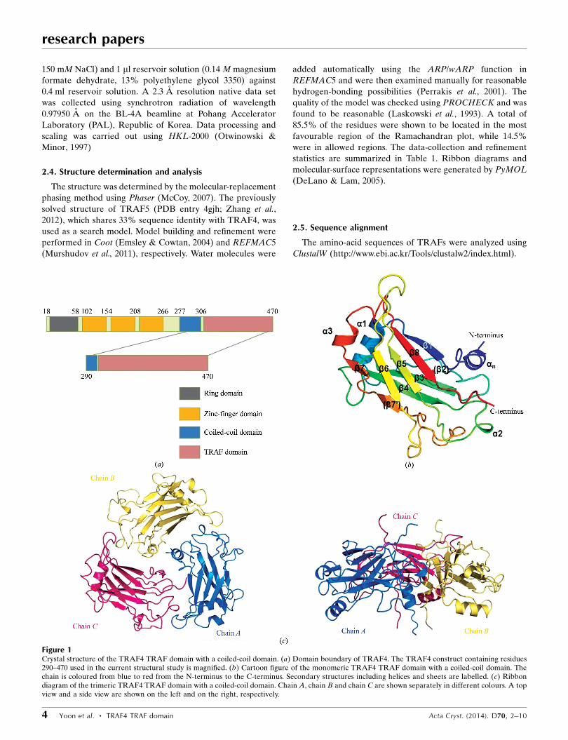
Figure 1Crystal structure of the TRAF4 TRAF domain with a coiled-coil domain. (a) Domain boundary of TRAF4. The TRAF4 construct containing residues290–470 used in the current structural study is magnified. (b) Cartoon figure of the monomeric TRAF4 TRAF domain with a coiled-coil domain. Thechain is coloured from blue to red from the N-terminus to the C-terminus. Secondary structures including helices and sheets are labelled. (c) Ribbondiagram of the trimeric TRAF4 TRAF domain with a coiled-coil domain. Chain A, chain B and chain C are shown separately in different colours. A topview and a side view are shown on the left and on the right, respectively.
2.6. PDB accession code
Coordinates and structural factors were deposited in the
Protein Data Bank as PDB entry 4k8u.
3. Results and discussion
3.1. Structure of the TRAF4 TRAF domain with a coiled-coildomain
TRAF4 is composed of several distinct domains, including a
RING domain, a zinc-finger domain, a coiled-coil domain and
a TRAF domain (Fig. 1a). The TRAF domain is located at the
C-terminus of TRAF4 and the coiled-coil domain is located
immediately before the TRAF domain. The TRAF domain
participates in protein–protein interactions in many important
signalling pathways.
The 2.3 A resolution crystal structure of the TRAF domain
with a coiled-coil domain comprising amino acids 290–470
was solved using the molecular-replacement (MR) method
and was refined to an Rwork of 23.7% and an Rfree of 28.1%.
The high-resolution structure of the TRAF4 TRAF domain
with a coiled-coil domain revealed that it is comprised of three
helices, �1–�3, eight sheets, �1–�8, and an extra helix, �N, at
the N-terminus (Fig. 1b). The overall fold was similar to that of
other TRAF domains. �2, which was found in the structures of
TRAF2 and TRAF6, was not detected in TRAF4, while �3,
which is absent only in TRAF2, was detected in TRAF4. �70,
which is located before �7, is only detected in TRAF4.
The structure of the TRAF4 TRAF domain is compact and
ordered in the central region, with several disordered regions.
The average B factor is 69 A2 (Table 1). Plotting individual B
factors for each residue showed that the stacked �-sheet in
research papers
Acta Cryst. (2014). D70, 2–10 Yoon et al. � TRAF4 TRAF domain 5
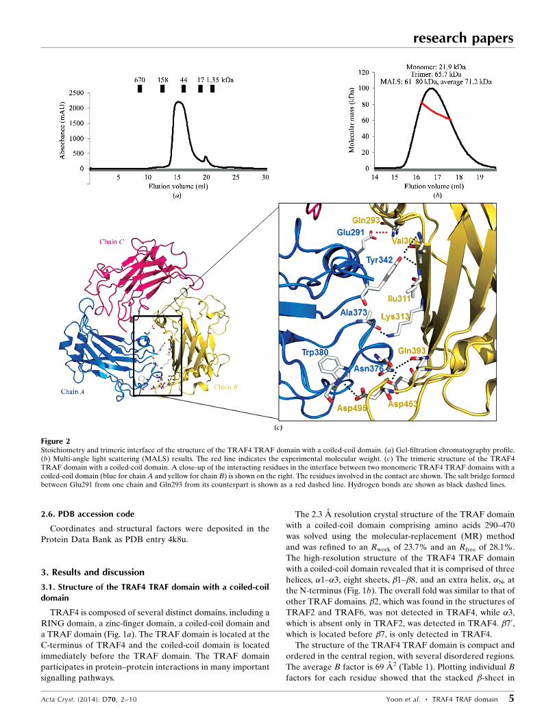
Figure 2Stoichiometry and trimeric interface of the structure of the TRAF4 TRAF domain with a coiled-coil domain. (a) Gel-filtration chromatography profile.(b) Multi-angle light scattering (MALS) results. The red line indicates the experimental molecular weight. (c) The trimeric structure of the TRAF4TRAF domain with a coiled-coil domain. A close-up of the interacting residues in the interface between two monomeric TRAF4 TRAF domains with acoiled-coil domain (blue for chain A and yellow for chain B) is shown on the right. The residues involved in the contact are shown. The salt bridge formedbetween Glu291 from one chain and Gln293 from its counterpart is shown as a red dashed line. Hydrogen bonds are shown as black dashed lines.
the central region has the lowest B factors, whereas the long
extended loop between �6 and �70 has the highest B factors
(Supplementary Fig. S11). The N- and C-terminal residues are
also more flexible with higher B factors.
There were three monomers in the asymmetric unit, chain
A, chain B and chain C (Fig. 1c). A model of chain A was built
from residues 291 to 468, while those of chain B and chain C
were built from residues 293 to 468 and from residues 295 to
470, respectively. Together, these chains formed a symmetric
trimer. The seven antiparallel �-sheets comprising residues
309–314, 345–351, 362–370, 386–390, 404–408, 439–441 and
456–462 were numbered �1, �3, �4, �5, �6 and �7, respectively
(Fig. 1b). The probable �-sheets which were detected in
TRAF2 and TRAF6, and comprised residues 331–337, are
indicated as (�2). An extra sheet comprising residues 434–436
is indicated as (�70). Four �-helices, �n (residues 291–302), �1
(residues 316–327), �2 (residues 375–377) and �3 (residues
442–446), were detected (Fig. 1b).
3.2. Stoichiometry of TRAF4 and the trimeric interface withinthe TRAF4 TRAF domain with a coiled-coil domain
Based on previously reported TRAF structures, the TRAF
domain was shown to exist as a trimer in solution (Park et al.,
1999; Zhang et al., 2012; Ye et al., 2002). In the crystals of
TRAF2 and TRAF6 the asymmetric unit contained a trimeric
molecule, while the asymmetric unit of the TRAF2 and
TRAF5 crystals contained a monomeric molecule (Park et al.,
1999; Zhang et al., 2012; Ye et al., 2002).
The TRAF4 TRAF domain with a coiled-coil domain that
was used in our structural study was overexpressed in bacteria
and was purified by affinity chromatography followed by gel-
filtration chromatography. On gel-filtration chromatography,
TRAF4 eluted at around 15 ml, indicating that it forms a
trimer in solution (Fig. 2a). The stoichiometry of TRAF4 was
accurately confirmed by multi-angle light scattering (MALS).
research papers
6 Yoon et al. � TRAF4 TRAF domain Acta Cryst. (2014). D70, 2–10
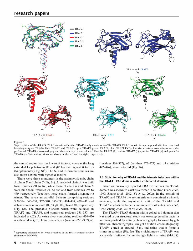
Figure 3Superposition of the TRAF4 TRAF domain with other TRAF family members. (a) The TRAF4 TRAF domain is superimposed with four structuralhomologues (grey, TRAF4; blue, TRAF2; red, TRAF3; cyan, TRAF5; green, TRAF6; blue, NALP1 PYD). Pairwise structural comparisons were alsoperformed. TRAF4 is coloured grey and the counterparts are coloured blue for TRAF2 (b), red for TRAF3 (c), cyan for TRAF5 (d) and green forTRAF6 (e). Side and top views are shown on the left and the right, respectively.
1 Supporting information has been deposited in the IUCr electronic archive(Reference: MN5037).
The calculated molecular weight of the monomeric TRAF4
TRAF domain with a coiled-coil domain (amino acids 290–
470) including the C-terminal His tag was 21.9 kDa and the
experimental molecular weight from MALS was 71.2 kDa
(0.8% fitting error), with a polydispersity of 1.000 (Fig. 2b).
According to the analysis using gel-filtration chromatography
and MALS, we concluded that TRAF4 exists as a trimer in
solution and that trimeric TRAF4, as detected in our crystal
structure, might be a biologically functional unit.
The trimeric TRAF4 TRAF domain with a coiled-coil
domain exhibited a typical mushroom shape, in which the
TRAF domain forms the cap and the coiled-coil domain forms
the stalk (Fig. 2c). The trimeric interface of TRAF4 is formed
by packing one end of the (�2)–�3 connecting loop, the �4–�5
connecting loop and �n of one monomer against an edge and a
face of the �5–�6 connecting loop and �1 of the neighbouring
monomer (Fig. 2c). Several hydrogen bonds formed between
Tyr342, Ala373, Asn376 and Trp380 from one TRAF molecule
(chain A) and Val309, Ile311, Lys313, Gln393, Asp453 and
Asp495 from the second molecule (chain B) and one main salt
bridge between Glu291 in one monomer (chain A) and Gln293
in the neighbouring monomer (chain B) are key interactions
for trimerization (Fig. 2c). The salt bridge in the trimeric
interface was located in the coiled-coil stalk region, indicating
that the coiled-coil domain is important for stabilization of
trimer formation. The total surface area of the three molecules
is around 27 438 A2, and 3938 A2 is buried upon complex
formation, which corresponds to an average of 1312 A2 per
molecule. An average surface area of 656 A2 is buried upon
dimer interface formation.
3.3. Comparison with other TRAF-domain structures
The DALI server was used to search for structurally similar
proteins to the TRAF4 TRAF domain (Holm & Sander, 1995)
and several structurally related proteins were identified
(Table 2). The top seven matches, which had Z-scores of
22.8–12.7, were (in order) TRAF2, TRAF3, TRAF5, TRAF6,
meprin, speckle-type POZ protein (SPOP) and HAUSP.
Pairwise structural alignments between the TRAF4 TRAF
domain and other TRAF domains showed that the positions
and the lengths of several loops in the TRAF4 TRAF domain
differed from those in other TRAF domains (Fig. 3). In
particular, two loops connecting �5–�6 and �6–�7 of the
TRAF4 TRAF domain were relatively longer than those in
other TRAFs (Fig. 3). The location of the �n helix of the
coiled-coil domain also differed in that it did not superimpose
well on the � helix in other TRAFs. These structural differ-
ences of TRAF4 might be critical to its functional differences
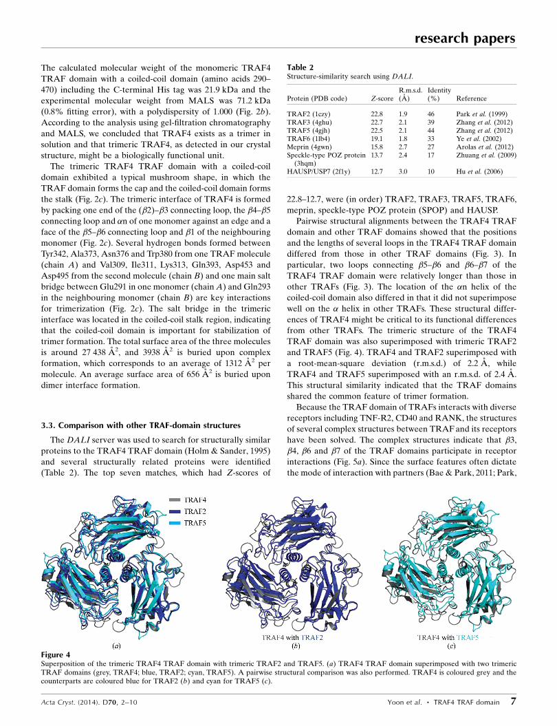
from other TRAFs. The trimeric structure of the TRAF4
TRAF domain was also superimposed with trimeric TRAF2
and TRAF5 (Fig. 4). TRAF4 and TRAF2 superimposed with
a root-mean-square deviation (r.m.s.d.) of 2.2 A, while
TRAF4 and TRAF5 superimposed with an r.m.s.d. of 2.4 A.
This structural similarity indicated that the TRAF domains
shared the common feature of trimer formation.
Because the TRAF domain of TRAFs interacts with diverse
receptors including TNF-R2, CD40 and RANK, the structures
of several complex structures between TRAF and its receptors
have been solved. The complex structures indicate that �3,
�4, �6 and �7 of the TRAF domains participate in receptor
interactions (Fig. 5a). Since the surface features often dictate
the mode of interaction with partners (Bae & Park, 2011; Park,
research papers
Acta Cryst. (2014). D70, 2–10 Yoon et al. � TRAF4 TRAF domain 7
Table 2Structure-similarity search using DALI.
Protein (PDB code) Z-scoreR.m.s.d.(A)
Identity(%) Reference
TRAF2 (1czy) 22.8 1.9 46 Park et al. (1999)TRAF3 (4ghu) 22.7 2.1 39 Zhang et al. (2012)TRAF5 (4gjh) 22.5 2.1 44 Zhang et al. (2012)TRAF6 (1lb4) 19.1 1.8 33 Ye et al. (2002)Meprin (4gwn) 15.8 2.7 27 Arolas et al. (2012)Speckle-type POZ protein
(3hqm)13.7 2.4 17 Zhuang et al. (2009)
HAUSP/USP7 (2f1y) 12.7 3.0 10 Hu et al. (2006)
Figure 4Superposition of the trimeric TRAF4 TRAF domain with trimeric TRAF2 and TRAF5. (a) TRAF4 TRAF domain superimposed with two trimericTRAF domains (grey, TRAF4; blue, TRAF2; cyan, TRAF5). A pairwise structural comparison was also performed. TRAF4 is coloured grey and thecounterparts are coloured blue for TRAF2 (b) and cyan for TRAF5 (c).
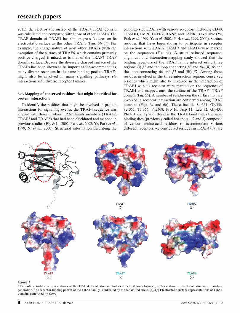
2011), the electrostatic surface of the TRAF4 TRAF domain
was calculated and compared with those of other TRAFs. The
TRAF domain of TRAF4 has similar gross features on its
electrostatic surface as the other TRAFs (Figs. 5b–5f). For
example, the charge nature of most other TRAFs (with the
exception of the surface of TRAF6, which contains primarily
positive charges) is mixed, as is that of the TRAF4 TRAF
domain surface. Because the diversely charged surface of the
TRAFs has been shown to be important for accommodating
many diverse receptors in the same binding pocket, TRAF4
might also be involved in many signalling pathways via
interactions with diverse receptor families.
3.4. Mapping of conserved residues that might be critical forprotein interactions
To identify the residues that might be involved in protein
interactions for signalling events, the TRAF4 sequence was
aligned with those of other TRAF family members (TRAF2,
TRAF3 and TRAF6) that had been elucidated and mapped in
previous studies (Ely & Li, 2002; Ye et al., 2002; Ye, Park et al.,
1999; Ni et al., 2000). Structural information describing the
complexes of TRAFs with various receptors, including CD40,
TRADD, LMP1, TNFR2, RANK and TANK, is available (Ye,
Park et al., 1999; Ye et al., 2002; Park et al., 1999, 2000). Surface
residues that have been shown to participate in receptor
interactions with TRAF2, TRAF3 and TRAF6 were marked
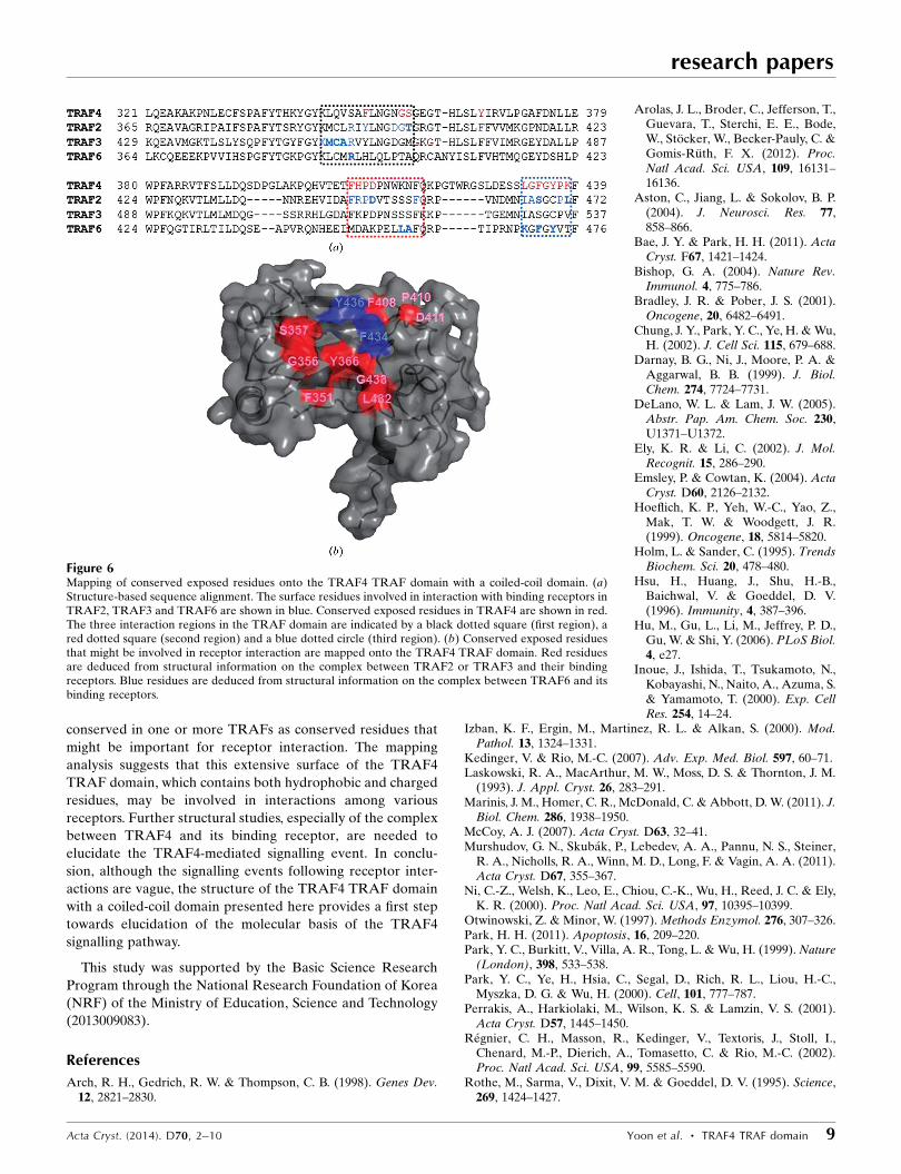
on the sequences (Fig. 6a). A structure-based sequence-
alignment and interaction-mapping study showed that the
binding receptors of the TRAF family interact using three
regions: (i) �3 and the loop connecting �3 and �4, (ii) �6 and
the loop connecting �6 and �7 and (iii) �7. Among those
residues involved in the three interaction regions, conserved
residues which might also be involved in the interaction of
TRAF4 with its receptor were marked on the sequence of
TRAF4 and mapped onto the surface of the TRAF4 TRAF
domain (Fig. 6b). A number of residues on the surface that are
involved in receptor interaction are conserved among TRAF
domains (Figs. 6a and 6b). These include Ser351, Gly356,
Ser357, Tyr366, Phe408, Pro410, Asp411, Leu432, Gly433,
Phe434 and Tyr436. Because the TRAF family uses the same
binding sites (previously called hot spots 1, 2 and 3) composed
of various amino-acid residues to accommodate various
different receptors, we considered residues in TRAF4 that are
research papers
8 Yoon et al. � TRAF4 TRAF domain Acta Cryst. (2014). D70, 2–10
Figure 5Electrostatic surface representations of the TRAF4 TRAF domain and its structural homologues. (a) Orientation of the TRAF domain for surfacegeneration. The receptor-binding pocket of the TRAF family is indicated by the red dotted circle. (b)–(f) Electrostatic surface representations of TRAFdomains generated by Coot.
conserved in one or more TRAFs as conserved residues that
might be important for receptor interaction. The mapping
analysis suggests that this extensive surface of the TRAF4
TRAF domain, which contains both hydrophobic and charged
residues, may be involved in interactions among various
receptors. Further structural studies, especially of the complex
between TRAF4 and its binding receptor, are needed to
elucidate the TRAF4-mediated signalling event. In conclu-
sion, although the signalling events following receptor inter-
actions are vague, the structure of the TRAF4 TRAF domain
with a coiled-coil domain presented here provides a first step
towards elucidation of the molecular basis of the TRAF4
signalling pathway.
This study was supported by the Basic Science Research
Program through the National Research Foundation of Korea
(NRF) of the Ministry of Education, Science and Technology
(2013009083).
References
Arch, R. H., Gedrich, R. W. & Thompson, C. B. (1998). Genes Dev.12, 2821–2830.
Arolas, J. L., Broder, C., Jefferson, T.,Guevara, T., Sterchi, E. E., Bode,W., Stocker, W., Becker-Pauly, C. &Gomis-Ruth, F. X. (2012). Proc.Natl Acad. Sci. USA, 109, 16131–16136.
Aston, C., Jiang, L. & Sokolov, B. P.(2004). J. Neurosci. Res. 77,858–866.
Bae, J. Y. & Park, H. H. (2011). ActaCryst. F67, 1421–1424.
Bishop, G. A. (2004). Nature Rev.Immunol. 4, 775–786.
Bradley, J. R. & Pober, J. S. (2001).Oncogene, 20, 6482–6491.
Chung, J. Y., Park, Y. C., Ye, H. & Wu,H. (2002). J. Cell Sci. 115, 679–688.
Darnay, B. G., Ni, J., Moore, P. A. &Aggarwal, B. B. (1999). J. Biol.Chem. 274, 7724–7731.
DeLano, W. L. & Lam, J. W. (2005).Abstr. Pap. Am. Chem. Soc. 230,U1371–U1372.
Ely, K. R. & Li, C. (2002). J. Mol.Recognit. 15, 286–290.
Emsley, P. & Cowtan, K. (2004). ActaCryst. D60, 2126–2132.
Hoeflich, K. P., Yeh, W.-C., Yao, Z.,Mak, T. W. & Woodgett, J. R.(1999). Oncogene, 18, 5814–5820.
Holm, L. & Sander, C. (1995). TrendsBiochem. Sci. 20, 478–480.
Hsu, H., Huang, J., Shu, H.-B.,Baichwal, V. & Goeddel, D. V.(1996). Immunity, 4, 387–396.
Hu, M., Gu, L., Li, M., Jeffrey, P. D.,Gu, W. & Shi, Y. (2006). PLoS Biol.4, e27.
Inoue, J., Ishida, T., Tsukamoto, N.,Kobayashi, N., Naito, A., Azuma, S.& Yamamoto, T. (2000). Exp. CellRes. 254, 14–24.
Izban, K. F., Ergin, M., Martinez, R. L. & Alkan, S. (2000). Mod.Pathol. 13, 1324–1331.
Kedinger, V. & Rio, M.-C. (2007). Adv. Exp. Med. Biol. 597, 60–71.Laskowski, R. A., MacArthur, M. W., Moss, D. S. & Thornton, J. M.
(1993). J. Appl. Cryst. 26, 283–291.Marinis, J. M., Homer, C. R., McDonald, C. & Abbott, D. W. (2011). J.
Biol. Chem. 286, 1938–1950.McCoy, A. J. (2007). Acta Cryst. D63, 32–41.Murshudov, G. N., Skubak, P., Lebedev, A. A., Pannu, N. S., Steiner,
R. A., Nicholls, R. A., Winn, M. D., Long, F. & Vagin, A. A. (2011).Acta Cryst. D67, 355–367.
Ni, C.-Z., Welsh, K., Leo, E., Chiou, C.-K., Wu, H., Reed, J. C. & Ely,K. R. (2000). Proc. Natl Acad. Sci. USA, 97, 10395–10399.
Otwinowski, Z. & Minor, W. (1997). Methods Enzymol. 276, 307–326.Park, H. H. (2011). Apoptosis, 16, 209–220.Park, Y. C., Burkitt, V., Villa, A. R., Tong, L. & Wu, H. (1999). Nature
(London), 398, 533–538.Park, Y. C., Ye, H., Hsia, C., Segal, D., Rich, R. L., Liou, H.-C.,
Myszka, D. G. & Wu, H. (2000). Cell, 101, 777–787.Perrakis, A., Harkiolaki, M., Wilson, K. S. & Lamzin, V. S. (2001).
Acta Cryst. D57, 1445–1450.Regnier, C. H., Masson, R., Kedinger, V., Textoris, J., Stoll, I.,
Chenard, M.-P., Dierich, A., Tomasetto, C. & Rio, M.-C. (2002).Proc. Natl Acad. Sci. USA, 99, 5585–5590.
Rothe, M., Sarma, V., Dixit, V. M. & Goeddel, D. V. (1995). Science,269, 1424–1427.
research papers
Acta Cryst. (2014). D70, 2–10 Yoon et al. � TRAF4 TRAF domain 9
Figure 6Mapping of conserved exposed residues onto the TRAF4 TRAF domain with a coiled-coil domain. (a)Structure-based sequence alignment. The surface residues involved in interaction with binding receptors inTRAF2, TRAF3 and TRAF6 are shown in blue. Conserved exposed residues in TRAF4 are shown in red.The three interaction regions in the TRAF domain are indicated by a black dotted square (first region), ared dotted square (second region) and a blue dotted circle (third region). (b) Conserved exposed residuesthat might be involved in receptor interaction are mapped onto the TRAF4 TRAF domain. Red residuesare deduced from structural information on the complex between TRAF2 or TRAF3 and their bindingreceptors. Blue residues are deduced from structural information on the complex between TRAF6 and itsbinding receptors.
Rothe, M., Wong, S. C., Henzel, W. J. & Goeddel, D. V. (1994). Cell,78, 681–692.
Shiels, H., Li, X., Schumacker, P. T., Maltepe, E., Padrid, P. A.,Sperling, A., Thompson, C. B. & Lindsten, T. (2000). Am. J. Pathol.157, 679–688.
Song, H. Y., Regnier, C. H., Kirschning, C. J., Goeddel, D. V. & Rothe,M. (1997). Proc. Natl Acad. Sci. USA, 94, 9792–9796.
Song, H. Y., Rothe, M. & Goeddel, D. V. (1996). Proc. Natl Acad. Sci.USA, 93, 6721–6725.
Xie, P. (2013). J. Mol. Signal. 8, 7.Ye, H. et al. (2002). Nature (London), 418, 443–447.Ye, X., Mehlen, P., Rabizadeh, S., VanArsdale, T., Zhang, H., Shin, H.,
Wang, J. J. L., Leo, E., Zapata, J., Hauser, C. A., Reed, J. C. &Bredesen, D. E. (1999). J. Biol. Chem. 274, 30202–30208.
Ye, H., Park, Y. C., Kreishman, M., Kieff, E. & Wu, H. (1999). Mol.Cell, 4, 321–330.
Yoon, J. H. & Park, H. H. (2013). Acta Cryst. F69, 1026–1028.Zapata, J. M., Lefebvre, S. & Reed, J. C. (2007). Adv. Exp. Med. Biol.
597, 188–201.Zapata, J. M., Pawlowski, K., Haas, E., Ware, C. F., Godzik, A. &
Reed, J. C. (2001). J. Biol. Chem. 276, 24242–24252.Zepp, J. A., Liu, C., Qian, W., Wu, L., Gulen, M. F., Kang, Z. & Li, X.
(2012). J. Immunol. 189, 33–37.Zhang, P., Reichardt, A., Liang, H. H., Aliyari, R., Cheng, D., Wang,
Y. Y., Xu, F., Cheng, G. H. & Liu, Y. F. (2012). Sci. Signal. 5, ra81.Zhuang, M., Calabrese, M. F., Liu, J., Waddell, M. B., Nourse, A.,
Hammel, M., Miller, D. J., Walden, H., Duda, D. M., Seyedin, S. N.,Hoggard, T., Harper, J. W., White, K. P. & Schulman, B. A. (2009).Mol. Cell, 36, 39–50.
research papers
10 Yoon et al. � TRAF4 TRAF domain Acta Cryst. (2014). D70, 2–10