structure of human ubiquitin-conjugating enzyme e2 g2 (ube2g2/ubc7)
Post on 06-Jul-2016
212 views
TRANSCRIPT
structural genomics communications
330 doi:10.1107/S1744309106009006 Acta Cryst. (2006). F62, 330–334
Acta Crystallographica Section F
Structural Biologyand CrystallizationCommunications
ISSN 1744-3091
Structure of human ubiquitin-conjugating enzymeE2 G2 (UBE2G2/UBC7)
Ryoichi Arai,a,b‡ Seiko
Yoshikawa,a‡ Kazutaka
Murayama,a,c Yuzuru Imai,d
Ryosuke Takahashi,d,e Mikako
Shirouzua,b and Shigeyuki
Yokoyamaa,b,f*
aProtein Research Group, RIKEN Genomic
Sciences Center, Tsurumi, Yokohama 230-0045,
Japan, bRIKEN SPring-8 Center, Harima Institute,
Sayo, Hyogo 679-5148, Japan, cTohoku
University Biomedical Engineering Research
Organization, Aoba, Sendai 980-8575, Japan,dRIKEN Brain Science Institute, Wako,
Saitama 351-0198, Japan, eDepartment of
Neurology, Graduate School of Medicine, Kyoto
University, Sakyo, Kyoto 606-8507, Japan, andfDepartment of Biophysics and Biochemistry,
Graduate School of Science, The University of
Tokyo, Bunkyo, Tokyo 113-0033, Japan
‡ These authors contributed equally to this
work.
Correspondence e-mail:
Received 22 December 2005
Accepted 10 March 2006
PDB Reference: human UBE2G2/UBC7, 2cyx,
r2cyxsf.
The human ubiquitin-conjugating enzyme E2 G2 (UBE2G2/UBC7) is involved
in protein degradation, including a process known as endoplasmic reticulum-
associated degradation (ERAD). The crystal structure of human UBE2G2/
UBC7 was solved at 2.56 A resolution. The UBE2G2 structure comprises a
single domain consisting of an antiparallel �-sheet with four strands, five
�-helices and two 310-helices. Structural comparison of human UBE2G2 with
yeast Ubc7 indicated that the overall structures are similar except for the long
loop region and the C-terminal helix. Superimposition of UBE2G2 on UbcH7 in
a c-Cbl–UbcH7–ZAP70 ternary complex suggested that the two loop regions of
UBE2G2 interact with the RING domain in a similar way to UbcH7. In
addition, the extra loop region of UBE2G2 may interact with the RING domain
or its neighbouring region and may be involved in the binding specificity and
stability.
1. Introduction
Ubiquitin-dependent protein degradation plays an important role in
the regulation of various cellular processes, including cell-cycle
progression, signal transduction, transcription, DNA repair and
protein quality control (Koepp et al., 1999; Laney & Hochstrasser,
1999). Ubiquitination involves the successive actions of the ubiquitin-
activating (E1), ubiquitin-conjugating (E2) and ubiquitin-protein
ligase enzymes (E3) (Hershko & Ciechanover, 1998; Pickart, 2001).
The E1 enzyme activates free ubiquitin and transfers it to E2 through
a thioester linkage between the ubiquitin C-terminus and an E2
active-site cysteine. The E3 enzyme recognizes its substrate and E2
and catalyzes the formation of an isopeptide bond between a lysine
"-amino group of the substrate (or ubiquitin) and the C-terminal
carboxyl group of ubiquitin Gly76. Over 30 human E2s have been
identified and they all contain a conserved �150 amino-acid catalytic
core. The E2 enzymes are grouped into four classes depending on the
presence and the location of additional sequences (Jentsch, 1992).
Some of these enzymes contain extra C-terminal and/or N-terminal
extensions from the core domain. The class I enzymes are the smallest
E2 enzymes and consist almost entirely of the conserved core
domain. The class II enzymes contain an extra C-terminal extension
from the core domain, while class III enzymes have an N-terminal
extension. The class IV enzymes contain both N- and C- terminal
extensions.
The human UBE2G2 gene encodes the ubiquitin-conjugating
enzyme E2 G2 (UBE2G2/UBC7), with a molecular weight of
18.6 kDa (165 amino acids). It was mapped to the region of human
chromosome 21q22.3 and its transcripts are ubiquitously expressed in
human tissues (Katsanis & Fisher, 1998; Rose et al., 1998). Human
UBE2G2 is a class I E2 enzyme. Recently, bacterial expression of His-
tagged human UBE2G2 was reported (Reyes et al., 2006). The human
UBE2G2 protein shares 100, 62, 47 and 27% identities to murine
UBE2G2/UBC7 (MmUBC7), yeast Ubc7, human UBE2G1 and
human UbcH7, respectively (Fig. 1a). The crystal structures of yeast
Ubc7 (Cook et al., 1997), a human E6AP–UbcH7 complex (Huang et# 2006 International Union of Crystallography
All rights reserved
al., 1999) and a human c-Cbl–UbcH7–ZAP-70 complex (Zheng et al.,
2000) have been reported. Functional studies have associated yeast
Ubc7 and MmUBC7 with the degradation of endoplasmic reticulum
(ER) substrates, a process known as ER-associated degradation
(ERAD; Jungmann et al., 1993; Fang et al., 2001; Tiwari & Weissman,
2001). Parkin, a gene product responsible for autosomal recessive
juvenile Parkinsonism (AR-JP), interacts with human UBE2G2/
UBC7 and UBC6 through its RING domain and specifically
ubiquitinates the Pael receptor in the presence of the E2s (Imai et al.,
2001). Furthermore, exogenous MmUBC7 mediates the ubiquitina-
tion and down regulation of both the inositol 1,4,5-triphosphate
receptor in human neuroblastoma cells (Webster et al., 2003) and the
human type 2 iodothyronine selenodeiodinase (Kim et al., 2003).
Recently, the interactions of human UBE2G2/UBC7 with some
RING-finger E3s, such as human HRD1 (Kikkert et al., 2004) and
TEB4 (Hassink et al., 2005), have been reported. To analyze the
structural and functional details of human UBE2G2/UBC7, which is
involved in important cellular processes, its structure must be
determined and compared with those of its homologues. Here, we
report the crystal structure of human UBE2G2/UBC7 at 2.56 A
resolution and discuss its structural aspects.
2. Materials and methods
2.1. Protein expression and purification
The human UBE2G2 gene (Imai et al., 2001) encoding human
ubiquitin-conjugating enzyme E2 G2 (UBE2G2/UBC7) was cloned
into a modified pENTR vector with a tobacco etch virus (TEV)
protease cleavage site, derived from pENTR1A (Invitrogen). The
expression vector pET/cMBP-UBE2G2 was constructed using
Gateway technology (Invitrogen) with pENTR/TEV-UBE2G2 and
pET/cMBP-GATEWAY bearing a T7 promoter, an N-terminal
maltose-binding protein (MBP) tag and a Gateway reading frame
cassette A (Invitrogen). The UBE2G2 protein was expressed as a
fusion with an N-terminal MBP tag and a TEV protease cleavage site
in Escherichia coli BL21(DE3). The protein was first purified on an
amylose-resin column (New England Biolabs) and the MBP tag was
then cleaved by His-tagged TEV protease, which was removed using
a HisTrap column (GE Healthcare). The protein was purified further
by Mono-Q and Superdex 75 column (GE Healthcare) chromato-
graphy steps. The yield of purified UBE2G2 protein was 8 mg per
litre of culture. The construct that was used for crystallization
contained the cloning artifact sequence GGSEF at the N-terminus.
structural genomics communications
Acta Cryst. (2006). F62, 330–334 Arai et al. � Human UBE2G2/UBC7 331
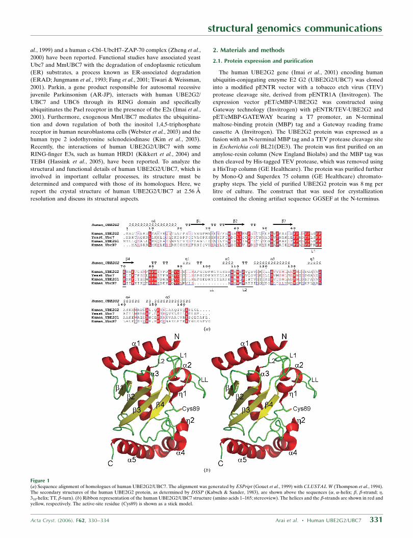
Figure 1(a) Sequence alignment of homologues of human UBE2G2/UBC7. The alignment was generated by ESPript (Gouet et al., 1999) with CLUSTAL W (Thompson et al., 1994).The secondary structures of the human UBE2G2 protein, as determined by DSSP (Kabsch & Sander, 1983), are shown above the sequences (�, �-helix; �, �-strand; �,310-helix; TT, �-turn). (b) Ribbon representation of the human UBE2G2/UBC7 structure (amino acids 1–165; stereoview). The helices and the �-strands are shown in red andyellow, respectively. The active-site residue (Cys89) is shown as a stick model.
2.2. Crystallization and data collection
Preliminary crystals of human UBE2G2 were obtained under
condition No. 42 (0.1 M Tris–HCl buffer pH 8.5 containing 1.5 M
ammonium sulfate and 12% glycerol) of the Crystal Screen 2 crystal
screening kit (Hampton Research) using the 96-well sitting-drop
vapour-diffusion method. The crystals of UBE2G2 used for structure
determination were obtained in drops composed of 1 ml 8.5 mg ml�1
protein solution (20 mM Tris–HCl buffer pH 8.0 containing 120 mM
NaCl, 2 mM DTT) and 1 ml reservoir solution (0.1 M Tris–HCl buffer
pH 8.1 containing 1.45 M ammonium sulfate and 12% glycerol;
Hampton Research) by the hanging-drop vapour-diffusion method
against 500 ml reservoir solution. A rod-like crystal (�350 � 100 �
100 mm) was obtained within a few days and was used for data
collection. The data collection was carried out at 100 K, with the
reservoir solution containing 27.5% glycerol as a cryoprotectant. The
diffraction data were collected at SPring-8 BL26B1 (Yamamoto et al.,
2002) and were recorded on a Jupiter 210 CCD detector (Rigaku).
All diffraction data were processed with the HKL2000 program suite
(Otwinowski & Minor, 1997).
2.3. Structure determination and refinement
The structure was solved by the molecular-replacement method
using MOLREP (Vagin & Teplyakov, 1997) with the yeast Ubc7
structure (PDB code 2ucz; Cook et al., 1997) as a search model. Data
in the resolution range 50–3.0 A were used in both rotation and
translation calculations, which gave an obvious solution with signifi-
cant contrast, resulting in three molecules in the asymmetric unit with
a Matthews coefficient (VM) of 3.83 A3 Da�1 and a solvent content of
67.91%. The model was corrected iteratively using O (Jones et al.,
1991) and was refined to 2.56 A using LAFIRE (Yao et al., 2006),
REFMAC5 (Murshudov et al., 1997) and Crystallography & NMR
System (CNS; Brunger et al., 1998). The crystallographic data and
refinement statistics are presented in Table 1. Since there was addi-
tional electron density, four residues of the cloning artifact sequence
at the N-terminus were also modelled. The final model includes 507
amino-acid residues of three UBE2G2 monomers and 23 water
molecules in the asymmetric unit. In the loop regions (residues 100–
106 and 131–133), the electron density corresponding to the side
chains was ambiguous, which increased the B factor. In addition,
relatively large areas of the molecular surface were exposed to the
solvent in the crystal as the solvent content was high. These features
resulted in the high average B factor. The quality of the model was
inspected using PROCHECK (Laskowski et al., 1993). The figures
were created using PyMOL (DeLano, 2005).
3. Results and discussion
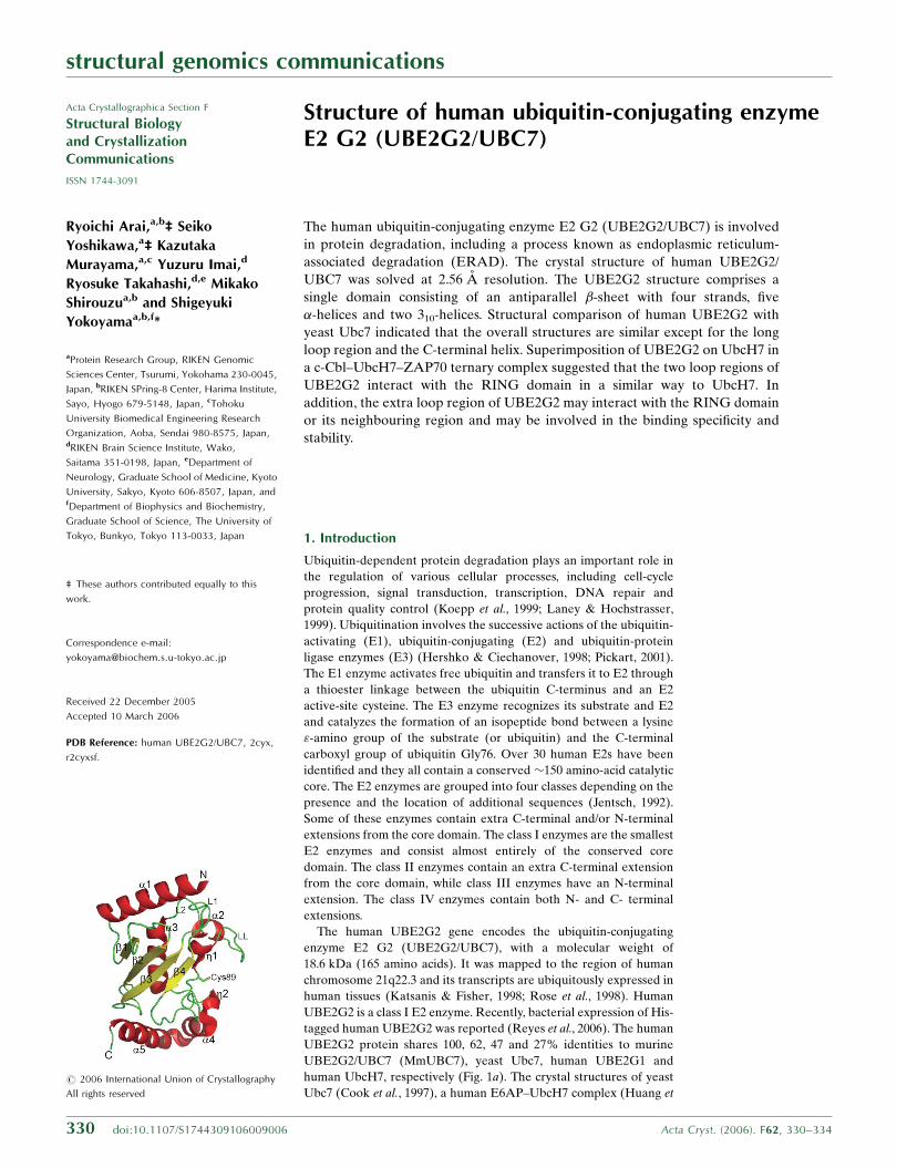
The crystal structure of human UBE2G2 comprises a single domain
consisting of an antiparallel �-sheet with four strands (�1–�4), five
�-helices (�1–�5) and two 310-helices (�1 and �2; Fig. 1b). The
ubiquitin-accepting residue Cys89 is located near �1. The overall
folding of UBE2G2 corresponds to the typical fold of ubiquitin-
conjugating enzymes. According to analytical ultracentrifugation, the
structural genomics communications
332 Arai et al. � Human UBE2G2/UBC7 Acta Cryst. (2006). F62, 330–334
Table 1X-ray data-collection and refinement statistics.
Values in parentheses are for the outer shell (2.65–2.56 A).
Data collectionSpace group P212121
Unit-cell parameters (A) a = 63.52, b = 87.61, c = 157.41Wavelength (A) 1.000Resolution (A) 50–2.56Total reflections 117935Unique reflections 28705Redundancy 4.1 (3.7)Completeness (%) 97.5 (84.6)I/�(I) 22.2 (4.2)Rsym† (%) 5.5 (29.3)
RefinementResolution (A) 49.43–2.56No. of reflections 28395No. of protein atoms 3996No. of water molecules 23Rwork (%) 22.8Rfree‡ (%) 26.2R.m.s.d. bond lengths (A) 0.009R.m.s.d. bond angles (�) 1.6Average B factor (A2) 75.7Ramachandran plot
Most favoured regions (%) 85.9Additional allowed regions (%) 14.1Generously allowed regions (%) 0.0Disallowed regions (%) 0.0
† Rsym =PjIi � Iavgj=
PIi , where Ii is the observed intensity and Iavg is the average
intensity. ‡ Rfree is calculated for 10% of randomly selected reflections excluded fromrefinement.
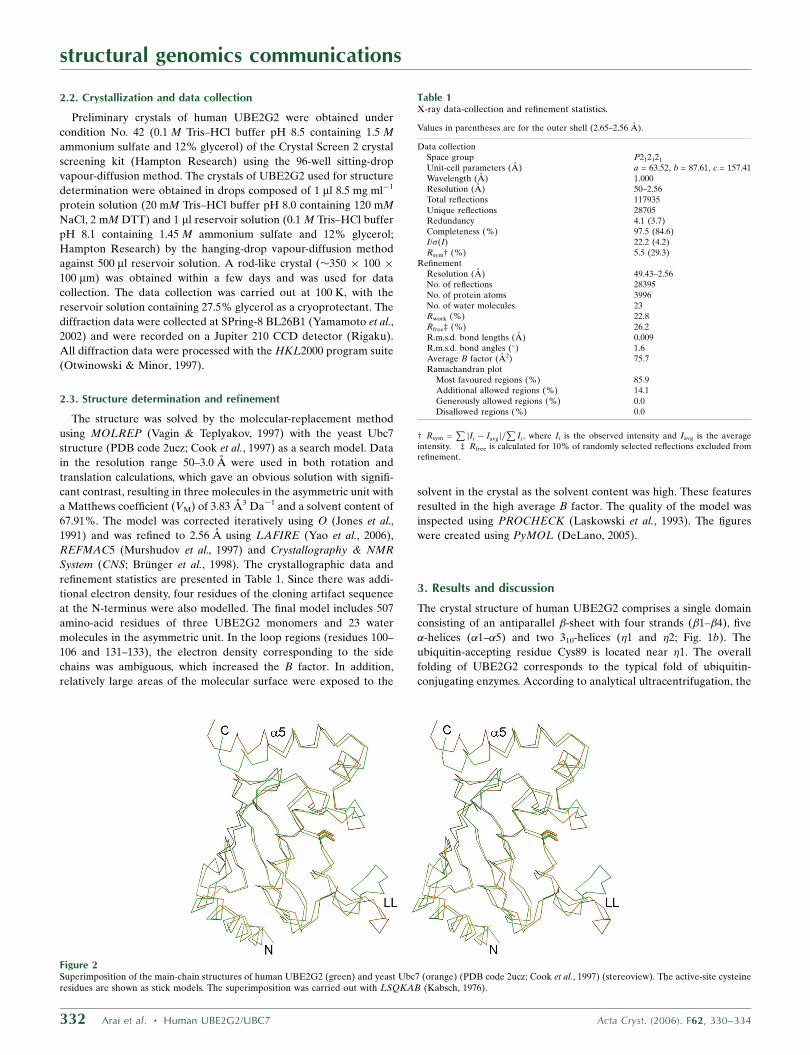
Figure 2Superimposition of the main-chain structures of human UBE2G2 (green) and yeast Ubc7 (orange) (PDB code 2ucz; Cook et al., 1997) (stereoview). The active-site cysteineresidues are shown as stick models. The superimposition was carried out with LSQKAB (Kabsch, 1976).
molecular weight of UBE2G2 was �18 kDa (data not shown), indi-
cating that the UBE2G2 protein exists as a monomer in solution.
Fig. 2 shows the superimposition of the main-chain structures of
human UBE2G2 and yeast Ubc7 (Cook et al., 1997). The overall
structure of UBE2G2 is remarkably similar to that of yeast Ubc7
(r.m.s.d. = 2.15 A over 164 C� atoms). The major differences between
human UBE2G2 and yeast Ubc7 are the structure of the long loop
(LL) region (95–106) and the angle of the C-terminal helix. The
C-terminal helix (�5) of UBE2G2 is closer to the �-sheet core region
than that of yeast Ubc7. The important interactions of UBE2G2 in
the contact region of the C-terminal helix and the core region are the
hydrophobic interactions among Phe54, Met77, Phe78, Ile154 and
Ile158 and the salt bridge between Glu76 and Lys161. The residues
Glu76, Ile154 and Ile158 are replaced with Ser76, Gln154 and Ser158
in yeast Ubc7, respectively, suggesting that the interactions of yeast
Ubc7 are weaker than those of UBE2G2. Consequently, the angle of
the C-terminal helix (�5) may change. Recently, the crystal structure
of the human ubiquitin-conjugating enzyme E2 G1 (UBE2G1), which
is another human homologue of yeast Ubc7, was deposited in the
PDB (PDB code 2awf). A structural comparison of UBE2G2 with
UBE2G1 revealed that the overall folding of UBE2G2 is similar to
that of UBE2G1 (r.m.s.d. = 1.12 A over 115 C� atoms), but in
UBE2G1 the residues 98–106 within the long loop (LL) region and
the C-terminal helices (�2, �4 and �5) were not located in the model
owing to disorder.
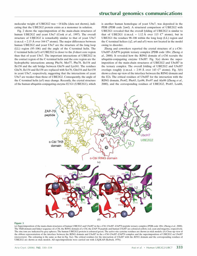
Zheng and coworkers reported the crystal structure of a c-Cbl–
UbcH7–ZAP70 peptide ternary complex (PDB code 1fbv; Zheng et
al., 2000). It revealed how the RING domain of c-Cbl recruits the
ubiquitin-conjugating enzyme UbcH7. Fig. 3(a) shows the super-
imposition of the main-chain structures of UBE2G2 and UbcH7 in
the ternary complex. The overall folding of UBE2G2 and UbcH7
overlaps roughly (r.m.s.d. = 2.95 A over 143 C� atoms). Fig. 3(b)
shows a close-up view of the interface between the RING domain and
the E2s. The critical residues of UbcH7 for the interaction with the
RING domain, Pro62, Phe63, Lys96, Pro97 and Ala98 (Zheng et al.,
2000), and the corresponding residues of UBE2G2, Pro65, Leu66,
structural genomics communications
Acta Cryst. (2006). F62, 330–334 Arai et al. � Human UBE2G2/UBC7 333
Figure 3(a) Superimposition of the main-chain structures of human UBE2G2 and UbcH7 in the c-Cbl–UbcH7–ZAP70 peptide ternary complex (PDB code 1fbv; Zheng et al., 2000).The TKB domain and linker sequence of c-Cbl, the RING domain of c-Cbl, the ZAP-70 peptide and human UbcH7 are coloured yellow, red, cyan and magenta, respectively.The zinc ions are indicated by grey spheres. The human UBE2G2 protein is coloured green. The active-site cysteine residues are shown as stick models. (b) Close-up view ofthe ribbon representation of the interface between the RING domain and UbcH7 in the c-Cbl–UbcH7–ZAP70 complex and the superimposition of UBE2G2 on UbcH7(stereoview). The colouring is the same as that in Fig. 3(a). The critical residues for the interaction of UbcH7 with the RING domain and the corresponding residues ofUBE2G2 are shown as stick models. All superimpositions were carried out with LSQKAB (Kabsch, 1976).
Ser111, Pro112 and Val113, overlap remarkably well (r.m.s.d. =
0.768 A over five C� atoms), suggesting that the L1 (64–70) and L2
(110–115) loops of UBE2G2 are involved in the interaction with the
RING domain in a similar way as UbcH7. This is consistent with the
previous results that Parkin binds to UBE2G2 as well as UbcH7 and
ubiquitinates substrates (Imai et al., 2000, 2001). In addition,
UBE2G2 has the extra long loop (LL) region (95–106), which is
probably located on the side near the RING domain. The B factor of
the LL region is relatively high, implying the possibility of confor-
mational flexibility. The LL region may interact with the RING
domain or its neighbouring region and may be involved in the binding
specificity and stability.
We thank Mr R. Akasaka and Dr M. Kukimoto-Niino for the
analytical ultracentrifugation, Mr S. Kamo for computer maintenance
and Ms A. Ishii, Ms K. Yajima, Ms M. Sunada and Ms T. Nakayama
for clerical assistance. We also thank Dr M. Yamamoto for data
collection at the RIKEN Structural Genomics beamline BL26B1 at
SPring-8. This work was supported by the RIKEN Structural
Genomics/Proteomics Initiative (RSGI), the National Project on
Protein Structural and Functional Analyses, the Ministry of Educa-
tion, Culture, Sports, Science and Technology of Japan.
References
Brunger, A. T., Adams, P. D., Clore, G. M., DeLano, W. L., Gros, P., Grosse-Kunstleve, R. W., Jiang, J.-S., Kuszewski, J., Nilges, M., Pannu, N. S., Read,R. J., Rice, L. M., Simonson, T. & Warren, G. L. (1998). Acta Cryst. D54,905–921.
Cook, W. J., Martin, P. D., Edwards, B. F., Yamazaki, R. K. & Chau, V. (1997).Biochemistry, 36, 1621–1627.
DeLano, W. L. (2005). PyMOL v.0.98. DeLano Scientific, South San Francisco,CA, USA.
Fang, S., Ferrone, M., Yang, C., Jensen, J. P., Tiwari, S. & Weissman, A. M.(2001). Proc. Natl Acad. Sci. USA, 98, 14422–14427.
Gouet, P., Courcelle, E., Stuart, D. I. & Metoz, F. (1999). Bioinformatics, 15,305–308.
Hassink, G., Kikkert, M., van Voorden, S., Lee, S. J., Spaapen, R., van Laar, T.,Coleman, C. S., Bartee, E., Fruh, K., Chau, V. & Wiertz, E. (2005). Biochem.J. 388, 647–655.
Hershko, A. & Ciechanover, A. (1998). Annu. Rev. Biochem. 67, 425–479.Huang, L., Kinnucan, E., Wang, G., Beaudenon, S., Howley, P. M., Huibregtse,
J. M. & Pavletich, N. P. (1999). Science, 286, 1321–1326.Imai, Y., Soda, M., Inoue, H., Hattori, N., Mizuno, Y. & Takahashi, R. (2001).
Cell, 105, 891–902.Imai, Y., Soda, M. & Takahashi, R. (2000). J. Biol. Chem. 275, 35661–35664.Jentsch, S. (1992). Annu. Rev. Genet. 26, 179–207.Jones, T. A., Zou, J. Y., Cowan, S. W. & Kjeldgaard, M. (1991). Acta Cryst. A47,
110–119.Jungmann, J., Reins, H. A., Schobert, C. & Jentsch, S. (1993). Nature
(London), 361, 369–371.Kabsch, W. (1976). Acta Cryst. A32, 922–923.Kabsch, W. & Sander, C. (1983). Biopolymers, 22, 2577–2637.Katsanis, N. & Fisher, E. M. (1998). Genomics, 51, 128–131.Kikkert, M., Doolman, R., Dai, M., Avner, R., Hassink, G., van Voorden, S.,
Thanedar, S., Roitelman, J., Chau, V. & Wiertz, E. (2004). J. Biol. Chem. 279,3525–3534.
Kim, B. W., Zavacki, A. M., Curcio-Morelli, C., Dentice, M., Harney, J. W.,Larsen, P. R. & Bianco, A. C. (2003). Mol. Endocrinol. 17, 2603–2612.
Koepp, D. M., Harper, J. W. & Elledge, S. J. (1999). Cell, 97, 431–434.Laney, J. D. & Hochstrasser, M. (1999). Cell, 97, 427–430.Laskowski, R. A., MacArthur, M. W., Moss, D. S. & Thornton, J. M. (1993). J.
Appl. Cryst. 26, 283–291.Murshudov, G. N., Vagin, A. A. & Dodson, E. J. (1997). Acta Cryst. D53,
240–255.Otwinowski, Z. & Minor, W. (1997). Methods Enzymol. 276, 307–326.Pickart, C. M. (2001). Annu. Rev. Biochem. 70, 503–533.Reyes, L. F., Sommer, C. A., Beltramini, L. M. & Henrique-Silva, F. (2006).
Protein Expr. Purif. 45, 324–328.Rose, S. A., Leek, J. P., Moynihan, T. P., Ardley, H. C., Markham, A. F. &
Robinson, P. A. (1998). Cytogenet. Cell Genet. 83, 98–99.Thompson, J. D., Higgins, D. G. & Gibson, T. J. (1994). Nucleic Acids Res. 22,
4673–4680.Tiwari, S. & Weissman, A. M. (2001). J. Biol. Chem. 276, 16193–16200.Vagin, A. & Teplyakov, A. (1997). J. Appl. Cryst. 30, 1022–1025.Webster, J. M., Tiwari, S., Weissman, A. M. & Wojcikiewicz, R. J. (2003). J.
Biol. Chem. 278, 38238–38246.Yamamoto, M., Kumasaka, T., Ueno, G., Ida, K., Kanda, H., Miyano, M. &
Ishikawa, T. (2002). Acta Cryst. A58, C302.Yao, M., Zhou, Y. & Tanaka, I. (2006). Acta Cryst. D62, 189–196.Zheng, N., Wang, P., Jeffrey, P. D. & Pavletich, N. P. (2000). Cell, 102, 533–539.
structural genomics communications
334 Arai et al. � Human UBE2G2/UBC7 Acta Cryst. (2006). F62, 330–334