structure and function of ubiquitin conjugating enzyme e2-25k: the tail is a core-dependent...
TRANSCRIPT
Structure and Function of Ubiquitin Conjugating Enzyme E2-25K: The Tail Is aCore-Dependent Activity Element†
Margaret T. Haldeman,‡,§ Gang Xia,| Eileen M. Kasperek,‡ and Cecile M. Pickart*,|
Department of Biochemistry, School of Medicine and Biomedical Sciences, State UniVersity of New York,Buffalo, New York 14214, and Department of Biochemistry, School of Hygiene and Public Health, Johns Hopkins UniVersity,
Baltimore, Maryland 21205
ReceiVed April 1, 1997; ReVised Manuscript ReceiVed June 16, 1997X
ABSTRACT: Individual members of the conserved family of ubiquitin conjugating enzymes (E2s) mediatethe ubiquitination and turnover of specific substrates of the ubiquitin-dependent degradation pathway.E2 proteins have a highly conserved core domain of∼150 amino acids which contains the active-siteCys. Certain E2s have unique terminal extensions, which are thought to contribute to selective E2 functionby interacting either with substrates or withtrans-acting factors such as ubiquitin-protein ligases (E3s).We used the mammalian ubiquitin conjugating enzyme E2-25K in a biochemical test of this hypothesis.The properties of two truncated derivatives show that the 47-residue tail of E2-25K is necessary for threeof the enzyme’s characteristic properties: high activity in the synthesis of unanchored K48-linkedpolyubiquitin chains; resistance of the active-site Cys residue to alkylation; and an unusual discriminationagainst noncognate (nonmammalian) ubiquitin activating (E1) enzymes. However, the tail is not sufficientto generate these properties, as shown by the characteristics of a chimeric enzyme in which the tail ofE2-25K was fused to the core domain of yeast UBC4. These and other results indicate that the specificbiochemical function of the tail is strongly dependent upon unique features of the E2-25K core domain.Thus, divergent regions within the conserved core domains of E2 proteins may be highly significant forfunction. Expression of truncated E2-25K as a glutathioneS-transferase (GST) fusion protein resulted inthe apparent recovery of E2-25K-specific properties, including activity in chain synthesis. However, thecatalytic mechanism utilized by the truncated fusion protein proved to be distinct from the mechanismutilized by the wild-type enzyme. The unexpected properties of the fusion protein were due to GST-induced dimerization. These results indicate the potential for self-association to modulate the polyubiquitinchain synthesis activities of E2 proteins, and indicate that caution should be applied in interpreting theactivities of GST fusion proteins.
Ubiquitin-dependent proteolysis is the predominant mech-anism for turnover of short-lived proteins in eukaryotic cells(1). The role of ubiquitin in proteolysis is that of a covalentsignal: ubiquitination allows target proteins to be recognizedby the 26S proteasome. The multienzyme ubiquitin pathwayis responsible for the turnover of key regulatory proteins,including mitotic cyclins (2, 3), transcription factors (4), andthe tumor suppressor protein p53 (5). The pathway alsodegrades misfolded proteins (6).Specificity in ubiquitin-mediated proteolysis appears to
arise primarily at the level of ubiquitination. In this process(1), the carboxyl terminus of ubiquitin (G76) is first activatedthrough ATP-dependent formation of a thiol ester with a Cysresidue of ubiquitin activating enzyme or E1.1 Ubiquitin isnext transferred to a Cys residue at the active site of aubiquitin conjugating enzyme or E2 protein. Althoughcertain E2 proteins can directly transfer ubiquitin to target
proteins [e.g., (7)], it appears that target protein ubiquitinationis usually accomplished following ubiquitin transfer fromthe E2 to a Cys residue at the active site of a ubiquitin-protein ligase or E3 (8). The E3 then transfers this ubiquitinto an internal Lys residue of the substrate. Conjugation ofadditional ubiquitins, in the form of a K48-linked polyubiq-uitin chain, renders the target protein especially susceptibleto degradation by the 26S proteasome (9).A large family of E2 proteins is characterized by a core
region consisting of∼150 amino acids that is conserved atthe level of both sequence and three-dimensional structure(10-13). Within this core region is a highly conservedactive-site motif containing the Cys residue that forms a thiolester with ubiquitin (10). Certain E2s have additionalregions, which may be up to∼125 residues in length,flanking the 15-kDa core region. These “extensions” aretypically divergent relative to comparable regions in otherE2 proteins within the same organism.Molecular genetic studies in the yeastSaccharomyces
cereVisiaehave shown that certain functions of the ubiquitinpathway involve specific members of the E2 protein family
† Funded by NIH Grant DK46984. C.M.P. is the recipient of aResearch Career Development Award from the NIH.* Correspondence should be addressed to this author at the Depart-
ment of Biochemistry, Johns Hopkins University, 615 N. Wolfe St.,Baltimore, MD 21205. Telephone: (410) 614-4554. Fax: (410) 955-2926. Email: [email protected].
‡ State University of New York.§ Present address: Department of Microbiology, The Ohio State
University, Columbus, OH 43210.| Johns Hopkins University.X Abstract published inAdVance ACS Abstracts,August 15, 1997.
1 Abbreviations: BSA, bovine serum albumin; DTT, dithiothreitol;GST, glutathioneS-transferase; E1, ubiquitin activating enzyme; E2,ubiquitin conjugating enzyme; E3, ubiquitin-protein ligase; IPTG,isopropyl â-D-thiogalactopyranoside; PMSF, phenylmethanesulfonylfluoride; TLCK, tosyllysyl chloromethyl ketone; Ub74, des-GlyGlyubiquitin (lacking the two C-terminal residues); Ub76, full-lengthubiquitin.
10526 Biochemistry1997,36, 10526-10537
S0006-2960(97)00750-2 CCC: $14.00 © 1997 American Chemical Society
(see Discussion). Such specificity could arise in one (orboth) of two ways: interaction of the E2 with the targetprotein, or interaction of the E2 with an E3 (or othertrans-acting factor). In several cases, it is apparently the E3 proteinthat interacts with a specific primary sequence element inthe target protein, while the E2 interacts with the E3 [e.g.,(14)]. However, there is evidence to suggest that certainE2s function in substrate recognition [e.g., (15-17)]. Re-gardless of the mechanistic basis of E2 specificity, theinteractions leading to such specificity will, in the simplestcase, involve regions of the E2 protein that are divergentamong the family members within a given organism. Thus,E2 terminal extensions may be specificity elements that directthe interactions of E2 proteins with E3 proteins and/orsubstrates (10, 18). Several lines of evidence support thishypothesis (see Discussion).Ubiquitin conjugating enzyme E2-25K is broadly ex-
pressed in mammalian tissues (19-22). In Vitro, E2-25Kis specific for ubiquitin as conjugation target, synthesizingunanchored polyubiquitin chains that are linked exclusivelyby K48-G76 isopeptide bonds (19). This activity, whichis unique to E2-25K among known mammalian E2s, issuggestive of a role in protein degradation, as is the highhomology between the core of E2-25K and the cores of theE2 proteins encoded by the yeastUBC1, UBC4, andUBC5genes (20). The latter three E2s form an essential subfamilythat functions in the turnover of short-lived proteins; UBC4and UBC5 are functionally redundant (23, 24). The biologi-cal function of E2-25K remains unknown.The 47-residue carboxyl-terminal tail of E2-25K is absent
in UBC4 and UBC5, and is dissimilar to the correspondingregion of UBC1. E2-25K, but not yeast UBC1, catalyzesthe synthesis of unanchored polyubiquitin chainsin Vitro(20). Based on this finding, we proposed that the tail ofE2-25K conferred activity toward ubiquitin as target protein.The studies presented here test this hypothesis throughanalyses of truncated and chimeric forms of E2-25K. Theresults support this hypothesis, but also show that specificfeatures of the E2-25K core domain are critical for activityand specificity.
EXPERIMENTAL PROCEDURES
Materials, Enzyme Preparations, and General Methods.Reagents and proteins were from Sigma unless statedotherwise. Bovine ubiquitin or recombinant K48R-ubiquitinwas radioiodinated to∼8000 cpm/pmol with chloramine-T(25). E1 was purified to electrophoretic homogeneity frombovine erythrocytes (26) or wheat germ (27). E2-20K waspurified from rabbit reticulocytes (26). Purified yeast E1was a gift of R. Kulka (Hebrew University, Jerusalem).Purified recombinant K48R-ubiquitin was provided by R.Beal (SUNY-Buffalo). The plasmid pRSUbD, encoding amutant ubiquitin with Asp as the 77th residue, was a gift ofC. Larsen and K. Wilkinson (Emory University). Expressionand purification of D77-ubiquitin were carried out byestablished procedures (28). Ub74was prepared as described(29). SDS-PAGE was carried out by the discontinuous slabmethod of Laemmli (30).Plasmid Preparations. Standard molecular biological
methods were used throughout (31). Inserts prepared byPCR were amplified with the proofreading thermostablepolymerases Vent (New England Biolabs) or pfu (Strat-agene). All mutations were verified by DNA sequencing,
either by PCR (fmol kit, Promega) or at the Hopkins Corefacility.(A) Plasmid pET3d-25K was generated by digesting
pOTS-25K (20) with NcoI andBamHI. The resulting insert,containing the coding sequence plus∼130 bp of 3′ untrans-lated sequence, was ligated intoNcoI/BamHI-digested pET3d.We recently determined that a sequencing error had misi-dentified residue 23 of E2-25K as Thr instead of Ser, asconfirmed by resequencing the original plasmid pUC19-25K2
(20). Residue 23 is also Ser in human E2-25K (15).(B) The plasmidpET3d-C170S,F174L-25Kwas generated
by a two-step PCR method (32). Plasmid pUC19-25K (20)was used as the template in PCR with mutagenic 5′ primerD1 (5′-GAA AAC CTA TCT GCT ATG GGC TTG-3′) and3′ flanking primer C2 which contained aBamHI site (20).The purified double-stranded product was used as the 3′primer in PCR with 5′ flanking primer C1 which containsanNcoI site (20). The final product was digested withNcoIandBamHI, and ligated into pET3d.(C) PlasmidpET3d-25K151was generated by PCR ampli-
fication using pUC19-25K as template with 5′ primer C1and 3′ primer C3 (5′-TAG CGG ATC CGC CTA CAC ATGTGC CCA A-3′). The resulting PCR product had a stopcodon after V151, followed by aBamHI site. It was ligatedinto SmaI-digested pGEM3Zf(-), then excised withNcoIandBamHI, and subcloned into pET3d.(D) The insert forpET3d-UBC4was prepared from a
coding insert derived from pPLUBC4 (provided by V. Chau,Wayne State University). A diluted aliquot of this insertwas amplified using 5′ primer H1 (5′-GGC TCT AGA GTCGAC CCA TGG CTT CTT CTA AAC G-3′) and a 3′ primer(5′-CCG AGG AGG GAT CCG CAT GCT TAT AC-3′) thatintroduced sites forNcoI and BamHI, respectively; theseprimers are mainly complementary to sequences flanking theUBC4 coding region. The appropriately digested insert wasligated into pET3d. Primer H1 introduced a Ser to Alamutation at residue 2 of UBC4 (Results). Relative to theoriginalUBC4gene (23), the gene in pPLUBC4 also carriedan A to G mutation at base 115, which created a Met to Valmutation at residue 39 (Results). These mutations did notaffect activity in ubiquitin thiol ester formation (Results);the M39V mutation is without effect on the growth ofS.cereVisiae(V. Chau, personal communication). Thein ViVoeffect of the S2A mutation has not been tested.(E) The plasmidpET3d-UBC4/25Kwas prepared as
follows. The UBC4 coding sequence was amplified asdescribed above, using 5′ primer H1 (above) and a 3′ primercarrying aBssHII site (5′-GGG TAC CGC ATG GCG CGCCCG CGT A-3′). The product was digested sequentiallywith NcoI andBssHII. The E2-25K tail region was amplifiedusing pUC19-25K as template, with 3′ flanking primer C2(carrying aBamHI site) and 5′ primer D2 carrying aBssHIIsite (5′-GCT GGC GCG CCA GTT TCT AGT CCA GAG-3′). The product was digested sequentially withBamHI andBssHII. The core- and tail-encoding inserts were combinedwith NcoI/BamHI-digested pET3d in a three-way ligation.We initially constructed a pET vector encoding an E2-14K/25K fusion by similar methods, but this protein wasquantitatively proteolyzed to an E2-14K-sized fragment underall expression conditions tested.3
2 L. Mastrandrea and C. Pickart, unpublished experiments.3 M. Haldeman and C. Pickart, unpublished experiments.
Structure and Function of E2-25K Biochemistry, Vol. 36, No. 34, 199710527
(F) To generatepGEX-25K, primers 25KN (5′-TCC ATGGGA GAC ATG GCC AAC-3′) and 25KC-200 (5′-TAAGGA TCC TTT CAG TTA CTC AGA AGC-3′) were usedto amplify the complete E2-25K coding sequence (pUC19-25K as template). The product was cut withBamHI, andthen ligated intoSmaI/BamHI-digested pGEM-3Zf(-) togenerate pGEM-25K. The insert was subcloned intoNcoI/BamHI-digested pGEX-2TK (Pharmacia) that had beenmodified to carry anNcoI site upstream of theBamHI site.This vector also specifies a thrombin cleavage site betweenthe GST and E2 moieties.(G) To generatepGEX-25K153, the insert was amplified
using pGEM-25K as template, with an M13 reverse primeras the 5′ flanking primer and 25KC-153 (5′-TAA GGA TCCTAA GCA TAC ACA TGT GC-3′) as the 3′ primer. Theappropriately digested insert was ligated into pGEX-2TK(above). The GST fusion protein encoded by this vectorterminates at A153 of E2-25K.Expression and Purification of Recombinant E2 Pro-
teins: (A) Soluble pET-Encoded Proteins.Proteins encodedby pET vectors were expressed inE. coli strain BL21(DE3)-pLysS as described previously (32), except that the inductiontemperature was 37°C. Cell pellets were lysed by resus-pending in buffer containing 50 mM Tris-HCl (24% base,pH 7.6), 1 mM EDTA,∼2 mM DTT, 0.1 mM TLCK, 1mM PMSF, 10µg/mL leupeptin (Boehringer), 10µg/mLsoybean trypsin inhibitor, and 0.4 mg/mL lysozyme (2 mLof buffer/g of cells). MgCl2 and DNase I were added to 10mM and 20 µg/mL, respectively, to digest DNA. Thesuspension was clarified by centrifugation at∼9000g (20min).For wild-type E2-25K and C170S,F174L-25K, we col-
lected proteins that precipitated from the 9000g supernatantbetween 40 and 80% saturation with ammonium sulfate, andthen carried out gradient elution (19, 33) from an FPLCmonoQ column (Pharmacia-LKB Biotech). These E2-25Kderivatives were 80-90% homogeneous following this two-step purification scheme, and were recovered at 10-50 mgof purified protein/L of cell suspension, with C170S,F174L-25K exhibiting severalfold higher expression.3
UBC4 and the UBC4/25K chimera were expressed atmuch lower levels, and were generally assayed in the 9000gsupernatant (above). In some cases, these enzymes werepartially purified by anion exchange chromatography. Crudelysate proteins were applied to a column of Q-Sepharose (8-15 mg of protein/mL of resin). For UBC4, we collected theunbound fraction. For E2-25K and UBC4/25K, the columnwas washed with several volumes of low-salt buffer, andthen eluted with 3 volumes of buffer containing 0.17 MNaCl.(B) Insoluble Protein: E2-25K151. This derivative parti-
tioned to inclusion bodies under all expression conditionstested. Inclusion bodies were purified by a modification ofa published procedure (34). The cell pellet from a 2 Lculturewas resuspended in several volumes of 20 mM Tris (24%base) containing 20% w/v sucrose and 1 mM EDTA. After10 min on ice, the cells were pelleted and frozen. One gramof cells was lysed by suspending in 10 mL of phosphate-buffered saline supplemented with 1 mM EDTA, 1 mM DTT,0.1 mM TLCK, 0.1 mM PMSF, 1µg/mL leupeptin, 1µg/mL soybean trypsin inhibitor, and 1µg/mL pepstatin. DNaseI (0.8 mg) and RNase A (2.6 mg) were added. After 10min at RT, the suspension was diluted with 40 mL morelysis buffer, and then centrifuged at 13000g (30 min). The
pellet was suspended in 60 mL of phosphate-buffered salinesupplemented with 25% w/v sucrose, 2.5 mM EDTA, and1% v/v Triton X-100. Following 10 min on ice, inclusionbodies were pelleted by centrifugation at 25000g (10 min).This washing step was repeated to generate the final inclusionbody preparation. Inclusion bodies were suspended in 20mL of denaturation buffer containing 50 mM Tris-HCl (50%base, pH 8.0), 5 M urea, and 5 mM EDTA. After 1 h onice, insoluble material was removed by centrifuging at12000g (30 min). To renature the denatured protein, thesupernatant was poured slowly into 200 mL of renaturingbuffer containing 50 mM Tris-HCl (50% base), 1 mM DTT,20% v/v glycerol, 0.1 mM PMSF, 10 mM TLCK, 1µg/mLtrypsin inhibitor, 1µg/mL leupeptin, and 0.1µg/mL pep-statin. The resulting suspension was stirred gently at 5°Covernight. Insoluble material was removed by centrifugation(above); the concentration of urea was reduced from 0.5 to0.1 M by concentration and dilution (Centricon-3, Amicon).In assays of E2-25K151, the final concentration of urea rangedfrom 20 mM (Figures 3 and 4, Results) to 40 mM (Table 1,Results). Control studies showed that the presence of 0.1M urea in the assay had no effect on diubiquitin synthesiscatalyzed by E2-25K.3 To provide a control for studies withE2-25K151, wild-type enzyme was similarly denatured andrefolded. Enzyme for this purpose was expressed frompOTS-25K inE. coli strain AR120 (20), since a higher yieldof inclusion bodies was obtained in this system. Cell pelletswere resuspended in lysis buffer (above) and lysed using aFrench press. The rest of the procedure was as describedabove, except that ovalbumin was added as carrier to enhanceprotein recovery during renaturation. For the experimentshown in Table 1 (Results), we diluted 2 mg of purifiedC170S,F174L-25K (below) into 1 mL of denaturation buffer.The rest of the protocol was as described above, except thatthe volumes were reduced 10-fold. The refolded E2 enzymeswere 80-90% homogeneous based on SDS-PAGE andCoomassie or silver staining (data not shown).(C) GST Fusion Proteins.These derivatives were ex-
pressed inE. coli strain BL21(DE3)pLysS (GST-25K200) orUT5600 (GST-25K153). Induction and lysis were carried outby procedures similar to those used for expression from thepET3d-25K vector (above), except that cells were grown atroom temperature instead of 37°C. Cell lysates were clarifedby centrifugation at 13000g for 10 min. The GST fusionproteins were purified using GSH-agarose according to aprotocol from Pharmacia-LKB Biotech. Typically, 0.2 mLof resin allowed the recovery of∼0.4 mg of fusion proteinfrom lysate derived from a 100-mL culture. The GSHconcentration in the eluate was reduced from 10 mM to<0.5mM by repeated dilution and reconcentration in a Centricon-10 device (Amicon), using a buffer of 50 mM Tris-HCl (24%base), 0.1 mM EDTA, and 0.2 mM DTT. The two GSTfusion proteins were 80-90% homogeneous based on SDS-PAGE and Coomassie staining (data not shown). To removethe GST moiety from purified GST-153, the protein (0.1 mg)was treated with 1.5 units of thrombin (U.S. Biochemicals)for 2 h atroom temperature, in 0.2 mL of phosphate-bufferedsaline containing 1 mM DTT. Following this treatment, GSTand uncleaved fusion protein were removed by passing theincubation through GSH-agarose. The cleaved E2 productwas designated E2-25K153. It bears 15 extra pGEX-encodedresidues at its N-terminus (GSRRASVESHMPMGD).Ubiquitin Thiol Ester Assay. Incubations (5-20 µL)
contained 50 mM Tris-HCl (24% base), 5 mM MgCl2, 2
10528 Biochemistry, Vol. 36, No. 34, 1997 Haldeman et al.
mM ATP, a creatine phosphate/kinase-based ATP-regenerat-ing system, 0.3 unit/mL inorganic pyrophosphatase, 20-100nM E1, and 2µM 125I-ubiquitin (pH 7.3, 37°C). Assayswere initiated by adding E2 (∼0.5-2 µM), and quenchedafter 3-5 min with sample buffer lacking mercaptoethanol.E2-ubiquitin adducts were detected by electrophoresis on12.5% SDS-PAGE gels, followed by autoradiography (7).In some cases, bands were exised and counted for quanti-tation. Controls showed that all of the E2-associated125I-ubiquitin was labile to treatment withâ-mercaptoethanol,unless stated otherwise.Diubiquitin Synthesis: Pulse-Chase Assay.Conditions
in the pulse incubation (5 min) were as described for thiolester formation (above). After removing an aliquot tomonitor thiol ester formation, the chase was initiated byadding a cocktail providing 1 mg/mL ubiquitin and 2.5-10mM EDTA, usually in the presence of 0.4 mg/mL ovalbuminas carrier. Aliquots were quenched at increasing times insample buffer lacking mercaptoethanol, followed by elec-trophoresis. After autoradiography, E2-ubiquitin bandswere excised from the dried gel and counted. Pseudo-first-order rate constants for diubiquitin synthesis were obtainedfrom semi-log plots of thiol ester radioactivityVersuschaseincubation time (19).Diubiquitin Synthesis: Continuous Assay.This assay
monitors the ligation of G76 of125I-K48R-ubiquitin (2µM)to K48 of D77-ubiquitin (0.25-5 mg/mL). Other conditionswere essentially the same as in the pulse of the pulse-chaseassay, except that the E2 concentration was usually 0.3µM.Aliquots (5µL) were quenched in an equal volume of samplebuffer and boiled; 5µL of the quenched sample waselectrophoresed on a 13.5% SDS-PAGE gel. Followingautoradiography, diubiquitin bands were excised and counted.Iodoacetamide InactiVation. Wild-type or mutant E2-25K
(∼1 µM) was preincubated for 10 min at 37°C withiodoacetamide (1-3 mM) in a buffer containing 14 mM Tris-HCl (24% base), 0.1 mM EDTA, and∼0.2 mg/mL carrierovalbumin (7µL volume). DTT (1µL) was then added toa final concentration that was 60% of the iodoacetamideconcentration. After 10 min more, thiol ester formation wasassayed by adding 2µL of a cocktail providing E1, MgATP,and125I-ubiquitin (above). For the control, iodoacetamideand DTT were premixed and incubated for 10 min; the E2was then added; after 20 min more, thiol ester formationwas assayed.Gel Filtration. Analytical gel filtration analysis of E2
proteins was carried out on a 0.7× 25 cm column ofSephacryl-200 (Pharmacia-LKB), in a buffer of 50 mM Tris-HCl (24% base), 0.1 mM EDTA, 0.4 mM DTT, and 0.1 mg/mL ovalbumin. In some runs, this buffer was supplementedwith 0.2 M NaCl. Columns were run at 5°C. Samples wereapplied in a volume of 0.2 mL; 0.27 mL fractions (9 dropseach) were collected into plastic tubes. The void volumewas determined with blue dextran. Standard proteins wereE1 (116 kDa), BSA (68 kDa), ovalbumin (43 kDa), carbonicanhydrase (30 kDa), and RNase S (13.7 kDa). The peak ofE1 was determined by thiol ester assay (above). The peaksof other standard proteins were determined by SDS-PAGEand Coomassie staining of fraction aliquots. E2 proteinswere applied to the column at a concentration of 1-3 µM.Elution was monitored by assaying 8µL aliquots of thefractions for E2-125I-ubiquitin thiol ester formation (with 50nM added E1, above). The E2-25K, UBC4, and UBC4/25Kpreparations analyzed in this fashion were partially purified,
while GST-25K200, GST-25K153, and E2-25K153were purified(above).
RESULTS
Continuous Assay for Diubiquitin Synthesis.Some of thederivatives of E2-25K used in the present work had lowactivity in polyubiquitin chain synthesis. To facilitate theanalysis of such derivatives, we developed a new assay,which monitors the conjugation of G76 of125I-K48R-ubiquitin to K48 of D77-ubiquitin. The resulting dimer isblocked at its proximal and distal termini, so labeleddiubiquitin is formed as the terminal product. Since thebackground rate is negligible and product formation is linearwith time and E2 concentration, the sensitivity of this assayis high. This contrasts with the pulse-chase assay used incertain experiments, which monitors diubiquitin formationduring a single turnover of the labeled E2-ubiquitin thiolester (19). The sensitivity of the pulse-chase assay is lowwhen chain synthesis activity is low, because the E2-ubiquitin thiol ester, which is not regenerated during theassay, hydrolyzes at a significant rate (33).Figure 1A and Figure 1B show the properties of the
continuous assay as applied to a mutant form of E2-25K inwhich C170 and F174 have been changed to Ser and Leu,respectively. These mutations were made in an effort toimprove the crystallization properties of E2-25K; the mutant
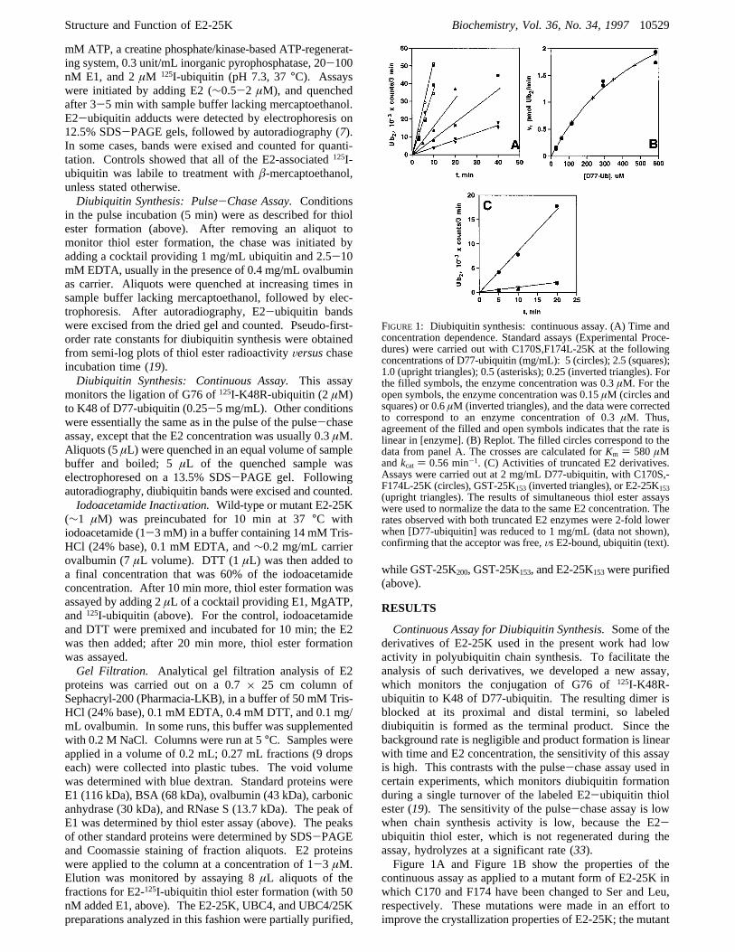
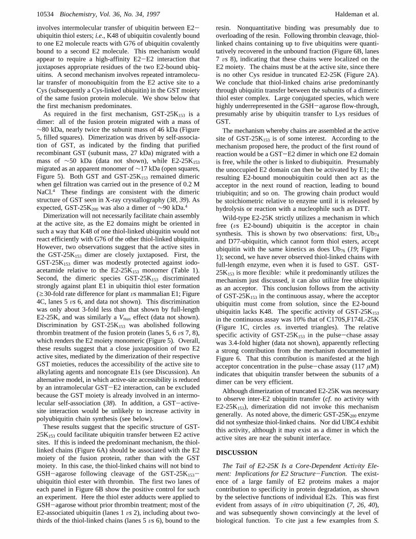
FIGURE 1: Diubiquitin synthesis: continuous assay. (A) Time andconcentration dependence. Standard assays (Experimental Proce-dures) were carried out with C170S,F174L-25K at the followingconcentrations of D77-ubiquitin (mg/mL): 5 (circles); 2.5 (squares);1.0 (upright triangles); 0.5 (asterisks); 0.25 (inverted triangles). Forthe filled symbols, the enzyme concentration was 0.3µM. For theopen symbols, the enzyme concentration was 0.15µM (circles andsquares) or 0.6µM (inverted triangles), and the data were correctedto correspond to an enzyme concentration of 0.3µM. Thus,agreement of the filled and open symbols indicates that the rate islinear in [enzyme]. (B) Replot. The filled circles correspond to thedata from panel A. The crosses are calculated forKm ) 580 µMandkcat ) 0.56 min-1. (C) Activities of truncated E2 derivatives.Assays were carried out at 2 mg/mL D77-ubiquitin, with C170S,-F174L-25K (circles), GST-25K153 (inverted triangles), or E2-25K153(upright triangles). The results of simultaneous thiol ester assayswere used to normalize the data to the same E2 concentration. Therates observed with both truncated E2 enzymes were 2-fold lowerwhen [D77-ubiquitin] was reduced to 1 mg/mL (data not shown),confirming that the acceptor was free,VsE2-bound, ubiquitin (text).
Structure and Function of E2-25K Biochemistry, Vol. 36, No. 34, 199710529
protein was expressed inE. coli at a higher level than wild-type E2-25K (Experimental Procedures). As shown inFigure 1A, with the mutant enzyme product formation inthe continuous assay was linear with time (Figure 1A). Therate at low concentrations of D77-ubiquitin (e100µM) wasdirectly proportional to the concentration of this acceptor(Figure 1B). At higher acceptor concentrations, there wasweak saturation, consistent withKm ) 580µM (line, Figure1B). The calculated value ofkcat/Km, 966 M-1 min-1 (kcat) 0.56 min-1), is similar to values of 500-1200 M-1 min-1
obtained for wild-type E2-25K (19, 33) and C170S,F174L-25K (data not shown) in the pulse-chase assay. Theseresults indicate that the C170S and F174L mutations are fullypermissive for biochemical activity, and in some of theexperiments described below, C170S,F174L-25K served asa positive control.The Tail of E2-25K Is Necessary, but Not Sufficient, for
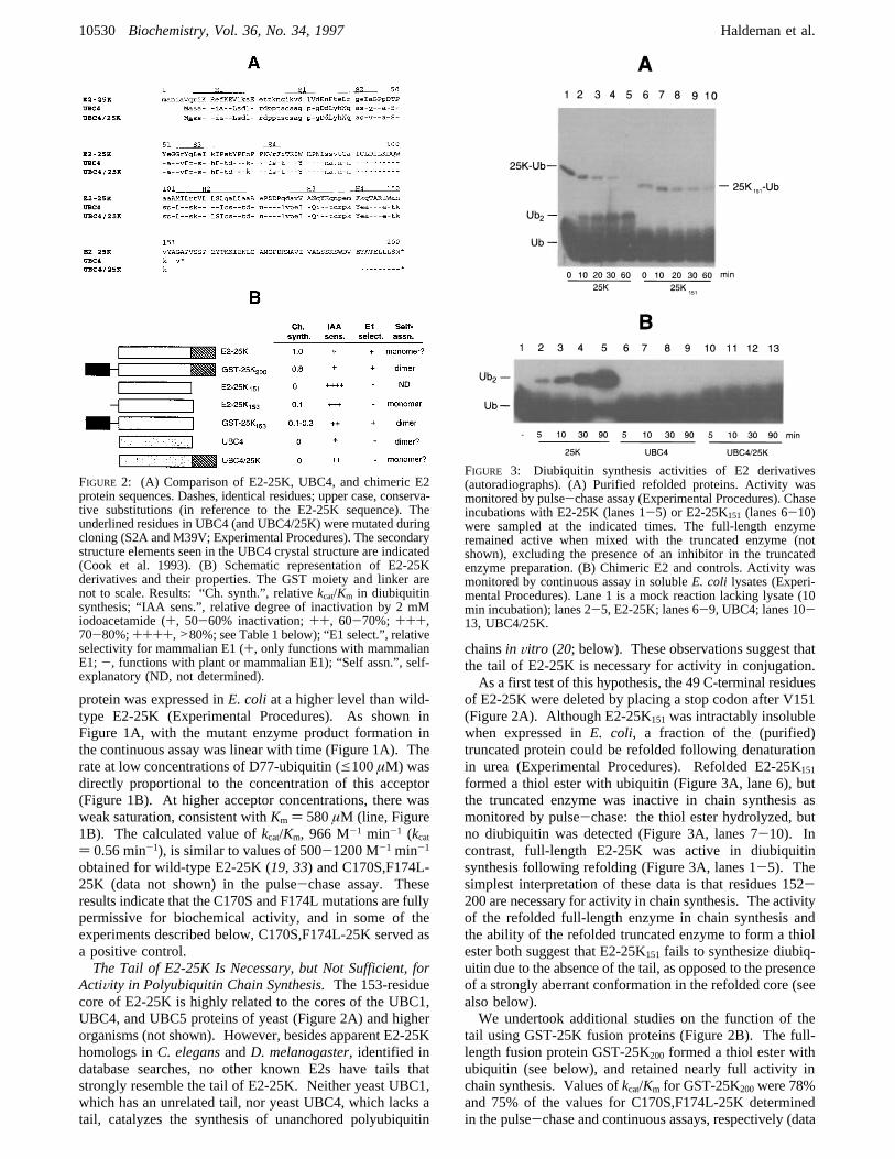
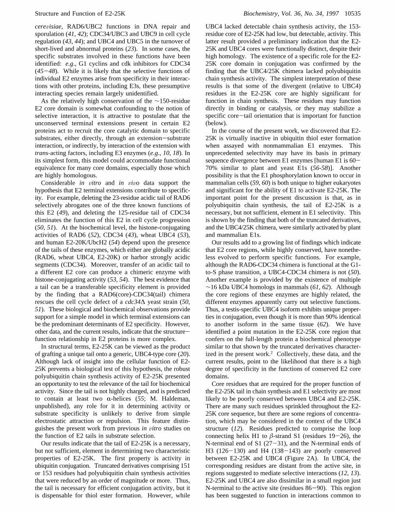
ActiVity in Polyubiquitin Chain Synthesis.The 153-residuecore of E2-25K is highly related to the cores of the UBC1,UBC4, and UBC5 proteins of yeast (Figure 2A) and higherorganisms (not shown). However, besides apparent E2-25Khomologs inC. elegansandD. melanogaster, identified indatabase searches, no other known E2s have tails thatstrongly resemble the tail of E2-25K. Neither yeast UBC1,which has an unrelated tail, nor yeast UBC4, which lacks atail, catalyzes the synthesis of unanchored polyubiquitin
chainsin Vitro (20; below). These observations suggest thatthe tail of E2-25K is necessary for activity in conjugation.As a first test of this hypothesis, the 49 C-terminal residues
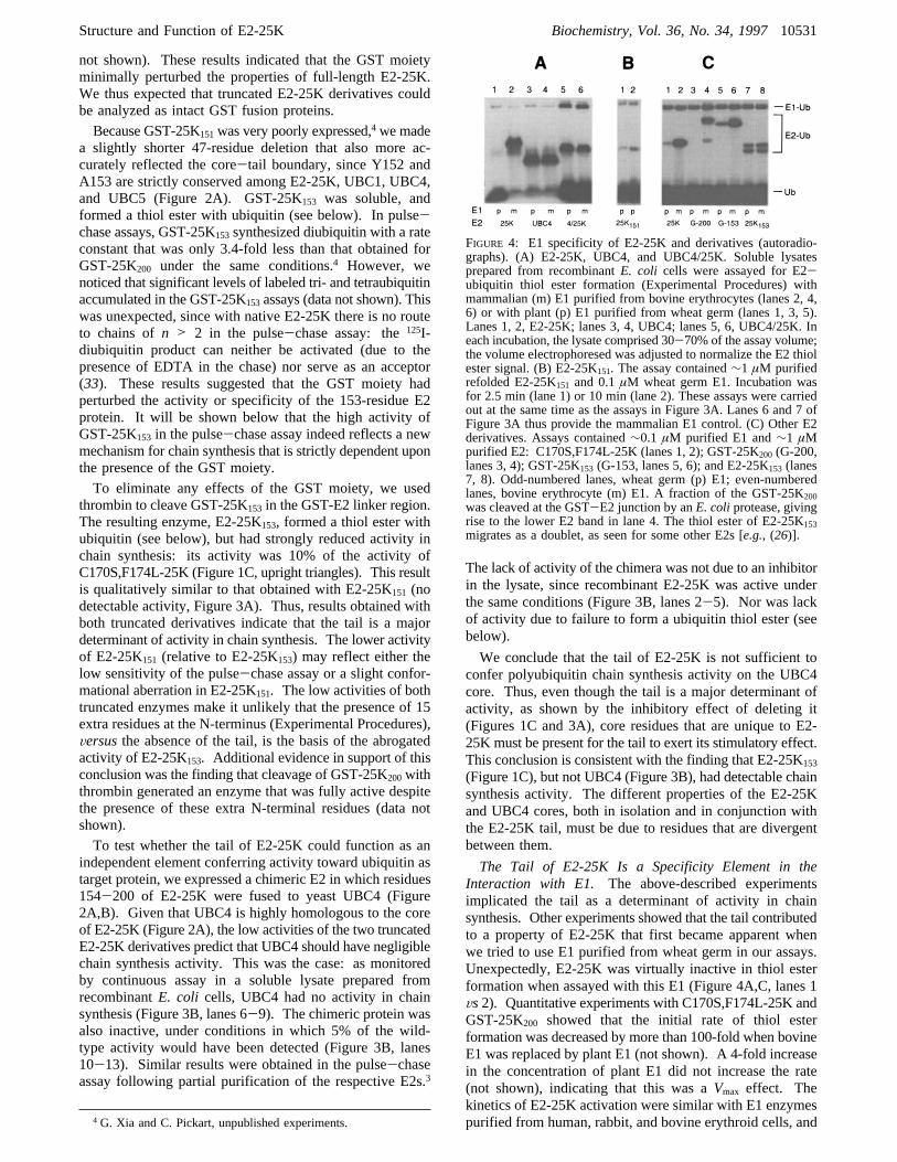
of E2-25K were deleted by placing a stop codon after V151(Figure 2A). Although E2-25K151was intractably insolublewhen expressed inE. coli, a fraction of the (purified)truncated protein could be refolded following denaturationin urea (Experimental Procedures). Refolded E2-25K151
formed a thiol ester with ubiquitin (Figure 3A, lane 6), butthe truncated enzyme was inactive in chain synthesis asmonitored by pulse-chase: the thiol ester hydrolyzed, butno diubiquitin was detected (Figure 3A, lanes 7-10). Incontrast, full-length E2-25K was active in diubiquitinsynthesis following refolding (Figure 3A, lanes 1-5). Thesimplest interpretation of these data is that residues 152-200 are necessary for activity in chain synthesis. The activityof the refolded full-length enzyme in chain synthesis andthe ability of the refolded truncated enzyme to form a thiolester both suggest that E2-25K151 fails to synthesize diubiq-uitin due to the absence of the tail, as opposed to the presenceof a strongly aberrant conformation in the refolded core (seealso below).We undertook additional studies on the function of the
tail using GST-25K fusion proteins (Figure 2B). The full-length fusion protein GST-25K200 formed a thiol ester withubiquitin (see below), and retained nearly full activity inchain synthesis. Values ofkcat/Km for GST-25K200were 78%and 75% of the values for C170S,F174L-25K determinedin the pulse-chase and continuous assays, respectively (data
FIGURE 2: (A) Comparison of E2-25K, UBC4, and chimeric E2protein sequences. Dashes, identical residues; upper case, conserva-tive substitutions (in reference to the E2-25K sequence). Theunderlined residues in UBC4 (and UBC4/25K) were mutated duringcloning (S2A and M39V; Experimental Procedures). The secondarystructure elements seen in the UBC4 crystal structure are indicated(Cook et al. 1993). (B) Schematic representation of E2-25Kderivatives and their properties. The GST moiety and linker arenot to scale. Results: “Ch. synth.”, relativekcat/Km in diubiquitinsynthesis; “IAA sens.”, relative degree of inactivation by 2 mMiodoacetamide (+, 50-60% inactivation;++, 60-70%; +++,70-80%;++++,>80%; see Table 1 below); “E1 select.”, relativeselectivity for mammalian E1 (+, only functions with mammalianE1;-, functions with plant or mammalian E1); “Self assn.”, self-explanatory (ND, not determined).
FIGURE 3: Diubiquitin synthesis activities of E2 derivatives(autoradiographs). (A) Purified refolded proteins. Activity wasmonitored by pulse-chase assay (Experimental Procedures). Chaseincubations with E2-25K (lanes 1-5) or E2-25K151 (lanes 6-10)were sampled at the indicated times. The full-length enzymeremained active when mixed with the truncated enzyme (notshown), excluding the presence of an inhibitor in the truncatedenzyme preparation. (B) Chimeric E2 and controls. Activity wasmonitored by continuous assay in solubleE. coli lysates (Experi-mental Procedures). Lane 1 is a mock reaction lacking lysate (10min incubation); lanes 2-5, E2-25K; lanes 6-9, UBC4; lanes 10-13, UBC4/25K.
10530 Biochemistry, Vol. 36, No. 34, 1997 Haldeman et al.
not shown). These results indicated that the GST moietyminimally perturbed the properties of full-length E2-25K.We thus expected that truncated E2-25K derivatives couldbe analyzed as intact GST fusion proteins.
Because GST-25K151was very poorly expressed,4 we madea slightly shorter 47-residue deletion that also more ac-curately reflected the core-tail boundary, since Y152 andA153 are strictly conserved among E2-25K, UBC1, UBC4,and UBC5 (Figure 2A). GST-25K153 was soluble, andformed a thiol ester with ubiquitin (see below). In pulse-chase assays, GST-25K153synthesized diubiquitin with a rateconstant that was only 3.4-fold less than that obtained forGST-25K200 under the same conditions.4 However, wenoticed that significant levels of labeled tri- and tetraubiquitinaccumulated in the GST-25K153assays (data not shown). Thiswas unexpected, since with native E2-25K there is no routeto chains ofn > 2 in the pulse-chase assay: the125I-diubiquitin product can neither be activated (due to thepresence of EDTA in the chase) nor serve as an acceptor(33). These results suggested that the GST moiety hadperturbed the activity or specificity of the 153-residue E2protein. It will be shown below that the high activity ofGST-25K153 in the pulse-chase assay indeed reflects a newmechanism for chain synthesis that is strictly dependent uponthe presence of the GST moiety.
To eliminate any effects of the GST moiety, we usedthrombin to cleave GST-25K153 in the GST-E2 linker region.The resulting enzyme, E2-25K153, formed a thiol ester withubiquitin (see below), but had strongly reduced activity inchain synthesis: its activity was 10% of the activity ofC170S,F174L-25K (Figure 1C, upright triangles). This resultis qualitatively similar to that obtained with E2-25K151 (nodetectable activity, Figure 3A). Thus, results obtained withboth truncated derivatives indicate that the tail is a majordeterminant of activity in chain synthesis. The lower activityof E2-25K151 (relative to E2-25K153) may reflect either thelow sensitivity of the pulse-chase assay or a slight confor-mational aberration in E2-25K151. The low activities of bothtruncated enzymes make it unlikely that the presence of 15extra residues at the N-terminus (Experimental Procedures),Versusthe absence of the tail, is the basis of the abrogatedactivity of E2-25K153. Additional evidence in support of thisconclusion was the finding that cleavage of GST-25K200withthrombin generated an enzyme that was fully active despitethe presence of these extra N-terminal residues (data notshown).
To test whether the tail of E2-25K could function as anindependent element conferring activity toward ubiquitin astarget protein, we expressed a chimeric E2 in which residues154-200 of E2-25K were fused to yeast UBC4 (Figure2A,B). Given that UBC4 is highly homologous to the coreof E2-25K (Figure 2A), the low activities of the two truncatedE2-25K derivatives predict that UBC4 should have negligiblechain synthesis activity. This was the case: as monitoredby continuous assay in a soluble lysate prepared fromrecombinantE. coli cells, UBC4 had no activity in chainsynthesis (Figure 3B, lanes 6-9). The chimeric protein wasalso inactive, under conditions in which 5% of the wild-type activity would have been detected (Figure 3B, lanes10-13). Similar results were obtained in the pulse-chaseassay following partial purification of the respective E2s.3
The lack of activity of the chimera was not due to an inhibitorin the lysate, since recombinant E2-25K was active underthe same conditions (Figure 3B, lanes 2-5). Nor was lackof activity due to failure to form a ubiquitin thiol ester (seebelow).
We conclude that the tail of E2-25K is not sufficient toconfer polyubiquitin chain synthesis activity on the UBC4core. Thus, even though the tail is a major determinant ofactivity, as shown by the inhibitory effect of deleting it(Figures 1C and 3A), core residues that are unique to E2-25K must be present for the tail to exert its stimulatory effect.This conclusion is consistent with the finding that E2-25K153
(Figure 1C), but not UBC4 (Figure 3B), had detectable chainsynthesis activity. The different properties of the E2-25Kand UBC4 cores, both in isolation and in conjunction withthe E2-25K tail, must be due to residues that are divergentbetween them.
The Tail of E2-25K Is a Specificity Element in theInteraction with E1. The above-described experimentsimplicated the tail as a determinant of activity in chainsynthesis. Other experiments showed that the tail contributedto a property of E2-25K that first became apparent whenwe tried to use E1 purified from wheat germ in our assays.Unexpectedly, E2-25K was virtually inactive in thiol esterformation when assayed with this E1 (Figure 4A,C, lanes 1Vs2). Quantitative experiments with C170S,F174L-25K andGST-25K200 showed that the initial rate of thiol esterformation was decreased by more than 100-fold when bovineE1 was replaced by plant E1 (not shown). A 4-fold increasein the concentration of plant E1 did not increase the rate(not shown), indicating that this was aVmax effect. Thekinetics of E2-25K activation were similar with E1 enzymespurified from human, rabbit, and bovine erythroid cells, and4 G. Xia and C. Pickart, unpublished experiments.
FIGURE 4: E1 specificity of E2-25K and derivatives (autoradio-graphs). (A) E2-25K, UBC4, and UBC4/25K. Soluble lysatesprepared from recombinantE. coli cells were assayed for E2-ubiquitin thiol ester formation (Experimental Procedures) withmammalian (m) E1 purified from bovine erythrocytes (lanes 2, 4,6) or with plant (p) E1 purified from wheat germ (lanes 1, 3, 5).Lanes 1, 2, E2-25K; lanes 3, 4, UBC4; lanes 5, 6, UBC4/25K. Ineach incubation, the lysate comprised 30-70% of the assay volume;the volume electrophoresed was adjusted to normalize the E2 thiolester signal. (B) E2-25K151. The assay contained∼1 µM purifiedrefolded E2-25K151 and 0.1µM wheat germ E1. Incubation wasfor 2.5 min (lane 1) or 10 min (lane 2). These assays were carriedout at the same time as the assays in Figure 3A. Lanes 6 and 7 ofFigure 3A thus provide the mammalian E1 control. (C) Other E2derivatives. Assays contained∼0.1 µM purified E1 and∼1 µMpurified E2: C170S,F174L-25K (lanes 1, 2); GST-25K200 (G-200,lanes 3, 4); GST-25K153 (G-153, lanes 5, 6); and E2-25K153 (lanes7, 8). Odd-numbered lanes, wheat germ (p) E1; even-numberedlanes, bovine erythrocyte (m) E1. A fraction of the GST-25K200was cleaved at the GST-E2 junction by anE. coliprotease, givingrise to the lower E2 band in lane 4. The thiol ester of E2-25K153migrates as a doublet, as seen for some other E2s [e.g., (26)].
Structure and Function of E2-25K Biochemistry, Vol. 36, No. 34, 199710531
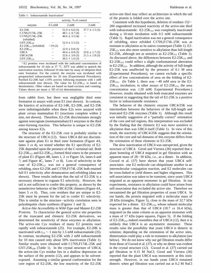
from rabbit liver, but there was negligible thiol esterformation in assays with yeast E1 (not shown). In contrast,the kinetics of activation of E2-14K, E2-20K, and E2-35Kwere indistinguishable when these E2s were assayed withplant Versusmammalian E1 (reactions complete within 2min, not shown). Therefore, E2-25K discriminates stronglyagainst noncognate (nonmammalian) E1 enzymes in the thiolester-forming reaction. This behavior is unique to E2-25Kamong known E2s.The structure of the E2-25K core is probably similar to
the structure of UBC4 (12). Since UBC4 did not discrimi-nate against plant E1 in thiol ester formation (Figure 4A,lanes 3Vs 4), we tested whether the E1 specificity of E2-25K depended upon the presence of the C-terminal tail. BothE2-25K151and E2-25K153 formed thiol esters in the presenceof plant E1 (Figure 4B, lanes 1, 2VsFigure 3A, lanes 6 and7; and Figure 4C, lanes 7Vs 8). Loss of selectivity in thecase of E2-25K151 was not a general consequence ofrefolding, since E2-25K and C170S,F174L-25K both retainedfull E1 selectivity after denaturation and refolding (data notshown). These results indicate that the tail of E2-25K is anecessary element in cognate E1 selectivity. However, thetail is not sufficient to confer this property, as shown by thenonselective behavior of the UBC4/25K chimera (Figure 4A,lanes 5Vs 6). Thus, core residues that are unique to E2-25K must be present for the tail to confer E1 selectivity.This is similar to the structure-activity correlation seen inpolyubiquitin chain synthesis (Figures 1 and 3).ActiVe-Site Accessibility in Wild-Type and Mutant E2-25K
Proteins. To characterize the general active site propertiesof the truncated and chimeric E2-25K derivatives, wedetermined the sensitivity of these enzymes to alkylation.The active-site Cys residues of many E2 proteins reactrapidly with iodoacetamide (25). For example, E2-20K isinactivated witht1/2∼ 1 min by 1.5 mM iodoacetamide (35).In contrast, incubating E2-25K with 2 mM iodoacetamidefor 10 min caused only about 50% inactivation (Table 1).Similar results were obtained with C170S,F174L-25K andGST-25K200 (Table 1). In the crystal structure of UBC4,the active-site Cys residue lies in a shallow depression onthe surface of the protein (12), and appears to be solvent-exposed. Assuming a similar general conformation for thecore region of E2-25K, the low reactivity of the E2-25K
active-site thiol may reflect an architecture in which the tailof the protein is folded over the active site.Consistent with this hypothesis, deletion of residues 152-
200 engendered increased reactivity of the active-site thiolwith iodoacetamide: E2-25K151was completely inactivatedduring a 10-min incubation with 0.5 mM iodoacetamide(Table 1). Rapid inactivation was not a general consequenceof refolding, since refolded C170S,F174L-25K was asresistant to alkylation as its native counterpart (Table 1). E2-25K153was also more sensitive to alkylation than full-lengthE2-25K, although not as sensitive as E2-25K151 (Table 1).As discussed above, the differences between E2-25K151 andE2-25K153 could reflect a slight conformational aberrationin E2-25K151. In addition, although the activity of full-lengthE2-25K was unaffected by the presence of 0.1 M urea(Experimental Procedures), we cannot exclude a specificeffect of low concentrations of urea on the folding of E2-25K151. (In Table 1, there was∼40 mM urea in the E2-25K151 incubation; in other assays of E2-25K151, the ureaconcentration wase20 mM; Experimental Procedures.)However, results obtained with both truncated enzymes areconsistent in suggesting that the tail of E2-25K is a positivefactor in iodoacetamide resistance.The behavior of the chimeric enzyme UBC4/25K was
intermediate between the behaviors of the full-length andtruncated E2-25K enzymes (Table 1). Although this resultwas initially suggestive of a “partially correct” orientationof the core and tail regions, this interpretation was excludedby the finding that the chimeric E2 wasmoresensitive toalkylation than was UBC4 itself (Table 1). In view of thisresult, the reactivity of UBC4/25K suggests that the orienta-tion of the core and tail domains of this enzyme differs fromthe orientation of these elements in E2-25K.The slow inactivation of UBC4 was unexpected, given the
structure of UBC4. Girod and Vierstra (36) reported that aplant homolog of UBC4 migrated in gel filtration with anapparent mass of 28-30 kDa,i.e., as a dimer. In addition,Gwozd et al. (37) have shown that yeast UBC4 self-associates: one E2 molecule can ubiquitinate another in anintermolecular conjugation reaction, and E2 molecules canbe cross-linked to yield dimers and higher oligomers. Thisself-association was taken to be transient, since yeast UBC4migrated as an apparent monomer in gel filtration. In ourexperiments, resistance to alkylation could have arisen fromself-association that occluded the active site. Therefore wereexamined the gel filtration properties of yeast UBC4. Inour hands, the protein migrated with a molecular mass of28 kDa (triangles, Figure 5), close to the mass of 32.7 kDaexpected for a dimer. E2-25K153, whose subunit molecularmass is greater than that of UBC4 (18.7Vs 16.4 kDa),migrated on the same column as an apparent monomer witha mass of 17 kDa (open squares, Figure 5). If the foldingof E2-25K153 indeed resembles that of UBC4 (12), then it isunlikely that UBC4 is an asymmetric monomer. Theseresults raise the possibility that yeast UBC4 is dimeric insolution; depending on the orientation of the active sites,dimerization could play a role in resistance to alkylation (seebelow). We do not know why our gel filtration results differfrom those of Gwozd et al. (37), or why no dimer was evidentin the crystal structure (12). Gwozd et al. (37) carried outgel filtration in 0.15 M NaCl; Girod and Vierstra (36)reported that the plant UBC4 was monomeric at this ionicstrength. However, in our hands yeast UBC4 remaineddimeric when gel filtration was carried out at 0.2 M NaCl
Table 1: Iodoacetamide Inactivationa
activity, % of control
enzyme 0.5 mM 2 mM 3 mM
E2-25K - 51.9( 6.0 (3) 37.7( 1.5 (3)C170S,F174L-25K - 48.1( 4.7 (4) -C170S,F174L-25K(refolded)
- 46.6( 3.5 (4) -
GST-25K200 - 52.4( 5.5 (5) -E2-25K151 (refolded) <10 (2) - -E2-25K153 - 22.9( 0.8 (3) -UBC4 - 44.4( 0.8 (3) 33.6( 1.7 (3)UBC4/25K - 32.6( 3.0 (3) 21.5( 1.8 (3)GST-25K153 - 30.5+ 1.6 (3) -a E2 proteins were incubated with the indicated concentration of
iodoacetamide for 10 min at 37°C. DTT was added to quench theinactivation reaction, followed 10 min later by assay of ubiquitin thiolester formation. For the control, the enzyme was incubated withprequenched iodoacetamide for 20 min (Experimental Procedures).Purified E2-20K hade10% activity following treatment with 1 mMiodoacetamide (not shown). The value for E2-25K151 is based ondensitometry. Other values are based on band excision and counting.Values shown are mean( SD of (n) determinations.
10532 Biochemistry, Vol. 36, No. 34, 1997 Haldeman et al.
(data not shown). It is possible that two mutations presentin the UBC4 used here, S2A and M39V (Experimentalprocedures), were significant with regard to self-association.Full-length E2-25K migrated with a molecular mass of
30 kDa, in agreement with previous results [circles, Figure5 (7)]. The UBC4/25K chimera comigrated with E2-25K(data not shown). The simplest interpretation of these datais that both enzymes are asymmetric monomers. If this istrue of E2-25K (subunit mass 22.5 kDa), then the tail mustmake a substantial contribution to asymmetry (cf. 17 kDafor E2-25K153). However, at present we cannot exclude amodel in which E2-25K and UBC4/25K are globular,symmetric dimers.Dimerization-Dependent Changes in the Mechanism and
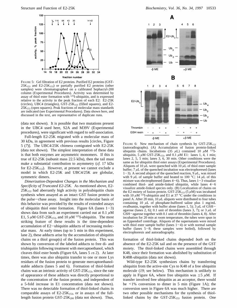
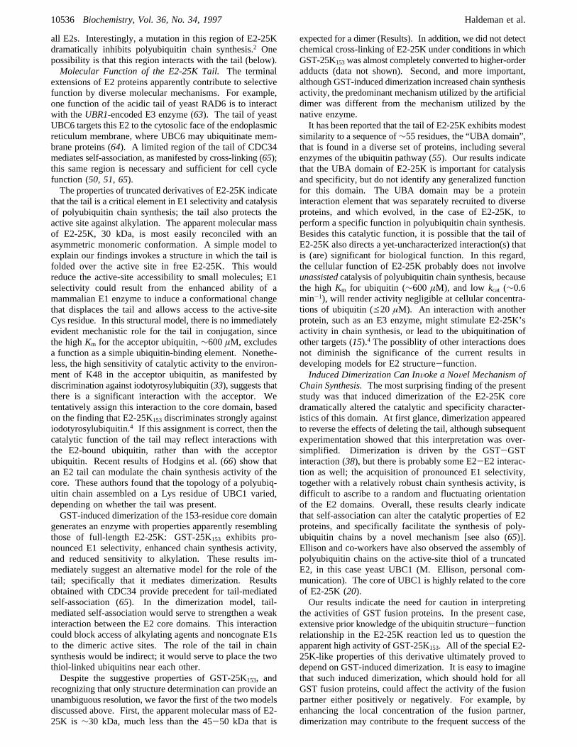
Specificity of Truncated E2-25K.As mentioned above, E2-25K153 had aberrantly high activity in polyubiquitin chainsynthesis when assayed as an intact GST fusion protein inthe pulse-chase assay. Insight into the molecular basis ofthis behavior was provided by the results of extended assaysof ubiquitin thiol ester formation. Figure 6A (lanes 1-3)shows data from such an experiment carried out at 0.1µME1, 5µM GST-25K153, and 10µM 125I-ubiquitin. The moststriking feature of these results is the time-dependentaccumulation of E2-ubiquitin adducts of increasing molec-ular mass. At early times (up to 5 min in this experiment,lane 2), these adducts arise by the accumulation of ubiquitinchains on a thiol group(s) of the fusion protein. This wasshown by conversion of the labeled adducts to free di- andtriubiquitin following treatment with mercaptoethanol, whichcleaves thiol ester bonds (Figure 6A, lanes 5Vs2). At latertimes, there was also ubiquitin transfer to one or more Lysresidues of the fusion protein to generate mercaptoethanol-stable adducts (lanes 5 and 6). Formation of thiol-linkedchains was an intrinsic activity of GST-25K153, since the rateof appearance of these adducts was directly proportional tothe concentration of the fusion protein, and independent ofa 5-fold increase in E1 concentration (data not shown).There was no detectable formation of thiol-linked chains incomparable assays of E2-25K153, or in assays of the full-length fusion protein GST-25K200 (data not shown). Thus,
formation of thiol-linked chains dependedboth on theabsence of the E2-25K tail and on the presence of the GSTmoiety. The thiol-linked chains were assembled throughK48, since their formation was abolished by substitution ofK48R-ubiquitin (data not shown).Wild-type E2-25K synthesizes chains by transferring
ubiquitin from the active-site Cys to K48 of a free ubiquitinmolecule (19; see below). This mechanism is unlikely toapply in Figure 6A, where free ubiquitin wase5 µM. Iftransfer involved free ubiquitin as an acceptor, there wouldbe <1% conversion to dimer in 5 min (Figure 1A); theconversion seen in Figure 6A was much higher. There aretwo other possible mechanisms for the synthesis of thiol-linked chains by the GST-25K153 fusion protein. One
FIGURE5: Gel filtration of E2 proteins. Purified E2 proteins (GST-25K153 and E2-25K153) or partially purified E2 proteins (othersamples) were chromatographed on a calibrated Sephacryl-200column (Experimental Procedures). Activity was determined byassay of thiol ester formation with125I-ubiquitin, and is expressedrelative to the activity in the peak fraction of each E2: E2-25K(circles), UBC4 (triangles), GST-25K153 (filled squares), and E2-25K153 (open squares). Peak fractions of molecular mass standardsare indicated (see Experimental Procedures). Data shown here, anddiscussed in the text, are representative of duplicate runs.
FIGURE 6: New mechanism of chain synthesis by GST-25K153(autoradiographs). (A) Accumulation of fusion protein-linkedubiquitin chains. Incubations (35µL) contained 10µM 125I-ubiquitin, 5µM GST-25K153, and 0.1µM E1: lanes 1, 4, 1 min;lanes 2, 5, 5 min; lanes 3, 6, 30 min. Other conditions were thesame as for ubiquitin thiol ester assays (Experimental Procedures).Aliquots of 10µL were quenched with 10µL of thiol ester samplebuffer; 7µL of the quenched incubation was electrophoresed (lanes1-3). A second aliquot of the quenched reaction, 9µL, was mixedwith 9 µL of sample buffer and heated to 100°C; 14 µL of thismixture was electrophoresed (lanes 4-6). Thus, lanes 1-3 visualizecombined thiol- and amide-linked ubiquitin, while lanes 4-6visualize amide-linked species only. (B) Localization of chains onthe E2 moiety of fusion protein. GST-25K153 (5 µM) was incubatedwith 10 µM 125I-ubiquitin and E1 at 37°C under the conditions inpanel A. After 20 min, 10µL aliquots were distributed to four tubescontaining 10µL of phosphate-buffered saline plus 1 mg/mLovalbumin, together with buffer alone (lanes 1, 5); 3µL of GSH-agarose (lanes 2, 6); 0.1 unit of thrombin (lanes 3, 7); or 3µL ofGSH-agarose together with 0.1 unit of thrombin (lanes 4, 8). Afterincubation for 20 min at room temperature, the tubes were spun inan Eppendorf centrifuge. Aliquots of the supernatants were mixedwith thiol ester sample buffer (lanes 1-4) or with normal samplebuffer (lanes 5-8; these samples were boiled), followed byelectrophoresis and autoradiography.
Structure and Function of E2-25K Biochemistry, Vol. 36, No. 34, 199710533
involves intermolecular transfer of ubiquitin between E2-ubiquitin thiol esters;i.e., K48 of ubiquitin covalently boundto one E2 molecule reacts with G76 of ubiquitin covalentlybound to a second E2 molecule. This mechanism wouldappear to require a high-affinity E2-E2 interaction thatjuxtaposes appropriate residues of the two E2-bound ubiq-uitins. A second mechanism involves repeated intramolecu-lar transfer of monoubiquitin from the E2 active site to aCys (subsequently a Cys-linked ubiquitin) in the GST moietyof the same fusion protein molecule. We show below thatthe first mechanism predominates.As required in the first mechanism, GST-25K153 is a
dimer: all of the fusion protein migrated with a mass of∼80 kDa, nearly twice the subunit mass of 46 kDa (Figure5, filled squares). Dimerization was driven by self-associa-tion of GST, as indicated by the finding that purifiedrecombinant GST (subunit mass, 27 kDa) migrated with amass of∼50 kDa (data not shown), while E2-25K153migrated as an apparent monomer of∼17 kDa (open squares,Figure 5). Both GST and GST-25K153 remained dimericwhen gel filtration was carried out in the presence of 0.2 MNaCl.4 These findings are consistent with the dimericstructure of GST seen in X-ray crystallography (38, 39). Asexpected, GST-25K200 was also a dimer of∼90 kDa.4
Dimerization will not necessarily facilitate chain assemblyat the active site, as the E2 domains might be oriented insuch a way that K48 of one thiol-linked ubiquitin would notreact efficiently with G76 of the other thiol-linked ubiquitin.However, two observations suggest that the active sites inthe GST-25K153 dimer are closely juxtaposed. First, theGST-25K153 dimer was modestly protected against iodo-acetamide relative to the E2-25K153 monomer (Table 1).Second, the dimeric species GST-25K153 discriminatedstrongly against plant E1 in ubiquitin thiol ester formation(g30-fold rate difference for plantVsmammalian E1; Figure4C, lanes 5Vs 6, and data not shown). This discriminationwas only about 3-fold less than that shown by full-lengthE2-25K, and was similarly aVmax effect (data not shown).Discrimination by GST-25K153 was abolished followingthrombin treatment of the fusion protein (lanes 5, 6Vs7, 8),which renders the E2 moiety monomeric (Figure 5). Overall,these results suggest that a close juxtaposition of two E2active sites, mediated by the dimerization of their respectiveGST moieties, reduces the accessibility of the active site toalkylating agents and noncognate E1s (see Discussion). Analternative model, in which active-site accessibility is reducedby an intramolecular GST-E2 interaction, can be excludedbecause the GST moiety is already involved in an intermo-lecular self-association (38). In addition, a GST-active-site interaction would be unlikely to increase activity inpolyubiquitin chain synthesis (see below).These results suggest that the specific structure of GST-
25K153 could facilitate ubiquitin transfer between E2 activesites. If this is indeed the predominant mechanism, the thiol-linked chains (Figure 6A) should be associated with the E2moiety of the fusion protein, rather than with the GSTmoiety. In this case, the thiol-linked chains will not bind toGSH-agarose following cleavage of the GST-25K153-ubiquitin thiol ester with thrombin. The first two lanes ofeach panel in Figure 6B show the positive control for suchan experiment. Here the thiol ester adducts were applied toGSH-agarose without prior thrombin treatment; most of theE2-associated ubiquitin (lanes 1Vs2), including about two-thirds of the thiol-linked chains (lanes 5Vs 6), bound to the
resin. Nonquantitative binding was presumably due tooverloading of the resin. Following thrombin cleavage, thiol-linked chains containing up to five ubiquitins were quanti-tatively recovered in the unbound fraction (Figure 6B, lanes7 Vs 8), indicating that these chains were localized on theE2 moiety. The chains must be at the active site, since thereis no other Cys residue in truncated E2-25K (Figure 2A).We conclude that thiol-linked chains arise predominantlythrough ubiquitin transfer between the subunits of a dimericthiol ester complex. Large conjugated species, which werehighly underrepresented in the GSH-agarose flow-through,presumably arise by ubiquitin transfer to Lys residues ofGST.The mechanism whereby chains are assembled at the active
site of GST-25K153 is of some interest. According to themechanism proposed here, the product of the first round ofreaction would be a GST-E2 dimer in which one E2 domainis free, while the other is linked to diubiquitin. Presumablythe unoccupied E2 domain can then be activated by E1; theresulting E2-bound monoubiquitin could then act as theacceptor in the next round of reaction, leading to boundtriubiquitin; and so on. The growing chain product wouldbe stoichiometric relative to enzyme until it is released byhydrolysis or reaction with a nucleophile such as DTT.Wild-type E2-25K strictly utilizes a mechanism in which
free (Vs E2-bound) ubiquitin is the acceptor in chainsynthesis. This is shown by two observations: first, Ub74
and D77-ubiquitin, which cannot form thiol esters, acceptubiquitin with the same kinetics as does Ub76 (19; Figure1); second, we have never observed thiol-linked chains withfull-length enzyme, even when it is fused to GST. GST-25K153 is more flexible: while it predominantly utilizes themechanism just discussed, it can also utilize free ubiquitinas an acceptor. This conclusion follows from the activityof GST-25K153 in the continuous assay, where the acceptorubiquitin must come from solution, since the E2-boundubiquitin lacks K48. The specific activity of GST-25K153in the continuous assay was 10% that of C170S,F174L-25K(Figure 1C, circlesVs. inverted triangles). The relativespecific activity of GST-25K153 in the pulse-chase assaywas 3.4-fold higher (data not shown), apparently reflectinga strong contribution from the mechanism documented inFigure 6. That this contribution is manifested at the highacceptor concentration in the pulse-chase assay (117µM)indicates that ubiquitin transfer between the subunits of adimer can be very efficient.Although dimerization of truncated E2-25K was necessary
to observe inter-E2 ubiquitin transfer (cf. no activity withE2-25K153), dimerization did not invoke this mechanismgenerally. As noted above, the dimeric GST-25K200enzymedid not synthesize thiol-linked chains. Nor did UBC4 exhibitthis activity, although it may exist as a dimer in which theactive sites are near the subunit interface.
DISCUSSION
The Tail of E2-25K Is a Core-Dependent ActiVity Ele-ment: Implications for E2 Structure-Function. The exist-ence of a large family of E2 proteins makes a majorcontribution to specificity in protein degradation, as shownby the selective functions of individual E2s. This was firstevident from assays ofin Vitro ubiquitination (7, 26, 40),and was subsequently shown convincingly at the level ofbiological function. To cite just a few examples fromS.
10534 Biochemistry, Vol. 36, No. 34, 1997 Haldeman et al.
cereVisiae, RAD6/UBC2 functions in DNA repair andsporulation (41, 42); CDC34/UBC3 and UBC9 in cell cycleregulation (43, 44); and UBC4 and UBC5 in the turnover ofshort-lived and abnormal proteins (23). In some cases, thespecific substrates involved in these functions have beenidentified: e.g., G1 cyclins and cdk inhibitors for CDC34(45-48). While it is likely that the selective functions ofindividual E2 enzymes arise from specificity in their interac-tions with other proteins, including E3s, these presumptiveinteracting species remain largely unidentified.As the relatively high conservation of the∼150-residue
E2 core domain is somewhat confounding to the notion ofselective interaction, it is attractive to postulate that theunconserved terminal extensions present in certain E2proteins act to recruit the core catalytic domain to specificsubstrates, either directly, through an extension-substrateinteraction, or indirectly, by interaction of the extension withtrans-acting factors, including E3 enzymes (e.g., 10, 18). Inits simplest form, this model could accommodate functionalequivalence for many core domains, especially those whichare highly homologous.Considerablein Vitro and in ViVo data support the
hypothesis that E2 terminal extensions contribute to specific-ity. For example, deleting the 23-residue acidic tail of RAD6selectively abrogates one of the three known functions ofthis E2 (49), and deleting the 125-residue tail of CDC34eliminates the function of this E2 in cell cycle progression(50, 51). At the biochemical level, the histone-conjugatingactivities of RAD6 (52), CDC34 (43), wheat UBC4 (53),and human E2-20K/UbcH2 (54) depend upon the presenceof the tails of these enzymes, which either are globally acidic(RAD6, wheat UBC4, E2-20K) or harbor strongly acidicsegments (CDC34). Moreover, transfer of an acidic tail toa different E2 core can produce a chimeric enzyme withhistone-conjugating activity (53, 54). The best evidence thata tail can be a transferable specificity element is providedby the finding that a RAD6(core)-CDC34(tail) chimerarescues the cell cycle defect of acdc34∆ yeast strain (50,51). These biological and biochemical observations providesupport for a simple model in which terminal extensions canbe the predominant determinants of E2 specificity. However,other data, and the current results, indicate that the structure-function relationship in E2 proteins is more complex.In structural terms, E2-25K can be viewed as the product
of grafting a unique tail onto a generic, UBC4-type core (20).Although lack of insight into the cellular function of E2-25K prevents a biological test of this hypothesis, the robustpolyubiquitin chain synthesis activity of E2-25K presentedan opportunity to test the relevance of the tail for biochemicalactivity. Since the tail is not highly charged, and is predictedto contain at least twoR-helices (55; M. Haldeman,unpublished), any role for it in determining activity orsubstrate specificity is unlikely to derive from simpleelectrostatic attraction or repulsion. This feature distin-guishes the present work from previousin Vitro studies onthe function of E2 tails in substrate selection.Our results indicate that the tail of E2-25K is a necessary,
but not sufficient, element in determining two characteristicproperties of E2-25K. The first property is activity inubiquitin conjugation. Truncated derivatives comprising 151or 153 residues had polyubiquitin chain synthesis activitiesthat were reduced by an order of magnitude or more. Thus,the tail is necessary for efficient conjugation activity, but itis dispensable for thiol ester formation. However, while
UBC4 lacked detectable chain synthesis activity, the 153-residue core of E2-25K had low, but detectable, activity. Thislatter result provided a preliminary indication that the E2-25K and UBC4 cores were functionally distinct, despite theirhigh homology. The existence of a specific role for the E2-25K core domain in conjugation was confirmed by thefinding that the UBC4/25K chimera lacked polyubiquitinchain synthesis activity. The simplest interpretation of theseresults is that some of the divergent (relative to UBC4)residues in the E2-25K core are highly significant forfunction in chain synthesis. These residues may functiondirectly in binding or catalysis, or they may stabilize aspecific core-tail orientation that is important for function(below).In the course of the present work, we discovered that E2-
25K is virtually inactive in ubiquitin thiol ester formationwhen assayed with nonmammalian E1 enzymes. Thisunprecedented selectivity may have its basis in primarysequence divergence between E1 enzymes [human E1 is 60-70% similar to plant and yeast E1s (56-58)]. Anotherpossibility is that the E1 phosphorylation known to occur inmammalian cells (59, 60) is both unique to higher eukaryotesand significant for the ability of E1 to activate E2-25K. Theimportant point for the present discussion is that, as inpolyubiquitin chain synthesis, the tail of E2-25K is anecessary, but not sufficient, element in E1 selectivity. Thisis shown by the finding that both of the truncated derivatives,and the UBC4/25K chimera, were similarly activated by plantand mammalian E1s.Our results add to a growing list of findings which indicate
that E2 core regions, while highly conserved, have nonethe-less evolved to perform specific functions. For example,although the RAD6-CDC34 chimera is functional at the G1-to-S phase transition, a UBC4-CDC34 chimera is not (50).Another example is provided by the existence of multiple∼16 kDa UBC4 homologs in mammals (61, 62). Althoughthe core regions of these enzymes are highly related, thedifferent enzymes apparently carry out selective functions.Thus, a testis-specific UBC4 isoform exhibits unique proper-ties in conjugation, even though it is more than 90% identicalto another isoform in the same tissue (62). We haveidentified a point mutation in the E2-25K core region thatconfers on the full-length protein a biochemical phenotypesimilar to that shown by the truncated derivatives character-ized in the present work.2 Collectively, these data, and thecurrent results, point to the likelihood that there is a highdegree of specificity in the functions of conserved E2 coredomains.Core residues that are required for the proper function of
the E2-25K tail in chain synthesis and E1 selectivity are mostlikely to be poorly conserved between UBC4 and E2-25K.There are many such residues sprinkled throughout the E2-25K core sequence, but there are some regions of concentra-tion, which may be considered in the context of the UBC4structure (12). Residues predicted to comprise the loopconnecting helix H1 toâ-strand S1 (residues 19-26), theN-terminal end of S1 (27-31), and the N-terminal ends ofH3 (126-130) and H4 (138-143) are poorly conservedbetween E2-25K and UBC4 (Figure 2A). In UBC4, thecorresponding residues are distant from the active site, inregions suggested to mediate selective interactions (12, 13).E2-25K and UBC4 are also dissimilar in a small region justN-terminal to the active site (residues 86-90). This regionhas been suggested to function in interactions common to
Structure and Function of E2-25K Biochemistry, Vol. 36, No. 34, 199710535
all E2s. Interestingly, a mutation in this region of E2-25Kdramatically inhibits polyubiquitin chain synthesis.2 Onepossibility is that this region interacts with the tail (below).Molecular Function of the E2-25K Tail.The terminal
extensions of E2 proteins apparently contribute to selectivefunction by diverse molecular mechanisms. For example,one function of the acidic tail of yeast RAD6 is to interactwith theUBR1-encoded E3 enzyme (63). The tail of yeastUBC6 targets this E2 to the cytosolic face of the endoplasmicreticulum membrane, where UBC6 may ubiquitinate mem-brane proteins (64). A limited region of the tail of CDC34mediates self-association, as manifested by cross-linking (65);this same region is necessary and sufficient for cell cyclefunction (50, 51, 65).The properties of truncated derivatives of E2-25K indicate
that the tail is a critical element in E1 selectivity and catalysisof polyubiquitin chain synthesis; the tail also protects theactive site against alkylation. The apparent molecular massof E2-25K, 30 kDa, is most easily reconciled with anasymmetric monomeric conformation. A simple model toexplain our findings invokes a structure in which the tail isfolded over the active site in free E2-25K. This wouldreduce the active-site accessibility to small molecules; E1selectivity could result from the enhanced ability of amammalian E1 enzyme to induce a conformational changethat displaces the tail and allows access to the active-siteCys residue. In this structural model, there is no immediatelyevident mechanistic role for the tail in conjugation, sincethe highKm for the acceptor ubiquitin,∼600µM, excludesa function as a simple ubiquitin-binding element. Nonethe-less, the high sensitivity of catalytic activity to the environ-ment of K48 in the acceptor ubiquitin, as manifested bydiscrimination against iodotyrosylubiquitin (33), suggests thatthere is a significant interaction with the acceptor. Wetentatively assign this interaction to the core domain, basedon the finding that E2-25K153 discriminates strongly againstiodotyrosylubiquitin.4 If this assignment is correct, then thecatalytic function of the tail may reflect interactions withthe E2-bound ubiquitin, rather than with the acceptorubiquitin. Recent results of Hodgins et al. (66) show thatan E2 tail can modulate the chain synthesis activity of thecore. These authors found that the topology of a polyubiq-uitin chain assembled on a Lys residue of UBC1 varied,depending on whether the tail was present.GST-induced dimerization of the 153-residue core domain
generates an enzyme with properties apparently resemblingthose of full-length E2-25K: GST-25K153 exhibits pro-nounced E1 selectivity, enhanced chain synthesis activity,and reduced sensitivity to alkylation. These results im-mediately suggest an alternative model for the role of thetail; specifically that it mediates dimerization. Resultsobtained with CDC34 provide precedent for tail-mediatedself-association (65). In the dimerization model, tail-mediated self-association would serve to strengthen a weakinteraction between the E2 core domains. This interactioncould block access of alkylating agents and noncognate E1sto the dimeric active sites. The role of the tail in chainsynthesis would be indirect; it would serve to place the twothiol-linked ubiquitins near each other.Despite the suggestive properties of GST-25K153, and
recognizing that only structure determination can provide anunambiguous resolution, we favor the first of the two modelsdiscussed above. First, the apparent molecular mass of E2-25K is ∼30 kDa, much less than the 45-50 kDa that is
expected for a dimer (Results). In addition, we did not detectchemical cross-linking of E2-25K under conditions in whichGST-25K153was almost completely converted to higher-orderadducts (data not shown). Second, and more important,although GST-induced dimerization increased chain synthesisactivity, the predominant mechanism utilized by the artificialdimer was different from the mechanism utilized by thenative enzyme.It has been reported that the tail of E2-25K exhibits modest
similarity to a sequence of∼55 residues, the “UBA domain”,that is found in a diverse set of proteins, including severalenzymes of the ubiquitin pathway (55). Our results indicatethat the UBA domain of E2-25K is important for catalysisand specificity, but do not identify any generalized functionfor this domain. The UBA domain may be a proteininteraction element that was separately recruited to diverseproteins, and which evolved, in the case of E2-25K, toperform a specific function in polyubiquitin chain synthesis.Besides this catalytic function, it is possible that the tail ofE2-25K also directs a yet-uncharacterized interaction(s) thatis (are) significant for biological function. In this regard,the cellular function of E2-25K probably does not involveunassistedcatalysis of polyubiquitin chain synthesis, becausethe highKm for ubiquitin (∼600 µM), and low kcat (∼0.6min-1), will render activity negligible at cellular concentra-tions of ubiquitin (e20 µM). An interaction with anotherprotein, such as an E3 enzyme, might stimulate E2-25K’sactivity in chain synthesis, or lead to the ubiquitination ofother targets (15).4 The possiblity of other interactions doesnot diminish the significance of the current results indeveloping models for E2 structure-function.Induced Dimerization Can InVoke a NoVel Mechanism of
Chain Synthesis.The most surprising finding of the presentstudy was that induced dimerization of the E2-25K coredramatically altered the catalytic and specificity character-istics of this domain. At first glance, dimerization appearedto reverse the effects of deleting the tail, although subsequentexperimentation showed that this interpretation was over-simplified. Dimerization is driven by the GST-GSTinteraction (38), but there is probably some E2-E2 interac-tion as well; the acquisition of pronounced E1 selectivity,together with a relatively robust chain synthesis activity, isdifficult to ascribe to a random and fluctuating orientationof the E2 domains. Overall, these results clearly indicatethat self-association can alter the catalytic properties of E2proteins, and specifically facilitate the synthesis of poly-ubiquitin chains by a novel mechanism [see also (65)].Ellison and co-workers have also observed the assembly ofpolyubiquitin chains on the active-site thiol of a truncatedE2, in this case yeast UBC1 (M. Ellison, personal com-munication). The core of UBC1 is highly related to the coreof E2-25K (20).Our results indicate the need for caution in interpreting
the activities of GST fusion proteins. In the present case,extensive prior knowledge of the ubiquitin structure-functionrelationship in the E2-25K reaction led us to question theapparent high activity of GST-25K153. All of the special E2-25K-like properties of this derivative ultimately proved todepend on GST-induced dimerization. It is easy to imaginethat such induced dimerization, which should hold for allGST fusion proteins, could affect the activity of the fusionpartner either positively or negatively. For example, byenhancing the local concentration of the fusion partner,dimerization may contribute to the frequent success of the
10536 Biochemistry, Vol. 36, No. 34, 1997 Haldeman et al.

“GST capture” technique that is widely used to detectprotein-protein interactions.
ACKNOWLEDGMENTWe thank Vince Chau and Art Haas for providing vectors
encoding UBC4 and E2-14K, respectively; Dick Kulka forproviding yeast E1; Te-Cheung Lee for providing themodified pGEX vector; Chris Larsen and Keith Wilkinsonfor providing pRSUbD; and Bill Cook for helpful discus-sions. We are grateful to Mike Ellison for helpful discussions,and permission to cite unpublished data.
REFERENCES1. Ciechanover, A. (1994)Cell 79, 13-21.2. Glotzer, M., Murray, A. W., and Kirschner, M. W. (1991)Nature 349, 132-138.
3. Hershko, A., Ganoth, D., Pehrson, J., Palazzo, R. E., andCohen, L. H. (1991)J. Biol. Chem. 266, 16376-16379.
4. Treier, M., Staszewski, L. M., and Bohmann, D. (1994)Cell78, 787-798.
5. Scheffner, M., Werness, B. A., Huibregtse, J. M., Levine, A.J., and Howley, P. M. (1990)Cell 63, 1129-1136.
6. Ciechanover, A., Finley, D., and Varshavsky, A. (1984)Cell37, 57-66.
7. Pickart, C. M., and Rose, I. A. (1985)J. Biol. Chem. 260,1573-1581.
8. Scheffner, M., Nuber, U., and Huibregtse, J. M. (1995)Nature373, 81-83.
9. Chau, V., Tobias, J. W., Bachmair, A., Marriott, D., Ecker,D. J., Gonda, D. K., and Varshavsky, A. (1989)Science 243,1576-1583.
10. Smith, S. E., Koegl, M., and Jentsch, S. (1996)Biol. Chem.377, 437-446.
11. Cook, W. J., Jeffrey, L. C., Sullivan, M. L., and Vierstra, R.D. (1992)J. Biol. Chem. 267, 15116-15121.
12. Cook, W. J., Jeffrey, L. C., Xu, Y., and Chau, V. (1993)Biochemistry 32, 13809-13817.
13. Cook, W. J., Martin, P. D., Edwards, B. F. P., Yamazaki, R.K., and Chau, V. (1997)Biochemistry 36, 1621-1627.
14. Varshavsky, A. (1992)Cell 69, 725-735.15. Kalchman, M. A., Graham, R. K., Xia, G., Koide, H. B.,
Hodgson, J. G., Graham, K. G., Goldberg, Y. P., Gietz, R.D., Pickart, C. M., and Hayden, M. R. (1996)J. Biol. Chem.271, 19385-19394.
16. Kovalenko, O. V., Plug, A. W., Haaf, T., Gonda, D. K., Ashley,T., Ward, D. C., Radding, C. M., and Golub, E. I. (1996)Proc.Natl. Acad. Sci. U.S.A. 93, 2958-2963.
17. Wang, Z.-Y., Qiu, Q.-Q., Seufert, W., Taguchi, T., Testa, J.R., Whitmore, S. A., Callen, D. F., Welsh, D., Shenk, T., andDeuel, T. F. (1996)J. Biol. Chem. 271, 24811-24816.
18. Jentsch, S., Seufert, W., Sommer, T., and Reins, H.-A. (1990)Trends Biol. Sci. 15, 105-108.
19. Chen, Z., and Pickart, C. M. (1990)J. Biol. Chem. 265,21835-21842.
20. Chen, Z., Niles, E. G., and Pickart, C. M. (1991)J. Biol. Chem.266, 15698-15704.
21. Haldeman, M. T., Finley, D., and Pickart, C. M. (1995)J.Biol. Chem. 270, 9507-9516.
22. Wefes, I., Mastrandrea, L. D., Haldeman, M., Koury, S. T.,Tamburlin, J., Pickart, C. M., and Finley, D. (1995)Proc. Natl.Acad. Sci. U.S.A. 92, 4982-4986.
23. Seufert, W., and Jentsch, S. (1990)EMBO J. 9, 543-550.24. Seufert, W., McGrath, J. P., and Jentsch, S. (1990)EMBO J.
9, 4535-4541.25. Hershko, A., Heller, H., Elias, S., and Ciechanover, A. (1983)
J. Biol. Chem. 258, 8206-8214.26. Pickart, C. M., and Vella, A. T. (1988)J. Biol. Chem. 263,
12028-12035.27. Sullivan, M. L., Callis, J., and Vierstra, R. D. (1990)Plant
Physiol. 94, 710-716.28. Beal, R., Deveraux, Q., Xia, G., Rechsteiner, M., and Pickart,
C. M. (1996)Proc. Natl. Acad. Sci. U.S.A. 93, 861-866.29. Wilkinson, K. D., and Audhya, T. K. (1981)J. Biol. Chem.
256, 9235-9241.30. Laemmli, U. K. (1970)Nature 227, 680-685.
31. Ausubel, F. J., Brent, R., Kingston, R. E., Moore, D. D.,Seidman, J. G., Smith, J. A., and Struhl, K., Eds. (1995)Current Protocols in Molecular Biology, Wiley, New York..
32. Li, J., and Pickart, C. M. (1995)Biochemistry 34, 15829-15837.
33. Pickart, C. M., Haldeman, M. T., Kasperek, E. M., and Chen,Z. (1992)J. Biol. Chem. 267, 14418-14423.
34. Lin, K., and Cheng, S. (1991)BioTechniques 11, 748-752.35. Klemperer, N. S., Berleth, E. S., and Pickart, C. M. (1989)
Biochemistry 28, 6035-6041.36. Girod, P.-A., and Vierstra, R. D. (1993)J. Biol. Chem. 268,
955-960.37. Gwozd, C. S., Arnason, T. G., Cook, W. J., Chau, V., and
Ellison, M. J. (1995)Biochemistry 34, 6296-6302.38. Ji, X., Zhang, P., Armstrong, R. N., and Gilliland, G. L. (1992)
Biochemistry 31, 10169-10184.39. Lim, K., Ho, J. X., Keeling, K., Gilliland, G. L., Ji, X., Ruker,
F., and Carter, D. C. (1994)Protein Sci. 3, 2233-2244.40. Haas, A. L., and Bright, P. M. (1988)J. Biol. Chem. 263,
13258-13267.41. Reynolds, P., Weber, S., and Prakash, L. (1985)Proc. Natl.
Acad. Sci. U.S.A. 82, 168-172.42. Jentsch, S., McGrath, J. P., and Varshavsky, A. (1987)Nature
329, 131-134.43. Goebl, M. G., Yochem, J., Jentsch, S., McGrath, J. P.,
Varshavsky, A., and Byers, B. (1988)Science 241, 1331-1334.
44. Seufert, W., Futcher, B., and Jentsch, S. (1995)Nature 373,78-81.
45. Schwob, E., Bohm, T., Mendenhall, M. D., and Nasmyth, K.(1994)Cell 79, 233-244.
46. Deshaies, R. J., Chau, V., and Kirschner, M. (1995)EMBO J.14, 303-312.
47. Pagano, M., Tam, S. W., Theodoras, A. M., Beer-Romero,P., Del Sal, G., Chau, V., Yew, P. R., Draetta, G. F., and Rolfe,M. (1995)Science 269, 682-685.
48. Yaglom, J., Linskens, M. H. K., Sadis, S., Rubin, D. M.,Futcher, B., and Finley, D. (1995)Mol. Cell. Biol. 15, 731-741.
49. Morrison, A., Miller, E. J., and Prakash, L. (1988)Mol. Cell.Biol. 8, 1179-1185.
50. Kolman, C. J., Toth, J., and Gonda, D. K. (1992)EMBO J.11, 3081-3090.
51. Silver, E. T., Gwozd, T. J., Ptak, C., Goebl, M., and Ellison,M. J. (1992)EMBO J. 11, 3091-3098.
52. Sung, P., Prakash, S., and Prakash, L. (1988)Genes DeV. 2,1476-1485.
53. Sullivan, M. L., and Vierstra, R. D. (1991)J. Biol. Chem. 266,23878-23885.
54. Kaiser, P., Mandl, S., Schweiger, M., and Schneider, R. (1995)FEBS Lett. 377, 193-196.
55. Hofmann, K., and Bucher, P. (1996)Trends Biochem. Sci. 21,172-173.
56. Hatfield, P. M., Callis, J., and Vierstra, R. D. (1990)J. Biol.Chem. 265, 15813-15817.
57. Handley, P. M., Mueckler, M., Siegel, N. R., Ciechanover,A., and Schwartz, A. L. (1991)Proc. Natl. Acad. Sci. U.S.A.88, 258-262.
58. McGrath, J. P., Jentsch, S., and Varshavsky, A. (1991)EMBOJ. 10, 227-236.
59. Cook, J. C., and Chock, P. B. (1995)Proc. Natl. Acad. Sci.U.S.A. 92, 3454-3457.
60. Stephen, A. G., Trausch-Azar, J. S., Ciechanover, A., andSchwartz, A. L. (1996)J. Biol. Chem. 271, 15608-15614.
61. Nuber, U., Schwarz, S., Kaiser, P., Schneider, R., andScheffner, M. (1996)J. Biol. Chem. 271, 2795-2800.
62. Wing, S. S., Bedard, N., Morales, C., Hingamp, P., and Trasler,J. (1996)Mol. Cell. Biol. 16, 4064-4072.
63. Dohmen, J. D., Madura, K., Bartel, B., and Varshavsky, A.(1991)Proc. Natl. Acad. Sci. U.S.A. 88, 7351-7355.
64. Sommer, T., and Jentsch, S. (1993)Nature 365, 176-179.65. Ptak, C., Prendergast, J. A., Hodgins, R., Kay, C. M., Chau,
V., and Ellison, M.J. (1994)J. Biol. Chem. 269, 26539-26545.66. Hodgins, R., Gwozd, C., Arnason, T., Cummings, M., and
Ellison, M. J. (1996)J. Biol. Chem. 271, 28766-28771.
BI970750U
Structure and Function of E2-25K Biochemistry, Vol. 36, No. 34, 199710537