strategies for differentiation, identification and typing ... · strategies for differentiation,...
TRANSCRIPT
309
Vet. Med. – Czech, 46, 2001 (11–12): 309–328 Review Article
Strategies for differentiation, identification and typingof medically important species of mycobacteria by molecularmethods
L. DVORSKÁ1, M. BARTOŠ1, G. MARTIN2, W. ERLER2, I. PAVLÍK1
1Veterinary Research Institute, Brno, Czech Republic2Federal Institute for Health Protection of Consumers and Veterinary Medicine, Department Jena, Germany
ABSTRACT: Molecular biology methods offer new opportunities to differentiate, identify and type bacterial species and stra-ins. These methods use the variability of nucleic sequences of genes such as 16S rDNA, beta subunit RNA-ase (rpoB), gyrase(gyrB), rDNA internal transcribed spacer and other genes. The aim of this paper is to provide comprehensive information aboutthe methods available to differentiate and identify species of mycobacteria at the DNA sequence level. The methods discussed inthe review include PCR, PCR-REA, sequencing analysis, spoligotyping and DNA fingerprinting. These methods have beenapplied to both the “universal” part of the genome and to specific mycobacterial genes.
Keywords: human, bovine and avian tuberculosis; paratuberculosis; avian mycobacteriosis, Johne’s diseases; 16S rDNA;internal transcribed spacer 16S-23S rDNA; insertion sequence; PCR; PCR-REA; RFLP; sequencing analysis, spoligotyping,repeat sequence
Abbreviations: aphC – gene encoding alkylhydroxyperoxidase C; ATCC – American Type Culture Collection; BCG – BacillusCalmette-Guérin; bp – base pair; CNCTC – The Czechoslovak National Collection of Type Cultures; CPM – conditionallypathogenic mycobacteria; BLAST – Basic Local Alignment Search Tool; DNA – deoxyribonucleic acid; dnaJ – gene encodingcold-shock protein DnaJ; DR – direct repeat; EMB – Etambutol; ETR – exact tandem repeats; FR – flanking region; gyrB –gene encoding beta subunit of gyrase; IS – insertion sequence; ITS – rDNA internal transcribed spacer; INH – Isoniazid; HIV/AIDS – Human Immune-Deficiency Virus/Acquired Immune Deficiency Syndrome; HPA – hybridisation protection assay; hsp– heat shock protein; katG – gene encoding catalase G; kDa – kilo Dalton; LCR – Ligase Chain Reaction; M. – Mycobacterium;MAC – M. avium complex; MDR–MTB – multi-drug-resistant strains of M. tuberculosis; MPTR – major polymorphic tandemrepeat; MTC – M. tuberculosis complex; mtp40 – gene encoding protein of M. tuberculosis; NBT – α-naphthyl butyrate; ORF –open reading frame; oxyR – gene encoding protein of oxidative stress response; PCR – polymerase chain reaction; PCR–REA =PRA – polymerase restriction analysis; PGRS – polymorphic GC–rich repeat sequence; PE – proteins with motifs of aminoacids Pro-Glu; pncA – gene encoding pyrazinamidase A; PPE – proteins with motifs of amino acid Pro-Pro-Glu; PZA – Pyrazi-namide; RNA – ribonucleic acid; rRNA – ribosomal RNA; rDNA – ribosomal DNA; RE – restriction endonuclease; REA –restriction endonuclease analysis; recA – gene encoding recombinase A; RFP – Rifampicin; RFLP – restriction fragment lengthpolymorphism; rpoB – gene encoding beta subunit RNA-ase; SDA – strand displacement amplification; spoligotyping – spaceroligotyping; Sp. – species; Subsp. – subspecies; STM – Streptomycin; TMA – transcription mediated amplification; VNTR –variable numbers of tandem repeats.
Partially supported by the Ministry of Agriculture of the Czech Republic (Grants No. QC0195 and No. MZe-M03-99-01), EC project SAC-ROHN (No. QLK2-CT-2000-00928) and the Ministry of Education, Youth and Sports of the Czech Republic (No. ME473).
CONTENTS
1. Introduction2. Commercial molecular methods for the identification
of medical important species of mycobacteria2.1. Methods for differentiation between species us-
ing the 16S rDNA sequence
2.1.1. Amplification and sequencing analysis ofthe 16S rDNA gene
2.1.2. Identification of MAC-species based on the16S rDNA gene
2.1.3. Restriction profile analysis of the 16S rDNAgene
2.2. Sequencing analysis of the recA gene

310
Review Article Vet. Med. – Czech, 46, 2001 (11–12): 309–328
1. INTRODUCTION
Even at the beginning of the twenty-first century, my-cobacterial infections in man remain a serious medicalproblem world-wide (Thoen and Steele, 1995; Grange,1996; Kubín, 1996). Apart from strains of the Myco-bacterium tuberculosis complex (MTC), which cause tu-berculosis, atypical mycobacteria, otherwise called con-ditionally pathogenic mycobacteria (CPM) can causehuman infections (mycobacterioses). The importance ofCPM has been increasing in the last two decades.M. avium complex (MAC), M. kansasii, M. xenopi,M. fortuitum and also other species are frequent causesof disease. The main reason for the dramatic increase ininfection caused by these species of mycobacteria is thecurrent world-wide HIV/AIDS pandemia. The clinicalsymptomatology of these diseases is no different fromclassical tuberculosis (Böttger, 1995; Grange, 1996;Bártů, 1998).
The basis for the unambiguous diagnosis of mycobac-terial infection and subsequent effective treatment is theisolation and identification of the bacterial species. Be-cause of the long generation time of mycobacteria, stan-dard cultivation and biochemical methods used toestablish phenotypic characteristics are time-consuming.Moreover, the phenotype of mycobacterial cultures is notstable, but demonstrates a striking variability, dependingon the cultivation conditions (Kirschner and Böttger,1998). Methods based on lipid analysis (gas chromatog-raphy, highly effective liquid chromatography or thin-layer chromatography) are technically demanding andexpensive and are therefore only used in a few special-ised laboratories.
Fast, easy and sensitive methods based on the detec-tion of genetic diversities have been developed in increas-ing numbers in the last decades and are now availablefor differentiation and identification of individual myco-
bacterial species and typing of individual strains for epi-demiological analyses. The terms differentiation, identi-fication and typing are often used with different sense inthe literature. In this paper we use the following defini-tions:1. differentiation – method to distinguish between sev-
eral species, example is the sequencing of the 16SrDNA,
2. identification – one specific form of differentiation,directed to a particular group of species, individual spe-cies or subtypes, such as the detection of MTC by thePCR amplification of IS6110 or the detection of M.a. paratuberculosis by PCR amplification of IS900,
3. typing – method for detailed description of individualstrains within a species which is necessary for epide-miological analysis and tracing of outbreak strains, ex-amples are PCR-RFLP methods based on specific ISelements, which are used for several species.The aim of this paper is to provide comprehensive in-
formation about the methods available for the differenti-ation and identification of mycobacterial species at theDNA level, which will be increasingly important formedical and veterinary laboratories in the future. Thiswork extends previous overviews: McFadden et al. (1987,Kirschner and Böttger (1998), Kremer et al. (1999) andAlgorithm for molecular differentiation of mycobacteria(Dostal et al., 2001, http://www.ridom.de/mycobacteria/).
2. COMMERCIAL MOLECULAR METHODSFOR THE IDENTIFICATION OF MEDICALIMPORTANT SPECIES OF MYCOBACTERIA
Two strategies have been developed into commercial-ly available test systems for differentiation and identifi-cation of mycobacteria. Both strategies rely on an initialnucleic acid amplification step. The systems differ in the
2.3. Sequencing analysis of the rpoB gene2.4. Restriction profile analysis of the gene for heat
shock protein hsp652.5. Amplification of the dnaJ gene2.6. Using gene sequences (katG, pncA, oxyR, aphC)
for the identification of mycobacteria2.7. Analysis of internal transcribed spacer (ITS)
16S-23S rDNA3. Methods for differentiation and identification of the
M. avium subtypes3.1. Insertion sequence IS900 specific for M. a. pa-
ratuberculosis3.2. Identification of avian tuberculosis caused by
IS901+ strains3.3. Identification of avian mycobacterioses caused by
IS1245+ strains
3.4. Differentiation of M. a. avium and M. a. paratuber-culosis using other methods
4. Methods of differentiation of the M. tuberculosis com-plex (MTC)
4.1. Detection of repeat sequences of DNA4.2. Differentiation and typing of MTC -strains by spo-
ligotyping4.3. Identification of M. tuberculosis and M. bovis by
the detection of specific genes (mtp40) and genevariation (oxyrR and pncA)
4.4. Differentiation of MTC-subspecies by restrictionanalysis of DNA sequence gyrB
4.5. Identification of Rifampicin-resistant M. tubercu-losis strains by oligonucleotide microchips
5. Conclusion6. References
311
Vet. Med. – Czech, 46, 2001 (11–12): 309–328 Review Article
detection of the amplicon. In the direct detection systemsthe amplicon is detected in biochemical reactions withspecies specific DNA probes. In contrast, the secondgroup of the tests is based on a specific reverse hybridis-ation step of the amplification product against fixed DNAof different mycobacteria species on a blotting membrane.
Following the first strategy, four commercial test sys-tems are available. Transcription Mediated Amplification(TMA) and Hybridisation Protection Assay (HPA) byGen-Probe Incorporated, Ligase Chain Reaction (LCR)followed by an antibody-antigen reaction to detect theligated probe (Abbott Laboratories), PCR and a biotin-avidin horseradish peroxidase detection system (RocheDiagnoistics) and the Strand Displacement Amplification(SDA) and homologous real-time detection system (Bec-ton Dickinson Probe TecET). All systems of this groupof tests are focused on a limited number of mycobacteri-al species, for example, the Accu-Probe system has beendesigned to identify M. tuberculosis, MAC-species,M. kansasii and M. gordonae (Gen-Probe Incorporated,San Diego, California, USA).
The target of the reverse hybridisation assays (grouptwo) is the 16S-23S rRNA spacer region which is ampli-fied in a PCR-reaction with a biotinylated primer sys-tem. The amplification products are subsequentlyhybridised to typing strips onto which parallel DNA probelines and control lines are fixed. After hybridisationstreptavidin labelled with alkaline phosphate is added andbound to any biotinylated hybrid previously formed. In-cubation with BCIP/NBT chromogen results in a purple/brown precipitate. The two commercially available sys-tems differentiate 12 mycobacterial species and MTC(HAIN Diagnostica, Nehren, Germany) and 8 mycobac-terial species and genus Mycobacterium (INNOGENET-ICS, Heiden, Germany), respectively. The performanceof both tests is very good, and the fast majority of isolateswas detected correctly. Both tests include a number of atyp-ical mycobacteria species of clinical importance whichare not covered by the other test systems (Boden et al.,1998; Tortoli et al., 2001).
2.1. Methods for differentiation between speciesusing the 16S rDNA sequence
2.1.1. Amplification and sequencing analysis of the16S rDNA gene
The 16S and 23S rRNA genes are particularly suitableas targets for identifying microorganisms down to thespecies level. With the exception of viruses, rRNA genesare found in all organisms. Ribosomal nucleic acids areconsidered to be phylogenetically meaningful moleculesthat provide a record of evolution. When organismsevolved into what we call domains, divisions, classes,families, genera, and species, these events were imprinted
in the sequences of rRNA. These imprints, or molecularsignatures, form the basis for identifying microorganisms(Kirschner and Böttger, 1998).
The sequence of the 16S rDNA gene is specific at thespecies level and is also a stable property of microorgan-isms. Hypervariable regions (between 129 bp – 276 bpand 430 bp – 495 bp) are particularly useful to resolvewithin-species variation. In one study (Kirschner andBöttger, 1998) so called “universal primers” UNB51 (5´-GAG TTT GAT CCT GGC TCA-3´) in position 8 bp to27 bp, and UNB800 (5´-GGA CTA CCA GGG TAT CTAAT-3´) in the position 806 bp to 787 bp were designedon the basis of the E. coli 16S rDNA gene sequence. Theresulting amplification product is 800 bp in size and maybe reamplified using primers UNB51 and UNB52 (5´-ACC GCG GCT GCT GGC AC-3´; position 533 bp –515 bp). The size of the reamplification product is 520 bp.UNB52 then serves as a primer for sequencing. By com-paring the mycobacterial sequence obtained with the uni-versal primers with published sequences of 16S rDNAusing the BLAST database (Basic Local AlignmentSearch Tool) it is possible to reliably identify a particu-lar bacterium. For example, the universal primers ampli-fication method is particularly useful to resolve groupsof M. gordonae which exhibits genome heterogeneitybetween the strains (Kirschner and Böttger, 1998). A dis-advantage of this identification method is that the close-ly related species M. kansasii and M. gastri have identical16S rDNA gene sequences (Kirschner and Böttger, 1998).A useful method to differentiate between these two spe-cies is to sequence the 16S-23S rDNA spacer region orto use the PRA method (Chapter 2.1.3.), described byTelenti et al. (1993).
2.1.2. Identification of MAC-species based on the16S rDNA gene
Wilton and Cousins (1992) described a method for thesimultaneous identification of genus, species and strainsof Mycobacterium sp. using conserved and variable se-quences of the 16S rDNA gene. By comparing 16S rDNAsequences of significant mycobacterial pathogens, theyfound variable regions specific for individual species.They used this information to develop a duplex amplifi-cation system, which makes it possible to identify thegenus Mycobacterium, and the species M. a. avium andM. intracellulare. By combining the primers for 16SrDNA with primers specific to the gene which encodesthe secretion protein MPB70 (specific for MTC), this sys-tem permits the detection and identification of clinicallyimportant mycobacteria in one single PCR.
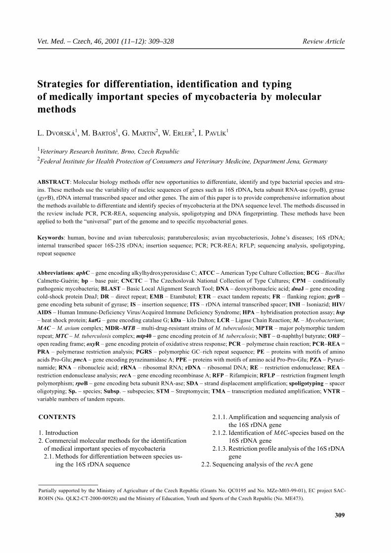
Through PCR amplification of conserved regions ofthe 16S rDNA gene using genus-specific oligonucleotidesMYCGEN-F (5´-AGA GTT TGA TCC TGG CTC AG-3´)and MYCGEN-R (5´-TGC ACA CAG GCC ACA AGG
312
Review Article Vet. Med. – Czech, 46, 2001 (11–12): 309–328
GA-3´) which produce a 1 030 bp fragment, it is pos-sible to identify the genus Mycobacterium. The combi-nation of the external primer MYCGEN-F with thespecies-specific primer MYCAV-R (5´-ACC AGA AGACAT GCG TCT TG-3´) results in a product of 180 bpwhich identifies the species M. a. avium and M. a. para-tuberculosis. Using the combination of the primer MY-CGEN-R with the species-specific primer MYCINT-F(5´-CCT TTA GGC GCA TGT CTT TA-3´), a 850 bpamplicon indicates M. intracellulare strains (Figure 1).A PCR system based on the above-mentioned primerscan be used to identify non-virulent strains of M. intrac-ellulare. The disadvantage of this method is that it doesnot distinguish between M. a. avium and M. a. paratu-berculosis, which, on the other hand illustrates the highsequence homology in these two species.
2.1.3. Restriction profile analysis of the 16S rDNAgene
The PCR-REA (or PRA) method is based on the com-bination of PCR amplification of the 16S rDNA gene andsubsequent restriction analysis. The primers MB-UZ1 (5´-GAC GAA CGC TGG CGG CGT GCT TAA C-3´) andMB-UZ2 (5´-CGT CCC AAT CGC CGA TC-3´) werederived from conserved regions of 16S rDNA by com-paring 15 mycobacterial species (Thierry et al., 1990).
On the basis of sequence comparison using the pro-grammes Gene Compar (Applied Maths, Version 4, Ko-rtrijk, Belgium) and GCG (Wisconsin Package, Version9.1, Genetics Computer Group, USA), we developed anidentification system, which permits the differentiationof most clinical significant mycobacterial species in twosteps. For amplification of the 16S rDNA gene primersaccording to Thierry et al. (1990) are used. The resultingPCR products, 1 300 bp in size, are digested using theREs Rsa I and Cfo I. From our sequence comparison re-sults we concluded that with RE Rsa I we can differenti-
ate between M. tuberculosis and M. bovis strains. In addi-tion, with Rsa I it is possible to distinguish species ofMAC (M. a. avium serotypes 1–3, 8–11 and 21,M. a. paratuberculosis) and M. intracellulare (serotypes7, 12–20, 22–28). Because of the similarity in the size ofrestriction fragments of species M. intracellulare,M. gordonae and M. ulcerans, obtained after digestionby Rsa I, we also digest the 1 300 bp PCR product withthe RE Cfo I. This method can be applied to differentiatethe M. ulcerans species from the M. terrae species. Ad-ditionally, it is possible to differentiate closely relatedspecies from the genera Corynebacterium, Rhodococcus,Gordonia and Nocardia which often appear in samplestogether with mycobacteria. The system we developeddoes not permit the differentiation between M. bovis andM. tuberculosis subsp. caprae. Variability of the DNAsequence of serotypes especially in species of theM. fortuitum complex, and the serotypes of M. kansasiiand M. chelonae also confounds their differentiation.
2.2. Sequencing analysis of the recA gene
The recA gene sequence is used as an alternative tothe sequence analysis of the gene 16S rDNA for the dif-ferentiation of mycobacteria (Blackwood et al., 2000).Protein RecA encoded by this gene exists in all bacteria.It has a role in homologous DNA recombination, DNAdamage repair and induction of the SOS response. Twofragments, A and B, were gained through the amplifica-tion of recA gene. Sequencing of the fragment A (915 bp– 970 bp) distinguished the species M. leprae, M. aurumand M. mucogenicum which indicated 75.7% similarity.Despite of 96% homology, the recA sequencing can dif-ferentiate the clinically important M. kansasii from theless clinically important M. gastri. Sequencing of the frag-ment B (about 1 kbp) distinguished the species M. xeno-pi, M. asiaticum, M. shimoidei and MTC.
2.3. Sequencing analysis of the rpoB gene
The polymorphism of the rpoB gene, which encodesthe beta subunit of RNA polymerase, was used to differ-entiate mycobacteria through DNA hybridisation andDNA sequence comparison (Lee et al., 2000). The vari-able region of rpoB in mycobacteria is suitable to be usedin a PCR-REA assay. This variable region of the rpoBgene is flanked by conserved sequences. They enable theamplification of the variable region using the same pairof primers for all mycobacterial species. The rpoB re-gion was amplified in 44 species of mycobacteria (Leeet al., 2000). The resulting amplification products, 360 bpin size, were subsequently digested using the REs Msp Iand Hae III. Most of the mycobacterial species could beseparated on the basis of these restriction profiles. In ad-
Figure 1. Annealing sites of genus- and species-specific primers on the16S rDNA gene in Mycobacterium sp. (Wilton and Cousins, 1992)
Gene - rDNA16S
MYCGEN-F MYCAV-R
MYCINT-F MYCGEN-R
1030 bp
850 bp180 bp
Conserved areas of mycobacterial DNA sequence Variable DNA sequence for rDNA genesSizes of amplification products

313
Vet. Med. – Czech, 46, 2001 (11–12): 309–328 Review Article
dition, species with several subtypes, such as M. gordo-nae, M. kansasii, M. celatum and M. fortuitum were dis-tinguished.
In the paper of Kim et al. (1999), the rpoB gene wassequenced in 44 species of mycobacteria. Slowly and fastgrowing mycobacterial species were differentiated aftercomparison of the 306 bp nucleotide sequence. The patho-genic M. kansasii was easily distinguished from non-pathogenic M. gastri, which is not, for example, possibleby sequencing of 16S rDNA. About 40 point mutations,deletions and insertions were discovered by sequencingrpoB. Point mutations occurred most frequently in thecodon, which encodes Ser (531) and His (526). The pointmutation Ser (531) to Leu dominated in approximately70% of Rifampicin-resistant clinical isolates of M. tu-berculosis.
2.4. Restriction profile analysis of the gene forheat shock protein hsp 65
Another possible alternative to commercial methodsdifferentiate the genus Mycobacterium is the amplifica-tion and subsequent restriction analysis of the gene whichencodes the heat shock protein hsp65, 65 kDa in size(Plikaytis et al., 1992; Telenti et al., 1993).
Stress proteins are an important component of the sur-face antigens of certain pathogens. Hsp65 contains bothspecies-specific epitopes and epitopes which regularlyoccur in different species of mycobacteria. The naturaloccurrence of conserved sequences of this gene allowsthe differentiation of mycobacteria through the restric-tion digestion of PCR products obtained with ‘univer-sal’ primers Tb11 (5´-ACC AAC GAT GGT GTG TCCAT-3´) and Tb12 (5´-CTT GTC GAA CCG CAT ACCCT-3´) (Telenti et al., 1993). A restriction map of theresulting PCR fragment, 441 bp in size, was constructedusing the REs Hae III and BstE II and GCG software(Wisconsin Package, Version 9.1, Genetics ComputerGroup, USA). In total, 33 different species of mycobac-teria were identified by this method (da Silva Rocha etal., 1999). On the basis of the two-step digestion (Hae IIIand BstE II) of reference strains, Telenti compiled a dif-ferentiation algorithm of mycobacteria at the level of spe-cies (Telenti et al., 1993).
The disadvantage of this method is its inability to dif-ferentiate members of the MTC. On the other hand, thismethod distinguishes individual subspecies of theM. fortuitum complex. It also separates MAC into M. a.avium and M. intracellulare species, as does the Accu-Probe system. A great benefit is the ability to distinguishbetween M. kansasii and M. gastri (Kirschner and Böt-tger, 1998). However, identification of a series of myco-bacteria is complicated because their PCR-REA profilesdo not fall into the algorithm compiled on the basis ofthe PCR-REA patterns of reference strains (da Silva
Rocha et al., 1999). Recently, new type strains were addedto the list of analysed species (Ergin et al., 2000; Brunel-lo et al., 2001).
2.5. Amplification of the dnaJ gene
The gene for dnaJ encodes a cold-shock protein of 389amino acids (MW = 41.2 kDa) (Nagai et al., 1990). Ithas been sequenced in 19 mycobacterial species (Take-waki et al., 1994). In accordance with Runyon’s classictyping (Runyon, 1959), which is based on the pigmenta-tion of colonies and growth speed, phylogenetic relationsby sequence comparisons of dnaJ were found inside thethree main groups: I, II and III. However, the speciesM. simiae, which is phylogenetically closer to Group IIIthan to Group I is an exception. Fast growing types ofmycobacteria, e.g. M. fortuitum and M. chelonae, did notform a coherent group that is designated by Runyon asgroup IV. It has been concluded from these results (Take-waki et al., 1994) that differentiation on the basis of thesequence of gene dnaJ relates more to the pigmentationof bacterial colonies than to the growth speed of individ-ual types. The same paper proposed a system of diges-tion of the gene for dnaJ using REs, which enable thedifferentiation of the majority of mycobacterial types(Takewaki et al., 1994).
In the paper of Uematsu et al. (1993), a 196 bp frag-ment of dnaJ gene was used to differentiate of 14 myco-bacterial species. Nested PCR was used to obtain theamplicon, and differentiation between species was car-ried out using PCR with selected REs. A similar strategyhas been used in another study to differentiate M. tuber-culosis from another 11 different species of mycobacte-ria (Inyaku et al., 1993).
In the laboratory in Brno, we use the method of Nagaiet al. (1990) to differentiate between most species of my-cobacteria. We amplify a specific region of dnaJ gene230 bp in size using the primers YNP9 (5´-GGG TGACGC GGC ATG GCC CA-3´) and YNP10 (5´-CGG GTTTCG TCG TAC TCC TT-3´). This is a favourable meth-od for the routine differentiation of most mycobacterialspecies because this region is specific to all species ofmycobacteria so far tested.
Based on the sequence of dnaJ, we discovered a meth-od to directly identify a member of the MAC down to thesubspecies level. PCR with dnaJ specific primers result-ed in a non-specific band of 533 bp. This band corre-sponds to the presence of insertion sequence IS901(confirmed by cloning and sequencing). IS901 is specif-ic for M. a. avium (serotypes 1–3), virulent for birds (Pav-lík et al., 2000b). Presence of the non-specific 533 bpamplicon in addition to the specific 230 bp fragment en-ables the positive identification of M. a. avium witha probability of 65.9%. Absence of the non-specific bandindicates that the bacterial strain is, with 99.4% proba-
314
Review Article Vet. Med. – Czech, 46, 2001 (11–12): 309–328
bility (169 out of 170 samples), IS901 negative. The non-specific amplicon arises as a result of the primer YNP-9binding to partly homologous sequences in IS901 (5´-CCG TAC CGG GT-3´, position 838 bp – 848 bp and5´-GCC CA-3´, position 1 371 bp – 1 367 bp, AccessionNo. X59272).
2.6. Using gene sequences (katG, pncA, oxyR,aphC) for the identification of mycobacteria
From what is known about the molecular-genetic ba-sis of sensitivity of mycobacteria to antituberculoticdrugs and other antibacterial medicaments, it is possi-ble to use polymorphism in the corresponding genes toidentify and differentiate between mycobacteria. Isoniaz-id (INH), Pyrazinamide (PZA), Streptomycin (STM),Rifampicin (RFP) and Etambutol (EMB) belong to thebasic line of antituberculotic drugs. This chapter willtherefore describe genes of mycobacteria which influ-ence the function of some of the chemotherapeutics men-tioned above and show variability within the genusMycobacterium.
GENE katG
Pathogenic mycobacteria can survive as intracellularparasites owing to the production of enzymes which de-grade the oxygen radicals that form part of the defencemechanism of the parasitised host (Miller et al., 1997).Mycobacteria produce catalases and peroxidases whichdegrade H2O2 (Knox, 1956). MAC species produce theenzymes KatE, KatG and AhpC. In contrast, only KatGis found in MTC species. Protein KatG is a heat-unsta-ble, H2O2 inducible enzyme with catalase as well as per-oxidase activity. It functions in the transformation of the“prodrug” INH into an active form. Polymorphism in thesurrounding regions of the gene katG was used for RFLPanalysis of sensitive or resistant M. tuberculosis strainsby Zhang et al. (1993). The sequence of a 75 bp repeatsurrounding MPTR, a 10 bp polymorphic tandem repeat,was used as a probe. Since the number of copies of the75 bp repeat is different in various strains, it is possibleto type between them relatively easily using the RFLPmethod.
In an other study (Brow et al., 1996), a 620 bp seg-ment of katG was used for MTC differentiation. This seg-ment was denatured and the renaturated single-strandedDNA was digested by the RE Cleavase I. This RE di-gests only a specific single-stranded DNA conformationformed on the basis of its nucleotide sequence. Thus, itis possible to use such digestion to identify point muta-tions by “structure fingerprints”. Structure fingerprintswere originally used for studying mutations leading toINH-resistance of M. tuberculosis isolates (Brow et al.,1996).
GENES pncA AND oxyR
PZA is an inactive form of drug which is converted toan active form by the enzyme pyrazinamidase (Pzase).Pzase is encoded by the gene pncA. The loss of Pzaseactivity, which is caused by deletion or substitution, isconnected to PZA-resistance (Cheng et al., 2000). To-gether with another gene oxyR, which encodes a proteinthat protects against the oxidative stress response of thehost‘s macrophages, these genes are used for the fast iden-tification of MTC strains. The polymorphism of pncA andoxyR genes are detected using the PCR-REA method. Itis possible to differentiate M. tuberculosis from M. bovisby using the RE Alu I (Yan et al., 1998). Through directsequencing of the 410 bp region of gene oxyR in 105strains of MTC, 29 strains were identified as M. bovis onthe basis of substitution in position 285 of the nucleotidewhich creates a unique restriction site for the RE Alu I.Therefore, it is also possible to differentiate the M. bovisspecies from other MTC species using the PCR-RFLPmethod with the RE Alu I.
GENE ahpC
Sequence analysis of alleles rpoB and ahpC was usedto study multi-drug-resistant M. tuberculosis (MDR-MTB) isolates in Scotland between 1990–1997. GeneahpC encodes a detoxifying enzyme which participatesin protection from oxidative metabolites. One MDR-MTBstrain out of these 715 Scottish strains had a synonymoussubstitution (ATT-ATC) in codon 6 and a very similarIS6110 RFLP profile to MDR-MTB belonging to a groupof strains “W”, originating from Asia. On the basis ofanamnestic data, it was found that this isolate indeed orig-inated from a patient of Asian origin (Fang et al., 1999b).
2.7. Analysis of internal transcribed spacer (ITS)16S-23S rDNA
The rRNA (rrn) genetic locus is very interesting froman evolutionary point of view. It is present in bothprokaryotes and eukaryotes. In prokaryotes, the rrn lo-cus contains genes for all three rRNA types: 16S, 23Sand 5S. Genes which encode the relevant rRNA in bac-teria are found in 1 to 11 copies (Gurtler and Stanisich,1996). On the chromosome, genes for rRNA are arrangedin groups. Each group of genes for rRNA is transcribedas an rRNA-transcription unit.
Genes for individual types of rRNA are separated byspacers, which demonstrate a high degree of sequenceand length variability at the level of genus as well as spe-cies. This diversity is caused by variations in the numberand types of rRNA sequences which are found inside thespacers. On the basis of substituting genes for tRNA,rRNA-transcription units are marked as: rrnA, rrnB,
315
Vet. Med. – Czech, 46, 2001 (11–12): 309–328 Review Article
rrnC, rrnD, rrnE a rrnF (Gurtler and Stanisich, 1996).The spacer region located between the genes for 16S and23S rRNA is extremely variable even in terms of closelyrelated taxonomic groups, which is a result of frequentinsertions and deletions in this region of the genome(Gurtler and Stanisich, 1996).
The polymorphism in the length and sequences of spac-ers in the rrn locus is used to differentiate various typesof prokaryotes (Barry et al., 1990). Jensen et al. (1993)amplified the spacer 16S-23S in 300 bacterial specieswhich belonged to 8 genera and 28 species or serotypes.For the purposes of amplification they used primer G1(5´-GAA GTC GTA ACA AGG-3´), whose sequence hadbeen derived from the highly conserved region 16S rDNAimmediately adjacent to the spacer 16S-23S, and primerL1 (5´-CAA GGC ATC CAC CGT-3´), which had beenderived from the most conserved part of the 23S rDNAsequence, located just behind the spacer. All 28 testedspecies displayed characteristic amplification profiles.
By sequence analysis of the internal transcribed spac-er (ITS) 16S-23S rDNA it is possible to separate certainspecies of mycobacteria into specific intraspecies taxonscalled “sequevars”, e.g. five sequevars were identifiedin the MAC species: MavA to MavE, which differ in oneor two nucleotides (Novi et al., 2000). ITS sequencingsupplements information acquired by sequencing 16SrDNA, which can be used to differentiate closely relatedspecies. A high sequence variability in the ITS of slowlygrowing mycobacteria has been noted (Roth et al., 1998).On the basis of this high degree of ITS variability, close-ly related species such as M. gastri and M. kansasii wereidentified, which cannot be differentiated using sequenceanalysis of the gene for 16S rDNA. However, ITS analy-sis failed in the discrimination between M. marinum andM. ulcerans, which share a high degree of genome re-latedness, also demonstrated by 16S rDNA analysis.Moreover, the same ITS sequences were discovered inmembers of the MTC, which includes phylogeneticallydistant bacterial species.
Roth et al. (1998) analysed the ITS sequence of 60strains of the genus Mycobacterium, which included 13species. Using PCR amplification with the primersEc16S.1390b (5´-TTG TAC ACA CCG CCC GTC A-3´)and Mb23S.44n (5´-TCT CGA TGC CAA GGC ATCCAC C-3´), which had been derived from the adjacentregions of the genes 16S rDNA and 23S rDNA, they de-tected a PCR product 480 bp in size in all 60 strains.Using sequence analysis they found that the spacer sizesof slowly growing species of mycobacteria are in therange from 235 nucleotides in M. xenopi to 285 inM. gastri. They were on average 75 nucleotides shorterthan in fast growing species of mycobacteria. On the ba-sis of this difference in ITS length it is possible to distin-guish visibly slowly growing and fast growing strains.Sectors with a high degree of sequence variability aredispensed over the whole spacer sequence. A conse-
quence of this significant variability was the discoveryof intraspecies sequence polymorphism in 4 of the 11species, in M. gastri designated as sequevars MgaA andMgaB, MAC (MavA to MavE), M. simiae (MsiA toMsiD) and M. xenopi (MxeA to MxeC).
Furthermore, a PCR-REA method on the basis of ITSwas used to distinguish MTC from MAC (Sansila et al.,1998). In a first PCR step, a 380 bp product was ampli-fied using primers 16SC (5´-TCG AAG GTG GGA TCGGC-3´) and 23SG (5´-GCG CCC TTA GAC ACT TAC-3´), which had been derived from adjacent sequences ofrDNA. Afterwards, this product was digested with REHae III, Msp I or BstX I (Sansila et al., 1998). After thedigestion with RE Hae III, unique PCR-REA profileswere obtained for different MAC species. PCR-REA con-stituents of M. intracellulare were similar to patterns ofM. scrofulaceum. A significant disadvantage of this meth-od was the inability to differentiate individual MTC sub-types.
A subsequent paper (Roth et al., 2000) describes a newdiagnostic algorithm for differentiation of mycobacteriaby PCR-REA. After amplification with primer Sp1 (5´-ACC TCC TTT CTA AGG AGC ACC-3´), which hadbeen derived from the beginning of the M. tuberculosisspacer sequence (Accession No. L15623) and primer Sp2derived from position 210 bp to 190 bp (5´-GAT GCTCGC AAC CAC TAT CCA-3´), amplicons of differentlengths were obtained. In slowly growing mycobacteria,the size of amplicons are between 200 bp and 330 bp, infast growing species the fragments were always longer than250 bp. Using the REs Hae III and Cfo I, it was possibleto differentiate slowly and fast growing mycobacteria. Fastgrowing species displayed PCR-RFLP fragments in therange 33 bp to 230 bp larger than slowly growing spe-cies (which produced products in the range 33 bp to175 bp) after digestion by RE Hae III. Digestion by RECfo I resulted in longer fragments from fast growing spe-cies, which contained a specific restriction site. A disad-vantage was that a great number of fast growing speciesof mycobacteria did not have a restriction site for RECfo I at all. In contrast, the amplified region of DNA ofall species of slowly growing bacteria were able to bedigested by this RE. The amplified region of DNA fromslowly growing bacteria carried a specific restriction sitefor RE Cfo I and formed shorter fragments divided intogroups A to D. For the final determination of speciesbelonging to the fast growing mycobacteria, either theRE Taq I was used, namely for the differentiation ofM. abscessus from M. chelonae I, II and III, or RE Ava IIfor the differentiation of M. porcinum from M. farcino-genes. REs Dde I, Taq I or Ava II were used for precisedifferentiation of slowly growing species of mycobacte-ria. It was necessary to use yet another step with certainspecies of this group of mycobacteria, namely digestionby REs Msp I (M. simiae) and Hinf I (M. kansasii). Thealgorithm for slowly growing species of mycobacteria
316
Review Article Vet. Med. – Czech, 46, 2001 (11–12): 309–328
thus became very complex and difficult to interpret. Prob-lems also arise with the fast growing species of myco-bacteria with the interpretation of so far unclearly definedspecies (Roth et al., 2000).
3. METHODS FOR DIFFERENTIATIONAND IDENTIFICATION OF THE M. AVIUMSUBTYPES
Currently used molecular techniques, especially theRFLP method, provide a new way of looking at strainsof individual MAC species (McFadden et al., 1987). In-sertion sequences, which vary in the number of copiesand their locations on the genome, are used as probes totype species (van Soolingen et al., 1998a). IS are char-acterised by a very low degree of mobility and limitedpolymorphism. Thus they have become an appropriatetool in the study of epidemiology of mycobacterial in-fections (Pavlík et al., 1999a; Dvorská et al., 2002).
The discovery of insertion sequences in mycobacteri-al genomes, e.g. IS900 in M. a. paratuberculosis (Green
et al., 1989), IS901 (Kunze et al., 1991), IS1245 (Guer-rero et al., 1995), IS1311 (Roiz et al., 1995) in the MACstrains was a major breakthrough in the study of myco-bacterial infections. A summary about mycobacterial ISsis given in our previous study (Dvorská et al., 1998). Up-to-date information about the number of mycobacterialinsertion sequences is available in the international data-base of IS at: http://pc4.sisc.ucl.ac.be/is.html
3.1. Insertion sequence IS900 specific forM. a. paratuberculosis
The occurrence of IS900 is limited only to the M. a.paratuberculosis genome, where it is found in 15 to 20copies (Green et al., 1989; Pavlík et al., 1995, 1999c;Bull et al., 2000). IS900 is 1451 bp in size and has oneopen reading frame (ORF) which encodes the protein p43.From an epidemiological point of view, IS900 is a high-ly stable marker used for precise identification ofM. a. paratuberculosis by the PCR method. Various lo-cations and numbers of IS900 on the genome are then
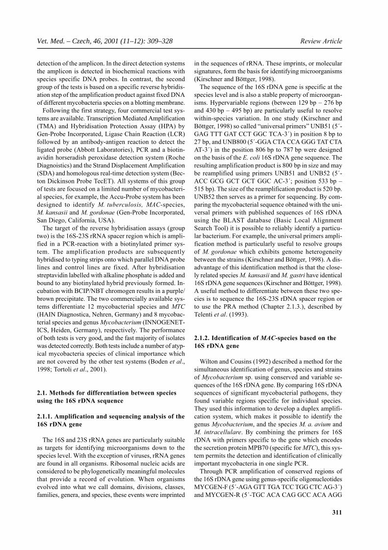
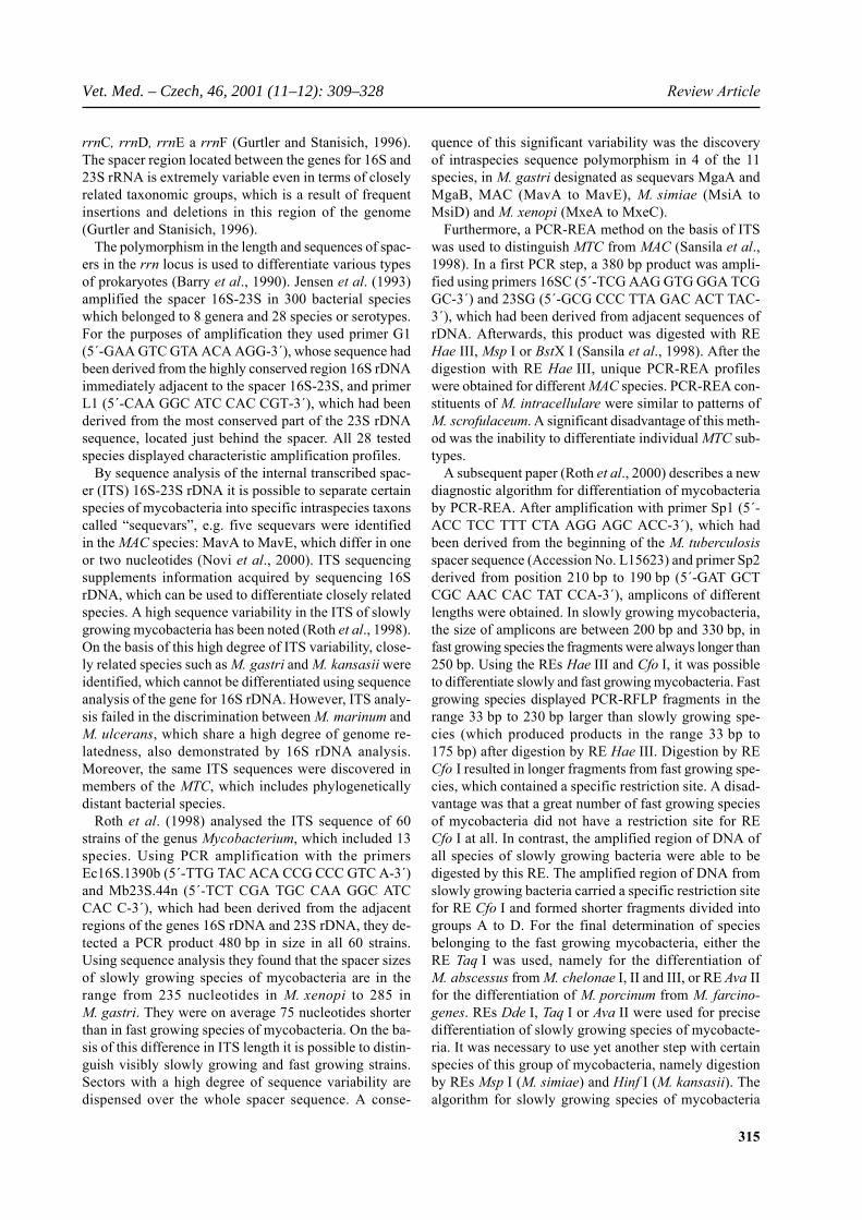
Figure 2. Diagnostic algorithm for the identification and differentiation of medically important mycobacterial species (Table 1)1internal transcribed space 16S-23S rDNA, MAC – M. avium complex
Slowly growing mycobacteria
Fast growing mycobacteria
Mycobacterium sp.dnaJ, 16S rDNA,
ITS 16S-23S1
rpoB
c
M. tuberculo-sis omplex
M. aviumcomplex
61101080
IS IS
M. tuberculosis
M. bovis
M. tuberculosis capreasubsp.
M.microtiM.africanum subtype I
mtp , pnc , kat , aph40 A G C
R Aoxy , pnc ,
?- unpublished designation of gene
M. leprearecA
gyrB
IS16S rDNA
1245
ISJ
901 dna
IS900 MAC
M. intracellulare
M. a. paratuberculosis
M. a. avium strains virulent for birds
16S rDNA
M. gastri
M. xenopiM. simiae
ITS16S-23S
1
M. gordonea
M. gordonea
M. kansasii
M. kansasii
M. kansasii
M. kansasii
rpoB
rpohsp
B65
hsp65
M. fortuitum
M. celatum
M. aurum
M. mucogenicum
317
Vet. Med. – Czech, 46, 2001 (11–12): 309–328 Review Article
used for the typing of individual strains using the RFLPmethod (Kunze et al., 1991; Pavlík et al., 1999c).
In the laboratory in Brno we drew up a standardisationscheme for typing individual strains of M. a. paratuber-culosis with the RFLP method using parallel digestionby two REs Pst I and BstE II (Pavlík et al., 1999c). Forthe description of a wide spectrum of different RFLPtypes we applied computer analysis of DNA fingerprintsusing the Gel Compar software (Applied Maths, Kortr-ijk, Belgium). The obtained results and identificationscheme are available at:http://www.vri.cz:/wwwrflptext.htm.
Thirteen Pst I RFLP types designated A to M wereidentified in 1 008 strains of M. a. paratuberculosis us-ing this system. Twenty RFLP types were detected byparallel digestion of chromosomal DNA by the REBstE II. Eighteen fingerprints: C1 to C3, C5, C7 to C20were included in a group of RFLP types designated withthe letter C (cattle). One RFLP type S1 was detected in agroup of RFLP types designated S (sheep) and one typeI1 was also detected in a group of RFLP type I (interme-diate). Previously described RFLP types C4, C6 and S2(Collins et al., 1990) and RFLP types S3 and I2 (deLisleet al., 1993) were included into our typing system afterscanning them from published figures. Using a combi-nation of results obtained after the parallel digestion byPst I and BstE II, a total of 28 different RFLP types weredifferentiated (Pavlík et al., 1999c). This method wasused in several epidemiological studies (Pavlík et al.,1994, 1996, 1999b,d, 2000a; Greig et al., 1999; Whit-lock et al., 1999).
3.2. Identification of avian tuberculosis caused byIS901+ strains
The insertion sequence IS901 was discovered by Kun-ze et al. (1991). It is 1472 bp in size, and contains oneORF for transponase, a protein of 401 amino acids. Thestability of IS901 in strains isolated primarily from clin-ical material from birds, domestic animals and from theenvironment is used for the rapid identification of IS901+strains using the PCR method. IS901 occurs on the ge-nome of virulent MAC strains in 2 to 13 copies (Kunzeet al., 1991; Ritacco et al., 1998), which we also con-firmed in 172 strains examined (Dvorská et al., 2002).
In in the laboratory in Brno, we explored the relation-ship between the presence of IS901 (PCR) and the viru-lence of these strains for birds in a biological experiment.A total 165 out of 738 (22.4%) strains caused genera-lised tuberculosis, 164 (99.4%) of them contained IS901.The remaining 573 (77.6%) strains were non-virulent;however, IS901 was present in 24 (4.2%) strains. As themajority of these strains were from collections, they mighthave lost virulence during several decades of storage.IS901 was demonstrated in all virulent field strains, which
enabled the replacement of biological experiments by thePCR method.
3.3. Identification of avian mycobacteriosescaused by IS1245+ strains
The presence of IS1245 has often been used to identi-fy MAC strains (Guerrero et al., 1995; Ritacco et al.,1998). IS1245 is limited to the species M. a. avium,M. a. paratuberculosis and M. a. silvaticum strains (so-called “wood pigeon” strains). Strains of M. intracellulareentirely lack this genomic element (Guerrero et al., 1995).IS1245 has been found in more than 20 copies in iso-lates from people and pigs. In contrast, MAC strains vir-ulent for poultry (containing IS901), contained only threecopies of this element, so-called “bird type” (Ritacco etal., 1998). Furthermore, it was observed that the numberof IS1245 copies on the genome relates to the serotypeof the given strain. The “bird type” (three IS1245 bands)contains strains of the serotypes 1 to 3. Strains of sero-types 4 to 6, 8 to 11 and 21 formed polymorphous IS1245RFLP profiles of 6 and 20 bands. Strains of serotypes 7,12 to 20, 22 to 28 did not contain element IS1245 at all(Ritacco et al., 1998).
3.4. Differentiation of M. a. avium andM. a. paratuberculosis using other methods
Eriks et al. (1996) developed a method to differentiateM. a. avium and M. a. paratuberculosis on the basis ofPCR-REA. They digested a 960 bp amplicon of the genehsp65 using the RE Pst I. It was possible to differenti-ate clinical isolates of both subspecies. The method wasparticularly useful to classify strain 18 of M. a. avium,which carried the insertion sequence IS901 and was in-correctly classified as M. a. paratuberculosis (Merkal,1979; Chiodini, 1993).
4. METHODS OF DIFFERENTIATINGTHE M. TUBERCULOSIS COMPLEX (MTC)
4.1. Detection of repeaed sequences of DNA
Members of the MTC (M. tuberculosis, M. africanum,M. bovis, M. bovis BCG, M. microti, M. tuberculosissubsp. caprae and M. canetti) are genetically closely re-lated taxons. The relatively high degree of intertype DNAhomology of MTC strains in the 16S rDNA sequence andof the 16S-23S spacer limits the differentiation of thesetypes using restriction analysis (Eisenach et al., 1988).Despite this genetic homology in the MTC, a relativelyhigh DNA polymorphism has been found in repetitiveDNA, which are ISs (Dvorská et al., 1998). Up till now,
318
Review Article Vet. Med. – Czech, 46, 2001 (11–12): 309–328
the following IS have been identified in MTC strains:IS6110 (Thierry et al., 1990), IS1081 (Collins andStephens, 1991), IS1547 (Fang et al., 1999a) and an IS-like element (Mariani et al., 1993).
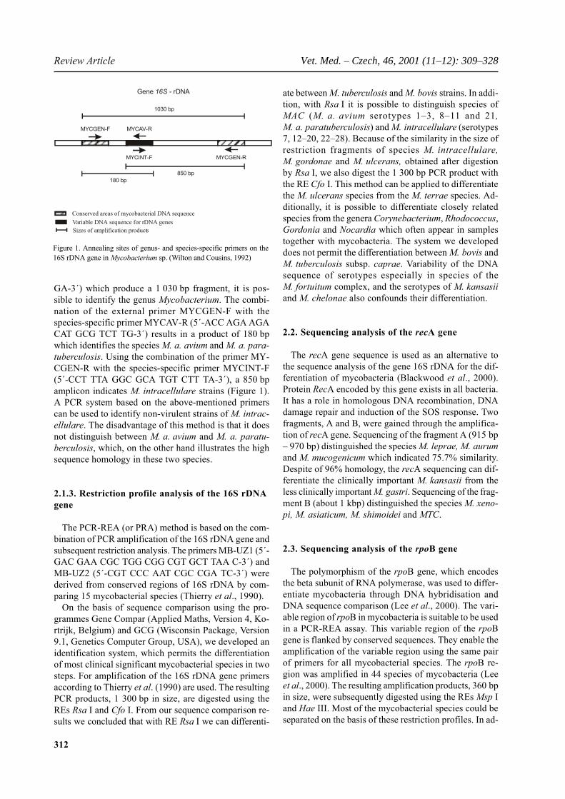
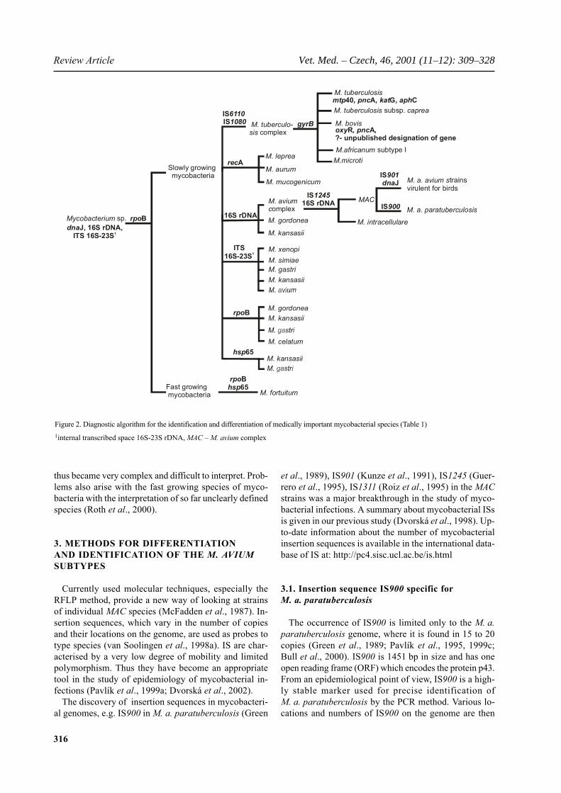
For MTC, IS6110 RFLP has become a widely used typ-ing method for epidemiological studies. An example oftypical pattern is given in Figure 3. In M. tuberculosisisolates (line 1) the large number of IS6110 copies onthe genome permits an excellent use of this element forstrain comparison in outbreak tracing. M. bovis strainsoften contain less than five IS6110 copies (Figure 3: BCGwith one copy and three examples of different cattle out-break strains from Germany). Therefore, the use ofIS6110 RFLP for epidemiological studies in M. bovis ismore limited and was more successful by combinationwith spoligotyping (Chapter 4.2.). As also M. tuberculosisstrains with a low number of IS6110 copies exist, theRFLP pattern can only be used for differentiation betweenthe species with a probability of error. In M. tuberculosissubsp. caprae (M. t. caprae) the usual copy number ofIS6110 appears to be higher than in M. bovis (last 4 lines).IS6110 is suitable to subtype spoligopattern which aremore conserved in M. t. caprae (unpublished observa-tion).
In contrast to IS6110, the use of the remaining IS ele-ments is limited by the small number of copies on thegenome and the low degree of polymorphism (van Sool-ingen et al., 1998b). However, six other types of shortrepetitive DNA with a varying degree of genetic diversi-ty and potential usefulness were identified: DR (Thierry
et al., 1990), PGRS (Ross et al., 1992), GTG and MPTR(Hermans et al., 1992), ETR and VNTR (Frothinghamand Meeker-O’Connell, 1998). Kremer et al. (1999) com-pared 12 current molecular methods to distinguish MTCsubtypes based on RFLP, DNA hybridisation, PCR andcombinations of these methods. In MTC, the intention ofsome methods is differentiation and typing in the senseof definition, as mentioned for IS6110 already and as willbe shown for spoligotyping later. By studying 60 MTCstrains, Kremer et al. (1999) showed that the IS6110RFLP and mixed-linker PCR have the greatest discrimi-natory capability in strains with a high number of IS6110copies. The mixed-linker PCR method is based on a prim-er specific to the IS6110 and a primer which is comple-mentary to the linker attached by ligation to restrictionfragments of genomic DNA. Strains with between 5 and7 IS6110 copies (M. bovis and M. tuberculosis strains iso-lated in Asia) may be best typed using VNTR, PGRS-RFLP, DR-RFLP or by spoligotyping. For strains withone or two IS6110 copies, the most effective methodsare PGRS-RFLP and DR-RFLP. Since two strains of MTCwere identified which did not carry at all IS6110, the spo-ligotyping method was applied to differentiate them.
In the paper of O’Brien et al. (2000), 60 strains ofM. bovis isolated from 1993 to 1998 from cattle, pigs,deer and badger contained a single copy of IS6110.A combination of PGRS-RFLP, DR-RFLP and spoligo-typing was used to type them. For DNA fingerprinting,a plasmid containing a 4 kb insert from restriction diges-tion of M. bovis DNA with the RE Alu I was used asa probe. By comparing the sequence of the insert withthe whole genome of M. tuberculosis strains H37Rv us-ing the BLAST database, it was found that it is 90% ho-mologous with the PPE gene (gene Rv0096). This geneconsists of a large number of repetitive 69 bp units. Theresulting fingerprints were readily distinguishable. 18types were distinguished using a DNA probe on the basisof a PPE gene, 11 types using the PGRS-RFLP method,10 types using DR-RFLP and 8 types using spoligotyp-ing. Using a combination of the hybridisation results withthe gene for PPE and DR-RFLP, 26 types were detected,and finally using a combination of all four methods, 28types were distinguished. For expediency and adequatetyping capabilities, the authors recommend the combi-nation PPE-DR.
4.2. Differentiation and typing of strains of theMTC by spoligotyping
Spoligotyping is based on DNA polymorphism at achromosomal locus, which is characterised by the pres-ence of a high number of conserved direct repeats, des-ignated as the DR region (Thierry et al., 1990). The directrepeats are 36 bp in size and are interrupted by DNA spac-ers of 35 bp to 41 bp. When the DR regions of several
Figure 3. IS6110 RFLP pattern of M. tuberculosis (line 1), M. bovis BCG(line 2), M. bovis (lines 3 to 5) and M. tuberculosis subsp. caprae (lines 6to 9)
1 2 3 4 5 6 7 8 9
4.0
3.9
3.8
3.7
3.6
3.5
3.4
3.3
3.2
3.1
3.0
2.9
log bp
319
Vet. Med. – Czech, 46, 2001 (11–12): 309–328 Review Article
Figu
re 4
. Spo
ligop
atte
rns t
ypic
al fo
r MTC
spec
ies a
nd su
btyp
es (v
an S
oolin
gen
et a
l., 1
997,
199
8b; A
rana
z et
al.,
199
9; Z
umar
rage
et a
l., 1
999;
Nie
man
n et
al.,
200
0b; S
ola
et a
l., 2
000;
Via
na-N
iero
et a
l., 2
001)

320
Review Article Vet. Med. – Czech, 46, 2001 (11–12): 309–328
strains were compared, it was noted that the order of spac-ers is nearly the same in all strains, but that there may bedeletions or insertions. The presence or absence of 43individual spacers may be detected using the spoligotyp-ing method.
For the purposes of spoligotyping, which is in fact areverse hybridisation, all 43 oligonucleotides are boundcovalently to a nylon membrane in parallel rows. Each ofthe oligonucleotides immobilised in this way representsthe unique sequence of a spacer which is a component ofthe DR region in MTC. In total, 37 oligonucleotides werederived from the sequences of spacers present in theM. tuberculosis H37Rv and the remaining six sequencesof oligonucleotides were derived from the spacers ofM. bovis BCG. These loaded membranes are commer-cially available.
To perform the method in the laboratory, initially theDR region including the various spacers has to be ampli-fied by PCR. The primers are derived from the DR se-quence. A biotinilated reverse primer is used to label thePCR products which after heat-denaturation are hybri-dised to the oligonucleotides on the membrane and de-tected by chemoluminescence. The presence of a spacerappears on the autoradiographic film as a signal in theshape of a black square. Individual strains are character-ised by a grouping of squares, which represent the exist-ing spacers.
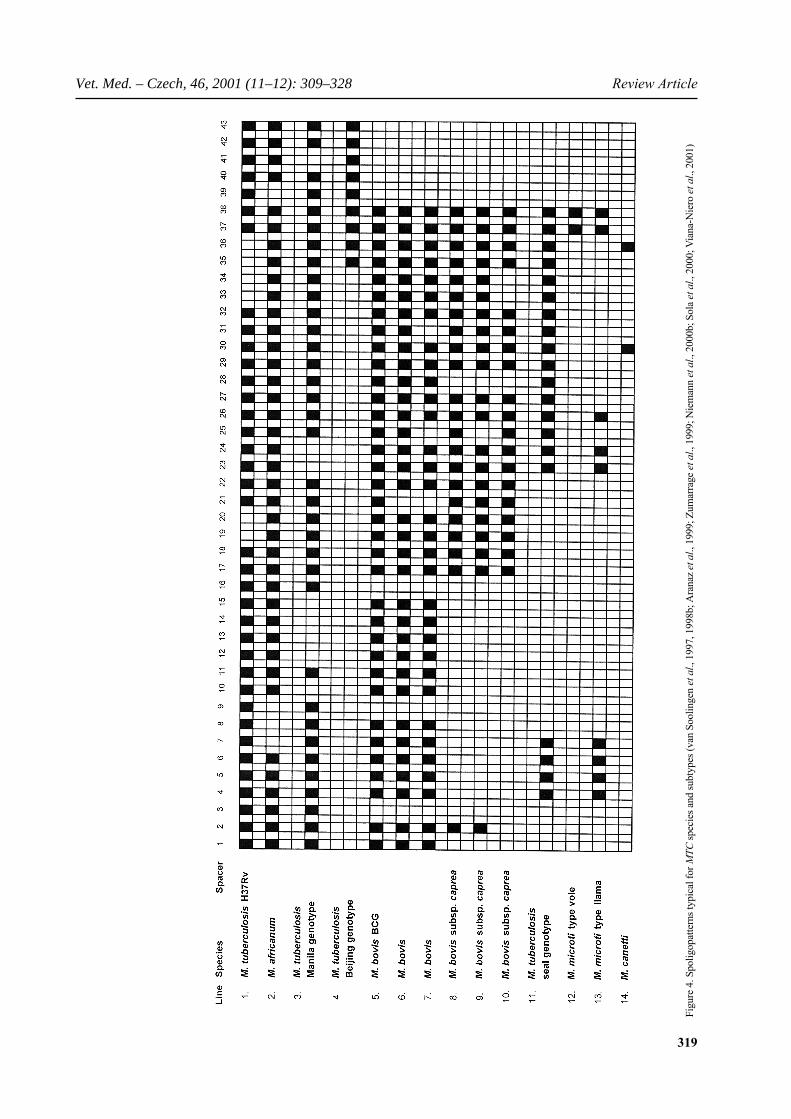
Spoligotyping is an excellent method for differentia-tion of MTC subtypes based on the presence and/or ab-sence of certain combinations of spacers. The types whichcan be differentiated are given in Figure 4 which sum-marises own results and the literature (van Soolingen etal., 1997, 1998b; Zumarraga et al., 1999; Aranaz et al.,1999; Niemann et al., 2000a,b; Sola et al., 2000; Viana-Niero et al., 2001). The M. tuberculosis subtypes (strainH37Rv is shown in line 1) are particularly characterisedby the presence of spacers 39–43. Individual differencesin M. tuberculosis strains occur in the presence/absenceof spacers 1–38. If the spacers 7, 8, 9 and 39 are missing,an isolate falls into subtype M. africanum (line 2); M.tuberculosis Manila genotype (line 3) is characterised bythe missing of spacer 41 and more than one of the lowerspacers. M. tuberculosis Beijing genotype (line 4) is char-acterised by the presence of spacers 35 to 43 only.
All following species in Figure 4 (lines 5 to 14) are char-acterised by the absence of spacers 3, 9, 16 and 39–43,which is typical for M. bovis. Included in the Figure 4are M. bovis BCG (line 5) and two isolates from cattleoutbreaks in Germany (lines 6 and 7). The differences inthe spoligopattern (spacers 21, 25 and 31) allow a strainidentical description. This illustrates that spoligotypingis not only a technique for differentiation but also fortyping of strains for epidemiological analyses. This is veryuseful for the subtypes M. tuberculosis, M. bovis and M.t. caprae. Subspecies M. t. caprae, represented by 3 dif-ferent isolates from cattle (lines 8 to 10), is particularly
characterised by the absence of spacers 3 to 16. The ab-sence of the spacers 3, 9, 16 and 39–43 illustrates theclose relationship to M. bovis.
In the last decades, 4 MTC subtypes have been identi-fied which differ significantly from typical MTC growthand biochemistry or have been found to be linked to in-dividual host species. Moreover, they are characterisedby unique spoligopatterns. M. tuberculosis seal genotype(line 11) was found in different species of seals in SouthAmerica (Zamarraga et al., 1999) and is characterisedby the absence of spacers 8 to 22. M. microti (line 12),originally isolated from tuberculosis in wild vole, hasbeen isolated also from other animal species and humantuberculosis cases (van Soolingen et al., 1998b). It isexisting in two types: M. microti presence of spacer 37and 38 only and the llama-type 37–38 plus 26, 23–24and 4–7. The species M. canetti, which has been isolatedin only a few occasions so far (van Soolingen et al., 1997)is characterised by the presence of only two spacers: 30and 36.
As spoligotyping is a PCR driven technique, only smallamounts of DNA are required for analysis. Therefore,spoligotyping is particularly suitable for the analysis ofslowly growing mycobacteria. It also permits the com-parison of strains which are not culturable after prolongedstorage. This can be of irreplaceable importance, as inthe case of relapses, when it is necessary to compare newstrains from patients whose earlier isolates can no longerbe cultivated.
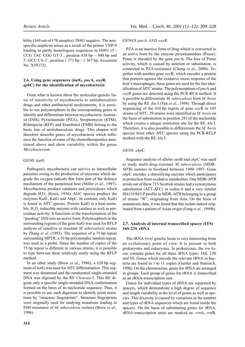
We used spoligotyping to examine the DNA of 24 deadisolates of M. bovis collected from 1965 to 1999 (Pavlíket al., 2001). 22 isolates from the Czech Republic, andone isolate from Slovakia and a Neotype M. bovis strainATCC 19210 were examined. The spoligopatterns wereevaluated using the software Gel Compare (AppliedMaths, Version 4.1, Kortrijk, Belgium). Seven spoligo-types with the working designates S1 to S7 were found(Figure 5). These were compared with the spoligotypesof 3 176 isolates from the RIVM (Netherlands Instituteof Public Health and the Environment, Bilthoven, TheNetherlands) database. The Neotype M. bovis strainstored in the Czech collection CNCTC (My310/82) since1974 was of the identical spoligotype S4 with an origi-nal Neotype strain from the USA. Isolates from the SouthAmerican capybara (Hydrochoerus hydrochaeris), im-ported from Germany in 1989, from cattle (isolated in1966, 1991 and 1994) and from three children with post-vaccination complications from the BCG vaccine wereof the most common spoligotype S1. Four unique spoli-gotypes S2, S3, S5 and S6 were identified in isolates fromthe Czech Republic from cattle (1965 and 1974), from afarm-reared red deer (Cervus elaphus) and from Slova-kia from cattle (1992). Czech isolates from a wild reddeer (1991), from cattle (1966, 1991, 1995) and from aneighty-year-old man (1999) were described to the spo-radically occurring spoligotype S7. Later it was found

321
Vet. Med. – Czech, 46, 2001 (11–12): 309–328 Review Article
that the S7 profile corresponds to the spoligotype M. t.caprae (Aranaz et al., 1999). In the Czech Republic, iso-lates of both unique spoligotypes and the most commonspoligotype in the RIVM database were found in infect-ed cattle focuses in the years 1965 to 1995 (Pavlík et al.,2001).
4.3. Differentiation of M. tuberculosis and M. bovisby the detection of specific genes (mtp40) and genevariation (oxyrR and pncA)
Strains of the species M. tuberculosis can be identifiedby the detection of the species-specific fragment mtp40using the PCR method with the primers PT1 (5´-CAACGC GCC GTC GGT GG-3´) and PT2 (5´-CCC CCCACG GCA CCG C-3´). The resulting 392 bp fragmentcorresponds to a surface antigen which is only present inthe genome of M. tuberculosis, and is not found in ge-nomes of other species of the MTC. This property wasused for the development of a specific, sensitive and rapiddiagnostic test by which infection caused by strains ofM. tuberculosis could be distinguished from infection bystrains of M. bovis (del Portillo et al., 1991; Herrera andSegovia, 1996). Using the PCR method of the genemtp40, it is also possible to detect strains of M. tubercu-losis which do not carry any copy of IS6110 in its ge-nome (Herrera and Segovia, 1996).
In samples with a negative amplification it is possible,through a subsequent amplification using primers specific
to M. bovis L1 (5´-CCC GCT GAT GCA AGT GCC-3´)and L2 (5´-CCC GCA CAT CCC AAC ACC-3´) to iden-tify this MTC species (Romero et al., 1999). It is alsopossible to distinguish individual MTC species throughsequence and restriction analyses of the genes oxyR, pncAa rpoB (Chapter 2.6.).
4.4. Differentiation of MTC-subspeciesby restriction analysis of DNA sequence gyrB
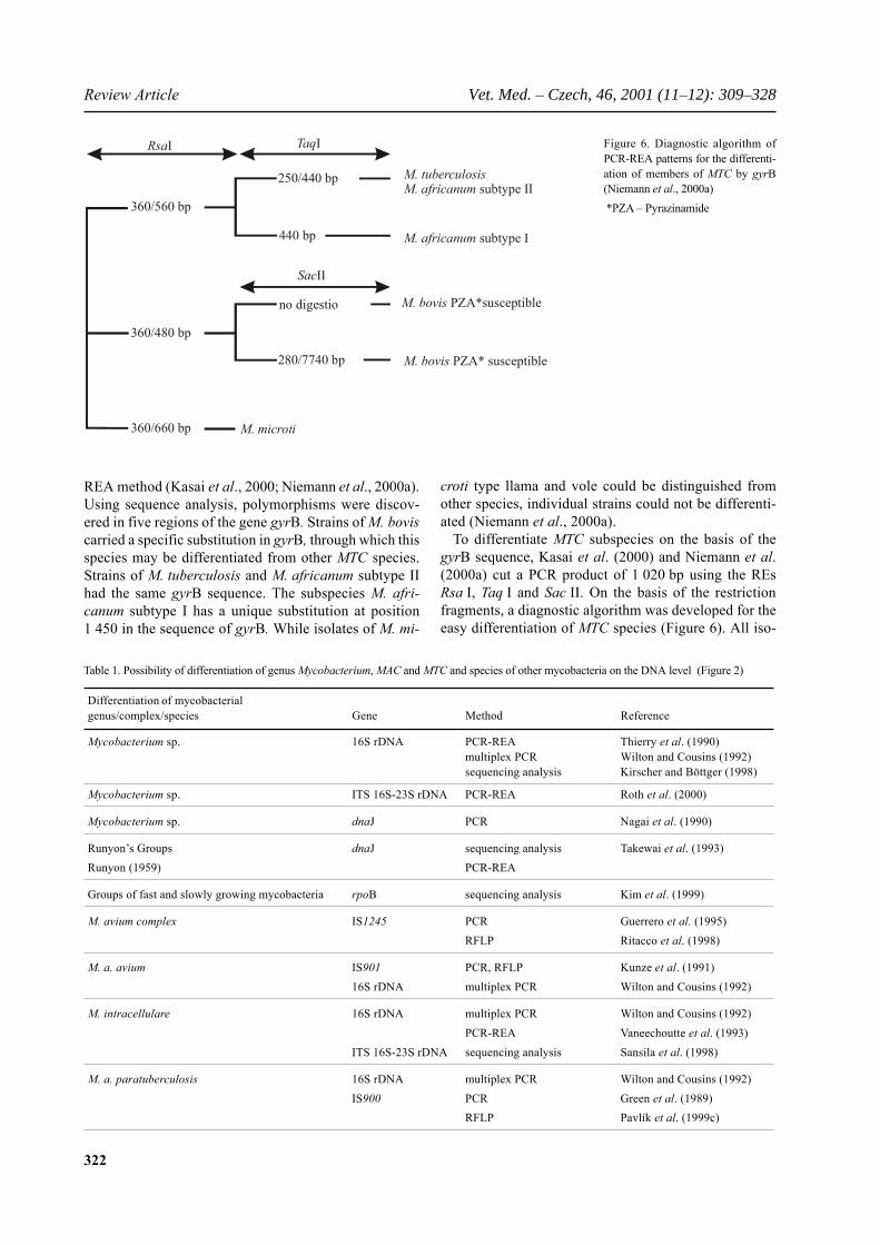
DNA gyrase is a topoisomerase II; a class of ATP-de-pendent enzymes which are capable of creating negativesuperhelixes from relaxed forms of covalently closed cir-cle (CCC) plasmid DNAs. Topoisomerases participate inthe regulation and progression of many important cellu-lar functions (particularly replication, transcription andrecombination). DNA gyrase is composed of two sub-units A and B, the encoding genes are gyrA and gyrB.Both genes are found in a 5 119 bp region of DNA. Theproteins GyrB (75 kDa) and GyrA (95 kDa) in mycobac-teria demonstrate 45–80% identity to the gyrases of otherbacteria (Unniraman and Nagaraja, 1999). Polymor-phisms in the sequence of gyrB were discovered and sub-sequently used to differentiate between MTC species.Amplification fragments, 1 200 bp in size, obtained fromclinical isolates of M. tuberculosis, M. bovis (PZA-sen-sitive and -resistant strains), M. africanum subtypes I andII and M. microti types vole and Ilama were sequenced(Niemann et al., 2000a) or examined using the PCR-
Figure 5. Dendrogram of spoligotypes of strains from the years 1965–1999 (Pavlík et al., 2001)
70 80 90 100
S1–S7 spoligotypes
S1
S2S3S4S5S6
S7
322
Review Article Vet. Med. – Czech, 46, 2001 (11–12): 309–328
Figure 6. Diagnostic algorithm ofPCR-REA patterns for the differenti-ation of members of MTC by gyrB(Niemann et al., 2000a) *PZA – Pyrazinamide
Table 1. Possibility of differentiation of genus Mycobacterium, MAC and MTC and species of other mycobacteria on the DNA level (Figure 2)
Differentiation of mycobacterialgenus/complex/species Gene Method Reference
Mycobacterium sp. 16S rDNA PCR-REA Thierry et al. (1990)multiplex PCR Wilton and Cousins (1992)sequencing analysis Kirscher and Böttger (1998)
Mycobacterium sp. ITS 16S-23S rDNA PCR-REA Roth et al. (2000)
Mycobacterium sp. dnaJ PCR Nagai et al. (1990)
Runyon’s Groups dnaJ sequencing analysis Takewai et al. (1993)Runyon (1959) PCR-REA
Groups of fast and slowly growing mycobacteria rpoB sequencing analysis Kim et al. (1999)
M. avium complex IS1245 PCR Guerrero et al. (1995)RFLP Ritacco et al. (1998)
M. a. avium IS901 PCR, RFLP Kunze et al. (1991)16S rDNA multiplex PCR Wilton and Cousins (1992)
M. intracellulare 16S rDNA multiplex PCR Wilton and Cousins (1992)PCR-REA Vaneechoutte et al. (1993)
ITS 16S-23S rDNA sequencing analysis Sansila et al. (1998)
M. a. paratuberculosis 16S rDNA multiplex PCR Wilton and Cousins (1992)IS900 PCR Green et al. (1989)
RFLP Pavlík et al. (1999c)
REA method (Kasai et al., 2000; Niemann et al., 2000a).Using sequence analysis, polymorphisms were discov-ered in five regions of the gene gyrB. Strains of M. boviscarried a specific substitution in gyrB, through which thisspecies may be differentiated from other MTC species.Strains of M. tuberculosis and M. africanum subtype IIhad the same gyrB sequence. The subspecies M. afri-canum subtype I has a unique substitution at position1 450 in the sequence of gyrB. While isolates of M. mi-
croti type llama and vole could be distinguished fromother species, individual strains could not be differenti-ated (Niemann et al., 2000a).
To differentiate MTC subspecies on the basis of thegyrB sequence, Kasai et al. (2000) and Niemann et al.(2000a) cut a PCR product of 1 020 bp using the REsRsa I, Taq I and Sac II. On the basis of the restrictionfragments, a diagnostic algorithm was developed for theeasy differentiation of MTC species (Figure 6). All iso-
360/560 bp
250/440 bp
440 bp
360/480 bp
no digestio
360/660 bp
280/7740 bp
RsaI TaqI
SacII
M. tuberculosisM. africanum subtype II
M. africanum subtype I
M. bovis PZA* susceptible
M. microti
M. bovis PZA*susceptible

323
Vet. Med. – Czech, 46, 2001 (11–12): 309–328 Review Article
Table 1 to be continued
Differentiation of mycobacterialgenus/complex/species Gene Method Reference
M. tuberculosis complex DR PCR, RFLP, spoligotyping Thierry et al. (1990, 1998b)van Soolingen et al. (1997)Zumarraga et al. (1999)Aranaz et al. (1999)Niemann et al. (2000b)Sola et al. (2000)Viana-Niero et al. (2001)
IS6110 PCR Thierry et al. (1990)RFLP Van Soolingen et al. (1991)
IS1081 PCR, RFLP Collins and Stephens (1991)PGRS PCR, RFLP Ross et al. (1992)MPTR, GTG PCR, RFLP Hermans et al. (1992)ETR PCR, RFLP Frothingham andVNTR PCR, RFLP Meeker–O’Connell (1998)gyrB sequencing analysis Niemann et al. (2000a)
PCR-REA Kasai et al. (2000)
M. bovis oxyR sequencing analysis Yan et al. (1998)pncA PCR-REA Yan et al. (1998)? PCR Romero et al. (1999)
M. tuberculosis mtp40 PCR del Portillo et al. (1991)pncA PCR-REA Yan et al. (1998)
M. tuberculosis katG structural fingerprinting Zhang et al. (1993)INH-resistant strains RFLP Brow et al. (1996)
M. tuberculosis MDR strains aphC sequencing analysis Fang et al. (1999)
M. fortuitum hsp65 PCR-REA Telenti et al. (1993)rpoB PCR-REA Kim et al. (1999)
Lee et al. (2000)
M. celatum rpoB PCR-REA Kim et al. (1999)Lee et al. (2000)
M. gordonae rpoB PCR-REA Kim et al. (1999)Lee et al. (2000)
16S rDNA sequencing analysis Kirscher and Böttger (1998)
M. kansasii hsp65 PCR-REA Telenti et al. (1993)16S rDNA sequencing analysis Kirscher and Böttger (1998)rpoB PCR-REA Kim et al. (1999)
sequencing analysis Lee et al. (2000)
M. gastri hsp65 PCR-REA Telenti et al. (1993)rpoB sequencing analysis Kim et al. (1999)
M. leprae recA sequencing analysis Blackwood et al. (2000)M. aurumM. mucogenicum
Sequevars M. avium ITS 16S-23S rDNA sequencing analysis Roth et al. (1998)Sequevars M. simiae Novi et al. (2000)Sequevars M. xenopiSequevars M. gastri
? – unpublished designation of gene; a. – avium; INH – Isoniazid; ITS - Internal Transcribed Spacer 16S-23S rDNA; MDR – multidrug-resistantstrains

324
Review Article Vet. Med. – Czech, 46, 2001 (11–12): 309–328
lates of M. tuberculosis displayed typical Rsa I-Taq IRFLP profiles. Likewise, isolates of PZA-resistantM. bovis created unique Rsa I-Sac II RFLP profiles. Tak-en together, the PCR-REA with the combination of REsdescribed above permits the easy and rapid differentia-tion between the species M. tuberculosis/M. africanumtype II, M. africanum type I, M. microti, and M. bovis.However, it does not differentiate the M. tuberculosisfrom M. africanum type II.
4.5. Identification of Rifampicin-resistantM. tuberculosis strains by oligonucleotide microchips
In response to the spread of MDR-MTB, there is a grow-ing interest in developing methods for its rapid identifi-cation. A new approach described by Mikhailovich et al.(2001) enables the identification of drug-resistant strainson microchips. These microchips are composed of oli-gonucleotide probes immobilised on polyacrylamideblocks. Using DNA hybridisation, PCR and also a ligase-chain reaction, analysis of the point mutations within avariable 81 bp long domain of the rpo gene was carriedout on a total of 42 oligonucleotides. This microchip ap-proach contains precisely the mutations in this domainwhich are responsible for 95% of the cases of RFP-resis-tance. Advantages include the exceptional speed of anal-ysis (approximately 1.5 hrs) and the sensitivity. Forexample, in a mixture of bacterial cells, as little as 1% ofMDR-MTB could be detected by the chip.
5. CONCLUSION
A limited number of biochemical tests are used to dif-ferentiate and identify mycobacteria and often difficul-ties occur. Since the mid-1980s, methods of molecularbiology have been developed in increasing numbers fordifferentiation and species identification of mycobacte-ria as well as for typing of individual strains for tracingof outbreaks and epidemiological studies (Table 1). Us-ing these methods, it is now possible to identify impor-tant veterinary and human pathogens. For example, theidentification and differentiation between individual sub-types and strains of the MTC can be carried out occuratelyusing the spoligotyping method. Individual types of theMAC are identified using the PCR method for detectingIS901, IS1245 and IS1311. The DNA fingerprinting meth-od, on the basis of ISs such as IS6110, IS900, IS901,IS1245, is used to seek for sources and vectors of thespread of medically important species of mycobacteria.
Acknowledgement
Authors are indebted to Janine Stubbs (Australian Na-tional University, Faculty of Biological Sciences, Can-berra, Australia) for critical reading of the manuscript.
6. REFERENCES
Aranaz A., Liebana E., Gomez-Mampaso E., Galan J.C.,Cousins D., Ortega A., Blazquez J., Baquero F., MateosA., Suarez G., Dominiguez L. (1999): Mycobacterium tu-berculosis subsp. caprae subsp. nov.: a taxonomic study ofa new member of the Mycobacterium tuberculosis complexisolated from goats in Spain. Int. J. Syst. Bacteriol., 49,1263–1273.
Barry T., Powell R., Gannon F. (1990): A general method togenerate DNA probes for microorganisms. Biotechnology,8, 233–236.
Bártů V. (1998): Pulmonary and extrapulmonary mycobacte-rioses (in Czech). Epidemiol. Mikrobiol. Imunol., 47, 20–22.
Blackwood K.S., He C., Gunton J., Turenne C.Y., Wolfe J.,Kabani A.M. ( 2000): Evaluation of recA sequences foridentification of Mycobacterium species. J. Clin. Micro-biol., 38, 2846–2852.
Boden D., Weizenegger M., Benz K., Ponstingl W., Heng-stler M., Rüsch-Gerdes S., Fahr A., Bartel J. (1998): Re-verse hybridization assay for rapid identification of myco-bacteria from cultured samples. Clin. Labor., 44, 687–692.
Böttger E.C. (1995): Mycobacteria and mycobacterioses (inGermany). Pneumologie, 49, 636–642.
Brow M.A., Oldenburg M.C., Lyamichev V., Heisler L.M.,Lyamicheva N., Hall J.G., Eagan N.J., Olive D.M., SmithL.M., Fors L., Dahlberg J.E. (1996): Differentiation ofbacterial 16S rRNA genes and intergenic regions and My-cobacterium tuberculosis katG genes by structure-specificendonuclease cleavage. J. Clin. Microbiol., 34, 3129–3137.
Brunello F., Ligozzi M., Cristelli E., Bonora S., Tortoli E.,Fontana, R. (2001): Identification of 54 mycobacterial spe-cies by PCR-restriction fragment length polymorphismanalysis of the hsp65 gene. J. Clin. Microbiol., 39, 2799–2806.
Bull T., Hermon-Taylor J., Pavlik I., El-Zaatari F., Tizard M.(2000): Characterisation of IS900 loci in Mycobacteriumavium subsp. paratuberculosis and development of multi-plex PCR typing. Microbiology, 146, 2185–2197.
Cheng S.J., Thibert L., Sanchez T., Heifets L., Zhang Y.(2000): pncA mutations as a major mechanism of Pyrazi-namide rezistance in Mycobacterium tuberculosis: spreadof a monoresistant strain in Quebec, Canada. Antimicrob.Agents Chemother., 44, 528–532.
Chiodini R.J. (1993): Abolish Mycobacterium paratubercu-losis Strain 18. J. Clin. Microbiol., 31, 1956–1957.
Collins D.M., Gabric D.M., deLisle G.W. (1990): Identifica-tion of two groups of Mycobacterium paratuberculosisstrains by restriction endonuclease analysis and DNA hy-bridization. J. Clin. Microbiol., 28, 1591–1596.
Collins D.M., Stephens D.M. (1991): Identification of an in-sertion sequence, IS1081, in Mycobacterium bovis. FEMSMicrobiol. Lett., 83, 11–16.
Da Silva Rocha A., da Costa Leite C., Torres H.M., de Miran-da A.B., Lopes M.Q.P., Degrave W.M., Suffys P.N. (1999):
325
Vet. Med. – Czech, 46, 2001 (11–12): 309–328 Review Article
Use of PCR-Restriction Fragment Length Polymorphismanalysis of the hsp65 gene for rapid identification of myco-bacteria in Brazil. J. Microbiol. Meth., 37, 223–229.
DeLisle G.W., Yates G., Collins D.M. (1993): Paratuberculo-sis in farmed deer: case reports and DNA characterizationof Mycobacterium paratuberculosis. J. Vet. Diagn. Invest.,5, 567–571.
Del Portillo P., Murillo L.A., Patarroyo M.E. (1991): Ampli-fication of a species-specific DNA fragment of Mycobacte-rium tuberculosis and its possible use in diagnosis. J. Clin.Microbiol., 29, 2163–2168.
Dostal S., Bergmann M., Richter E., Roth A., Niemann S.,Harmsen D. (2001): Algorithm for molecular differentia-tion of mycobacteria. http://www.ridom.de/mycobacteria/.
Dvorská L., Havelková M., Bartoš M., Bártl J., Pavlík I.(1998): Insertion sequences of mycobacteria and their usein the study of epidemiology of mycobacterial infections(in Czech). Vet. Med. – Czech, 44, 233–251.
Dvorská L., Mátlová L., Bartoš M., Weston R.T., Parmová I.,van Soolingen D., Pavlík I. (2002): Standardisation of Re-striction Fragment Length Polymorphism for Mycobacteri-um avium IS901+ ve strains and virulence for birds. J. Mi-crobiol. Meth., under preparation.
Eisenach K.D., Crawford J.T., Bates J.H. (1988): RepetitiveDNA sequences as probes for Mycobacterium tuberculosis.J. Clin. Microbiol., 26, 2240–2245.
Ergin A., Kocagoz T., Us D. (2000): Evaluation of 120 my-cobacterial strains isolated from clinical specimens to thespecies level by polymerase chain reaction-restriction en-zyme analysis. Scand. J. Infect. Dis., 32, 657–662
Eriks I.S., Kirsten T.M., Besser T.E., Cantor G.H., Kapur V.(1996): Rapid differentiation of Mycobacterium avium byPCR and Restriction Enzyme Analysis. J. Clin. Microbiol.,3, 734–737.
Fang Z., Doig C., Morrison N., Watt B., Forbes K.J. (1999a):Characterization of IS1547, a new member of IS900 fami-ly from the Mycobacterium tuberculosis complex, and itsassociation with IS6110. J. Bacteriol., 3, 1021–1024.
Fang Z., Doig C., Rayner A., Kenna D.T., Watt B., ForbesK.J. (1999b): Molecular evidence for heterogeneity of themultiple-drug-resistant Mycobacterium tuberculosis popu-lation in Scotland (1990 to 1997). J. Clin. Microbiol., 37,1003–1006.
Frothingham R., Meeker-O´Connell W.A. (1998): Genetic di-versity in the Mycobacterium tuberculosis complex basedon variable numbers of tandem DNA repeats. Microbiolo-gy, 144, 1189–1196.
Grange J.M. (1996): Mycobacteria and Human Disease. 2nded. Arnold London. 230 pp.
Green E.P., Tizard M.L.V., Moss M.T., Thompson J., Winter-bourne D.J., McFadden J.J., Hermon-Taylor J. (1989): Se-quence and characteristics of IS900, an insertion elementidentified in a human Crohn’s disease isolate of Mycobac-terium paratuberculosis. Nucl. Acids Res., 17, 9063–9073.
Greig A., Stevenson K., Henderson D., Perez V., Hughes V.,Pavlik I., Hines I., McKendrick M.E., Sharp J.M. (1999):
An epidemiological study of paratuberculosis in wild rab-bits in Scotland. J. Clin. Microbiol., 37, 1746–1751.
Guerrero C., Bernasconi J., Burki D., Bodmer T., Telenti A.(1995): A novel insertion element from Mycobacteriumavium, IS1245, is a specific target for analysis of strain re-latedness. J. Clin. Microbiol., 33, 304–307.
Gurtler V., Stanisich V.A. (1996): New approaches to typingand identification of bacteria using the 16S-23S rDNAspacer region. Microbiology, 142, 3–16.
Hermans P.W., van Soolingen D., van Embden J.D. (1992):Characterization of a major polymorphic tandem repeat inMycobacterium tuberculosis and its potential use in the ep-idemiology of Mycobacterium kansasii and Mycobacteri-um gordonae. J. Bacteriol., 174, 4157–4165.
Herrera E.A., Segovia J.D. (1996): Evaluation of mtp40 ge-nomic fragment amplification for specific detection of My-cobacterium tuberculosis in clinical specimens. J. Clin.Microbiol., 34, 1108–1113.
Inyaku K., Hiyama K., Ishioka S., Inamizu T., Yamakido M.(1993): Rapid detection and identification of mycobacteriain sputum samples by nested Polymerase Chain Reactionand Restriction Fragment Length Polymorphisms of dnaJheat shock protein gene. Hiroshima J. Med. Sci., 42, 21–31.
Jensen M.A., Webster J.A., Straus N. (1993): Rapid identifi-cation of bacteria on the basis of Polymerase Chain Reac-tion-amplified ribosomal DNA spacer polymorphisms.Appl. Environ. Microbiol., 59, 945–952.
Kasai H., Ezaki T., Harayama S. (2000): Differentiation ofphylogenetically related slowly growing mycobacteria bytheir gyrB sequences. J. Clin. Microbiol., 38, 301–308.
Kim B.J., Lee S.H., Lyu M.A., Kim S.J., Bai G.H., ChaeG.T., Kim E.C., Cha C.Y., Kook Y.H. (1999): Identifica-tion of mycobacterial species by comparative sequenceanalysis of the RNA polymerase gene (rpoB). J. Clin. Mi-crobiol., 37, 1714–1720.
Kirschner P., Böttger E.C. (1998): Species identification ofmycobacteria using rDNA sequencing. Meth. Mol. Biol.,101, 349–361.
Knox R. (1956): The growth of Mycobacterium tuberculosisin semi-solid agar media. J. Gen. Microbiol., 15, 259–371.
Kremer K., van Soolingen D., Frothingham R., Haas W.H.,Hermans P.W., Martin C., Palittapongarnpim P., PlikaytisB.B., Riley L.W., Yakrus M.A., Musser J.M., van EmbdenJ.D. (1999): Comparison of methods based on differentmolecular epidemiological markers for typing of Mycobac-terium tuberculosis complex strains: interlaboratory studyof discriminatory power and reproducibility. J. Clin. Mi-crobiol., 37, 2607–2618.
Kubín M. (1996): Tuberculosis semper viva (in Czech). Čas.Lék. Čes., 135, 39–42.
Kunze Z.M., Wall S., Appelberg R., Silva M.T., Portaels F.,McFadden J.J. (1991): IS901, a new member of a wide-spread class of atypical insertion sequences, is associatedwith pathogenicity in Mycobacterium avium. Mol. Micro-biol., 5, 2265–2272.
326
Review Article Vet. Med. – Czech, 46, 2001 (11–12): 309–328
Lee H., Park H.J., Cho S.N., Bai G.H., Kim S.J. (2000): Spe-cies identification of mycobacteria by PCR-RestrictionFragment Length Polymorphism of the rpoB gene. J. Clin.Microbiol., 38, 2966–2971.
Mariani F., Piccolella E., Colizzi V., Rappuoli R., Gross R.(1993): Characterization of an IS-like element from Myco-bacterium tuberculosis. J. Gen. Microbiol., 139, 1767–1772.
McFadden J.J., Butcher P.D., Chiodini R.J., Hermon-TaylorJ. (1987): Determination of genome size and DNA homol-ogy between an unclassified Mycobacterium species isolat-ed from patients with Crohn’s disease and other mycobac-teria. J. Gen. Microbiol., 133, 211–214.
Merkal, R.S. (1979): Proposal of ATCC 19698 as the Neo-type strain of Mycobacterium paratuberculosis In: Bergeyet al. (1923). Int. J. Syst. Bacteriol., 29, 263–264.
Mikhailovich V., Lapa S., Gryadunov D., Sobolev A., Strizh-kov B., Chernyh N., Skotnikova O., Irtuganova O., MorozA., Litvinov V., Vladimirskii M., Perelman M., Chernous-ova L., Erokhin V., Zasedatelev A., Mirzabekov A. (2001):Identification of Rifampin-resistant Mycobacterium tuber-culosis strains by hybridization, PCR, and ligase detectionreaction on oligonucleide microchips. J. Clin. Microbiol.,7, 2531–2540.
Miller R.A., Crawford J.T., Shinnick T.S. (1997): Role of ox-idants in microbial pathophysiology. Clin. Microbiol. Rev.,10, 1–18.
Nagai R., Takewaki S., Wada A., Okuzumi K., Tobita A., Oh-kubo A. (1990): Rapid detection and identification of myco-bacterial DNA by PCR. Rinsho Byori, 38, 1247–1253.
Niemann S., Harmsen D., Rüsch-Gerdes S., Richter E.(2000a): Differentiation of clinical Mycobacterium tuber-culosis complex isolates by gyrB DNA sequence polymor-phism analysis. J. Clin. Microbiol., 38, 3231–3234.
Niemann S., Richter E., Rüsch-Gerdes S. (2000b): Differen-tiation among members of the Mycobacterium tuberculosiscomplex by molecular and biochemical features: Evidencefor two pyrazinamide-susceptible subtypes of M.bovis. J.Clin. Microbiol., 38, 152–157
Novi C., Rindi L., Lari N., Garzelli C. (2000): Moleculartyping of Mycobacterium avium isolates by sequencing ofthe 16S-23S rDNA internal transcribed spacer and compar-ison with IS1245-based fingerprinting. J. Med. Microbiol.,49, 1091–1095.
O’Brien R., Flynn O., Costello E., O’Grady D., Rogers M.(2000): Identification of a novel DNA probe for strain typ-ing Mycobacterium bovis by Restriction Fragment LengthPolymorphism analysis. J. Clin. Microbiol., 38, 1723–1730.
Pavlík I., Bejčková L., Fixa B., Komárková O., Bedrna J.(1994): DNA fingerprinting as a tool for epidemiologicalstudies of paratuberculosis in ruminants and Crohn’s dis-ease. In: Proceedings of the Fourth International Collo-quium on Paratuberculosis, 17–21nd July, 1994, Cam-bridge, UK. ISBN 0-9633043-2-1 (pbk.), 279–289.
Pavlík I., Bejčková L., Pavlas M., Rozsypalová Z., KoskováS. (1995): Characterization by restriction endonucleaseanalysis and DNA hybridization using IS900 of bovine,ovine, caprine and human dependent strains of Mycobacte-rium paratuberculosis isolated in various localities. Vet.Microbiol., 45, 311–318.
Pavlík I., Horváthová A., Bártl J., Rychlík I. (1996): Study ofpathogenesis and epidemiology of paratuberculosis usingDNA fingerprinting. In: Proceedings of the Fifth Interna-tional Colloquium on Paratuberculosis, September 29–Oc-tober 4, 1996, Madison, Wisconsin, USA. ISBN 0-9633043-3-x (pbk.), 202–211.
Pavlík I., Bártl J., Franta V., Mátlová L., Dvorská L., YayoAyele W., Bureš F., Smolík J., Kostka F. (1999a): The roleof peat in the development of tuberculous lesions in por-cine lymph nodes (in Czech). Veterinářství, 49, 466–468.
Pavlík I., Bölske G., Englund S., Dvorská L., du Maine R.,Švastová P., Viske D., Parmová I., Bažant J. (1999b): Useof DNA fingerprinting for epidemiological studies ofparatuberculosis in Sweden and the Czech Republic. In:Proceedings of the Sixth International Colloquium onParatuberculosis, 14–18th February, 1999, Melbourne,Victoria, Australia. ISBN 0-9633043-4-8 (pbk.), 176–187.
Pavlík I., Horváthová A., Dvorská L., Bártl J., Švastová P.,du Maine R., Rychlík I. (1999c): Standardisation of Re-striction Fragment Length Polymorphism analysis for My-cobacterium avium subspecies paratuberculosis. J. Micro-biol. Meth., 38, 155–167.
Pavlík I., Horváthová A., Dvorská L., Švastová P., du MaineR., Fixa B., Rychlík I. (1999d): Homogeneity/heterogene-ity of Mycobacterium avium subsp. paratuberculosisstrains: correlation between RFLP-type and source (ani-mal, environment, human). In: Proceedings of the Sixth In-ternational Colloquium on Paratuberculosis, 14-18th Feb-ruary, 1999, Melbourne, Victoria, Australia. ISBN 0-9633043-4-8 (pbk.), 321–329.
Pavlík I., Bártl J., Dvorská L., Švastová P., du Maine R.,Macháčková M., Yayo Ayele W., Horváthová A. (2000a):Epidemiology of paratuberculosis in wild ruminants stud-ied by Restriction Fragment Length Polymorphism in theCzech Republic during the period 1995–1998. Vet. Micro-biol., 77, 231–251.
Pavlík I., Švastová P., Bártl J., Dvorská L., Rychlík I. (2000b):Relationship between IS901 in the Mycobacterium aviumcomplex strains isolated from birds, animals, humans, andthe environment and virulence for poultry. Clin. Diag. Lab.Immunol., 7, 212–217.
Pavlík I., Bureš F., Janovský P., Pečinka P., Fischer O., Bar-toš M., Dvorská L., Kremer K., van Soolingen D. (2001):The last seat of Mycobacterium bovis infection in theCzech Republic (in Czech). Veterinářství, 51, 19–23.
Plikaytis B.B., Plikaytis B.D., Yakrus M.A., Butler M.A.,Woodley C.L., Silcox V.A., Shinnick T.M. (1992): Differ-entiation of slowly growing Mycobacterium species, in-cluding Mycobacterium tuberculosis, by gene amplifica-
327
Vet. Med. – Czech, 46, 2001 (11–12): 309–328 Review Article
tion and Restriction Fragment Length Polymorphism anal-ysis. J. Clin. Microbiol., 30, 1815–1822.
Ritacco V., Kremer K., van der Laan T., Pijnenburg J.E., deHaas P.E.W., van Soolingen D. (1998): Use of IS901 andIS1245 in RFLP typing of Mycobacterium avium complex:relatedness among serovar reference strains, human andanimal isolates. Int. J. Tuberc. Lung. Dis., 2, 242–251.
Roiz M.P., Palenque E., Gerrero C., Garcia M.J. (1995): Useof Restriction Fragment Length Polymorphism as a geneticmarker typing Mycobacterium avium strains. J. Clin. Mi-crobiol., 33, 1389–1391.
Romero R.E., Garzon D.L., Mejia G.A., Monroy W., Patar-royo M.E., Murillo L.A. (1999): Identification of Myco-bacterium bovis in bovine clinical samples by PCR spe-cies-specific primers. Can. J. Vet. Res., 63, 101–106.
Ross B.C., Raios K., Jackson K., Dwyer B. (1992): Molecu-lar cloning of a highly repeated DNA element from Myco-bacterium tuberculosis and its use as an epidemiologicaltool. J. Clin. Microbiol., 30, 942–946.
Roth A., Fischer M., Hamid M.E., Michalke S., Ludwig W.,Mauch H. (1998): Differentiation of phylogenetically relat-ed slowly growing mycobacteria based on 16S-23S rRNAgene internal transcribed spacer sequences. J. Clin. Micro-biol., 36, 139–147.
Roth A., Reischl U., Streubel A., Naumann L., KroppenstedtR.M., Habicht M., Fischer M., Mauch H. (2000): Novel di-agnostic algorithm for identification of mycobacteria usinggenus-specific amplification of the 16S-23S rRNA genespacer and restriction endonucleases. J. Clin. Microbiol.,38, 1094–1104.
Runyon E.H. (1959): Mycobacterial classification. Anony-mous mycobacteria in pulmonary disease. Med. Clin.News Amer., 43, 273–290.
Sansila A., Hongmanee P., Chuchottaworn C., Rienthong S.,Rienthong D., Palittapongarnpim P. (1998): Differentiationbetween Mycobacterium tuberculosis and Mycobacteriumavium by amplification of the 16S-23S ribosomal DNAspacer. J. Clin. Microbiol., 36, 2399–2403.
Sola C., Filliol I., Legrand E., Rastogi N. (2000): Recent de-velopments of spoligotyping as applied to the study of epi-demiology, biodiversity and molecular phylogeny of theMycobacterium tuberculosis complex. Pathol. Biol., 48,921–932.
Takewaki S., Okuzumi P., Manabe I., Tanimura M., Miyamu-ra K., Nakahara K., Yazaki Y., Ohkubo A., Nagai R.(1994): Nucleotide sequence comparison of the mycobac-terial dnaJ gene and PCR-Restriction Fragment LengthPolymorphism analysis for identification of mycobacterialspecies. Int. J. Syst. Bacteriol., 44, 159–166.
Telenti A., Marchesi F., Balz F., Bally F., Böttger E.C., Bod-mer T. (1993): Rapid identification of mycobacteria to thespecies level by Polymerase Chain Reaction and Restric-tion Enzyme Analysis. J. Clin. Microbiol., 31, 175–178.
Thierry D., Cave M.D., Eisenach K.D., Crawford J.T., BatesJ.H., Gicquel B., Guesdon J.L. (1990): IS6110, an IS-like
element of the Mycobacterium tuberculosis complex. Nucl.Acids Res., 18, 188.
Thoen C.O., Steele J.H. (1995): Mycobacterium bovis infec-tion in animals and humans. Iowa State University Press.355 pp.
Tortoli E., Nanetti A., Piersimoni C., Cichero P., Farina C.,Mucignat G., Scarparo C., Bartolini L., Valentini R., NistaD., Gesu G., Tosi C.P., Crovatto M., Brusarosco G. (2001):Performance assessment of new multiplex probe assay foridentification of mycobacteria. J. Clin. Microbiol., 39,1079–1084.
Uematsu K., Sata T., Satoh Y., Maeda Y., Wakabayashi T.,Kurata T., Miki R., Chiba N., Ishikawa K. (1993): Detec-tion and identification of mycobacterial DNA with sputaand AIDS samples by nested-Polymerase Chain Reaction(nested-PCR). Kansenshogaku Zasshi, 67, 421–428.
Unniraman S., Nagaraja V. (1999): Regulation of DNA gy-rase operon in Mycobacterium smegmatis: a distinct mech-anism of relaxation stimulated transcription. Gen. Cells, 4,697-706.
Vaneechoute M., De Beenhouwer H., Claeys G., Verschrae-gen G., De Rouck A., Paepe N., Elaichouni A., Portaels F.(1993): Identification of Mycobacterium species by usingamplified ribosomal DNA restriction analysis. J. Clin. Mi-crobiol., 31, 2061–2065.
Van Soolingen D., Hoogenboezem T., de Haas P.E.W., Her-mans P.W.M., Koedam M.A., Teppema K.S., Brennan P.J.,Besra G.S., Portaels F., Top J., Schouls L.M., van EmbdenJ.D.A. (1997): A novel pathogenic taxon of the Mycobac-terium-tuberculosis complex, Canetti: characterization ofan exceptional isolate from Africa. Int. J. System. Bacteri-ol., 47, 1236–1245.
Van Soolingen D., Bauer J., Ritacco J., Leao S.C., Pavlík I.,Vincent V., Rastogi N., Gori A., Bodmer T., Garzelli C.,Garcia M.J. (1998a): IS1245 Restriction Fragment LengthPolymorphism typing of Mycobacterium avium isolates:proposal for standardization. J. Clin. Microbiol., 36, 3051–3054.
Van Soolingen D., van der Zanden A.G.M., de Haas P.E.W.,Noordhoek G.T., Kiers A., Foudraine N.A., Portaels F.,Kolk A.H.J., Kremer K., van Embden J.D.A. (1998b): Di-agnosis of Mycobacterium microti infections among hu-mans by using novel genetic markers, J. Clin. Microbiol.,36, 1840–1845.
Viana-Niero C., Gutierrez C., Sola C., Filliol I., BoulahbalF., Vincent V., Rastogi N. (2001): Genetic diversity of My-cobacterium africanum clinical isolates based on IS6110-restriction fragment length polymorphism analysis, spolig-otyping, and variable number of tandem DNA repeats. J.Clin. Microbiol., 39, 57–65.
Whitlock R.H., West S.R., Layton B., Ellingson J., Stabel J.,Rossiter C., Buergelt C., Ginn P., Pavlík I., Collins M.T.,Juste R., Habecker P. (1999): Paratuberculosis in Bison:a comparison of PCR, culture, serology and histopatholo-gy. In: Proceedings of the Sixth International Colloquium

328
Review Article Vet. Med. – Czech, 46, 2001 (11–12): 309–328
on Paratuberculosis, 14–18th February, 1999, Melbourne,Victoria, Australia. ISBN 0-9633043-4–8 (pbk.), 424–438.
Wilton S., Cousins D. (1992): Detection and identification ofmultiple mycobacterial pathogens by DNA amplificationin a single tube. PCR Meth. Appl., 1, 269–273.
Yan J.J., Chen F.F., Jin Y.T., Chang K.C., Wu J.J., WangY.W., Su I.J. (1998): Differentiation of BCG-induced lym-phadenitis from tuberculosis in lymph node biopsy speci-mens by molecular analyses of pncA and oxyR. J. Pathol.,184, 102.
Zhang Y., Garbe T., Young D. (1993): Transformation withkatG restores Isoniazid-sensitivity in Mycobacterium tu-berculosis isolates resistant to a range of drug concentra-tions. Mol. Microbiol., 8, 521–524.
Zumarraga M.J., Bernardelli A., Bastida R., Quse V., Lou-reiro J., Cataldi A., Bigi F., Alito A., Ramos M.C., SamperS., Otal I., Martin C., Romano M.I. (1999): Molecularcharacterization of mycobacteria isolated from seals. Mi-crobiol., 145, 2519–2526.
Received: 01–08–09Accepted after corrections: 02–01–07
Corresponding Authors:
Assoc. Prof. MVDr. Ivo Pavlík, CSc., Veterinary Research Institute, Hudcova 70, 621 32 Brno, Czech RepublicTel. +420 5 41 32 12 41, fax +420 5 41 21 12 29, e-mail: [email protected]