statistical signal processing approach …ethesis.nitrkl.ac.in/5944/1/e-111.pdf · statistical...
TRANSCRIPT
STATISTICAL SIGNAL PROCESSING APPROACH
TO SEGMENT PRIMARY COMPONENTS FROM
PATHOLOGICAL PHONOCARDIOGRAM (PCG)
D Sandeep Vara Sankar Roll No. : 212EC6197
Department Of Electronics and Communication Engineering
National Institute Of Technology, Rourkela
Rourkela, Odisha -769008, INDIA.
June 2014
STATISTICAL SIGNAL PROCESSING APPROACH
TO SEGMENT PRIMARY COMPONENTS FROM
PATHOLOGICAL PHONOCARDIOGRAM (PCG)
A Thesis submitted in partial fulfillment of the requirement for the degree of
Master of Technology
In
Electronics and Communication Engineering
Specialization: Signal and Image Processing
Submitted by
D Sandeep Vara Sankar
Roll No. : 212EC6197
Under the guidance of
Prof. L. P. ROY
Department Of Electronics and Communication Engineering
National Institute Of Technology, Rourkela
Rourkela, Odisha -769008, INDIA.
June 2014

Declaration
I hereby declare that
1) The work presented in this paper is original and has been done by myself under the
guidance of my supervisor.
2) The work has not been submitted to any other Institute for any degree or diploma.
3) The data used in this work is taken from only free sources and its credit has been cited
in references.
4) The materials (data, theoretical analysis, and text) used for this work has been given
credit by citing them in the text of the thesis and their details in the references.
5) I have followed the thesis guidelines provided by the Institute.
D Sandeep Vara Sankar
2nd june 2014
Department Of Electronics and Communication Engineering
National Institute Of Technology, Rourkela
Rourkela, Odisha -769008, INDIA.
Certificate
This is to endorse that the work presented in the thesis entitled Statistical Signal Processing
Approach to Segment Primary Components from Pathological Phonocardiogram by
Diddi Sandeep Vara Sankar is a record of an original research work carried out by him during
2013-2014 under my supervision and guidance in partial fulfillment of the requirement for the
award of the degree of Master of Technology in Electronics and Communication Engineering
with Signal and Image Processing as specialization, National Institute of Technology,
Rourkela. To the best of my knowledge, this thesis has not been submitted for any degree or
diploma elsewhere.
Place: NIT Rourkela Dr. Lakshi Prosad Roy
Date: 02-05-2014 Assistant professor
STATISTICAL SIGNAL PROCESSING APPROACH TO SEGMENT PRIMARY COMPONENTS FROM
PATHOLOGICAL PHONOCARDIOGRAM (PCG)
I | P a g e
D Sandeep Vara Sankar
ACKNOWLEDGEMENT
Behind every achievement lies an unfathomable sea of gratitude of those who energized
me, without whom it would never have come into existence.
It is my immense pleasure to express my gratitude, regards, and heartfelt respect to Prof.
Lakshi Prosad Roy, Department of Electronics and Communication Engineering, NIT Rourkela
for his endless and extreme support during and beyond the tenure of the project work. Hs advices
have always lighted up my path whenever I have struck in my work.
I would like to express my gratitude and respect to Prof. S. Meher, Prof. K. K. Mahapatra,
Prof. S. K. Patra, Prof. S. Ari, Prof. A. K. Sahoo, Prof. U. K. Sahoo, Prof. P. Singh, Prof. S. K.
Behera, Prof. S. K. Das, Prof. A. K. Swain, Prof. S. Maiti, and Prof. S. Hiremath for their support
and guidance throughout my M.Tech course duration. I would also like to thank all the faculty and
staff of ECE department, NIT Rourkela for their support and help in providing the resources which
are required.
I would like to thank my Ph.d Scholars, Dheeren Ku Mahapatra, and Bibhuti Bhusan
Pradhan for their selfless support and guidance during my project. Along with them I would like
to thank Bharat, Gowtham, Sudhakar, Ravi Kumar, Naresh Kumar, Venkatesh, bhargav,
Sudarshan, Yashwanth, Siba Prasad, and all other friends who encouraged and supported me in
every step of my career and personal life.
Last but not least, I would like to express my love, respect and gratitude to my parents,
Hima Bindu, sisters (especially Sudha, Madhu, and Bhavana) and all my family members, who
have always been with me in every decision I have made, without whom I would have never been
able to achieve whatsoever I could have till date.
D Sandeep Vara Sankar
STATISTICAL SIGNAL PROCESSING APPROACH TO SEGMENT PRIMARY COMPONENTS FROM
PATHOLOGICAL PHONOCARDIOGRAM (PCG)
II | P a g e
D Sandeep Vara Sankar
ABSTRACT
Cardiac disorders has become pretty common in the current world. On an average 30% of
the global deaths are due to cardiovascular diseases. This signifies the need for having research
with greater concentration in this field. Despite of the availability of many advanced techniques
like electrocardiography (ECG), Echocardiography and Carotid pulse, listening to the heart sounds
has become one of the orthodox approach which is being performed from long ago, often named
as auscultation methodology or Phonocardiogram. This methodology is the primary tool for the
health care physicians to screen the patients for heart pathology. However, to master, it needs a lot
of experience and knowledge. Yet the non-availability of advanced techniques at every door step
and its cost made this orthodox approach to survive. The proposed study is to make the health care
physicians to diagnose the pathology using phonocardiography in an effective manner. The study
uses the statistics of the signal information in the form of variance. The proposed technique uses
filtering and decimation as preprocessing method to limit the low frequency noises/disturbances
and to concentrate only on the components of interest (i.e. S1 and S2). The preprocessed signal is
wavelet analyzed and synthesized followed by principal component analysis to extract necessary
features which resembles the information of S1 and S2. A proposed splitting algorithm is processed
to the featured signal to separate the phonocardiogram signal into series of cardiac cycles and
energy envelope is calculated for the same featured signal. By using the information of the cardiac
cycles and energy envelopes, segmentation of S1 and S2 from pathological phonocardiogram is
performed. The results show that the proposed technique does not rely on any time-frequency
parameter which effects the performance of the study. Hence a novel technique based on statistical
analysis has been proposed to detect the primary components (S1 and S2) from pathological
phonocardiogram with less computational effort and better accuracy.
STATISTICAL SIGNAL PROCESSING APPROACH TO SEGMENT PRIMARY COMPONENTS FROM
PATHOLOGICAL PHONOCARDIOGRAM (PCG)
III | P a g e
D Sandeep Vara Sankar
Contents
Acknowledgement I
Abstract II
Contents III
List of Figures IV
List of tables V
1. INTRODUCTION 1
1.1. Motivation 4
1.2. Objective 5
1.3. Thesis Outline 5
2. CARDIAC ANATOMY 6
2.1.Physiology of heart 7
2.2.Heart Sounds 8
2.3.Primary Heart Sounds 9
2.3.1. First Heart Sound (S1) 9
2.3.2. Second Heart Sound (S2) 9
2.4.Extra Heart Sounds 10
2.4.1. Third Heart Sound 10
2.4.2. Fourth Heart Sound 10
2.5.Murmurs 11
2.5.1. Aortic Stenosis 11
2.5.2. Mitral Stenosis 11
2.5.3. Mitral Regurgitation 13
2.5.4. Tetralogy of Fallot 14
2.5.5. Aortic Regurgitation 16
2.5.6. Tricuspid Stenosis 17
STATISTICAL SIGNAL PROCESSING APPROACH TO SEGMENT PRIMARY COMPONENTS FROM
PATHOLOGICAL PHONOCARDIOGRAM (PCG)
IV | P a g e
D Sandeep Vara Sankar
3. THEORETICAL BACKGROUND 18
3.1.Fourier Analysis 19
3.2.Time-Frequency Analysis 20
3.2.1. Short time/term Fourier transform 21
3.2.2. Wavelet Analysis 22
3.2.2.1.Continuous wavelet transform 23
3.2.2.2. Discrete wavelet transform 24
3.2.3. Wavelet synthesis 26
3.3.Principal Component Analysis (PCA) 27
4. SEGMENTATION OF HSs USING STATISTICAL ANALYSIS 30
4.1.Pre-processing 31
4.2.Wavelets 36
4.2.1. Mother wavelet 36
4.2.2. Wavelet Decomposition 37
4.2.3. Wavelet synthesis 42
4.3.Principal Component Analysis (PCA) 48
4.4.Separation of HS signal into Cardiac Cycles 55
4.5.Envelope Extraction of the Featured signal 60
4.6.Segmentation of Primary Components 63
4.6.1. Missing peak detection 63
4.6.2. Detection of S1 and S2 from murmured PCG signal 63
5. PERFORMANCE ANALYSIS 69
5.1.Performance Analysis 70
5.2.Identification of S1 and S2 72
CONCLUSION 78
DISSEMINATION 79
REFERENCES 80
STATISTICAL SIGNAL PROCESSING APPROACH TO SEGMENT PRIMARY COMPONENTS FROM
PATHOLOGICAL PHONOCARDIOGRAM (PCG)
V | P a g e
D Sandeep Vara Sankar
List of Figures
Fig 2.1 Typical view of Heart. 7
Fig 2.2 Location of valves and their auscultation positions. 8
Fig 2.3 Healthy PCG signal. 9
Fig 2.4 PCG signal for 3rd HS. 10
Fig 2.5 PCG signal for 4th HS. 11
Fig 2.6 PCG signal for aortic stenosis. 11
Fig 2.7 PCG signal for mitral stenosis. 12
Fig 2.8 PCG signal for mitral regurgitation. 13
Fig 2.9 PCG signal for pulmonic stenosis. 15
Fig 2.10 PCG signal for aortic regurgitation. 16
Fig 2.11. PCG signal for tricuspid stenosis. 17
Fig 3.1. Healthy HS signal and its Fourier transform. 20
Fig 3.2. Short time Fourier transform 21
Fig 3.3. STFT for a healthy heart sound using Hamming window of size 512 22
Fig 3.4. Wavelet filter bank. 24
Fig 3.5. Implementation of three level wavelet decomposition. 25
Fig 3.6. Implementation of two level wavelet synthesis 26
Fig 3.7. One level decomposition and synthesis wavelet 26
Fig 3.8. Principal component analysis subspace 28
Fig 4. Block diagram for segmenting and identifying principal components. 30
Fig 4.1. HS signal and its frequency spectrum for healthy heart. 32
Fig 4.2. HS signal and its frequency spectrum for third HS. 32
STATISTICAL SIGNAL PROCESSING APPROACH TO SEGMENT PRIMARY COMPONENTS FROM
PATHOLOGICAL PHONOCARDIOGRAM (PCG)
VI | P a g e
D Sandeep Vara Sankar
Fig 4.3. HS signal and its frequency spectrum for fourth HS. 33
Fig 4.4. HS signal and its frequency spectrum for Aortic stenosis. 33
Fig 4.5. HS signal and its frequency spectrum for mitral stenosis. 34
Fig 4.6. HS signal and its frequency spectrum for Aortic regurgitation. 34
Fig 4.7. HS signal and its frequency spectrum for mitral regurgitation. 35
Fig 4.8. HS signal and its frequency spectrum for pulmonic stenosis. 35
Fig 4.9. HS signal and its frequency spectrum for tricuspid stenosis. 36
Fig 4.10. Wavelet decomposition for healthy HS signal. 38
Fig 4.11. Wavelet decomposition for a patient with third HS signal. 38
Fig 4.12. Wavelet decomposition for a patient with fourth HS signal. 39
Fig 4.13. Wavelet decomposition for a patient with aortic stenosis. 39
Fig 4.14. Wavelet decomposition for a patient with mitral stenosis. 40
Fig 4.15. Wavelet decomposition for a patient with aortic regurgitation. 40
Fig 4.16. Wavelet decomposition for a patient with mitral regurgitation. 41
Fig 4.17. Wavelet decomposition for a patient with pulmonic stenosis. 41
Fig 4.18. Wavelet decomposition for a patient with tricuspid stenosis. 42
Fig 4.19. Reconstructed HS signal for healthy patient. 43
Fig4.20. Reconstructed HS signal for third HS patient. 43
Fig 4.21. Reconstructed HS signal for fourth HS patient. 44
Fig 4.22. Reconstructed HS signal for patient with aortic stenosis. 44
Fig 4.23. Reconstructed HS signal for patient with mitral stenosis. 45
Fig 4.24. Reconstructed HS signal for patient with aortic regurgitation. 45
Fig 4.25. Reconstructed HS signal for patient with mitral regurgitation. 46
Fig 4.26. Reconstructed HS signal for patient with pulmonic stenosis. 46
Fig 4.27. Reconstructed HS signal for patient with tricuspid stenosis. 47
STATISTICAL SIGNAL PROCESSING APPROACH TO SEGMENT PRIMARY COMPONENTS FROM
PATHOLOGICAL PHONOCARDIOGRAM (PCG)
VII | P a g e
D Sandeep Vara Sankar
Fig 4.28. Healthy HS signal projected on principal components. 49
Fig 4.29. 3rd HS signal projected on principal components. 50
Fig 4.30. 4th HS signal projected on principal components. 50
Fig 4.31. Aortic stenosis signal projected on principal components. 51
Fig 4.32. Mitral stenosis signal projected on principal components. 51
Fig 4.33. Aortic regurgitation signal projected on principal components. 52
Fig 4.34. Mitral regurgitation signal projected on principal components. 52
Fig 4.35. Pulmonic stenosis signal projected on principal components. 53
Fig 4.36. Tricuspid stenosis signal projected on principal components. 53
Fig 4.37. Flow diagram for finding start and end positions of cardiac cycles. 56
Fig 4.38. Flow diagram for Block A. 57
Fig 4.39. Individual cardiac cycle for person with healthy HS signal. 58
Fig 4.40. Individual cardiac cycle for patient with 3rd HS signal. 58
Fig 4.41. Individual cardiac cycle for patient with 4th HS signal. 59
Fig 4.42. Individual cardiac cycle for patient with aortic stenosis. 59
Fig 4.43. Individual cardiac cycle for patient with mitral stenosis. 59
Fig 4.44. Individual cardiac cycle for patient with tricuspid stenosis. 60
Fig 4.45. Comparison of different envelope methods. 60
Fig 4.46. Extracted envelope energy for healthy HS. 62
Fig 4.47. Extracted envelope energy for patient with pulmonic stenosis. 62
Fig 4.48. S1 and S2 detected for healthy person. 64
Fig 4.49. S1 and S2 detected for patient with 3rd HS. 65
Fig 4.50. S1 and S2 detected for patient with 4th HS. 65
Fig 4.51. S1 and S2 detected for patient with aortic stenosis. 66
Fig 4.52. S1 and S2 detected for patient with mitral stenosis. 66
STATISTICAL SIGNAL PROCESSING APPROACH TO SEGMENT PRIMARY COMPONENTS FROM
PATHOLOGICAL PHONOCARDIOGRAM (PCG)
VIII | P a g e
D Sandeep Vara Sankar
Fig 4.53. S1 and S2 detected for patient with aortic regurgitation. 67
Fig 4.54. S1 and S2 detected for patient with mitral regurgitation. 67
Fig 4.55. S1 and S2 detected for patient with pulmonic stenosis. 68
Fig 5.1. Envelope energy and threshold level for tricuspid stenosis. 71
Fig 5.2. Identified primary components after segmentation for healthy person. 72
Fig 5.3. Identified primary components after segmentation for 3rd HS patient. 73
Fig 5.4. Identified primary components after segmentation for 4th HS patient. 74
Fig 5.5. Identified primary components after segmentation for aortic stenosis patient. 75
Fig 5.6. Identified primary components after segmentation for mitral stenosis patient. 75
Fig 5.7. Identified primary components after segmentation for aortic regurgitation patient. 76
Fig 5.8. Identified primary components after segmentation for mitral regurgitation patient. 76
Fig 5.9. Identified primary components after segmentation for pulmonic stenosis patient. 77
List of Tables
Table 1: Summary of the principal component analysis. 53
Table 2. Summary of segmentation algorithm. 71

STATISTICAL SIGNAL PROCESSING APPROACH TO SEGMENT PRIMARY COMPONENTS FROM
PATHOLOGICAL PHONOCARDIOGRAM (PCG)
1 | P a g e
D Sandeep Vara Sankar
CHAPTER 1
Introduction
STATISTICAL SIGNAL PROCESSING APPROACH TO SEGMENT PRIMARY COMPONENTS FROM
PATHOLOGICAL PHONOCARDIOGRAM (PCG)
2 | P a g e
D Sandeep Vara Sankar
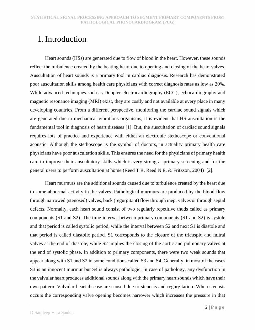
1. Introduction
Heart sounds (HSs) are generated due to flow of blood in the heart. However, these sounds
reflect the turbulence created by the beating heart due to opening and closing of the heart valves.
Auscultation of heart sounds is a primary tool in cardiac diagnosis. Research has demonstrated
poor auscultation skills among health care physicians with correct diagnosis rates as low as 20%.
While advanced techniques such as Doppler-electrocardiography (ECG), echocardiography and
magnetic resonance imaging (MRI) exist, they are costly and not available at every place in many
developing countries. From a different perspective, monitoring the cardiac sound signals which
are generated due to mechanical vibrations organisms, it is evident that HS auscultation is the
fundamental tool in diagnosis of heart diseases [1]. But, the auscultation of cardiac sound signals
requires lots of practice and experience with either an electronic stethoscope or conventional
acoustic. Although the stethoscope is the symbol of doctors, in actuality primary health care
physicians have poor auscultation skills. This ensures the need for the physicians of primary health
care to improve their auscultatory skills which is very strong at primary screening and for the
general users to perform auscultation at home (Reed T R, Reed N E, & Fritzson, 2004) [2].
Heart murmurs are the additional sounds caused due to turbulence created by the heart due
to some abnormal activity in the valves. Pathological murmurs are produced by the blood flow
through narrowed (stenosed) valves, back (regurgitant) flow through inept valves or through septal
defects. Normally, each heart sound consist of two regularly repetitive thuds called as primary
components (S1 and S2). The time interval between primary components (S1 and S2) is systole
and that period is called systolic period, while the interval between S2 and next S1 is diastole and
that period is called diastolic period. S1 corresponds to the closure of the tricuspid and mitral
valves at the end of diastole, while S2 implies the closing of the aortic and pulmonary valves at
the end of systolic phase. In addition to primary components, there were two weak sounds that
appear along with S1 and S2 in some conditions called S3 and S4. Generally, in most of the cases
S3 is an innocent murmur but S4 is always pathologic. In case of pathology, any dysfunction in
the valvular heart produces additional sounds along with the primary heart sounds which have their
own pattern. Valvular heart disease are caused due to stenosis and regurgitation. When stenosis
occurs the corresponding valve opening becomes narrower which increases the pressure in that
STATISTICAL SIGNAL PROCESSING APPROACH TO SEGMENT PRIMARY COMPONENTS FROM
PATHOLOGICAL PHONOCARDIOGRAM (PCG)
3 | P a g e
D Sandeep Vara Sankar
particular chamber. Due to increase in pressure the valve muscle becomes stiffer and leaflet loses
its agility resulting in reduced amount blood flow through it. In case of regurgitation, the valve
leaflet doesn’t close properly letting the blood to flow backward across the valve which increase
the burden in that valve. This kind of flow of blood is referred to as regurgitant flow. Most of the
defects have murmurs as systolic ejection murmur (e.g. Aortic stenosis), mid-systolic murmur (e.g.
pulmonic stenosis) pan systolic murmur (e.g. mitral regurgitation), mid diastolic rumbling murmur
(e.g. Mitral stenosis), early diastolic murmur (e.g. aortic regurgitation, pulmonary regurgitation).
These individual patterns of murmur distributions resemble the need for diagnostic aid in
this area, both for training and clinical use. The first step in developing such a system is the
segmentation of heart sounds into diagnostically relevant components. This is a challenging task
because of the non-stationary nature of heart sounds and variability seen between the patients (even
with in the same patient over time). So automatic analysis and diagnosis of HSs is required and
many researchers are paying much concentration on this field [2].
Many have studied the application of short time Fourier transform (STFT) as one of the
way to account for the non-stationarity of the signal. A basic understanding is provided on non-
stationary nature of the HSs using short time Fourier transform (STFT) in time-frequency plane
by Abdelghani Djebbari and Fethi Bereksi reguig [3] which gives a trade-off between time and
frequency resolution due to its fixed window length. The trade-off due to fixed window length is
eliminated by the introduction of wavelet transform. J.J Lee, S.M Lee, I.Y kim, H.K Min and S.H
Hong [4] showed the comparison between STFT and wavelet transform and stated that wavelet
transform acts as the primary step in the feature extraction of HSs.
A method for segmenting HS components based on HS envelogram is proposed by H.
Liang, S. Lukkarinen and L. Hartimo [5] provides a good idea for understanding structure and
characteristics of HS signal. Shannon energy is used as a medium for calculating the envelope of
the energy of HSs. The algorithm shows promising results but failed in the presence of high
intensity murmurs and/or noises.
Time-frequency analysis are probably the most widely used techniques for analyzing HS
signals. Segmentation of some pathological PCG signals using time-frequency approach is
proposed D. Boutana, M. Benidir, and B. Barkat [6]. In addition to segmentation the work permits
STATISTICAL SIGNAL PROCESSING APPROACH TO SEGMENT PRIMARY COMPONENTS FROM
PATHOLOGICAL PHONOCARDIOGRAM (PCG)
4 | P a g e
D Sandeep Vara Sankar
to extract useful information in terms of features for diagnosis and recognition of pathology. The
method gives promising results but restricted to abnormalities which have murmur concentration
only in systolic phase.
Time-frequency analysis does not work well in cases where primary components and
murmurs are inseparable, so a probabilistic approach using hidden Markov model (HMM) was
proposed by L. G. Gamero, and R. Watrous [7], to model systolic and diastolic durations, which
are further used to detect the presence of primary components (S1 and S2). However, the study
needs the QRS peak values of the simultaneous ECG recording as reference for detection of HSs
without which the algorithm doesn’t work.
Although a model-based approach has the ease of algorithmic implementation, it is
sensitive to modelling (such as the choice of the wavelet) and model parameter estimation (like
determining the parameters of an HMM). So a homomorphic filtering based self-organizing
probabilistic model approach was proposed by Gill D, Gavrieli N, and Intrator N [8], to identify
and detect primary HSs. Homomorphic filtering is advantageous due to its scalable smoothening
property and its ability in handling the splitting problem and serrated peaks and noisy environment.
Wavelet analysis has become one of the extensively used technique in analysing PCG
signals. Wavelets has the capability of analysing the signals, extracting features, detecting specific
events of interest, classification and component segmentation. The study of J.J Lee & co…
discussed above [4] has given clear understanding of the capability of wavelets in extracting the
features. Christer Ahlstrom et al in [9] proposed a feature based systolic murmur classification
which uses wavelet as one of the deciding factor for detecting murmur and segmenting HSs. M.A.R
Santos and M. Souza in [10] demonstrated that Daubechies (4-19), Meyer and Morlet are the
family of wavelets which are capable for analysing PCG signals. These wavelets have the property
of orthogonality which reduces the correlation problems.
1.1. Motivation Detection of cardiac disorders at the early stages makes the people to get rid of those
vicious problems and lead a peaceful life. When cost effectiveness is of concern, ECG and
Echocardiogram cannot be afforded by the common people for general check-up and the
availability of these facilities are restricted to only towns and cities. These difficulties makes the
STATISTICAL SIGNAL PROCESSING APPROACH TO SEGMENT PRIMARY COMPONENTS FROM
PATHOLOGICAL PHONOCARDIOGRAM (PCG)
5 | P a g e
D Sandeep Vara Sankar
general auscultation methodology to become easiest diagnostic technique for pathology detection.
For the last few decades, researches have been made to device techniques to segment primary
components from pathological phonocardiogram (PCG). Some techniques provides better
segmentation results but on the cost of algorithm complexity and price. These researches and the
typical behavior of heart sounds (HSs) gives motivation to work for the betterment of the available
methods and overcome its drawbacks in the novel technique.
1.2. Objective The main objective of this work is to propose a robust segmentation algorithm based on
statistical signal processing which, is capable of identifying key cardiac events, reduces the
computational complexity and has ability to address almost all cardiac disorders. The study uses
statistical parameters in segmenting two primary components (S1 and S2) from others. The
presented segmentation technique uses a proposed splitting algorithm to separate the PCG signal
into series of cardiac cycles and also to identify the boundaries of the primary components.
1.3. Thesis Outline The thesis is organized into 5 chapters. The first chapter speaks about, the introduction of
heart sounds and some of the discussions made by the researchers in their studies. The second
chapter gives a brief idea of the anatomy of heart sounds and their corresponding murmurs. The
third chapter briefs the background of the basic concepts used in this study. The fourth chapter
describes the proposed techniques and their corresponding results and finally discussion on the
entire study is made in the fifth chapter followed by conclusion to the complete work and
references.
STATISTICAL SIGNAL PROCESSING APPROACH TO SEGMENT PRIMARY COMPONENTS FROM
PATHOLOGICAL PHONOCARDIOGRAM (PCG)
6 | P a g e
D Sandeep Vara Sankar
CHAPTER 2
Cardiac anatomy
STATISTICAL SIGNAL PROCESSING APPROACH TO SEGMENT PRIMARY COMPONENTS FROM
PATHOLOGICAL PHONOCARDIOGRAM (PCG)
7 | P a g e
D Sandeep Vara Sankar
2. Cardiac Anatomy
2.1. Physiology of heart
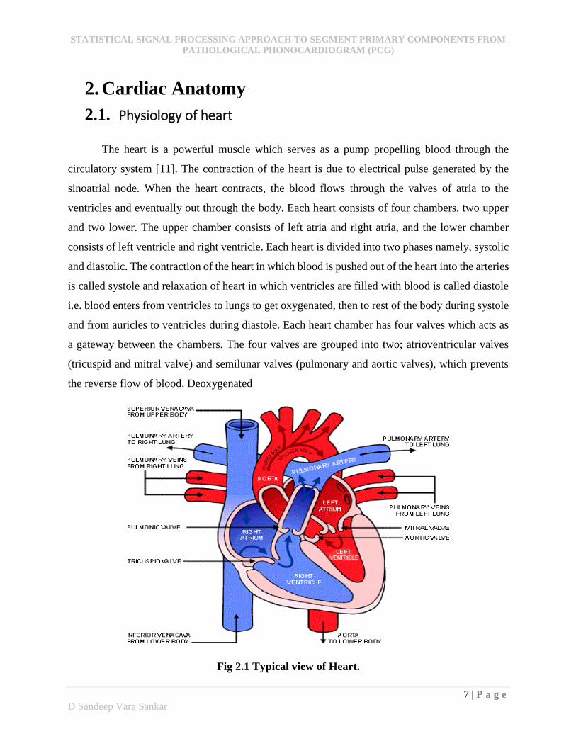
The heart is a powerful muscle which serves as a pump propelling blood through the
circulatory system [11]. The contraction of the heart is due to electrical pulse generated by the
sinoatrial node. When the heart contracts, the blood flows through the valves of atria to the
ventricles and eventually out through the body. Each heart consists of four chambers, two upper
and two lower. The upper chamber consists of left atria and right atria, and the lower chamber
consists of left ventricle and right ventricle. Each heart is divided into two phases namely, systolic
and diastolic. The contraction of the heart in which blood is pushed out of the heart into the arteries
is called systole and relaxation of heart in which ventricles are filled with blood is called diastole
i.e. blood enters from ventricles to lungs to get oxygenated, then to rest of the body during systole
and from auricles to ventricles during diastole. Each heart chamber has four valves which acts as
a gateway between the chambers. The four valves are grouped into two; atrioventricular valves
(tricuspid and mitral valve) and semilunar valves (pulmonary and aortic valves), which prevents
the reverse flow of blood. Deoxygenated
Fig 2.1 Typical view of Heart.
STATISTICAL SIGNAL PROCESSING APPROACH TO SEGMENT PRIMARY COMPONENTS FROM
PATHOLOGICAL PHONOCARDIOGRAM (PCG)
8 | P a g e
D Sandeep Vara Sankar
blood from the body parts enters the right atrium and is passed to the right ventricle through the
tricuspid valve and is ejected into the lungs through pulmonic valve for oxygenation. The
Oxygenated blood from the lungs enters the left atrium is passed to left ventricle through mitral
valve and is then ejected into the aorta.
2.2. Heart Sounds
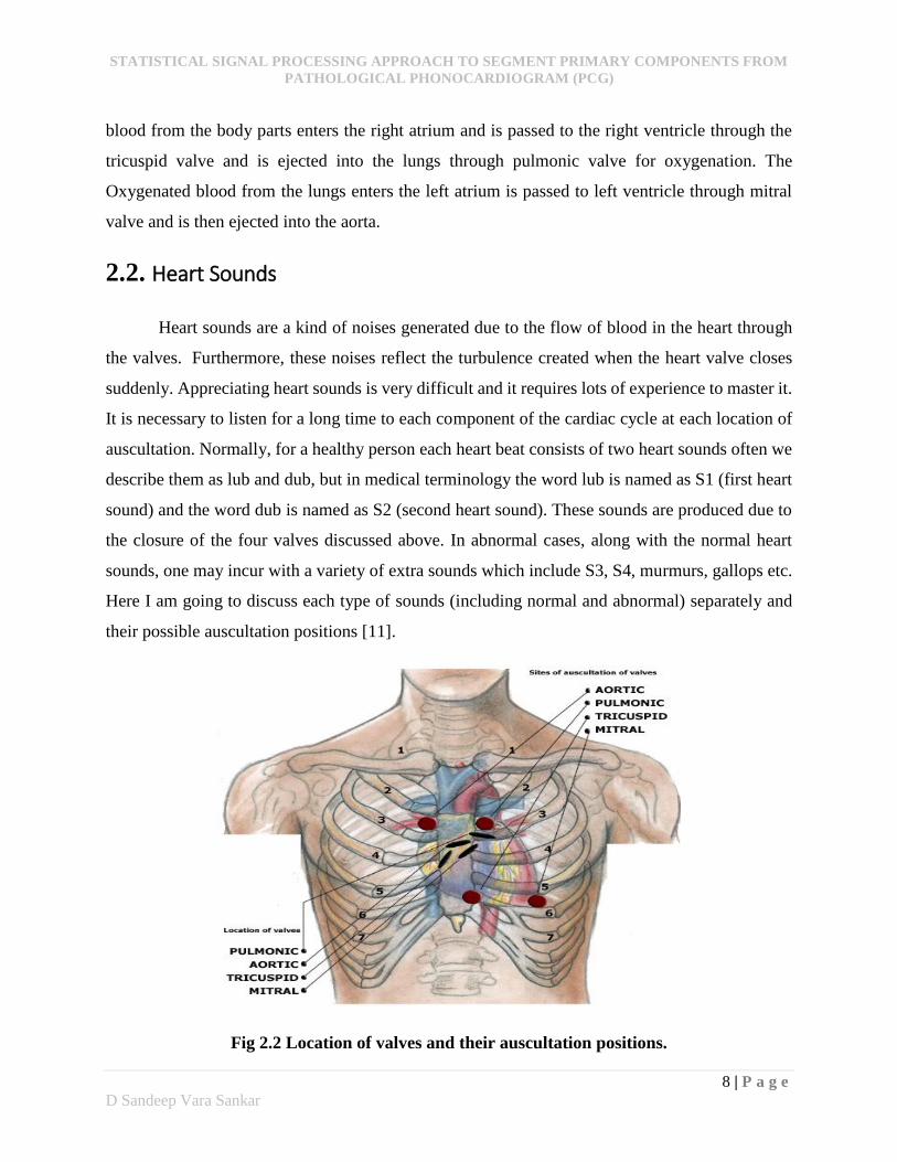
Heart sounds are a kind of noises generated due to the flow of blood in the heart through
the valves. Furthermore, these noises reflect the turbulence created when the heart valve closes
suddenly. Appreciating heart sounds is very difficult and it requires lots of experience to master it.
It is necessary to listen for a long time to each component of the cardiac cycle at each location of
auscultation. Normally, for a healthy person each heart beat consists of two heart sounds often we
describe them as lub and dub, but in medical terminology the word lub is named as S1 (first heart
sound) and the word dub is named as S2 (second heart sound). These sounds are produced due to
the closure of the four valves discussed above. In abnormal cases, along with the normal heart
sounds, one may incur with a variety of extra sounds which include S3, S4, murmurs, gallops etc.
Here I am going to discuss each type of sounds (including normal and abnormal) separately and
their possible auscultation positions [11].
Fig 2.2 Location of valves and their auscultation positions.
STATISTICAL SIGNAL PROCESSING APPROACH TO SEGMENT PRIMARY COMPONENTS FROM
PATHOLOGICAL PHONOCARDIOGRAM (PCG)
9 | P a g e
D Sandeep Vara Sankar
2.3. Primary Heart Sounds
2.3.1. First heart sound (S1):
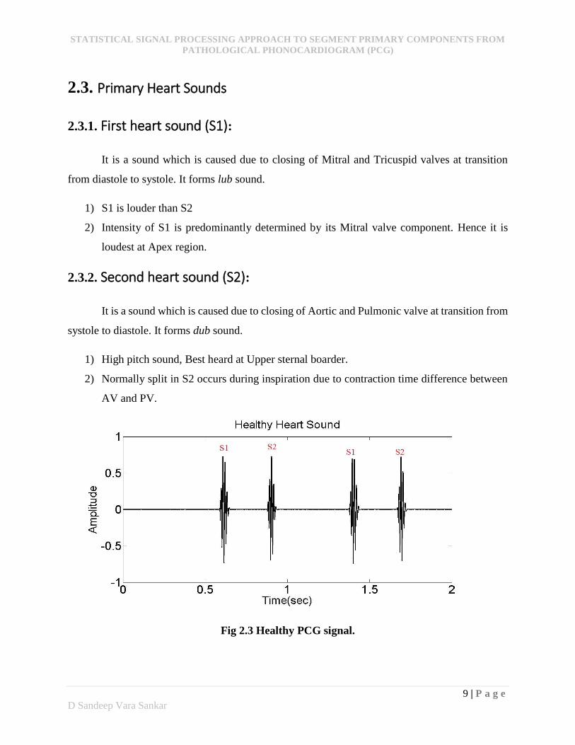
It is a sound which is caused due to closing of Mitral and Tricuspid valves at transition
from diastole to systole. It forms lub sound.
1) S1 is louder than S2
2) Intensity of S1 is predominantly determined by its Mitral valve component. Hence it is
loudest at Apex region.
2.3.2. Second heart sound (S2):
It is a sound which is caused due to closing of Aortic and Pulmonic valve at transition from
systole to diastole. It forms dub sound.
1) High pitch sound, Best heard at Upper sternal boarder.
2) Normally split in S2 occurs during inspiration due to contraction time difference between
AV and PV.
Fig 2.3 Healthy PCG signal.
STATISTICAL SIGNAL PROCESSING APPROACH TO SEGMENT PRIMARY COMPONENTS FROM
PATHOLOGICAL PHONOCARDIOGRAM (PCG)
10 | P a g e
D Sandeep Vara Sankar
2.4. Extra Heart Sounds
2.4.1. Third heart sound (S3):
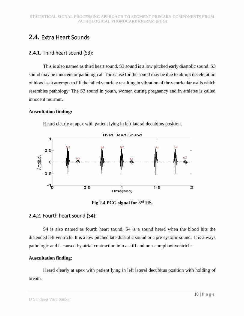
This is also named as third heart sound. S3 sound is a low pitched early diastolic sound. S3
sound may be innocent or pathological. The cause for the sound may be due to abrupt deceleration
of blood as it attempts to fill the failed ventricle resulting in vibration of the ventricular walls which
resembles pathology. The S3 sound in youth, women during pregnancy and in athletes is called
innocent murmur.
Auscultation finding:
Heard clearly at apex with patient lying in left lateral decubitus position.
Fig 2.4 PCG signal for 3rd HS.
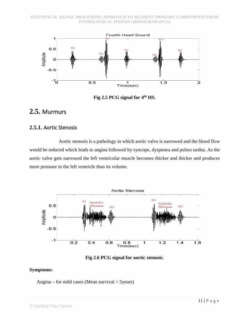
2.4.2. Fourth heart sound (S4):
S4 is also named as fourth heart sound. S4 is a sound heard when the blood hits the
distended left ventricle. It is a low pitched late diastolic sound or a pre-systolic sound. It is always
pathologic and is caused by atrial contraction into a stiff and non-compliant ventricle.
Auscultation finding:
Heard clearly at apex with patient lying in left lateral decubitus position with holding of
breath.
STATISTICAL SIGNAL PROCESSING APPROACH TO SEGMENT PRIMARY COMPONENTS FROM
PATHOLOGICAL PHONOCARDIOGRAM (PCG)
11 | P a g e
D Sandeep Vara Sankar
Fig 2.5 PCG signal for 4th HS.
2.5. Murmurs
2.5.1. Aortic Stenosis
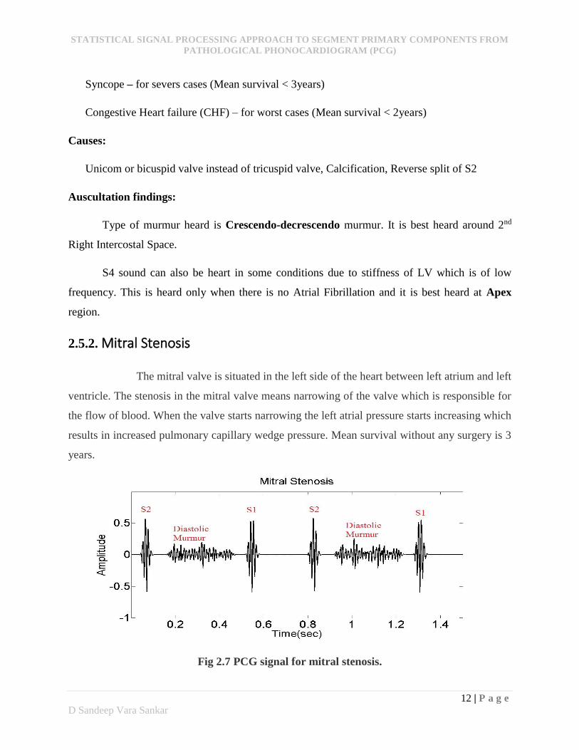
Aortic stenosis is a pathology in which aortic valve is narrowed and the blood flow
would be reduced which leads to angina followed by syncope, dyspnoea and pulses tardus. As the
aortic valve gets narrowed the left ventricular muscle becomes thicker and thicker and produces
more pressure in the left ventricle than its volume.
Fig 2.6 PCG signal for aortic stenosis.
Symptoms:
Angina – for mild cases (Mean survival < 5years)
STATISTICAL SIGNAL PROCESSING APPROACH TO SEGMENT PRIMARY COMPONENTS FROM
PATHOLOGICAL PHONOCARDIOGRAM (PCG)
12 | P a g e
D Sandeep Vara Sankar
Syncope – for severs cases (Mean survival < 3years)
Congestive Heart failure (CHF) – for worst cases (Mean survival < 2years)
Causes:
Unicom or bicuspid valve instead of tricuspid valve, Calcification, Reverse split of S2
Auscultation findings:
Type of murmur heard is Crescendo-decrescendo murmur. It is best heard around 2nd
Right Intercostal Space.
S4 sound can also be heart in some conditions due to stiffness of LV which is of low
frequency. This is heard only when there is no Atrial Fibrillation and it is best heard at Apex
region.
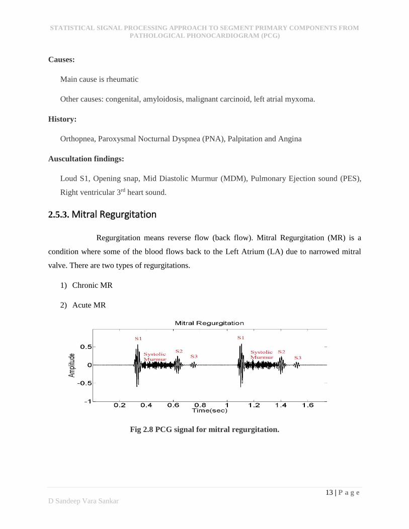
2.5.2. Mitral Stenosis
The mitral valve is situated in the left side of the heart between left atrium and left
ventricle. The stenosis in the mitral valve means narrowing of the valve which is responsible for
the flow of blood. When the valve starts narrowing the left atrial pressure starts increasing which
results in increased pulmonary capillary wedge pressure. Mean survival without any surgery is 3
years.
Fig 2.7 PCG signal for mitral stenosis.
STATISTICAL SIGNAL PROCESSING APPROACH TO SEGMENT PRIMARY COMPONENTS FROM
PATHOLOGICAL PHONOCARDIOGRAM (PCG)
13 | P a g e
D Sandeep Vara Sankar
Causes:
Main cause is rheumatic
Other causes: congenital, amyloidosis, malignant carcinoid, left atrial myxoma.
History:
Orthopnea, Paroxysmal Nocturnal Dyspnea (PNA), Palpitation and Angina
Auscultation findings:
Loud S1, Opening snap, Mid Diastolic Murmur (MDM), Pulmonary Ejection sound (PES),
Right ventricular 3rd heart sound.
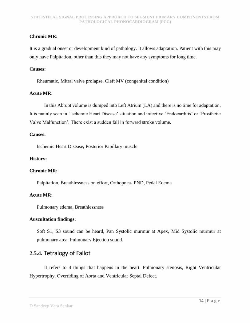
2.5.3. Mitral Regurgitation
Regurgitation means reverse flow (back flow). Mitral Regurgitation (MR) is a
condition where some of the blood flows back to the Left Atrium (LA) due to narrowed mitral
valve. There are two types of regurgitations.
1) Chronic MR
2) Acute MR
Fig 2.8 PCG signal for mitral regurgitation.
STATISTICAL SIGNAL PROCESSING APPROACH TO SEGMENT PRIMARY COMPONENTS FROM
PATHOLOGICAL PHONOCARDIOGRAM (PCG)
14 | P a g e
D Sandeep Vara Sankar
Chronic MR:
It is a gradual onset or development kind of pathology. It allows adaptation. Patient with this may
only have Palpitation, other than this they may not have any symptoms for long time.
Causes:
Rheumatic, Mitral valve prolapse, Cleft MV (congenital condition)
Acute MR:
In this Abrupt volume is dumped into Left Atrium (LA) and there is no time for adaptation.
It is mainly seen in ‘Ischemic Heart Disease’ situation and infective ‘Endocarditis’ or ‘Prosthetic
Valve Malfunction’. There exist a sudden fall in forward stroke volume.
Causes:
Ischemic Heart Disease, Posterior Papillary muscle
History:
Chronic MR:
Palpitation, Breathlessness on effort, Orthopnea- PND, Pedal Edema
Acute MR:
Pulmonary edema, Breathlessness
Auscultation findings:
Soft S1, S3 sound can be heard, Pan Systolic murmur at Apex, Mid Systolic murmur at
pulmonary area, Pulmonary Ejection sound.
2.5.4. Tetralogy of Fallot
It refers to 4 things that happens in the heart. Pulmonary stenosis, Right Ventricular
Hypertrophy, Overriding of Aorta and Ventricular Septal Defect.
STATISTICAL SIGNAL PROCESSING APPROACH TO SEGMENT PRIMARY COMPONENTS FROM
PATHOLOGICAL PHONOCARDIOGRAM (PCG)
15 | P a g e
D Sandeep Vara Sankar
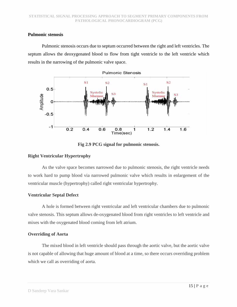
Pulmonic stenosis
Pulmonic stenosis occurs due to septum occurred between the right and left ventricles. The
septum allows the deoxygenated blood to flow from tight ventricle to the left ventricle which
results in the narrowing of the pulmonic valve space.
Fig 2.9 PCG signal for pulmonic stenosis.
Right Ventricular Hypertrophy
As the valve space becomes narrowed due to pulmonic stenosis, the right ventricle needs
to work hard to pump blood via narrowed pulmonic valve which results in enlargement of the
ventricular muscle (hypertrophy) called right ventricular hypertrophy.
Ventricular Septal Defect
A hole is formed between right ventricular and left ventricular chambers due to pulmonic
valve stenosis. This septum allows de-oxygenated blood from right ventricles to left ventricle and
mixes with the oxygenated blood coming from left atrium.
Overriding of Aorta
The mixed blood in left ventricle should pass through the aortic valve, but the aortic valve
is not capable of allowing that huge amount of blood at a time, so there occurs overriding problem
which we call as overriding of aorta.
STATISTICAL SIGNAL PROCESSING APPROACH TO SEGMENT PRIMARY COMPONENTS FROM
PATHOLOGICAL PHONOCARDIOGRAM (PCG)
16 | P a g e
D Sandeep Vara Sankar
Causes and symptoms:
Only possible cause is congenital and the symptoms may be dyspnea, poor weight gain,
cyanotic and tet spells.
Auscultation finding:
Pulmonic stenosis can be known physically by examining for the harsh systolic ejection
murmur at the ‘upper left sternal boarder’.
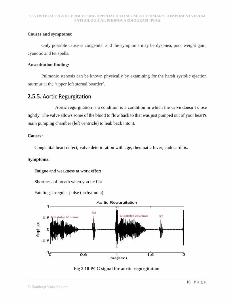
2.5.5. Aortic Regurgitation
Aortic regurgitation is a condition is a condition in which the valve doesn’t close
tightly. The valve allows some of the blood to flow back to that was just pumped out of your heart's
main pumping chamber (left ventricle) to leak back into it.
Causes:
Congenital heart defect, valve deterioration with age, rheumatic fever, endocarditis.
Symptoms:
Fatigue and weakness at work effort
Shortness of breath when you lie flat.
Fainting, Irregular pulse (arrhythmia).
Fig 2.10 PCG signal for aortic regurgitation.
STATISTICAL SIGNAL PROCESSING APPROACH TO SEGMENT PRIMARY COMPONENTS FROM
PATHOLOGICAL PHONOCARDIOGRAM (PCG)
17 | P a g e
D Sandeep Vara Sankar
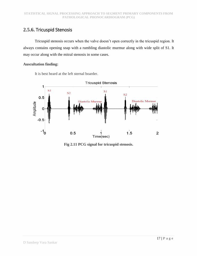
2.5.6. Tricuspid Stenosis
Tricuspid stenosis occurs when the valve doesn’t open correctly in the tricuspid region. It
always contains opening snap with a rumbling diastolic murmur along with wide split of S1. It
may occur along with the mitral stenosis in some cases.
Auscultation finding:
It is best heard at the left sternal boarder.
Fig 2.11 PCG signal for tricuspid stenosis.

STATISTICAL SIGNAL PROCESSING APPROACH TO SEGMENT PRIMARY COMPONENTS FROM
PATHOLOGICAL PHONOCARDIOGRAM (PCG)
18 | P a g e
D Sandeep Vara Sankar
CHAPTER 3
Theoretical background
STATISTICAL SIGNAL PROCESSING APPROACH TO SEGMENT PRIMARY COMPONENTS FROM
PATHOLOGICAL PHONOCARDIOGRAM (PCG)
19 | P a g e
D Sandeep Vara Sankar
3. Basic Theory
Signals carries prodigious amounts of data in which finding an important information is
more difficult. It is necessary to process the signal in such a way that only few coefficients reveals
the necessary information. These requirements opened the door for the discovery of huge jungle
of new transforms. In this work, to segment heart sound signals an extension of methods based on
time-frequency will be investigated. In order to have a successful segmentation algorithm, it is
necessary to understand the basic concepts behind the techniques. This chapter constitutes
fundamentals of Fourier theory followed by time-frequency analysis like windowed Fourier
transform and Wavelet analysis. Finally it will be concluded with principal component analysis
concept.
3.1. Fourier Analysis
Fourier analysis is useful everywhere in the fields of mathematics and physics because the
time invariant convolution operators are diagonalized [12]. It is a method which defines the
periodic waveforms by the sum of trigonometric functions. The representation of the Fourier
analysis for any energy function x(t) which is of finite duration is given by sum of sinusoids ejωt
as:
dextx tj)(2
1)( (3.1)
The amplitude x(ω) for each of the sinusoidal wave function ejωt is equal to correlation of ejωt with
x, which can also be called as Fourier transform:
dtetxx tj )()( (3.2)
The more regular the function x(t), the faster will be the decay of the amplitude )(x , when the
frequency of ω is increased.
STATISTICAL SIGNAL PROCESSING APPROACH TO SEGMENT PRIMARY COMPONENTS FROM
PATHOLOGICAL PHONOCARDIOGRAM (PCG)
20 | P a g e
D Sandeep Vara Sankar
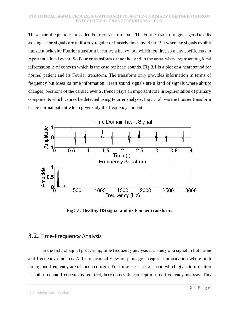
These pair of equations are called Fourier transform pair. The Fourier transform gives good results
as long as the signals are uniformly regular or linearly time-invariant. But when the signals exhibit
transient behavior Fourier transform becomes a heavy tool which requires so many coefficients to
represent a local event. So Fourier transform cannot be used in the areas where representing local
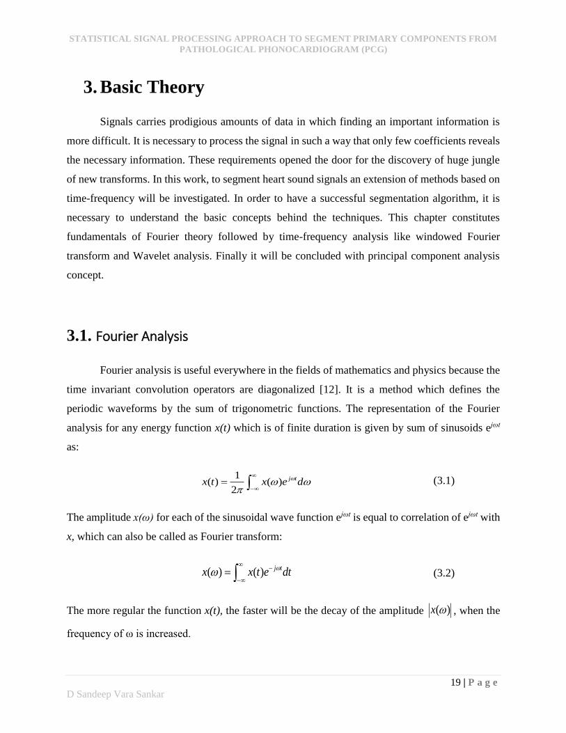
information is of concern which is the case for heart sounds. Fig 3.1 is a plot of a heart sound for
normal patient and its Fourier transform. The transform only provides information in terms of
frequency but loses its time information. Heart sound signals are a kind of signals where abrupt
changes, positions of the cardiac events, trends plays an important role in segmentation of primary
components which cannot be detected using Fourier analysis. Fig 3.1 shows the Fourier transform
of the normal patient which gives only the frequency content.
Fig 3.1. Healthy HS signal and its Fourier transform.
3.2. Time-Frequency Analysis
In the field of signal processing, time frequency analysis is a study of a signal in both time
and frequency domains. A 1-dimensional view may not give required information where both
timing and frequency are of much concern. For those cases a transform which gives information
in both time and frequency is required, here comes the concept of time frequency analysis. This
STATISTICAL SIGNAL PROCESSING APPROACH TO SEGMENT PRIMARY COMPONENTS FROM
PATHOLOGICAL PHONOCARDIOGRAM (PCG)
21 | P a g e
D Sandeep Vara Sankar
analysis uses time localization technique which plays an important role in speech and sound
processing. The main issue is to understand how to adapt time-frequency component to signal
processing. In this context Heisenberg uncertainty principle plays a promising role.
3.2.1. Short time/term Fourier transform
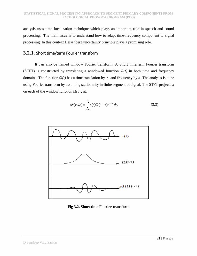
It can also be named window Fourier transform. A Short time/term Fourier transform
(STFT) is constructed by translating a windowed function Ω(t) in both time and frequency
domains. The function Ω(t) has a time translation by and frequency by α. The analysis is done
using Fourier transform by assuming stationarity in finite segment of signal. The STFT projects x
on each of the window function Ω( , α):
.)()(),( dtettxsx ti (3.3)
Fig 3.2. Short time Fourier transform
STATISTICAL SIGNAL PROCESSING APPROACH TO SEGMENT PRIMARY COMPONENTS FROM
PATHOLOGICAL PHONOCARDIOGRAM (PCG)
22 | P a g e
D Sandeep Vara Sankar
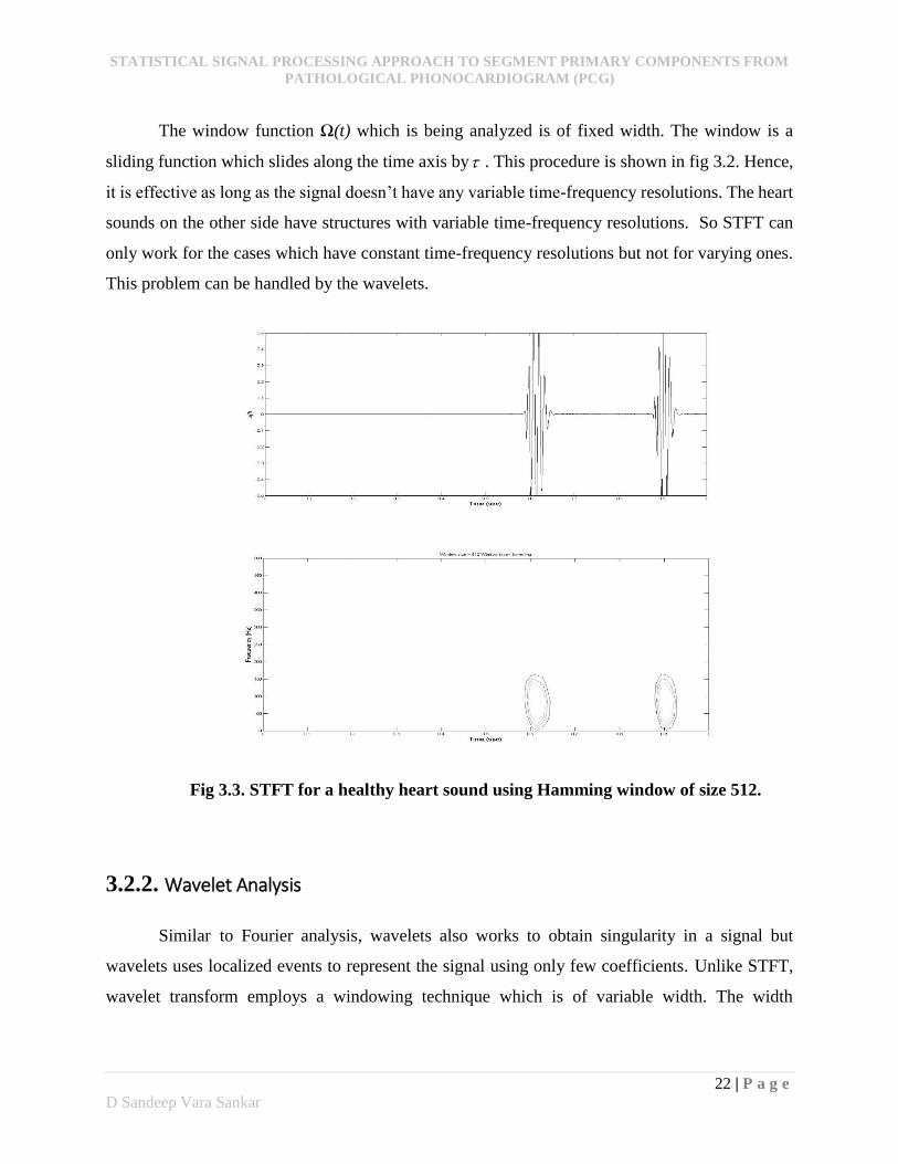
The window function Ω(t) which is being analyzed is of fixed width. The window is a
sliding function which slides along the time axis by . This procedure is shown in fig 3.2. Hence,
it is effective as long as the signal doesn’t have any variable time-frequency resolutions. The heart
sounds on the other side have structures with variable time-frequency resolutions. So STFT can
only work for the cases which have constant time-frequency resolutions but not for varying ones.
This problem can be handled by the wavelets.
Fig 3.3. STFT for a healthy heart sound using Hamming window of size 512.
3.2.2. Wavelet Analysis
Similar to Fourier analysis, wavelets also works to obtain singularity in a signal but
wavelets uses localized events to represent the signal using only few coefficients. Unlike STFT,
wavelet transform employs a windowing technique which is of variable width. The width
STATISTICAL SIGNAL PROCESSING APPROACH TO SEGMENT PRIMARY COMPONENTS FROM
PATHOLOGICAL PHONOCARDIOGRAM (PCG)
23 | P a g e
D Sandeep Vara Sankar
variability allows wavelets to work for different frequency resolutions. Based on the applications,
wavelets are divided into two types: continuous and discrete wavelet transform.
3.2.2.1. Continuous wavelet transform
Wavelets offer the best representation for non-stationary signals. Here, large amplitude
wavelet coefficients can detect and measure short high frequency variations because of the narrow
time localization at high frequencies, so it gives better time resolution and at low frequencies their
time resolution is lower, but they have a better frequency resolution. This modification of time and
frequency resolution is adapted to represent sounds with sharp attacks or signals that have much
frequency variations. These sharp attacks and high frequency variations are the exact
characteristics of a HS signal.
A wavelet is constructed from a mother wavelet of zero average which is dilated with a
scale parameter s, and translated by :
,0)( dtt (3.4)
The continuous wavelet transform of x at any scale s and position is the projection of x on the
corresponding wavelet coefficient:
.)(1
)(),( * dts
t
stxswx
(3.5)
It represents one-dimensional signals by highly redundant time-scale coefficients in ( , s) [12].
The scale parameter, s, is inversely proportional to the frequency which is defined as
1s (3.6)
Where ω is the frequency in Hertz.
From the wavelet perspective, scaling factor either dilates or compress the wavelet. Larger scales
dilate the wavelet which highlights the slow variation activities in the signal and small scales
compress the wavelet which retrieves the transient behaviour of the signal. The continuous wavelet
STATISTICAL SIGNAL PROCESSING APPROACH TO SEGMENT PRIMARY COMPONENTS FROM
PATHOLOGICAL PHONOCARDIOGRAM (PCG)
24 | P a g e
D Sandeep Vara Sankar
transform measures the similarity between the signal and the wavelet by continuously translating
and scaling the mother wavelet. So to represent the coefficients an infinite number of wavelets are
required which increases the redundancy and hence is impractical. It also uses 2-Dimension for
dealing with 1-Dimension entity which gives extreme redundant information. Discrete wavelet
transform works for those cases where redundancy is of much concern.
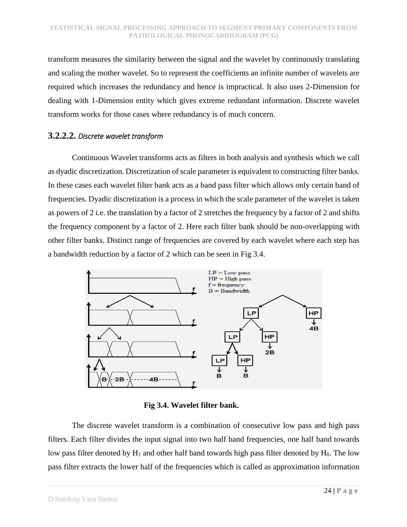
3.2.2.2. Discrete wavelet transform
Continuous Wavelet transforms acts as filters in both analysis and synthesis which we call
as dyadic discretization. Discretization of scale parameter is equivalent to constructing filter banks.
In these cases each wavelet filter bank acts as a band pass filter which allows only certain band of
frequencies. Dyadic discretization is a process in which the scale parameter of the wavelet is taken
as powers of 2 i.e. the translation by a factor of 2 stretches the frequency by a factor of 2 and shifts
the frequency component by a factor of 2. Here each filter bank should be non-overlapping with
other filter banks. Distinct range of frequencies are covered by each wavelet where each step has
a bandwidth reduction by a factor of 2 which can be seen in Fig 3.4.
Fig 3.4. Wavelet filter bank.
The discrete wavelet transform is a combination of consecutive low pass and high pass
filters. Each filter divides the input signal into two half band frequencies, one half band towards
low pass filter denoted by H1 and other half band towards high pass filter denoted by H0. The low
pass filter extracts the lower half of the frequencies which is called as approximation information
STATISTICAL SIGNAL PROCESSING APPROACH TO SEGMENT PRIMARY COMPONENTS FROM
PATHOLOGICAL PHONOCARDIOGRAM (PCG)
25 | P a g e
D Sandeep Vara Sankar
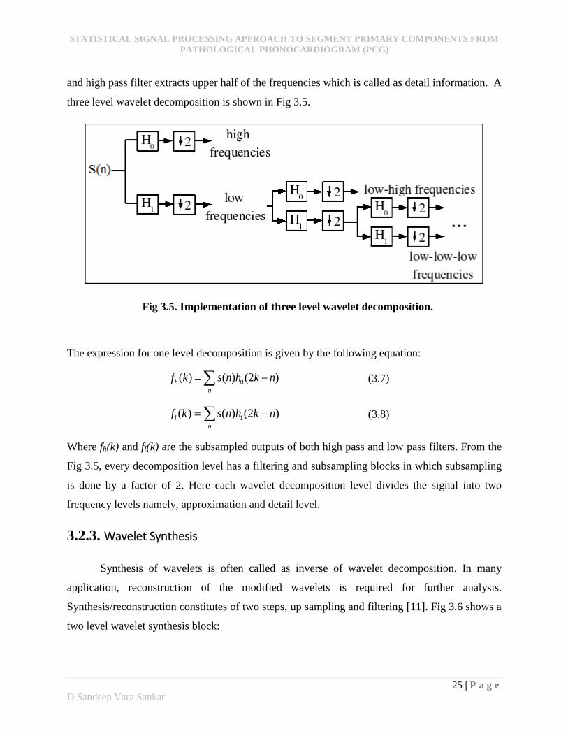
and high pass filter extracts upper half of the frequencies which is called as detail information. A
three level wavelet decomposition is shown in Fig 3.5.
Fig 3.5. Implementation of three level wavelet decomposition.
The expression for one level decomposition is given by the following equation:
n
h nkhnskf )2()()( 0 (3.7)
n
l nkhnskf )2()()( 1 (3.8)
Where fh(k) and fl(k) are the subsampled outputs of both high pass and low pass filters. From the
Fig 3.5, every decomposition level has a filtering and subsampling blocks in which subsampling
is done by a factor of 2. Here each wavelet decomposition level divides the signal into two
frequency levels namely, approximation and detail level.
3.2.3. Wavelet Synthesis
Synthesis of wavelets is often called as inverse of wavelet decomposition. In many
application, reconstruction of the modified wavelets is required for further analysis.
Synthesis/reconstruction constitutes of two steps, up sampling and filtering [11]. Fig 3.6 shows a
two level wavelet synthesis block:
STATISTICAL SIGNAL PROCESSING APPROACH TO SEGMENT PRIMARY COMPONENTS FROM
PATHOLOGICAL PHONOCARDIOGRAM (PCG)
26 | P a g e
D Sandeep Vara Sankar
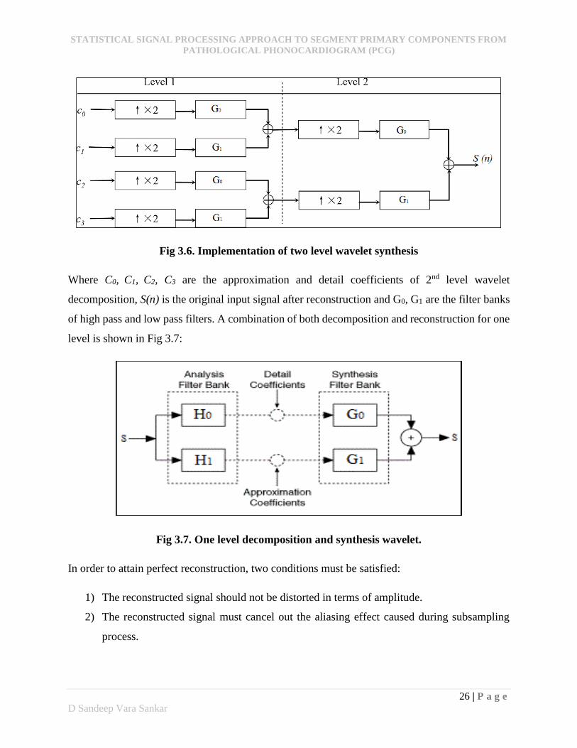
Fig 3.6. Implementation of two level wavelet synthesis
Where C0, C1, C2, C3 are the approximation and detail coefficients of 2nd level wavelet
decomposition, S(n) is the original input signal after reconstruction and G0, G1 are the filter banks
of high pass and low pass filters. A combination of both decomposition and reconstruction for one
level is shown in Fig 3.7:
Fig 3.7. One level decomposition and synthesis wavelet.
In order to attain perfect reconstruction, two conditions must be satisfied:
1) The reconstructed signal should not be distorted in terms of amplitude.
2) The reconstructed signal must cancel out the aliasing effect caused during subsampling
process.
STATISTICAL SIGNAL PROCESSING APPROACH TO SEGMENT PRIMARY COMPONENTS FROM
PATHOLOGICAL PHONOCARDIOGRAM (PCG)
27 | P a g e
D Sandeep Vara Sankar
3.3. Principal component analysis (PCA)
PCA is a method for analysing data sets of high dimension, revealing patterns and
highlights the similarities and differences. Although PCA can be used for various types of analysis,
here the emphasis will be on data reduction and feature extraction. In data sets with many variable
it is obvious that more than one variable is measuring the same driving force. PCA generates a
new set of variables called principal components(PC). Each PC is a linear combination of the
original variables. The purpose of using this is to remove redundancy of information and replace
a group of variables which measures the same information with a single new variable called
principal component (PC). The calculation of the principle component is essentially equivalent to
performing the singular value decomposition (SVD) on a data set, X which is described in [13,14].
The process is outlined below as:
1) Calculate the covariance matrix for the data set X. The matrix is of size m × n where rows
denote the variable and columns denotes the observation for each variable.
2) Calculate the eigenvalues, λi (i=1,2,…,m) of the covariance matrix.
3) Now calculate the eigenvectors of the corresponding eigenvalues, Ui (i=1,2,…,m).
4) Order the eigenvalues in descending order and align the corresponding eigenvectors based
on the eigenvalues. These forms principal components and principle eigenvalues.
A=diag(σ1, σ2, σ3,-----, σm) where σ1 >σ2 > σ3 >-----, >σm
PC=[PC1 PC2 ------------ PCm]. (3.9)
Where PCi is the Eigen vector that corresponds to Eigen value σi.
Properties of PCA
There are infinite number of ways to determine an orthogonal basis for a particular set of
data. Based on its properties PCA provides great benefits in feature extraction problems.
STATISTICAL SIGNAL PROCESSING APPROACH TO SEGMENT PRIMARY COMPONENTS FROM
PATHOLOGICAL PHONOCARDIOGRAM (PCG)
28 | P a g e
D Sandeep Vara Sankar
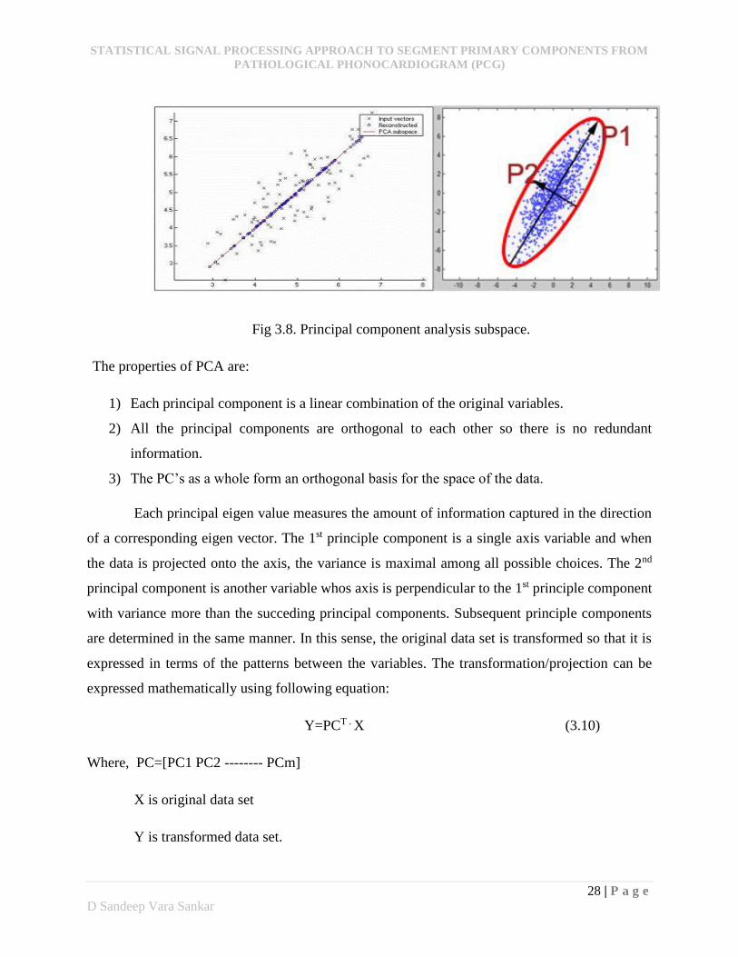
Fig 3.8. Principal component analysis subspace.
The properties of PCA are:
1) Each principal component is a linear combination of the original variables.
2) All the principal components are orthogonal to each other so there is no redundant
information.
3) The PC’s as a whole form an orthogonal basis for the space of the data.
Each principal eigen value measures the amount of information captured in the direction
of a corresponding eigen vector. The 1st principle component is a single axis variable and when
the data is projected onto the axis, the variance is maximal among all possible choices. The 2nd
principal component is another variable whos axis is perpendicular to the 1st principle component
with variance more than the succeding principal components. Subsequent principle components
are determined in the same manner. In this sense, the original data set is transformed so that it is
expressed in terms of the patterns between the variables. The transformation/projection can be
expressed mathematically using following equation:
Y=PCT . X (3.10)
Where, PC=[PC1 PC2 -------- PCm]
X is original data set
Y is transformed data set.
STATISTICAL SIGNAL PROCESSING APPROACH TO SEGMENT PRIMARY COMPONENTS FROM
PATHOLOGICAL PHONOCARDIOGRAM (PCG)
29 | P a g e
D Sandeep Vara Sankar
CHAPTER 4
Segmentation
Algorithm
STATISTICAL SIGNAL PROCESSING APPROACH TO SEGMENT PRIMARY COMPONENTS FROM
PATHOLOGICAL PHONOCARDIOGRAM (PCG)
30 | P a g e
D Sandeep Vara Sankar
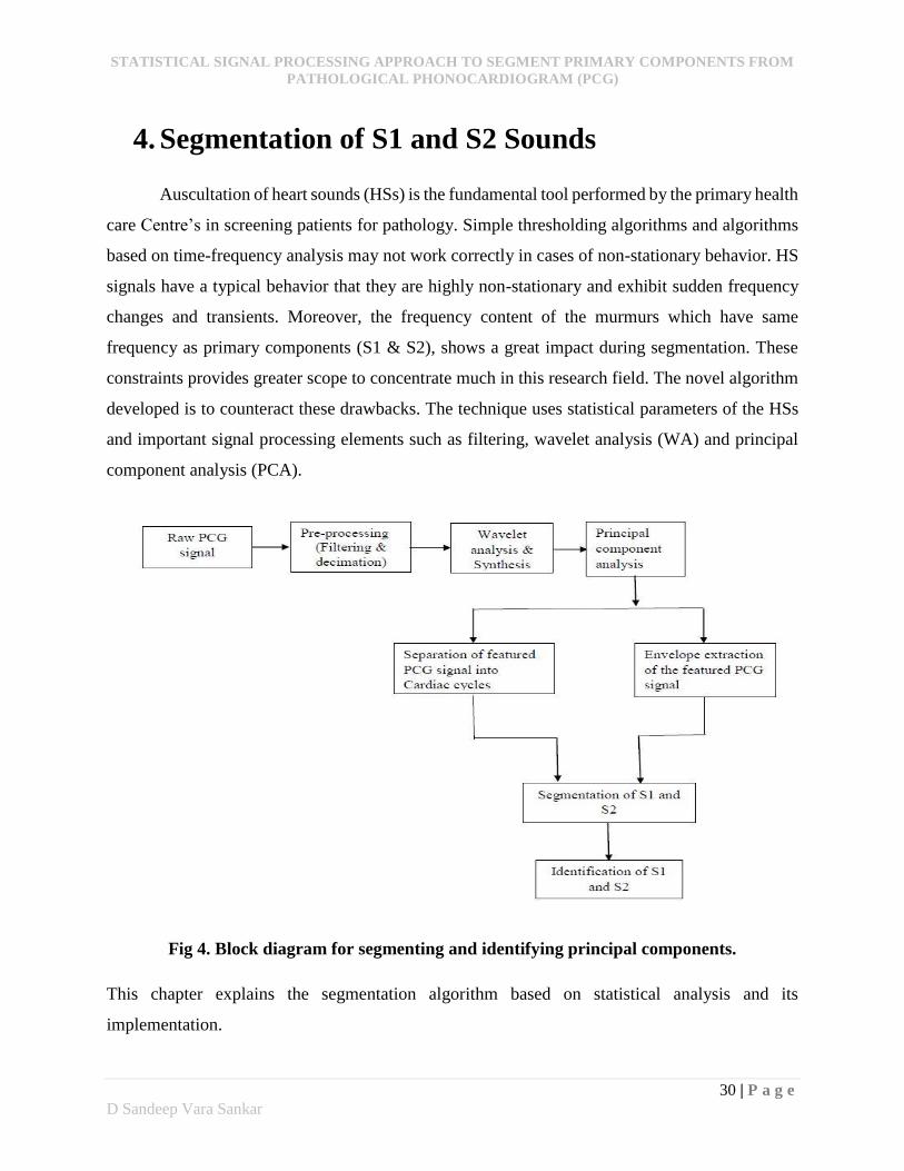
4. Segmentation of S1 and S2 Sounds
Auscultation of heart sounds (HSs) is the fundamental tool performed by the primary health
care Centre’s in screening patients for pathology. Simple thresholding algorithms and algorithms
based on time-frequency analysis may not work correctly in cases of non-stationary behavior. HS
signals have a typical behavior that they are highly non-stationary and exhibit sudden frequency
changes and transients. Moreover, the frequency content of the murmurs which have same
frequency as primary components (S1 & S2), shows a great impact during segmentation. These
constraints provides greater scope to concentrate much in this research field. The novel algorithm
developed is to counteract these drawbacks. The technique uses statistical parameters of the HSs
and important signal processing elements such as filtering, wavelet analysis (WA) and principal
component analysis (PCA).
Fig 4. Block diagram for segmenting and identifying principal components.
This chapter explains the segmentation algorithm based on statistical analysis and its
implementation.
STATISTICAL SIGNAL PROCESSING APPROACH TO SEGMENT PRIMARY COMPONENTS FROM
PATHOLOGICAL PHONOCARDIOGRAM (PCG)
31 | P a g e
D Sandeep Vara Sankar
4.1. Pre-processing
Filtering
During acquisition process there is a possibility of noise interference. The noises may be
due to respiratory tracts in the human being, environmental disturbances or even the measuring
equipment itself. The frequency content of these noises would be less than 40Hz. It is necessary
to remove these noises at initial state for better boundary detection of S1 and S2. So a filtering
technique is performed to remove those low frequency noises using high pass filter (HPF) with a
cut-off frequency of 40Hz.
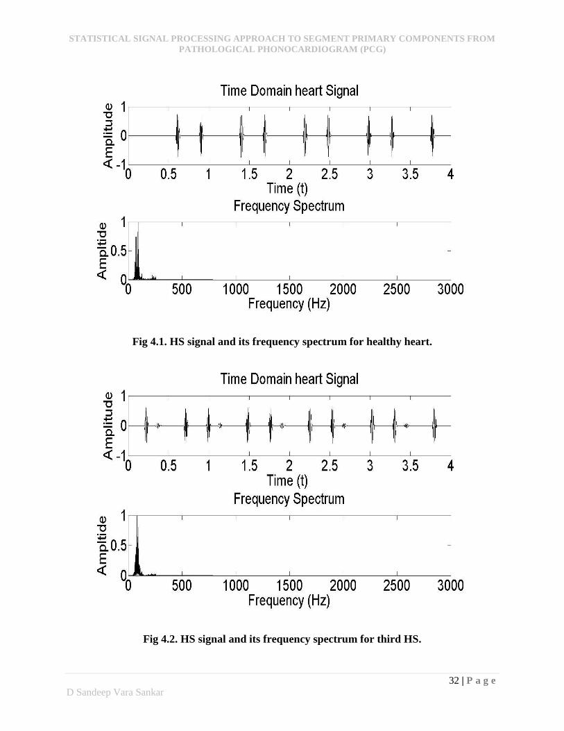
Decimation
The phonocardiographic signal records used in this study are sampled at a sampling
frequency of 22050Hz. Researchers have demonstrated that most of the primary components have
a frequency ranges from 40 to 200Hz which are due to the turbulence of blood flow in the heart
valves and the murmurs due to valvular dysfunctions have a frequency range up to 600Hz. This
shows that it is unnecessary to consider the frequencies which are not of clinical significance for
analysis and diagnosis purposes. The justification of the studies are shown in below figures (4.1 –
4.9). In each figure the PCG signal is shown on the top followed by its frequency spectrum. So
further processing is required for the HS signal so that the frequencies of interest are only present
for further processing. This is done by decimation. Decimation employs an eighth-order low pass
Chebyshev type I filter with cut-off frequency of 0.8*(Fs/2)/r, where Fs is the sampling frequency
and r is the decimation factor. Here ‘r’ is chosen to obtain sample rate of 700Hz. It filters the HS
in both forward and reverse directions to remove all phase distortions.
STATISTICAL SIGNAL PROCESSING APPROACH TO SEGMENT PRIMARY COMPONENTS FROM
PATHOLOGICAL PHONOCARDIOGRAM (PCG)
32 | P a g e
D Sandeep Vara Sankar
Fig 4.1. HS signal and its frequency spectrum for healthy heart.
Fig 4.2. HS signal and its frequency spectrum for third HS.
STATISTICAL SIGNAL PROCESSING APPROACH TO SEGMENT PRIMARY COMPONENTS FROM
PATHOLOGICAL PHONOCARDIOGRAM (PCG)
33 | P a g e
D Sandeep Vara Sankar
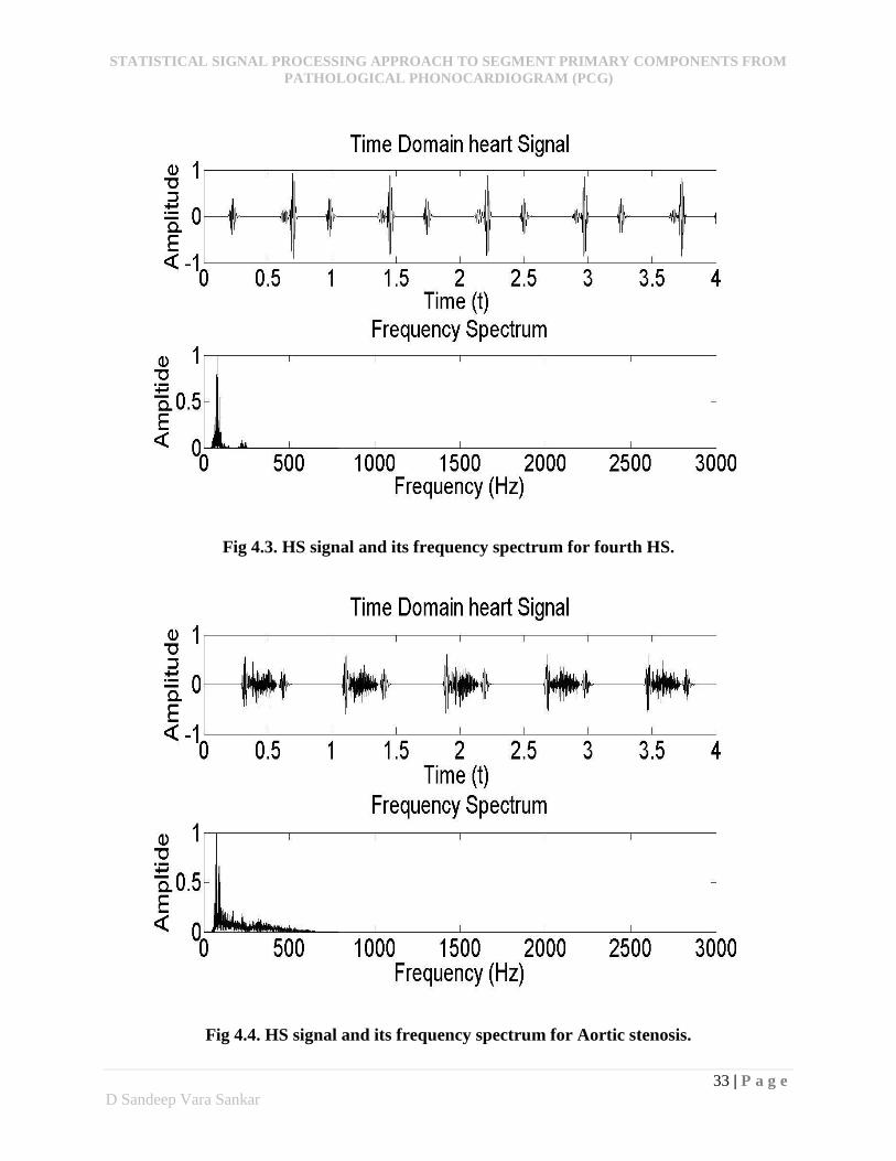
Fig 4.3. HS signal and its frequency spectrum for fourth HS.
Fig 4.4. HS signal and its frequency spectrum for Aortic stenosis.
STATISTICAL SIGNAL PROCESSING APPROACH TO SEGMENT PRIMARY COMPONENTS FROM
PATHOLOGICAL PHONOCARDIOGRAM (PCG)
34 | P a g e
D Sandeep Vara Sankar
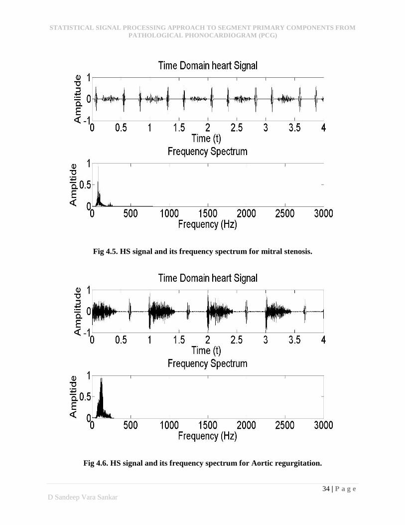
Fig 4.5. HS signal and its frequency spectrum for mitral stenosis.
Fig 4.6. HS signal and its frequency spectrum for Aortic regurgitation.
STATISTICAL SIGNAL PROCESSING APPROACH TO SEGMENT PRIMARY COMPONENTS FROM
PATHOLOGICAL PHONOCARDIOGRAM (PCG)
35 | P a g e
D Sandeep Vara Sankar
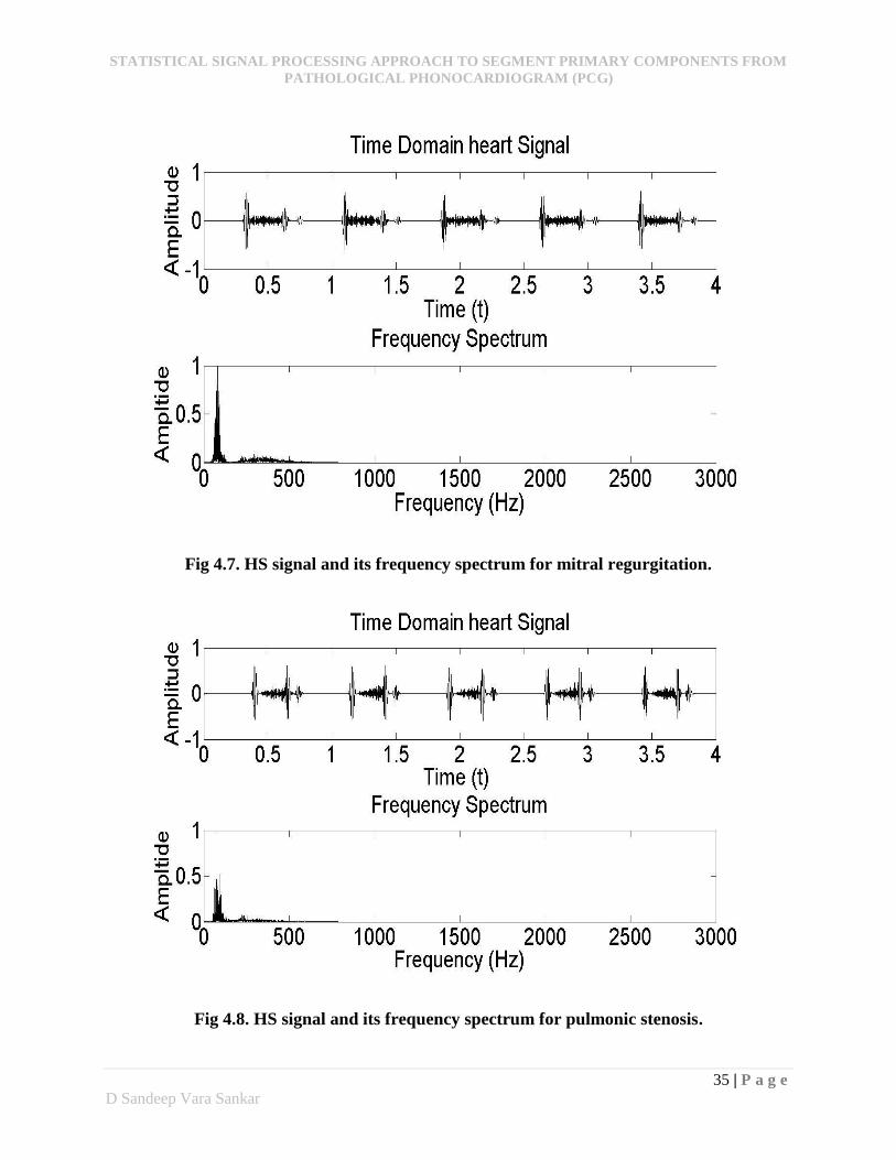
Fig 4.7. HS signal and its frequency spectrum for mitral regurgitation.
Fig 4.8. HS signal and its frequency spectrum for pulmonic stenosis.

STATISTICAL SIGNAL PROCESSING APPROACH TO SEGMENT PRIMARY COMPONENTS FROM
PATHOLOGICAL PHONOCARDIOGRAM (PCG)
36 | P a g e
D Sandeep Vara Sankar
Fig 4.9. HS signal and its frequency spectrum for tricuspid stenosis.
From the above figures, it is an evident that all the important events of the PCG signal lies
below 700Hz frequencies and also it is observed that no data is available at very low frequency
area which depicts no important information is present in that frequency range. So it is inevitable
to filter and decimate the PCG signal before processing.
4.2 Wavelets
Wavelets are capable of handling rapidly changing transient signals because of their scaling
and translation property. Due to non-stationary behavior of the HSs, it is unlikely that a single
decomposition level serve the purpose of capturing the energy of the primary components for
different pathologies.
STATISTICAL SIGNAL PROCESSING APPROACH TO SEGMENT PRIMARY COMPONENTS FROM
PATHOLOGICAL PHONOCARDIOGRAM (PCG)
37 | P a g e
D Sandeep Vara Sankar
4.2.1 Mother wavelet
Though wavelet plays an important role in analyzing the structure of the coefficients, it is
necessary to choose best wavelet, which we call as ‘mother’ wavelet. The wavelet basis is selected
from the previous studies done by the scholars M.A.R Santos and M. Souza [10] and Tang Chu
Lin [14]. Both of them showed that Daubechies and Meyer wavelets are good candidates for HS
signal analysis.
From the results of Tang Chu Lin, Daubechies-15 (db) is chosen as the mother wavelet for
decomposing the HS signals. Daubechies wavelet has a property of orthogonality which is used to
remove the redundant information and also it has strong resemblance of the primary components
of HS signal.
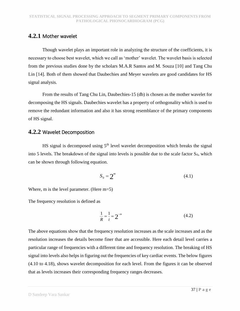
4.2.2 Wavelet Decomposition
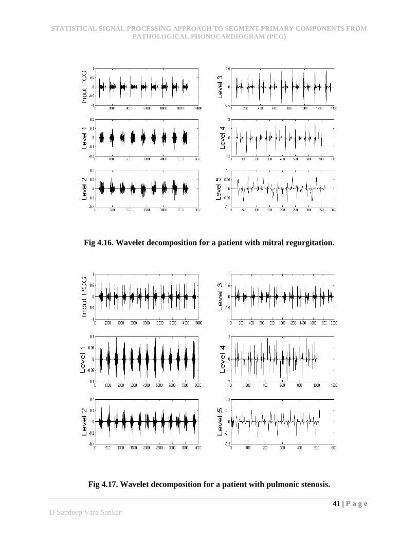
HS signal is decomposed using 5th level wavelet decomposition which breaks the signal
into 5 levels. The breakdown of the signal into levels is possible due to the scale factor S0, which
can be shown through following equation.
20
mS (4.1)
Where, m is the level parameter. (Here m=5)
The frequency resolution is defined as
211 m
iR
(4.2)
The above equations show that the frequency resolution increases as the scale increases and as the
resolution increases the details become finer that are accessible. Here each detail level carries a
particular range of frequencies with a different time and frequency resolution. The breaking of HS
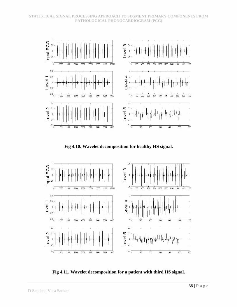
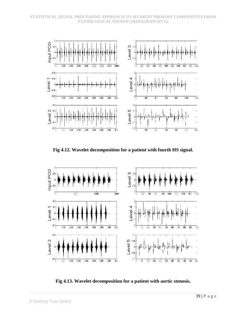
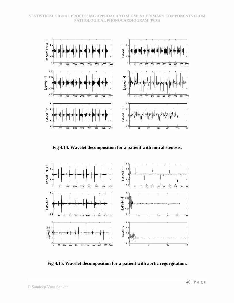
signal into levels also helps in figuring out the frequencies of key cardiac events. The below figures
(4.10 to 4.18), shows wavelet decomposition for each level. From the figures it can be observed
that as levels increases their corresponding frequency ranges decreases.
STATISTICAL SIGNAL PROCESSING APPROACH TO SEGMENT PRIMARY COMPONENTS FROM
PATHOLOGICAL PHONOCARDIOGRAM (PCG)
38 | P a g e
D Sandeep Vara Sankar
Fig 4.10. Wavelet decomposition for healthy HS signal.
Fig 4.11. Wavelet decomposition for a patient with third HS signal.
STATISTICAL SIGNAL PROCESSING APPROACH TO SEGMENT PRIMARY COMPONENTS FROM
PATHOLOGICAL PHONOCARDIOGRAM (PCG)
39 | P a g e
D Sandeep Vara Sankar
Fig 4.12. Wavelet decomposition for a patient with fourth HS signal.
Fig 4.13. Wavelet decomposition for a patient with aortic stenosis.
STATISTICAL SIGNAL PROCESSING APPROACH TO SEGMENT PRIMARY COMPONENTS FROM
PATHOLOGICAL PHONOCARDIOGRAM (PCG)
40 | P a g e
D Sandeep Vara Sankar
Fig 4.14. Wavelet decomposition for a patient with mitral stenosis.
Fig 4.15. Wavelet decomposition for a patient with aortic regurgitation.
STATISTICAL SIGNAL PROCESSING APPROACH TO SEGMENT PRIMARY COMPONENTS FROM
PATHOLOGICAL PHONOCARDIOGRAM (PCG)
41 | P a g e
D Sandeep Vara Sankar
Fig 4.16. Wavelet decomposition for a patient with mitral regurgitation.
Fig 4.17. Wavelet decomposition for a patient with pulmonic stenosis.

STATISTICAL SIGNAL PROCESSING APPROACH TO SEGMENT PRIMARY COMPONENTS FROM
PATHOLOGICAL PHONOCARDIOGRAM (PCG)
42 | P a g e
D Sandeep Vara Sankar
Fig 4.18. Wavelet decomposition for a patient with tricuspid stenosis.
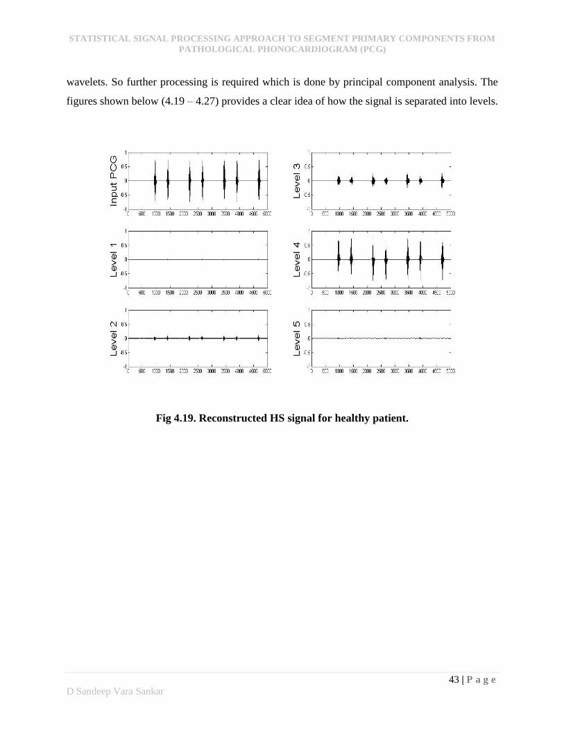
4.2.3 Wavelet Synthesis
The wavelet decomposition analysis of HS signals illustrate the information captured in
each detail level. In order to obtain best temporal resolution, it is necessary that each of the
coefficient vector should be used to synthesize the original HS signal. The representation of the
signal components determines the accuracy in segmenting the events. Furthermore the accuracy
levels of the segmented components gets diminished due to presence of unwanted elements (either
noise or/and murmur) in a signal. The purpose of wavelet synthesis is to obtain a set of featured
signals at the highest temporal resolution. From the feature set, each point in the pre-processed HS
signal can be viewed as a point in a 5-dimensional space where each dimension corresponds to a
detail level. If we consider each sample point as a random variable then there would be five
observations per variable. The five synthesized signals generate a feature set for the original HS
signal. These features cannot be used as the final set of features for segmentation because obtaining
the best feature for each sample point from five obtained observations is not possible using
STATISTICAL SIGNAL PROCESSING APPROACH TO SEGMENT PRIMARY COMPONENTS FROM
PATHOLOGICAL PHONOCARDIOGRAM (PCG)
43 | P a g e
D Sandeep Vara Sankar
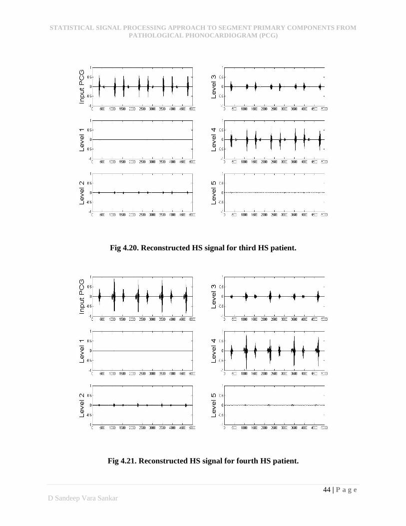
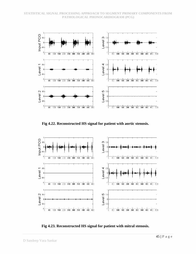
wavelets. So further processing is required which is done by principal component analysis. The
figures shown below (4.19 – 4.27) provides a clear idea of how the signal is separated into levels.
Fig 4.19. Reconstructed HS signal for healthy patient.
STATISTICAL SIGNAL PROCESSING APPROACH TO SEGMENT PRIMARY COMPONENTS FROM
PATHOLOGICAL PHONOCARDIOGRAM (PCG)
44 | P a g e
D Sandeep Vara Sankar
Fig 4.20. Reconstructed HS signal for third HS patient.
Fig 4.21. Reconstructed HS signal for fourth HS patient.
STATISTICAL SIGNAL PROCESSING APPROACH TO SEGMENT PRIMARY COMPONENTS FROM
PATHOLOGICAL PHONOCARDIOGRAM (PCG)
45 | P a g e
D Sandeep Vara Sankar
Fig 4.22. Reconstructed HS signal for patient with aortic stenosis.
Fig 4.23. Reconstructed HS signal for patient with mitral stenosis.
STATISTICAL SIGNAL PROCESSING APPROACH TO SEGMENT PRIMARY COMPONENTS FROM
PATHOLOGICAL PHONOCARDIOGRAM (PCG)
46 | P a g e
D Sandeep Vara Sankar
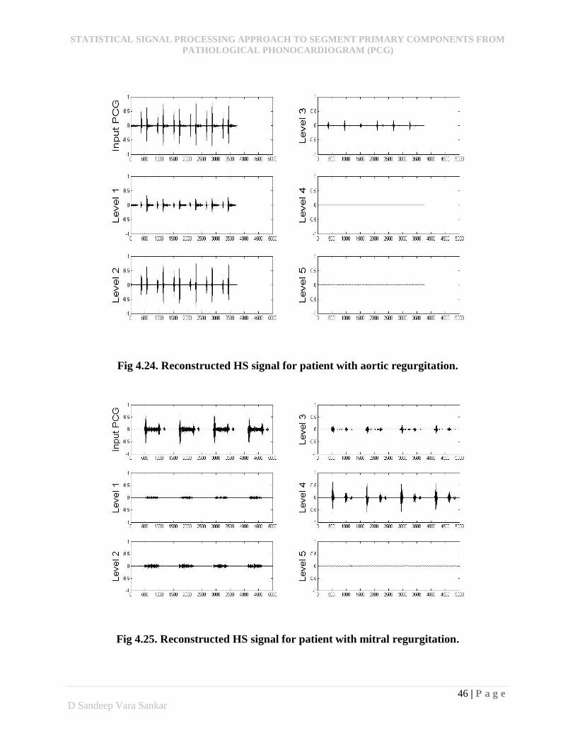
Fig 4.24. Reconstructed HS signal for patient with aortic regurgitation.
Fig 4.25. Reconstructed HS signal for patient with mitral regurgitation.
STATISTICAL SIGNAL PROCESSING APPROACH TO SEGMENT PRIMARY COMPONENTS FROM
PATHOLOGICAL PHONOCARDIOGRAM (PCG)
47 | P a g e
D Sandeep Vara Sankar
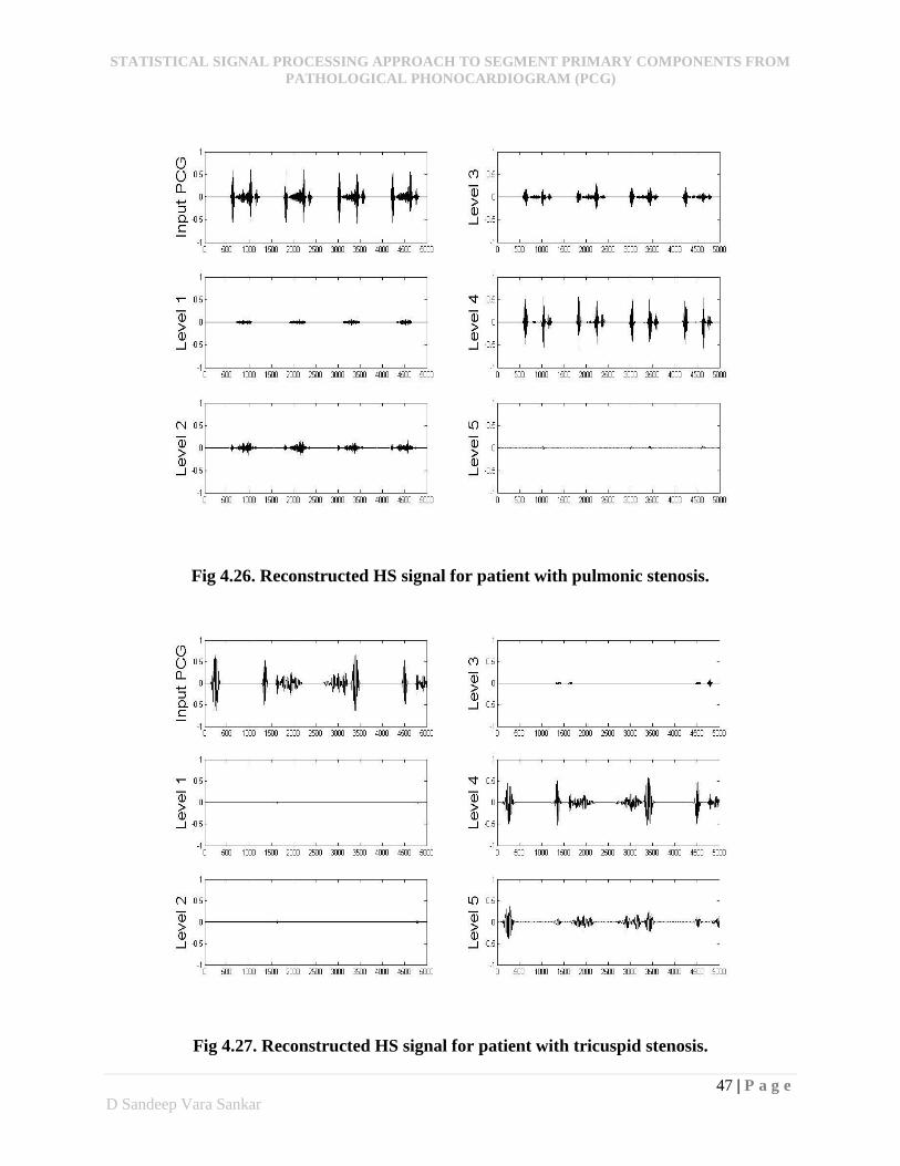
Fig 4.26. Reconstructed HS signal for patient with pulmonic stenosis.
Fig 4.27. Reconstructed HS signal for patient with tricuspid stenosis.
STATISTICAL SIGNAL PROCESSING APPROACH TO SEGMENT PRIMARY COMPONENTS FROM
PATHOLOGICAL PHONOCARDIOGRAM (PCG)
48 | P a g e
D Sandeep Vara Sankar
From the figures, we can conclude that level 1 doesn’t carry any information about HS
which can be neglected. Level 2, 3, 4 & 5 carries the information of the HSs and in that level 3
and level 4 contains maximum information content of the HS components than the other two.
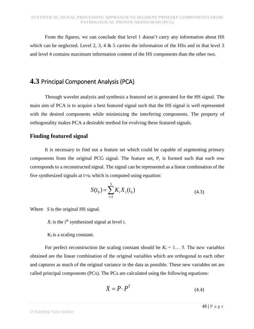
4.3 Principal Component Analysis (PCA)
Through wavelet analysis and synthesis a featured set is generated for the HS signal. The
main aim of PCA is to acquire a best featured signal such that the HS signal is well represented
with the desired components while minimizing the interfering components. The property of
orthogonality makes PCA a desirable method for evolving these featured signals.
Finding featured signal
It is necessary to find out a feature set which could be capable of segmenting primary
components from the original PCG signal. The feature set, P, is formed such that each row
corresponds to a reconstructed signal. The signal can be represented as a linear combination of the
five synthesized signals at t=t0 which is computed using equation:
5
1
00 )()(i
ii tXKtS (4.3)
Where S is the original HS signal.
Xi is the ith synthesized signal at level i.
Ki is a scaling constant.
For perfect reconstruction the scaling constant should be Ki = 1… 5. The new variables
obtained are the linear combination of the original variables which are orthogonal to each other
and captures as much of the original variance in the data as possible. These new variables set are
called principal components (PCs). The PCs are calculated using the following equations:
TPPX (4.4)
STATISTICAL SIGNAL PROCESSING APPROACH TO SEGMENT PRIMARY COMPONENTS FROM
PATHOLOGICAL PHONOCARDIOGRAM (PCG)
49 | P a g e
D Sandeep Vara Sankar
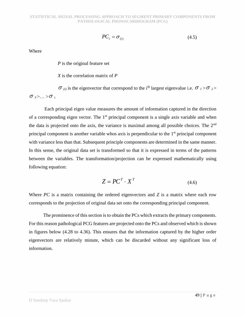
)(iiPC (4.5)
Where
P is the original feature set
X is the correlation matrix of P
(i) is the eigenvector that correspond to the ith largest eigenvalue i.e. 1 > 2 >
3 >… > i.
Each principal eigen value measures the amount of information captured in the direction
of a corresponding eigen vector. The 1st principal component is a single axis variable and when
the data is projected onto the axis, the variance is maximal among all possible choices. The 2nd
principal component is another variable whos axis is perpendicular to the 1st principal component
with variance less than that. Subsequent principle components are determined in the same manner.
In this sense, the original data set is transformed so that it is expressed in terms of the patterns
between the variables. The transformation/projection can be expressed mathematically using
following equation:
TT XPCZ (4.6)
Where PC is a matrix containing the ordered eigenvectors and Z is a matrix where each row
corresponds to the projection of original data set onto the corresponding principal component.
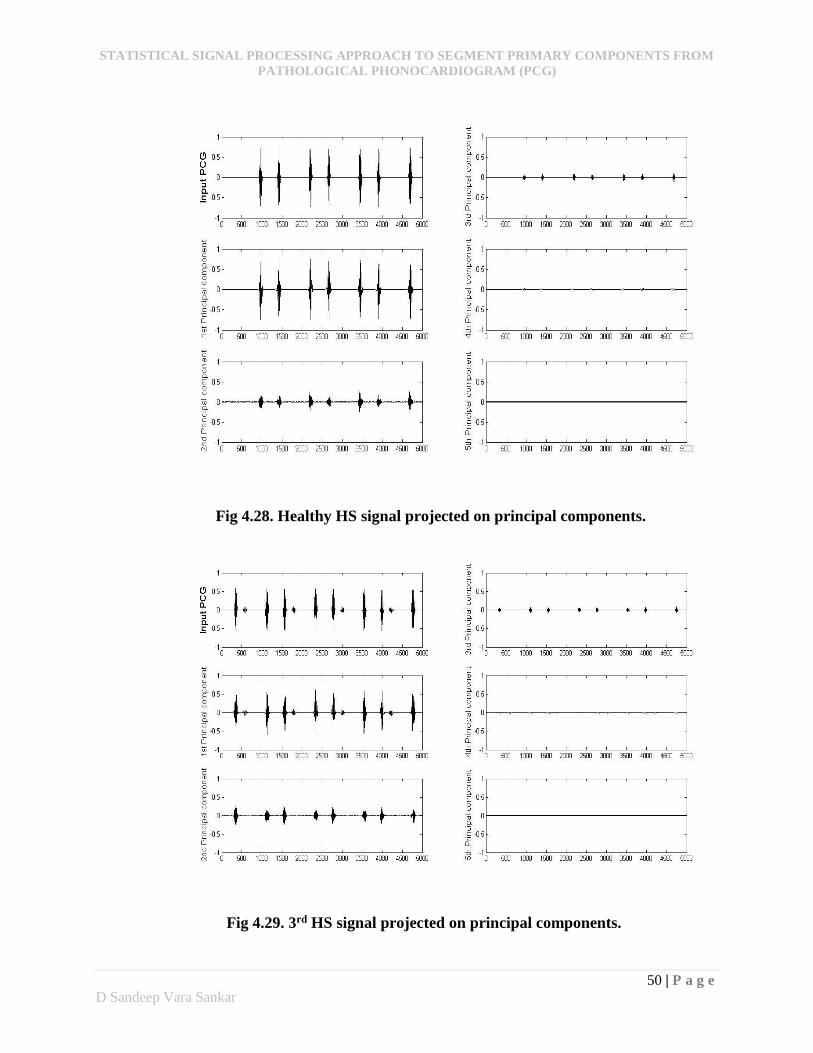
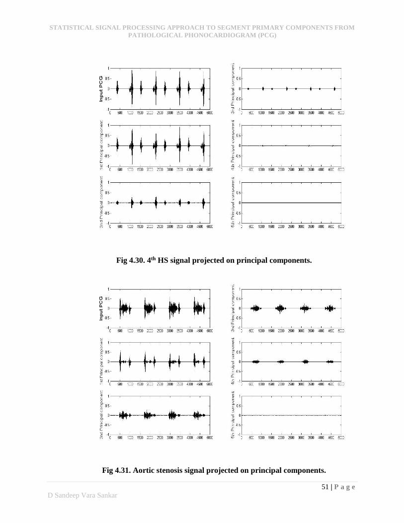
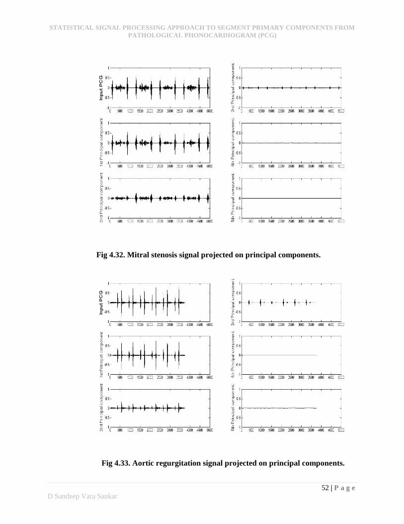
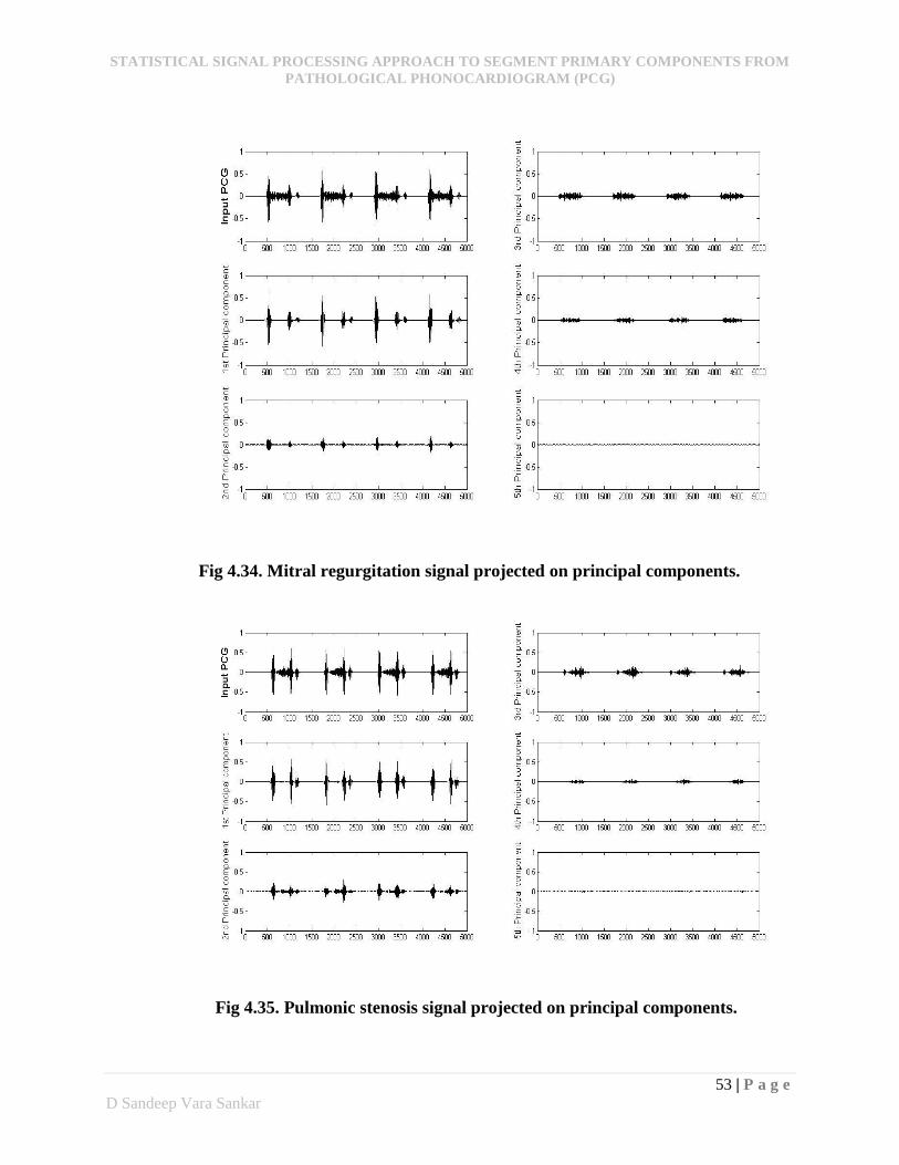
The prominence of this section is to obtain the PCs which extracts the primary components.
For this reason pathological PCG features are projected onto the PCs and observed which is shown
in figures below (4.28 to 4.36). This ensures that the information captured by the higher order
eigenvectors are relatively minute, which can be discarded without any significant loss of
information.
STATISTICAL SIGNAL PROCESSING APPROACH TO SEGMENT PRIMARY COMPONENTS FROM
PATHOLOGICAL PHONOCARDIOGRAM (PCG)
50 | P a g e
D Sandeep Vara Sankar
Fig 4.28. Healthy HS signal projected on principal components.
Fig 4.29. 3rd HS signal projected on principal components.
STATISTICAL SIGNAL PROCESSING APPROACH TO SEGMENT PRIMARY COMPONENTS FROM
PATHOLOGICAL PHONOCARDIOGRAM (PCG)
51 | P a g e
D Sandeep Vara Sankar
Fig 4.30. 4th HS signal projected on principal components.
Fig 4.31. Aortic stenosis signal projected on principal components.
STATISTICAL SIGNAL PROCESSING APPROACH TO SEGMENT PRIMARY COMPONENTS FROM
PATHOLOGICAL PHONOCARDIOGRAM (PCG)
52 | P a g e
D Sandeep Vara Sankar
Fig 4.32. Mitral stenosis signal projected on principal components.
Fig 4.33. Aortic regurgitation signal projected on principal components.
STATISTICAL SIGNAL PROCESSING APPROACH TO SEGMENT PRIMARY COMPONENTS FROM
PATHOLOGICAL PHONOCARDIOGRAM (PCG)
53 | P a g e
D Sandeep Vara Sankar
Fig 4.34. Mitral regurgitation signal projected on principal components.
Fig 4.35. Pulmonic stenosis signal projected on principal components.
STATISTICAL SIGNAL PROCESSING APPROACH TO SEGMENT PRIMARY COMPONENTS FROM
PATHOLOGICAL PHONOCARDIOGRAM (PCG)
54 | P a g e
D Sandeep Vara Sankar
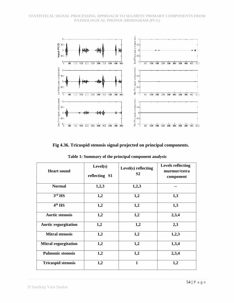
Fig 4.36. Tricuspid stenosis signal projected on principal components.
Table 1: Summary of the principal component analysis
Heart sound Level(s)
reflecting S1
Level(s) reflecting
S2
Levels reflecting
murmur/extra
component
Normal 1,2,3 1,2,3 --
3rd HS 1,2 1,2 1,3
4th HS 1,2 1,2 1,3
Aortic stenosis 1,2 1,2 2,3,4
Aortic regurgitation 1,2 1,2 2,3
Mitral stenosis 1,2 1,2 1,2,3
Mitral regurgitation 1,2 1,2 1,3,4
Pulmonic stenosis 1,2 1,2 2,3,4
Tricuspid stenosis 1,2 1 1,2
STATISTICAL SIGNAL PROCESSING APPROACH TO SEGMENT PRIMARY COMPONENTS FROM
PATHOLOGICAL PHONOCARDIOGRAM (PCG)
55 | P a g e
D Sandeep Vara Sankar
The summary of the PCA analysis in table 1 shows that most of the primary components
are concentrated in 1st and 2nd PCs and murmurs are concentrated in all 1, 2, 3 and 4 PCs. From
the observations of the above figures, the primary components although concentrates in both 1st
and 2nd PCs, their most of the energy/information content is present in 1st PC only. In the same
way although the murmurs have their role in all the 4 PCs, their most energy/information content
is seen in 2nd and 3rd Pcs only. As the aim of this study is only to segment and identify primary
components, the 1st PC which has maximum information content of primary components is chosen
for further analysis and the remaining PCs are neglected.
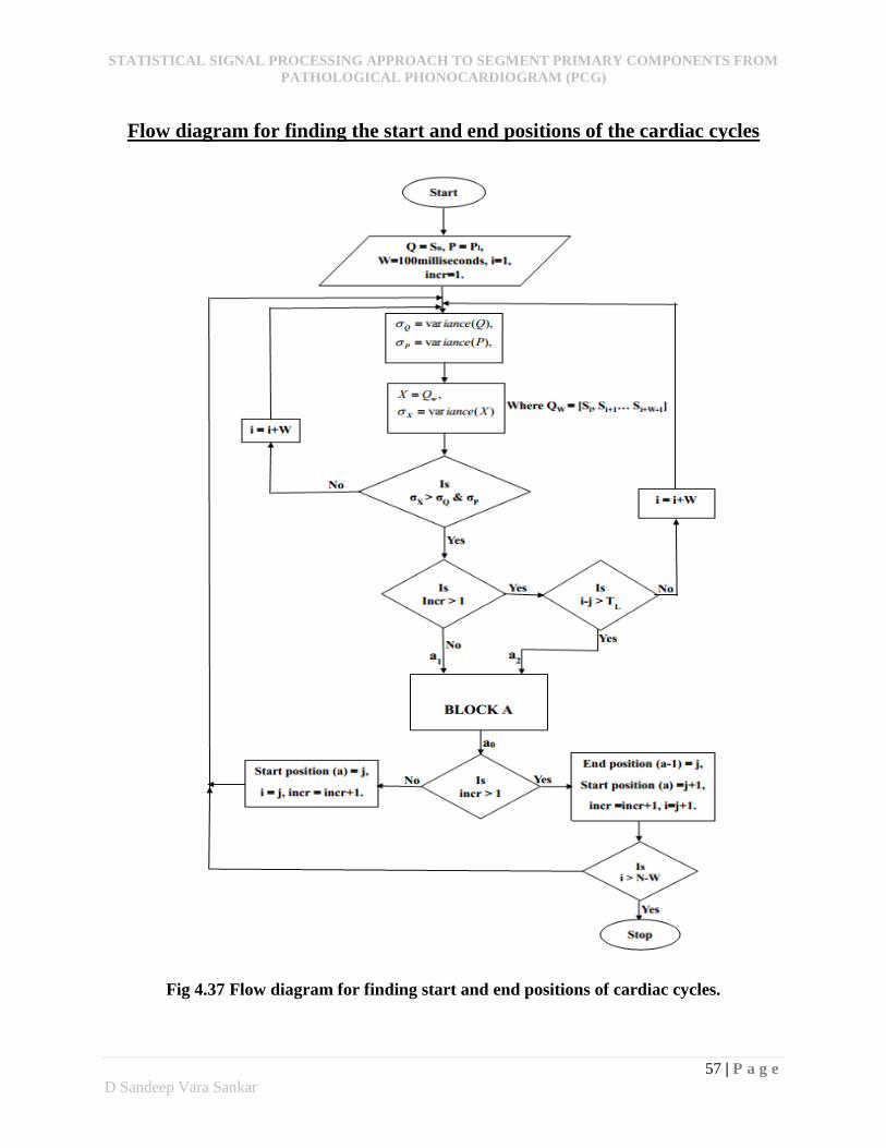
4.4 Separation of HS signal into Cardiac Cycles
Each HS signal is a combination of many cardiac cycles and separating the whole PCG
signal into series of cardiac cycles is of clinical importance. After examining several pathological
PCGs it is not obvious to conclude that segmentation techniques which uses amplitude
thresholding works better for the cases of murmur dominated HSs and primary components
diminished HSs. In order to combat these problems segmentation based on cardiac cycle is
proposed in this study. Each cardiac cycle consists of S1, systole, S2 and diastole. In order to
divide the entire HS signal into combination of these four phases, a well-defined process which
can be capable of picking exact boundary positions is prerequisite. So an algorithm based on
statistical analysis is proposed to gratify the above conditions, which is called as a splitting
algorithm. The algorithm constitutes of two important variance parameters, global variance and
local variance. According to the algorithm, the variances of the raw PCG signal and the 1st PC of
PCA analysed PCG signal are the global variances and the variance of each windowed segment of
the 1st PC of the PCA analysed HS signal is the local variance. Each local variance is compared
with the global variances which acts as thresholds and accordingly further processing is computed.
The entire algorithm is assembled in the form of flow chart which is shown in figures 4.37 & 4.38.
Here each cardiac cycle will have one start position and one end position. Variances are
represented in the form of σ(.) where (.) represents corresponding signal parameter. Sn is the PCG
signal, where n is the nth sample of the PCG signal, Pl is the 1st PC of the PCA analysed HS signal,
where l is the lth sample of the signal. N is the length of the raw HS signal, and L is the length of
STATISTICAL SIGNAL PROCESSING APPROACH TO SEGMENT PRIMARY COMPONENTS FROM
PATHOLOGICAL PHONOCARDIOGRAM (PCG)
56 | P a g e
D Sandeep Vara Sankar
the 1st PC of the PCA analysed HS signal. In this algorithm, firstly, procedure for finding the start
position of the very 1st cardiac cycle is carried followed by the search for the corresponding end
position of the cycle. The end position is ensured by a special condition which depends on the
sampling rate (fs) and the average heart rate (avgrate) possibility for an abnormal person.
avgratefT sL where, fs = 1575 samples per second.
Here the variable ‘TL’ denotes the lower limit of the average cardiac cycle. This value is not
particular to any individual but in general it is approximately constant. For this study the ‘TL’ is
taken as 100beats per minute (1 beat per 0.6 second). Once the end position is obtained, the next
start position can be written as the position next to the corresponding end position and the
algorithm continues for the search of next end position. The repetition process continues until the
length becomes equal to L – window size (W).
STATISTICAL SIGNAL PROCESSING APPROACH TO SEGMENT PRIMARY COMPONENTS FROM
PATHOLOGICAL PHONOCARDIOGRAM (PCG)
57 | P a g e
D Sandeep Vara Sankar
Flow diagram for finding the start and end positions of the cardiac cycles
Fig 4.37 Flow diagram for finding start and end positions of cardiac cycles.
STATISTICAL SIGNAL PROCESSING APPROACH TO SEGMENT PRIMARY COMPONENTS FROM
PATHOLOGICAL PHONOCARDIOGRAM (PCG)
58 | P a g e
D Sandeep Vara Sankar
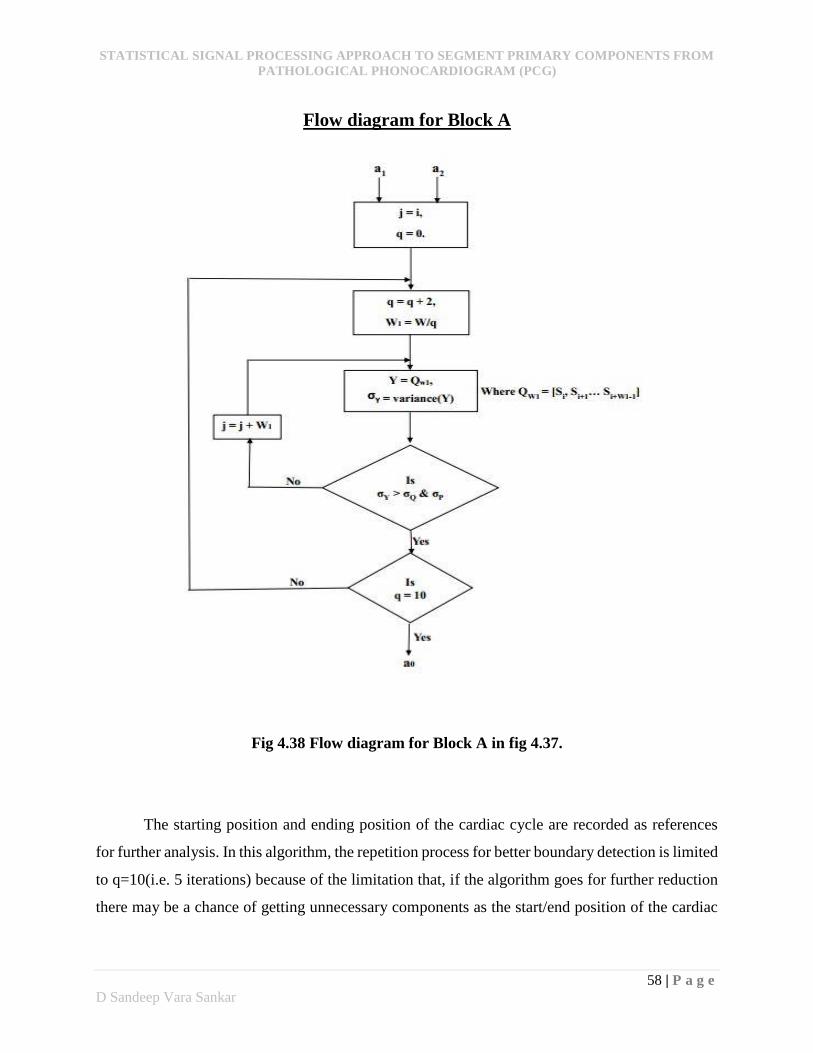
Flow diagram for Block A
Fig 4.38 Flow diagram for Block A in fig 4.37.
The starting position and ending position of the cardiac cycle are recorded as references
for further analysis. In this algorithm, the repetition process for better boundary detection is limited
to q=10(i.e. 5 iterations) because of the limitation that, if the algorithm goes for further reduction
there may be a chance of getting unnecessary components as the start/end position of the cardiac
STATISTICAL SIGNAL PROCESSING APPROACH TO SEGMENT PRIMARY COMPONENTS FROM
PATHOLOGICAL PHONOCARDIOGRAM (PCG)
59 | P a g e
D Sandeep Vara Sankar
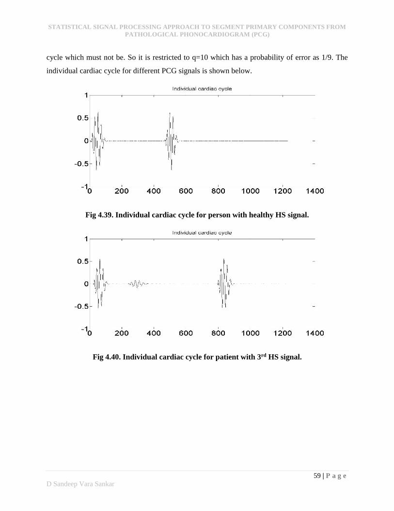
cycle which must not be. So it is restricted to q=10 which has a probability of error as 1/9. The
individual cardiac cycle for different PCG signals is shown below.
Fig 4.39. Individual cardiac cycle for person with healthy HS signal.
Fig 4.40. Individual cardiac cycle for patient with 3rd HS signal.
STATISTICAL SIGNAL PROCESSING APPROACH TO SEGMENT PRIMARY COMPONENTS FROM
PATHOLOGICAL PHONOCARDIOGRAM (PCG)
60 | P a g e
D Sandeep Vara Sankar
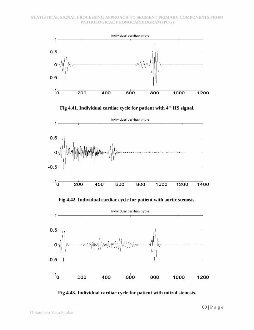
Fig 4.41. Individual cardiac cycle for patient with 4th HS signal.
Fig 4.42. Individual cardiac cycle for patient with aortic stenosis.
Fig 4.43. Individual cardiac cycle for patient with mitral stenosis.
STATISTICAL SIGNAL PROCESSING APPROACH TO SEGMENT PRIMARY COMPONENTS FROM
PATHOLOGICAL PHONOCARDIOGRAM (PCG)
61 | P a g e
D Sandeep Vara Sankar
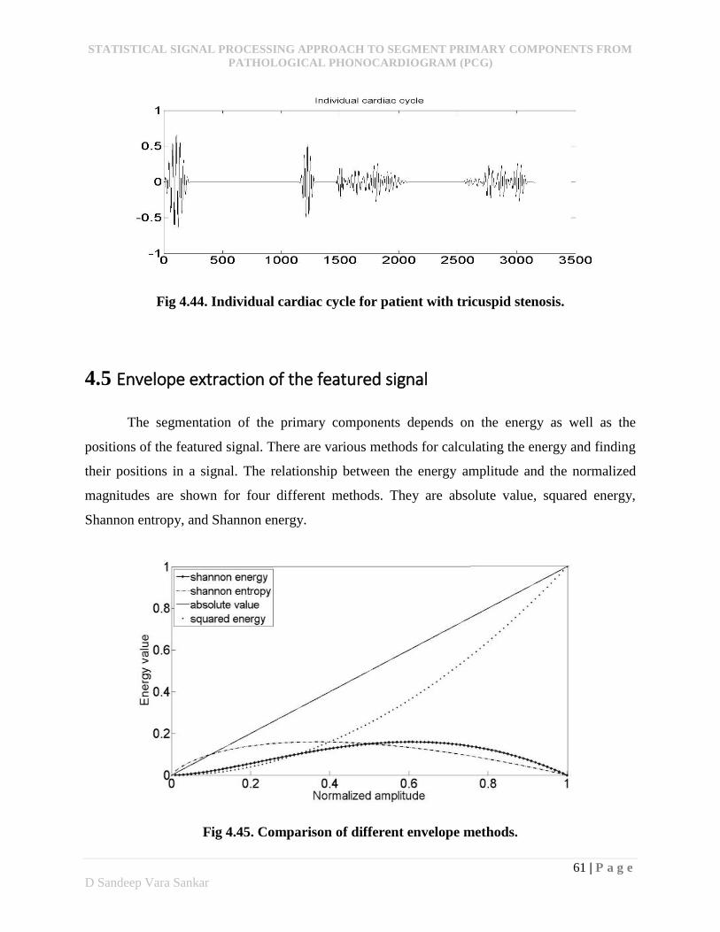
Fig 4.44. Individual cardiac cycle for patient with tricuspid stenosis.
4.5 Envelope extraction of the featured signal
The segmentation of the primary components depends on the energy as well as the
positions of the featured signal. There are various methods for calculating the energy and finding
their positions in a signal. The relationship between the energy amplitude and the normalized
magnitudes are shown for four different methods. They are absolute value, squared energy,
Shannon entropy, and Shannon energy.
Fig 4.45. Comparison of different envelope methods.
STATISTICAL SIGNAL PROCESSING APPROACH TO SEGMENT PRIMARY COMPONENTS FROM
PATHOLOGICAL PHONOCARDIOGRAM (PCG)
62 | P a g e
D Sandeep Vara Sankar
The absolute value weighs all intensities in a signal equally. The squared energy obscures
the low intensity values while increasing the high intensities exponentially. The Shannon entropy
magnifies the energy levels from low to medium and diminishes the energy levels from medium
to high. The Shannon energy magnifies the medium intensity values while diminishing the high
and low intensity values which can be shown in fig 4.37. From the observations, we have chosen
to use Shannon energy for its property of attenuating the effects of low energy noises. The Shannon
energy is calculated for the short segments of 40millisecond window size with 50% overlapping.
The Shannon energy is calculated using following equation:
)(log1 2
1
2 iPiPN
EsegN
iseg
(4.6)
Where
Nseg is the number of samples in the 40millisecond segment.
The positions of the energy levels captured by the Shannon energy which are shown in figures
below are stored for further analysis.
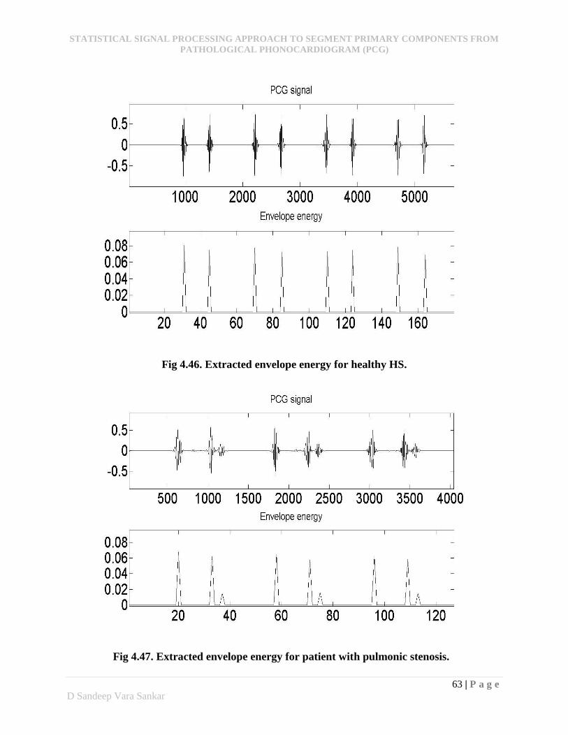
STATISTICAL SIGNAL PROCESSING APPROACH TO SEGMENT PRIMARY COMPONENTS FROM
PATHOLOGICAL PHONOCARDIOGRAM (PCG)
63 | P a g e
D Sandeep Vara Sankar
Fig 4.46. Extracted envelope energy for healthy HS.
Fig 4.47. Extracted envelope energy for patient with pulmonic stenosis.
STATISTICAL SIGNAL PROCESSING APPROACH TO SEGMENT PRIMARY COMPONENTS FROM
PATHOLOGICAL PHONOCARDIOGRAM (PCG)
64 | P a g e
D Sandeep Vara Sankar
4.6 Segmentation of primary components
Cardiac anatomy states that the location of occurrence of the murmurs depends on the type
of pathology, and the intensity level of the murmur depends on the position of auscultation. So it
is unlikely to conclude that every pathology has a specific type of murmur at specific location. To
combat this problem first we should segment the stationary components which are common for all
pathologies. These stationary components are called primary components or components of
interest (S1 and S2). Primary components are the components which has a specific pattern and a
specific distribution [15]. Segmentation of primary components is done using the records which
are stored from previous two sections. The records include cardiac cycle positions and envelope
positions.
4.6.1 Missing peak detection
During some pathological conditions there may be a chance that the intensity level of either
S1 or S2 get buried due to valve dysfunctionality. So it is necessary to detect those peaks initially.
For this the difference between the envelope positions are calculated and compared with the
cardiac cycle duration of that particular cardiac event. As each cardiac cycle consists of S1, systole,
S2 and diastolic phases, it is mandatory that the difference between consecutive envelope positions
should always be less than the cardiac duration. If this condition is not satisfied, it clearly states
that one of the primary component is missing and further process is done to extract the missing
peak. In this process only missing components are extracted.
4.6.2 Detection of S1 and S2 from murmured PCG signal
The main aim of this work is to separate primary components from murmur/noise corrupted
HS signal. In this analysis along with the stored records from envelope extraction and cardiac cycle
separation, two additional references are taken i.e. difference between consecutive envelopes and
time period of the individual energy components. Using these four data sets and two thresholds
which are derived from above data sets, primary components are segmented.
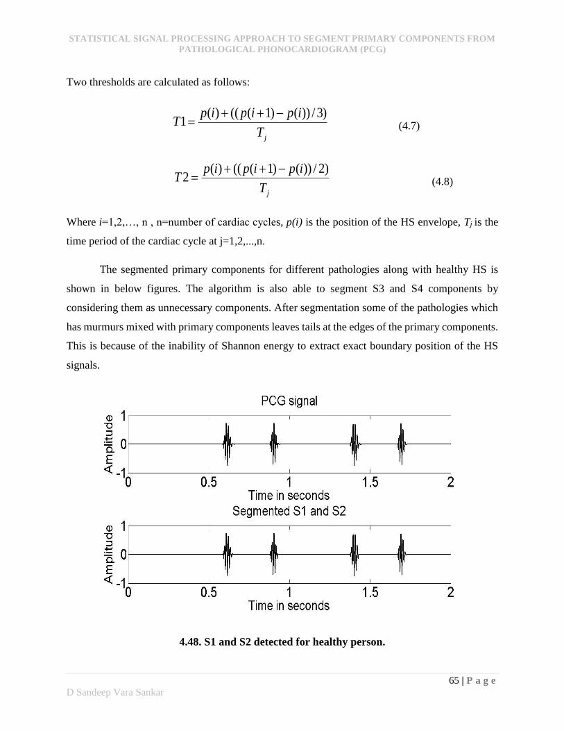
STATISTICAL SIGNAL PROCESSING APPROACH TO SEGMENT PRIMARY COMPONENTS FROM
PATHOLOGICAL PHONOCARDIOGRAM (PCG)
65 | P a g e
D Sandeep Vara Sankar
Two thresholds are calculated as follows:
jT
ipipipT
)3/))()1((()(1
(4.7)
jT
ipipipT
)2/))()1((()(2
(4.8)
Where i=1,2,…, n , n=number of cardiac cycles, p(i) is the position of the HS envelope, Tj is the
time period of the cardiac cycle at j=1,2,...,n.
The segmented primary components for different pathologies along with healthy HS is
shown in below figures. The algorithm is also able to segment S3 and S4 components by
considering them as unnecessary components. After segmentation some of the pathologies which
has murmurs mixed with primary components leaves tails at the edges of the primary components.
This is because of the inability of Shannon energy to extract exact boundary position of the HS
signals.
4.48. S1 and S2 detected for healthy person.
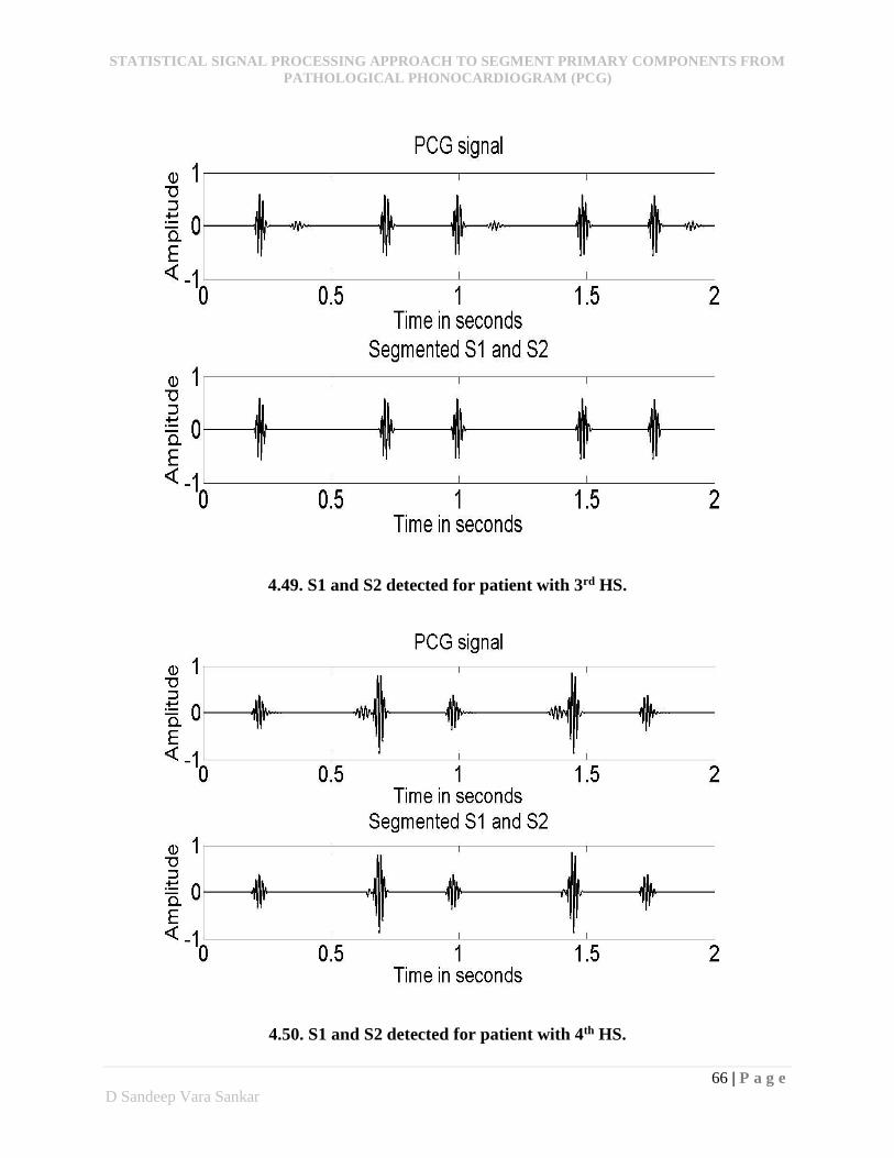
STATISTICAL SIGNAL PROCESSING APPROACH TO SEGMENT PRIMARY COMPONENTS FROM
PATHOLOGICAL PHONOCARDIOGRAM (PCG)
66 | P a g e
D Sandeep Vara Sankar
4.49. S1 and S2 detected for patient with 3rd HS.
4.50. S1 and S2 detected for patient with 4th HS.
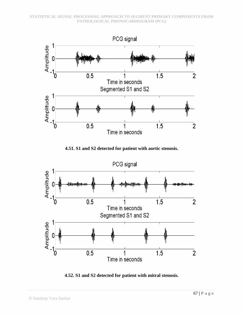
STATISTICAL SIGNAL PROCESSING APPROACH TO SEGMENT PRIMARY COMPONENTS FROM
PATHOLOGICAL PHONOCARDIOGRAM (PCG)
67 | P a g e
D Sandeep Vara Sankar
4.51. S1 and S2 detected for patient with aortic stenosis.
4.52. S1 and S2 detected for patient with mitral stenosis.
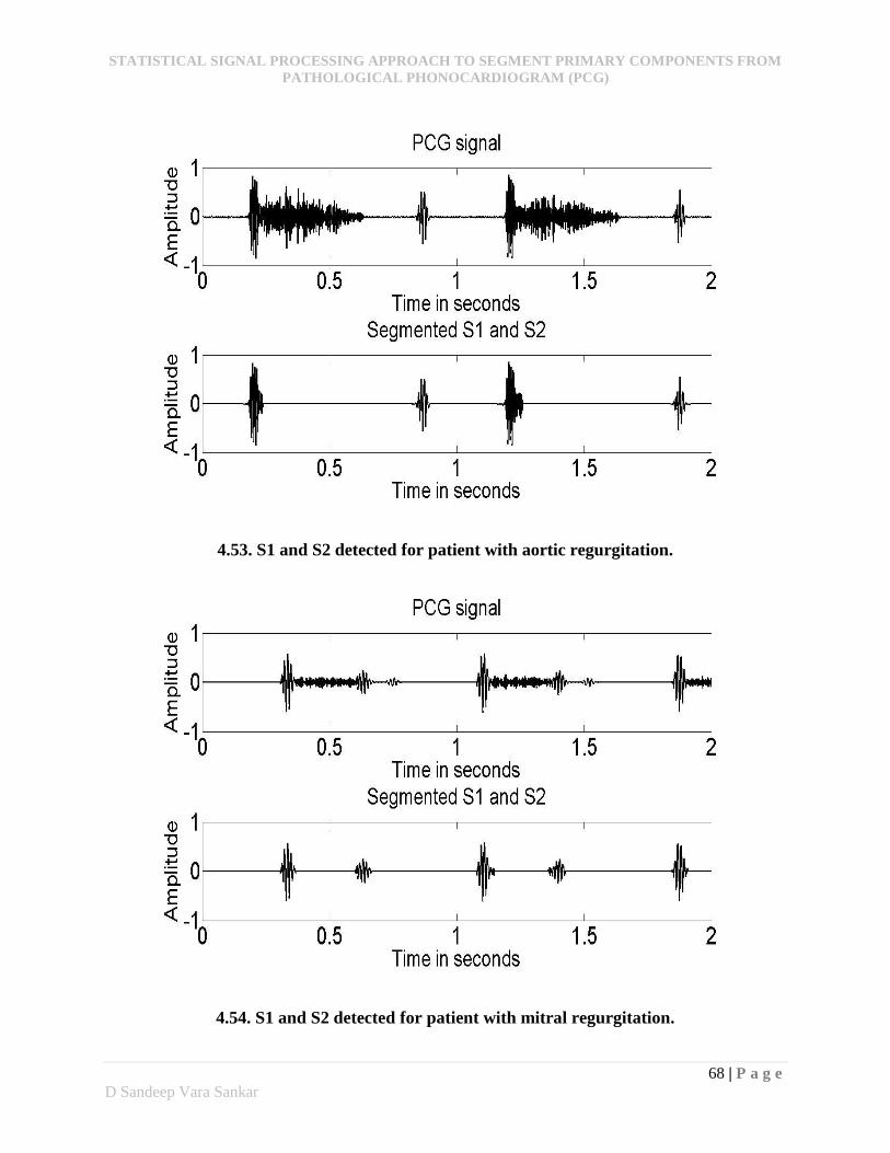
STATISTICAL SIGNAL PROCESSING APPROACH TO SEGMENT PRIMARY COMPONENTS FROM
PATHOLOGICAL PHONOCARDIOGRAM (PCG)
68 | P a g e
D Sandeep Vara Sankar
4.53. S1 and S2 detected for patient with aortic regurgitation.
4.54. S1 and S2 detected for patient with mitral regurgitation.
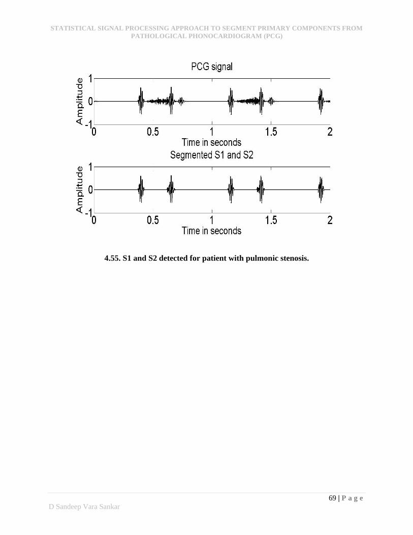
STATISTICAL SIGNAL PROCESSING APPROACH TO SEGMENT PRIMARY COMPONENTS FROM
PATHOLOGICAL PHONOCARDIOGRAM (PCG)
69 | P a g e
D Sandeep Vara Sankar
4.55. S1 and S2 detected for patient with pulmonic stenosis.
STATISTICAL SIGNAL PROCESSING APPROACH TO SEGMENT PRIMARY COMPONENTS FROM
PATHOLOGICAL PHONOCARDIOGRAM (PCG)
70 | P a g e
D Sandeep Vara Sankar
CHAPTER 5
Performance analysis
STATISTICAL SIGNAL PROCESSING APPROACH TO SEGMENT PRIMARY COMPONENTS FROM
PATHOLOGICAL PHONOCARDIOGRAM (PCG)
71 | P a g e
D Sandeep Vara Sankar
Heart sounds are non-stationary and exhibits sudden frequency transients from one cardiac cycle
to another cardiac cycle. Many researchers describe that the normal heart rate as regular, but more
careful analysis reveals that healthy individuals have heart rate that fluctuates considerably even
at rest [15]. This depicts the typical nature of the PCG for any analysis. Despite of any pathology
each cardiac event has two components (S1 and S2) as common and is quite stationary. The main
focus of this study is to highlight the stationary components which we call them as primary
components (S1 and S2).
5.1. Performance Analysis
The performance analysis of this study is very difficult to evaluate. But when observing all
pathologies, the proposed technique doesn’t work quite well in cases where the lower limit for
average cardiac period (TL) condition fails to pick the exact cardiac cycle boundaries. This is
because of the presence of some unnecessary components which has valid energy (i.e. > global
threshold) between the lower limit point and the actual cardiac end point. This problem arises in
tricuspid stenosis which is shown in below figure 5.1. In this figure, the murmur pertains at the
end of the cardiac cycle where the average lower limit of the cardiac cycle (TL) is crossed. As in
this case, the murmur energy is greater than threshold; the algorithm takes that murmur as valid
component and treats as the ending position of the cardiac cycle which gives false representation
for the cardiac cycles.
This problem can be eliminated by modelling the signal by using estimators. The primary
components always have a stationary behaviour, so it has a constant frequency range from which
the non-stationary components can be eliminated and make the algorithm still valid in these cases.
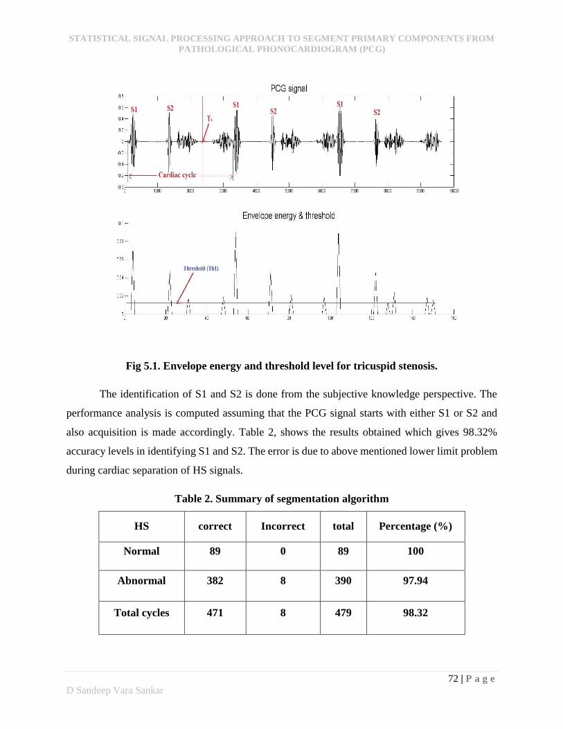
STATISTICAL SIGNAL PROCESSING APPROACH TO SEGMENT PRIMARY COMPONENTS FROM
PATHOLOGICAL PHONOCARDIOGRAM (PCG)
72 | P a g e
D Sandeep Vara Sankar
Fig 5.1. Envelope energy and threshold level for tricuspid stenosis.
The identification of S1 and S2 is done from the subjective knowledge perspective. The
performance analysis is computed assuming that the PCG signal starts with either S1 or S2 and
also acquisition is made accordingly. Table 2, shows the results obtained which gives 98.32%
accuracy levels in identifying S1 and S2. The error is due to above mentioned lower limit problem
during cardiac separation of HS signals.
Table 2. Summary of segmentation algorithm
HS correct Incorrect total Percentage (%)
Normal 89 0 89 100
Abnormal 382 8 390 97.94
Total cycles 471 8 479 98.32
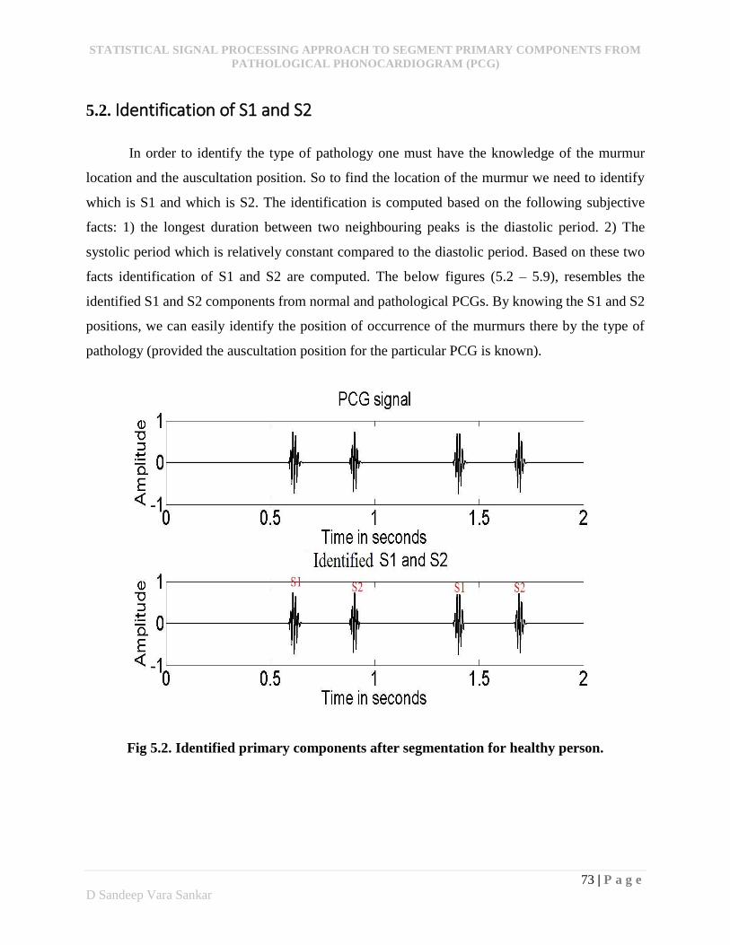
STATISTICAL SIGNAL PROCESSING APPROACH TO SEGMENT PRIMARY COMPONENTS FROM
PATHOLOGICAL PHONOCARDIOGRAM (PCG)
73 | P a g e
D Sandeep Vara Sankar
5.2. Identification of S1 and S2
In order to identify the type of pathology one must have the knowledge of the murmur
location and the auscultation position. So to find the location of the murmur we need to identify
which is S1 and which is S2. The identification is computed based on the following subjective
facts: 1) the longest duration between two neighbouring peaks is the diastolic period. 2) The
systolic period which is relatively constant compared to the diastolic period. Based on these two
facts identification of S1 and S2 are computed. The below figures (5.2 – 5.9), resembles the
identified S1 and S2 components from normal and pathological PCGs. By knowing the S1 and S2
positions, we can easily identify the position of occurrence of the murmurs there by the type of
pathology (provided the auscultation position for the particular PCG is known).
Fig 5.2. Identified primary components after segmentation for healthy person.
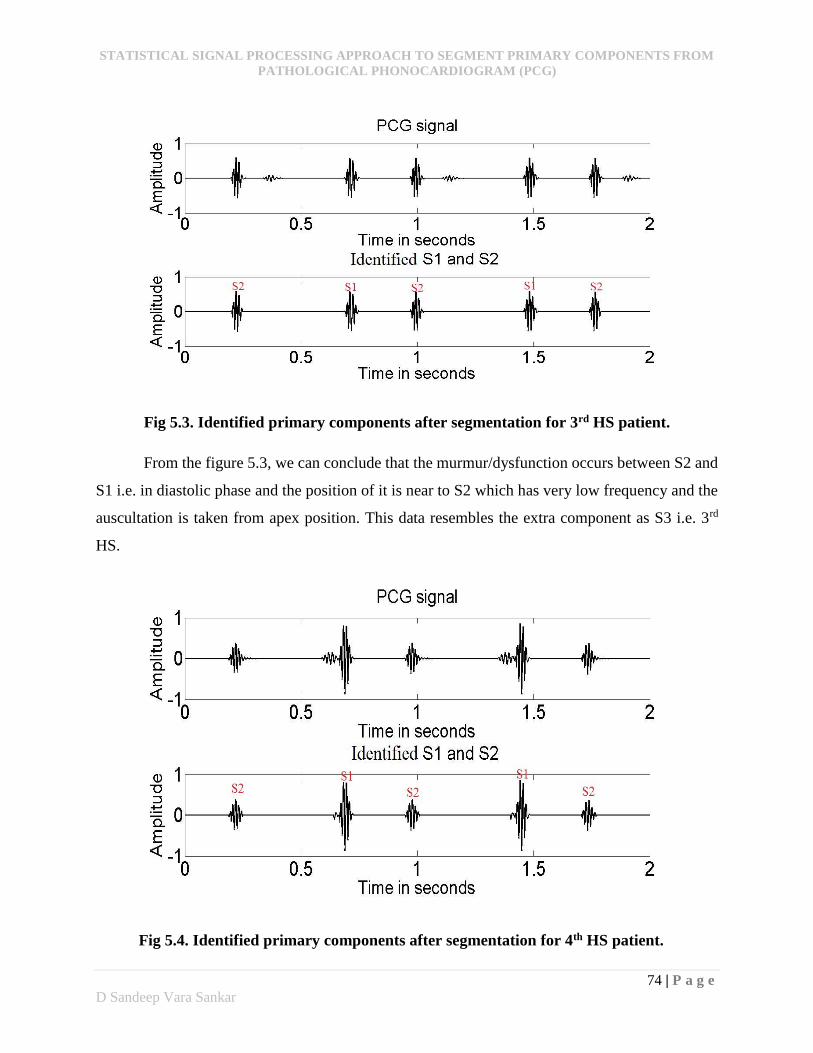
STATISTICAL SIGNAL PROCESSING APPROACH TO SEGMENT PRIMARY COMPONENTS FROM
PATHOLOGICAL PHONOCARDIOGRAM (PCG)
74 | P a g e
D Sandeep Vara Sankar
Fig 5.3. Identified primary components after segmentation for 3rd HS patient.
From the figure 5.3, we can conclude that the murmur/dysfunction occurs between S2 and
S1 i.e. in diastolic phase and the position of it is near to S2 which has very low frequency and the
auscultation is taken from apex position. This data resembles the extra component as S3 i.e. 3rd
HS.
Fig 5.4. Identified primary components after segmentation for 4th HS patient.
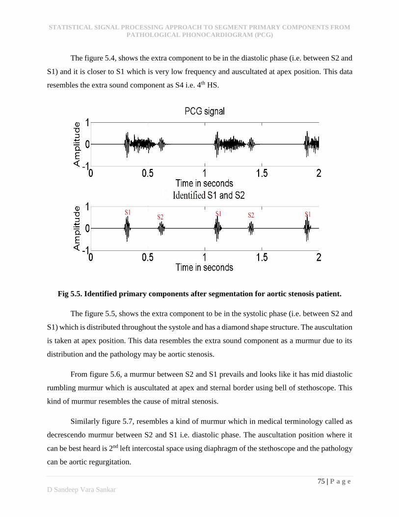
STATISTICAL SIGNAL PROCESSING APPROACH TO SEGMENT PRIMARY COMPONENTS FROM
PATHOLOGICAL PHONOCARDIOGRAM (PCG)
75 | P a g e
D Sandeep Vara Sankar
The figure 5.4, shows the extra component to be in the diastolic phase (i.e. between S2 and
S1) and it is closer to S1 which is very low frequency and auscultated at apex position. This data
resembles the extra sound component as S4 i.e. 4th HS.
Fig 5.5. Identified primary components after segmentation for aortic stenosis patient.
The figure 5.5, shows the extra component to be in the systolic phase (i.e. between S2 and
S1) which is distributed throughout the systole and has a diamond shape structure. The auscultation
is taken at apex position. This data resembles the extra sound component as a murmur due to its
distribution and the pathology may be aortic stenosis.
From figure 5.6, a murmur between S2 and S1 prevails and looks like it has mid diastolic
rumbling murmur which is auscultated at apex and sternal border using bell of stethoscope. This
kind of murmur resembles the cause of mitral stenosis.
Similarly figure 5.7, resembles a kind of murmur which in medical terminology called as
decrescendo murmur between S2 and S1 i.e. diastolic phase. The auscultation position where it
can be best heard is 2nd left intercostal space using diaphragm of the stethoscope and the pathology
can be aortic regurgitation.
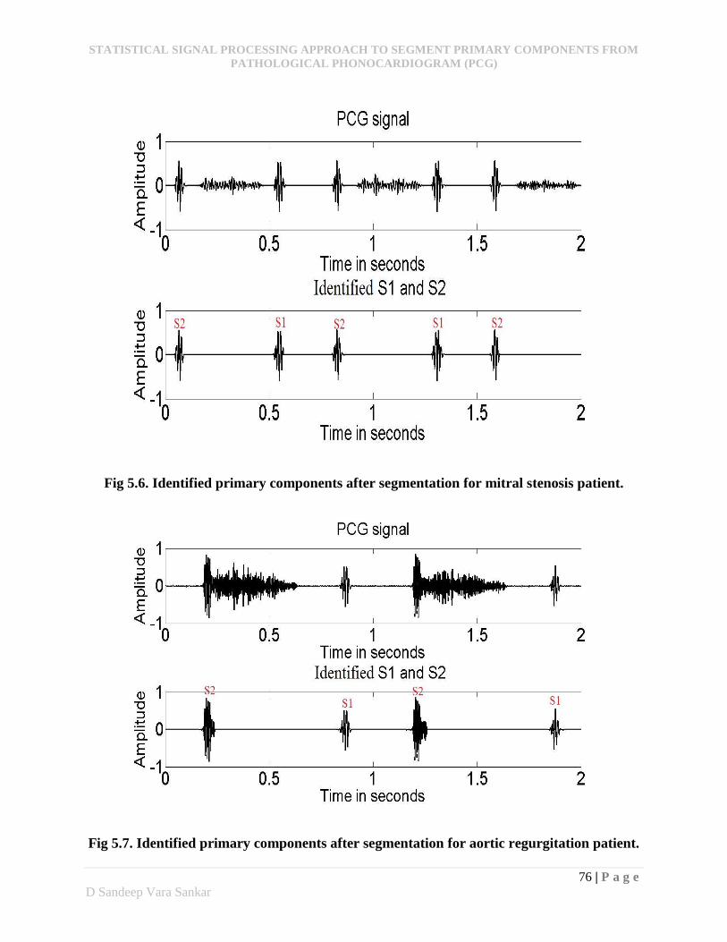
STATISTICAL SIGNAL PROCESSING APPROACH TO SEGMENT PRIMARY COMPONENTS FROM
PATHOLOGICAL PHONOCARDIOGRAM (PCG)
76 | P a g e
D Sandeep Vara Sankar
Fig 5.6. Identified primary components after segmentation for mitral stenosis patient.
Fig 5.7. Identified primary components after segmentation for aortic regurgitation patient.
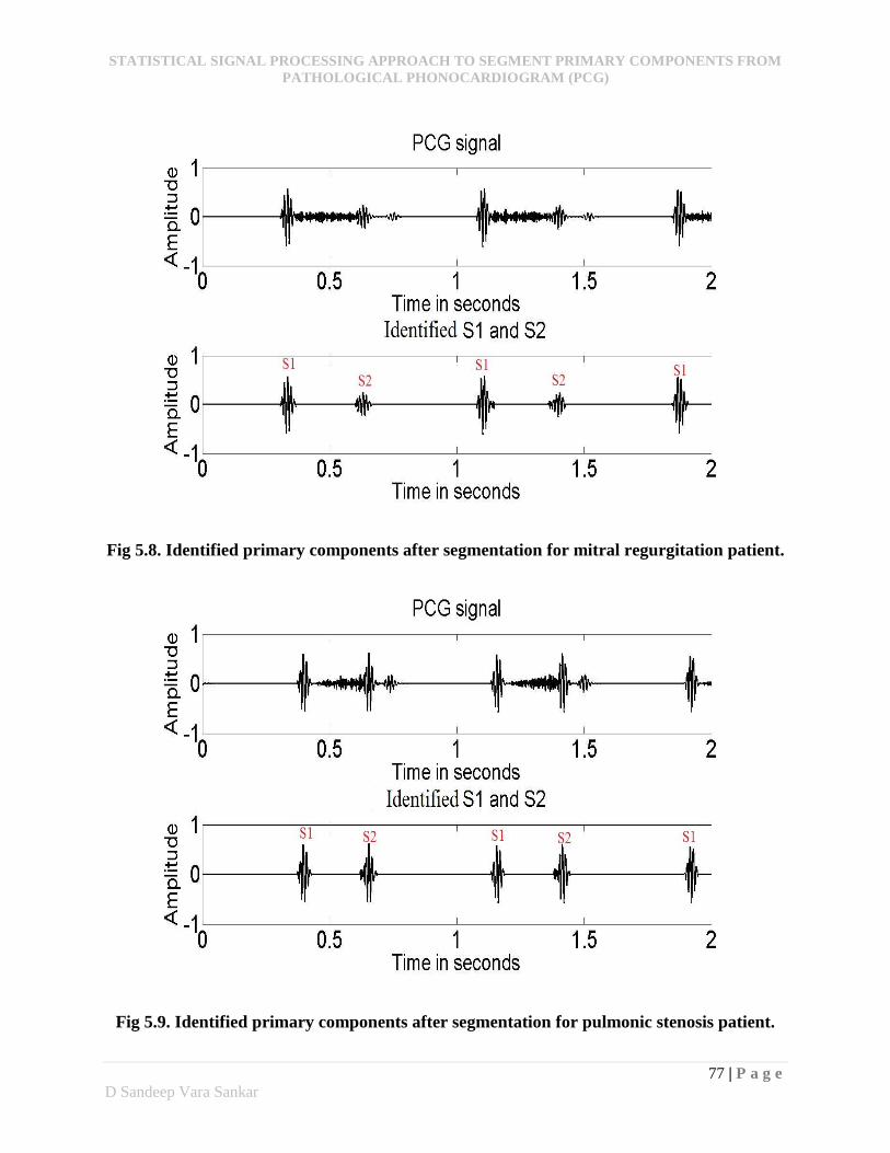
STATISTICAL SIGNAL PROCESSING APPROACH TO SEGMENT PRIMARY COMPONENTS FROM
PATHOLOGICAL PHONOCARDIOGRAM (PCG)
77 | P a g e
D Sandeep Vara Sankar
Fig 5.8. Identified primary components after segmentation for mitral regurgitation patient.
Fig 5.9. Identified primary components after segmentation for pulmonic stenosis patient.
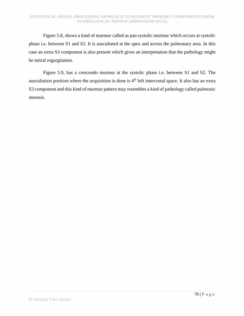
STATISTICAL SIGNAL PROCESSING APPROACH TO SEGMENT PRIMARY COMPONENTS FROM
PATHOLOGICAL PHONOCARDIOGRAM (PCG)
78 | P a g e
D Sandeep Vara Sankar
Figure 5.8, shows a kind of murmur called as pan systolic murmur which occurs at systolic
phase i.e. between S1 and S2. It is auscultated at the apex and across the pulmonary area. In this
case an extra S3 component is also present which gives an interpretation that the pathology might
be mitral regurgitation.
Figure 5.9, has a crescendo murmur at the systolic phase i.e. between S1 and S2. The
auscultation position where the acquisition is done is 4th left intercostal space. It also has an extra
S3 component and this kind of murmur pattern may resembles a kind of pathology called pulmonic
stenosis.
STATISTICAL SIGNAL PROCESSING APPROACH TO SEGMENT PRIMARY COMPONENTS FROM
PATHOLOGICAL PHONOCARDIOGRAM (PCG)
79 | P a g e
D Sandeep Vara Sankar
Conclusion
A segmentation algorithm which uses statistical analysis as a major tool for segmenting
primary components from abnormal PCG is presented in this study. The study uses wavelets and
principal component analysis for extracting the relevant features for processing segmentation
algorithm. The peeling algorithm used to separate PCG signal into series of cardiac cycles signifies
the importance and feasibility of statistical approach in phonocardiographic analysis. The
algorithm gives 98.32% of accuracy in identifying the primary components from variety of
pathologies. However, the error probability of 1/9, obtained during peeling algorithm doesn’t
concede much accuracy distraction. The presented study is simple and can be capable of picking
up low energy components without any additional effort.
The identification of exact end positions of the cardiac cycle becomes difficult in some
pathological conditions because of the propagation of murmur near to the end position. So further
processing is required for exact boundary detection by using valid estimators.
STATISTICAL SIGNAL PROCESSING APPROACH TO SEGMENT PRIMARY COMPONENTS FROM
PATHOLOGICAL PHONOCARDIOGRAM (PCG)
80 | P a g e
D Sandeep Vara Sankar
Dissemination
[1] D Sandeep Vara Sankar, Lakshi Prosad Roy, “Principal Component Analysis (PCA)
Approach to Segment Primary Components from Pathological Phonocardiogram”,
Proceedings of 2014 IEEE International Conference on Communication and Signal
Processing, APEC, Tamil Nadu, pp. 1023-1027, April 2014.
STATISTICAL SIGNAL PROCESSING APPROACH TO SEGMENT PRIMARY COMPONENTS FROM
PATHOLOGICAL PHONOCARDIOGRAM (PCG)
81 | P a g e
D Sandeep Vara Sankar
References
[1]. Samjin Choi, Zhongwei Jiang, “Comparison of Envelope Extraction Algorithms for
Cardiac Sound Signal Segmentation”, Expert Systems with Applications, 2008, Edition
34, Issue 2, pp. 1056-1069.
[2]. Reed T R, Reed N E and Fritzson P, “Heart sound analysis for symptom detection and
computer-aided diagnosis,” Simul. Model. Practice Theory, 12 (2004), pp.129–146.
[3]. Abdelghani Djebbari and Fethi Bereksi Reguig, “Short-Time Fourier Transform
Analysis of the Phonocardiogram Signal”, 7th IEEE International Conference, 2000,
volume 2, pp. 844-847.
[4]. Jung Jun Lee, Sang Min Lee, In Young Kim, Hong Ki Min, Seung Hong Hong,
“Comparison between Short Time Fourier Transform and Wavelet Transform for
Feature Extraction of Heart Sound”, Proceedings of IEEE Region 10 Conference,
TENCON, 1999, volume 2, pp. 1547-1550.
[5]. H. Liang, S. Lukkarinen, and I. Hartimo, “Heart sound segmentation algorithm based
on heart sound envelogram,” Computers in Cardiology, vol. 24, 1997, pp. 105-108.
[6]. D. Boutana, M. Benidir, and B. Barkat, “segmentation and identification of some
pathological phonocardiogram signals using time-frequency analysis”, IET Signal
Processing, 2005, volume 5, pp. 527-537.
[7]. L. G. Gamero, R. Watrous. “Detection of the first and second heart sound using
probabilistic models,” Proceedings of the 25' Annual International Conference of the
IEEE EMBS, Cancun, Mexico, 17-21 Sept 2003, Vol.3, pp. 2877-2880.
[8]. D Gill, N Gavrieli, N Intrator. “Detection and identification of heart sounds using
homomorphic envelogram and self-organizing probabilistic model,” Computers in
Cardiology 2005, pp. 957−960.
[9]. C Ahlstrom, P Hult, P Rask, J.E Karlsson, E Nylander, U Dahlstrom, P Ask, “Feature
Extraction for Systolic Heart Murmur Classification”, Annals of Biomedical
Engineering, November 2006, volume 34, pp. 1666-1677.
STATISTICAL SIGNAL PROCESSING APPROACH TO SEGMENT PRIMARY COMPONENTS FROM
PATHOLOGICAL PHONOCARDIOGRAM (PCG)
82 | P a g e
D Sandeep Vara Sankar
[10]. M.A.R Santos and M. Souza, “Detection of First and Second Cardiac Sounds Based on
Time-Frequency Analysis”, proceedings 23rd EMBS conference, Istanbul, 2001, pp.
1915-1918.
[11]. Paul A, Laizzo (Ed), “Handbook of Cardiac Anatomy, Physiology, and Devices”, 2009,
2nd Edition.
[12]. Stephane Mallat, “A wavelet Tour of Signal Processing, The Sparse Way”, An Imprint
of Elsevier, 2009, 3rd Edition.
[13]. Jonathon Shlens, “A Tutorial on Principal Component Analysis,” December 10, 2005,
Version 2.
[14]. Lin, Tang Chu, “Phonocardiogram Segmentation”, Thesis (M.S),University of Hawaii
at Manoa, 2005.
[15]. L. Goldberger, David R. Rigney and Bruce J. west, “Chaos and Fractals in Human
Physiology,” Scientific American, February 1990, pp.421-435.
[16]. Tilkian, A., and M. Conover, “Understanding Heart Sounds and Murmurs: With an
Introduction to Lung Sounds,” 4th ed. Philadelphia, W. B. Saunders Co., 2001.
[17]. Ergen, B., and Y. Tatar, “Time-frequency analysis of phonocardiogram,”
MEASUREMENT 2003, Fourth International Conference on Measurement, 2003, pp.
222-225.
[18]. Kumar, D., P. Carvalho, M. Antunes, J. Henriques, M. Maldonado, R. Schmidt, and J.
Habetha, “Wavelet transform and simplicity based heart murmur segmentation,” Proc.
Computers in Cardiology, Valencia, Spain, 2006, pp. 173–176.
[19]. R.M. Rangayyan and R.J. Lehner, 1988, “Phonocardiogram signal processing: A
review,” CRC Critical Reviews in Biomedical Engineering, Vol. 15, Issue 3, pp. 211-
236.
[20]. Hadjileontiadis L.J, Rekanos I.T, “Detection of Explosive Lungs and Bowel Sounds by
means of Fractal Dimension”. IEEE Signal Processing Letters, 2003, pp. 311–314.
[21]. www.easyauscultation.com.