staphylococcal and streptococcal superantigen exotoxinscmr.asm.org/content/26/3/422.full.pdf ·...
TRANSCRIPT

Staphylococcal and Streptococcal Superantigen Exotoxins
Adam R. Spaulding,a Wilmara Salgado-Pabón,a Petra L. Kohler,b* Alexander R. Horswill,a Donald Y. M. Leung,c Patrick M. Schlieverta,b
Department of Microbiology, University of Iowa Carver College of Medicine, Iowa City, Iowa, USAa; Department of Microbiology, University of Minnesota Medical School,Minneapolis, Minnesota, USAb; Department of Pediatrics, National Jewish Health, Denver, Colorado, USAc
SUMMARY . . . . . . . . . . . . . . . . . . . . . . . . . . . . . . . . . . . . . . . . . . . . . . . . . . . . . . . . . . . . . . . . . . . . . . . . . . . . . . . . . . . . . . . . . . . . . . . . . . . . . . . . . . . . . . . . . . . . . . . . . . . . . . . . . . . . . . . . . . . . . . . . . .422INTRODUCTION . . . . . . . . . . . . . . . . . . . . . . . . . . . . . . . . . . . . . . . . . . . . . . . . . . . . . . . . . . . . . . . . . . . . . . . . . . . . . . . . . . . . . . . . . . . . . . . . . . . . . . . . . . . . . . . . . . . . . . . . . . . . . . . . . . . . . . . . . . . .422THE SUPERANTIGEN FAMILY . . . . . . . . . . . . . . . . . . . . . . . . . . . . . . . . . . . . . . . . . . . . . . . . . . . . . . . . . . . . . . . . . . . . . . . . . . . . . . . . . . . . . . . . . . . . . . . . . . . . . . . . . . . . . . . . . . . . . . . . . . . . . . .423SUPERANTIGEN BENEFITS TO MICROBES . . . . . . . . . . . . . . . . . . . . . . . . . . . . . . . . . . . . . . . . . . . . . . . . . . . . . . . . . . . . . . . . . . . . . . . . . . . . . . . . . . . . . . . . . . . . . . . . . . . . . . . . . . . . . . . . . .424BIOCHEMISTRY AND STRUCTURES OF SUPERANTIGENS. . . . . . . . . . . . . . . . . . . . . . . . . . . . . . . . . . . . . . . . . . . . . . . . . . . . . . . . . . . . . . . . . . . . . . . . . . . . . . . . . . . . . . . . . . . . . . . . . .424ROLE OF SUPERANTIGENS IN HUMAN DISEASES . . . . . . . . . . . . . . . . . . . . . . . . . . . . . . . . . . . . . . . . . . . . . . . . . . . . . . . . . . . . . . . . . . . . . . . . . . . . . . . . . . . . . . . . . . . . . . . . . . . . . . . . .428
Introduction . . . . . . . . . . . . . . . . . . . . . . . . . . . . . . . . . . . . . . . . . . . . . . . . . . . . . . . . . . . . . . . . . . . . . . . . . . . . . . . . . . . . . . . . . . . . . . . . . . . . . . . . . . . . . . . . . . . . . . . . . . . . . . . . . . . . . . . . . . . . . .428Staphylococcal Menstrual TSS . . . . . . . . . . . . . . . . . . . . . . . . . . . . . . . . . . . . . . . . . . . . . . . . . . . . . . . . . . . . . . . . . . . . . . . . . . . . . . . . . . . . . . . . . . . . . . . . . . . . . . . . . . . . . . . . . . . . . . . . . . . .428Outside-In Signaling Mechanism Results in Staphylococcal mTSS . . . . . . . . . . . . . . . . . . . . . . . . . . . . . . . . . . . . . . . . . . . . . . . . . . . . . . . . . . . . . . . . . . . . . . . . . . . . . . . . . . . . . . .430TSST-1 Production of TSS . . . . . . . . . . . . . . . . . . . . . . . . . . . . . . . . . . . . . . . . . . . . . . . . . . . . . . . . . . . . . . . . . . . . . . . . . . . . . . . . . . . . . . . . . . . . . . . . . . . . . . . . . . . . . . . . . . . . . . . . . . . . . . . . .430Nonmenstrual Staphylococcal TSS . . . . . . . . . . . . . . . . . . . . . . . . . . . . . . . . . . . . . . . . . . . . . . . . . . . . . . . . . . . . . . . . . . . . . . . . . . . . . . . . . . . . . . . . . . . . . . . . . . . . . . . . . . . . . . . . . . . . . . .431Streptococcal TSS . . . . . . . . . . . . . . . . . . . . . . . . . . . . . . . . . . . . . . . . . . . . . . . . . . . . . . . . . . . . . . . . . . . . . . . . . . . . . . . . . . . . . . . . . . . . . . . . . . . . . . . . . . . . . . . . . . . . . . . . . . . . . . . . . . . . . . . .431Animal Models of TSS . . . . . . . . . . . . . . . . . . . . . . . . . . . . . . . . . . . . . . . . . . . . . . . . . . . . . . . . . . . . . . . . . . . . . . . . . . . . . . . . . . . . . . . . . . . . . . . . . . . . . . . . . . . . . . . . . . . . . . . . . . . . . . . . . . . .432Staphylococcal Superantigen Food Poisoning . . . . . . . . . . . . . . . . . . . . . . . . . . . . . . . . . . . . . . . . . . . . . . . . . . . . . . . . . . . . . . . . . . . . . . . . . . . . . . . . . . . . . . . . . . . . . . . . . . . . . . . . . . .432Staphylococcal Pneumonia and Superantigens . . . . . . . . . . . . . . . . . . . . . . . . . . . . . . . . . . . . . . . . . . . . . . . . . . . . . . . . . . . . . . . . . . . . . . . . . . . . . . . . . . . . . . . . . . . . . . . . . . . . . . . . . .433Staphylococcal Infective Endocarditis and Superantigens . . . . . . . . . . . . . . . . . . . . . . . . . . . . . . . . . . . . . . . . . . . . . . . . . . . . . . . . . . . . . . . . . . . . . . . . . . . . . . . . . . . . . . . . . . . . . . .433Staphylococcal Sepsis and Superantigens . . . . . . . . . . . . . . . . . . . . . . . . . . . . . . . . . . . . . . . . . . . . . . . . . . . . . . . . . . . . . . . . . . . . . . . . . . . . . . . . . . . . . . . . . . . . . . . . . . . . . . . . . . . . . . .435Superantigens in Atopic Dermatitis . . . . . . . . . . . . . . . . . . . . . . . . . . . . . . . . . . . . . . . . . . . . . . . . . . . . . . . . . . . . . . . . . . . . . . . . . . . . . . . . . . . . . . . . . . . . . . . . . . . . . . . . . . . . . . . . . . . . . .435Pathobiology Underlying AD . . . . . . . . . . . . . . . . . . . . . . . . . . . . . . . . . . . . . . . . . . . . . . . . . . . . . . . . . . . . . . . . . . . . . . . . . . . . . . . . . . . . . . . . . . . . . . . . . . . . . . . . . . . . . . . . . . . . . . . . . . . . .435Mechanisms by Which Superantigens Drive AD Inflammation. . . . . . . . . . . . . . . . . . . . . . . . . . . . . . . . . . . . . . . . . . . . . . . . . . . . . . . . . . . . . . . . . . . . . . . . . . . . . . . . . . . . . . . . . . .435Management of AD . . . . . . . . . . . . . . . . . . . . . . . . . . . . . . . . . . . . . . . . . . . . . . . . . . . . . . . . . . . . . . . . . . . . . . . . . . . . . . . . . . . . . . . . . . . . . . . . . . . . . . . . . . . . . . . . . . . . . . . . . . . . . . . . . . . . . .435Streptococcal Superantigens and Delayed Sequelae . . . . . . . . . . . . . . . . . . . . . . . . . . . . . . . . . . . . . . . . . . . . . . . . . . . . . . . . . . . . . . . . . . . . . . . . . . . . . . . . . . . . . . . . . . . . . . . . . . . .435
MECHANISMS TO INTERFERE WITH SUPERANTIGEN ACTIONS AND PRODUCTION . . . . . . . . . . . . . . . . . . . . . . . . . . . . . . . . . . . . . . . . . . . . . . . . . . . . . . . . . . . . . . . . . . . . . .436Treatment of Infections with Antibiotics and Supportive Care To Allow Clearance of Bacteria and Kidney Elimination of Superantigens . . . . . . . . . . . . . . .436Preventing Superantigen Production by Mucosal Microbicides Added to Tampons and Surfaces . . . . . . . . . . . . . . . . . . . . . . . . . . . . . . . . . . . . . . . . . . . . . . . . . . . . . .437Passive Vaccination against Superantigens. . . . . . . . . . . . . . . . . . . . . . . . . . . . . . . . . . . . . . . . . . . . . . . . . . . . . . . . . . . . . . . . . . . . . . . . . . . . . . . . . . . . . . . . . . . . . . . . . . . . . . . . . . . . . . .437Active Vaccination against S. aureus and Superantigens . . . . . . . . . . . . . . . . . . . . . . . . . . . . . . . . . . . . . . . . . . . . . . . . . . . . . . . . . . . . . . . . . . . . . . . . . . . . . . . . . . . . . . . . . . . . . . . . .437
FOR THE FUTURE . . . . . . . . . . . . . . . . . . . . . . . . . . . . . . . . . . . . . . . . . . . . . . . . . . . . . . . . . . . . . . . . . . . . . . . . . . . . . . . . . . . . . . . . . . . . . . . . . . . . . . . . . . . . . . . . . . . . . . . . . . . . . . . . . . . . . . . . . . .437ACKNOWLEDGMENTS. . . . . . . . . . . . . . . . . . . . . . . . . . . . . . . . . . . . . . . . . . . . . . . . . . . . . . . . . . . . . . . . . . . . . . . . . . . . . . . . . . . . . . . . . . . . . . . . . . . . . . . . . . . . . . . . . . . . . . . . . . . . . . . . . . . . . .438REFERENCES . . . . . . . . . . . . . . . . . . . . . . . . . . . . . . . . . . . . . . . . . . . . . . . . . . . . . . . . . . . . . . . . . . . . . . . . . . . . . . . . . . . . . . . . . . . . . . . . . . . . . . . . . . . . . . . . . . . . . . . . . . . . . . . . . . . . . . . . . . . . . . . .438AUTHOR BIOS . . . . . . . . . . . . . . . . . . . . . . . . . . . . . . . . . . . . . . . . . . . . . . . . . . . . . . . . . . . . . . . . . . . . . . . . . . . . . . . . . . . . . . . . . . . . . . . . . . . . . . . . . . . . . . . . . . . . . . . . . . . . . . . . . . . . . . . . . . . . . .446
SUMMARY
This review begins with a discussion of the large family of Staph-ylococcus aureus and beta-hemolytic streptococcal pyrogenic toxinT lymphocyte superantigens from structural and immunobiologi-cal perspectives. With this as background, the review then dis-cusses the major known and possible human disease associationswith superantigens, including associations with toxic shock syn-dromes, atopic dermatitis, pneumonia, infective endocarditis,and autoimmune sequelae to streptococcal illnesses. Finally, thereview addresses current and possible novel strategies to preventsuperantigen production and passive and active immunizationstrategies.
INTRODUCTION
Staphylococcus aureus is a Gram-positive, catalase-positive,coagulase-positive, facultative aerobe that is a major cause of
many kinds of illnesses throughout the world. In 2007, based ondata collected in 2005, the Centers for Disease Control and Pre-vention (CDC) and their collaborators published a report statingthat S. aureus is the most significant cause of serious infectiousdiseases and infectious disease deaths in the United States (1). S.aureus can cause a wide variety of infections, ranging from rela-
tively benign furuncles and soft tissue abscesses to others that arelife-threatening, such as infective endocarditis, necrotizing (hem-orrhagic) pneumonia, sepsis, and toxic shock syndrome (TSS)(2–12). The ability of S. aureus to be such a capable pathogen,while at the same time appearing as part of the human normalflora, resides largely in the myriad of cell surface and secretedvirulence factors that the organism produces (7). Estimates sug-gest that 30 to 40% of the human population are asymptomati-cally colonized at any given time on one or more of their mucosalsurfaces; up to 70% of people may be transiently colonized (7, 10).Importantly, people who are colonized by S. aureus have a higherrisk of infection than noncolonized persons.
Streptococcus pyogenes (group A streptococcus) is also a Gram-positive coccus, but the organism is a catalase-negative, aerotoler-
Address correspondence to Patrick M. Schlievert, [email protected].
* Present address: Petra L. Kohler, 3M Corporate Research Materials Laboratory, St.Paul, Minnesota, USA.
Copyright © 2013, American Society for Microbiology. All Rights Reserved.
doi:10.1128/CMR.00104-12
422 cmr.asm.org Clinical Microbiology Reviews p. 422–447 July 2013 Volume 26 Number 3
on July 19, 2018 by guesthttp://cm
r.asm.org/
Dow
nloaded from
ant anaerobe. Like S. aureus, group A streptococci are highly as-sociated with serious infections and deaths in humans, primarilyalso due to their myriad cell surface and secreted virulence factors(4, 13–17). Group A streptococci are considered primary humanpathogens in that initial exposure to the organisms usually resultsin acute illness, typically manifested as pharyngitis or impetigo(18). There are an estimated 10 million cases of pharyngitis in theUnited States each year. The organisms can also cause life-threat-ening illnesses such as TSS with or without necrotizing fasciitisand myositis (19, 20), and the organisms are associated with de-velopment of autoimmune diseases such as rheumatic fever (21),acute glomerulonephritis (22), and guttate psoriasis (23). GroupA streptococci may be asymptomatically carried by up to 10 to20% of humans, usually after having overt infections (24).
Other beta-hemolytic streptococci, including group B, C, andG strains, also have the ability to cause serious human illnesses,including streptococcal TSS with or without necrotizing fasciitisand myositis (25–37). Additionally, group B streptococci are wellknown to cause neonatal sepsis and meningitis, and group C andG strains cause pharyngitis.
This review discusses a highly important family of secretedvirulence factors produced by both organisms and additionally bycertain strains of group B, C, and G streptococci. This family,referred to as pyrogenic toxin superantigens or more simply su-perantigens, overstimulates many immune processes that allowthese organisms to cause serious human illnesses (4, 8, 15, 38). Thereview will focus on S. aureus and group A streptococci since moredata are available for these organisms, but the key principles arealso relevant to the pathogenesis of other beta-hemolytic strepto-cocci that produce superantigens. It is important to note thatwhereas other beta-hemolytic streptococci secrete superantigens,other staphylococci (coagulase negative) of human origin so fardo not secrete detectable superantigens (39). Coagulase-negativestaphylococci from other animals may produce superantigens (40,41). It is not our intent to discuss cell surface virulence factors ofthese organisms, though we recognize that both organisms relyheavily on production of numerous cell surface, as well as se-creted, exoproteins in order to colonize the host and cause seriousillnesses (7, 8, 38, 42, 43). The cell surface virulence factors includethe large families of microbial surface components recognizingadhesive matrix molecules (MSCRAMMs) and immunoglobulinFc-binding proteins which are important for host colonizationand for the interference with local host immune responses (42,43). The secreted virulence factors, in addition to superantigens,include multiple cytolysins, proteases, nucleases, and lipases thatwe will mention only in relation to superantigen involvement indisease processes.
The major goal of this review is to present new information onsuperantigen disease associations and novel ways to interfere withthe production and activities of superantigens. Superantigens arecritical to development of TSS and likely other cardiovascular andvascular diseases associated with S. aureus and beta-hemolyticstreptococci. We and our clinical colleagues have described 25novel superantigen-associated illnesses, making it is likely thatagents that interfere with superantigen production and activitywill greatly impact human medicine (8, 44, 45).
THE SUPERANTIGEN FAMILY
Superantigens are an extraordinary family of nonglycosylatedlow-molecular-weight exoproteins. They are secreted by all hu-
man-pathogenic S. aureus and group A streptococci that we havetested (�8,000), with secretion dependent on a cleavable signalpeptide. Superantigens have molecular sizes ranging from 19,000to 30,000 Da (8). The proteins are unusually resistant to heat (forexample, most remain biologically active despite boiling for 1 h),they are generally resistant to proteolysis (for example, by trypsinand pepsin) and acids (such as stomach acid), and they are highlyresistant to desiccation (toxic shock syndrome toxin 1 [TSST-1]remains biologically active after being dried on petri dishes formore than one year) (8, 15, 38). Their biological toxicity and en-vironmental stability have resulted in some superantigens beingcategorized as select agents of bioterrorism.
S. aureus strains secrete from 1 to 23 of at least 24 serologicallydistinct superantigens, and group A streptococcal strains have theability to produce up to 11 superantigens (8, 15, 38). For example,we have one S. aureus strain in our collection that produces 23superantigens, lacking only the ability to produce TSST-1 (46).The only S. aureus strains that we are aware of that do not secretesuperantigens are NCTC 8325-4 and its variant strains (RN4220,RN6390, and RN450); this makes the restriction-less strainRN4220 a highly useful organism for cloning superantigen genes.The S. aureus superantigens include TSST-1, the staphylococcalenterotoxins (SEs) (serotypes A, Bn, Cn [where n refers to multiplevariants], D, E, and G), and the SE-like (SE-l) superantigens (se-rotypes H, I, and J to X) (4, 8, 15, 38, 47). It is important to notethat there is no SE serotype F (SEF) or SE-l protein serotype F (SE-lF) designation. The name SEF was retired from use as a result ofthe renaming of staphylococcal pyrogenic exotoxin C (PE C) andSEF as TSST-1 in 1984 (48).
The SE superantigens are defined by emetic activity when in-gested by humans or when given orally to monkeys (47). TSST-1was originally thought to have emetic activity when purified byBergdoll et al. (3), but that activity was later shown to result fromSEA contamination. TSST-1 lacks emetic activity and lacks thecystine loop structure thought to be important for emetic activityof SEs (49). It is also important to note that TSST-1 was so namedto recognize its principal association with TSS and to allow for thepossibility that TSST variants may arise (for example, TSST-2,etc.). To date, there are no human TSST-1 variants, though thereis a protein referred to as TSST-ovine that has 7 amino acid dif-ferences from TSST-1 (50). TSST-ovine is biologically inactivewhen tested against human lymphocytes, but the protein is activeagainst lymphocytes from sheep (50). The SE-like proteins eitherlack emetic activity or have not been tested (47). Several, includingSE-l H, SE-l K, SE-l L, and SE-l Q, have been tested and are non-emetic; the remaining SE-l proteins have not been tested (51–54).Almost all of the staphylococcal superantigens are encoded onvariable genetic DNA elements, with the exception of SE-l X,which is encoded on the core chromosome (8, 55). SEA is encodedby the sea gene located on a bacteriophage (56), and SED is en-coded by the sed gene found on a plasmid (57), but most staphy-lococcal superantigens are encoded by genes on S. aureus patho-genicity islands (SaPIs) (8).
Like other exoproteins and cell surface virulence factors, staph-ylococcal superantigens are under complex regulatory control, in-cluding by global regulators such as agr, sae, rot, and srr. It is notour intent to discuss these DNA regulatory elements in detail.Later in this review, we discuss selected regulatory elements as theypertain to novel agents that prevent superantigen production.
Neutralization of Superantigens
July 2013 Volume 26 Number 3 cmr.asm.org 423
on July 19, 2018 by guesthttp://cm
r.asm.org/
Dow
nloaded from

Readers are encouraged to read important reviews that discussglobal regulatory pathways in detail (58–60).
Group A streptococci also may produce large numbers of su-perantigens. These were originally known as scarlet fever toxins orerythrogenic toxins due to their abilities to cause the scarlet feverrash (discussed in detail later in this review) but have more re-cently been referred to as streptococcal pyrogenic exotoxins(SPEs) (8, 15, 61, 62). Group A streptococci can produce up to 11serologically distinct superantigens (8, 15, 63–67). The strepto-coccal superantigens include SPE (serotypes A, C, and G to M),streptococcal superantigen (SSA), and streptococcal mitogenicexotoxin Zn (SMEZn) (8, 15). All of the streptococcal superanti-gens are encoded by genes located within bacteriophages, exclud-ing SPE G and SMEZ, which are encoded on the core chromosome(8, 15, 18). There is another SPE, designated SPE B, whose genespeB is also encoded in the core chromosome (68). Althoughshown to have superantigen activity, this protein is clearly a cys-teine protease based on activities and structure determination (62,69, 70). Its superantigenic activity has also been controversial,with observed superantigen activity possibly being the result ofcontaminants (71). However, it is also possible that the moleculehas superantigen activity due to regions of the protein not in-volved in protease activity. There is precedent for this to occur, asanother protein, streptococcal M protein, has both recognizedantiphagocytic and superantigen activities (72, 73). It thus seemslikely that the protein is both a cysteine protease and an atypicalsuperantigen. SPE B will not be discussed further in this reviewexcept as related to poststreptococcal acute glomerulonephritis.
As noted above, pyrogenic toxin superantigens are not limitedto S. aureus and group A streptococci. In fact, a number of otherorganisms produce superantigens. Recent reports of coagulase-negative staphylococci of animal origin (40, 41) and other beta-hemolytic streptococci, namely, groups B, C, and G (4, 8, 15, 25,31, 33, 74–76), have been published. Superantigens of group Bstreptococci have not been purified. Some superantigens of groupC streptococci appear to be unique, whereas others, and thosefrom coagulase-negative staphylococci and group G streptococci,are identical or nearly identical to those from S. aureus and groupA streptococcal strains. Superantigens have also been reported tooccur in Mycoplasma arthritidis, Yersinia enterocolitica, Yersiniapseudotuberculosis, Plasmodium falciparum, Clostridium perfrin-gens, Candida albicans, and Toxoplasma gondii (77–82). Interest-ingly, a superantigen has even been found in the rhizomes of thestinging nettle, Urtica dioica (83).
The global regulation of SPEs has not been extensively studied.As noted above, the majority are encoded on bacteriophages.However, SPEs that have been studied, such as SPE A and SPE C,are produced primarily in the post-exponential phase/early sta-tionary phase of streptococcal growth. This corresponds to thetime of maximal production of major staphylococcal superanti-gens such as TSST-1, SEB, and SEC (84, 85) by S. aureus. In un-published studies, we have shown that the gene speA, encodingSPE A, is regulated by endogenous S. aureus global toxin regula-tors, such as agr and srrAB, when speA is cloned into S. aureus.Additionally, the SPE A protein, which shares highly significantprimary sequence similarity to SEB and SEC, is highly resistant toS. aureus proteases when the protein is produced in S. aureus. Incontrast, neither the SPE B nor the SPE C protein is stably pro-duced when their genes are cloned into S. aureus, with both pro-teins being quickly proteolyzed to small fragments. These data
suggest that group A streptococci may have acquired the SPE Agene from bacteriophage transduction from S. aureus or a com-mon ancestral organism. The SPE B and SPE C genes do not ap-pear to have originated from S. aureus or a recent ancestor.
SUPERANTIGEN BENEFITS TO MICROBES
S. aureus and group A streptococci are highly successful patho-gens. This almost certainly is because these two organisms aremultidimensional in disease causation, producing a myriad of cellsurface and secreted virulence factors, among these being supe-rantigens. It is interesting to ask what the potential microbial sur-vival benefit from superantigen production by S. aureus and groupA streptococci is. We hypothesize the following. Both S. aureusand group A streptococci colonize and cause infections in thepresence of immune systems; neither organism resides in otherenvironments in nature. Thus, it is most likely that superantigens,and the majority of other virulence factors, are selected becausethey interfere with normal immune function, increasing thechances of survival and transmission for both organisms.
It can be envisioned that there are three levels of protectionafforded to both organisms by their virulence factors. First, supe-rantigens interfere with immune system function systemically(globally), while both organisms initiate disease from initial infec-tion sites. It is well recognized that women who develop menstrualTSS (mTSS) do not produce protective antibodies against TSST-1or other staphylococcal products during or upon recovery fromtheir illnesses (86). The continued use of tampons by such womenplaces them at high risk of recurrent illnesses (86). It has also beenshown that production of multiple superantigens by strains ismore toxic than production of a single superantigen (87, 88), pre-sumably through overactivation of multiple T cell populations. Ithas been shown that the suppression of antibody production bysuperantigens results at least in part from gamma interferon(IFN-�) production by overactivated CD4 T cells (4, 89). Addi-tionally, it has been shown that the massive production of tumornecrosis factor (TNF), a proinflammatory cytokine, by immunecells exposed to superantigens counterintuitively suppresses nor-mal phagocytic cell infiltration into infection sites (90). This sup-pression of phagocytic cell infiltration has been noted in bothstaphylococcal and streptococcal TSS cases (90–93). The majormechanism of immunity to both pathogens is thought to be anti-body neutralization of toxins combined with antibody-comple-ment opsonization and microbial killing by phagocytic cells; theproduction of superantigens interferes with both activities by in-terfering with antibody production and phagocytic cell che-motaxis. Second, both organisms also produce cytotoxins that actlocally to kill immune cells that have come into infection sites inspite of the presence of superantigens. Third, the organisms pro-duce cell surface virulence factors that provide the last line ofdefense against the immune system. The combination of thesemechanisms effectively allows the organisms to persist in theirhosts and increase the likelihood of transmission.
BIOCHEMISTRY AND STRUCTURES OF SUPERANTIGENS
Superantigens were initially known by their first described biolog-ical activities (4, 94). Thus, SEs were known as enterotoxins due totheir causation of staphylococcal food poisoning (95, 96). SPEswere defined as erythrogenic toxin (SPE A) and scarlet fever toxinsdue to their abilities to induce the scarlet fever rash (61, 94, 97).Unfortunately, the scientific and medical communities adopted
Spaulding et al.
424 cmr.asm.org Clinical Microbiology Reviews
on July 19, 2018 by guesthttp://cm
r.asm.org/
Dow
nloaded from

these as the only activities of the SEs and SPEs. Thus, when SPEswere first identified as a family of pyrogenic exotoxins, it wasthought they were different from erythrogenic toxin and scarletfever toxins (94). In 1979, Schlievert et al. showed that the rash ofscarlet fever, as caused by erythrogenic toxin, depended on lym-phocyte activation (now known as superantigenicity) superim-posed on preexistent delayed hypersensitivity (61). Thus, it wasunlikely that humans or animal models of human illnesses wouldshow a scarlet fever rash upon initial SPE or other superantigenexposure. This is consistent with scarlet fever being more preva-lent in certain geographic areas of the United States, with regionswith more cases of group A streptococcal infections having morecases of scarlet fever (98). Additionally, it is consistent with the factthat many women who develop menstrual TSS have had at leastone episode of TSS in which the rash, a defining criterion of TSS,is absent, prior to the full-blown episode in which the rash ispresent. Lastly, this is also consistent with the observations thatmany individuals with streptococcal TSS or persons with non-menstrual staphylococcal TSS, with only single exposures to TSSorganisms, do not show rashes (99). In contrast, women who havemenstrual periods with continuous vaginal colonization by S. au-reus are likely to have monthly repeated exposures to TSST-1.
For many years, the superantigens TSST-1, SEs, SE-l proteins,and SPEs were known as pyrogenic toxins, since pyrogenicity wasa shared activity of all of the proteins (4). Additionally, all of theproteins shared the unusual activity of enhancement of suscepti-bility to lethal shock caused by lipopolysaccharide (LPS) by up to106-fold (100, 101). These became the defining properties of thefamily. It had been known since the 1970s that pyrogenic toxinswere highly potent stimulators of T lymphocyte proliferation, in-dependent of antigen specificity, and that this property was im-portant in human illness (9, 61, 87, 102, 103).
The term superantigen was coined by Marrack and Kappler toemphasize the novel way that pyrogenic toxins stimulate T cellproliferation (104). Superantigens cross bridge T cell receptors(TCRs) with major histocompatibility complex class II (MHC II)molecules on antigen-presenting cells (APCs) in a relatively non-specific manner, inducing highly significant proliferation of Tcells and activation of APCs such as macrophages (8, 105, 106).Typically, antigens stimulate approximately 1 in 10,000 T cellsthrough their specific interaction with MHC II and processed pep-tides displayed on the surface of APCs. In contrast, superantigensstimulate up to 50% of T cells, inducing in T cells and macro-phages what is often referred to as a “cytokine storm” that definesmany symptoms of TSS (discussed below). There are four prop-erties that define staphylococcal and streptococcal superantigensand distinguish them from conventional peptide antigens (107):(i) superantigens elicit strong primary responses which are not
seen with conventional peptide antigens; (ii) the variable part ofthe �-chain of the TCR (V�-TCR) is sufficient for recognition ofa superantigen, which is not the case with peptide antigens; (iii)MHC II proteins are required for superantigen stimulation of Tcells, but the interactions are not class II restricted; and (iv) supe-rantigens interact with MHC II as unprocessed molecules. It ispossible for strong conventional antigens to appear as weak supe-rantigens, so it is necessary to demonstrate that putative superan-tigens have these four activities.
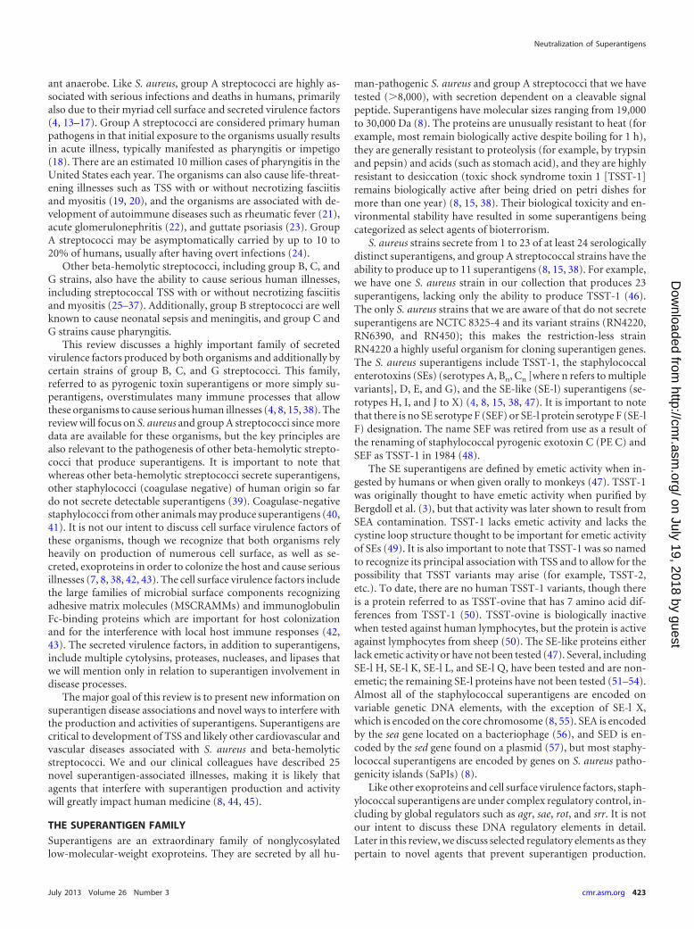
Numerous structural and mutational analyses have providedan impressive amount of information regarding the three-dimen-sional structures and host cell interactions of superantigens. Stud-ies that have examined the crystal structures of superantigensdetermined that superantigens contain a conserved overall struc-ture made of two major protein domains: an amino-terminal oli-gosaccharide/oligonucleotide binding (O/B) fold, comprised of a�-barrel, and a carboxy-terminal �-grasp domain made of antipa-rallel �-strands, with domains connected by a central, diagonal�-helix (108, 109). Based on small variations in this common corestructure, superantigens can be categorized into 5 major groups(Table 1; representatives are shown in Fig. 1 and 2).
Group I superantigens are characterized by TSST-1 (Fig. 1),but the group also includes TSST-ovine and the recently describedSE-l X (Table 1) (8, 55, 110, 111). Group I superantigens haveunique primary amino acid sequences compared to those of theother superantigens. These superantigens have only the corestructure, lacking the emetic cystine loop of SEs and the extra loopof 15 amino acids in group V SE-l superantigens (8). Group Isuperantigens contain only one MHC II binding site, the low-affinity MHC II binding site in their O/B folds that interacts withthe �-chains of MHC II molecules (Fig. 1) (112). In its binding toMHC II, TSST-1 also interacts with the antigenic peptide locatedin the peptide-binding groove of the molecule. The V�-TCRbinding site of TSST-1 is known; this binding site is located on thetop back of the molecule in the standard view, in a groove formedbetween the O/B fold and �-grasp domains (Fig. 1) (113, 114). Atthis time, we do not know the location of either the MHC II orV�-TCR site of SE-l X.
The group II superantigens are characterized primarily bySEBn, SECn, and SPE A1-4, in addition to others (Table 1) (8, 49).These superantigens contain the core superantigen structure plusa cystine loop that has a varying 10- to 19-amino-acid sequenceseparating the cysteine residues (49, 115). Importantly, thoughthe cystine loop is required for emetic activity, its presence doesnot guarantee emesis. Indeed, SPE A contains a cystine loop, likeother superantigens in this group; however, it has no emetic ac-tivity (49, 115). The lack of emesis of SPE A has been suggested tobe due to the presence of a third cysteine in the loop that results in
TABLE 1 Structural features of staphylococcal and streptococcal superantigens
Group Superantigens MHC II bindingCystine loop (lengthin amino acids) Other feature
I TSST-1, SE-l X, TSST-ovine Low-affinity site �-chain No Unique amino acidsequence
II SEB, SEC, SEG, SE-l U, SE-l W, SPE A, SSA Low-affinity site �-chain Yes (10–19)III SEA, SED, SEE, SE-I H, SE-l J, SE-l N to SE-l P Low-affinity site �-chain, high-affinity site �-chain Yes (9)IV SPE C, SPE G, SPE, J, SMEZ Low-affinity site �-chain, high-affinity site �-chain NoV SE-l I, SE-l K to SE-l M SE-l Q to SE-l T, SE-l
V, SPE HLow-affinity site �-chain, high-affinity site �-chain No 15-amino acid loop
insertion
Neutralization of Superantigens
July 2013 Volume 26 Number 3 cmr.asm.org 425
on July 19, 2018 by guesthttp://cm
r.asm.org/
Dow
nloaded from
an abnormal cystine loop being formed (115, 116). Additionally, ithas been shown in SEC that changing the two cysteines to alanineresults in loss of emetic activity, but changing the residues to ser-ine does not (49). Thus, it appears that the conformation of the SEstructure, as regulated by cystine loop amino acids, may be moreimportant in determining emesis than the actual presence of thecystine loop itself. Similar to group I superantigens, group II su-perantigens contain only one MHC II site, the low-affinity,�-chain MHC II binding site, and this interaction does not de-pend on interaction with the antigenic peptide within the MHC IIpeptide-binding groove (109, 117, 118). The V�-TCR binding siteof the group II superantigens is located on the top front of thesuperantigens (109, 119–121). Even though group I and group IIsuperantigens have only one MHC II site, the low-affinity site,their interaction with V�-TCRs is on opposite sides of the supe-rantigens. This makes group I tricomplex interactions appear asthree beads on a string, whereas group II tricomplex interactionsappear as the superantigen forming a wedge between MHC II andV�-TCR. This means that MHC II molecules on APCs cannotsimultaneously contact group I superantigens and TCRs, unlikethe standard simultaneous interaction of MHC II molecules withantigenic peptides and TCRs.
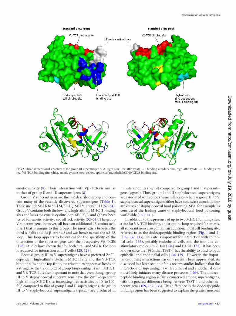
Group III superantigens include SEA (Fig. 2), SED, and SEE,among others (Table 1) (8). The group III superantigens contain acystine loop like the group II superantigens, and thus those testedhave been shown to have emetic activity; however the loop of thegroup III superantigens is always nine amino acids long (8). Im-
portantly, the group III superantigens contain the low-affinity�-chain MHC II binding site on their O/B folds, but they alsocontain a second, high-affinity site, referred to as a Zn2�-depen-dent MHC II binding site, in their �-grasp domains (Fig. 2) (8,122). The Zn2�-dependent high-affinity site interacts with the�-chains of MHC II molecules (8, 122). The presence of two MHCII sites allows the superantigens to cross bridge MHC II moleculeson adjacent APCs and increase superantigen activity (123). Thepresence of the Zn2�-dependent high-affinity site on these supe-rantigens makes them 10- to 100-fold more active overall in caus-ing cytokine production from T cells and APCs than those supe-rantigens that have only the low-affinity MHC II site. Group IIIsuperantigens bind V�-TCRs in the site comparable to that forgroup II superantigens (Fig. 2, top front in the groove between theO/B fold and �-grasp domains) (123, 124).
The group IV superantigens are produced by group A strep-tococci but not S. aureus and are characterized by SPE C, SPEG, SPE J, and SMEZ (Table 1) (8). These superantigens containboth low (�-chain)- and high (�-chain)-affinity MHC II bind-ing sites; detailed studies have shown that the SPE C high-affinity MHC II site is similar to the site found in SEA (124–126). It has been suggested that the superantigen SMEZ is themost potent superantigen (127); however, this group IV super-antigen, which contains both low-affinity and Zn2�-dependenthigh-affinity sites, shares expected activity comparable to thatof other superantigens that have both MHC II sites (8, 15).Group IV superantigens lack the cystine loop required for
FIG 1 Three-dimensional structure of the group I superantigen TSST-1. Light blue, low-affinity MHC II binding site; red, V�-TCR binding site; yellow,epithelial/endothelial/CD40/CD28 binding site.
Spaulding et al.
426 cmr.asm.org Clinical Microbiology Reviews
on July 19, 2018 by guesthttp://cm
r.asm.org/
Dow
nloaded from
emetic activity (8). Their interaction with V�-TCRs is similarto that of group II and III superantigens (8).
Group V superantigens are the last described group and con-tain many of the recently discovered superantigens (Table 1).These include SE-l K to SE-l M, SE-l Q, SE-l V, and SPE H (52–54).Group V contains both the low- and high-affinity MHC II bindingsites and lacks the emetic cystine loop. SE-l K, L, and Q have beentested for emetic activity, and all lack activity (52–54). The groupV superantigens, however, all have an additional 15-amino-acidinsert that is unique to this group. The insert exists between thethird �-helix and the �-strand 8 and was hence named the �3-�8loop. This loop appears to be critical for the specificity of theinteraction of the superantigens with their respective V�-TCRs(128). Studies have shown that for both SPE I and SE-l K, the loopis required for interaction with T cells (128, 129).
Because group III to V superantigens have a preferred Zn2�-dependent high-affinity �-chain MHC II site and the V�-TCRbinding sites on the top front, the tricomplex appears as beads ona string like the tricomplex of group I superantigens with MHC IIand V�-TCR. It is also important to note that even though groupIII to V staphylococcal superantigens have the Zn2�-dependenthigh-affinity MHC II site, increasing their activities by 10- to 100-fold compared to that of group I and II superantigens, the groupIII to V staphylococcal superantigens typically are produced in
minute amounts (pg/ml) compared to group I and II superanti-gens (�g/ml). Thus, group I and II staphylococcal superantigensare associated with serious human illnesses, whereas group III to Vstaphylococcal superantigens either have no disease association orare causes of staphylococcal food poisoning. SEA, for example, isconsidered the leading cause of staphylococcal food poisoningworldwide (130, 131).
In addition to the presence of up to two MHC II binding sites,a site for V�-TCR binding, and a cystine loop required for emesis,all superantigens also contain an additional host cell binding site,referred to as the dodecapeptide binding region (Fig. 1 and 2)(109, 132, 133). This site is important for interaction with epithe-lial cells (133), possibly endothelial cells, and the immune co-stimulatory molecules CD40 (134) and CD28 (135). It has beenknown since the 1980s that TSST-1 has the ability to bind to bothepithelial and endothelial cells (136–139). However, the impor-tance of these interactions has only recently been appreciated. Asdiscussed in a later section of this review, studies indicate that theinteraction of superantigens with epithelial and endothelial cellsmost likely initiates many disease processes (109). The dodeca-peptide binding region is fairly conserved among superantigens,with the greatest difference being between TSST-1 and other su-perantigens (109, 132, 133). This difference in the dodecapeptidebinding region has been suggested to explain the greater mucosal
FIG 2 Three-dimensional structure of the group III superantigen SEA. Light blue, low-affinity MHC II binding site; dark blue, high-affinity MHC II binding site;red, V�-TCR binding site; white, emetic cystine loop; yellow, epithelial/endothelial/CD40/CD28 binding site.
Neutralization of Superantigens
July 2013 Volume 26 Number 3 cmr.asm.org 427
on July 19, 2018 by guesthttp://cm
r.asm.org/
Dow
nloaded from

epithelium penetration of TSST-1 than of other superantigens(109, 133, 140) and thus TSST-1’s unique association with men-strual, vaginal TSS (115). The dodecapeptide binding region islocated at the base of the central, diagonal �-helix (Fig. 1 and 2)(109).
ROLE OF SUPERANTIGENS IN HUMAN DISEASES
Introduction
While superantigens are classically defined clinically by their abil-ities to cause staphylococcal and streptococcal TSS, they have beenassociated with many other illnesses caused by S. aureus and groupA streptococci (Table 2). All serious illnesses caused by S. aureusand group A streptococci have significant vascular-cardiovasculareffects on the host, including capillary leak syndromes, in the formof TSS, sepsis, infective endocarditis, pneumonia, and osteomy-elitis. Since superantigens are produced by all pathogenic S. aureusand group A streptococcal strains, these toxins should be consid-ered to be contributory to all such serious illnesses. We will discussselected superantigen-associated illnesses in this review. Readersare encouraged to use Table 2 as a source of references that discussother possible disease associations.
Staphylococcal Menstrual TSS
The most well-recognized illness caused by superantigens is TSS,which is a potentially life-threatening illness usually caused by S.aureus or group A streptococci. However, group B, C, and G beta-
hemolytic streptococci also cause TSS cases. Staphylococcal TSS,as originally described, was most often seen in women during theirmenstrual periods and is referred to as menstrual TSS (mTSS) (93,141). mTSS refers only to the association with menstruation anddoes not refer to the site of S. aureus isolation; approximately 95%of mTSS cases are menstrual with vaginal colonization, whereasthe remaining 5% are menstrual with nonvaginal colonization,occurring with almost any other type of infection. Most men-strual, vaginal staphylococcal TSS cases are associated with tam-pon use (93, 141), with higher incidence associated with highertampon absorbency (86, 93, 141). The incidence of mTSS peakedin the early 1980s, when the incidence was as high as 10/100,000women (86). The incidence of this illness is lower today, at ap-proximately 1 to 3/100,000 women (142–145), but importantly,the illness continues to occur; it is alarming that the illness isconsiderably less recognized today since there has not been mediaattention for many years. mTSS is defined as occurring within 2 to3 days of onset of menstruation, during menstruation, and within2 to 3 days after menstruation. S. aureus reaches its highest num-bers on days 2 to 3 of menstruation, which precedes by 1 day thepeak onset of TSS (days 3 to 4) (143). On day 2 to 3 of menstrua-tion, S. aureus counts vaginally as measured by numbers on tam-pons may exceed 1011 per tampon (143). TSST-1 amounts of ashigh as 100 �g may be present on soiled tampons (146). Counter-intuitively, most TSST-1 in tampons is present in regions of tam-pons that lack menstrual blood (146). Studies have shown that
TABLE 2 Human illnesses caused by or associated with superantigens
Human illnesses Associated superantigen(s) (reference[s])
Staphylococcal menstrual TSS TSST-1 (3, 9, 48)Staphylococcal nonmenstrual TSS Primarily TSST-1, SEB, SEC (145, 200, 327)
Soft tissue infection associated Primarily TSST-1, SEB, SEC (145, 200, 327)Postrespiratory viral Primarily TSST-1 but also SEB, SEC, and SE-l X (5, 55, 152, 154)Purpura fulminans Primarily TSST-1, SEB, SEC (154, 155)Extreme pyrexia Any (156)Recalcitrant erythematous desquamating syndrome in AIDS Any (153)Anaphylactic TSST-1Kawasaki-like TSST-1, SEB, SEC (328–332)Scleroderma-likeAcute-onset rheumatoid arthritisNeonatal exanthematous TSST-1 (333, 334)
Streptococcal illnessesScarlatina and scarlet fever SPEs (97)Erysipleas SPEsSevere invasive disease without hypotension SPEs (199)TSS with or without necrotizing fasciitis/myositis SPE A and C (19, 20, 64, 92, 211, 213, 218, 219, 224, 335), SPE B (218, 224, 335, 336),
others (67, 199)Purpura fulminans Any (337)Kawasaki-like Any (338)
Staphylococcal food poisoning SEs (95, 339)Guttate psoriasis SPEs (23, 284)Atopic dermatitis Any (46)Severe nasal polyposis Any staphylococcal (340)Obsessive compulsive disorder and other nervous system disorders SPEs (286)Acute rheumatic fever SPEs (67, 88, 221)Acute glomerulonephritis SPE B (88, 288–290, 341)Perineal erythema Any staphylococcal (342)Desquamative inflammatory vaginitis Any staphylococcal (326)Sudden infant death syndrome Any (343, 344)
Spaulding et al.
428 cmr.asm.org Clinical Microbiology Reviews
on July 19, 2018 by guesthttp://cm
r.asm.org/
Dow
nloaded from

blood prevents production of TSST-1 by S. aureus strains (147).Today, the highest incidence of mTSS is in adolescents of ages 12to 15, whereas in the early 1980s, the highest incidence was inwomen of ages 15 to 21 (144). The reason for this change toyounger mTSS patients is unknown.
Staphylococcal TSS is defined by the following criteria (93, 141,148, 149): high fever, hypotension, erythematous (scarlet fever-like) rash, peeling of the skin often during recovery, and any threemultiorgan components (often seen as flu-like symptoms, includ-ing vomiting and diarrhea), i.e., blood, central nervous system,gastrointestinal system, liver, mucous membranes, muscular sys-tem, and renal system. Serology tests for measles, leptospirosis,and Rocky Mountain spotted fever are negative, as are blood andcerebrospinal fluid tests for organisms other than S. aureus. It isnow recognized that often one of the defining criteria is not pres-ent; this illness is considered probable staphylococcal TSS (99).The defining criterion most often missing is the scarlet fever-likerash that occurs in association with preexistent delayed hypersen-sitivity to staphylococcal superantigens (61, 150). However, casesof probable TSS in which each defining criterion has been absenthave been described. If more than one criterion is absent and othercauses ruled out, the illness can be considered toxin-mediateddisease (151).
As soon as definitive mTSS was described, it was recognized thatthe majority of patients with the illness did not meet the full cri-teria for TSS that had been established to perform epidemiologicstudies by the CDC (141). Additionally, it is now recognized thatthe majority of staphylococcal TSS cases are not menstrual andvaginally associated. Multiple additional nonmenstrual categoriesof staphylococcal TSS are recognized, including the more com-monly seen postrespiratory viral pneumonia-associated TSS(Table 2) (152). This illness may progress exceptionally rapidly,being lethal in a matter of hours, and in children this illness isassociated with an extremely high case-fatality rate (up to 90%)(152). Other categories of staphylococcal TSS include (i) illnessassociated with any type of soft tissue infection; (ii) recalcitranterythematous desquamating syndrome, an unrelenting TSS inAIDS patients that results in death after as many as 70 days ofillness (153); (iii) purpura fulminans, a rapidly progressing TSSillness associated with clotting abnormalities and purpuric rash(154, 155); (iv) extreme pyrexia syndrome, an illness with a 100%case-fatality rate and fevers in excess of 108°F (156); and (v) ana-phylactic TSS, an acute illness that has a 100% case-fatality rateand high cardiac eosinophilia. The last illness may be a severe formof chronic TSS episodes in patients who develop atopic dermatitis(AD) rashes instead of the characteristic scarlet fever-like rash.Importantly, each year new categories of staphylococcal TSS areidentified, including, most recently, a rediscovered, severe entero-colitis TSS illness (157, 158). It is interesting that staphylococcalsuperantigens were once considered to be common causes of en-terocolitis. With the recognition and association of enterocolitiswith Clostridium difficile infection, the staphylococcal superanti-gen association quickly disappeared; however, it is now increas-ingly rerecognized that enterocolitis cases can be associated withstaphylococcal superantigens in the absence of C. difficile (157,158).
Staphylococcal superantigens are not evenly distributed amongTSS cases. Nearly all mTSS is caused by TSST-1-producing S. au-reus strains (3, 9, 145). Rare mTSS cases appear to be associatedwith production of SE-l G and SEI (159). The reason for the high
association of TSST-1 with mTSS is not completely clear, but itlikely depends on at least three factors: (i) TSST-1 is produced inhigh concentrations relative to the majority of other superanti-gens, (ii) TSST-1 has greater mucosal surface-penetrating abilitythan other superantigens (115, 140), and (iii) large numbers ofmucosal S. aureus strains produce TSST-1 (160). It is important torecognize that when first identified as causing mTSS, S. aureusstrains highly associated with illness belonged to the bacterio-phage type 29/52 complex (161, 162). These strains with the abilityto produce TSST-1 emerged as major clones in 1972 (161, 162),such that today essentially 100% of these organisms produceTSST-1. As many as 25% of persons colonized on mucosal sur-faces with S. aureus may be colonized with TSST-1-producingstrains (160). We recognize these strains today by other, newertyping mechanisms, including pulsed-field gel electrophoresis(PFGE), as primarily type USA200 and staphylococcal protein Atype and/or multilocus sequence type as primarily clonal complex30. In this review, we use the CDC USA200 designation to refer tothese strains. USA200 strains appear to be highly adapted to mu-cosal surface colonization, with infections resulting from TSST-1production and/or microbial spread from those surfaces. For ex-ample, �95% of USA200 strains have a mutation in the alpha-toxin gene that greatly reduces production of the cytolysin (163–165). Alpha-toxin is highly toxic to humans and is the mostinflammatory protein produced by S. aureus strains (163). Whilealpha-toxin is critical for production of furuncles and soft tissueabscesses originating from skin infections, production of high lev-els of the cytolysin would be expected to result in highly lethalinfections if produced on mucosal surfaces; it is thus likely that thereduction in alpha-toxin production by USA200 strains has al-lowed them to colonize mucosal surfaces effectively. This mucosalniche location of USA200 strains, with consequent diseases origi-nating from those mucosal surfaces, has been underappreciated,as evidenced by a recent publication suggesting that USA200strains are less virulent than historic skin-adapted strains referredto as bacteriophage type 80/81 (166).
USA200 strains secrete large amounts of TSST-1 that allowsthem to cause mTSS (and nonmenstrual TSS) (3, 9). Strains mayproduce 3 to 20 �g/ml in vitro in broth cultures but up to 16,000�g/ml in vitro in tampons as biofilms (167). The environmentalfactors that allow USA200 strains to produce TSST-1 have beenestablished; these are similar for most other superantigens. Theseconditions include growth in complex media containing animalprotein with low glucose (glucose functions as a catabolite repres-sor of exotoxin production), neutral pH (as expected vaginallyduring menstruation), temperature of 37°C to 40°C, and oxygenbalanced with CO2 (84, 168). As noted previously, blood compo-nents, and more specifically hemoglobin peptides, negatively af-fect production of TSST-1 (147). The introduction of oxygen intothe human vagina, a typically anaerobic environment, by tamponsis now considered the major reason for the tampon associationwith mTSS (84, 168, 169). The introduction of oxygen would alsoexplain the major association of mTSS with higher-absorbencytampons in that those tampons introduce more oxygen. Studies ofregulation of TSST-1 production by oxygen led to the identifica-tion of a global regulator of TSST-1 production called staphylo-coccal respiratory response (Srr) A/B (170). This important two-component system functions as a repressor of TSST-1 production(and that of other exotoxins) when oxygen levels are low. Therepressor function under anaerobic conditions appears to be
Neutralization of Superantigens
July 2013 Volume 26 Number 3 cmr.asm.org 429
on July 19, 2018 by guesthttp://cm
r.asm.org/
Dow
nloaded from
dominant over all other global regulators of exotoxin production(170). One other factor that increases TSST-1 production hasbeen identified, the surfactant pluronic L-92, which was present inone tampon in the early 1980s (171, 172). It is hypothesized thatpluronic L-92 alters staphylococcal two-component system sig-naling that upregulates TSST-1 production by as much as 8-fold.It has also been suggested that tampons composed of all cottonreduce TSST-1 production compared to that with tampons com-posed of cotton-rayon blends or all rayon (173). These studies arerefuted by multiple, carefully performed studies that fail to find areduction in TSST-1 production by all-cotton tampons (174–177).
Outside-In Signaling Mechanism Results in StaphylococcalmTSS
S. aureus typically colonizes the vaginal mucosal surface, resultingin TSST-1 production and penetration through the mucosa.TSST-1-producing S. aureus strains accomplish this feat by amechanism called “outside-in signaling,” where initial interac-tions with epithelial cells promote TSST-1 penetration and re-cruitment and stimulation of immune cells (109). As noted pre-viously in this review, TSST-1 exhibits enhanced mucosal surfacepenetration compared to other superantigens (115, 140). In a rab-bit model of vaginal TSS, TSST-1 was the most lethal, compared toSEC and the streptococcal superantigen SPE A (115). Porcine vag-inal ex vivo models, which nearly completely mimic the humanvaginal mucosa, have shown that TSST-1 alone is able to penetratethe vaginal mucosa (178, 179), but low levels of the cytolysin al-pha-toxin greatly enhance penetration.
Human and porcine vaginal mucosae are composed of nonke-ratinized, stratified squamous epithelium with a thickness of 10 to20 cell layers. The cell layers at the top are flattened and relativelysenescent, while the deeper layers are more cuboidal and are met-abolically active. It appears that TSST-1, alone and as enhanced bycytolysin production, penetrates these mucosae through stimula-tion of chemokine production by epithelial cells (109, 180, 181).The combined effects of TSST-1, cytolysin-induced inflamma-tion, and cytolysin toxicity likely contribute to destabilizationof the stratified squamous epithelial barrier, allowing TSST-1 ac-cess to the deeper layers of the mucosal epithelium where thesuperantigen can directly interact with epithelial cells close to thebasement membrane (109). Approximately 104 TSST-1 receptorsites per cell have been demonstrated on primary human epithelialcells and immortalized human vaginal epithelial cells (137, 180).Of these, CD40 and possibly an additional, unknown receptorbind TSST-1 (134, 182). TSST-1 induces the production of pro-inflammatory chemokines interleukin-8 (IL-8) (CCL8) andMIP-3� (CCL20) in human vaginal epithelial cells in vitro by amechanism dependent on signaling via ADAM17 and epithelialgrowth factor receptor (180, 182). These chemokines attract neu-trophils and other immune cells, including T cells and macro-phages, to infection sites. In line with this, immune cell recruit-ment to the subepithelial mucosa has been shown to occur in theex vivo porcine vaginal model (178, 180). Hence, the concertedaction of TSST-1 and low levels of cytolysins, such as alpha-toxin,results in mucosal epithelium inflammation and increased perme-ability, followed by TSST-1 penetration and induction of chemo-kines by metabolically active epithelial cells and finally recruit-ment of immune cells to the submucosa. This outside-in signalingmechanism provides TSST-1 accessibility to a sufficient pool of T
cells and macrophages to elicit the cytokine storm characteristic ofmTSS, IL-1� and TNF-� (produced by macrophages) andTNF-�, IL-2, and IFN-� (produced by T cells). It is noteworthythat TSST-1 has the ability to induce TSS from other mucosalsurfaces such as intestinal and airway surfaces; it is likely that thesame outside-in signaling mechanism contributes to TSST-1 pro-duction of TSS from those surfaces. Additionally, it is likely thatsimilar outside-in signaling mechanisms explain the productionof other microbial infections from mucosal surfaces, such as hu-man immunodeficiency virus (HIV) infections, as we recentlyproposed (183). In the simian immunodeficiency virus (SIV)model of HIV infection, SIV is proinflammatory to epithelial cells,leading to barrier disruption and recruitment of T cells that be-come infected.
TSST-1 Production of TSS
Once TSST-1 is produced and T cells and macrophages becomeactivated to secrete a cytokine storm, the cascade of events visiblyseen as TSS begins. Fever depends on TSST-1 and/or cytokinestimulation of the hypothalamic fever response control center(101, 184, 185). The most severe symptom associated with TSS ishypotension, which may progress to shock and death, resultingfrom capillary leakage. TSST-1 induces vascular injury in part bythe combined effect of toxin-induced systemic release of vasoac-tive mediators such as TNF-� and TNF-� (8, 105), synergy withother molecules such as LPS (186–188), and direct toxic interac-tion with the vascular endothelium (138, 139). The identity of theendothelial cell receptor for TSST-1 is currently unknown andunder investigation. However, it is clear that fluid replacement tooffset capillary leakage is required for management of TSS cases inhumans (93, 141) and rabbit models (189).
An interesting and potentially important property of superan-tigens is their ability to enhance the lethality of LPS by up to106-fold. This mechanism depends on synergistic TNF produc-tion in the presence of both superantigens and LPS (188) and onthe impaired LPS clearance function of the liver in the presence ofsuperantigens (190). Although the LPS enhancement mechanismis not universally accepted as contributing to TSS, it is intriguingfor many reasons. Humans and rabbits are approximately equallysusceptible to TSST-1 and TSS, and both have high numbers ofLPS-containing Gram-negative intestinal and vaginal flora (191).In contrast, mice are approximately 1011 times more resistant toTSST-1 lethality on a per-gram basis than rabbits and humans(191), and mice are less colonized by LPS-containing Gram-neg-ative bacteria. Rabbits become approximately 1,000 times moresusceptible to TSST-1 at 8 months of age than young rabbits, andthis corresponds to the same time as rabbits become 1,000 timesmore susceptible to the lethal effects of LPS. Typically in cases ofmTSS, S. aureus is cultured vaginally together with Escherichia coli(143, 146, 192), and these E. coli organisms may provide LPS thatpenetrates into the circulation. Once together in the circulation,the combination of TSST-1 and LPS may synergize to cause en-hanced TNF production and consequent enhanced capillary leak-age associated with TSS (188). E. coli vaginally may also providetryptophan or tryptophan precursors needed by the majority ofTSS S. aureus organisms, 75% of which are tryptophan auxo-trophs because their operons encoding proteins for tryptophanproduction are disrupted by insertion of the pathogenicity island(SaPI-2) carrying the TSST-1 gene (193–195).
The ability of S. aureus to cause mTSS depends also on the
Spaulding et al.
430 cmr.asm.org Clinical Microbiology Reviews
on July 19, 2018 by guesthttp://cm
r.asm.org/
Dow
nloaded from
immune status and genetics of colonized women. It has been longappreciated that women with low levels of or no antibodies toTSST-1 are serosusceptible to mTSS, whereas those individualswith antibodies, particularly the IgG4 subclass, appear to be pro-tected (3, 196–198). There is an age-dependent appearance of an-tibodies to TSST-1 in humans, with 80% of humans having anti-bodies to the superantigen by 12 years of age (196, 197). Theremaining 20% of serosusceptible individuals are among thosewho develop TSS. Importantly, these 20% appear not to be able toproduce protective antibodies to TSST-1 and thus remain suscep-tible to TSS recurrences (86). The failure to develop antibodies inserosusceptible women likely results from the TSS cytokine stormpreventing B cell function (87, 199). The same lack of antibodyproduction has been seen in approximately 50% of rabbits testedin a model of human TSS (134). In contrast, 100% of rabbitsrespond with production of neutralizing antibodies when chal-lenged with a nontoxic mutant of TSST-1, referred to as TSST-1(G31S/S32P). These studies indicate that the failure to developantibodies is not due to genetic nonresponsiveness but rather isdue to native TSST-1 effects on the immune system. A final im-portant point to mention relative to mTSS is that some womenwho have TSST-1 present in tampons and who lack antibodies toTSST-1 do not develop mTSS (146). This indicates that thesewomen may lack an epithelial cell receptor for TSST-1, whichleads to a failure in the outside-in signaling mechanism. If allknown factors are taken into account, such as the percentage ofwomen using tampons, the percentage of women who fail to makeantibodies to TSST-1, the percentage of women with TSST-1-pro-ducing S. aureus vaginally, and the percentage of women lacking aneeded epithelial cell receptor for TSST-1 penetration, the pre-dicted incidence of mTSS should be approximately 5/100,000,which is the approximate incidence seen.
Nonmenstrual Staphylococcal TSS
As noted above, nonmenstrual staphylococcal TSS occurs in asso-ciation with nearly any type of staphylococcal infection. As withmTSS, not all superantigens are equally associated with nonmen-strual TSS. Studies indicate that 50% of nonmenstrual TSS casesare caused by USA200 and related strains producing TSST-1 (145,200). The remaining 50% of strains nearly always produce thesuperantigen SEB or SEC (145, 200). The reason for the associa-tion of these three superantigens with most TSS cases, whethermTSS or nonmenstrual TSS, is their high level of production com-pared to that of other superantigens. SEB and SEC are produced ingreater concentrations than even TSST-1 by strains, i.e., 25 to 100�g/ml in vitro in broth cultures and up to 20,000 �g/ml in vitro intampon biofilm cultures. However, it is important to rememberthe greater mucosa-penetrating ability of TSST-1 than of SEB andSEC (115).
Whereas TSST-1 is restricted to USA200 and related strains of S.aureus, SEB and SEC may be produced by both USA200 andUSA400 strains. As many as 15 to 30% of mTSS, USA200 strains ofS. aureus coproduce TSST-1 and SEC; it is highly unusual, how-ever, to isolate strains that coproduce TSST-1 and SEB, thoughrare strains have been identified (46). Interestingly, the strainsproducing both TSST-1 and SEC do not appear to be more lethalin mTSS than strains that only produce TSST-1. This is almostcertainly because (i) critical medical intervention prevents lethal-ity and (ii) TSS strains may produce TSST-1 amounts alone or inthe presence of SEC in excess of 100,000 TSS-inducing doses per
tampon (146, 167). Thus, just production of TSST-1 alone ap-pears to be in vast excess of that needed to cause illness. It has beenshown that amounts of superantigens as low as 0.1 �g may induceTSS symptoms in humans (201).
USA400 strains are the major clones of S. aureus that produceeither SEB or SEC; rare strains may coproduce SEB and SEC. Inthe 1990s, community-associated methicillin-resistant strains ofS. aureus (CA-MRSA) were first identified in children (6). Many ofthese strains were USA400 CA-MRSA, as evidenced by the de-scription of children in the Upper Midwest who succumbed tosuch infections and through characterization of many other infec-tions associated with such strains (5, 154, 202). CA-MRSAUSA400 strains, as well as their methicillin-sensitive S. aureus(MSSA) counterparts, are primarily causes of skin and soft tissueinfections, but these organisms cause a highly fatal TSS-like illnesswhen present in the lungs and bloodstream. Additionally, thesestrains are common causes of all forms of nonmenstrual TSS (145,200), accounting for up to 50% of cases.
Additional comments need to be made regarding TSS strainsassociated with nonmenstrual illness. Nearly one-half of cases areassociated with TSST-1 production. In the United States, the ma-jority, but not all, of these TSST-1-producing organisms areMSSA. However, in other countries, TSST-1-positive MRSAstrains appear to be more common (203–205). Given their in-creased presence on mucosal surfaces today as opposed to 1980(160), it is possible that TSST-1-positive MRSA strains will con-tinue to increase in numbers in the United States. USA400 strainswere the initially identified causes of CA-MRSA necrotizing(hemorrhagic) pneumonia and sepsis. Pneumonia and sepsis donot preclude the patients from also simultaneously having TSS,the symptoms of which are usually present. USA400 strains weremore recently displaced in many, but not all, regions of the UnitedStates by USA300 CA-MRSA with ability to cause necrotizingpneumonia with TSS-like symptoms (206, 207). These strainsmost often fail to produce TSST-1, SEB, or SEC (207, 208). How-ever, the strains produce a newly recognized superantigen, SE-l X,which has been linked to necrotizing pneumonia in rabbit modelstudies (55). Additionally, these strains appear to produce a dele-tion derivative of TSST-1, as associated with extreme pyrexia syn-drome (156). The possible involvement of staphylococcal supe-rantigens in pneumonia will be discussed further in a later sectionof this review.
Streptococcal TSS
In 1987 (19) and in 1989 in the most definitive clinical study (20),streptococcal TSS was described. This illness is most often associ-ated with group A streptococcal infection associated with breaksin the skin, such as minor cuts (19, 20, 91) or chicken pox lesionsin children (209, 210), but may be associated with nearly any typeof group A streptococcal infection. It may be important that in astreptococcal TSS outbreak in southeastern Minnesota, as manyas 35% of children had pharyngitis caused by M3 streptococci,whereas patients with streptococcal TSS caused by the same or-ganism primarily had infections associated with breaks in the skin(91). It remains unclear why this difference in infectious processesoccurs, but it may be related to reduced streptococcal superanti-gen penetration of mucosal barriers (115).
Streptococcal TSS is defined by the following criteria: isolationof group A streptococci (either from a sterile site, indicating adefinitive case, or from a nonsterile site, indicating a probable
Neutralization of Superantigens
July 2013 Volume 26 Number 3 cmr.asm.org 431
on July 19, 2018 by guesthttp://cm
r.asm.org/
Dow
nloaded from
case), hypotension, and two or more of the conditions adultrespiratory distress syndrome, coagulopathy, erythematous mac-ular rash, liver complications, renal dysfunction, and soft tissuenecrosis. These criteria are similar to those for staphylococcal TSS(though simplified), except for three major differences: (i) strep-tococcal TSS with necrotizing fasciitis and myositis is often seenwith accompanying bloodstream sepsis in which the causative or-ganisms localize in deep tissue sites of preexistent damage, such asbruises (19, 20, 211–214), whereas staphylococcal TSS is mostoften associated with localized nonbloodstream infections/colo-nizations such as of the vaginal mucosa in mTSS (86, 93, 141, 152);(ii) streptococcal TSS with necrotizing fasciitis and myositis isassociated with severe pain of the primary site of localized infec-tion (bruises or apparent muscle tears), and this may be masked byuse of nonsteroidal anti-inflammatory agents (20, 211–214); and(iii) streptococcal TSS is typically associated with necrotizing fas-ciitis and myositis, even though cases also occur in the absence ofnecrotizing fasciitis and myositis, and as such streptococcal TSSmay have a case-fatality rate of 50 to 100% (20, 211–214). Untilrecently, necrotizing fasciitis and myositis were not seen or wereuncommon with staphylococcal TSS; it is noteworthy that recentstudies now suggest that S. aureus also has acquired the ability tocause TSS that includes necrotizing fasciitis and myositis (215,216). Soon after the recognition of group A streptococcal TSS,studies recognized that other beta-hemolytic streptococci couldcause the same illness, primarily group B, C, and G streptococci.
Recent studies of streptococcal TSS indicate that multiple sub-sets of illness may develop, with a continuum from mild scarlatinato life-threatening illness. In the early 1900s, scarlet fever was rec-ognized as a potentially life-threatening illness (217). Indeed,many hospitals had isolation wings to sequester patients with theillness. In the 1950s, severe scarlet fever was no longer a serioushealth threat in the United States, with the illness taking on formsof milder scarlet fever without hypotension and even milder scar-latina. In the mid-1980s, severe scarlet fever returned with theappearance of streptococcal TSS, described initially in patientsfrom the Rocky Mountain West and then becoming recognizedworldwide (19, 20). Today, we recognize that streptococcal TSSmay occur with or without necrotizing fasciitis and/or myositis,but it is also recognized that severe invasive streptococcal disease(for example, sepsis) may occur without hypotension but with orwithout necrotizing fasciitis and/or myositis (148). A recent studyhas shown that the spectrum of these acute streptococcal diseasesresults in part from the degree of cytokine storm provoked by thecausative organisms, with stronger responses leading to TSS, in-termediate responses leading to invasive diseases without TSS,and mild responses leading to pharyngitis and mild scarlatinalillnesses (199).
As with staphylococcal TSS, not all superantigens produced bygroup A streptococci are equally associated with streptococcalTSS. As originally described, streptococcal TSS was associated pri-marily with M1 and M3 streptococci, and these two M types con-tinue to dominate (19, 20, 91, 211, 213, 214, 218, 219). However,other M types also are clearly associated, including M type 18strains that are also highly associated with development of rheu-matic fever (220–223).
Just as certain M types are highly associated with streptococcalTSS, certain superantigens are more often associated than others.Initially, SPE A was the leading superantigen associated because ofits production by causative M1, M3, and M18 strains (19, 20, 91,
92, 210, 214, 218, 219, 224). However, other major SPEs, such asSPE C, and streptococcal mitogenic exotoxin Z are also associatedwith cases (67, 92, 199, 220, 221, 224–226). Because SPE B (cys-teine protease) is in the group A streptococcal core genome, thisexoprotein is also associated with streptococcal TSS, and particu-larly its importance in M1 strains has been thoroughly investi-gated (226, 227).
Although group A streptococcal TSS is often associated withbreaks in the skin, many cases have body site origins that are un-known (148). Some of these are almost certain to originate frommucosal surfaces where group A streptococci often cause diseases,for example, pharyngitis. Outside signaling mechanisms similar tothose seen in mTSS may take place to induce streptococcal TSS. Inthese cases, infection of the oral mucosa initiates a cascade ofevents that allows penetration not only of the streptococcal supe-rantigens but also of the bacteria, leading to sepsis (109, 181).Proinflammatory cytokine/chemokine induction of human vagi-nal epithelial cells and mucosal surface penetration studies (usedas models for the nonkeratinized, stratified squamous epitheliumof the oral mucosa) have also been done on SPE A and the group Astreptococcal cytolysin streptolysin O (SLO), with outcomes sim-ilar to those obtained with S. aureus TSST-1 and alpha-toxin (109,181). The major difference is that SLO directly damages the toplayers of the mucosal tissue without provoking as strong an in-flammatory response as alpha-toxin. The effect is 2-fold: (i) itallows penetration of SPEs and direct interaction with epithelialcells to elicit production of proinflammatory mediators, immunecell recruitment, and induction of TSS (analogous to the case forTSST-1), and (ii) it enhances bacterial penetration and systemicdissemination, which might explain the presence of the organismsin the bloodstream during streptococcal TSS.
Animal Models of TSS
Superantigens clearly cause TSS. This statement is supported bythe association of superantigens and causative bacteria with hu-man illnesses. Additionally, superantigens cause TSS symptoms inanimal models. For example, studies have shown that administra-tion of staphylococcal and streptococcal superantigens in subcu-taneously implanted miniosmotic pumps duplicates the symp-toms of TSS in rabbits (228, 229). It is important to remember thatmice are highly resistant to superantigens unless the liver is dam-aged first with D-galactosamine (230), which causes liver necrosis,a feature not seen in cases of human TSS. Additionally, superan-tigens cause lethal TSS in rabbits as applied intrapulmonarily(231). Studies with the use of isogenic strains that differ only inproduction of superantigens (232) and the use of active and pas-sive immunization against specific superantigens to protect rab-bits (44, 134, 231) conclusively establish that superantigens causeTSS. Recent studies have begun using HLA humanized mice, butthe usefulness of these animals remains unclear (233, 234). Thestudies do, however, demonstrate that HLA class II moleculesstrongly control superantigenic responses. Finally, superantigensdirectly injected into humans cause TSS symptoms (201).
Staphylococcal Superantigen Food Poisoning
We discussed the presence of the emetic loop in some superanti-gens (SEs) in a previous section. Not all superantigens are emetic,but SEs, including most commonly SEA, cause 24- to 48-h epi-sodes of retching, vomiting, and diarrhea every 15 to 30 min,without fever, after human or nonhuman primate ingestion of
Spaulding et al.
432 cmr.asm.org Clinical Microbiology Reviews
on July 19, 2018 by guesthttp://cm
r.asm.org/
Dow
nloaded from

nanogram quantities of SEs (95, 96). The lack of fever with staph-ylococcal food poisoning likely results from combinations of non-pyrogenic quantities required to cause emesis and failure of SEs toexhibit high mucosa penetration. Emetic activity has been shownin studies to be independent of superantigenicity (51, 115).Through their emetic activity, S. aureus SEs are primary causes oftoxin-mediated food-borne illness and the second leading cause offood-borne illness overall (235). Domestic cats and nonhumanprimates have been used extensively in studies of the SE causationof food poisoning; a house musk shrew model is a newly devel-oped animal model used to investigate SE emetic activity (236,237).
While much of the mechanism of the ability of SEs to inducefood poisoning remains unknown, recent studies to examine theeffects of SEA on human intestinal epithelial cells demonstratedthat SEA induces increases in the intracellular calcium concentra-tion of these cells (238). Using the house musk shrew model, Hu etal. demonstrated that SEA induces the release of serotonin in theintestine to cause emesis (239). Other work suggests that the vagusnerve is involved, where SEs stimulate the vagus nerve, therebyactivating the sympathetic nervous system (240). For further in-formation on mechanisms and outbreaks of staphylococcal foodpoisoning, readers are referred to other reviews (4, 241).
Staphylococcal food poisoning is a self-limiting illness that israrely if ever fatal. However, this incapacitating activity of SEs mayhave been primarily responsible for SEs, such as SEB, being listedas select agents of bioterrorism. It is noteworthy that during the1950s and 1960s, the United States stockpiled tons of SEB yearly asits major bioweapon. Additionally, because it is difficult to dena-ture superantigens and unnecessary to weaponize them for themto be taken orally or intrapulmonarily, SEs pose a significant haz-ard by these two routes. Likewise, there is no evidence to indicatethat humans can be vaccinated against this activity.
Staphylococcal Pneumonia and Superantigens
There are an estimated 70,000 cases of S. aureus pneumonia in theUnited States each year. Because all pathogenic S. aureus strainsproduce high levels of superantigens, these toxins contribute tosevere pneumonia, as demonstrated in animal models of humandisease (44, 134, 231). Even though pneumonia is an illness thatdescribes the primary infection site, the illness does not excludepneumonia-associated staphylococcal TSS, since TSS is defined asa collection of symptoms rather than a body site of infection.
In 1987, MacDonald et al. (152) described postinfluenza TSSassociated with the superantigens TSST-1 and SEB. The illnessdoes not require influenza virus infection, in that cases of pneu-monia-associated TSS occur in association with many other upperrespiratory viral infections and even asthma. This illness occurseach year, usually during the winter months, throughout theworld. Some investigators propose that postinfluenza TSS is thesame as Thucydides syndrome, recognized as the plague of Athensin 430 BC (242). In the study by MacDonald et al., there was a 90%case-fatality rate in children, all associated with TSST-1-produc-ing USA200 strains. One strain from the sole surviving child was aUSA400 strain producing SEB. One of us (P.M.S.) has tested largenumbers of other strains from children with postinfluenza TSS,and the majority of the S. aureus strains belonged to the USA200clonal group and produced TSST-1.
In 1999, the CDC and colleagues published a report on fourfatal cases of S. aureus necrotizing (hemorrhagic) pneumonia in
children associated with the recently emergent USA400 clonalgroup of CA-MRSA (5). Two of these isolates produced SEB andtwo produced SEC, the expected superantigens produced byUSA400 strains. Subsequent studies have shown that these twosuperantigens are nearly always present in USA400 CA-MRSAstrains (202).
More recently, investigators have studied the ability of theUSA300 clonal group of CA-MRSA to cause necrotizing pneumo-nia (206, 243–246). Studies performed in mice have suggested aleading role for alpha-toxin (166, 243–246). This toxin is highlyinflammatory, leading to significant lung congestion. However,these prior studies have not evaluated the role of superantigens,since mice are not susceptible to the lethal effects of superantigens(191).
A rabbit model of necrotizing pneumonia conclusively showedthat superantigens are critical for the development of lethal pul-monary illness associated with staphylococcal infection (231). Pu-rified SEB and SEC, when installed intrapulmonarily, inducehemorrhagic lung tissue, respiratory distress, and lethal TSS (231).Intrabroncheal administration of CA-MRSA strains of theUSA200 (TSST-1-positive) and USA400 (SEB- and SEC-positive)clonal types in rabbits induces lung pathology and lethality similarto those seen with individual administration of superantigens(231). The newly discovered SE-like protein serotype X superan-tigen (55), encoded by at least USA300, and as well USA100 andUSA400 clonal types, appears to be critical for the development ofnecrotizing pneumonia and lethal TSS in rabbits. Furthermore,vaccination against TSST-1, SEB, or SEC provides complete pro-tection against highly lethal doses of S. aureus strains producingthe respective superantigens (231). While cytolysins contribute tolung pathology in rabbits, evidence suggests that they are not thecause of the fatal outcomes associated with staphylococcal pneu-monia. Vaccination against alpha-toxin alone does not protectrabbits from lethal pulmonary illness, and CA-MRSA USA200strains that do not produce alpha-toxin still cause fatal disease(134).
We believe that the outside-in signaling mechanism also appliesfor TSS developed from lung infections. However, during pneu-monia, superantigens encounter only a single cell layer, typicallythe bronchial epithelium. Primary human bronchial epithelialcells stimulated with TSST-1 express high levels of the proinflam-matory molecules TNF-� and IL-8 (247). The simplicity of thesetissues, compared to the stratified structure of vaginal mucosalsurfaces, would allow for penetration of superantigens that usuallydo not exhibit great mucosal penetration, which is consistent withthe association of S. aureus SEB and SEC with one-half of non-menstrual TSS cases associated with the organism. Furthermore,direct superantigen cytotoxic effects on the pulmonary endothe-lial cells could contribute to pulmonary edema, necrosis, and re-spiratory distress (139).
Collectively, the above data indicate there are two pathways tofatal pneumonia, one in which alpha-toxin (and likely other cyto-lysins) causes high-level intrapulmonary inflammation and one inwhich superantigens cause pneumonia TSS (Fig. 3). It is likely thatboth mechanisms contribute.
Staphylococcal Infective Endocarditis and Superantigens
Infective endocarditis is a serious, life-threatening infection of theheart endothelium characterized by the formation on heart valvesof “cauliflower-like” vegetations composed of microorganisms
Neutralization of Superantigens
July 2013 Volume 26 Number 3 cmr.asm.org 433
on July 19, 2018 by guesthttp://cm
r.asm.org/
Dow
nloaded from

and host factors (248). S. aureus is a major cause of infective en-docarditis worldwide (249). Infective endocarditis occurs mostoften at sites of preexisting heart damage, usually involving valves.Damaged sites become coated with platelets and other host fac-
tors, such that in regions of turbulence, S. aureus can adhere andgrow. As many as 50% of infective endocarditis patients may suc-cumb, and up to one-half of the patients may develop strokes andmetastatic abscesses. Valve failure, strokes, and sepsis associatedwith metastatic abscesses may lead to death in patients.
Recent studies have shown that as many as 90% of infectiveendocarditis isolates of S. aureus belong to the USA200 clonalgroup, all of which produce TSST-1, among other virulence fac-tors (11, 250). Pragman et al. showed that TSST-1 is critical fordevelopment of infective endocarditis as tested in rabbits with theuse of isogenic strains, one strain positive for TSST-1 and the otherstrain negative for TSST-1 (251). Only one rabbit administeredthe TSST-1-negative strain developed a vegetation, which con-tained only 100 CFU. In contrast, the TSST-1-positive straincaused nearly 7 logs more CFU in cardiac vegetations. In a recentstudy with the use of passive neutralization of SEC in CA-MRSAUSA400 strain MW2, administration of V�-TCRs specific for SECprevented development of cardiac vegetations (252). Finally, ac-tive immunization studies targeting superantigens and cytolysinseffectively prevented USA200 S. aureus infective endocarditis(134).
There are multiple possible explanations for superantigen rolesin infective endocarditis (Fig. 4). Staphylococci initially colonizedamaged endothelium to initiate infective endocarditis (Fig. 4A).Staphylococcal superantigens (Fig. 4B) may then interact withhost cells to prevent endothelium wound healing through directeffects on endothelial cells (139). Studies have shown that TSST-1is toxic to porcine aortic endothelial cells (139). Superantigensmay also systemically dysregulate the immune system and preventclearance of the growing S. aureus valve infections. Finally, supe-rantigens cause mild or major capillary leakage dependent on su-perantigen concentration. Fluid and electrolyte replacement canbe used in humans and rabbit models (189) to prevent the lethaleffects of capillary leakage. It is possible that mild hypotension ormild capillary leakage not even manifested as hypotension can
FIG 3 Model for S. aureus production of pneumonia. The presence of S.aureus in the lungs leads to inflammation due to alpha-toxin and superanti-gens and to TSS due to superantigens, both leading to anoxia, hypotension,and possibly death.
FIG 4 Model of possible roles of S. aureus superantigens in infective endocarditis. S. aureus may colonize damaged endothelium of heart valves (A) and, throughcombinations of superantigenicity (immune dysfunction), superantigen-induced failure of endothelium healing (maintenance of endothelial damage), andinduction of capillary leakage, may facilitate vegetation formation (B).
Spaulding et al.
434 cmr.asm.org Clinical Microbiology Reviews
on July 19, 2018 by guesthttp://cm
r.asm.org/
Dow
nloaded from
alter blood flow in sites of preexisting heart damage to enhancevegetation formation.
Staphylococcal Sepsis and Superantigens
Both staphylococcal pneumonia and infective endocarditis pa-tients develop sepsis as a consequence of their infections, and sig-nificant numbers of patients develop sepsis following surgery. It islikely that many systemic effects in these patients result from su-perantigen activities on the host, since the infecting strains arelikely to produce the toxins. Investigators have shown that cer-tain superantigens, notably SEA, TSST-1, and SEC, are overrepre-sented in sepsis cases compared to nonsepsis cases (253–255).However, it is unlikely that superantigens are directly produced inthe bloodstream of patients, since hemoglobin peptides inhibitsuperantigen production (147). It is more likely that superanti-gens are produced in focal sites of infection protected from hemo-globin peptides and then secreted into the bloodstream.
Superantigens in Atopic Dermatitis
Atopic dermatitis (AD), is a chronic relapsing, highly pruritic,inflammatory skin disease that is often the prelude to develop-ment of food allergy, asthma, and allergic rhinitis (256, 257). S.aureus skin infections exacerbate skin disease in patients with ADand modify the host response to environmental allergens and viralpathogens (258, 259). Recent studies suggest that host-pathogeninteractions stemming from the production of S. aureus-derivedvirulence factors, such as superantigens and alpha-toxin, contrib-ute greatly to the skin inflammation seen in AD (260, 261).
Pathobiology Underlying AD
AD patients have various systemic and skin immune abnormali-ties, including elevated total serum IgE and sensitization to aller-gens, elevated T helper (Th2)-type cytokine expression in acutelesions, and increased numbers of T cells expressing the skin hom-ing receptor cutaneous lymphocyte-associated antigen (256, 257).These patients also have a defect in terminal differentiation oftheir keratinocytes leading to reduced expression of barrier pro-teins such as filaggrin, as well as decreased expression of epidermalantimicrobial peptides needed for innate immune responsesagainst invading bacteria and viruses (262, 263). The reduction inbarrier proteins may be due to a combination of mutations ingenes encoding skin barrier proteins such as filaggrin (264, 265),as well as downregulation of epithelial differentiation protein lev-els by Th2-type cytokines and IL-22 (266, 267). A critical linkbetween the barrier defect in AD patients with filaggrin gene(FLG) mutations and Th2 polarization could be explained in partby enhanced allergen penetration through damaged epidermis ac-companied by increased production of thymic stromal lympho-poietin (TSLP) by keratinocytes, leading to a Th2-type milieu(268, 269). Loss of filaggrin has been linked to enhanced antigenpenetration into the skin and increased S. aureus and viral growthin the skin, as well as susceptibility to the cytotoxic effects of staph-ylococcal toxins, e.g., alpha-toxin (270–272). AD patients withmore polarized Th2-type disease with allergies and asthma andincreased biomarkers, including serum IgE and TSLP, have beenreported to have more severe skin disease complicated by micro-bial infections (273). These observations suggest that FLG muta-tions and Th2 responses enhance S. aureus colonization and createa niche for S. aureus infection in AD skin. Once S. aureus hascolonized AD skin, it perpetuates its survival by increasing Th2-
mediated responses that suppress antimicrobial responses inatopic skin (274).
Mechanisms by Which Superantigens Drive ADInflammation
The mechanisms by which S. aureus induces skin inflammation inAD are under active investigation. A number of staphylococcalproducts, including superantigens, alpha-toxin, peptidoglycan,and lipoteichoic acid have been observed to activate cells involvedin the pathogenesis of AD (10). Due to their highly potent immu-nostimulatory activity, much of the focus has been on the role ofsuperantigens in the pathogenesis of AD (105). Support for a roleof superantigens in AD includes the following. (i) The severity ofAD correlates with the number of superantigen-secreting S. au-reus organisms colonizing the skin (275). Utilizing a murinemodel of skin inflammation, S. aureus superantigens plus aller-gens have been shown to have an additive effect in driving cuta-neous inflammation (276). (ii) Most AD patients make IgE anti-bodies directed against superantigens found on their skin (277),and the presence of these IgE antibodies to superantigens corre-lates with skin disease severity (278). Basophils and skin mast cellsfrom patients with antisuperantigen IgE release histamine uponexposure to the relevant superantigen but not in response to su-perantigens to which they have no specific IgE. (iii) The superan-tigen SEB, applied to the skin, can induce eczematoid skin changes(260). The ability of superantigens to induce eczema is likely re-lated to their capacities to bind to MHC II molecules on APCs orto stimulate T cells to produce proinflammatory cytokines (105).(iv) We have demonstrated that superantigens induce T cell ex-pression of the skin homing receptor via stimulation of IL-12 pro-duction (279). (v) After stimulation by the superantigen SEB, Tregulatory cells lose their immunosuppressive activity, suggestinga novel mechanism by which superantigens could augment T cellactivation in patients with AD (280). (vi) Superantigens inducecorticosteroid resistance, suggesting that several mechanisms existby which superantigens increase AD severity (281). Interestingly,superantigens also selectively induce T cells to secrete IL-31, ahighly pruritogenic cytokine that regulates filaggrin expressionand is produced by Th2 cells (282). Thus, superantigens may con-tribute to the pathogenesis of AD via multiple immune pathwaysleading to skin inflammation.
Management of AD
The treatment of AD requires a multipronged approach that in-cludes improvement in skin barrier function, reduction of the skininflammatory response, and control of S. aureus infection withjudicious use of antibiotics (283). Overuse of antibiotics in man-aging AD can result in MRSA infection with organisms that com-plicate control of skin disease. Given the key role that superanti-gens and alpha-toxin play in contributing to skin inflammation inAD, the development of vaccines directed against these staphylo-coccal toxins would be a welcome addition to the treatment arma-mentarium for these patients.
Streptococcal Superantigens and Delayed Sequelae
Group A streptococci are well recognized for the ability to causedelayed-sequela autoimmune diseases. We will provide a brief dis-cussion of the possible association of streptococcal superantigenswith three autoimmune diseases. In addition to causing TSS andrelated illnesses, streptococcal superantigens are associated with
Neutralization of Superantigens
July 2013 Volume 26 Number 3 cmr.asm.org 435
on July 19, 2018 by guesthttp://cm
r.asm.org/
Dow
nloaded from

the autoimmune disease guttate psoriasis (23, 284). This illnessfollows group A streptococcal illnesses, with susceptible patientsdeveloping characteristic lesions at 7 to 15 days postinfection. Pa-tients exhibit T cell receptor skewing that is typical of the super-antigen SPE C; T cell skewing is caused by superantigen overacti-vation of selected V�-containing T cells such that these cells areoverrepresented (skewed). The mechanism of superantigen in-volvement in guttate and possibly other forms of psoriasis meritsfurther study.
Throughout history, M18 group A streptococci have causedrheumatic fever. In the United States today, these M18 strains arethe most common causes of this autoimmune disease (222, 223).Features of rheumatic fever include evidence of multiple priorgroup A streptococcal infections, fever, heart abnormalities, ar-thritis, and Sydenham’s chorea (222). There is a clear associationof rheumatic fever with immunologic cross-reactivity to strepto-coccal antigens, with M protein most often cited (21). Studies haveshown that all rheumatogenic streptococci produce the superan-tigen SPE C, including all M18 strains (67, 88, 221). Additionally,it has been shown that SPE C has the ability to penetrate the blood-brain barrier, with possible central nervous system effects resem-bling Sydenham’s chorea (285). Although not definitively estab-lished, it has been suggested that SPE C and other M18-associatedsuperantigens may enhance immune cross-reactivity to M proteinsand in this way be associated with the autoimmune disease (67, 285)and even other illnesses such as pediatric autoimmune neuropsychi-atric disorders associated with streptococci (PANDAS) (286). Ithas also been demonstrated that superantigens have the ability toinduce arthritis in an animal model of rheumatoid arthritis (287).
In 1979, Schlievert et al. (88) noted the association of SPE B(cysteine protease) production by group A streptococcal strainsand the development of acute glomerulonephritis. Recent studieshave shown that SPE B localizes in kidney deposits associated withthe illness (288–290). Our unpublished studies with animals indi-cate that systemic treatment of rabbits with purified SPE B leads toproteinuria and hematuria, clinical features of acute glomerulo-nephritis. The role of other superantigens in this autoimmunedisease is unknown.
MECHANISMS TO INTERFERE WITH SUPERANTIGENACTIONS AND PRODUCTION
There are multiple ways to interfere with superantigen-associatedillnesses. These include (i) treatment of infections with antibioticsand supportive care to allow clearance of bacteria and kidneyelimination of superantigens, (ii) preventing superantigen pro-duction by mucosal microbicides added to tampons and surfaces,(iii) passive immunization against superantigens, and (iv) activevaccination against S. aureus and superantigens.
Treatment of Infections with Antibiotics and SupportiveCare To Allow Clearance of Bacteria and Kidney Eliminationof Superantigens
The standard of care for treatment of superantigen illnesses beginswith initial evaluation for potential sources of infection. For ex-ample, it is necessary to perform a vaginal and throat examination,remove tampons or other devices (such as contraceptive dia-phragms or nasal packing following surgery), and culture for S.aureus and beta-hemolytic streptococci, most often group A.Wounds should be evaluated for infection, noting that manytimes these infection sites may be difficult to find, as TSS microbes
often do not cause significant inflammation (90–92, 291). Forexample, we are aware of postsurgical TSS cases where the incisionsites have completely healed over, without signs of infection, yethaving abscesses of nearly 300 ml. These abscesses, although oftenthought to be anaerobic, are not; instead they have 60% of theoxygen content of air. Thus, they serve as excellent potential sitesfor superantigen production. Thorough examination of skin andsoft tissues should be performed, paying special attention to pain-ful areas such as bruises. Cultures of blood and other sites shouldbe obtained. Early surgical intervention is extremely important,particularly in cases of streptococcal TSS (20, 45, 91, 92, 211, 213,219, 292) and cases of staphylococcal necrotizing fasciitis (215,216). Magnetic resonance imaging (MRI) may be useful to iden-tify deep soft tissue damage and instruct on surgical procedures.Supportive care for hypotension is also of major importance. Pa-tients may require large amounts of intravenous fluids, vasopres-sors, and management of acute renal failure, acute respiratorydistress syndrome, disseminated intravascular coagulation, ormyocardial suppression. It is of critical importance to maintainkidney function, since this appears to be the major way for hu-mans to eliminate superantigens.
The standard of care for treatment of all superantigen diseasesalso includes the use of antibiotics to treat acute infections andreduce the incidence of recurrences. Nearly all S. aureus strains inthe United States are resistant to penicillin and ampicillin, butmany strains (MSSA) are susceptible to most other �-lactam an-tibiotics as well as other antibiotics. The antibiotic clindamycinhas been shown to reduce production of superantigens by S. au-reus and group A streptococci at antibiotic concentrations belowthose necessary to kill the pathogens (293–296). The percentagesof methicillin-resistant S. aureus (MRSA) strains are increasing,and we now recognize both hospital-associated MRSA (HA-MRSA) and CA-MRSA, though the boundary between thesegroups is blurring. HA-MRSA strains typically exhibit broad an-timicrobial resistance based on the presence of a large DNA ele-ment, referred to as staphylococcal cassette chromosome mec typeII (SCCmec II), which encodes methicillin and other resistances.In contrast, CA-MRSA strains, as originally described, containsmaller SCCmec DNAs, such as SCCmec IV. CA-MRSA strains,although resistant to �-lactam antibiotics, are often susceptible toother antibiotics. Small numbers of vancomycin-resistant S. au-reus strains have been identified (297).
It is important in mTSS cases, particularly, to remind patientsthat they will not develop neutralizing antibodies upon recoveryfrom illness (165). Thus, they remain susceptible to recurrences;we know of women who have had six recurrent, severe mTSSepisodes. These recurrences most often occur in women who con-tinue to use tampons, but they also occur in milder forms inwomen who discontinue tampon use (86). To reduce the inci-dence of mTSS recurrence, patients should be advised not to usetampons. It should be recognized that approximately 1% of men-struating women have group A streptococci vaginally, and thusthese individuals are at risk of streptococcal TSS from that infec-tion site. Such cases are not tampon associated, since group Astreptococci grow independent of oxygen. It is interesting to notethat some women develop mTSS prior to the appearance of men-strual blood yet within the two-day window of time to be classifiedas mTSS. Tests of tampons indicate that their vaginal pH has al-ready risen to near neutrality, suggesting that the role of hormonechanges may be to bring mucosal pH to the neutrality required for
Spaulding et al.
436 cmr.asm.org Clinical Microbiology Reviews
on July 19, 2018 by guesthttp://cm
r.asm.org/
Dow
nloaded from
superantigen production. Pregnant women appear to be at risk ofstreptococcal TSS as well, with a high case-fatality rate for bothmother and fetus (292). All persons should be advised to receiveyearly influenza vaccination to prevent the highly fatal postinflu-enza TSS.
Preventing Superantigen Production by MucosalMicrobicides Added to Tampons and Surfaces
With the occurrence of the mTSS epidemic in the 1980s, therehave been many efforts made to develop tampons and other de-vices used on mucosal surfaces that prevent production of supe-rantigens, particularly TSST-1. For example, Kotex tampons con-tain a surfactant, cetiol, that reduces TSST-1 production (298). In1992, Schlievert et al. published that the potential tampon additiveglycerol monolaurate (GML) reduces TSST-1 production at con-centrations that do not inhibit bacterial growth but kills S. aureusand most other vaginal pathogens at higher concentrations (299).These studies resulted in additional work that demonstrates thatGML inhibits TSST-1 production through interference with bac-terial signal transduction (300), reduces S. aureus and group Bstreptococci vaginally while selecting for the normal flora lactoba-cilli, and reduces vaginal inflammation as measured by reductionsin IL-8 production (301); IL-8 is a chemokine that specificallyattracts neutrophils to infection sites. In more recent studies, it hasbeen shown that GML stabilizes and is anti-inflammatory to mu-cosal epithelial cells as well as other immune cells (302, 303). Thestudies suggest that GML, in addition to preventing TSST-1 pro-duction, stabilize the host mucosa to prevent outside-in signaling(109). GML is safe as used vaginally in 5% (50-mg/ml) gels forperiods of up to 6 months, as tested in rhesus macaques (304).Studies suggest that GML can also be used to reduce simultane-ously bacterial vaginosis pathogens and Candida infections vagi-nally (305). GML has been used effectively to prevent SIV trans-mission with a high-dose challenge in a rhesus macaque model(183). Finally, it appears that GML both interferes with bacterialsignal transduction to prevent exotoxin production and dissipatesthe potential across plasma membranes to kill pathogens (167);the reason for the resistance of lactobacilli to GML appears to bethat they use GML as a cross-reactive quorum-sensing mechanismthat actually stimulates growth (167). Collectively, these and otherstudies have led to GML being incorporated into o.b. optiBalancetampons in Europe. Other molecules that could be applied tomucosal surfaces and devices to reduce the risk of staphylococcaland streptococcal illnesses have also been identified (306–309),including agents that prevent superantigen production and thosethat stabilize mucosal surfaces.
Passive Vaccination against Superantigens
It has been known for many years that intravenous immunoglob-ulin (IVIG) preparations usually have antibodies against all staph-ylococcal and streptococcal superantigens (310, 311). This hasbeen confirmed by numerous investigators (45, 312, 313). A de-finitive study performed by Kaul et al. (45) demonstrated a highlysignificant reduction in the case-fatality rate of streptococcal TSSwith use of IVIG. A similar study has not been performed fortreating staphylococcal TSS with IVIG, but there are many exam-ples reported to P.M.S. of individual patients responding to suchtherapy. It is now standard practice in some hospitals to adminis-ter vancomycin and IVIG to children with S. aureus pneumonia. Anote of caution in regard to the use of IVIG is merited. We are
aware of a case of streptococcal TSS where the patient was treatedwith IVIG and responded so quickly that the decision was made todiscontinue additional IVIG treatment. The patient immediatelyrelapsed, and IVIG treatment was reinstated. In calculations of therelative amounts of antibodies in IVIG against major superanti-gens, knowing that enzyme-linked immunosorbent assay (ELISA)antibody titers of �40 in humans appear to be required for pro-tection from superantigens and knowing the blood volume of hu-mans, it appears that IVIG has just sufficient antibodies to supe-rantigens to provide protection. Because of the high cost of IVIGand the occasional shortages that have occurred, there is a need todevelop additional antibody and T cell receptor-based therapies.
Studies have been initiated to develop monoclonal antibodiesagainst certain superantigens that can be humanized (314, 315).These studies are in their infancy, but it is expected that in thefuture such antibody cocktails may replace the need for the costlyIVIG. Multiple studies have also been performed that show thatengineered high-affinity (picomolar affinity) V�-TCRs, which areuniquely specific to binding and inactivating superantigens, pre-vent fever and lethality due to concurrent treatment or pretreat-ment with superantigens or staphylococci and streptococci thatproduce them, as tested in rabbit models (44, 231, 252, 316, 317).These reagents have also been shown in multiple rabbit studies tobe important in preventing lethality due to staphylococcal pneu-monia (231) and in preventing vegetation formation in infectiveendocarditis (252).
Active Vaccination against S. aureus and Superantigens
There have been multiple trials of human vaccines against S. au-reus, and all of these have failed. The primary antigens used forvaccination have been staphylococcal cell surface antigens. Ourrecent study suggests that organisms such as those causing infec-tive endocarditis aggregate to facilitate disease causation (318,319). The same studies suggest that antibodies against cell surfaceantigens of the organisms further aggregate the organisms andmay facilitate their abilities to cause disease (318, 319). Thus, it ispossible that continued emphasis on development of cell surface-based vaccines will result in continued failures. We have recentlytaken a different approach, noting that our prior studies (231)indicate that combinations of secreted superantigens and alpha-toxin are required for S. aureus to cause illnesses as tested in rab-bits. With that in mind, we have prepared vaccine toxoids andshown that immunization of rabbits protects against high-dose S.aureus intrapulmonary and intravenous challenge in an infectiveendocarditis model (134). The data suggest that S. aureus requiresits secreted virulence factors to initiate diseases and may requirethem for colonization.
Similar studies suggest that toxoid vaccines may be produced tovaccinate against group A streptococcal superantigens (320, 321).However, major studies to vaccinate against group A streptococci,like for S. aureus, are directed against surface antigens, particularlyM protein (322–324) and C5a peptidase (325). These studies arealso in their infancy.
FOR THE FUTURE
As noted above, we continue each year to identify new superanti-gen-associated illnesses. For example, we have just identified casesof desquamative inflammatory vaginitis that are associated withsuperantigen production by S. aureus (326). It appears likely thatthe full extent of superantigen-associated illnesses will be recog-
Neutralization of Superantigens
July 2013 Volume 26 Number 3 cmr.asm.org 437
on July 19, 2018 by guesthttp://cm
r.asm.org/
Dow
nloaded from

nized only when effective vaccines against S. aureus and group Astreptococci are developed and with their use we observe whatillnesses disappear. With the recognition that rabbits can be com-pletely protected from challenge with all major PFGE clonalgroups of S. aureus by superantigen and cytolysin toxoids, we mayhave a path now to develop a safe vaccine for human use. Toxoidsare expected to stimulate neutralizing antibodies that prevent se-rious diseases without concerns about microbial aggregation.These studies should continue.
Additionally, it is useful to develop novel strategies to neutral-ize superantigens or prevent their formation. It is clear that IVIGcan reduce the case-fatality rate of streptococcal TSS and likelystaphylococcal TSS. If less expensive reagents such as monoclonalantibodies and V�-TCRs that are effective and inexpensive can bedeveloped, they may be applicable to diseases, including nonseri-ous illnesses.
Finally, although many superantigen-associated illnesses arelinked with superantigenicity, there are many symptoms of TSSand other illnesses, where the mechanisms of toxicity remain un-clear. These should be explored in detail.
ACKNOWLEDGMENTS
We gratefully acknowledge Jeffrey Kavanaugh for his help with preparingFig. 1 and 2.
This research was supported by U.S. Public Health Service grants R01AI074283, R01 AR41256, and U54 AI57153. P.M.S. is a member of theGreat Lakes Regional Center of Excellence in Biodefense and EmergingInfectious Diseases.
REFERENCES1. Klevens RM, Morrison MA, Nadle J, Petit S, Gershman K, Ray S,
Harrison LH, Lynfield R, Dumyati G, Townes JM, Craig AS, Zell ER,Fosheim GE, McDougal LK, Carey RB, Fridkin SK. 2007. Invasivemethicillin-resistant Staphylococcus aureus infections in the UnitedStates. JAMA 298:1763–1771.
2. Que YA, Moreillon P. 2011. Infective endocarditis. Nat. Rev. Cardiol.8:322–336.
3. Bergdoll MS, Crass BA, Reiser RF, Robbins RN, Davis JP. 1981. A newstaphylococcal enterotoxin, enterotoxin F, associated with toxic-shock-syndrome Staphylococcus aureus isolates. Lancet i:1017–1021.
4. Bohach GA, Fast DJ, Nelson RD, Schlievert PM. 1990. Staphylococcaland streptococcal pyrogenic toxins involved in toxic shock syndromeand related illnesses. Crit. Rev. Microbiol. 17:251–272.
5. Centers for Disease Control. 1999. Four pediatric deaths from commu-nity-acquired methicillin-resistant Staphylococcus aureus—Minnesotaand North Dakota, 1997–1999. JAMA 282:1123–1125.
6. Herold BC, Immergluck LC, Maranan MC, Lauderdale DS, GaskinRE, Boyle-Vavra S, Leitch CD, Daum RS. 1998. Community-acquiredmethicillin-resistant Staphylococcus aureus in children with no identifiedpredisposing risk. JAMA 279:593–598.
7. Lowy FD. 1998. Staphylococcus aureus infections. N. Engl. J. Med. 339:520 –532.
8. McCormick JK, Yarwood JM, Schlievert PM. 2001. Toxic shock syn-drome and bacterial superantigens: an update. Annu. Rev. Microbiol.55:77–104.
9. Schlievert PM, Shands KN, Dan BB, Schmid GP, Nishimura RD. 1981.Identification and characterization of an exotoxin from Staphylococcusaureus associated with toxic-shock syndrome. J. Infect. Dis. 143:509 –516.
10. Schlievert PM, Strandberg KL, Lin YC, Peterson ML, Leung DY. 2010.Secreted virulence factor comparison between methicillin-resistant andmethicillin-sensitive Staphylococcus aureus, and its relevance to atopicdermatitis. J. Allergy Clin. Immunol. 125:39 – 49.
11. Xiong YQ, Fowler VG, Yeaman MR, Perdreau-Remington F, Kre-iswirth BN, Bayer AS. 2009. Phenotypic and genotypic characteristics ofpersistent methicillin-resistant Staphylococcus aureus bacteremia in vitroand in an experimental endocarditis model. J. Infect. Dis. 199:201–208.
12. Navarre WW, Schneewind O. 1999. Surface proteins of gram-positivebacteria and mechanisms of their targeting to the cell wall envelope.Microbiol. Mol. Biol. Rev. 63:174 –229.
13. Cole JN, Barnett TC, Nizet V, Walker MJ. 2011. Molecular insight intoinvasive group A streptococcal disease. Nat. Rev. Microbiol. 9:724 –736.
14. Kreikemeyer B, McIver KS, Podbielski A. 2003. Virulence factor regu-lation and regulatory networks in Streptococcus pyogenes and their impacton pathogen-host interactions. Trends Microbiol. 11:224 –232.
15. McCormick J, Peterson ML, Schlievert PM. 2006. Toxins and superan-tigens of group A streptococci, p 47–58. In Fischetti VA, Novick RP,Ferretti JJ, Portnoy DA, Rood JI (ed), Gram-positive pathogens. ASMPress, Washington, DC.
16. Molloy EM, Cotter PD, Hill C, Mitchell DA, Ross RP. 2011. Strepto-lysin S-like virulence factors: the continuing sagA. Nat. Rev. Microbiol.9:670 – 681.
17. Navarre WW, Schneewind O. 1994. Proteolytic cleavage and cell wallanchoring at the LPXTG motif of surface proteins in gram-positive bac-teria. Mol. Microbiol. 14:115–121.
18. Schlievert PM, Bohach GA. 2007. Staphyoloccocal and streptococcalsuperantigens: an update, p 21–36. In Kotb MA, Fraseer JD (ed), Supe-rantigens: molecular basis for their role in human diseases. ASM Press,Washington DC.
19. Cone LA, Woodard DR, Schlievert PM, Tomory GS. 1987. Clinical andbacteriologic observations of a toxic shock-like syndrome due to Strep-tococcus pyogenes. N. Engl. J. Med. 317:146 –149.
20. Stevens DL, Tanner MH, Winship J, Swarts R, Ries KM, SchlievertPM, Kaplan E. 1989. Severe group A streptococcal infections associatedwith a toxic shock-like syndrome and scarlet fever toxin A. N. Engl. J.Med. 321:1–7.
21. Cunningham MW. 2012. Streptococcus and rheumatic fever. Curr.Opin. Rheumatol 24:408 – 416.
22. Marshall CS, Cheng AC, Markey PG, Towers RJ, Richardson LJ, FaganPK, Scott L, Krause VL, Currie BJ. 2011. Acute post-streptococcalglomerulonephritis in the Northern Territory of Australia: a review of 16years data and comparison with the literature. Am. J. Trop. Med. Hyg.85:703–710.
23. Leung DY, Travers JB, Giorno R, Norris DA, Skinner R, Aelion J,Kazemi LV, Kim MH, Trumble AE, Kotb M, et al. 1995. Evidence fora streptococcal superantigen-driven process in acute guttate psoriasis. J.Clin. Invest. 96:2106 –2112.
24. Roberts AL, Connolly KL, Kirse DJ, Evans AK, Poehling KA, PetersTR, Reid SD. 2012. Detection of group A streptococcus in tonsils frompediatric patients reveals high rate of asymptomatic streptococcal car-riage. BMC Pediatr. 12:3.
25. Shimomura Y, Okumura K, Murayama SY, Yagi J, Ubukata K, KirikaeT, Miyoshi-Akiyama T. 2011. Complete genome sequencing and anal-ysis of a Lancefield group G Streptococcus dysgalactiae subsp. equisimilisstrain causing streptococcal toxic shock syndrome (STSS). BMCGenomics 12:17.
26. Sendi P, Johansson L, Norrby-Teglund A. 2008. Invasive group Bstreptococcal disease in non-pregnant adults: a review with emphasis onskin and soft-tissue infections. Infection 36:100 –111.
27. Sendi P, Graber P, Johansson L, Norrby-Teglund A, Zimmerli W.2007. Streptococcus agalactiae in relapsing cellulitis. Clin. Infect. Dis. 44:1141–1142.
28. Ekelund K, Skinhoj P, Madsen J, Konradsen HB. 2005. Invasive groupA, B, C and G streptococcal infections in Denmark 1999-2002: epidemi-ological and clinical aspects. Clin. Microbiol. Infect. 11:569 –576.
29. Gardam MA, Low DE, Saginur R, Miller MA. 1998. Group B strepto-coccal necrotizing fasciitis and streptococcal toxic shock-like syndromein adults. Arch. Intern. Med. 158:1704 –1708.
30. Hussain SM, Luedtke GS, Baker CJ, Schlievert PM, Leggiadro RJ.1995. Invasive group B streptococcal disease in children beyond earlyinfancy. Pediatr. Infect. Dis. J. 14:278 –281.
31. Schlievert PM, Gocke JE, Deringer JR. 1993. Group B streptococcaltoxic shock-like syndrome: report of a case and purification of an asso-ciated pyrogenic toxin. Clin. Infect. Dis. 17:26 –31.
32. Schlievert PM, Varner MW, Galask RP. 1983. Endotoxin enhancementas a possible etiology of early-onset group B beta-hemolytic streptococcalsepsis in the newborn. Obstet. Gynecol. 61:588 –592.
33. Wagner JG, Schlievert PM, Assimacopoulos AP, Stoehr JA, Carson PJ,Komadina K. 1996. Acute group G streptococcal myositis associated
Spaulding et al.
438 cmr.asm.org Clinical Microbiology Reviews
on July 19, 2018 by guesthttp://cm
r.asm.org/
Dow
nloaded from

with streptococcal toxic shock syndrome: case report and review. Clin.Infect. Dis. 23:1159 –1161.
34. Kittang BR, Bruun T, Langeland N, Mylvaganam H, Glambek M,Skrede S. 2011. Invasive group A, C and G streptococcal disease inwestern Norway: virulence gene profiles, clinical features and outcomes.Clin. Microbiol. Infect. 17:358 –364.
35. Kittang BR, Langeland N, Skrede S, Mylvaganam H. 2010. Two un-usual cases of severe soft tissue infection caused by Streptococcus dysga-lactiae subsp. equisimilis. J. Clin. Microbiol. 48:1484 –1487.
36. Igwe EI, Shewmaker PL, Facklam RR, Farley MM, van Beneden C,Beall B. 2003. Identification of superantigen genes speM, ssa, and smeZ ininvasive strains of beta-hemolytic group C and G streptococci recoveredfrom humans. FEMS Microbiol. Lett. 229:259 –264.
37. Proft T, Webb PD, Handley V, Fraser JD. 2003. Two novel superanti-gens found in both group A and group C Streptococcus. Infect. Immun.71:1361–1369.
38. Dinges MM, Orwin PM, Schlievert PM. 2000. Exotoxins of Staphylo-coccus aureus. Clin. Microbiol. Rev. 13:16 –34.
39. Kreiswirth BN, Schlievert PM, Novick RP. 1987. Evaluation of coagu-lase-negative staphylococci for ability to produce toxic shock syndrometoxin 1. J. Clin. Microbiol. 25:2028 –2029.
40. Madhusoodanan J, Seo KS, Remortel B, Park JY, Hwang SY, Fox LK,Park YH, Deobald CF, Wang D, Liu S, Daugherty SC, Gill AL, BohachGA, Gill SR. 2011. An enterotoxin-bearing pathogenicity island inStaphylococcus epidermidis. J. Bacteriol. 193:1854 –1862.
41. Park JY, Fox LK, Seo KS, McGuire MA, Park YH, Rurangirwa FR,Sischo WM, Bohach GA. 2011. Detection of classical and newly de-scribed staphylococcal superantigen genes in coagulase-negative staphy-lococci isolated from bovine intramammary infections. Vet. Microbiol.147:149 –154.
42. Foster TJ, Hook M. 1998. Surface protein adhesins of Staphylococcusaureus. Trends Microbiol. 6:484 – 488.
43. Patti JM, Allen BL, McGavin MJ, Hook M. 1994. MSCRAMM-mediated adherence of microorganisms to host tissues. Annu. Rev. Mi-crobiol. 48:585– 617.
44. Buonpane RA, Churchill HR, Moza B, Sundberg EJ, Peterson ML,Schlievert PM, Kranz DM. 2007. Neutralization of staphylococcal en-terotoxin B by soluble, high-affinity receptor antagonists. Nat. Med. 13:725–729.
45. Kaul R, McGeer A, Norrby-Teglund A, Kotb M, Schwartz B, O’RourkeK, Talbot J, Low DE. 1999. Intravenous immunoglobulin therapy forstreptococcal toxic shock syndrome—a comparative observationalstudy. The Canadian Streptococcal Study Group. Clin. Infect. Dis. 28:800 – 807.
46. Schlievert PM, Case LC, Strandberg KL, Abrams BB, Leung DY. 2008.Superantigen profile of Staphylococcus aureus isolates from patients withsteroid-resistant atopic dermatitis. Clin. Infect. Dis. 46:1562–1567.
47. Lina G, Bohach GA, Nair SP, Hiramatsu K, Jouvin-Marche E, Mari-uzza R. 2004. Standard nomenclature for the superantigens expressed byStaphylococcus. J. Infect. Dis. 189:2334 –2336.
48. Bergdoll MS, Schlievert PM. 1984. Toxic-shock syndrome toxin. Lancetii:691.
49. Hovde CJ, Marr JC, Hoffmann ML, Hackett SP, Chi YI, Crum KK,Stevens DL, Stauffacher CV, Bohach GA. 1994. Investigation of the roleof the disulphide bond in the activity and structure of staphylococcalenterotoxin C1. Mol. Microbiol. 13:897–909.
50. Lee PK, Kreiswirth BN, Deringer JR, Projan SJ, Eisner W, Smith BL,Carlson E, Novick RP, Schlievert PM. 1992. Nucleotide sequences andbiologic properties of toxic shock syndrome toxin 1 from ovine- andbovine-associated Staphylococcus aureus. J. Infect. Dis. 165:1056 –1063.
51. Harris TO, Grossman D, Kappler JW, Marrack P, Rich RR, Betley MJ.1993. Lack of complete correlation between emetic and T-cell-stimulatory activities of staphylococcal enterotoxins. Infect. Immun. 61:3175–3183.
52. Orwin PM, Fitzgerald JR, Leung DY, Gutierrez JA, Bohach GA,Schlievert PM. 2003. Characterization of Staphylococcus aureus entero-toxin L. Infect. Immun. 71:2916 –2919.
53. Orwin PM, Leung DY, Donahue HL, Novick RP, Schlievert PM. 2001.Biochemical and biological properties of staphylococcal enterotoxin K.Infect. Immun. 69:360 –366.
54. Orwin PM, Leung DY, Tripp TJ, Bohach GA, Earhart CA, OhlendorfDH, Schlievert PM. 2002. Characterization of a novel staphylococcal
enterotoxin-like superantigen, a member of the group V subfamily ofpyrogenic toxins. Biochemistry 41:14033–14040.
55. Wilson GJ, Seo KS, Cartwright RA, Connelley T, Chuang-Smith ON,Merriman JA, Guinane CM, Park JY, Bohach GA, Schlievert PM,Morrison WI, Fitzgerald JR. 2011. A novel core genome-encoded su-perantigen contributes to lethality of community-associated MRSAnecrotizing pneumonia. PLoS Pathog. 7:e1002271. doi:10.1371/journal.ppat.1002271.
56. Betley MJ, Lofdahl S, Kreiswirth BN, Bergdoll MS, Novick RP. 1984.Staphylococcal enterotoxin A gene is associated with a variable geneticelement. Proc. Natl. Acad. Sci. U. S. A. 81:5179 –5183.
57. Bayles KW, Iandolo JJ. 1989. Genetic and molecular analyses of the geneencoding staphylococcal enterotoxin D. J. Bacteriol. 171:4799 – 4806.
58. Novick RP. 2003. Autoinduction and signal transduction in the regula-tion of staphylococcal virulence. Mol. Microbiol. 48:1429 –1449.
59. Pragman AA, Schlievert PM. 2004. Virulence regulation in Staphylococ-cus aureus: the need for in vivo analysis of virulence factor regulation.FEMS Immunol. Med. Microbiol. 42:147–154.
60. Yarwood JM, Schlievert PM. 2003. Quorum sensing in Staphylococcusinfections. J. Clin. Invest. 112:1620 –1625.
61. Schlievert PM, Bettin KM, Watson DW. 1979. Reinterpretation of theDick test: role of group A streptococcal pyrogenic exotoxin. Infect. Im-mun. 26:467– 472.
62. Tomai MA, Schlievert PM, Kotb M. 1992. Distinct T-cell receptor Vbeta gene usage by human T lymphocytes stimulated with the strepto-coccal pyrogenic exotoxins and pep M5 protein. Infect. Immun. 60:701–705.
63. Banks DJ, Porcella SF, Barbian KD, Beres SB, Philips LE, Voyich JM,DeLeo FR, Martin JM, Somerville GA, Musser JM. 2004. Progresstoward characterization of the group A streptococcus metagenome:complete genome sequence of a macrolide-resistant serotype M6 strain.J. Infect. Dis. 190:727–738.
64. Beres SB, Sylva GL, Barbian KD, Lei B, Hoff JS, Mammarella ND, LiuMY, Smoot JC, Porcella SF, Parkins LD, Campbell DS, Smith TM,McCormick JK, Leung DY, Schlievert PM, Musser JM. 2002. Genomesequence of a serotype M3 strain of group A streptococcus: phage-encoded toxins, the high-virulence phenotype, and clone emergence.Proc. Natl. Acad. Sci. U. S. A. 99:10078 –10083.
65. Ferretti JJ, McShan WM, Ajdic D, Savic DJ, Savic G, Lyon K, Prime-aux C, Sezate S, Suvorov AN, Kenton S, Lai HS, Lin SP, Qian Y, JiaHG, Najar FZ, Ren Q, Zhu H, Song L, White J, Yuan X, Clifton SW,Roe BA, McLaughlin R. 2001. Complete genome sequence of an M1strain of Streptococcus pyogenes. Proc. Natl. Acad. Sci. U. S. A. 98:4658 –4663.
66. Nakagawa I, Kurokawa K, Yamashita A, Nakata M, Tomiyasu Y,Okahashi N, Kawabata S, Yamazaki K, Shiba T, Yasunaga T, HayashiH, Hattori M, Hamada S. 2003. Genome sequence of an M3 strain ofStreptococcus pyogenes reveals a large-scale genomic rearrangement ininvasive strains and new insights into phage evolution. Genome Res.13:1042–1055.
67. Smoot LM, McCormick JK, Smoot JC, Hoe NP, Strickland I, Cole RL,Barbian KD, Earhart CA, Ohlendorf DH, Veasy LG, Hill HR, LeungDY, Schlievert PM, Musser JM. 2002. Characterization of two novelpyrogenic toxin superantigens made by an acute rheumatic fever clone ofStreptococcus pyogenes associated with multiple disease outbreaks. Infect.Immun. 70:7095–7104.
68. Bohach GA, Hauser AR, Schlievert PM. 1988. Cloning of the gene,speB, for streptococcal pyrogenic exotoxin type B in Escherichia coli.Infect. Immun. 56:1665–1667.
69. Hauser AR, Schlievert PM. 1990. Nucleotide sequence of the strepto-coccal pyrogenic exotoxin type B gene and relationship between thetoxin and the streptococcal proteinase precursor. J. Bacteriol. 172:4536 –4542.
70. Kagawa TF, Cooney JC, Baker HM, McSweeney S, Liu M, Gubba S,Musser JM, Baker EN. 2000. Crystal structure of the zymogen form ofthe group A Streptococcus virulence factor SpeB: an integrin-bindingcysteine protease. Proc. Natl. Acad. Sci. U. S. A. 97:2235–2240.
71. Gerlach D, Reichardt W, Fleischer B, Schmidt KH. 1994. Separation ofmitogenic and pyrogenic activities from so-called erythrogenic toxintype B (streptococcal proteinase). Zentralbl Bakteriol. 280:507–514.
72. Tomai M, Kotb M, Majumdar G, Beachey EH. 1990. Superantigenicityof streptococcal M protein. J. Exp. Med. 172:359 –362.
73. Pahlman LI, Olin AI, Darenberg J, Morgelin M, Kotb M, Herwald H,
Neutralization of Superantigens
July 2013 Volume 26 Number 3 cmr.asm.org 439
on July 19, 2018 by guesthttp://cm
r.asm.org/
Dow
nloaded from

Norrby-Teglund A. 2008. Soluble M1 protein of Streptococcus pyogenestriggers potent T cell activation. Cell. Microbiol. 10:404 – 414.
74. Assimacopoulos AP, Stoehr JA, Schlievert PM. 1997. Mitogenic factorsfrom group G streptococci associated with scarlet fever and streptococcaltoxic shock syndrome. Adv. Exp. Med. Biol. 418:109 –114.
75. Paillot R, Darby AC, Robinson C, Wright NL, Steward KF, AndersonE, Webb K, Holden MT, Efstratiou A, Broughton K, Jolley KA,Priestnall SL, Marotti Campi MC, Hughes MA, Radford A, Erles K,Waller AS. 2010. Identification of three novel superantigen-encodinggenes in Streptococcus equi subsp. zooepidemicus, szeF, szeN, and szeP.Infect. Immun. 78:4817– 4827.
76. Zell C, Resch M, Rosenstein R, Albrecht T, Hertel C, Gotz F. 2008.Characterization of toxin production of coagulase-negative staphylo-cocci isolated from food and starter cultures. Int. J. Food Microbiol.127:246 –251.
77. Boubou MI, Collette A, Voegtle D, Mazier D, Cazenave PA, Pied S.1999. T cell response in malaria pathogenesis: selective increase in T cellscarrying the TCR V(beta)8 during experimental cerebral malaria. Int.Immunol. 11:1553–1562.
78. Bowness P, Moss PA, Tranter H, Bell JI, McMichael AJ. 1992. Clos-tridium perfringens enterotoxin is a superantigen reactive with human Tcell receptors V beta 6.9 and V beta 22. J. Exp. Med. 176:893– 896.
79. Denkers EY, Caspar P, Sher A. 1994. Toxoplasma gondii possesses asuperantigen activity that selectively expands murine T cell receptor Vbeta 5-bearing CD8� lymphocytes. J. Exp. Med. 180:985–994.
80. Rink L, Kirchner H. 1992. Mycoplasma arthritidis-derived superantigen.Chem. Immunol. 55:137–145.
81. Ito Y, Abe J, Yoshino K, Takeda T, Kohsaka T. 1995. Sequence analysisof the gene for a novel superantigen produced by Yersinia pseudotuber-culosis and expression of the recombinant protein. J. Immunol. 154:5896 –5906.
82. Stuart PM, Munn RK, DeMoll E, Woodward JG. 1995. Characteriza-tion of human T-cell responses to Yersinia enterocolitica superantigen.Hum. Immunol. 43:269 –275.
83. Galelli A, Truffa-Bachi P. 1993. Urtica dioica agglutinin. A superanti-genic lectin from stinging nettle rhizome. J. Immunol. 151:1821–1831.
84. Schlievert PM, Blomster DA. 1983. Production of staphylococcal pyro-genic exotoxin type C: influence of physical and chemical factors. J. In-fect. Dis. 147:236 –242.
85. Recsei P, Kreiswirth B, O’Reilly M, Schlievert P, Gruss A, Novick RP.1986. Regulation of exoprotein gene expression in Staphylococcus aureusby agar. Mol. Gen. Genet. 202:58 – 61.
86. Osterholm MT, Davis JP, Gibson RW, Mandel JS, Wintermeyer LA,Helms CM, Forfang JC, Rondeau J, Vergeront JM. 1982. Tri-statetoxic-state syndrome study. I. Epidemiologic findings. J. Infect. Dis. 145:431– 440.
87. Schlievert PM. 1983. Alteration of immune function by staphylococcalpyrogenic exotoxin type C: possible role in toxic-shock syndrome. J.Infect. Dis. 147:391–398.
88. Schlievert PM, Bettin KM, Watson DW. 1979. Production of pyrogenicexotoxin by groups of streptococci: association with group A. J. Infect.Dis. 140:676 – 681.
89. Poindexter NJ, Schlievert PM. 1986. Suppression of immunoglobulin-secreting cells from human peripheral blood by toxic-shock-syndrometoxin-1. J. Infect. Dis. 153:772–779.
90. Fast DJ, Schlievert PM, Nelson RD. 1988. Nonpurulent response totoxic shock syndrome toxin 1-producing Staphylococcus aureus. Rela-tionship to toxin-stimulated production of tumor necrosis factor. J. Im-munol. 140:949 –953.
91. Cockerill FR, III, MacDonald KL, Thompson RL, Roberson F, KohnerPC, Besser-Wiek J, Manahan JM, Musser JM, Schlievert PM, Talbot J,Frankfort B, Steckelberg JM, Wilson WR, Osterholm MT. 1997. Anoutbreak of invasive group A streptococcal disease associated with highcarriage rates of the invasive clone among school-aged children. JAMA277:38 – 43.
92. Cockerill FR, III, Thompson RL, Musser JM, Schlievert PM, Talbot J,Holley KE, Harmsen WS, Ilstrup DM, Kohner PC, Kim MH, Frank-fort B, Manahan JM, Steckelberg JM, Roberson F, Wilson WR. 1998.Molecular, serological, and clinical features of 16 consecutive cases ofinvasive streptococcal disease. Southeastern Minnesota StreptococcalWorking Group. Clin. Infect. Dis. 26:1448 –1458.
93. Davis JP, Chesney PJ, Wand PJ, LaVenture M. 1980. Toxic-shock
syndrome: epidemiologic features, recurrence, risk factors, and preven-tion. N. Engl. J. Med. 303:1429 –1435.
94. Watson DW. 1960. Host-parasite factors in group A streptococcal infec-tions. Pyrogenic and other effects of immunologic distinct exotoxinsrelated to scarlet fever toxins. J. Exp. Med. 111:255–284.
95. Bergdoll MS. 1988. Monkey feeding test for staphylococcal enterotoxin.Methods Enzymol. 165:324 –333.
96. McCormick JK, Bohach GA, Schlievert PM. 2003. Pyrogenic, lethal,and emetic properties of superantigens in rabbits and primates. MethodsMol. Biol. 214:245–253.
97. Dick GF, Dick GH. 1983. Landmark article Jan 26, 1924: the etiology ofscarlet fever. JAMA 250:3096.
98. Rantz LA, Boisvert PJ, Spink WW. 1946. The Dick test in militarypersonnel, with special reference to the pathogenesis of the skin reaction.N. Engl. J. Med. 235:39 – 43.
99. Reingold AL, Hargrett NT, Dan BB, Shands KN, Strickland BY,Broome CV. 1982. Nonmenstrual toxic shock syndrome: a review of 130cases. Ann. Intern. Med. 96:871– 874.
100. Kim YB, Watson DW. 1970. A purified group A streptococcal pyrogenicexotoxin. Physiochemical and biological properties including the en-hancement of susceptibility to endotoxin lethal shock. J. Exp. Med. 131:611– 622.
101. Schlievert PM, Watson DW. 1978. Group A streptococcal pyrogenicexotoxin: pyrogenicity, alteration of blood-brain barrier, and separationof sites for pyrogenicity and enhancement of lethal endotoxin shock.Infect. Immun. 21:753–763.
102. Schlievert PM, Bettin KM, Watson DW. 1977. Purification and char-acterization of group A streptococcal pyrogenic exotoxin type C. Infect.Immun. 16:673– 679.
103. Schlievert PM, Schoettle DJ, Watson DW. 1980. Ganglioside andmonosaccharide inhibition of nonspecific lymphocyte mitogenicity bygroup A streptococcal pyrogenic exotoxins. Infect. Immun. 27:276 –279.
104. Marrack P, Kappler J. 1990. The staphylococcal enterotoxins and theirrelatives. Science 248:705–711.
105. Kotzin BL, Leung DY, Kappler J, Marrack P. 1993. Superantigens andtheir potential role in human disease. Adv. Immunol. 54:99 –166.
106. Li H, Llera A, Malchiodi EL, Mariuzza RA. 1999. The structural basis ofT cell activation by superantigens. Annu. Rev. Immunol. 17:435– 466.
107. Leung DYM, Huber BT, Schlievert PM. 1997. Historical perspective ofsuperantigens and their biological activities, p 1–14. In Leung DYM,Huber BT, Schlievert PM (ed.), Superantigens: molecular biology, im-munology, and relevance to human disease. Marcel Dekker, Inc., NewYork, NY.
108. Mitchell DT, Levitt DG, Schlievert PM, Ohlendorf DH. 2000. Struc-tural evidence for the evolution of pyrogenic toxin superantigens. J. Mol.Evol. 51:520 –531.
109. Brosnahan AJ, Schlievert PM. 2011. Gram positive bacterial superanti-gen outside-in signaling causes toxic shock syndrome. FEBS J. 278:4649 –4667.
110. Acharya KR, Passalacqua EF, Jones EY, Harlos K, Stuart DI, BrehmRD, Tranter HS. 1994. Structural basis of superantigen action in-ferred from crystal structure of toxic-shock syndrome toxin-1. Na-ture 367:94 –97.
111. Prasad GS, Earhart CA, Murray DL, Novick RP, Schlievert PM,Ohlendorf DH. 1993. Structure of toxic shock syndrome toxin 1. Bio-chemistry 32:13761–13766.
112. Kim J, Urban RG, Strominger JL, Wiley DC. 1994. Toxic shock syn-drome toxin-1 complexed with a class II major histocompatibility mol-ecule HLA-DR1. Science 266:1870 –1874.
113. McCormick JK, Tripp TJ, Llera AS, Sundberg EJ, Dinges MM, Mari-uzza RA, Schlievert PM. 2003. Functional analysis of the TCR bindingdomain of toxic shock syndrome toxin-1 predicts further diversity inMHC class II/superantigen/TCR ternary complexes. J. Immunol. 171:1385–1392.
114. Moza B, Varma AK, Buonpane RA, Zhu P, Herfst CA, Nicholson MJ,Wilbuer AK, Seth NP, Wucherpfennig KW, McCormick JK, KranzDM, Sundberg EJ. 2007. Structural basis of T-cell specificity and activa-tion by the bacterial superantigen TSST-1. EMBO J. 26:1187–1197.
115. Schlievert PM, Jablonski LM, Roggiani M, Sadler I, Callantine S,Mitchell DT, Ohlendorf DH, Bohach GA. 2000. Pyrogenic toxin super-antigen site specificity in toxic shock syndrome and food poisoning inanimals. Infect. Immun. 68:3630 –3634.
116. Earhart CA, Vath GM, Roggiani M, Schlievert PM, Ohlendorf DH.
Spaulding et al.
440 cmr.asm.org Clinical Microbiology Reviews
on July 19, 2018 by guesthttp://cm
r.asm.org/
Dow
nloaded from
2000. Structure of streptococcal pyrogenic exotoxin A reveals a novelmetal cluster. Protein Sci. 9:1847–1851.
117. Jardetzky TS, Brown JH, Gorga JC, Stern LJ, Urban RG, Chi YI,Stauffacher C, Strominger JL, Wiley DC. 1994. Three-dimensionalstructure of a human class II histocompatibility molecule complexedwith superantigen. Nature 368:711–718.
118. Papageorgiou AC, Collins CM, Gutman DM, Kline JB, O’Brien SM,Tranter HS, Acharya KR. 1999. Structural basis for the recognition ofsuperantigen streptococcal pyrogenic exotoxin A (SpeA1) by MHC classII molecules and T-cell receptors. EMBO J. 18:9 –21.
119. Fields BA, Malchiodi EL, Li H, Ysern X, Stauffacher CV, SchlievertPM, Karjalainen K, Mariuzza RA. 1996. Crystal structure of a T-cellreceptor beta-chain complexed with a superantigen. Nature 384:188 –192.
120. Leder L, Llera A, Lavoie PM, Lebedeva MI, Li H, Sekaly RP, BohachGA, Gahr PJ, Schlievert PM, Karjalainen K, Mariuzza RA. 1998. Amutational analysis of the binding of staphylococcal enterotoxins B andC3 to the T cell receptor beta chain and major histocompatibility com-plex class II. J. Exp. Med. 187:823– 833.
121. Li H, Llera A, Tsuchiya D, Leder L, Ysern X, Schlievert PM, Karj-alainen K, Mariuzza RA. 1998. Three-dimensional structure of thecomplex between a T cell receptor beta chain and the superantigen staph-ylococcal enterotoxin B. Immunity 9:807– 816.
122. Petersson K, Thunnissen M, Forsberg G, Walse B. 2002. Crystal struc-ture of a SEA variant in complex with MHC class II reveals the ability ofSEA to crosslink MHC molecules. Structure 10:1619 –1626.
123. Kozono H, Parker D, White J, Marrack P, Kappler J. 1995. Multiplebinding sites for bacterial superantigens on soluble class II MHC mole-cules. Immunity 3:187–196.
124. Antonsson P, Wingren AG, Hansson J, Kalland T, Varga M, DohlstenM. 1997. Functional characterization of the interaction between the su-perantigen staphylococcal enterotoxin A and the TCR. J. Immunol. 158:4245– 4251.
125. Li Y, Li H, Dimasi N, McCormick JK, Martin R, Schuck P, SchlievertPM, Mariuzza RA. 2001. Crystal structure of a superantigen bound tothe high-affinity, zinc-dependent site on MHC class II. Immunity 14:93–104.
126. Hudson KR, Tiedemann RE, Urban RG, Lowe SC, Strominger JL,Fraser JD. 1995. Staphylococcal enterotoxin A has two cooperative bind-ing sites on major histocompatibility complex class II. J. Exp. Med. 182:711–720.
127. Unnikrishnan M, Altmann DM, Proft T, Wahid F, Cohen J, Fraser JD,Sriskandan S. 2002. The bacterial superantigen streptococcal mitogenicexotoxin Z is the major immunoactive agent of Streptococcus pyogenes. J.Immunol. 169:2561–2569.
128. Gunther S, Varma AK, Moza B, Kasper KJ, Wyatt AW, Zhu P,Rahman AK, Li Y, Mariuzza RA, McCormick JK, Sundberg EJ. 2007.A novel loop domain in superantigens extends their T cell receptor rec-ognition site. J. Mol. Biol. 371:210 –221.
129. Brouillard JN, Gunther S, Varma AK, Gryski I, Herfst CA, RahmanAK, Leung DY, Schlievert PM, Madrenas J, Sundberg EJ, McCormickJK. 2007. Crystal structure of the streptococcal superantigen SpeI andfunctional role of a novel loop domain in T cell activation by group Vsuperantigens. J. Mol. Biol. 367:925–934.
130. Argudin MA, Mendoza MC, Rodicio MR. 2010. Food poisoning andStaphylococcus aureus enterotoxins. Toxins (Basel) 2:1751–1773.
131. Sospedra I, Soler C, Manes J, Soriano JM. 2011. Analysis of staphylo-coccal enterotoxin A in milk by matrix-assisted laser desorption/ionization-time of flight mass spectrometry. Anal. Bioanal. Chem. 400:1525–1531.
132. Arad G, Levy R, Hillman D, Kaempfer R. 2000. Superantigen antago-nist protects against lethal shock and defines a new domain for T-cellactivation. Nat. Med. 6:414 – 421.
133. Brosnahan AJ, Schaefers MM, Amundson WH, Mantz MJ, Squier CA,Peterson ML, Schlievert PM. 2008. Novel toxic shock syndrome toxin-1amino acids required for biological activity. Biochemistry 47:12995–13003.
134. Spaulding AR, Lin YC, Merriman JA, Brosnahan AJ, Peterson ML,Schlievert PM. 2012. Immunity to Staphylococcus aureus secreted pro-teins protects rabbits from serious illnesses. Vaccine 30:5099 –5109.
135. Arad G, Levy R, Nasie I, Hillman D, Rotfogel Z, Barash U, Supper E,Shpilka T, Minis A, Kaempfer R. 2011. Binding of superantigen toxinsinto the CD28 homodimer interface is essential for induction of cytokine
genes that mediate lethal shock. PLoS Biol. 9:e1001149. doi:10.1371/journal.pbio.1001149.
136. Kushnaryov VM, Bergdoll MS, MacDonald HS, Vellinga J, Reiser R.1984. Study of staphylococcal toxic shock syndrome toxin in humanepithelial cell culture. J. Infect. Dis. 150:535–545.
137. Kushnaryov VM, MacDonald HS, Reiser R, Bergdoll MS. 1984. Staph-ylococcal toxic shock toxin specifically binds to cultured human epithe-lial cells and is rapidly internalized. Infect. Immun. 45:566 –571.
138. Kushnaryov VM, MacDonald HS, Reiser RF, Bergdoll MS. 1989.Reaction of toxic shock syndrome toxin 1 with endothelium of humanumbilical cord vein. Rev. Infect. Dis. 11(Suppl 1):S282–S288.
139. Lee PK, Vercellotti GM, Deringer JR, Schlievert PM. 1991. Effects ofstaphylococcal toxic shock syndrome toxin 1 on aortic endothelial cells.J. Infect. Dis. 164:711–719.
140. Hamad AR, Marrack P, Kappler JW. 1997. Transcytosis of staphylo-coccal superantigen toxins. J. Exp. Med. 185:1447–1454.
141. Shands KN, Schmid GP, Dan BB, Blum D, Guidotti RJ, Hargrett NT,Anderson RL, Hill DL, Broome CV, Band JD, Fraser DW. 1980.Toxic-shock syndrome in menstruating women: association with tam-pon use and Staphylococcus aureus and clinical features in 52 cases. N.Engl. J. Med. 303:1436 –1442.
142. Gaventa S, Reingold AL, Hightower AW, Broome CV, Schwartz B,Hoppe C, Harwell J, Lefkowitz LK, Makintubee S, Cundiff DR, et al.1989. Active surveillance for toxic shock syndrome in the United States,1986. Rev. Infect. Dis. 11(Suppl 1):S28 –S34.
143. Schlievert PM, Case LC, Strandberg KL, Tripp TJ, Lin YC, PetersonML. 2007. Vaginal Staphylococcus aureus superantigen profile shift from1980 and 1981 to 2003, 2004, and 2005. J. Clin. Microbiol. 45:2704 –2707.
144. DeVries AS, Lesher L, Schlievert PM, Rogers T, Villaume LG, DanilaR, Lynfield R. 2011. Staphylococcal toxic shock syndrome—2006: epi-demiology, clinical features, and molecular characteristics. PLoS One6:e22997. doi:10.1371/journal.pone.0022997.
145. Schlievert PM, Tripp TJ, Peterson ML. 2004. Reemergence of staphy-lococcal toxic shock syndrome in Minneapolis-St. Paul, Minnesota, dur-ing the 2000-2003 surveillance period. J. Clin. Microbiol. 42:2875–2876.
146. Schlievert PM, Nemeth KA, Davis CC, Peterson ML, Jones BE. 2010.Staphylococcus aureus exotoxins are present in vivo in tampons. Clin.Vaccine Immunol. 17:722–727.
147. Schlievert PM, Case LC, Nemeth KA, Davis CC, Sun Y, Qin W, WangF, Brosnahan AJ, Mleziva JA, Peterson ML, Jones BE. 2007. Alpha andbeta chains of hemoglobin inhibit production of Staphylococcus aureusexotoxins. Biochemistry 46:14349 –14358.
148. Schlievert PM, Bohach GA. 2007. Staphylococcal and streptococcalsuperantigens: an update, p 21–36. In Kotb M, Fraser, JD. (ed), Superan-tigens: molecular basis for their role in human diseases. ASM Press,Washington, DC.
149. Todd JK, Kapral FA, Fishaut M, Welch TR. 1978. Toxic shock syn-drome associated with phage group 1 staphylococci. Lancet ii:1116 –1118.
150. John CC, Niermann M, Sharon B, Peterson ML, Kranz DM, SchlievertPM. 2009. Staphylococcal toxic shock syndrome erythroderma is asso-ciated with superantigenicity and hypersensitivity. Clin. Infect. Dis. 49:1893–1896.
151. Parsonnet J. 1998. Case definition of staphylococcal TSS: a proposedrevision incorporating laboratory findings. Int. Congr. Symp. Ser.229:15.
152. MacDonald KL, Osterholm MT, Hedberg CW, Schrock CG, PetersonGF, Jentzen JM, Leonard SA, Schlievert PM. 1987. Toxic shock syn-drome. A newly recognized complication of influenza and influenzalikeillness. JAMA 257:1053–1058.
153. Cone LA, Woodard DR, Byrd RG, Schulz K, Kopp SM, Schlievert PM.1992. A recalcitrant, erythematous, desquamating disorder associatedwith toxin-producing staphylococci in patients with AIDS. J. Infect. Dis.165:638 – 643.
154. Kravitz G, Dries DJ, Peterson ML, Schlievert PM. 2005. Purpurafulminans due to Staphylococcus aureus. Clin. Infect. Dis. 40:941–947.
155. Adem PV, Montgomery CP, Husain AN, Koogler TK, Arangelovich V,Humilier M, Boyle-Vavra S, Daum RS. 2005. Staphylococcus aureussepsis and the Waterhouse-Friderichsen syndrome in children. N. Engl.J. Med. 353:1245–1251.
156. Assimacopoulos AP, Strandberg KL, Rotschafer JH, Schlievert PM.2009. Extreme pyrexia and rapid death due to Staphylococcus aureus in-fection: analysis of 2 cases. Clin. Infect. Dis. 48:612– 614.
Neutralization of Superantigens
July 2013 Volume 26 Number 3 cmr.asm.org 441
on July 19, 2018 by guesthttp://cm
r.asm.org/
Dow
nloaded from
157. Kotler DP, Sandkovsky U, Schlievert PM, Sordillo EM. 2007. Toxicshock-like syndrome associated with staphylococcal enterocolitis in anHIV-infected man. Clin. Infect. Dis. 44:e121– e123.
158. Lin Z, Kotler DP, Schlievert PM, Sordillo EM. 2010. Staphylococcalenterocolitis: forgotten but not gone? Dig. Dis. Sci. 55:1200 –1207.
159. Jarraud S, Cozon G, Vandenesch F, Bes M, Etienne J, Lina G. 1999.Involvement of enterotoxins G and I in staphylococcal toxic shock syn-drome and staphylococcal scarlet fever. J. Clin. Microbiol. 37:2446 –2449.
160. Kuehnert MJ, Kruszon-Moran D, Hill HA, McQuillan G, McAllisterSK, Fosheim G, McDougal LK, Chaitram J, Jensen B, Fridkin SK,Killgore G, Tenover FC. 2006. Prevalence of Staphylococcus aureus nasalcolonization in the United States, 2001-2002. J. Infect. Dis. 193:172–179.
161. Altemeier WA, Lewis S, Schlievert PM, Bjornson HS. 1981. Studies ofthe staphylococcal causation of toxic shock syndrome. Surg. Gynecol.Obstet. 153:481– 485.
162. Altemeier WA, Lewis SA, Schlievert PM, Bergdoll MS, Bjornson HS,Staneck JL, Crass BA. 1982. Staphylococcus aureus associated with toxicshock syndrome: phage typing and toxin capability testing. Ann. Intern.Med. 96:978 –982.
163. Lin YC, Anderson MJ, Kohler PL, Strandberg KL, Olson ME, HorswillAR, Schlievert PM, Peterson ML. 2011. Proinflammatory exoproteincharacterization of toxic shock syndrome Staphylococcus aureus. Bio-chemistry 50:7157–7167.
164. O’Reilly M, Kreiswirth B, Foster TJ. 1990. Cryptic alpha-toxin gene intoxic shock syndrome and septicaemia strains of Staphylococcus aureus.Mol. Microbiol. 4:1947–1955.
165. Schlievert PM, Osterholm MT, Kelly JA, Nishimura RD. 1982. Toxinand enzyme characterization of Staphylococcus aureus isolates from pa-tients with and without toxic shock syndrome. Ann. Intern. Med. 96:937–940.
166. Deleo FR, Kennedy AD, Chen L, Bubeck Wardenburg J, KobayashiSD, Mathema B, Braughton KR, Whitney AR, Villaruz AE, MartensCA, Porcella SF, McGavin MJ, Otto M, Musser JM, Kreiswirth BN.2011. Molecular differentiation of historic phage-type 80/81 and con-temporary epidemic Staphylococcus aureus. Proc. Natl. Acad. Sci. U. S. A.108:18091–18096.
167. Schlievert PM, Peterson ML. 2012. Glycerol monolaurate antibacterialactivity in broth and biofilm cultures. PLoS One 7:e40350. doi:10.1371/journal.pone.0040350.
168. Yarwood JM, Schlievert PM. 2000. Oxygen and carbon dioxide regula-tion of toxic shock syndrome toxin 1 production by Staphylococcus au-reus MN8. J. Clin. Microbiol. 38:1797–1803.
169. Hill DR, Brunner ME, Schmitz DC, Davis CC, Flood JA, SchlievertPM, Wang-Weigand SZ, Osborn TW. 2005. In vivo assessment ofhuman vaginal oxygen and carbon dioxide levels during and post men-ses. J. Appl. Physiol. 99:1582–1591.
170. Yarwood JM, McCormick JK, Schlievert PM. 2001. Identification of anovel two-component regulatory system that acts in global regulation ofvirulence factors of Staphylococcus aureus. J. Bacteriol. 183:1113–1123.
171. Schlievert PM. 1996. Effect of Merocel vaginal sponge on growth ofStaphylococcus aureus and production of toxic shock syndrome-associated toxins. J. Am. Coll Surg. 183:19 –24.
172. Melish ME, Murata S, Fukunaga C, Frogner K, McKissick C. 1989.Vaginal tampon model for toxic shock syndrome. Rev. Infect. Dis.11(Suppl 1):S238 –S246.
173. Tierno PM, Jr, Hanna BA. 1989. Ecology of toxic shock syndrome:amplification of toxic shock syndrome toxin 1 by materials of medicalinterest. Rev. Infect. Dis. 11(Suppl 1):S182–S186.
174. Parsonnet J, Modern PA, Giacobbe KD. 1996. Effect of tampon com-position on production of toxic shock syndrome toxin-1 by Staphylococ-cus aureus in vitro. J. Infect. Dis. 173:98 –103.
175. Schlievert PM. 1995. Comparison of cotton and cotton/rayon tamponsfor effect on production of toxic shock syndrome toxin. J. Infect. Dis.172:1112–1114.
176. Schlievert PM. 1996. Tampons and toxic shock syndrome. Med. J. Aust.164:635– 636.
177. Fischetti VA, Chapman F, Kakani R, James J, Grun E, Zabriskie JB.1989. Role of air in growth and production of toxic shock syndrometoxin 1 by Staphylococcus aureus in experimental cotton and rayon tam-pons. Rev. Infect. Dis. 11(Suppl. 1):S176 –S181.
178. Davis CC, Kremer MJ, Schlievert PM, Squier CA. 2003. Penetration oftoxic shock syndrome toxin-1 across porcine vaginal mucosa ex vivo:
permeability characteristics, toxin distribution, and tissue damage. Am.J. Obstet. Gynecol. 189:1785–1791.
179. Squier CA, Mantz MJ, Schlievert PM, Davis CC. 2008. Porcine vaginaex vivo as a model for studying permeability and pathogenesis in mucosa.J. Pharm. Sci. 97:9 –21.
180. Peterson M, Ault K, Kremer MJ, Klingelhutz AJ, Davis CC, Squier CA,Schlievert PM. 2005. Innate immune system is activated by stimulationof vaginal epithelial cells with Staphylococcus aureus and toxic shock syn-drome toxin-1. Infect. Immun. 73:2164 –2174.
181. Brosnahan AJ, Mantz MJ, Squier CA, Peterson ML, Schlievert PM.2009. Cytolysins augment superantigen penetration of stratified mucosa.J. Immunol. 182:2364 –2373.
182. Breshears LM, Schlievert PM, Peterson ML. 2012. A disintegrin andmetalloproteinase 17 (ADAM17) and epidermal growth factor receptor(EGFR) signaling drive the epithelial response to Staphylococcus aureustoxic shock syndrome toxin-1 (TSST-1). J. Biol. Chem. 287:32578 –32587.
183. Li Q, Estes JD, Schlievert PM, Duan L, Brosnahan AJ, Southern PJ,Reilly CS, Peterson ML, Schultz-Darken N, Brunner KG, Nephew KR,Pambuccian S, Lifson JD, Carlis JV, Haase AT. 2009. Glycerol mono-laurate prevents mucosal SIV transmission. Nature 458:1034 –1038.
184. Schlievert PM, Bettin KM, Watson DW. 1978. Effect of antipyretics ongroup A streptococcal pyrogenic exotoxin fever production and ability toenhance lethal endotoxin shock. Proc. Soc. Exp. Biol. Med. 157:472– 475.
185. Schlievert PM, Watson DW. 1979. Biogenic amine involvement inpyrogenicity and enhancement of lethal endotoxin shock by group Astreptococcal pyrogenic exotoxin. Proc. Soc. Exp. Biol. Med. 162:269 –274.
186. Schlievert PM. 1982. Enhancement of host susceptibility to lethal endo-toxin shock by staphylococcal pyrogenic exotoxin type C. Infect. Immun.36:123–128.
187. Stone RL, Schlievert PM. 1987. Evidence for the involvement of endo-toxin in toxic shock syndrome. J. Infect. Dis. 155:682– 689.
188. Dinges MM, Schlievert PM. 2001. Comparative analysis of lipopolysac-charide-induced tumor necrosis factor alpha activity in serum and lethal-ity in mice and rabbits pretreated with the staphylococcal superantigentoxic shock syndrome toxin 1. Infect. Immun. 69:7169 –7172.
189. Lee PK, Deringer JR, Kreiswirth BN, Novick RP, Schlievert PM. 1991.Fluid replacement protection of rabbits challenged subcutaneous withtoxic shock syndrome toxins. Infect. Immun. 59:879 – 884.
190. Schlievert PM, Bettin KM, Watson DW. 1980. Inhibition of ribonucleicacid synthesis by group A streptococcal pyrogenic exotoxin. Infect. Im-mun. 27:542–548.
191. Schlievert PM. 2009. Cytolysins, superantigens, and pneumonia due tocommunity-associated methicillin-resistant Staphylococcus aureus. J. In-fect. Dis. 200:676 – 678.
192. Chow AW, Percival-Smith R, Bartlett KH, Goldring AM, Morrison BJ.1986. Vaginal colonization with Escherichia coli in healthy women. De-termination of relative risks by quantitative culture and multivariate sta-tistical analysis. Am. J. Obstet. Gynecol. 154:120 –126.
193. Musser JM, Schlievert PM, Chow AW, Ewan P, Kreiswirth BN, Ros-dahl VT, Naidu AS, Witte W, Selander RK. 1990. A single clone ofStaphylococcus aureus causes the majority of cases of toxic shock syn-drome. Proc. Natl. Acad. Sci. U. S. A. 87:225–229.
194. Chu MC, Melish ME, James JF. 1989. Growth of toxic shock syndrometoxin 1-producing, tryptophan-requiring strains of Staphylococcus au-reus associated with the presence of Escherichia coli. Rev. Infect. Dis.11(Suppl 1):S101–S103.
195. Chu MC, Melish ME, James JF. 1985. Tryptophan auxotypy associatedwith Staphylococcus aureus that produce toxic-shock-syndrome toxin. J.Infect. Dis. 151:1157–1158.
196. Vergeront JM, Stolz SJ, Crass BA, Nelson DB, Davis JP, Bergdoll MS.1983. Prevalence of serum antibody to staphylococcal enterotoxin Famong Wisconsin residents: implications for toxic-shock syndrome. J.Infect. Dis. 148:692– 698.
197. Parsonnet J, Hansmann MA, Delaney ML, Modern PA, Dubois AM,Wieland-Alter W, Wissemann KW, Wild JE, Jones MB, Seymour JL,Onderdonk AB. 2005. Prevalence of toxic shock syndrome toxin 1-pro-ducing Staphylococcus aureus and the presence of antibodies to this su-perantigen in menstruating women. J. Clin. Microbiol. 43:4628 – 4634.
198. Kansal R, Davis C, Hansmann M, Seymour J, Parsonnet J, Modern P,Gilbert S, Kotb M. 2007. Structural and functional properties of anti-
Spaulding et al.
442 cmr.asm.org Clinical Microbiology Reviews
on July 19, 2018 by guesthttp://cm
r.asm.org/
Dow
nloaded from
bodies to the superantigen TSST-1 and their relationship to menstrualtoxic shock syndrome. J. Clin. Immunol. 27:327–338.
199. Kotb M, Norrby-Teglund A, McGeer A, El-Sherbini H, Dorak MT,Khurshid A, Green K, Peeples J, Wade J, Thomson G, Schwartz B, LowDE. 2002. An immunogenetic and molecular basis for differences inoutcomes of invasive group A streptococcal infections. Nat. Med.8:1398 –1404.
200. Schlievert PM, Kim MH. 1991. Reporting of toxic shock syndromeStaphylococcus aureus in 1982 to 1990. J. Infect. Dis. 164:1245–1246.
201. Giantonio BJ, Alpaugh RK, Schultz J, McAleer C, Newton DW, Shan-non B, Guedez Y, Kotb M, Vitek L, Persson R, Gunnarsson PO,Kalland T, Dohlsten M, Persson B, Weiner LM. 1997. Superantigen-based immunotherapy: a phase I trial of PNU-214565, a monoclonalantibody-staphylococcal enterotoxin A recombinant fusion protein, inadvanced pancreatic and colorectal cancer. J. Clin. Oncol. 15:1994 –2007.
202. Fey PD, Said-Salim B, Rupp ME, Hinrichs SH, Boxrud DJ, Davis CC,Kreiswirth BN, Schlievert PM. 2003. Comparative molecular analysis ofcommunity- or hospital-acquired methicillin-resistant Staphylococcusaureus. Antimicrob. Agents Chemother. 47:196 –203.
203. Kim JS, Kim HS, Song W, Cho HC, Lee KM, Kim EC. 2007. Molecularepidemiology of methicillin-resistant Staphylococcus aureus isolates withtoxic shock syndrome toxin and staphylococcal enterotoxin C genes.Korean J. Lab Med. 27:118 –123.
204. Nagao M, Okamoto A, Yamada K, Hasegawa T, Hasegawa Y, Ohta M.2009. Variations in amount of TSST-1 produced by clinical methicillinresistant Staphylococcus aureus (MRSA) isolates and allelic variation inaccessory gene regulator (agr) locus. BMC Microbiol. 9:52.
205. Robert J, Tristan A, Cavalie L, Decousser JW, Bes M, Etienne J,Laurent F. 2011. Panton-Valentine leukocidin-positive and toxic shocksyndrome toxin 1-positive methicillin-resistant Staphylococcus aureus: aFrench multicenter prospective study in 2008. Antimicrob. Agents Che-mother. 55:1734 –1739.
206. Centers for Disease Control. 2003. Methicillin-resistant Staphylococcusaureus infections in correctional facilities—Georgia, California, andTexas, 2001-2003. MMWR Morb. Mortal. Wkly. Rep. 52:992–996.
207. Francis JS, Doherty MC, Lopatin U, Johnston CP, Sinha G, Ross T,Cai M, Hansel NN, Perl T, Ticehurst JR, Carroll K, Thomas DL,Nuermberger E, Bartlett JG. 2005. Severe community-onset pneumoniain healthy adults caused by methicillin-resistant Staphylococcus aureuscarrying the Panton-Valentine leukocidin genes. Clin. Infect. Dis. 40:100 –107.
208. Diep BA, Gill SR, Chang RF, Phan TH, Chen JH, Davidson MG, LinF, Lin J, Carleton HA, Mongodin EF, Sensabaugh GF, Perdreau-Remington F. 2006. Complete genome sequence of USA300, an epi-demic clone of community-acquired meticillin-resistant Staphylococcusaureus. Lancet 367:731–739.
209. Vugia DJ, Peterson CL, Meyers HB, Kim KS, Arrieta A, Schlievert PM,Kaplan EL, Werner SB. 1996. Invasive group A streptococcal infectionsin children with varicella in Southern California. Pediatr. Infect. Dis. J.15:146 –150.
210. Bradley JS, Schlievert PM, Sample TG, Jr. 1991. Streptococcal toxicshock-like syndrome as a complication of varicella. Pediatr. Infect. Dis. J.10:77–79.
211. Davies HD, McGeer A, Schwartz B, Green K, Cann D, Simor AE, LowDE. 1996. Invasive group A streptococcal infections in Ontario, Canada.Ontario Group A Streptococcal Study Group. N. Engl. J. Med. 335:547–554.
212. Hoge CW, Schwartz B, Talkington DF, Breiman RF, MacNeill EM,Englender SJ. 1993. The changing epidemiology of invasive group Astreptococcal infections and the emergence of streptococcal toxic shock-like syndrome. A retrospective population-based study. JAMA 269:384 –389.
213. Demers B, Simor AE, Vellend H, Schlievert PM, Byrne S, Jamieson F,Walmsley S, Low DE. 1993. Severe invasive group A streptococcal in-fections in Ontario, Canada: 1987-1991. Clin. Infect. Dis. 16:792– 800.
214. Schlievert PM, Kotb MY, Stevens DL. 2000. Streptococcal superanti-gens: streptococcal toxic shock syndrome. In Cunningham MW, Fuji-nami RS (ed), Effects of microbes on the immune system. LippincottWilliams & Wilkins, Philadelphia, PA.
215. Miller LG, Perdreau-Remington F, Rieg G, Mehdi S, Perlroth J, BayerAS, Tang AW, Phung TO, Spellberg B. 2005. Necrotizing fasciitiscaused by community-associated methicillin-resistant Staphylococcusaureus in Los Angeles. N. Engl. J. Med. 352:1445–1453.
216. Regev A, Weinberger M, Fishman M, Samra Z, Pitlik SD. 1998.Necrotizing fasciitis caused by Staphylococcus aureus. Eur. J. Clin. Micro-biol. Infect. Dis. 17:101–103.
217. Birkhaug KE. 1925. Studies in scarlet fever. II. Studies on the use ofconvalescent scarlet fever serum and Dochez scarlatinal antistreptococ-cic serum for the treatment of scarlet fever. Bull. Johns Hopkins Hosp.36:134 –171.
218. Cleary PP, Kaplan EL, Handley JP, Wlazlo A, Kim MH, Hauser AR,Schlievert PM. 1992. Clonal basis for resurgence of serious Streptococcuspyogenes disease in the 1980s. Lancet 339:518 –521.
219. Belani K, Schlievert PM, Kaplan EL, Ferrieri P. 1991. Association ofexotoxin-producing group A streptococci and severe disease in children.Pediatr. Infect. Dis. J. 10:351–354.
220. Cavalieri SJ, Allais JM, Schlievert PM, Dworzack DL, Clark RB. 1989.Group A streptococcal peritonitis in a patient undergoing continuousambulatory peritoneal dialysis. Am. J. Med. 86:249 –250.
221. Goshorn SC, Schlievert PM. 1989. Bacteriophage association of strep-tococcal pyrogenic exotoxin type C. J. Bacteriol. 171:3068 –3073.
222. Kaplan EL, Hill HR. 1987. Return of rheumatic fever: consequences,implications, and needs. J. Pediatr. 111:244 –246.
223. Veasy LG, Tani LY, Daly JA, Korgenski K, Miner L, Bale J, Kaplan EL,Musser JM, Hill HR. 2004. Temporal association of the appearance ofmucoid strains of Streptococcus pyogenes with a continuing high inci-dence of rheumatic fever in Utah. Pediatrics 113:e168 –172.
224. Hauser AR, Stevens DL, Kaplan EL, Schlievert PM. 1991. Molecularanalysis of pyrogenic exotoxins from Streptococcus pyogenes isolates as-sociated with toxic shock-like syndrome. J. Clin. Microbiol. 29:1562–1567.
225. Kapur V, Nelson K, Schlievert PM, Selander RK, Musser JM. 1992.Molecular population genetic evidence of horizontal spread of two allelesof the pyrogenic exotoxin C gene (speC) among pathogenic clones ofStreptococcus pyogenes. Infect. Immun. 60:3513–3517.
226. Norrby-Teglund A, Thulin P, Gan BS, Kotb M, McGeer A, AnderssonJ, Low DE. 2001. Evidence for superantigen involvement in severe groupa streptococcal tissue infections. J. Infect. Dis. 184:853– 860.
227. Carroll RK, Musser JM. 2011. From transcription to activation: howgroup A streptococcus, the flesh-eating pathogen, regulates SpeB cys-teine protease production. Mol. Microbiol. 81:588 – 601.
228. Lee PK, Schlievert PM. 1989. Quantification and toxicity of group Astreptococcal pyrogenic exotoxins in an animal model of toxic shocksyndrome-like illness. J. Clin. Microbiol. 27:1890 –1892.
229. Parsonnet J, Gillis ZA, Richter AG, Pier GB. 1987. A rabbit model oftoxic shock syndrome that uses a constant, subcutaneous infusion oftoxic shock syndrome toxin 1. Infect. Immun. 55:1070 –1076.
230. Silverstein R. 2004. D-Galactosamine lethality model: scope and limita-tions. J. Endotoxin Res. 10:147–162.
231. Strandberg KL, Rotschafer JH, Vetter SM, Buonpane RA, Kranz DM,Schlievert PM. 2010. Staphylococcal superantigens cause lethal pulmo-nary disease in rabbits. J. Infect. Dis. 202:1690 –1697.
232. de Azavedo JC, Foster TJ, Hartigan PJ, Arbuthnott JP, O’Reilly M,Kreiswirth BN, Novick RP. 1985. Expression of the cloned toxic shocksyndrome toxin 1 gene (tst) in vivo with a rabbit uterine model. Infect.Immun. 50:304 –309.
233. Nooh MM, El-Gengehi N, Kansal R, David CS, Kotb M. 2007. HLAtransgenic mice provide evidence for a direct and dominant role of HLAclass II variation in modulating the severity of streptococcal sepsis. J.Immunol. 178:3076 –3083.
234. Rajagopalan G, Sen MM, David CS. 2004. In vitro and in vivo evalua-tion of staphylococcal superantigen peptide antagonists. Infect. Immun.72:6733– 6737.
235. Le Loir Y, Baron F, Gautier M. 2003. Staphylococcus aureus and foodpoisoning. Genet. Mol. Res. 2:63–76.
236. Hu DL, Omoe K, Shimoda Y, Nakane A, Shinagawa K. 2003. Inductionof emetic response to staphylococcal enterotoxins in the house muskshrew (Suncus murinus). Infect. Immun. 71:567–570.
237. Hu DL, Omoe K, Shimura H, Ono K, Sugii S, Shinagawa K. 1999.Emesis in the shrew mouse (Suncus murinus) induced by peroral andintraperitoneal administration of staphylococcal enterotoxin A. J. FoodProt. 62:1350 –1353.
238. Hu DL, Suga S, Omoe K, Abe Y, Shinagawa K, Wakui M, Nakane A.2005. Staphylococcal enterotoxin A modulates intracellular Ca2� signalpathway in human intestinal epithelial cells. FEBS Lett. 579:4407– 4412.
239. Hu DL, Zhu G, Mori F, Omoe K, Okada M, Wakabayashi K, Kaneko
Neutralization of Superantigens
July 2013 Volume 26 Number 3 cmr.asm.org 443
on July 19, 2018 by guesthttp://cm
r.asm.org/
Dow
nloaded from
S, Shinagawa K, Nakane A. 2007. Staphylococcal enterotoxin inducesemesis through increasing serotonin release in intestine and it is down-regulated by cannabinoid receptor 1. Cell. Microbiol. 9:2267–2277.
240. Sugiyama H, Hayama T. 1965. Abdominal viscera as site of emeticaction for staphylococcal enterotoxin in the monkey. J. Infect. Dis. 115:330 –336.
241. Hennekinne JA, De Buyser ML, Dragacci S. 2012. Staphylococcus aureusand its food poisoning toxins: characterization and outbreak investiga-tion. FEMS Microbiol. Rev. 36:815– 836.
242. Langmuir AD, Worthen TD, Solomon J, Ray CG, Petersen E. 1985.The Thucydides syndrome. A new hypothesis for the cause of the plagueof Athens. N. Engl. J. Med. 313:1027–1030.
243. Bubeck Wardenburg J, Bae T, Otto M, Deleo FR, Schneewind O. 2007.Poring over pores: alpha-hemolysin and Panton-Valentine leukocidin inStaphylococcus aureus pneumonia. Nat. Med. 13:1405–1406.
244. Bubeck Wardenburg J, Palazzolo-Ballance AM, Otto M, SchneewindO, DeLeo FR. 2008. Panton-Valentine leukocidin is not a virulencedeterminant in murine models of community-associated methicillin-resistant Staphylococcus aureus disease. J. Infect. Dis. 198:1166 –1170.
245. Bubeck Wardenburg J, Schneewind O. 2008. Vaccine protection againstStaphylococcus aureus pneumonia. J. Exp. Med. 205:287–294.
246. Kobayashi SD, Malachowa N, Whitney AR, Braughton KR, GardnerDJ, Long D, Bubeck Wardenburg J, Schneewind O, Otto M, Deleo FR.2011. Comparative analysis of USA300 virulence determinants in a rab-bit model of skin and soft tissue infection. J. Infect. Dis. 204:937–941.
247. Aubert V, Schneeberger D, Sauty A, Winter J, Sperisen P, Aubert JD,Spertini F. 2000. Induction of tumor necrosis factor alpha and interleu-kin-8 gene expression in bronchial epithelial cells by toxic shock syn-drome toxin 1. Infect. Immun. 68:120 –124.
248. Spaulding AR, Satterwhite EA, Lin YC, Chuang-Smith ON, Frank KL,Merriman JA, Schaefers MM, Yarwood JM, Peterson ML, Schlievert PM.2012. Differential ability of Staphylococcus aureus to cause infective endocar-ditis and lethal sepsis in rabbits. Front. Cell Infect. Microbiol. 2:18.
249. Bashore TM, Cabell C, Fowler V, Jr. 2006. Update on infective endo-carditis. Curr. Probl. Cardiol. 31:274 –352.
250. Nienaber JJ, Sharma Kuinkel BK, Clarke-Pearson M, Lamlertthon S,Park L, Rude TH, Barriere S, Woods CW, Chu VH, Marin M, Buk-ovski S, Garcia P, Corey GR, Korman T, Doco-Lecompte T, MurdochDR, Reller LB, Fowler VG, Jr. 2011. Methicillin-susceptible Staphylo-coccus aureus endocarditis isolates are associated with clonal complex 30genotype and a distinct repertoire of enterotoxins and adhesins. J. Infect.Dis. 204:704 –713.
251. Pragman AA, Yarwood JM, Tripp TJ, Schlievert PM. 2004. Character-ization of virulence factor regulation by SrrAB, a two-component systemin Staphylococcus aureus. J. Bacteriol. 186:2430 –2438.
252. Mattis DM, Spaulding AR, Chuang-Smith ON, Sundberg EJ, Schliev-ert PM, Kranz DM. 2013. Engineering a soluble high-affinity receptordomain that neutralizes staphylococcal enterotoxin C in rabbit models ofdisease. Protein Eng. Des. Sel. 26:133–142.
253. Dauwalder O, Thomas D, Ferry T, Debard AL, Badiou C, VandeneschF, Etienne J, Lina G, Monneret G. 2006. Comparative inflammatoryproperties of staphylococcal superantigenic enterotoxins SEA and SEG:implications for septic shock. J. Leukoc. Biol. 80:753–758.
254. Ferry T, Thomas D, Genestier AL, Bes M, Lina G, Vandenesch F,Etienne J. 2005. Comparative prevalence of superantigen genes in Staph-ylococcus aureus isolates causing sepsis with and without septic shock.Clin. Infect. Dis. 41:771–777.
255. Becker K, Friedrich AW, Lubritz G, Weilert M, Peters G, Von Eiff C.2003. Prevalence of genes encoding pyrogenic toxin superantigens andexfoliative toxins among strains of Staphylococcus aureus isolated fromblood and nasal specimens. J. Clin. Microbiol. 41:1434 –1439.
256. Boguniewicz M, Leung DY. 2011. Atopic dermatitis: a disease of alteredskin barrier and immune dysregulation. Immunol. Rev. 242:233–246.
257. Guttman-Yassky E, Nograles KE, Krueger JG. 2011. Contrasting patho-genesis of atopic dermatitis and psoriasis. I. Clinical and pathologic con-cepts. J. Allergy Clin. Immunol. 127:1110 –1118.
258. Laouini D, Kawamoto S, Yalcindag A, Bryce P, Mizoguchi E, OettgenH, Geha RS. 2003. Epicutaneous sensitization with superantigen inducesallergic skin inflammation. J. Allergy Clin. Immunol. 112:981–987.
259. Kawakami Y, Tomimori Y, Yumoto K, Hasegawa S, Ando T, Tagaya Y,Crotty S, Kawakami T. 2009. Inhibition of NK cell activity by IL-17allows vaccinia virus to induce severe skin lesions in a mouse model ofeczema vaccinatum. J. Exp. Med. 206:1219 –1225.
260. Skov L, Olsen JV, Giorno R, Schlievert PM, Baadsgaard O, Leung DY.2000. Application of staphylococcal enterotoxin B on normal and atopicskin induces up-regulation of T cells by a superantigen-mediated mech-anism. J. Allergy Clin. Immunol. 105:820 – 826.
261. Brauweiler AM, Bin L, Kim BE, Oyoshi MK, Geha RS, Goleva E, LeungDYM. 2013. Filaggrin dependent secretion of sphingomyelinase protectsagainst staphylococcal alpha toxin induced keratinocyte death. J. AllegyClin. Immunol. 131:421– 427.
262. Ong PY, Ohtake T, Brandt C, Strickland I, Boguniewicz M, Ganz T,Gallo RL, Leung DY. 2002. Endogenous antimicrobial peptides and skininfections in atopic dermatitis. N. Engl. J. Med. 347:1151–1160.
263. Broccardo CJ, Mahaffey S, Schwarz J, Wruck L, David G, SchlievertPM, Reisdorph NA, Leung DY. 2011. Comparative proteomic profilingof patients with atopic dermatitis based on history of eczema herpeticuminfection and Staphylococcus aureus colonization. J. Allergy Clin. Immu-nol. 127:186 –193.
264. Margolis DJ, Apter AJ, Gupta J, Hoffstad O, Papadopoulos M, Camp-bell LE, Sandilands A, McLean WH, Rebbeck TR, Mitra N. 2012. Thepersistence of atopic dermatitis and filaggrin (FLG) mutations in a USlongitudinal cohort. J. Allergy Clin. Immunol. 130:912–917.
265. Irvine AD, McLean WH, Leung DY. 2011. Filaggrin mutations associ-ated with skin and allergic diseases. N. Engl. J. Med. 365:1315–1327.
266. Howell MD, Gao P, Kim BE, Lesley LJ, Streib JE, Taylor PA, ZaccaroDJ, Boguniewicz M, Beck LA, Hanifin JM, Schneider LC, Hata TR,Gallo RL, Kaplan MH, Barnes KC, Leung DY. 2011. The signal trans-ducer and activator of transcription 6 gene (STAT6) increases the pro-pensity of patients with atopic dermatitis toward disseminated viral skininfections. J. Allergy Clin. Immunol. 128:1006 –1014.
267. Gittler JK, Shemer A, Suarez-Farinas M, Fuentes-Duculan J, GulewiczKJ, Wang CQ, Mitsui H, Cardinale I, de Guzman Strong C, KruegerJG, Guttman-Yassky E. 2012. Progressive activation of T(H)2/T(H)22cytokines and selective epidermal proteins characterizes acute andchronic atopic dermatitis. J. Allergy Clin. Immunol. 130:1344 –1354.
268. Ziegler SF. 2012. Thymic stromal lymphopoietin and allergic disease. J.Allergy Clin. Immunol. 130:845– 852.
269. Nakajima S, Igyarto BZ, Honda T, Egawa G, Otsuka A, Hara-ChikumaM, Watanabe N, Ziegler SF, Tomura M, Inaba K, Miyachi Y, KaplanDH, Kabashima K. 2012. Langerhans cells are critical in epicutaneoussensitization with protein antigen via thymic stromal lymphopoietin re-ceptor signaling. J. Allergy Clin. Immunol. 129:1048 –1055.
270. Kawasaki H, Nagao K, Kubo A, Hata T, Shimizu A, Mizuno H,Yamada T, Amagai M. 2012. Altered stratum corneum barrier andenhanced percutaneous immune responses in filaggrin-null mice. J. Al-lergy Clin. Immunol. 129:1538 –1546.
271. Miajlovic H, Fallon PG, Irvine AD, Foster TJ. 2010. Effect of filaggrinbreakdown products on growth of and protein expression by Staphylo-coccus aureus. J. Allergy Clin. Immunol. 126:1184 –1190.
272. Bin L, Kim BE, Brauweiler A, Goleva E, Streib J, Ji Y, Schlievert PM,Leung DY. 2012. Staphylococcus aureus alpha-toxin modulates skin hostresponse to viral infection. J. Allergy Clin. Immunol. 130:683– 691.
273. Beck LA, Boguniewicz M, Hata T, Schneider LC, Hanifin J, Gallo R,Paller AS, Lieff S, Reese J, Zaccaro D, Milgrom H, Barnes KC, LeungDY. 2009. Phenotype of atopic dermatitis subjects with a history of ec-zema herpeticum. J. Allergy Clin. Immunol. 124:260 –269.
274. Kisich KO, Carspecken CW, Fieve S, Boguniewicz M, Leung DY. 2008.Defective killing of Staphylococcus aureus in atopic dermatitis is associ-ated with reduced mobilization of human beta-defensin-3. J. AllergyClin. Immunol. 122:62– 68.
275. Boguniewicz M, Leung DY. 2010. Recent insights into atopic dermatitisand implications for management of infectious complications. J. AllergyClin. Immunol. 125:4 –15.
276. Herz U, Schnoy N, Borelli S, Weigl L, Kasbohrer U, Daser A, Wahn U,Kottgen E, Renz H. 1998. A human-SCID mouse model for allergicimmune response bacterial superantigen enhances skin inflammationand suppresses IgE production. J. Invest. Dermatol. 110:224 –231.
277. Leung DY, Harbeck R, Bina P, Reiser RF, Yang E, Norris DA, HanifinJM, Sampson HA. 1993. Presence of IgE antibodies to staphylococcalexotoxins on the skin of patients with atopic dermatitis. Evidence for anew group of allergens. J. Clin. Invest. 92:1374 –1380.
278. Bunikowski R, Mielke M, Skarabis H, Herz U, Bergmann RL, Wahn U,Renz H. 1999. Prevalence and role of serum IgE antibodies to the Staph-ylococcus aureus-derived superantigens SEA and SEB in children withatopic dermatitis. J. Allergy Clin. Immunol. 103:119 –124.
Spaulding et al.
444 cmr.asm.org Clinical Microbiology Reviews
on July 19, 2018 by guesthttp://cm
r.asm.org/
Dow
nloaded from

279. Leung DY, Gately M, Trumble A, Ferguson-Darnell B, Schlievert PM,Picker LJ. 1995. Bacterial superantigens induce T cell expression of theskin-selective homing receptor, the cutaneous lymphocyte-associatedantigen, via stimulation of interleukin 12 production. J. Exp. Med. 181:747–753.
280. Ou LS, Goleva E, Hall C, Leung DY. 2004. T regulatory cells in atopicdermatitis and subversion of their activity by superantigens. J. AllergyClin. Immunol. 113:756 –763.
281. Leung DY, Hanifin JM, Pariser DM, Barber KA, Langley RG, Schliev-ert PM, Abrams B, Hultsch T. 2009. Effects of pimecrolimus cream 1%in the treatment of patients with atopic dermatitis who demonstrate aclinical insensitivity to topical corticosteroids: a randomized, multicen-tre vehicle-controlled trial. Br. J. Dermatol. 161:435– 443.
282. Cornelissen C, Marquardt Y, Czaja K, Wenzel J, Frank J, Luscher-Firzlaff J, Luscher B, Baron JM. 2012. IL-31 regulates differentiationand filaggrin expression in human organotypic skin models. J. AllergyClin. Immunol. 129:426 – 433.
283. Guttman-Yassky E, Nograles KE, Krueger JG. 2011. Contrasting patho-genesis of atopic dermatitis and psoriasis. II. Immune cell subsets andtherapeutic concepts. J. Allergy Clin. Immunol. 127:1420 –1432.
284. Baker BS, Bokth S, Powles A, Garioch JJ, Lewis H, Valdimarsson H,Fry L. 1993. Group A streptococcal antigen-specific T lymphocytes inguttate psoriatic lesions. Br. J. Dermatol. 128:493– 499.
285. Schlievert PM, Gray ED. 1989. Group A streptococcal pyrogenic exo-toxin (scarlet fever toxin) type A and blastogen A are the same protein.Infect. Immun. 57:1865–1867.
286. Kim SW, Grant JE, Kim SI, Swanson TA, Bernstein GA, Jaszcz WB,Williams KA, Schlievert PM. 2004. A possible association of recurrentstreptococcal infections and acute onset of obsessive-compulsive disor-der. J. Neuropsychiatry Clin. Neurosci. 16:252–260.
287. Schwab JH, Brown RR, Anderle SK, Schlievert PM. 1993. Superantigencan reactivate bacterial cell wall-induced arthritis. J. Immunol. 150:4151– 4159.
288. Batsford SR, Mezzano S, Mihatsch M, Schiltz E, Rodriguez-Iturbe B.2005. Is the nephritogenic antigen in post-streptococcal glomerulonephritispyrogenic exotoxin B (SPE B) or GAPDH? Kidney Int. 68:1120–1129.
289. Parra G, Rodriguez-Iturbe B, Batsford S, Vogt A, Mezzano S, Olav-arria F, Exeni R, Laso M, Orta N. 1998. Antibody to streptococcalzymogen in the serum of patients with acute glomerulonephritis: a mul-ticentric study. Kidney Int. 54:509 –517.
290. Cu GA, Mezzano S, Bannan JD, Zabriskie JB. 1998. Immunohisto-chemical and serological evidence for the role of streptococcal proteinasein acute post-streptococcal glomerulonephritis. Kidney Int. 54:819 – 826.
291. Fast DJ, Schlievert PM, Nelson RD. 1989. Toxic shock syndrome-associated staphylococcal and streptococcal pyrogenic toxins are potent in-ducers of tumor necrosis factor production. Infect. Immun. 57:291–294.
292. Nyberg SL, Schlievert PM, Saul W, Johnson JR, Ferrieri P, WatkinsVS. 1995. Successful management of a serious group A streptococcalinfection during the third trimester of pregnancy. Clin. Infect. Dis. 21:1058 –1059.
293. Schlievert PM, Kelly JA. 1984. Clindamycin-induced suppression of toxic-shock syndrome-associated exotoxin production. J. Infect. Dis. 149:471.
294. Stevens DL, Bryant AE, Hackett SP. 1995. Antibiotic effects on bacterialviability, toxin production, and host response. Clin. Infect. Dis.20(Suppl. 2):S154 –S157.
295. Stevens DL, Gibbons AE, Bergstrom R, Winn V. 1988. The Eagle effectrevisited: efficacy of clindamycin, erythromycin, and penicillin in thetreatment of streptococcal myositis. J. Infect. Dis. 158:23–28.
296. Stevens DL, Ma Y, Salmi DB, McIndoo E, Wallace RJ, Bryant AE.2007. Impact of antibiotics on expression of virulence-associated exo-toxin genes in methicillin-sensitive and methicillin-resistant Staphylo-coccus aureus. J. Infect. Dis. 195:202–211.
297. Chang S, Sievert DM, Hageman JC, Boulton ML, Tenover FC, DownesFP, Shah S, Rudrik JT, Pupp GR, Brown WJ, Cardo D, Fridkin SK.2003. Infection with vancomycin-resistant Staphylococcus aureus con-taining the vanA resistance gene. N. Engl. J. Med. 348:1342–1347.
298. Schlievert PM, Blomster DA, Kelly JA. 1984. Toxic shock syndromeStaphylococcus aureus: effect of tampons on toxic shock syndrome toxin1 production. Obstet. Gynecol. 64:666 – 671.
299. Schlievert PM, Deringer JR, Kim MH, Projan SJ, Novick RP. 1992.Effect of glycerol monolaurate on bacterial growth and toxin production.Antimicrob. Agents Chemother. 36:626 – 631.
300. Projan SJ, Brown-Skrobot S, Schlievert PM, Vandenesch F, Novick
RP. 1994. Glycerol monolaurate inhibits the production of beta-lactamase, toxic shock toxin-1, and other staphylococcal exoproteins byinterfering with signal transduction. J. Bacteriol. 176:4204 – 4209.
301. Strandberg KL, Peterson ML, Schaefers MM, Case LC, Pack MC,Chase DJ, Schlievert PM. 2009. Reduction in Staphylococcus aureusgrowth and exotoxin production and in vaginal interleukin 8 levels dueto glycerol monolaurate in tampons. Clin. Infect. Dis. 49:1711–1717.
302. Peterson ML, Schlievert PM. 2006. Glycerol monolaurate inhibits theeffects of gram-positive select agents on eukaryotic cells. Biochemistry45:2387–2397.
303. Witcher KJ, Novick RP, Schlievert PM. 1996. Modulation of immunecell proliferation by glycerol monolaurate. Clin. Diagn. Lab. Immunol.3:10 –13.
304. Schlievert PM, Strandberg KL, Brosnahan AJ, Peterson ML, Pambuc-cian SE, Nephew KR, Brunner KG, Schultz-Darken NJ, Haase AT.2008. Glycerol monolaurate does not alter rhesus macaque (Macaca mu-latta) vaginal lactobacilli and is safe for chronic use. Antimicrob. AgentsChemother. 52:4448 – 4454.
305. Strandberg KL, Peterson ML, Lin YC, Pack MC, Chase DJ, SchlievertPM. 2010. Glycerol monolaurate inhibits Candida and Gardnerella vagi-nalis in vitro and in vivo but not Lactobacillus. Antimicrob. Agents Che-mother. 54:597– 601.
306. Schaefers MM, Breshears LM, Anderson MJ, Lin YC, Grill AE, Pa-nyam J, Southern PJ, Schlievert PM, Peterson ML. 2012. Epithelialproinflammatory response and curcumin-mediated protection fromstaphylococcal toxic shock syndrome toxin-1. PLoS One 7:e32813. doi:10.1371/journal.pone.0032813.
307. Lin YC, Schlievert PM, Anderson MJ, Fair CL, Schaefers MM,Muthyala R, Peterson ML. 2009. Glycerol monolaurate and dodecylg-lycerol effects on Staphylococcus aureus and toxic shock syndrome tox-in-1 in vitro and in vivo. PLoS One 4:e7499. doi:10.1371/journal.pone.0007499.
308. McNamara PJ, Syverson RE, Milligan-Myhre K, Frolova O, SchroederS, Kidder J, Hoang T, Proctor RA. 2009. Surfactants, aromatic andisoprenoid compounds, and fatty acid biosynthesis inhibitors suppressStaphylococcus aureus production of toxic shock syndrome toxin 1. An-timicrob. Agents Chemother. 53:1898 –1906.
309. Schlievert PM. 2007. Chitosan malate inhibits growth and exotoxin produc-tion of toxic shock syndrome-inducing Staphylococcus aureus strains andgroup A streptococci. Antimicrob. Agents Chemother. 51:3056–3062.
310. Meissner HC, Schlievert PM, Leung DY. 1994. Mechanisms of immu-noglobulin action: observations on Kawasaki syndrome and RSV pro-phylaxis. Immunol. Rev. 139:109 –123.
311. Schlievert PM. 2001. Use of intravenous immunoglobulin in the treat-ment of staphylococcal and streptococcal toxic shock syndromes andrelated illnesses. J. Allergy Clin. Immunol. 108:107S–110S.
312. Norrby-Teglund A, Basma H, Andersson J, McGeer A, Low DE, Kotb M.1998. Varying titers of neutralizing antibodies to streptococcal superantigensin different preparations of normal polyspecific immunoglobulin G: impli-cations for therapeutic efficacy. Clin. Infect. Dis. 26:631–638.
313. Norrby-Teglund A, Kaul R, Low DE, McGeer A, Andersson J, Ander-sson U, Kotb M. 1996. Evidence for the presence of streptococcal-superantigen-neutralizing antibodies in normal polyspecific immuno-globulin G. Infect. Immun. 64:5395–5398.
314. Varshney AK, Wang X, Cook E, Dutta K, Scharff MD, Goger MJ, FriesBC. 2011. Generation, characterization, and epitope mapping of neutral-izing and protective monoclonal antibodies against staphylococcal en-terotoxin B-induced lethal shock. J. Biol. Chem. 286:9737–9747.
315. Singh PK, Agrawal R, Kamboj DV, Gupta G, Boopathi M, Goel AK,Singh L. 2010. Construction of a single-chain variable-fragment anti-body against the superantigen staphylococcal enterotoxin B. Appl. Envi-ron. Microbiol. 76:8184 – 8191.
316. Yang X, Buonpane RA, Moza B, Rahman AK, Wang N, Schlievert PM,McCormick JK, Sundberg EJ, Kranz DM. 2008. Neutralization of mul-tiple staphylococcal superantigens by a single-chain protein consisting ofaffinity-matured, variable domain repeats. J. Infect. Dis. 198:344 –348.
317. Wang N, Mattis DM, Sundberg EJ, Schlievert PM, Kranz DM. 2010. Asingle, engineered protein therapeutic agent neutralizes exotoxins fromboth Staphylococcus aureus and Streptococcus pyogenes. Clin. Vaccine Im-munol. 17:1781–1789.
318. McCormick JK, Tripp TJ, Dunny GM, Schlievert PM. 2002. Formation ofvegetations during infective endocarditis excludes binding of bacterial-specific host antibodies to Enterococcus faecalis. J. Infect. Dis. 185:994–997.
Neutralization of Superantigens
July 2013 Volume 26 Number 3 cmr.asm.org 445
on July 19, 2018 by guesthttp://cm
r.asm.org/
Dow
nloaded from

319. Schlievert PM, Chuang-Smith ON, Peterson ML, Cook LC, DunnyGM. 2011. Enterococcus faecalis endocarditis severity in rabbits is reducedby IgG Fabs interfering with aggregation substance. PLoS One 5:e13194.doi:10.1371/journal.pone.0013194.
320. Roggiani M, Stoehr JA, Leonard BA, Schlievert PM. 1997. Analysis oftoxicity of streptococcal pyrogenic exotoxin A mutants. Infect. Immun.65:2868 –2875.
321. Roggiani M, Stoehr JA, Olmsted SB, Matsuka YV, Pillai S, OhlendorfDH, Schlievert PM. 2000. Toxoids of streptococcal pyrogenic exotoxinA are protective in rabbit models of streptococcal toxic shock syndrome.Infect. Immun. 68:5011–5017.
322. Bauer MJ, Georgousakis MM, Vu T, Henningham A, Hofmann A,Rettel M, Hafner LM, Sriprakash KS, McMillan DJ. 2012. Evaluation ofnovel Streptococcus pyogenes vaccine candidates incorporating multipleconserved sequences from the C-repeat region of the M-protein. Vaccine30:2197–2205.
323. Dale JB, Penfound TA, Chiang EY, Walton WJ. 2011. New 30-valent Mprotein-based vaccine evokes cross-opsonic antibodies against non-vaccine serotypes of group A streptococci. Vaccine 29:8175– 8178.
324. Penfound TA, Chiang EY, Ahmed EA, Dale JB. 2010. Protective effi-cacy of group A streptococcal vaccines containing type-specific and con-served M protein epitopes. Vaccine 28:5017–5022.
325. Cleary PP, Matsuka YV, Huynh T, Lam H, Olmsted SB. 2004. Immu-nization with C5a peptidase from either group A or B streptococci en-hances clearance of group A streptococci from intranasally infected mice.Vaccine 22:4332– 4341.
326. Pereira N, Edlind TD, Schlievert PM, Nyirjesy P. 2013. Vaginal toxicshock reaction triggering desquamative inflammatory vaginitis. J. Low.Genit. Tract. Dis. 17:88 –91.
327. Schlievert PM. 1986. Staphylococcal enterotoxin B and toxic-shock syn-drome toxin-1 are significantly associated with non-menstrual TSS. Lan-cet i:1149 –1150.
328. Hall M, Hoyt L, Ferrieri P, Schlievert PM, Jenson HB. 1999. Kawasakisyndrome-like illness associated with infection caused by enterotoxinB-secreting Staphylococcus aureus. Clin. Infect. Dis. 29:586 –589.
329. Leung DY, Meissner HC, Fulton DR, Murray DL, Kotzin BL, Schliev-ert PM. 1993. Toxic shock syndrome toxin-secreting Staphylococcus au-reus in Kawasaki syndrome. Lancet 342:1385–1388.
330. Leung DY, Meissner HC, Fulton DR, Quimby F, Schlievert PM. 1995.Superantigens in Kawasaki syndrome. Clin. Immunol. Immunopathol.77:119 –126.
331. Leung DY, Meissner HC, Shulman ST, Mason WH, Gerber MA, GlodeMP, Myones BL, Wheeler JG, Ruthazer R, Schlievert PM. 2002. Prev-
alence of superantigen-secreting bacteria in patients with Kawasaki dis-ease. J. Pediatr. 140:742–746.
332. Leung DY, Sullivan KE, Brown-Whitehorn TF, Fehringer AP, Allen S,Finkel TH, Washington RL, Makida R, Schlievert PM. 1997. Associa-tion of toxic shock syndrome toxin-secreting and exfoliative toxin-secreting Staphylococcus aureus with Kawasaki syndrome complicated bycoronary artery disease. Pediatr. Res. 42:268 –272.
333. Kikuchi K, Takahashi N, Piao C, Totsuka K, Nishida H, Uchiyama T.2003. Molecular epidemiology of methicillin-resistant Staphylococcus aureusstrains causing neonatal toxic shock syndrome-like exanthematous diseasein neonatal and perinatal wards. J. Clin. Microbiol. 41:3001–3006.
334. Takahashi N. 2003. Neonatal toxic shock syndrome-like exanthematousdisease (NTED). Pediatr. Int. 45:233–237.
335. Schlievert PM, Assimacopoulos AP, Cleary PP. 1996. Severe invasivegroup A streptococcal disease: clinical description and mechanisms ofpathogenesis. J. Lab. Clin. Med. 127:13–22.
336. Aziz RK, Pabst MJ, Jeng A, Kansal R, Low DE, Nizet V, Kotb M. 2004.Invasive M1T1 group A streptococcus undergoes a phase-shift in vivo toprevent proteolytic degradation of multiple virulence factors by SpeB.Mol. Microbiol. 51:123–134.
337. Dhodapkar K, Corbacioglu S, Chang MW, Karpatkin M, DiMichele D.2000. Purpura fulminans caused by group A beta-hemolytic streptococ-cus sepsis. J. Pediatr. 137:562–567.
338. Matsubara K, Fukaya T. 2007. The role of superantigens of group AStreptococcus and Staphylococcus aureus in Kawasaki disease. Curr.Opin. Infect. Dis. 20:298 –303.
339. McCormick JK, Schlievert PM. 2003. Expression, purification, and detec-tion of novel streptococcal superantigens. Methods Mol. Biol. 214:33–43.
340. Bernstein JM, Ballow M, Schlievert PM, Rich G, Allen C, Dryja D.2003. A superantigen hypothesis for the pathogenesis of chronic hyper-plastic sinusitis with massive nasal polyposis. Am. J. Rhinol. 17:321–326.
341. Cu GA, Mezzano S, Zabriskie JB. 1997. Role of streptococcal proteinasein acute post-streptococcal glomerulonephritis. Adv. Exp. Med. Biol.418:137–140.
342. Manders SM, Heymann WR, Atillasoy E, Kleeman J, Schlievert PM.1996. Recurrent toxin-mediated perineal erythema. Arch. Dermatol.132:57– 60.
343. Lindsay J. 1996. Infectious agents and sudden infant death syndrome(SIDS): an update. Mol. Med. Today 2:94 –95.
344. Newbould MJ, Malam J, McIllmurray JM, Morris JA, Telford DR,Barson AJ. 1989. Immunohistological localisation of staphylococcaltoxic shock syndrome toxin (TSST-1) antigen in sudden infant deathsyndrome. J. Clin. Pathol. 42:935–939.
Adam R. Spaulding received his Ph.D. from theDepartment of Microbiology at the Universityof Iowa Carver College of Medicine. He re-ceived his B.S. degree in microbiology from theUniversity of Georgia in 2007. Mr. Spauldingentered graduate school in the fall of 2008,when he joined Dr. Patrick Schlievert’s labora-tory. He is studying Staphylococcus aureus, su-perantigens, and their role in virulence, with afocus on the development of a vaccine.
Wilmara Salgado-Pabón obtained her Ph.D.from the University of Wisconsin—Madison inthe laboratory of Dr. Joseph P. Dillard. Herstudies focused on the molecular mechanismsof DNA secretion in the bacterial pathogenNeisseria gonorrhoeae. After completing her de-gree, Dr. Salgado-Pabón received a 3-year fel-lowship to perform research at Institut Pasteur,France, in the laboratory of Dr. Philippe J. San-sonetti. There, her research focused on the invivo visualization of host-pathogen interactionsby two-photon microscopy, specifically, the manipulation of the immuneresponse by the bacterial pathogen Shigella flexneri. Dr. Salgado-Pabón iscurrently a postdoctoral associate at the University of Iowa in the laboratoryof Dr. Patrick M. Schlievert, studying the pathogenic mechanisms of staph-ylococcal superantigens in important human illnesses such as infectiveendocarditis.
Spaulding et al.
446 cmr.asm.org Clinical Microbiology Reviews
on July 19, 2018 by guesthttp://cm
r.asm.org/
Dow
nloaded from

Petra L. Kohler earned her undergraduate de-gree with a major in molecular biology in 2003at the University of Wisconsin—Madison. Shethen continued her studies at the University ofWisconsin, earning a Ph.D. in microbiology in2008 with a thesis focused on bacterial patho-genesis. After completing graduate studies, Dr.Kohler carried out postdoctoral studies onGram-positive pathogens at the University ofMinnesota Medical School in the laboratory ofDr. Patrick Schlievert. Dr. Kohler is working asa senior microbiologist in the Corporate Research Laboratories at 3M Com-pany in St. Paul, MN, where she has been since 2010.
Alexander R. Horswill was born in 1973 inRochester, MN. He studied bacteriology at theUniversity of Wisconsin—Madison as an un-dergraduate and continued on as a graduatestudent in the laboratory of Dr. Jorge Escalante-Semerena. He completed his Ph.D. in 2001, re-searching short-chain fatty acid catabolism inSalmonella. For postdoctoral studies, he joinedthe group of Dr. Stephen J. Benkovic at Penn-sylvania State University and studied enzymeengineering and peptide cyclization with in-teins. In 2005, he joined the Microbiology Department at the University ofIowa as an Assistant Professor, and he was promoted to Associate Professorin 2010. His research program focuses on Staphylococcus aureus, with anemphasis on peptide quorum-sensing mechanisms and the characterizationof quorum-quenching agents. His group also investigates S. aureus biofilmmaturation and dispersal, as well as host-pathogen interactions.
Donald Y. M. Leung is Professor of Pediatrics atthe University of Colorado Denver School ofMedicine. He received his Ph.D. (1975) andM.D. (1977) degrees from the University ofChicago. He completed a pediatric internship/residency at The Children’s Hospital MedicalCenter in Boston and was a fellow in allergy-immunology at Boston Children’s Hospitalfrom 1979 to 1981. He joined the faculty at Har-vard Medical School with appointments as As-sistant Professor (1983) and Associate Professor(1987) in pediatrics. Since 1989, Dr. Leung has been Head of Pediatric Al-lergy-Immunology at National Jewish Health, and since 1994 he has beenDirector of the University of Colorado Clinical and Translation ResearchCenter Satellite at National Jewish Health. From 1995 to 2005, he was therecipient of an NIH/NHLBI merit award for Kawasaki syndrome studies. In2010, he was appointed principal investigator of a multicenter NIH/NIAIDatopic dermatitis research network to study mechanisms underlying infec-tions in atopic dermatitis.
Patrick M. Schlievert received his bachelor’s(geology/general science, 1971) and Ph.D. (mi-crobiology, 1976) degrees from the Universityof Iowa. He was a postdoctoral associate at theUniversity of Minnesota from 1976 to 1979,studying streptococcal pyrogenic toxins. In1979, Dr. Schlievert accepted a faculty positionat UCLA, where he discovered toxic shock syn-drome (TSS) toxin 1 and demonstrated whytampons are associated with TSS. Dr. Schlievertreturned to the University of Minnesota, wherehe became a full professor in 1990. In 2011, he accepted the position of Chairof Microbiology at the University of Iowa. Dr. Schlievert and colleagues havedescribed multiple superantigen-associated illnesses, including streptococ-cal TSS in 1987. His research continues in the description of new diseases,drug discovery, and vaccine development. He received the University ofMinnesota’s highest honor for teaching microbiology and immunology tomedical students and was a member of the Academic Health Center Acad-emy of Excellence in Medical Research.
Neutralization of Superantigens
July 2013 Volume 26 Number 3 cmr.asm.org 447
on July 19, 2018 by guesthttp://cm
r.asm.org/
Dow
nloaded from