stacey parkinson's thesis · sommaire en francais vi sommaire en francais la maladie de...
TRANSCRIPT

ACKNOWLEDGEMENTS i
ACKNOWLEDGEMENTS
This author is grateful to all these generous people for their contribution to this thesis.
Don Williams, DOMP my brilliant thesis advisor for his time, support, and advice in bringing this endeavor to fruition. Thomas Hein, my thesis partner for keeping the inspiration going and never losing your sense of humor. Peter Lewycky, BSc, MEng, PEng for turning the data into to statistics.
Pam Ward for making the statistics understandable.
Marshal Linfoot, for his expertise in formatting.
Arielle Berger, for her time and effort with editing.
Judy Hauserman, for always supporting me and teaching me that I am capable of anything. This would not have been possible without you. My partner, Gillian Farnsworth whose patience, love and support made this possible.

THESIS ADVISOR ii
THESIS ADVISOR
Don Williams, D.O.M.P
Toronto, Ontario, Canada

HYPOTHESIS iii
HYPOTHESIS
Cranial osteopathic treatment in conjunction with exercise will produce an
improvement in the balance of subjects with Parkinson’s disease, as measured using the
Berg Balance Scale. This difference will be greater (p<.05) than that of the control group
using exercise only.

ABSTRACT iv
ABSTRACT
Parkinson’s disease is the second leading neurodegenerative disease in the adult
population in Canada. It is characterized by cardinal motor symptoms which include
tremor, rigidity, bradykinesia and postural instability. Postural instability predisposes
Parkinson’s patients to an increased risk of falls. Falling in Parkinson’s patients is
associated with reduction in quality of life, expense to the individual and the healthcare
system, and morbidity.
Postural instability in Parkinson’s is now believed to be rooted within the basal
ganglia in the central nervous system. The treatment of postural instability has thus far
been limited to medication and physiotherapy. Cranial osteopathy, in that it focuses on
the mobility of the cranial bones, may provide a unique, non-invasive, therapeutic
alternative that could result in improvement in balance in Parkinson’s patients.
The objective of this pilot study was to determine the effect of cranial
osteopathic treatment on balance in patients with Parkinson’s disease. Balance was
measured using a functional performance test, known as the Berg Balance Scale (BBS).
The study design followed a single blind between-group design. A mixed gender
group with ages ranging from 55- 82 with a diagnosis of idiopathic Parkinson’s disease
were studied. Subjects were randomly assigned to either a control or experimental group.
The experimental group consisted of seven subjects and the control group consisted of
four subjects with idiopathic Parkinson’s disease. A power analysis determined that a
sample size of n=16 was needed to have statistical significance on the Berg Balance Scale
within this population.
The control group participated in a four week exercise program designed to
increase stability and balance. The experimental group received four cranial osteopathic

ABSTRACT v
treatments in addition to participating in the four week exercise program. A pre-test and
post-test assessment using the Berg Balance Scale was administered on initiation and
completion of the study at two levels of severity of Parkinson’s as measured on the
Hoehn and Yahr rating scale. To provide added insight, an osteopathic assessment of the
severity of lesions was conducted.
The result of this preliminary study showed a greater improvement in the Berg
Balance Scale score for the group receiving osteopathic treatment compared to the
control group participants who received only exercise (p=.028). This result was not
dependent on level of severity of Parkinson’s as measured on the Hoehn and Yahr scale
(p=.87).
Additionally, 17 of 38 variables measured pre-treatment and post-treatment on the
lesion severity scale and 29 of 38 variables measured pre-treatment and post-treatment on
the vitality scale showed improvement for the group receiving osteopathic treatment,
while control group participants showed no change on either measure.
While the results are interpreted as promising for the use of osteopathic treatment
in improving balance in Parkinson’s patients, the research calls for caution in
interpretation given the small sample size which did not meet the requirements of the
power analysis. This paper concludes with a call for further research on the efficacy of
cranial osteopathy in treatment of Parkinson’s.

SOMMAIRE EN FRANCAIS vi
SOMMAIRE EN FRANCAIS
La maladie de Parkinson est la deuxième maladie neurodégénérative leader chez
les adultes au Canada. Il se caractérise par des symptômes moteurs cardinales qui
comprennent des tremblements, rigidité, bradykinésie ainsi qu’une instabilité posturale.
Cette instabilité posturale prédispose les patients atteints de la maladie de Parkinson à un
risque accru de chutes. Chutes chez les patients atteints de la maladie de Parkinson est
associée à la morbidité, une réduction de la qualité de vie ainsi qu’un stress financière
chez la personne et le système de soins de santé.
On suppose maintenant que la cause de l’instabilité posturale est enracinée dans
les noyaux gris centrales du système nerveux central. Le traitement de l'instabilité
posturale a jusqu'à présent été limité aux médicaments ainsi que la physiothérapie.
L'ostéopathie crânienne, en ce qu'il se concentre sur la mobilité des os crâniens, peut-être
fournir une alternative thérapeutique unique et non invasive, qui pourrait se traduire par
une amélioration de l'équilibre chez les patients atteints de la maladie de Parkinson.
L'objectif de cette étude pilote était de déterminer l'effet du traitement
d'ostéopathique crânien sur l'équilibre des patients atteints de la maladie de Parkinson.
L’equilibre a été mesurée à l'aide d'un test de performance fonctionnelle : l'échelle
d'équilibre de Berg.
L’etude a suivit une methodologie utilisant « single-blind » avec un groupe
d’hommes et de femmes âges de 55-82 ans avec un diagnostic de la maladie de
Parkinson idiopathique. Par tirage au sort, nous avons constitués un groupe contrôle et un
groupe expérimental. Le groupe expérimental se composait de sept sujets et le groupe de
contrôle se composait de quatre sujets atteints de la maladie de Parkinson idiopathique.
SOMMAIRE EN FRANCAIS vii
Une analyse a détermine qu'un pouvoir de n = 16 était nécessaire pour que l'étude aye une
signification statistique.
Le groupe de contrôle a participé à un programme d’exercice pendant quatre
semaines, qui a été conçu pour accroître la stabilité et l'équilibre. Le groupe expérimental
a reçu quatre traitements d'ostéopathie crâniens en plus de participer au programme
d'exercice. Une évaluation pré et post-test a été administre en utilisant l'échelle d'équilibre
Berg. Ce test a été administrée a l'initiation et a l'achèvement de l'étude à deux niveaux
de gravité de la maladie de Parkinson, mesurée par l'échelle de Hoehn et Yahr. Pour
fournir de l'information supplémentaire, une evaluation de la gravité des lésions
d'ostéopathie a été effectuée.
Le résultat de cette étude préliminaire a montré une grande amélioration dans les
resultats du test de l’Echelle de Berg dans le groupe recevant les traitements
ostéopathique comparé aux participants de groupe de contrôle (p =. 028) quelle que soit
le niveau de gravité de la maladie de Parkinson, mesurée sur l'échelle de Hoehn et Yahr
(p =. 87)
En outre, 17 de 38 variables mesurées pré et post traitement sur l'échelle de
gravité de lésion et 29 de 38 variables mesurées pré et post traitement sur l'échelle de
vitalité a montré une amélioration pour le groupe experimentale, tandis que il y avait
aucun changement sur chaque mesure avec les sujets du groupe contrôle.
Bien que les résultats sont interprétés comme prometteur pour l'utilisation du
traitement ostéopathique dans l'amélioration de l'équilibre chez les patients atteints de la
maladie de Parkinson, la recherche appelle à la prudence dans l'interprétation des donnée,
car la taille de l'échantillon ne respectait pas les exigences. Cet article se termine par un

SOMMAIRE EN FRANCAIS viii
appel de poursuivre les recherches sur l'efficacité de l'ostéopathie crânienne dans le
traitement de la maladie de Parkinson.
TABLE OF CONTENTS ix
TABLE OF CONTENTS
I Acknowledgements ....................................................................................................................... i
II Thesis Advisor ............................................................................................................................. ii
III Hypothesis .................................................................................................................................. iii
IV Abstract ....................................................................................................................................... iv
V Sommaire En Francais ................................................................................................................ vi
VI Table of Contents ........................................................................................................................ ix
VII Table of Figures ......................................................................................................................... xii
VIII Table of Tables ......................................................................................................................... xiii
1 Chapter One: Introduction ...................................................................................................................... 14
2 Chapter Two: Literature Review ............................................................................................................ 17
2.1 Diagnosis of Idiopathic Parkinson’s Disease ................................................................................. 17 2.2 Etiology of Parkinson’s Disease .................................................................................................... 19
2.2.1 Genetics ................................................................................................................................ 19 2.2.2 Enviromental/Neurotoxins ................................................................................................... 20 2.2.3 Oxidative Stress ................................................................................................................... 21
2.3 Pathogenesis of Idiopathic Parkinson’s Disease ............................................................................ 21 2.4 Pharmacological Treatment of Parkinson’s Disease ...................................................................... 22
2.4.1 Dopamine Precursor/Levodopa ........................................................................................... 22 2.4.2 Dopamine Agonists .............................................................................................................. 24
2.5 Neuroprotection ............................................................................................................................. 25 2.6 Surgical Intervention ...................................................................................................................... 26
2.6.1 Deep Brain Stimulation ........................................................................................................ 26 2.6.2 Neural Transplantation ......................................................................................................... 27
2.7 Physical Therapy ............................................................................................................................ 28 2.8 Theories on Postural Instability in Parkinson’s Disease ................................................................ 31
2.8.1 Changing Set ........................................................................................................................ 32 2.8.2 Postural Inflexibility ............................................................................................................ 33 2.8.3 Freezing ................................................................................................................................ 34
2.9 Summary Of Non-Osteopathic Literature Review ......................................................................... 35 2.10 Osteopathic Literature on Parkinson’s Disease ............................................................................. 36 2.11 Osteopathic Studies on Parkinson’s ............................................................................................... 36 2.12 Neurological Osteopathic Literature .............................................................................................. 38 2.13 Cranial Validity .............................................................................................................................. 41
2.13.1 The Inherent Mobility of the Brain and Spinal Cord ........................................................... 41 2.13.2 The Fluctuation of Cerebrospinal Fluid ............................................................................... 43 2.13.3 The Mobility of Intracranial and Intraspinal Membranes .................................................... 44 2.13.4 The Articular Mobility of the Cranial Bones ....................................................................... 45
2.14 Summary of Osteopathic Literature Review .................................................................................. 46
3 Chapter Three: Osteopathic Justification ................................................................................................ 48
3.1 The Principle of the Role of the Artery is Absolute ...................................................................... 50

TABLE OF CONTENTS x
3.1.1 Anatomy of Arterial Supply to the Basal Ganglia ............................................................... 52 3.1.2 Venous Drainage .................................................................................................................. 55 3.1.3 Anatomy of the Venous Sinuses .......................................................................................... 56 3.1.4 Cerebrospinal Fluid .............................................................................................................. 58
3.2 The Principle of Structure Governs Function ................................................................................ 59 3.2.1 Basal Ganglia Anatomy ....................................................................................................... 60 3.2.2 Embryology .......................................................................................................................... 63 3.2.3 Development of the Human Brain ....................................................................................... 63
3.3 The Principle of Autoregulation .................................................................................................... 64 3.3.1 Neuroplasticity ..................................................................................................................... 64 3.3.2 Neuroprotection ................................................................................................................... 66
3.4 The Body as a Functional unit ....................................................................................................... 66 3.4.1 Osteopathic Techniques ....................................................................................................... 68
3.5 Conclusion to Osteopathic Justification ......................................................................................... 69
4 Chapter Four: Research Methodology .................................................................................................... 70
4.1 Type Of Research ........................................................................................................................... 70 4.2 Target Population ........................................................................................................................... 70 4.3 Inclusion Criteria ............................................................................................................................ 71 4.4 Exclusion Criteria .......................................................................................................................... 71 4.5 Independent Variable ..................................................................................................................... 72 4.6 Dependent Variables ...................................................................................................................... 72 4.7 Control And Exercise ..................................................................................................................... 72 4.8 Measuring Tool .............................................................................................................................. 74
4.8.1 Description ........................................................................................................................... 74 4.8.2 Berg Balance Scale Validity ................................................................................................ 74 4.8.3 Equipment Used ................................................................................................................... 75
4.9 Recruitment Methodology ............................................................................................................. 76 4.10 Telephone Screening ...................................................................................................................... 78 4.11 Randomization ............................................................................................................................... 78 4.12 Procedure ....................................................................................................................................... 79 4.13 Description Of Clinical Environment And Treatment Room ........................................................ 83 4.14 Osteopathic Treatment ................................................................................................................... 86 4.15 Ethics .............................................................................................................................................. 87
5 Chapter Five: Data Analysis and Results ............................................................................................... 89
5.1 Osteopathic Evaluation Analysis ................................................................................................... 92
6 Chapter Six: Discussion ........................................................................................................................ 106
6.1 Discussion of Results Based on Osteopathic Justification ........................................................... 107 6.2 Discussion about Osteopathic Assessment .................................................................................. 108 6.3 Discussion of Hoehn and Yahr Results ........................................................................................ 110
6.3.1 Discussion on the Hoehn and Yahr Rating Scale .............................................................. 111 6.4 Discussion Of the Berg Balance Scale ......................................................................................... 113 6.5 Discussion of Intervention ........................................................................................................... 114 6.6 Discussion of the Exercise Protocol ............................................................................................. 115

TABLE OF CONTENTS xi
6.7 Discussion Related To Literature Review ................................................................................... 115 6.8 Discussion of Observations .......................................................................................................... 116 6.9 Discussion Related to Pilot Study ................................................................................................ 118
7 Self –Critique ........................................................................................................................................ 121
7.1 Recruitment .................................................................................................................................. 121 7.1.1 Critique Of Sample Size .................................................................................................... 122
7.2 Critique of Randomization ........................................................................................................... 122 7.3 Critique of Inclusion/Exclusion Criteria ...................................................................................... 123 7.4 Critique of Outcome Measures .................................................................................................... 124 7.5 Critique of the Osteopathic Assessment Form ............................................................................. 124 7.6 Recommendations for Future Research ....................................................................................... 125
8 Conclusions .......................................................................................................................................... 127
IX References ................................................................................................................................ 130
X Appendix A: Consent Form ..................................................................................................... 277
XI Appendix B: Hoehn and Yahr Scale ........................................................................................ 279
XII Appendix C: Exercise Protocal ................................................................................................ 280
XIII Appendix D: Berg Balance Scale ............................................................................................ 290
XIV Appendix E: Physician Letter .................................................................................................. 294
XV Appendix F: Business Card ..................................................................................................... 295
XVI Appendix G: Recruitment Notice ............................................................................................ 296
XVII Appendix H: Telephone Interview .......................................................................................... 297
XVIII Appendix I: Data Log .............................................................................................................. 299
XIX Appendix J: Medical Questionnaire ........................................................................................ 300
XX Appendix K: Privacy Policy .................................................................................................... 303
XXI Appendix L: Appointment Form ............................................................................................. 304
XXII Appendix M: Osteopathic Assessment Form .......................................................................... 305
XXIII Appendix N: Technique Descriptions ..................................................................................... 309
XXIV Appendix O: Canadian College of Osteopathy Methodology ................................................. 314
XXV Appendix P: Statistician Letter ................................................................................................ 315
XXVI Appendix Q: Lesion Classification Correlation Tables ........................................................... 316
XXVII Appendix R: Vitality Correlation Tables ................................................................................. 346
XXVIII Appendix S: Pilot Study Approval Letter ................................................................................ 379
XXIX Appendix T: Proposal ............................................................................................................. 380

TABLE OF FIGURES xii
TABLE OF FIGURES
Figure: 1. Inferior view: Arterial supply to the brain (Netter, 1989). ........................................................ 53 Figure: 2. Arteries of the base of the brain and brain stem (Fix, 2009, p.39). ........................................... 54 Figure: 3. Coronal section through cerebral hemisphere at internal capsule and thalamus, showing
distribution of arterial supply (Fix, 2009, p.40). ........................................................................ 55 Figure: 4. Deep veins of the brain: Superior view (Netter, 1989) .............................................................. 57 Figure: 5. Lateral view of right cerebral hemisphere dissected to show the position of the basal nuclei
(Nolte, 1981). ............................................................................................................................. 61 Figure: 6. Horizontal section of the cerebrum showing the relationship between the lentiform nucleus, the
caudate nucleus, the thalamus, and the internal capsule (Nolte, 1981). .................................... 62 Figure: 7. Diagram showing the relationship between the lentiform nucleus, the caudate nucleus, the
thalamus, and the internal capsule as seen from the left side (Nolte, 1981). ............................. 62 Figure: 8. PT Fitness stopwatch, product number 84-0794-0, 1/100th second accuracy. Used in a number
of the tasks within the Berg Balance Scale. ............................................................................... 75 Figure: 9. 40 cm ruler used to measure length of arm reach measured in centimeters as listed in item eight
in the Berg Balance Scale .......................................................................................................... 76 Figure: 10. Ikea step stool 23 cm from ground as used in item twelve on the BBS. Seen with Ikea slipper
used in item nine in the Berg Balance Scale .............................................................................. 76 Figure: 11. Envelopes for subject randomization ......................................................................................... 79 Figure: 12. Hallway extending to treatment room ....................................................................................... 83 Figure: 13. Gym where exercise protocol was administered ....................................................................... 84 Figure: 14. Treatment room ......................................................................................................................... 85 Figure: 15. Treatment room showing desk ................................................................................................... 85 Figure: 16. Treatment outline ....................................................................................................................... 87 Figure: 17. Randomization based on the Hoehn and Yahr Rating Scale ................................................... 110 Figure: 18. Hoehn and Yahr Original Scale (Goetz, et al., 2004, p. 1021) ................................................ 112 Figure: 19. Modified Hoehn and Yahr scale (Goetz, et al., 2004, p. 1021) ............................................... 112
TABLE OF TABLES xiii
TABLE OF TABLES
Table 1: Techniques and Proposed Effects .................................................................................................... 68 Table 2: Hoehn and Yahr Rating Per Group .................................................................................................. 89 Table 3: Generalized Linear Model Information ........................................................................................... 90 Table 4: Categorical Variable Information .................................................................................................... 90 Table 5: Continuous Variable Information .................................................................................................... 90 Table 6: Tests of Model Effects ..................................................................................................................... 90 Table 7: Pairwise Comparison 1 .................................................................................................................... 91 Table 8: Pairwise Comparsion 2 .................................................................................................................... 91 Table 9: Pairwise Comparison 3 .................................................................................................................... 91 Table 10: Lesion Variable Severity ............................................................................................................... 93 Table 11: Results of Lesion Classification. ................................................................................................... 97 Table 12: Vitality Severity Variables ......................................................................................................... 100 Table 13: Results of Vitality Severity ......................................................................................................... 104

CHAPTER ONE: INTRODUCTION 14
1 CHAPTER ONE: INTRODUCTION
The objective of this research was to assess the effects of cranial osteopathy on
balance in people with Parkinson’s disease.
Parkinson’s disease is the second leading neurodegenerative condition, second
only to Alzheimers, in the adult population within Canada (Martin, Suchowersky, Kovacs
Burns, & Jonsson, 2010). In Canada, approximately 100,000 people are living with
Parkinson’s disease. Although Parkinson’s can affect anyone during their adult life, the
incidence of Parkinson’s disease increases with age. One in every 1000 people will
develop Parkinson’s, with that figure increasing to 1 in 100 between the ages of 60 and
80 (De Lau & Breteler, 2006).
This is of particular importance as the geriatric population in Canada continues to
grow. Statistics Canada ("Population estimates and projections ", 2008) estimated that the
population greater than sixty-five years of age will increase from 4.4 million today to
almost 7 million by the year 2021. It has also been projected by Statistics Canada that
between 1991 and 2016, there will be a 92% increase in the number of people over the
age of 65 living with Parkinson’s (Canada, 2011).
Parkinson’s disease is clinically defined as a progressive disorder characterized by
tremor, rigidity, bradykinesia and postural instability. Pathologically, there is neuronal
loss within the substantia nigra of the basal ganglia, which affects dopamine production.
(Martin, et al., 2010; Martinez-Martin, 1994; Nutt, Hammerstad, & Gancher, 1992).
Parkinson’s disease is characterized by an impairment of postural reflexes, thereby
reducing stability and leading to disturbances in balance. Balance issues predispose
Parkinson’s patients to falling which threatens injury, immobility and loss of
independence (Ashburn, Stack, Pickering, & Ward, 2001).

CHAPTER ONE: INTRODUCTION 15
The risk of falling is a major concern for those living with Parkinson’s disease.
Ashburn et al. (2001) found incidence of falling in the subjects with Parkinson’s disease
was three times higher than that reported among the age matched control group. An
analysis of six studies on falling in Parkinson’s disease, determined that occurrence of
falls in this population is between 40%-70% (Pickering et al., 2007). Subjects with
Parkinson’s that had fallen in the past 12 months were twice the proportion of that within
the general population (Ashburn, et al., 2001).
Falls are associated with morbidity, reduced quality of life, mortality, and expense
to healthcare systems and the individual. Falls in the general elderly population account
for two thirds of all injury-related hospital admissions and three quarters of injury-related
days of hospital care (Jaglal, Sherry, & Schatzker, 1996). The cost of caring for elderly
patients due to falls is between $10 and $20 billion dollars (U.S.) per year (Tibbits,
1996).
One common result of falling is hip fractures, which leads to restrictions of
mobility and activity, and eventually, to a loss of independence. In Parkinson’s, hip
fractures due to falling occur more frequently. One study concluded that Parkinson’s
patients suffered 66% more hip fractures compared to healthy controls (Johnell, Melton,
Atkinson, O'Fallon, & Kurland, 1992). Taggart and Crawford (1995) found a 10% lower
bone mineral density in Parkinson’s subjects compared to healthy age matched controls.
U.S. statistics estimated that hip fractures due to falls cost $7.3 billion U.S. in 1989
(Jaglal, et al., 1996). Canada reported an annual cost of $650 million from hip fracture
implications with an estimated rise to $2.4 billion by 2041 (Wiktorowicz, Goeree,
Papaioannou, Adachi, & Papadimitropoulos, 2001).

CHAPTER ONE: INTRODUCTION 16
Balance control, which was once thought of as a single system of a fixed set of
reflexes, is now seen as a complex motor skill derived from the interaction of many
sensorimotor processes. This evolution of knowledge has changed our understanding of
postural instability in Parkinson’s disease. There is now a strong alternative view that
postural instability in Parkinson’s disease is not a result of dysfunctional peripheral
reflexes, but rather is caused by neuronal loss within the basal ganglia.
Cranial osteopathy is a unique form of treatment because of its believed effects on
the central nervous system. It is for this reason that this study endeavored to assess the
effects of cranial osteopathy on Parkinson’s disease. The following study was designed as
a single blinded between-group study. The original study proposal intended to employ a
fully powered study of 16 subjects. However, despite considerable efforts and time, a full
sample was not possible to generate. With the permission of the thesis committee’s chair,
Jane Stark, DOMP, this thesis was re-titled as a pilot study.
The Berg Balance scale was employed to assess the balance of Parkinson’s
subjects pre-intervention and post-intervention. Both the control and experimental
groups participated in an exercise program prescribed by the Parkinson’s Society of
Canada and administered by an independent physiotherapist. In addition, the
experimental group also received four cranial osteopathic treatments. The results of this
study demonstrated a positive effect of cranial osteopathy on balance in Parkinson’s
patients. However, one must caution that the power was small and the groups uneven.
Thus these positive results would indicate further study is warranted.

CHAPTER TWO: LITERATURE REVIEW 17
2 CHAPTER TWO: LITERATURE REVIEW
Balance can be defined as the ability to maintain the center of gravity over the
base of support. One of the roles of the basal ganglia is to maintain the neurons within the
motor cortex in a state of readiness for action. This enables the postural muscles to
engage in order to maintain the center of gravity during movement. In Parkinson’s
disease, the inability to maintain balance is thought to be rooted in these motor cortex
neurons. The dysfunction of these neurons disrupts anticipatory postural muscles by
decreasing the timing and size of muscle activation (Smithson, Morris, & Iansek, 1998)
2.1 DIAGNOSIS OF IDIOPATHIC PARKINSON’S DISEASE
There is currently no reliable test to diagnose Parkinson’s disease. Instead, the
diagnosis of Parkinson’s disease is primarily based on clinical symptoms. The patient
must present with at least two of the following symptoms: bradykinesia, resting tremor,
rigidity and postural instability. The onset of symptoms appears asymmetric, presenting
in one limb and spreading to the other limb unilaterally. When they are available, PET
and CT scans are used to determine a pathological diagnosis which includes degeneration
of the substantia nigra and the presence of Lewy bodies within the substantia nigra.
However, these diagnostic tests are not always readily available and therefore it is
common to prescribe levodopa if Parkinson’s disease is suspected (De Lau & Breteler,
2006). A positive response to this medication is thought to be a verification of diagnosis.
The Lewy body is an eosinophilic cytoplasmic inclusion body that is found within
remaining neurons of effected nuclei (Nutt, 1992). They differ in size and shape
depending on their location however all contain a filamentous cytoskeleton with a dense
eosinophilic core and a surrounding halo (Forno, 1996). Lewy bodies are thought to form
when substantial cellular degradation is present and there is an accumulation of protein

CHAPTER TWO: LITERATURE REVIEW 18
(Jenner & Olanow, 1998). Although Lewy bodies have been studied extensively there is
no consensus as to their role in Parkinson’s disease. One possible theory is that they may
aid in the removal of damaged protein (Martin, et al., 2010).
The presence of Lewy bodies is not limited to Parkinson’s disease as they appear
in other disorders involving neuronal loss. However the distribution of Lewy bodies is
specific to Parkinson’s disease. Lewy bodies are associated with Parkinson’s disease
when found within the substantia nigra, the hypothalamus, and the mesolimbic and
mesocortical pathways. More specifically, Lewy bodies are present in the dorsal motor
nucleus of the vagus, the hypothalamus, the nucleus basalis of Meynert (NBM), the locus
ceruleus (LC), the Edinger-Westphal nucleus in the midbrain, the raphe nuclei, cerebral
cortex, and autonomic ganglia (Jager, Hartog, & Bethlem, 1960).
Parkinsonism is a description of symptoms and although idiopathic Parkinson’s
disease is the primary cause there are other conditions that cause Parkinsonism. Other
conditions that are considered when determining diagnosis include: essential tremor,
multiple system atrophy, progressive supranuclear palsy, cortico-basal ganglionic
degeneration, dementia with Lewy bodies, and vascular Parkinson’s (Martin, et al.,
2010).
A report of the Quality Standards Subcommittee of the American Academy of
Neurology concluded that if any of a number of clinical features appear at early onset a
diagnosis of idiopathic Parkinson’s disease is unlikely. The clinical features include: falls
during initial appearance of symptoms, poor response to levodopa, symmetrical clinical
symptoms, rapid progression, lack of tremor, or dysautonomia (Suchowersky, Reich, et
al., 2006).

CHAPTER TWO: LITERATURE REVIEW 19
2.2 ETIOLOGY OF PARKINSON’S DISEASE
Although the prevalence of Parkinson’s disease is extensive, the causes are still
relatively unknown. Current popular theory is that Parkinson’s disease is not caused by
one single factor but rather by a multitude of factors. The following is a review of some
of the most prevalent theories.
2.2.1 GENETICS
A monogenetic cause has been widely refuted in recent years. Although a variety
of mutated genes have been discovered, they only account for 10% of Parkinson’s cases
(Hardy, Cookson, & Singleton, 2003). However, a genetic predisposition is widely
accepted as a high risk factor for Parkinson’s disease. Various studies have been
conducted on familial etiology of Parkinson’s disease, and there is an accepted theory
that some genes, in combination with environmental factors make individuals more
susceptible to Parkinson’s (De Lau & Breteler, 2006).
Thirteen genetic loci have been identified in Parkinson’s disease (De Lau &
Breteler, 2006). These genetic loci are considered to be Mendelian, meaning that
Parkinson’s disease results from changes to a single gene that is inherited from the
previous generation. One example of this is the PARK1 gene that encodes a particular
pre-synaptic protein that can be neurotoxic if not formed properly (Gwinn-Hardy, 2002).
In conjunction with these thirteen genetic loci, five areas have been identified to
be associated with susceptibility to Parkinson’s disease. These include chromosomes
5,6,8,9 and 17 (Scott et al., 2001).
In the future, genetics may play a role in early intervention, or perhaps pre-
symptomatic neuroprotective treatment (Gwinn-Hardy, 2002).

CHAPTER TWO: LITERATURE REVIEW 20
2.2.2 ENVIROMENTAL/NEUROTOXINS
Support for the neurotoxic theory was promoted in 1983 when a group of heroin
users injected a synthetic form of heroin that caused Parkinsonism. An extensive study on
these patients concluded that the causative substance was 1-methyl-4-phenyl-1,2,3,6-
tetrahydropyridine (MPTP) (Langston & Ballard, 1984). The pathological examination
showed destruction of the dopamine neurons of the substantia nigra. All of the subjects
progressed into an idiopathic Parkinson’s state within 10 years of initial onset (Parkes,
1986).
MPTP is unique in the specificity of its destructive actions which differs from
other toxic exposure. Other toxic substances cause degeneration throughout the brain
with slightly varying symptoms. MPTP also differs in its responsiveness to treatment as
seen with levodopa treatment. A lower dosage of levodopa, produces stronger and more
prevalent side effects. Another, differing factor from idiopathic Parkinson’s disease is the
absence of Lewy bodies.
MPTP studies on primates have replicated the destruction of the nigro-striatal
dopamine system. They also displayed all the cardinal signs of idiopathic Parkinson’s
disease, as well as effectively responding to levodopa. Administering MPTP to rodents
also produced destruction of the nigro-striatal system, but without corresponding motor
impairments (Javitch, D'Amato, Strittmatter, & Snyder, 1985; Parkes, 1986). These
studies weren’t meant to prove the hypothesis of toxicity as a causative factor in
Parkinson’s disease. However, this toxicity hypothesis has been refuted because it
doesn’t replicate the underlying progressive path of human Parkinson’s disease.

CHAPTER TWO: LITERATURE REVIEW 21
2.2.3 OXIDATIVE STRESS
Oxidative stress occurs when the burden of oxyradicals is greater than the body’s
antioxidant ability. The mitochondria produce ATP and generate oxygen used within
individual cells. The byproduct of this process is oxyradicals which are highly reactive
and have the potential to destroy tissue. In normality, the mitochondria have antioxidant
defenses, however in Parkinson’s disease there is an abnormal increase in oxyradicals
that could possibly overtax the mitochondria (Jenner, 2003). The potential side effects of
this pathogenic mechanism is two-fold: firstly, an increase in oxyradicals will cause an
increase in cellular death, and secondly, a potential decrease in ATP production will
decrease cellular oxygen. It is important to note, however, that research has been
inconclusive as to whether oxidative stress is due to mitochondrial dysfunction or vice
versa (Jenner & Olanow, 1998).
2.3 PATHOGENESIS OF IDIOPATHIC PARKINSON’S DISEASE
Parkinson’s disease is characterized as a movement disorder resulting from
deficiency of dopamine in the motor control pathways of the central nervous system.
Dopamine is produced in the substantia nigra of the basal ganglia. Dopamine
affects two motor pathways within the basal ganglia. These pathways control the activity
of the globua pallidus interna, which has inhibitory connections to the thalamus. The
thalamus has excitatory influence over the pathways to the motor cortex. Dopamine
reduces the inhibitory activity of the globua pallidus interna, which in turn allows for the
thalamus to facilitate movement (Fix, 2009; Martin, et al., 2010).
In Parkinson’s disease, the neurons that produce dopamine degenerate. This loss
of dopamine neurotransmitter results in an imbalance of the indirect and direct motor
pathways. This imbalance causes the globua pallidus interna to be in a constant excitatory

CHAPTER TWO: LITERATURE REVIEW 22
state, which inhibits the thalamic outflow and therefore inhibit movement (Forno, 1996;
Lang & Lozano, 1998).
2.4 PHARMACOLOGICAL TREATMENT OF PARKINSON’S DISEASE
All pharmaceutical options to date treat Parkinson’s disease symptomatically and
therefore can be quite limiting. Although, drug treatment is extremely effective at treating
many of the cardinal symptoms, it has little effect on balance and postural stability
(Koller, Glatt, Vetere-Overfield, & Hassanein, 1989). There have been no breakthroughs
in finding a treatment to slow the degeneration of the neurons that produce dopamine
(Rocchi, Chaiari, & Horak, 2002). However, advancements in pharmaceutical treatment
have provided information on the pathogenesis of Parkinson’s disease. This is most
prevalent with the discovery of the dopamine precursor levodopa. Still the most common
medication prescribed for Parkinson’s disease, it substantiated the theory of the substantia
nigra degeneration causing dopamine deficiency (Forno, 1996; Martin, et al., 2010).
2.4.1 DOPAMINE PRECURSOR/LEVODOPA
Parkinson’s disease is a pathology characterized by the depletion of dopamine.
However, administering dopamine does not effectively replenish levels because it cannot
cross the blood-brain barrier. Pharmaceutical research discovered that levodopa, the
metabolic precursor to dopamine, does cross the blood-brain barrier. In order to prevent
systemic and peripheral conversion to dopamine, levodopa is normally administered in
conjunction with a decarboxylase inhibitor such as carbidopa. Carbidopa does not cross
the blood-brain barrier and therefore does not interfere with the conversion of levodopa
to dopamine within the CNS. In Canada, this combination is offered in a single pill called
Sinemet. Sinemet “reduces the amount of levodopa required for optimum therapeutic
benefit by about 75-80%, permits an earlier response to therapy, reduces the incidence of

CHAPTER TWO: LITERATURE REVIEW 23
nausea, vomiting and cardiac arrhythmias” (Compendium of pharmaceuticals and
specialties: The Canadian drug reference for health professionals, 2009, p. 2138).
Levodopa has been well recognized as the most effective treatment for
Parkinson’s disease since the late 1960’s (Goetz, Poewe, Rascol, & Sampaio, 2005;
Martin, et al., 2010). It significantly reduces bradykinesia, rigidity and tremor but has no
effect on postural stability (Koller, et al., 1989) . In a more recent study, the effects of
levodopa on postural sway were measured in Parkinson’s patients during quiet stance.
The subjects had all received deep brain stimulation as well as levodopa treatment. They
were measured under four conditions: off levodopa and off stimulation; deep brain
stimulation only; levodopa only; and, finally, both levodopa and electrode stimulation.
This study concluded that postural sway abnormalities increase with levodopa as opposed
to the other three conditions (Rocchi, et al., 2002).
Further substantiation that levodopa is ineffective at improving balance in
Parkinson’s disease can be seen in post-encephalitic Parkinson’s, where the initial
symptom is falling. Post-encephalitic Parkinson’s is characterized by degeneration of the
globus pallidus, the area of the basal ganglia responsible for the righting reflex. Patients
with post-encephalitic Parkinson’s are unresponsive to levodopa, suggesting that this
medication is not effective in treating the postural instability component of Parkinson’s
(Koller, et al., 1989).
Although levodopa is currently the most effective treatment for the cardinal signs
of Parkinson’s disease, there are some significant side effects. The most troublesome are
the motor complications such as on-off phenomenon and dyskinesia, which tend to be
brought on from long term use. One study concluded that the prevalence of these side

CHAPTER TWO: LITERATURE REVIEW 24
effects is 50% of those who have taken levodopa for five years (Lang & Lozano, 1998).
The on-off phenomenon is the alteration between an effective response to the medication
and little or no response. As the disease progresses this phenomenon can be quite sudden,
causing a postural instability or loss of balance (Nutt, et al., 1992).
Dyskinesia usually presents as a twisting or writhing motion during peak dosage
when Parkinson’s motor symptoms are minimal (Martin, et al., 2010). This may occur as
a response to excessive stimulation of dopamine receptors. With disease progression this
dyskinesia is sometimes present throughout the entire dosage. In addition to dyskinesia
the non-motor side effects include nausea, sudden onset of sleep, vomiting, depression,
and psychotic episodes (Compendium of pharmaceuticals and specialties: The Canadian
drug reference for health professionals, 2009).
2.4.2 DOPAMINE AGONISTS
Dopamine agonists work to stimulate the dopamine receptors. Initially dopamine
agonists were prescribed as an adjunct to levodopa, primarily for those who experience
motor complications from the levodopa. Dopamine agonists are now also prescribed as a
monotherapy but a review by the American Academy of Neurology noted that levodopa
was more effective in treating the motor symptoms of Parkinson’s disease (Suchowersky,
Gronseth, et al., 2006). Dopamine agonists are more effective in the treatment of rigidity
and bradykinesia, but similar to levodopa it has also shown little effect on postural
instability (Suchowersky, Gronseth, et al., 2006).
The original dopamine agonists, bromocriptine and peroglide, were ergot
derivatives and had severe side effects such as retroperitoneal or pleural fibrosis and
cardiac valvulopathy. Peroglide has since been taken off the market in North America
(Martin, et al., 2010). Two new forms of agonist, ropinirole and pramipexole, are now

CHAPTER TWO: LITERATURE REVIEW 25
available in Canada with fewer severe side effects than the originals. However, these
recent additions are associated with a greater frequency of side effects then levodopa. The
most common side effects include: hallucinations, somnolence, leg edema and dizziness
(Suchowersky, Gronseth, et al., 2006). Dopamine agonists are also known to have
adverse effects on impulse control, which can lead to compulsive buying, pathological
gambling and sexual addiction (Weintraub et al., 2006). A recent meta-analysis of
dopamine agonists found that using them in early Parkinson’s disease can reduce
symptoms without the unwanted motor complications of levodopa (Baker et al., 2009).
However, it also found that the adverse effects caused higher withdrawal rates from the
these pharmaceutical studies (Baker, et al., 2009).
2.5 NEUROPROTECTION
Neuroprotection refers to something that would slow the rate of progression of
Parkinson’s disease. Currently, nothing has been developed that has proven successful
(Hirsch, 2007). However, assessing neuroprotective mechanisms can be challenging due
to difficulty in determining the rate of progression. Simply monitoring the rate of
symptomatic development would be flawed considering that most Parkinson’s patients
are using pharmaceutical therapies. Tracking the amount of neuronal decline is the only
valid way to measure neuroprotection. Several studies have been done to assess
neuroprotective qualities of dopamine agonists using neuroimaging as surrogate markers.
Clarke and Guttman (2002) found that the effects of pharmacological intervention on
dopamine transport obscured neuroimaging measures. Therefore, it has been determined
that there is not enough evidence to support the use of radiotracer imaging in Parkinson’s
disease. Currently, the only method of measuring the quantity of neurons, with a strong
degree of validity, is by postmortem dissection. Hence there are currently no conclusive

CHAPTER TWO: LITERATURE REVIEW 26
methods to assess neuroprotective agents in a living patient, although this hasn’t impeded
the scientific community from researching methods of neuroprotection (Olanow &
Jankovic, 2005b; Winkler, Sauer, Lee, & Bjorklund, 1996).
Selegiline was thought to provide neuroprotection by decreasing free radical
production. This medication was designed as an antidepressant but in the 1980s was
found to prevent Parkinsonism symptoms after MPTP injection. Clinical trials showed
that selegiline delayed the need for levodopa (Myllyla, Sotaniemi, Vuorinem, &
Heinonen, 1992). However, the American Academy of Neurology stated that there wasn’t
enough conclusive evidence to suggest that selegiline had neuroprotective properties
(Suchowersky, Gronseth, et al., 2006). Pilot studies on creatine, minocycline and
coenzyme 10, have all had promising results, but further study is needed (Martin, et al.,
2010).
2.6 SURGICAL INTERVENTION
2.6.1 DEEP BRAIN STIMULATION
Deep brain stimulation has become a widely accepted treatment for Parkinson’s
disease. It is often used in conjunction with pharmaceutical treatment when the patient is
suffering from fluctuating “off” phases, severe dyskinesia or advanced Parkinson’s
disease that is unresponsive to medication (Ostergaard, Sunde, & Dupont, 2002). Deep
brain stimulation involves the implantation of electrodes within the ventralis intermedius
nucleus of the thalamus (Vim), the posteroventral portion of the globus pallidus interna
(GPi), or the subthalamic nucleus (STN). Deep brain stimulation to the GPi and the STN
is effective for treating tremor, rigidity and dyskinesias, whereas deep brain stimulation
to the Vim primarily affects tremor (Weaver, Stern, & Follett, 2006).

CHAPTER TWO: LITERATURE REVIEW 27
Candidates for this treatment tend to be younger and have less co-morbidity, with
severe motor fluctuation despite drug treatment. A patient’s prior positive responsiveness
to levodopa, that over time has become less effective, is considered to be a good
indication that this surgical intervention will be effective (Martin, et al., 2010).
Deep brain stimulation is one of the few treatments for Parkinson’s disease that
has shown improvement in postural instability. A recent study that evaluated the effects
of deep brain stimulation on 26 patients with advanced Parkinson’s disease found
significant improvement in postural instability as measured by part III of the Unified
Parkinson’s Disease Rating Scale (Ostergaard, et al., 2002). Rocci et al. (2002) found
impressive results when studying postural sway by measuring the centre of foot pressure
in open eyed, quiet stance of Parkinson’s subjects who had deep brain stimulation. The
mean velocity of centre of foot trajectory was very similar to that of the control subjects
(p>0.05). A five year follow-up study on bilateral subthalamic nucleus stimulation
showed an improvement in postural stability as well as gait and freezing episodes during
the “off” phase (Krack et al., 2003). However, balance and gait deteriorated during the
“on” phase.
2.6.2 NEURAL TRANSPLANTATION
Neural implantation surgeries are currently being used in clinical trials however,
they are still considered to be in the investigational stage. The most common and widely
controversial neural transplantation surgery is human fetal mesencephalic cells.
Individual trials report varying results on the effects of controlling motor
symptoms in Parkinson’s patients. A review of 23 clinical studies concluded that there
was insufficient evidence to confirm the efficacy of neural fetal transplantation for
control of motor symptoms, prevention of motor symptoms and the prevention of disease
CHAPTER TWO: LITERATURE REVIEW 28
progression ("Surgical treatment for Parkinson's disease: Neural transplantation," 2002).
However, this review did note that post mortem studies have shown that fetal cells
survive after implantation.
Ethical concerns over the use of human fetal cells, have spurred researchers to
experiment with alternate neural cells. The most common alternative was the
implantation of the patient’s adrenal medullary cells or cervical sympathetic ganglion
cells in to the substantia nigra. However, poor results have largely caused researchers to
abandon this surgery (Olanow & Jankovic, 2005a).
2.7 PHYSICAL THERAPY
Physical therapy is often used as an adjunct therapy to pharmacological treatment.
Physical therapy intervention in Parkinson’s addresses muscle strength, motor
coordination, gait training, mobility, flexibility and balance. The American Physical
Therapy Association outlined a model of physical therapy for Parkinson’s disease. It
proposes a progression of treatment beginning with relaxation, breathing exercises,
passive muscle stretching, active range of motion, postural alignment, weight shifting,
balance responses, gait activities and home-based exercises (Schenkman et al., 1989).
Auditory, visual and external cueing techniques are commonly used in
physiotherapy intervention. This is thought to by-pass the basal ganglia by utilizing the
frontal lobe to control sequential movement (Morris, 2000). One small study that
substantiates the hypothesis of external cueing assessed 14 Parkinson’s disease subjects
with minimal to moderate balance instability. These subjects were asked to balance on a
rubber inflated disc that was placed over a force plate. The subjects were assessed three
times. Initially they were asked to stand still for a control baseline, then they were asked

CHAPTER TWO: LITERATURE REVIEW 29
to focus on minimizing movements of their feet, which assessed internal focus, and lastly
the subjects were asked to focus on minimizing movements of the disc, which was meant
to simulate an external focus condition. This study concluded that there was less postural
sway in the external focus group compared to the other two groups (Wulf, Landers,
Lewthwaite, & Tollner, 2009).
Although physiotherapy is part of a standard treatment of Parkinson’s, there is
varying evidence to support this protocol. A review of eight clinical studies concluded
that physical therapy for Parkinson’s patients is possibly useful. It stated that
physiotherapy should result in motor gains but that the gains most probably will not
continue after therapy has ceased ("Physical and occupational therapy in Parkinson's
disease," 2002). A Cochrane review of randomized controlled trials found that there was
insufficient evidence to support or refute the effect of physiotherapy (Deane, Jones,
Clarke, & Playford, 2001). Another meta-analysis that critically reviewed 12 studies
concluded that Parkinson’s patients benefited from physical therapy when used as an
adjunct to medication. This research specifically found benefits in activities of daily
living, walking speed, stride length and quality of living. This review did not look at
balance and it did not find statistically significant changes in neurological symptoms such
as bradykinesia, tremor and rigidity (Goede, Keus, Kwakkel, & Wagenaar, 2001).
Overall, the effectiveness of physiotherapy in treating balance in Parkinson’s
disease has not been substantiated in reviews and meta-analysis. This may be due to
methodological concerns with studies that resulted in exclusion from the reviews (Deane,
et al., 2001; Goede, et al., 2001). Many of the studies involving physiotherapy and
Parkinson’s do show improvements in gait and quality of life.

CHAPTER TWO: LITERATURE REVIEW 30
A large study that assessed the effectiveness of an inpatient rehabilitation program
for people with Parkinson`s disease showed significant improvements in the Functional
Independence Measure that is comprised of motor and cognitive sections. However, the
intervention within this study included not only physical therapy, but also a neurologist
specializing in neuro-rehabilation and movement disorders, speech pathologist,
occupational therapist, nurses and case managers. There was no long term follow-up to
determine if the results continued when the subjects returned to their home setting (Ellis
et al., 2008).
One study found significant improvement in bradykinesia (p=0.009), and rigidity
(p=0.005) after a four-week exercise program. There was no change to resting or active
tremor (Comella, Stebbins, Brown-Toms, & Goetz, 1994). In a controlled trial on home-
based exercise and the reduction of falls in Parkinsonian subjects, there were lower rates
of repeated falls after 8 weeks (p=0.0004) and 6 months (p=0.007) (Ashburn et al., 2007).
However, there was no difference between the groups on the Berg Balance Scale.
A recent (small) study measuring the effect of physiotherapy on balance had
positive results. This research compared two groups: the first completed a balance
exercise program and the second completed the same balance exercise program in
addition to strength training. Balance was assessed using dynamic posturography pre-
intervention and post-intervention and at a four-week follow-up. The results showed that
both balance training and strength training increased balance and latency to falls and that
the effects lasted for a four-week period (Hirsh, Toole, Maitland, & Rider, 2003). A
refutation of this study included the lack of control group, the small sample size and the
validity of the posturography (Grimbergen, Munneke, & Bloem, 2004).

CHAPTER TWO: LITERATURE REVIEW 31
2.8 THEORIES ON POSTURAL INSTABILITY IN PARKINSON’S DISEASE
One of the clinical signs of advancing idiopathic Parkinson’s is postural
instability. This postural instability results in falls and, consequently, injury and loss of
independence. Falling comes at great cost to the Parkinson’s patient. A great deal of
research has been done to identify the reasons for postural dyscontrol and therefore
falling in people with Parkinson’s (Horak, Nutt, & Nashner, 1992). However, despite
this, the pathophysiology of postural instability still remains an inadequately understood
process in Parkinson’s disease.
Postural dyscontrol in Parkinson’s disease may be due to a single factor or a
combination of factors such as abnormal muscle firing, postural inflexibility, and/or
freezing.
One of the roles of the basal ganglia is to maintain the neurons within the motor
cortex in a state of readiness for action. This enables the postural muscles to engage in
order to maintain the centre of gravity during movement. This readiness for action is a
form of behavioural plasticity that is referred to as set and changing set. Set is the
nervous systems immediate response to any given situation based on previous
experiences. A changing set refers to the ability of the nervous system to respond to a
change in condition or context. The ability to change set quickly is necessary for
adaptation to changes in conditions. This allows for a more appropriate and efficient
response necessary to provide balance (Chong & Horak, 1998).
In Parkinson’s disease, the inability to maintain balance is thought to be rooted in
these motor cortex neurons. The dysfunction of these neurons disrupts anticipatory
postural muscles by decreasing the timing and size of muscle activation (Smithson, et al.,
1998).

CHAPTER TWO: LITERATURE REVIEW 32
2.8.1 CHANGING SET
The hypothesis that changing set is difficult for Parkinson’s patients has been
tested in various recent studies as a possible cause for postural instability.
One study was designed to assess postural set changes in subjects with
Parkinson’s disease compared to Alzheimer’s subjects as well as a third group of healthy
age-matched controls. The first experiment compared tibialis anterior activation while
rising up into dorsiflexion (toe stance) during standing without support compared to the
same action with support. The control group and the Alzheimer’s subjects reduced the
activity of tibialis anterior while coming into a toe stance while holding onto a support.
The Parkinson’s subjects were unable to inhibit tibialis anterior initially; however after
several trials set changes were made (Chong & Horak, 1998).
These same subjects were subjected to a second study designed to assess postural
set changes using surface perturbation. The subjects sat on a stool, initially with their feet
planted on the ground and secondarily with their feet dangling above the ground. A
backwards translation was placed through the support surface. Normally, to maintain an
upright position a forward sway of the trunk occurs and the soleus is activated when the
feet are planted. In the Parkinson’s subjects, the soleus activated pointlessly when the
legs were dangling, providing further evidence that set changing is difficult in patients
with Parkinson’s disease. Medication did not have an effect on set change (Chong &
Horak, 1998).
Another study designed to assess the hypothesis that basal ganglia dysfunction,
such as Parkinson’s, impairs the ability to change set quickly also supported this
hypothesis. This study involved two experiments, a sensorimotor set experiment and a
cognitive set experiment. Ten subjects with idiopathic Parkinson’s disease were

CHAPTER TWO: LITERATURE REVIEW 33
compared with ten healthy young subjects and ten healthy older subjects. In the
sensorimotor experiment the gastrocnemius response was measured during backward
translation. Parkinson’s subjects had difficulty suppressing gastrocnemius to the first
translation but response improved as the same test continued. This suggested to the
authors that Parkinson’s disease subjects had slow set changing response that recovered
with continued practice. These results were similar when the subjects experienced a
change from translation to rotation (Chong, Horak, & Woollacott, 2000).
In the cognitive set experiment subjects were given instructions to give or resist
perturbations, while the amplitude to their responses was measured. The results of this
experiment showed that subjects had greater difficulty with the instruction to resist
surface perturbations as opposed to the instruction to give in. The researchers
hypothesized that this may be due to the opportunity for the subjects to prepare to let go
to muscular contraction when asked to give in. This contrasts the instruction of resisting
for the subject was unaware as to the direction of surface perturbations, and therefore did
not have the opportunity to prepare. Both instructions showed greater delay in the
Parkinson’s subjects (Chong, et al., 2000).
Chong et al. (2000) concluded that normal postural response latency and
continued postural instability are a result of difficulty changing set.
2.8.2 POSTURAL INFLEXIBILITY
Postural inflexibility is a widely used term to describe one of the causes for falling
in Parkinson’s disease. It denotes inter-segmental stiffening that occurs in Parkinson’s
patients. This stiffening decreases mobility and therefore provides some stability by
decreasing sway. However, according to Gruneberg, Bloem, Honegger, and Allum

CHAPTER TWO: LITERATURE REVIEW 34
(2004) this stiffening also removes the ability to have flexible responses to changes in
environment, which predisposes the Parkinson’s patient to falls.
In contrast with the previous statement, Mitchell, Collins, De Luca, Burrows, and
Lipsitz (1995) hypothesized that postural instability in Parkinson’s disease is due to the
increase in postural sway. Mediolateral sway is associated with falling and poor
performance on balance assessment. A controlled trial measuring 22 Parkinson’s subjects
against 24 age-matched control subjects analyzed postural sway during open eyed quiet
stance. It concluded that there is an increase in mediolateral (ML) postural sway in
Parkinson’s subjects. Interestingly, they also found a decrease in anteroposterior (AP)
sway in Parkinson’s subjects compared to healthy control subjects. From these findings
the researches postulated that the lack of AP sway is a sign of postural inflexibility and
may contribute to the increase of compensatory ML sway (Mitchell, et al., 1995).
The absence of protected arm motions was tested by assessing PD subjects on a
sudden rotating platform. It was seen that instead of arm flexion the subjects adducted
their arms. This doesn’t explain postural instability but may provide insight as to why
Parkinson’s patients sustain more injury due to falls, including hip fractures (Carpenter,
Allum, & Honegger, 2004).
2.8.3 FREEZING
Freezing of gait is associated with a high incidence of falling in patients with
Parkinson’s disease (Bloem, Hausdorff, Visser, & Giladi, 2004). Freezing is a peculiar
phenomenon that refers to the sudden inability to move, which occurs most typically
during gait. It is often associated with the action of turning during the “off” state of
medication. One study that highlighted the difficulty in turning compared healthy elderly
subjects to Parkinson’s subjects. Control subjects performing a 360-degree turn took

CHAPTER TWO: LITERATURE REVIEW 35
fewer than six steps as opposed to Parkinson’s subjects, who took 20 steps to turn
(Yekutiel, Pinhasov, Shahar, & Sroka, 1991). Bloem et al (2004) found that the majority
of falls in Parkinson’s occur forwards. The authors postulated that this may be due to
freezing of gait. The actual mechanism behind freezing is not well understood.
2.9 SUMMARY OF NON-OSTEOPATHIC LITERATURE REVIEW
Although much research has been done in the pathogenesis, etiology and
treatment of Parkinson’s disease, a great deal is still unknown. Parkinson’s disease is a
neurodegenerative disease with its pathological origins based within the basal ganglia of
the encephalon. It is characterized by tremor, rigidity, bradykinesia and postural
instability. Current theories on the etiology of Parkinson’s disease include toxicity,
environmental factors, oxidative stress model, and genetic predisposition. The current
theories on the causes of Parkinson’s disease provide an insight into how osteopathic
treatment may be beneficial as a conjunct treatment in the care of Parkinson’s patients.
The treatment of postural instability in Parkinson’s disease is very limited. The
majority of people living with Parkinson’s are being treated with pharmaceuticals. The
medications available for treatment of Parkinson’s disease are effective in temporarily
curbing symptoms of tremor, bradykinesia and rigidity, but have little effect on the
treatment of postural instability. All the pharmaceuticals available in the treatment of
Parkinson’s disease have mild to major side effects and none have been proven to slow
the progression of the disease.
Deep brain stimulation is a surgical procedure that has shown some improvement
in the treatment of postural instability. However, this surgery is only available to younger
candidates with severe motor fluctuations and like all surgeries it is invasive and comes
with risks. Neural fetal transplantation is another treatment option, but similar to deep
CHAPTER TWO: LITERATURE REVIEW 36
brain stimulation, it is not available to all patients. Ethical concerns have arisen over this
procedure and therefore it is not available everywhere.
Physical therapy is a common adjunct treatment to pharmaceutical intervention.
Studies have indicated various results on the treatment of balance in Parkinson’s disease.
Postural instability in Parkinson’s disease is thought to be rooted in one or a combination
of factors - difficulty with changing set, postural inflexibility and freezing phenomenon.
These all occur due to the neuronal loss of the basal ganglia.
2.10 OSTEOPATHIC LITERATURE ON PARKINSON’S DISEASE
A.T. Still discussed shaking palsy, which was described as a “shaking and tremor
in muscles, accompanied by in-coordination” (1910,p.139). Still refuted all theories on
the causes of shaking palsy at the time, and reasoned that there was a mechanical cause.
A.T. Still states that “shaking palsy is an effect of a cause, producing atrophy of the
whole system from the eighth rib to the atlas, by shutting off the blood, cerebrospinal and
other fluids that nourish the nervous system” (1910,p.140).
2.11 OSTEOPATHIC STUDIES ON PARKINSON’S
Four studies were reviewed that specifically dealt with Parkinson’s disease and
manual osteopathy. None of these studies looked at balance.
One study was designed to determine if there was a commonality between cranial
strain patterns in subjects with idiopathic Parkinson’s disease against normal age-
matched controls. The results found a higher frequency of bilateral occipitoatlantal
compression (p<.02) and bilateral occipitomastoid compression (p<.05) among the
subjects with Parkinson’s disease (Rivera-Martineza, Wells, & Capobianco, 2002). This
study suggested that osteopathy in the cranial field may contribute significantly to the
management of patients with neurological disorders and that there is a basis for further

CHAPTER TWO: LITERATURE REVIEW 37
study to examine the effectiveness of cranial osteopathy. Rivera-Martineza et al. (2002)
postulated that cranial osteopathic manipulation will not regenerate the affected areas of
the brain, but may provide enough support to decrease the progression of the disease
process.
A refute to this study focused its criticism on the small sample size and the inter-
reliability of cranial examination (Boehm, Lawner, & McFee, 2003). It should be noted
here that inter-reliability and intra-reliability are limitations of all cranial osteopathic
studies. Hartman and Norton (2002) critically reviewed six published studies on inter-
reliability in the cranial field and found only one had statistically significant results. A
separate study found high intra-observer reliability of cranial strain patterns (k=0.67)
(Halma et al., 2008). There is much difficulty in proving inter-reliability and intra-
reliability and this challenge will continue to plague osteopathic cranial research until
addressed.
The second study of the four aforementioned research studies measured gait in
people with mild to moderate Parkinson’s. This study showed an increase in stride length
(p<0.02) and cadence (p<0.005) after a single standardized 30 minute osteopathic
treatment (Wells et al., 1999). A power of n=28 was employed in this study. Twenty of
the subjects had idiopathic Parkinson’s disease and eight were healthy age-matched
control subjects. The subjects with idiopathic Parkinson’s disease were placed in equal
groupings, ten in the experimental and ten in the sham group. The remaining eight
healthy controls were treated with the same osteopathic protocol. The single osteopathic
treatment was a set protocol consisting of 14 osteopathic techniques however, only one
technique was a cranial osteopathic technique, which was an occipitoatlanto release.
CHAPTER TWO: LITERATURE REVIEW 38
A third study on the effectiveness of osteopathic manipulative treatment for
Parkinson’s disease was set up as a single-blind placebo controlled study. The sample
size of 27 subjects placed 14 subjects in the treatment group while 13 received a sham
osteopathic treatment. The outcome was measured using the Unified Parkinson’s Disease
Rating Scale and the Quality of Life Inventory (PDQ-39). This study revealed no
statistically significant difference between the treatment and sham group, however the
authors cited a trend towards improvement that was clinically significant (Snider et al.,
2007). The treatment protocol was not included in the report obtained; therefore it is
difficult to extract what the osteopathic manipulative treatment entailed.
An unpublished thesis from the Weiner Schule fur Osteopathie studied the effects
of two osteopathic treatments on 19 patients with idiopathic Parkinson’s disease. The
osteopathic treatment was unspecified and individualized. The results showed a 10%
increase in the speed of gait post-treatment (Petzl, 2004).
2.12 NEUROLOGICAL OSTEOPATHIC LITERATURE
With such little research on the effects of Osteopathy on Parkinson’s disease, it is
necessary to extrapolate from various osteopathic neurological studies.
Greenman and McPartland (1995) used cranial osteopathy to examine and treat 55
subjects that had experienced a traumatic brain injury. All but one patient had a decrease
in the primary respiratory mechanism, and cranial strain patterns were found in 95% of
the subjects. Although the study did not note the primary respiratory mechanism (PRM)
rate that was used as a comparison, it did cite four other studies that had determined
normal PRM rate between 12.47 and 11.9 cycles per minute. The subjects with traumatic
brain injuries averaged 7.2 cycles per minute. This study concluded that cranial
osteopathy in traumatic brain injuries can be useful, but unfortunately it did not discuss

CHAPTER TWO: LITERATURE REVIEW 39
the particular benefits for this population. Instead the paper focused on the 5% that had
adverse reactions to the treatment (Greenman & McPartland, 1995).
Frymann has contributed a great deal to the study of cranial osteopathy and
neurological changes. In one such study, Frymann extracted clinical data to determine if
there was a traumatic pattern in the craniosacral mechanism in children with learning
difficulties. The cranial strain patterns were noted in 103 children with learning
difficulties compared to 106 children without learning difficulties. Lateral strain lesions
were found in 86.4% of children with learning difficulties compared to 70.7% in the
unaffected group. Likewise, it was noted that 46.5% of vertical strains were present in the
affected group compared to 37.7% in the control group (Frymann, 1976).
Clinical experience motivated Frymann, Carney, and Springall (1992) to study the
effects of osteopathic treatment on the neurological development in children. The
children were assigned to either a medical group, which included medical or structural
issues without neurological symptoms, or a neurological group, which consisted of
children with known neurological issues. These included poor academic performance,
behavioural issues, abnormal neuromotor function and delayed development or learning.
All subjects received 6 to 12 treatments, although the waiting-list group was initially used
as a baseline before treatment was administered. The measuring tool used was Houle’s
Profile of Development, which measures mobility, manual dexterity, speech, visual
ability, auditory competence and perceptive tactility. In total, 43 of the original 186
subjects completed the study in its entirety and 13 subjects in the wait-list group
completed the study. High attrition rates may be due to the length of study, the waiting
period for some participants and relying on parents and caretakers. The results of the
CHAPTER TWO: LITERATURE REVIEW 40
experimental neurological group showed a significant increase in scores between the tests
(P<.01) whereas the waiting-list neurological group’s scores slightly decreased between
testing. Interestingly, there was little difference between the pre-testing and post-testing
of both the experimental medical group compared to the waiting-list medical group.
Follow-up testing was administered several months after treatment was completed. The
results were significant in that they showed a continuation of the positive effects of
osteopathic treatment (P<.001) (Frymann, et al., 1992).
Lassovetskaia, who was inspired by Frymann’s research, added to the body of
knowledge by studying 96 children with delayed academic performance. They were
treated with cranial osteopathy over a six to twelve week period. The results showed a
significant increase in academic performance compared to students with similar academic
delay that did not receive treatment (Lassovetskaia, 2002). This research has been cited in
numerous works, however this author was unable to utilize this paper as it was published
in Russian and a translation is not available.
A recent pilot study focused on the effectiveness of cranial osteopathy and
myofascial release compared to acupuncture on children with spastic cerebral palsy.
There were 55 subjects in total, with 15 in the osteopathic intervention and 18 in the
acupuncture intervention. The remaining 22 subjects were placed in the wait-list control
and participated in non-therapeutic play for the duration of the study. After the
intervention groups had completed their study, the wait-list group was randomized and
put through the same intervention protocol. These results were then added to the overall
statistics for this study. Each subject in the osteopathic intervention was treated 10 times
over a 24-week period, while the acupuncture subjects had 30 sessions over 24 weeks.

CHAPTER TWO: LITERATURE REVIEW 41
The results showed that osteopathic intervention was statistically significant (P<.05) in
the Gross Motor Function Measurement and the Functional Independence Measure for
Children. It did not, however, show significant difference in the other nine outcome
measures. The author stated that three of the outcome measures used were unreliable or
irreproducible. The acupuncture group showed no significant differences (Duncan, 2007).
2.13 CRANIAL VALIDITY
All of the previously cited studies had some component of cranial osteopathy in
the intervention portion of the research, however none endeavored to use cranial
osteopathy as the sole means of treatment. This research is using cranial osteopathy as
complete treatment methodology to elicit changes in the balance of Parkinson’s patients.
It therefore seems prudent to review research on the validity of cranial osteopathy.
Cranial osteopathy is based on Sutherland’s model of the five phenomena that
make up the primary respiratory mechanism (PRM). For the purpose of this research we
will look at the first four.
2.13.1 THE INHERENT MOBILITY OF THE BRAIN AND SPINAL CORD
There is a fair amount of supporting evidence to substantiate motion of the central
nervous system. This is the least controversial of all the elements that make up the
primary respiratory mechanism. However, prior to the scientific evidence, osteopaths
postulated the mechanism of PRM.
In Osteopathy in the Cranial Field, Magoun noted that during the embryonic
formation of the cerebral cortex the neural tube “curls up like ram’s horns” (1976, p. 24).
He postulated that the central nervous system inherently moves in by coiling and
uncoiling similar to this embryonic formation.

CHAPTER TWO: LITERATURE REVIEW 42
In the 1960s, Frymann set up a series of experiments to record a third rhythm
within the living cranium, slower than the respiratory or cardiac rhythm. Frymann (1971)
saw the need for instrumentation and got an electronics engineer to design a device that
was sensitive enough to pick up mechanical recordings of this rhythm. Recording of
cranial motion ranged from 0.0005 to 0.001 inch. In one of the experiments, cranial
rhythm was recorded in conjunction with respiratory rhythm, which was monitored using
a pneumogram. The cranial rhythm was found to be slower chest motion, with the
maximum cranial motion occurring during interrupted respiration.
More recent research substantiating this theory has been done through the medical
community. The lack of osteopathic research in this field is possibly due to the lack of
funding.
Maier, Hardy and Jolesz (1994), used magnetic resonance imagining (MRI) in
real time to capture periodic motion of the brain. The imaging showed the hemispheres of
the brain squeezing the ventricles at a velocity of 1mm per sec with a recoil that appeared
slower. However, these researchers found the motion of the brain and cerebrospinal fluid
moving in frequency with the heart rate, which is considerably faster than the primary
respiratory rhythm.
In a separate study, cine echo-planar MR imaging was used to determine the
intrinsic pulsations of the brain parenchyma. The movement was measured in conjunction
with the cardiac cycle and the researchers noted that there was rapid brain motion during
systole with a slow diastolic recovery. Although this provides some evidence of inherent
motion of the CNS, it differs from PRM in that PRM has an equal length flexion and
extension phase. This study also found the motion of the brain primarily occurred in

CHAPTER TWO: LITERATURE REVIEW 43
cephalocaudal and lateral directions. The velocity was recorded at 2mm/sec in the
brainstem and 1.5mm/sec in the central thalami (Poncelet, Wedeen, Weisskoff, & Cohen,
1992).
Another study that utilized MR imaging technology found that the highest
velocity of movement in the brain occurred during systole at the basal ganglia at a rate of
1.0 mm per sec and the brain stem at 1.5 mm per second. The researchers concluded that
the encephalon moves in a piston-like action due to arterial expansion, which in turn
causes expansion of the brain that compresses the ventricular system pushing
cerebrospinal fluid into the spinal cord (Greitz et al., 1992).
2.13.2 THE FLUCTUATION OF CEREBROSPINAL FLUID
The movement of cerebrospinal fluid through the ventricles, brain and spinal cord
is a well recognized mechanism. The concept of cerebrospinal fluid fluctuation has often
been noted in associated research on the movement of the brain and spinal cord. In one
such study, Maier et al. (1994) determined that the cerebrospinal fluid had periodic
motion as seen in MR imaging. It was also noted during this imaging that the
cerebrospinal fluid moved in a cephalic direction when the subjects were instructed to
cough (Maier, Hardy, & Jolesz, 1994).
Moskalenko, a large contributor to the knowledge of intracranial hemodynamics,
collaborated with Naumenko in the 1960s to provide us with specific research analyzing
the connection between the fluctuation of cerebrospinal fluid and the dynamics of blood.
Using a Plethysmography on canines, the researchers measured the blood volume and
cerebrospinal fluid within the cranium, as well as the intracranial pressure. Results
showed that 10% of the total cerebrospinal fluid shifts from the cerebrum to the spinal
cavity with every exhalation, returning on inspiration. A much slower shift in location
CHAPTER TWO: LITERATURE REVIEW 44
was seen moving at a frequency of 30 to 40 times per hour. They conclude that there is a
constant shift of cerebrospinal fluid that occurs between the cranial and spinal cavities
(Moskalenko, 1980).
In a more recent study, intracranial hemodynamics in human subjects were
assessed using the bioimpedance method to measure volume ratios within the cranial
cavity and transcranial ultrasound to measure variation of blood flow within the middle
cerebral artery. The results found slow oscillations that were related to cerebrospinal fluid
and cerebral circulation (Moskalenko et al., 2001).
2.13.3 THE MOBILITY OF INTRACRANIAL AND INTRASPINAL MEMBRANES
The intracranial and intraspinal membranes consist of the dural membranes
surrounding the spinal cord, the falx cerebri and the tentorium cerebelli. The continuity of
the dural tissue is well documented; however, the motion as described in the concept of
the primary respiratory rhythm has minimal supporting evidence. Practitioners of cranial
osteopathy utilize this continuity of tissue and the reciprocal tension membrane it forms
to treat cranial, cervical and sacral mal-alignments. Sutherland wrote that the structures
attach to all bones of the neurocranium and are responsible for their movement
(Sutherland).
Kostopoulos and Keramidas (1992) tested out the hypothesis that cranial
techniques have an impact on dural membranes. They used an embalmed cadaver in
which the brain tissue was removed but the intracranial membranes were left intact.
Instrumentation was used to detect elongation of the falx during various cranial
techniques. The results were significant, showing a 1.44mm elongation with a frontal lift,
a 1.08mm due to a parietal lift, -.033mm during a sphenobasilar compression, and
0.28mm during a sphenobasilar decompression.

CHAPTER TWO: LITERATURE REVIEW 45
A radiology study on fixed spinal cords showed pulsating motions of the spinal
cord during MR imaging. It concluded that cord motion was decreased in subjects with
tethering or cord compression compared to normal control subjects. The pulsative
velocity particularly slowed down at the level of cervical and upper thoracic vertebrae
(Levy et al., 1988).
2.13.4 THE ARTICULAR MOBILITY OF THE CRANIAL BONES
It has been postulated that the role of mobility with the cranial bones is necessary
as a protective mechanism for the brain during times of increased intracranial pressure
due to volume fluidic changes.
A preliminary study of cranial bone mobility in six anesthetized adult squirrel
monkeys had promising results. A hole was drilled into the parietal bone without
disrupting the dura and an eye screw was placed in the bone. A transducer was attached
to the screw to monitor parietal bone displacement. Blood pressure, heart rate and
respiratory rate were also measured. The results showed a significant displacement of
parietal bones when various pressures were applied to the monkeys. In addition, a cranial
bone displacement pattern emerged that cycled between 5-7 per min and was not
associated with the respiratory or heart rate (Micheal, 1975).
An artificial cerebrospinal fluid was injected into the lateral ventricles in
anesthetized cats and an instrument was applied to either side of the sagittal suture. The
instrumentation showed that the cranial bones moved as the intracranial volume
increased. The authors postulated that cranial compliance is dependent on the mobility of
blood and cerebrospinal fluid and the movement of cranial bones (Heisey & Adams,
1993).

CHAPTER TWO: LITERATURE REVIEW 46
NASA has done considerable research on intracranial pressures that are associated
with nausea, headaches and projectile vomiting due to microgravity exposure. A non-
invasive ultrasound was developed to measure intracranial pressures by detecting skull
movements (Ueno et al., 1998). This device was used in a study with six healthy
volunteers placed in various tilt positions and their cranial diameter pulsations were
measured in conjunction with arterial blood pressure. The results showed that cranial
diameter was significantly altered (p< 0.001) with a tilt angle and there was no
correspondence to arterial blood pressure (Ueno, Ballard, Macias, Yost, & Hargens,
2003).
Moskalenko et al. studied the skulls of 23 patients using serial x-rays and nucleo-
paramagnetic resonance (NMR) tomograms. A radiopaque solution was injected into the
carotid followed by a series of imaging for 35-45 seconds at a rate of two pictures per
second. The analysis of the x-ray data revealed a periodic “dislocation” of the cranial
bones with an amplitude that ranged from 0.38 – 0.21 mm. The NMR images resulted in
a similar amplitude, however it was an incomplete analysis due to the short duration of
the imaging. A significant dislocation of the cranial bones (1mm) was seen after injection
of the radioplaque solution (Moskalenko et al., 1999).
2.14 SUMMARY OF OSTEOPATHIC LITERATURE REVIEW
There is very little research on Parkinson’s disease and osteopathy and even fewer
studies with a focus on cranial osteopathy. This researcher was able to indentify four
studies that specifically looked at Parkinson’s and osteopathy. Although, some of the
results looked promising, none evaluated balance or used cranial osteopathy as a sole
treatment method. A variety of other studies were found that supported the use of cranial
osteopathy in other neurological conditions, such as traumatic brain injuries, learning

CHAPTER TWO: LITERATURE REVIEW 47
disabilities and cerebral palsy. From these studies concepts were extrapolated that may
apply to the use of cranial osteopathy for the treatment of balance in Parkinson’s disease.
Research on the efficacy of cranial osteopathy is limited. The current literature
review endeavored to examine cranial osteopathy as a viable treatment option. Research
suggests that the main criticism of cranial osteopathy is existence of the primary
respiratory mechanism. Examining literature that supports or refutes Sutherland’s model
of the phenomena that constitute the primary respiratory mechanism provides an avenue
into this discussion. Many studies were reviewed that point to the existence of the
osteopathic concept of the primary respiratory mechanism.

CHAPTER THREE: OSTEOPATHIC JUSTIFICATION 48
3 CHAPTER THREE: OSTEOPATHIC JUSTIFICATION
Balance can be defined as the ability to maintain the centre of gravity over the
base of support. Postural equilibrium is the condition in which all the forces acting on the
body are balanced such that the centre of mass is controlled relative to the base of support
(Horak, Henry, & Shumway-Cook, 1997). However, the action of balance itself is multi-
faceted.
Balance control was once thought of as a single system of a fixed set of reflexes
that responded to sensory stimuli. Research on postural response to surface perturbations
has provided a different view of balance control (Horak, et al., 1997). It is now seen as an
adaptable, learned complex motor skill derived from the interaction of many
sensorimotor processes. This evolution of knowledge has changed the understanding of
postural instability in Parkinson’s disease. There is now a strong alternative view that
postural instability in Parkinson’s disease is not a result of dysfunctional peripheral
reflexes, but rather is caused by neuronal loss within the basal ganglia (Chong, et al.,
2000; Horak, et al., 1997).
Postural instability in Parkinson’s disease has been related to difficulty with
changing set, freezing and axial stiffness. Difficulty changing set and freezing are
impairments that occur within the central nervous system primarily within the basal
ganglia. Hirsch (2009) suggested that intensive targeted training may slow or stop the
progression of Parkinson’s disease by promoting neurorestoration and reorganization.
Traditionally, postural instability in Parkinson’s disease has been managed by
physiotherapy. This treatment focus was primarily on proprioceptive training of hip, knee
and ankle reflexes, postural alignment, and increasing strength and flexibility. Although
the results were varied, there were no long-term gains ("Physical and occupational
CHAPTER THREE: OSTEOPATHIC JUSTIFICATION 49
therapy in Parkinson's disease," 2002). Our new understanding of balance has adjusted
the physiotherapy approach to postural dyscontrol in Parkinson’s. Current models use
central nervous system compensatory strategies such as auditory and visual cueing,
behavioral therapy as well as exercise-based therapy (Goodwin, Richards, Taylor, Taylor,
& Campbell, 2008). There is a growing postulation that a multi-faceted approach is
required to treat balance disorder with Parkinson’s disease.
Cranial osteopathy is a unique treatment option in that it may have an influence
on the central nervous system. Sutherland’s principles of the primary respiratory
mechanism or cranial mechanism demonstrate this connection with the central nervous
system (Magoun, 1976). These principles include: fluctuation of cerebrospinal fluid,
mobility of the reciprocal tension membrane, inherent motility of the brain and spinal
cord, mobility of the cranial bones, and involuntary motion of the sacrum between the
ilia. The relationship between the principles of the primary respiratory mechanism and
their influence on the central nervous system will be discussed in detail within this
chapter.
It follows that if cranial osteopathy has the potential to affect the central nervous
system this treatment option would be of greater efficacy when focused on pathologies
where the primary cause is rooted in the central nervous system. The focus of this
research is to assess the hypothesis that cranial osteopathy will be effective in the
treatment of balance in Parkinson’s patients due to its targeted action at the level of the
central nervous system. There are a number of reasons that would suggest that cranial
osteopathy would be beneficial in this regard.
CHAPTER THREE: OSTEOPATHIC JUSTIFICATION 50
Researchers hypothesize that toxicity is a contributing factor in the etiology of
Parkinson’s disease. Cranial osteopathy could be beneficial in that it may soften cranial
restrictions which may allow for an increase in circulation and venous drainage as well as
assist in the normalization of cerebrospinal fluid. All of these components could promote
the removal of toxins and reduce inflammation within the basal ganglia. In addition, an
increase in cerebral circulation and normalization of cerebrospinal fluid may allow for an
improved environment for neuroplasticity to take place. Neuroplasticity of the brain
denotes the ability for the brain to self repair and is often discussed in Parkinson’s
literature (Hirsch & Farley, 2009). In theory, if the brain has the ability to regenerate or
reorganize neural pathways, then the minimizing cranial osteopathic lesions should
promote this restoration.
As noted in the literature review, neuroprotection is a topic of much interest in
Parkinson’s research. To date, pharmaceuticals have been the therapy of choice to
promote neuroprotection. By increasing the suppleness of the cranium and increasing
cerebral circulation, osteopathy, in theory may be able to contribute to the removal of free
radicals by helping to decrease oxidative stress and promote neuroprotection.
The principles of osteopathy provide an avenue into the justification of this
research and further expand on the rationale presented above.
3.1 THE PRINCIPLE OF THE ROLE OF THE ARTERY IS ABSOLUTE
Ever since MPTP was found to elicit Parkinson’s symptoms in the late 1980s,
much research has been done on environmental toxic exposure and the incidence of
Parkinson’s disease (Kontakos & Stokes, 2000). Research on additional noxious agents
have shown that carbon monoxide poisoning affects the basal ganglia, while chronic

CHAPTER THREE: OSTEOPATHIC JUSTIFICATION 51
magnesium exposure, hydrocyanic acid, and copper cause focal changes in the brain
(Martin, et al., 2010).
Although studies on toxic exposure have had varying results, there is a common
understanding in the scientific community that environmental factors play a role in
Parkinson’s disease (Forno, 1996; Hirsch, 2007; Martin, et al., 2010).
The osteopathic principle of the role of the artery implies that fluidic flow is
essential to a homeostatic state and therefore stasis or obstruction of circulation will lead
to cell death. This principle is of utmost importance when applied to the brain. Because
the brain requires more oxygen and glucose than any other organ, vascular insufficiency
will result in necrosis, with devastating results (Nolte, 1981). Cranial osteopathy may
have an effect on the arterial supply, venous drainage and the flow of cerebrospinal fluid
within the cranium. This was highlighted by Magoun (1976) when he noted the
importance of using cranial osteopathy to eliminate circulatory stasis in the brain. The
removal of structural impedances allows for optimal circulatory flow as suggested by
Moskalenko’s (1980) research, which found that the movement of cranial bones had an
effect on vascular circulation in the brain.
The use of cranial osteopathy in Parkinson’s patients can potentially increase
blood flow to the effected basal ganglia, contributing to the removal of toxins. This is of
particular importance considering that research has shown continued degeneration long
after initial exposure to MPTP, suggesting that toxic substances released by glial cells
continues to occur and may contribute to the degeneration of the basal ganglia in
Parkinson’s disease (Hirsch, 2007).

CHAPTER THREE: OSTEOPATHIC JUSTIFICATION 52
In one study of post mortem dissections, the basal ganglia showed significant
increase in the blood vessel branches of the substantia nigra pars compacta compared to
control dissections. The researchers postulate that the increased number of blood vessels
may be due to a neuroprotective mechanism that contributes to the increase of
elimination of toxins (McGeer & McGeer, 2004). This further shows that optimal
vascularization to the basal ganglia and surrounding tissues will contribute to the removal
of potentially damaging toxins.
3.1.1 ANATOMY OF ARTERIAL SUPPLY TO THE BASAL GANGLIA
Magoun stated that the “basal ganglia in the center of the cerebral hemispheres,
having to do with muscle rigidity and tremor, receive their blood from the arteries in
intimate relation to the body of the sphenoid bone and hence are subject to the effects of
shifts in the bone” (1968, p.41). Therefore, for the purpose of this study, the vasculature
innervating the basal ganglia must be taken into consideration.
Arterial supply to the brain comes from the internal carotid arteries and the two
vertebral arteries. The internal carotid arteries bifurcate into the middle and anterior
cerebral arteries, but prior to this bifurcation it branches off to form the anterior chorodial
artery and the posterior communicating artery. The anterior chorodial artery supplies
some portions of the internal capsule, the globus pallidus, caudate nucleus and the
amygdala of the basal ganglia as well as supplying the optic tract, the choroid plexus of
the lateral ventricle and part of the cerebral peduncle (Fix, 2009; Nolte, 1981).

CHAPTER THREE: OSTEOPATHIC JUSTIFICATION 53
Figure: 1. Inferior view: Arterial supply to the brain (Netter, 1989).
Some of the blood supply to the basal ganglia comes directly off of the circle of
Willis, which is located at the base of the cranium surrounding the sella turcia of the
sphenoid. The circle of Willis is an arterial loop consisting of the anterior cerebral, the
internal carotid and the posterior cerebral arteries. The posterior cerebral artery, which
extends off of the basilar artery, provides blood to the mid-brain, including the substantia
nigra (Fix, 2009).
The middle cerebral artery supplies blood to the sensory and motor regions
around the central fissure (Gray, 2003). The middle cerebral artery passes along the lesser
wing of the sphenoid and can be subjected to mechanical pressure with a torsion or
sidebending rotation lesion of the sphenobasilar symphysis, causing an alteration of the
circulation to the sensory and motor regions around the central fissure (Kimberly, 1954;
Magoun, 1976). The anterior choroidal artery, which supplies most of the basal ganglia,

CHAPTER THREE: OSTEOPATHIC JUSTIFICATION 54
is usually a branch of the internal carotid, however in some cases it branches off of the
middle cerebral artery (Nolte, 1981).
Figure: 2. Arteries of the base of the brain and brain stem (Fix, 2009, p.39).

CHAPTER THREE: OSTEOPATHIC JUSTIFICATION 55
Figure: 3. Coronal section through cerebral hemisphere at internal capsule and thalamus, showing distribution of arterial supply (Fix, 2009, p.40).
3.1.2 VENOUS DRAINAGE
Magoun noted that the main cause of circulatory stasis within the brain is
impeded vascular drainage due to the anatomical vulnerability of the venous vessels
(1976). These vessels are particularly vulnerable to distortion because unlike other veins,
they do not have a muscular wall to maintain their shape.
The venous drainage of the basal ganglia enters the straight sinus via the great
cerebral vein (Gray, 2003). Therefore a lesion of the straight sinus could have an effect
on the drainage of the basal ganglia. Ninety-five percent of the venous drainage occurs

CHAPTER THREE: OSTEOPATHIC JUSTIFICATION 56
through the jugular foramina, which can also be restricted due to muscular, dural, or
fascial tensions (Frick, 1991). A venous impedance in this area can also affect cranial
nerves IX, X and XI as they exit the jugular foramina (Magoun, 1976). This is of
particular importance, for Rivera-Martineza et al. (2002) noted various cranial lesions in
Parkinson’s patients and found an increased incidence of occiptomastoid lesions, which
can impede the venous drainage via the jugular vein.
Magoun stated that “any retardation of venous drainage can be a grave
predisposing factor to pathology in the central nervous system” (1976, p. 96). Any
impedance in venous drainage will cause a backflow, resulting in ischemia to the delicate
neural tissue. For this reason it is essential to address the venous flow of the cranium and
encephalon of patients with Parkinson’s disease.
3.1.3 ANATOMY OF THE VENOUS SINUSES
The venous blood exits from the dorsal aspect of the brain from two
interconnected systems of cerebral veins. The Galenic system, or great cerebral venous
system, lies deep within the brain and receives venous drainage from the internal
structures of the entire brain. The superficial veins, or meningeal veins, comprise the
second system, which drains the superficial aspect of the cerebral hemispheres into the
superior sagittal sinus (Kaplan & Ford, 1966; Nolte, 1981).
Although these systems join at the straight sinus, for the purpose of this
justification the focus will be on the deep galenic system. The paired internal cerebral
veins are the chief veins that are found deep in the brain. They are formed from the septal
vein and the thalamostriate vein at the interventricular foramen. The thalamostriate vein
is responsible for the drainage of most of the thalamus and the caudate nucleus. This vein
also receives the choroidal vein, which drains the choroid plexus of the lateral ventricles.

CHAPTER THREE: OSTEOPATHIC JUSTIFICATION 57
The internal cerebral veins journey through the transverse cerebral fissure and fuse to
form the great cerebral vein of the vein of Galen. The great cerebral vein travels a short
distance superiorly to join the inferior sagittal sinus together to form the straight sinus.
Prior to forming the straight sinus, the great cerebral vein is joined by the paired basilar
veins. The middle cerebral vein, which is responsible for drainage of the insula and the
inferior portion of the basal ganglia, drains into the basilar veins (Nolte, 1981). The
middle cerebral vein follows the medial surface of the temporal lobe, travelling around
the cerebral peduncle before joining the basilar veins and great vein. The majority of the
venous drainage then exits at the straight sinus, which drains into the internal jugular
veins (Frick, 1991; Nolte, 1981).
Figure: 4. Deep veins of the brain: Superior view (Netter, 1989)

CHAPTER THREE: OSTEOPATHIC JUSTIFICATION 58
3.1.4 CEREBROSPINAL FLUID
Still stated that “ the cerebrospinal fluid is the highest known element that is
contained in the human body and unless the brain furnishes this fluid in abundance a
disabled condition on the body will remain” (Still, 1986, p. 39).
The role of cerebrospinal fluid in the pathogenesis or treatment of Parkinson’s
disease is not commonly discussed in the medical literature. However, the importance of
cerebrospinal fluid on the health of the central nervous system is commonly discussed in
osteopathic literature. Becker described the cerebrospinal fluid as a dielectric envelope
that acts as an insulating substance through which electrical forces occur. When there is a
change in chemical makeup or restriction of movement there is a loss of this protective
character, and therefore a change in nerve impulses. Becker stated that “as long as
cerebrospinal fluid is normal in physical characteristics and freely fluctuating, there will
be normal discharge of nerve impulses from the brain and spinal cord” (Becker, 1948a,
p.43).
Becker (1948a) hypothesized that pathology can develop due to the loss of the
normal dielectric envelope in the brain. In conclusion, Becker stated that the use of
cranial manipulative treatment could restore the normal dielectric envelope for the nerve
impulses. Based on Becker’s conclusions, one can hypothesis, that restoration of the
normal dielectric envelope of the cerebrospinal fluid could promote neuroplasticity and
neuroprotection within Parkinson’s patients.
Although Becker’s work is important to the osteopathic profession, one must not
place too much credence on it for he was not an expert on cerebrospinal fluid.
In addition to the cerebrospinal fluid’s effect on neural impulses, it also has an
impact on the physiology of the brain. Magoun stated “should any part of the craniosacral

CHAPTER THREE: OSTEOPATHIC JUSTIFICATION 59
mechanism fail to function normally, the accompanying cerebrospinal fluid stasis leads to
chemical change, accumulation of metabolites, local cell pathology and the perversion of
physiology called disease” (1976, p. 96).
On post-mortem evaluation, researchers found cytokines in the cerebrospinal fluid
of Parkinson’s patients (McGeer & McGeer, 2004). This has led researchers to determine
that an inflammatory process occurs within the encephalon of those afflicted with
Parkinson’s disease, but whether this inflammation is a cause or consequence of
Parkinson’s disease is undetermined (De Lau & Breteler, 2006; McGeer & McGeer,
2004). However, the presence of pro-inflammatory cytokines and active inflammation
can be linked to neuronal loss (McGeer & McGeer, 1997). In fact, Jenner and Olanow
(2006) raised the possibility that inflammation may be a primary cause in some cases of
Parkinson’s disease.
Research suggested that targeting the inflammatory process may have
neuroprotective capabilities (De Lau & Breteler, 2006). Magoun (1976) stated that
compression of the fourth ventricle can reduce edema and venous congestion, which can
be compromised by an occipital malposition. Thus, cranial osteopathy may provide an
avenue to address inflammation within the encephalon.
3.2 THE PRINCIPLE OF STRUCTURE GOVERNS FUNCTION
This osteopathic principle suggests that physiological health is dependent upon
the anatomical structure being sound. Magoun (1968) stated “even a minimal lack of
motion sooner or later will bring about dysfunction or even disease.” The cranial bones
are the container or the structure for the central nervous system. Based on this concept,
an osteopathic lesion within the cranial bones may affect the function of the tissue that
lies beneath it. This concept is of particular importance for this study, where cranial

CHAPTER THREE: OSTEOPATHIC JUSTIFICATION 60
osteopathy is employed to effect changes within the basal ganglia and therefore the
balance of Parkinson’s patients.
The principle of structure governs function within the cranium was outlined in a
preliminary study in which magnetic resonance imaging was used to determine changes
in the fornix and corpus collosum. The results showed that when pressure was applied
externally to the maxilla and the bregma the shape of the fornix changed by 4mm and the
corpus collosum changed by 5mm (Pick, 1994). This study highlights the effects that
cranial osteopathy may have on the structure that lies beneath the cranial bones. Although
these results are promising, it should be noted that a single cadaver was used for this
study.
3.2.1 BASAL GANGLIA ANATOMY
The prosencephalon is the forebrain and consists of the hypothalamus, the
thalamencephalon (in the diencephalon), the basal ganglia and the cerebral cortex (in the
telecephalon). The basal ganglia are a paired structure consisting of two groups of
cerebral nuclei that are situated in both the left and right cerebral hemispheres. The basal
ganglia includes the amygdala, the caudate, the putmen and the globus pallidus. These
nuclei are located laterally and slightly anterior to the thalamus and medial to the lateral
ventricles (Frick, Leonhardt, & Starck, 1991).

CHAPTER THREE: OSTEOPATHIC JUSTIFICATION 61
Figure: 5. Lateral view of right cerebral hemisphere dissected to show the position of the basal nuclei (Nolte, 1981).
The thalamus lies central to the basal ganglia. The caudate nucleus of the basal
ganglia is a “C” shaped structure that encircles the other nuclei of the basal ganglia. The
anterior portion of the caudate nucleus is connected to the more central structure, the
putamen, by a series of bridges. Medial to the putamen lies the globus pallidus. The
caudate is within close proximity to the lateral wall of the lateral ventricles. At the end of
the tail-like caudate lies the almond-shaped amygdale. The amygdala lies within the
temporal lobe just anterior to the inferior horn of the lateral ventricle. The amygdala is
considered to be both a structure of the basal ganglia and the limbic system. Between the
putamen and the thalamus lies the globus pallidus. Together, the caudate and the putamen
are clinically known as the striatum. However, in international terminology the globus
pallidus is also included in the striatum (Frick, 1991).

CHAPTER THREE: OSTEOPATHIC JUSTIFICATION 62
Figure: 6. Horizontal section of the cerebrum showing the relationship between the lentiform nucleus, the caudate nucleus, the thalamus, and the internal capsule (Nolte, 1981).
Figure: 7. Diagram showing the relationship between the lentiform nucleus, the caudate nucleus, the thalamus, and the internal capsule as seen from the left side (Nolte, 1981).

CHAPTER THREE: OSTEOPATHIC JUSTIFICATION 63
Although the basal ganglia lies deep in the grey matter of the telencephalon, it is
in relation to the cranial base and therefore may be affected by cranial base dysfunctions.
The hypothalamus, which is in anatomical relation to the basal ganglia, is subject to
distortion due to occipital malpositions (Magoun, 1968).
3.2.2 EMBRYOLOGY
Although Parkinson’s disease is a neurodegenerative disease that primarily occurs
in the later years of life, it is significant to have an understanding of the embryonic
origins of the basal ganglia. Firstly, knowledge of osteogenesis will provide an
understanding of the connections between the structure and its function. Secondly, it may
provide an understanding of pre-existing cranial lesions that have had an impact on the
health of the central nervous system.
Magoun (1968) stated that pathology due to skull distortion may occur at the
perinatal stage. This is due to the malleability of the fetal cranium, which causes cranial
integrity to be maintained by the dural membranes and the external periosteum. For this
reason, distortion of the tentorium cerebelli and the falx cerebri are quite common during
the birth process. Deformation of the dural membranes may have a detrimental impact on
the cerebrum and the cerebellum.
3.2.3 DEVELOPMENT OF THE HUMAN BRAIN
The brain develops from the cranial portion of the neural tube and separates into
three primary brain vesicles that form the prosencephalon (forebrain), mesencephalon
(mid-brain), and the rhombencephalon (hind-brain). The forebrain divides into the
telecephalon and the diencephalon during the fifth week of gestation (Moore & Persaud,
2003).

CHAPTER THREE: OSTEOPATHIC JUSTIFICATION 64
The caudate nucleus and the putamen arise from a single embryological structure,
the ganglionic hillock. The ganglionic hillock is divided in two by tracts coming from the
internal capsule. Post maturation, the caudate nucleus and the putamen are referred to as
the striatum and function as one unit (Moore & Persaud, 2003).
The globus pallidus derives from the diencephalon. The internal capsule pushes
the globus pallidus against the putamen and although they are in close proximity their
origin and function differ (Frick, 1991).
3.3 THE PRINCIPLE OF AUTOREGULATION
The osteopathic principle of autoregulation denotes that the body has an inherent
ability to heal itself. The osteopath’s role is to restore mobility in order to provide an
optimal environment for the body to move towards homeostasis or balance.
Parkinson’s disease is an imbalance of inhibitory and facilitatory neurons, caused
by a deterioration of the dopaminergic neurons that project from the substantia nigra of
the mid-brain to the striatum (Fix, 2009). This imbalance reduces the activation of the
motor cortex, producing the cardinal symptoms of Parkinson’s disease. Cranial
osteopathic treatment may provide a treatment method to minimize lesions that impede
homeostasis. The current research study postulates that providing cranial osteopathic
treatment to people afflicted with Parkinson’s disease will provide an optimal
environment for the brain to autoregulate and move towards homeostasis.
3.3.1 NEUROPLASTICITY
Neuroplasticity, or cortico remapping, refers to the brain’s ability to reorganize or
repair in order to compensate for injury or pathology. Currently, Parkinson’s research has
steered towards neuroplasticity, specifically synaptic plasticity of the dopamine
producing neurons of the basal ganglia (Calabresi, Picconi, Parnetti, & Di Filippo, 2006).

CHAPTER THREE: OSTEOPATHIC JUSTIFICATION 65
Picconi et al. (2003) demonstrated that levodopa was able to restore a form of synaptic
plasticity referred to as long-term depression. There has been recent preliminary research
that suggests exercise effects neuroplasticity in Parkinson’s disease (Hirsch & Farley,
2009).
Similar to neuroplasticity, the osteopathic concept of autoregulation denotes the
ability for the body to repair and reorganize to facilitate health. Current medical research
on neuroplasticity in Parkinson’s disease substantiates the osteopathic concept of
autoregulation. This provides an avenue into the justification of the hypothesis that by
creating a more balanced environment for the brain it will have a more optimal
environment for neuroplasticity to occur.
This hypothesis is highlighted in Frymann’s (1992) research on children with
varying neurological impairments including poor academic performance, behavioural
issues, abnormal neuromotor function and delayed development or learning. The results
of this single-blinded, cross-over study showed a significant increase in scores on the
Houle’s Profile of Development test (P<.01). This study highlights that osteopathic
treatment has an effect on the function of the central nervous system.
The concept of reorganization or cortico remapping may also be utilized by
promoting an environment that would give the body a greater opportunity to heal itself by
recruiting dopamine from other sites. Research has discovered that dopamine is also
produced within the frontal lobe and the adrenal glands (Rakshi et al., 1999). Although
adrenal gland transplantation into the brain has had varying results in curbing symptoms
of Parkinson’s, the recruitment of dopamine from the frontal lobe may provide different
results.

CHAPTER THREE: OSTEOPATHIC JUSTIFICATION 66
3.3.2 NEUROPROTECTION
Neuroprotection is an essential component for consideration in a degenerative
disease such as Parkinson’s. Unfortunately, the clinical symptoms of Parkinson’s disease
often do not appear until 60%-80% loss of substantia nigra neurons (Adler & Ahlskog,
2000). This statistic demonstrates the importance of the need for neuroprotection of the
remaining neurons within the basal ganglia. Loss of postural stability in Parkinson’s
patients is associated with an advancing disease process. By providing an environment
for neuroprotection, cranial osteopathy may contribute to the slowing of disease
progression and postural instability.
In theory, eliminating osteopathic cranial lesions may contribute to the removal of
neuronal damaging free radicals.
3.4 THE BODY AS A FUNCTIONAL UNIT
The human body is a functional unit with the brain having a leading role as the
center of control. Magoun stated that “the central nervous system and the pituitary gland
control the rest of the body therefore structural deviations in the cranium are of the
utmost importance” (1968, p. 186). From this statement it can be postulated that cranial
osteopathic treatment has a global effect on the neural system of the body.
Osteopathy works under the premise that functional health of the central nervous
system is influenced by the primary respiratory mechanism outlined by William Garner
Sutherland. This is a recognized osteopathic concept considered to be responsible for the
maintenance of homeostasis (Magoun, 1976). One of the phenomena of the primary
respiratory mechanism is the mobility of the intracranial and intraspinal membranes or
the corelink. Anatomically, the dura lines the interior of the cranium and attaches to the
foramen magnum, the first to third cervical vertebra, and the second sacral segment

CHAPTER THREE: OSTEOPATHIC JUSTIFICATION 67
(Gray, 2003). Due to the continuity of the dural membrane a cranial lesion may have an
effect at the sacrum (Druelle, 1985).
The anterior/posterior and the posterior/anterior lines of gravity begin at the
foramen magnum of the occiput. The center of gravity exists at the level of the third
lumbar vertebra, where the anterior/posterior and posterior/anterior lines cross. In order
to maintain the center of gravity these two lines of gravity must be in balance. It is
therefore essential that the occiput be in alignment with the rest of the body (Wernham,
1956).
Parkinsonian patients often present with a concavity to the less affected side,
causing a scoliotic posture (Nutt, et al., 1992). Unilateral spasm or hypertonicity of SCM
will draw the ipsilateral occipital condyle anterior.
Another typical presentation of Parkinsonian posture is the “stooped” or “simian”
position, in which the body moves towards flexion (Nutt, et al., 1992). Cervical flexion is
countered by the attempt to correct the horizontal line of vision causing the atlas to draw
anterior on the posterior occipital condyles.
Sustained tension of the cervical musculature attaching to the occiput and
temporal bones can cause dysfunction at the cranial base. Rectus capitus posterior minor
and ligamentum flava and ligamentum nuchae has filmaments which attach to the dura
(Mitchell, Humphreys, & O'Sullivan, 1998). This suggests that there is a relationship
between the meninges and spinal dynamics.
The fascial chains of the body attach onto the cranium. Paoletti (2006) stated that
the external fascia chains play a role in maintaining posture, while the central fascial
chain plays a role in supporting functions. The cervical fascia attaches to the base of the

CHAPTER THREE: OSTEOPATHIC JUSTIFICATION 68
cranium and is continuous with the dura via the foramina (Page, 1952). A pull on the
fascia from a cranial lesion of the base will change the reciprocal tension membrane
causes changes to the position of the sacrum (Magoun, 1976).
3.4.1 OSTEOPATHIC TECHNIQUES
The cranial osteopathic techniques that were utilized in the current study were
chosen for their possible influence on circulation. Increasing circulation may assist in
decreasing inflammation and toxicity which could promote a healthier environment
within the encephalon. This could provide an avenue for autoregulatory action, such as
re-mapping and optimizing the health of basal ganglia. Although imperial evidence does
not exist to substantiate the efficacy of these cranial techniques, they are frequently
employed in osteopathic treatment to achieve circulatory effects. The following table
depicts the techniques used within the current study, their indications and possible
outcomes as taught by the Canadian College of Osteopathy (Druelle & Forget, 2000).
Table 1: Techniques and Proposed Effects
Techniques Indications/Proposed Effects
Venous Sinus Increases drainage Increase vitality
EV4, Posterior fossa Opens membranes of the base of the cranium Restores straight sinus, increasing drainage
CV4 Increase fluid drive Decrease inflammatory process Influence the autonomic nervous system Balances membranes
Parietal lift Balance membranes and fluids Release membranous sutural lesions of the vault Decompresses the encephalon against the base of the cranium To access the sensorimotor cortex
Bilateral temporal rocking Stimulation of the thalamus and central nuclei Harmonization of membranes and fluids Increase vitality Increase venous drainage Increase resiliency of the encephalon
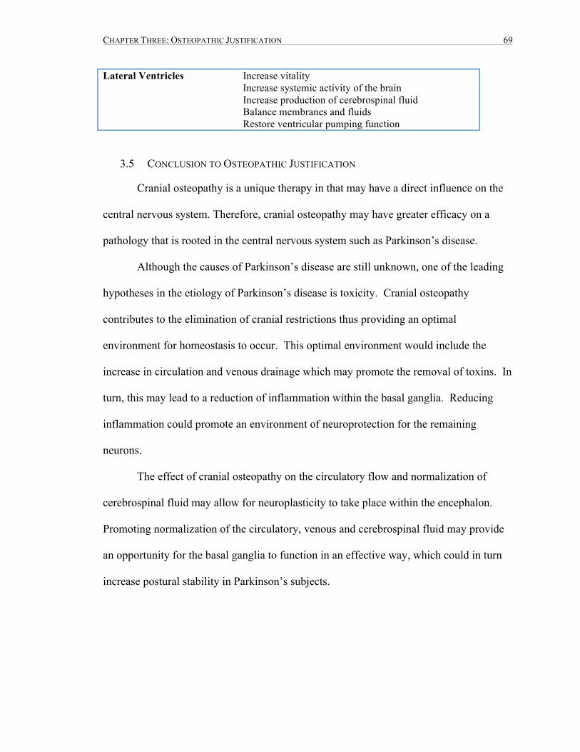
CHAPTER THREE: OSTEOPATHIC JUSTIFICATION 69
Lateral Ventricles Increase vitality Increase systemic activity of the brain Increase production of cerebrospinal fluid Balance membranes and fluids Restore ventricular pumping function
3.5 CONCLUSION TO OSTEOPATHIC JUSTIFICATION
Cranial osteopathy is a unique therapy in that may have a direct influence on the
central nervous system. Therefore, cranial osteopathy may have greater efficacy on a
pathology that is rooted in the central nervous system such as Parkinson’s disease.
Although the causes of Parkinson’s disease are still unknown, one of the leading
hypotheses in the etiology of Parkinson’s disease is toxicity. Cranial osteopathy
contributes to the elimination of cranial restrictions thus providing an optimal
environment for homeostasis to occur. This optimal environment would include the
increase in circulation and venous drainage which may promote the removal of toxins. In
turn, this may lead to a reduction of inflammation within the basal ganglia. Reducing
inflammation could promote an environment of neuroprotection for the remaining
neurons.
The effect of cranial osteopathy on the circulatory flow and normalization of
cerebrospinal fluid may allow for neuroplasticity to take place within the encephalon.
Promoting normalization of the circulatory, venous and cerebrospinal fluid may provide
an opportunity for the basal ganglia to function in an effective way, which could in turn
increase postural stability in Parkinson’s subjects.

CHAPTER FOUR: RESEARCH METHODOLOGY 70
4 CHAPTER FOUR: RESEARCH METHODOLOGY
4.1 TYPE OF RESEARCH
This study was a randomized single blinded between-group design which
examined the effect of cranial osteopathy on the balance in Parkinson’s subjects. It
consisted of a control group that entailed exercise only and an experimental group that
received exercise and four cranial osteopathic treatments. The tool utilized to measure
balance was the Berg Balance Scale. A pre-intervention and post-intervention assessment
using the Berg Balance Scale was administered to both groups. In addition an osteopathic
evaluation of the cranial system was administered to both groups pre-intervention and
post-intervention by the evaluating researcher.
4.2 TARGET POPULATION
The target population was comprised of eleven volunteers (five male and six
female) between the ages of fifty-five and eighty-two that had a previous diagnosis of
idiopathic Parkinson’s disease. A power analysis performed for the research proposal of
this study determined a power of sixteen (8 per group) was needed to show statistical
power (80% and an alpha = 0.05). A number of studies employing the Berg Balance
Scale were used to determine this power. A synthesis of these papers revealed that
Parkinson’s patients in earlier stage of Parkinson’s, determined using the Hohen and
Yahr scale or the Unified Parkinson’s disease rating scale, scored around 50 on the Berg
Balance Scale and that physical exercise produced a negligible improvement in the Berg
Balance Scale. It was felt that an improvement of two points on the Berg Balance Scale
would show significant change.

CHAPTER FOUR: RESEARCH METHODOLOGY 71
4.3 INCLUSION CRITERIA
Participants included in this study fit the following criteria:
• Primary diagnosis of idiopathic Parkinson’s disease by a physician;
• Can ambulate with or without an assisting device;
• Between the ages of 55 and 90 years old;
• Signed consent form, See Appendix A: Consent Form
• Completed medical questionnaire; rating between stage II and stage IV on the
Hoehn and Yahr Scale. See Appendix B: Hoehn and Yahr Scale.
4.4 EXCLUSION CRITERIA
If any of the following criteria were present in potential subject they would have
been excluded from this study.
• Central nervous system problems other than Parkinson’s: stroke, Multiple
Sclerosis, Muscular Dystrophy;
• Traumatic brain injury;
• Previous osteopathic treatment;
• Receiving any kind of other manual therapy concurrently to avoid potential
confounds;
• Diagnosed with balance disorder;
• History of encephalitis, cerebral vascular disease, neoplasm;
• Uncorrected visual impairment;
• Altered medication during study;
• Any condition that would contraindicate manual treatment or the ability to
complete the outcome tasks.
CHAPTER FOUR: RESEARCH METHODOLOGY 72
4.5 INDEPENDENT VARIABLE
The independent variable in this research was the exercise protocol and cranial
osteopathic treatment plus the exercise protocol. Although guidelines had been set out to
ensure each subject in the experimental group received some uniformity within the
treatment, each individual was different and therefore various techniques were employed
to the subjects.
One of the main criticisms of cranial osteopathic research is the lack of inter-rater
reliability and intra-rater reliability (Hartman & Norton, 2002). In order to curb some of
this concern an independent Osteopath validated the hands of Stacey Hauserman and
Thomas Hein, prior to commencement of this study. On September 8th 2009 at the
Canadian College of Osteopathy, Toronto, the researchers were asked to evaluate four
structures and write down the lesions they perceived. Brad McCutcheon, D.O.M.P,
Principle of the Canadian College of Osteopathy, had previously evaluated the volunteer
and recorded his findings. The findings were compared and Brad McCutcheon
determined the researcher’s hands to be validated.
4.6 DEPENDENT VARIABLES
The only dependent variable within this research was the Berg Balance Scale
scores for the subjects’ pre-assessment and post-assessment.
4.7 CONTROL AND EXERCISE
Each subject participated in four weekly, one hour exercise sessions that were
published by the Parkinson’s Society of Canada. (See Appendix C: Exercise Protocol)
The exercise sessions were run by an independent physiotherapist, Hermina Vas. The
exercise regimen was included primarily as an incentive to encourage participation and

CHAPTER FOUR: RESEARCH METHODOLOGY 73
was not the primary variable under study. As such the type of exercise was restricted to a
basic and commonly recognized level for all subjects in the study.
The physiotherapist was instructed to assist each subject through the exercise
protocol by reading directly from the instruction booklet. The first section included a
brief description on good postural habits, as outlined on page three of the exercise
package. Included in this description were three demonstrations of healthy posture in
standing, sitting, and supine positions. Three postural alignment exercises were also
preformed and included, spinal roll downs while sitting, rhomboid and middle trapezius
strengthening with prone, forward head postural alignment.
The second portion included seven stretching exercises. The physiotherapist gave
the exact instructions written in the guidelines. If the subject needed clarification the
physiotherapist would stray from the specific wording to assist the subject. The stretching
exercises targeted the following muscles; spinal rotators, latissimus dorsi, pectoralis
major, anterior deltoids, iliopsoas and quadriceps, soleus and gastrocnemius. All
stretching was maintained for 30 seconds with breath, within the subjects’ limitations.
The third portion of the exercise protocol included the strengthening portion. A
series of movements with isometric contraction targeting the upper body and quadriceps
were preformed, followed by deltoid strengthening with a two pound weight. A series of
movements to target the lower body were then preformed while holding onto a chair for
support. Lastly, a side stepping motion and marching activity were performed
The goal was for all subjects to be able to complete this exercise protocol
however the physiotherapist removed the side stepping component for two of the subjects
due to concern over safety. This did not seem to affect their outcome measures.

CHAPTER FOUR: RESEARCH METHODOLOGY 74
4.8 MEASURING TOOL
The Berg Balance Scale (BBS) is an objective functional performance assessment
which measures a person’s ability to maintain stability while performing tasks. It is
commonly used for the elderly population, and people with Parkinson’s disease, stroke,
and other various balance impairment. The Berg Balance Scale is an inexpensive
assessment tool that can be performed in any clinical setting. It is often used to evaluate
the risk of falling, and to monitor progression of physical therapy (Thorbahn & Newton,
1996).
4.8.1 DESCRIPTION
The BBS is a 14 item scale that can be performed within a 20 minute time
interval. Each item on the scale is scored out of five points; zero being the lowest level of
function and four being the highest. There is a total score of 56. All of the 14 items on the
BBS are task orientated, and come with a small set of instructions that is relayed to the
subjects performing the tasks. The tasks progress from sitting to standing, standing with
feet together, to tandem standing and single-leg standing. Lower ratings are given if the
subject needs assistance, specific supervision, cueing, and if the time or distance
measurements are not met. A detailed description of the Berg Balance scale can be found
in Appendix C: Berg Balance Scale
Two studies on the BBS on the predictability of falls in the elderly confirmed that
those who scored lower than 45 were more likely to fall than those who scored above 45
(Kornetti, Fritz, Chiu, Light, & Velozo, 2004; Thorbahn & Newton, 1996).
4.8.2 BERG BALANCE SCALE VALIDITY
A review of literature on various functional balance assessment tools indicated the
Berg Balance Scale is used as a gold standard to validate other balance measures
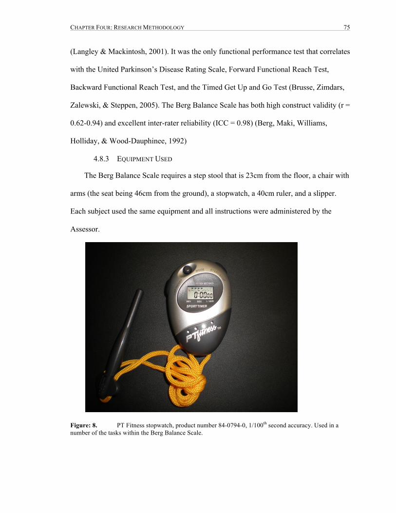
CHAPTER FOUR: RESEARCH METHODOLOGY 75
(Langley & Mackintosh, 2001). It was the only functional performance test that correlates
with the United Parkinson’s Disease Rating Scale, Forward Functional Reach Test,
Backward Functional Reach Test, and the Timed Get Up and Go Test (Brusse, Zimdars,
Zalewski, & Steppen, 2005). The Berg Balance Scale has both high construct validity (r =
0.62-0.94) and excellent inter-rater reliability (ICC = 0.98) (Berg, Maki, Williams,
Holliday, & Wood-Dauphinee, 1992)
4.8.3 EQUIPMENT USED
The Berg Balance Scale requires a step stool that is 23cm from the floor, a chair with
arms (the seat being 46cm from the ground), a stopwatch, a 40cm ruler, and a slipper.
Each subject used the same equipment and all instructions were administered by the
Assessor.
Figure: 8. PT Fitness stopwatch, product number 84-0794-0, 1/100th second accuracy. Used in a number of the tasks within the Berg Balance Scale.

CHAPTER FOUR: RESEARCH METHODOLOGY 76
Figure: 9. 40 cm ruler used to measure length of arm reach measured in centimeters as listed in item eight in the Berg Balance Scale
Figure: 10. Ikea step stool 23 cm from ground as used in item twelve on the BBS. Seen with Ikea slipper used in item nine in the Berg Balance Scale
4.9 RECRUITMENT METHODOLOGY
Recruitment occurred within the greater Toronto area. Contacts were initially
made with Dr. Guttmann of the Movement Disorder Center at Markham Stouffville
CHAPTER FOUR: RESEARCH METHODOLOGY 77
Hospital and Dr. Adams at Baycrest Hospital. Both neurologist’s were initially
enthusiastic regarding this study and offered to refer patients once the study was
approved. Further contact was made after the research was approved for a study.
Regretfully, Dr. Guttmann and Dr. Adams were unable to act on their earlier enthusiasm
due to the lack of a formal ethics review board at the College of Osteopathy and a
possible conflict with a rehabilitation study at the Movement Disorder Centre. Physician
letters were sent out to various physicians in the north end of Toronto without any
response. (See Appendix D: Physician Letter) Business cards with contact information
and study details were placed with flyers in numerous health centers, assisted living
homes and retirement homes within Toronto. (See Appendix E: Business Card, See
Appendix F: Recruitment Notice) A few calls were received from this source but the
potential subjects felt the distance was too far to travel. Personal contact was made with
the organizer of Sutherland-Chan Massage School Parkinson outreach clinic. The
organizer was concerned about breaching their privacy policies but she did agree to hand
out business cards containing the study details on an individual basis. However, no
subjects were attained through this source. The researchers attended the 2010 Parkinson’s
Walk in Toronto and left cards on the information tables.
Both researchers emailed their respective data base of patients to inform them
about the study. The patients were asked to pass on the information to anyone that they
believe might benefit from this research. Seven inquiries and four subjects were attained
from this source.
Direct contact was made with the organizers of five separate Parkinson’s support
groups in the Greater Toronto Area. From these contacts an arrangement was made to

CHAPTER FOUR: RESEARCH METHODOLOGY 78
make a presentation to the Thornhill Parkinson’s support group. Many of the participants
in the group were interested in becoming subjects in the current study.
Overall, the majority of inquires and subsequent subjects were attained from
direct referral from both former subjects and the researchers personal database. In total 21
people called to inquire about the study. Three of which were declined due to ambulatory
issues because they would not be able to complete the outcome measures. One potential
subject was declined due to a possible Parkinson’s Plus syndrome. A diagnosis of
idiopathic Parkinson’s disease was specified for inclusion into the study. Another subject
was accepted after the initial screening, but did not show up for her first appointment.
The clinic receptionist reached this potential subject a week later and was told she was
longer interested in participating. The remaining five potential subjects were either
unable or unwilling to attend five consecutive weeks of the study.
4.10 TELEPHONE SCREENING
Interested subjects called the clinic number and received a semi-scripted interview
by the clinic receptionist. (See Appendix G: Telephone Interview) This interviewed
determined whether the potential subject met the initial criteria for the study. If the
subject did not meet the criteria outlined in the inclusion and exclusion section, they were
thanked for the call and rejected for the study. If the potential subject met the criteria they
were scheduled for their first appointment.
4.11 RANDOMIZATION
During the initial appointment each patient was asked to randomly choose an
envelope from a table top, where the envelopes had been placed an equal distance apart.
Each envelope contained either a red or a black card. Eight of each color playing card

CHAPTER FOUR: RESEARCH METHODOLOGY 79
were placed facing down in envelopes and sealed at the commencement of this research.
The black cards represented the experimental group and red cards the control group.
Two researchers participated in the study. A coin toss prior to commencement of
this research determined that Stacey Hauserman was to be the treating Osteopath
(Osteopath 2) and Thomas Hein was to be the Assessor (Osteopath 1).
After the subject selected the envelope they handed it to the clinic receptionist.
The receptionist passed the envelope to Osteopath 2 who then removed the card from the
envelope and recorded the participant’s data into the data log. (See Appendix H: Data
Log)
Figure: 11. Envelopes for subject randomization
4.12 PROCEDURE
Once the subjects had passed the initial telephone screening, the receptionist
booked the subject in for the next available appointment in the study. The subject then

CHAPTER FOUR: RESEARCH METHODOLOGY 80
arrived at 9:45am on the allotted date. They were then asked to fill out a medical
questionnaire and an informed consent form in the clinic waiting room. (See Appendix I:
Medical Questionnaire, See Appendix A: Informed Consent) Osteopath 1 (the assessor)
introduced himself and brought the potential subject into the physiotherapy clinic.
Osteopath 1 reviewed the medical history to ensure there were no conflicts with the
study. He then gave a brief description of the study and the expectations of both the
patient and the researchers were outlined. The subject was informed of the privacy
policy, their right to withdrawal from the study at any time and forfeit of participation if
there were any changes that excluded them from the study. The subjects were also
informed of the need for their medication to remain consistent throughout the duration of
the study. If the subject’s medication required alteration during the study they were asked
to disclose this information to the researchers. A copy of the privacy policy statement
compliant with PIPEDA guidelines was given to the subject. (See Appendix J: Privacy
Policy)
Osteopath 1 preformed an evaluation to determine the subjects staging of the
Hoehn and Yahr Scale. The Hoehn and Yahr Scale is a simple, well known rating scale
that determines the stage of Parkinson’s disease. It was used in this study to determine
that some postural instability was present in the subjects. If the subject’s staging was
within II to IV they were accepted into the study.
Osteopath 1 then administered the 14 item Berg Balance Scale in the main gym of
the PhysioActive clinic. The subject was then asked to choose an envelope from a table
top and the results of the randomization were recorded in the data log as described in the
above section. Osteopath 1 was not privy to this information. Osteopath 2 then

CHAPTER FOUR: RESEARCH METHODOLOGY 81
determined the dates and times for the remaining appointments. If the subject was
assigned to the experimental group they were asked to come back for four consecutive
weekly two hour sessions. If they were assigned to the control group they were asked to
come back for four consecutive weekly sessions for one hour each week. The last session
included the post-evaluation using the Berg Balance Scale and an osteopathic assessment
preformed by Osteopath 1.
Osteopath 1 escorted the subject to the treatment room where an osteopathic
evaluation was preformed. Upon completion the subject was brought back into the gym
for their first exercise session with an independent physiotherapist Hermina Vas,
Registered Physiotherapist.
The information of the subjects remaining appointments were recorded by
Osteopath 2 on a patient appointment form and handed to the subject privately. (See
Appendix K: Appointment Form)
If the subject was placed in the experimental group, on their next appointment
they were greeted by the receptionist and escorted to the treatment room where Osteopath
2 was waiting to commence the subject’s first 60 minute cranial osteopathic treatment.
Upon completion of the first treatment, Osteopath 2 escorted the subject out to the gym
for their second exercise session with the physiotherapist. This sequence was repeated for
the subject’s third and fourth visit to the clinic.
It should be noted that in order to ensure blinding Osteopath 1 was not on the
premise during the subjects’ second to fourth appointments. However, Osteopath 1 was
on site during the first and fifth appointments. During the study hours a sign reading
Parkinson’s Study in Session was placed on the door of the treatment room regardless of

CHAPTER FOUR: RESEARCH METHODOLOGY 82
whether an osteopathic treatment was being preformed. Osteopath 1 was privy to the
knowledge that the sign would always be on the door during study hours. This was to
ensure that if Osteopath 1 walked by the treatment room he would not know whether a
treatment was being preformed. Also prior to commencement of the study Osteopath 1
was informed that Osteopath 2 would be in the treatment room during the study hours.
This ensured that if by chance Osteopath 1 were to accidently see Osteopath 2 at the
clinic it would not confound blinding.
On the subject’s final visit the subject was escorted to the treatment room to be
met by Osteopath 2 for the fourth and final cranial osteopathic treatment. At the end of
the treatment the subject was asked to go back to the waiting room and take a seat.
Fifteen minutes after the treatment, Osteopath 1 met the subject in the waiting room and
escorted him/her to the gym for a post-study assessment with the Berg Balance Scale.
This was followed by an osteopathic assessment to determine the severity of lesions and
vitality. The data was recorded on the osteopathic assessment form. (See Appendix L:
Osteopathic Assessment Form)
If the subject was placed in the control group, they returned for their second
appointment and were greeted by the receptionist. The receptionist then escorted the
subject to the gym for their second exercise session with the physiotherapist. Upon
completion of each session the subject escorted themselves out. On the final appointment
the subject was greeted by the receptionist and asked to take a seat. Osteopath 1 then
escorted the subject into the gym for the post-assessment of the Berg Balance Scale,
followed by an osteopathic assessment.
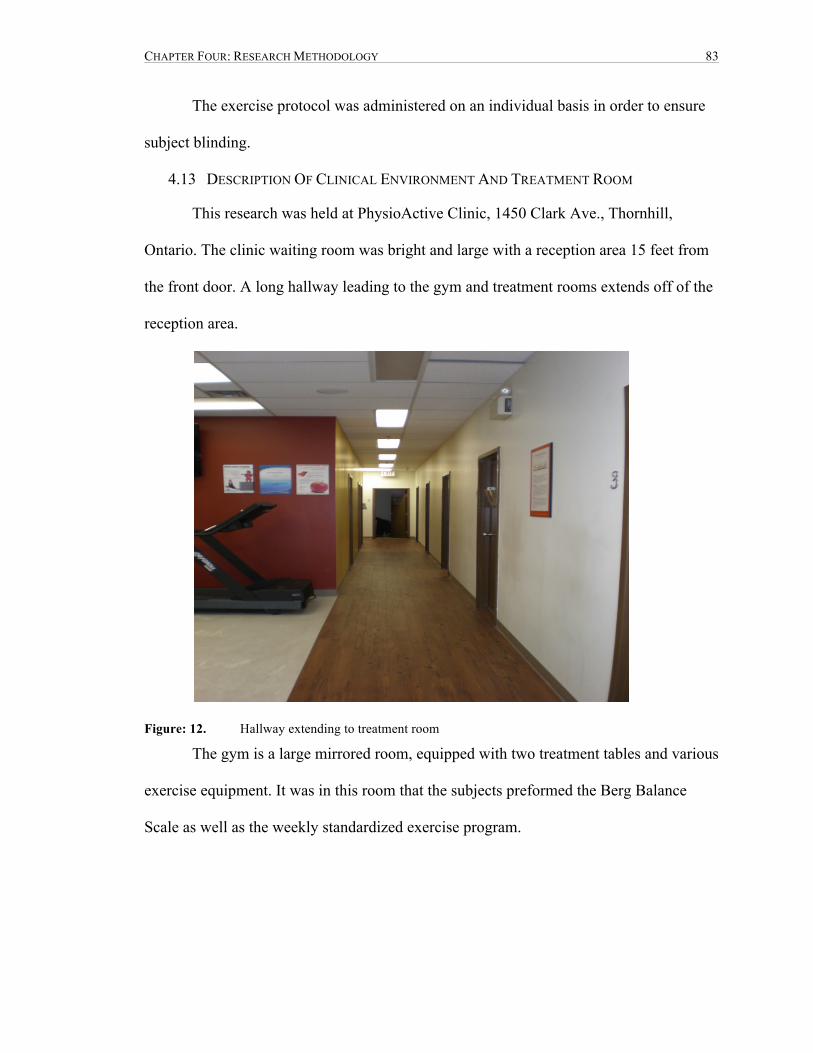
CHAPTER FOUR: RESEARCH METHODOLOGY 83
The exercise protocol was administered on an individual basis in order to ensure
subject blinding.
4.13 DESCRIPTION OF CLINICAL ENVIRONMENT AND TREATMENT ROOM
This research was held at PhysioActive Clinic, 1450 Clark Ave., Thornhill,
Ontario. The clinic waiting room was bright and large with a reception area 15 feet from
the front door. A long hallway leading to the gym and treatment rooms extends off of the
reception area.
Figure: 12. Hallway extending to treatment room
The gym is a large mirrored room, equipped with two treatment tables and various
exercise equipment. It was in this room that the subjects preformed the Berg Balance
Scale as well as the weekly standardized exercise program.

CHAPTER FOUR: RESEARCH METHODOLOGY 84
Figure: 13. Gym where exercise protocol was administered
All osteopathic treatments were preformed in a treatment room at the end of the
hallway. The room is large and equipped with a Kor Innovations Euro Lift 2000
hydraulic table, a small desk, a stool and a chair. The subjects often used the chair to
remove their shoes before getting onto the table. The room had high windows for privacy
and natural light.
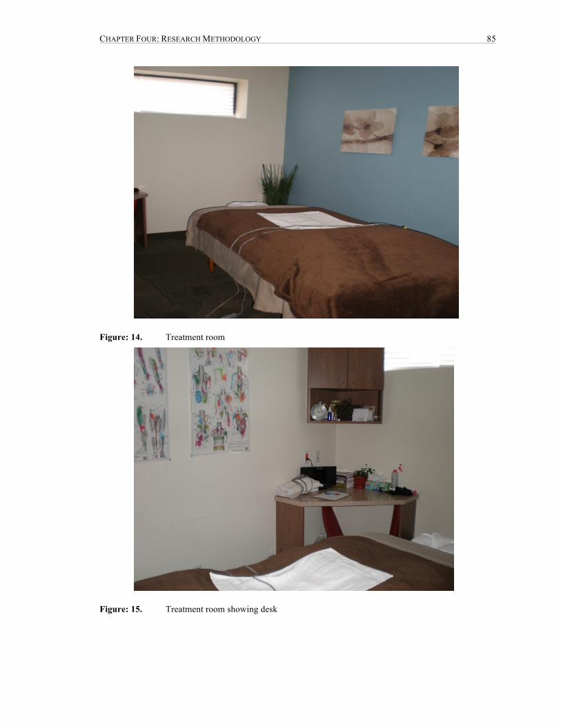
CHAPTER FOUR: RESEARCH METHODOLOGY 85
Figure: 14. Treatment room
Figure: 15. Treatment room showing desk

CHAPTER FOUR: RESEARCH METHODOLOGY 86
4.14 OSTEOPATHIC TREATMENT
The subjects assigned to the experimental group received four, weekly, one hour
exercise sessions supervised by an independent physiotherapist. In conjunction to the
exercise protocol the experimental group also received four, one hour cranial osteopathic
treatments. The first treatment was aimed at clearing non-physiological lesions that were
impeding the craniosacral mechanism. This included, but was not limited to the pelvis
and the spinal column. This was followed with a venous sinus technique to increase
vitality of the primary respiratory mechanism and to increase drainage and encourage
optimal circulation. All techniques listed in this section are written in further detail in
Appendix N: Technique Descriptions. Strict adherence to the Canadian College of
Osteopathy (CCO) methodology was followed. See Appendix O: Canadian College of
Osteopathy Methodology.
The second treatment addressed the cranial sphere and was based on the individual’s
cranial lesions, prioritized using the CCO methodology. Therefore, non-physiological
without respect to the axis lesions within the cranial base, C1 and C2 were addressed. In
addition, a posterior fossa technique, an EV4 and CV4 were administered to increase
overall fluidic drive.
The focus of the third treatment was to restore the cranial axis and increase vascular
flow. A parietal lift was used during this treatment to balance membranes, access the
sensorimotor cortex and to apply a pumping effect to the encephalon.
The final treatment addressed the endocranium in some capacity. A bilateral
temporal rocking technique was used to stimulate the central nuclei and the thalamus.
This was followed by lateral ventricle technique to increase the systemic activity of the

CHAPTER FOUR: RESEARCH METHODOLOGY 87
brain (Druelle & Forget, 2000). A local, regional and global integration was given
following each treatment.
Upon completion of the final treatment, the subject was re-assessed by the Assessor
using the Berg Balance Scale and an osteopathic evaluation.
Figure: 16. Treatment outline
4.15 ETHICS
In this study, each subject was required to read and sign an informed consent
form. This form indicated that the subject was able to terminate their participation at any
time, for any reason, without consequence. The subject was informed that they were
randomly placed into either a control or experimental group and that both groups
received a form of treatment. Furthermore, subjects were also informed that at no time
before, during or after their participation were they to be made aware of the treatment
Treatment 1 Clearing , venous sinus
Treatment 2 Cranial base, posterior fossa, CV4, EV4
Treatment 3 Increase vascular flow and restore cranial axis
Parietal liC
Treatment 4 Bilateral temporal rocking, lateral ventricles

CHAPTER FOUR: RESEARCH METHODOLOGY 88
group to which they had been assigned. All interventions were thought to be of minimum
risk of harm.
All of the subjects’ personal information is and will remain confidential. The
subjects were asked to read a privacy statement that was compliant with the PIPEDA
guidelines. The interventions used were low risk to health and the inclusion/exclusion
criteria further was designed to reduced risk to subjects with significant co-morbidities.
There was no monetary incentive used to attract subjects. The author declares that no
conflict of interest exists.

CHAPTER FIVE: DATA ANALYSIS AND RESULTS 89
5 CHAPTER FIVE: DATA ANALYSIS AND RESULTS
The data for the current study was analyzed by statistician Peter Lewycky. Mr.
Lewycky also provided the pre-analysis for the proposal portion of this study. All
analysis, tables and graphs were done using SPSS Release 16. A letter from Peter
Lewycky, validating his contribution to this work can be found in Appendix P:
Statistician Letter.
The data for the hypothesis that cranial osteopathy will improve balance in
Parkinson’s subjects were analyzed using the Wald Chi Square method of analysis. In
the primary analysis, results were analyzed to determine the effect of cranial osteopathic
treatment on Berg Balance scale (BBS) in subjects in the control group versus the
experimental group. Both groups saw improvement in the BBS score (BBS pre to BBS
post) whether treatment was exercise or exercise plus cranial osteopathic treatment.
However, improvement was greater in the experimental group receiving osteopathic
treatment in conjunction with exercise. This result was significant at the p=.028 level.
Table 2: Hoehn and Yahr Rating Per Group

CHAPTER FIVE: DATA ANALYSIS AND RESULTS 90
Table 3: Generalized Linear Model Information
Table 4: Categorical Variable Information
Table 5: Continuous Variable Information
Table 6: Tests of Model Effects
Participants in the study were classified on the Hoehn and Yahr rating scale of
Parkinson’s severity. All participants were at either stage two or stage three, indicating
some postural instability. The Hoehn and Yahr rating was analyzed to determine whether
cranial osteopathic treatment may have a differential effect on BBS depending upon
severity level of Parkinson’s disease. The statistical test returned a probability value of
p=0.87, suggesting no interaction between treatment and severity level of Parkinson’s
disease. This result indicates that cranial osteopathic treatment on BBS shows positive
improvement regardless of the staging level of Hoehn and Yahr.
An additional analysis was performed to determine whether cranial osteopathic
treatment within a given severity level of Parkinson’s may result in greater improvement.

CHAPTER FIVE: DATA ANALYSIS AND RESULTS 91
This test was almost statistically significant at p=.055. While one cannot conclude a true
effect, the data would suggest that the difference in treatment effect is most pronounced
at the greater Hoehn and Yahr severity level 3.
The following three tables are pairwise comparisons of estimated marginal means
based on the original scale of the dependent variable BBS post scores.
Table 7: Pairwise Comparison 1
a. The mean difference is significant at the .05 level.
Table 8: Pairwise Comparsion 2
Table 9: Pairwise Comparison 3

CHAPTER FIVE: DATA ANALYSIS AND RESULTS 92
5.1 OSTEOPATHIC EVALUATION ANALYSIS
An osteopathic assessment was also conducted in an effort to gain greater insight
into the effects of cranial osteopathic treatment on the participants. The assessor was
unaware of whether the subjects were in the experimental or control group. The tables
below show the results of measures of both vitality and cranial lesion classifications.
This first table provides descriptive statistics on 38 evaluated anatomical
structures, assessed at both pre-treatment and post-treatment stages for both experimental
and control groups on cranial lesion severity. The rating scale is based on the
methodology used and taught at the Canadian College of Osteopathy. Each structure was
assigned a number from 0 to 4 representing the severity of the lesion. On this scale, 0
represents no lesion, 1 represents a physiological lesion, 2 represents a non-physiological
lesion with respect to the axis, 3 represents a non-physiological lesion without respect to
the axis, and 4 represents a compaction. In this table all structures that were assessed
pre-intervention and post-intervention are listed. It is divided into control and
experimental groups so a visual comparison can be made. The mean provides an
arithmetic average of severity of lesion per structure within each group. The standard
deviation is a measurement of variability which shows the amount of variation from the
average. It is commonly used to measure confidence in the statistical conclusion.

CHAPTER FIVE: DATA ANALYSIS AND RESULTS 93
Table 10: Lesion Variable Severity

CHAPTER FIVE: DATA ANALYSIS AND RESULTS 94
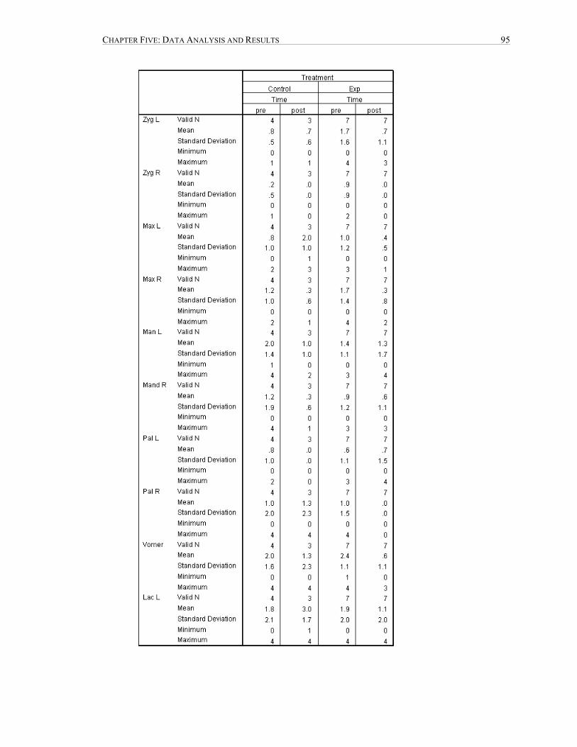
CHAPTER FIVE: DATA ANALYSIS AND RESULTS 95

CHAPTER FIVE: DATA ANALYSIS AND RESULTS 96
A detailed statistical analysis was performed to show the relationship between
pre-intervention lesion classification scores and post-intervention lesion classification
scores. This analysis was done using the Somers’d test to evaluate change in the pre-
classification and post-classification scores of both the control and experimental groups.
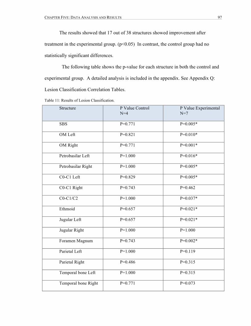
CHAPTER FIVE: DATA ANALYSIS AND RESULTS 97
The results showed that 17 out of 38 structures showed improvement after
treatment in the experimental group. (p<0.05) In contrast, the control group had no
statistically significant differences.
The following table shows the p-value for each structure in both the control and
experimental group. A detailed analysis is included in the appendix. See Appendix Q:
Lesion Classification Correlation Tables.
Table 11: Results of Lesion Classification.
Structure P Value Control N=4
P Value Experimental N=7
SBS P=0.771 P=0.005*
OM Left P=0.821 P=0.010*
OM Right P=0.771 P=0.001*
Petrobasilar Left P=1.000 P=0.016*
Petrobasilar Right P=1.000 P=0.005*
C0-C1 Left P=0.829 P=0.005*
C0-C1 Right P=0.743 P=0.462
C0-C1/C2 P=1.000 P=0.037*
Ethmoid P=0.657 P=0.021*
Jugular Left P=0.657 P=0.021*
Jugular Right P=1.000 P=1.000
Foramen Magnum P=0.743 P=0.002*
Parietal Left P=1.000 P=0.119
Parietal Right P=0.486 P=0.315
Temporal bone Left P=1.000 P=0.315
Temporal bone Right P=0.771 P=0.073

CHAPTER FIVE: DATA ANALYSIS AND RESULTS 98
Frontal Bone Left P=1.000 P=0.033*
Frontal Bone Right P=0.657 P=0.021*
Occipital Squama P=0.771 P=0.002*
Sphenoid G Wing P=1.000 P=0.510
Zygoma Left P=1.000 P=0.122
Zygoma Right P=1.000 P=0.070
Maxilla Left P=0.200 P=0.478
Maxilla Right P=0.314 P=0.021*
Mandible Left P=0.486 P=0.781
Mandible Right P=0.829 P=0.780
Palantine Left P=0.429 P=1.000
Palantine Right P=1.000 P=0.192
Vomer P=0.571 P=0.013*
Lacrimal Left P=0.400 P=0.592
Lacrimal Right P=1.000 P=0.731
Nasal Left P=0.429 P=0.286
Nasal Right P=1.000 P=0.192
Sacrum P=0.771 P=0.012*
L5/S1 P=0.971 P=0.202
Ilium Left P=1.000 P=0.315
Ilium Right P=0.686 P=0.0318
C3 P=0.571 P=0.021*
*represents a statistically significant improvement
An additional assessment measure looked at each of the 38 anatomical structures
pre and post treatment as measured on a scale of vitality. The vitality scale was measured

CHAPTER FIVE: DATA ANALYSIS AND RESULTS 99
using scores ranging from 0-3, with 0 representing no vitality and 3 representing normal
vitality. The table below shows descriptive statistics for each of the 38 variables on the
vitality measure.

CHAPTER FIVE: DATA ANALYSIS AND RESULTS 100
Table 12: Vitality Severity Variables

CHAPTER FIVE: DATA ANALYSIS AND RESULTS 101

CHAPTER FIVE: DATA ANALYSIS AND RESULTS 102

CHAPTER FIVE: DATA ANALYSIS AND RESULTS 103
Again, a Somers’d test was used to determine the effect of treatment on vitality
before and after treatment in both the experimental and control groups on all 38 variables.
Out of the 38 structures evaluated 29 showed statistically significant
improvements between the pre-vitality and post-vitality scores in the experimental group.

CHAPTER FIVE: DATA ANALYSIS AND RESULTS 104
There was no statistical significance in any structure between the pre-vitality and post-
vitality scores in the control group.
The following table shows the p-value for the vitality measure for each structure
in both the control and experimental group. This table depicts the essential results of a
more detail analysis that can be seen in Appendix R: Vitality Correlation Tables
Table 13: Results of Vitality Severity
Structure/Variable P-Value Control N=4
P-Value Experimental N=7
SBS P=1.000 P=0.001*
OM Left P=0.429 P=0.015*
OM Right P=0.771 P=0.001*
Petrobasilar Left P=0.143 P=0.012*
Petrobasilar Right P=0.486 P=0.003*
C0-C1 Left P=0.829 P=0.003*
C0-C1 Right P=0.486 P=0.040*
C0-C1/C2 P=0.686 P=0.031*
Ethmoid P=0.400 P=0.012*
Jugular Left P=0.429 P=0.009*
Jugular Right P=1.000 P=0.035*
Foramen Magnum P=0.257 P=0.001*
Parietal Left P=0.486 P=0.020*
Parietal Right P=0.486 P=0.020*
Temporal bone Left P=0.143 P=0.269
Temporal bone Right P=0.486 P=0.004*
Frontal Bone Left P=0.657 P=0.009*
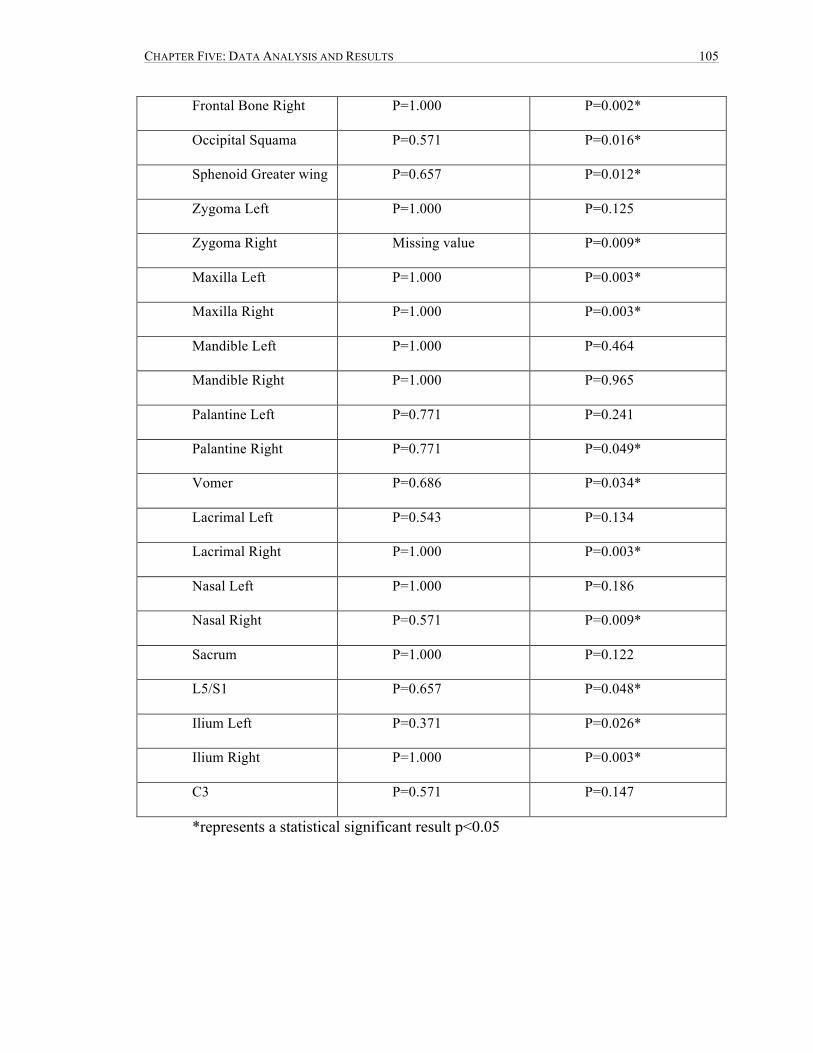
CHAPTER FIVE: DATA ANALYSIS AND RESULTS 105
Frontal Bone Right P=1.000 P=0.002*
Occipital Squama P=0.571 P=0.016*
Sphenoid Greater wing P=0.657 P=0.012*
Zygoma Left P=1.000 P=0.125
Zygoma Right Missing value P=0.009*
Maxilla Left P=1.000 P=0.003*
Maxilla Right P=1.000 P=0.003*
Mandible Left P=1.000 P=0.464
Mandible Right P=1.000 P=0.965
Palantine Left P=0.771 P=0.241
Palantine Right P=0.771 P=0.049*
Vomer P=0.686 P=0.034*
Lacrimal Left P=0.543 P=0.134
Lacrimal Right P=1.000 P=0.003*
Nasal Left P=1.000 P=0.186
Nasal Right P=0.571 P=0.009*
Sacrum P=1.000 P=0.122
L5/S1 P=0.657 P=0.048*
Ilium Left P=0.371 P=0.026*
Ilium Right P=1.000 P=0.003*
C3 P=0.571 P=0.147
*represents a statistical significant result p<0.05
CHAPTER SIX: DISCUSSION 106
6 CHAPTER SIX: DISCUSSION
The hypothesis for this study was that cranial osteopathic treatment in conjunction
with exercise will produce a greater improvement in the balance of subjects with
Parkinson’s disease, as compared to a control group receiving only exercise. The
resulting post-treatment Berg Balance Scale scores showed a greater improvement for the
experimental group than for the control group (p=.028)
While the study results were favorable with respect to the positive effects of
cranial osteopathic treatment on balance in Parkinson’s subjects, and would suggest that
the null hypothesis can be rejected, the researcher calls for caution in interpretation due to
the small sample size (n=11) and to the uneven groups. The experimental group, with
seven participants, had greater power to detect an effect than the control group, with four
participants. This skewed distribution of subjects to experimental and control groups was
an outcome of the randomization procedure which was based on a planned sample size of
16 subjects. The randomization procedure is discussed in greater detail in section 4.11.
With a larger sample size it is possible that additional subjects in the control
group may have shown improvements similar to that seen in the experimental group, thus
eradicating any differential effect postulated to be due to cranial osteopathic treatment in
the experimental group participants.
It is therefore recommended that this research be interpreted as a pilot study only,
and that further research be conducted with a larger sample size.
While this study should be regarded as a pilot, the discussion section addresses
possible reasons for the positive effect under an assumption that the result was real rather
than a function of chance. The reader is encouraged to consider this rationale, in the hope
that future researchers may find it provocative and may draw further hypotheses worth

CHAPTER SIX: DISCUSSION 107
exploring.
6.1 DISCUSSION OF RESULTS BASED ON OSTEOPATHIC JUSTIFICATION
Cranial osteopathic treatment has been critically scrutinized both within and
outside of the osteopathic profession. It is nonetheless widely used in clinical practice
and taught to student osteopaths throughout Canada. This research endeavored to
demonstrate the efficacy of cranial osteopathy in improving balance in Parkinson’s
patients. Parkinson’s disease is a progressive neurological disease with its neuronal
destruction based within the encephalon. Cranial osteopathy may have beneficial effects
in treatment of a pathology that is based within the central nervous system.
The cranial osteopathic treatment may have produced the resulting balance
improvement in the experimental subjects due primarily to its effect in increasing
circulation to the basal ganglia. In cerebral pathology, arterial insufficiency causes an
ischemic state which can lead to necrosis of the neural cells (Nolte, 1981). Moskalenko’s
research found that the removal of structural impedances, using cranial osteopathic
treatment, had an effect in improving circulation in the brain. The cranial osteopathic
treatment may have had the effect of removing structural impedances thus increasing
circulation in and to the basal ganglia. This increased blood flow to the basal ganglia
may allow a more optimal environment for neuronal activity and a resulting increase in
postural stability.
Researchers believe that toxicity is one of the leading contributing factors to
Parkinson’s disease. Although the actual effects of techniques such as venous sinuses are
not well documented it has been suggested within the teachings at the Canadian College
of Osteopathy that this technique has an effect on the venous drainage of the cranium
(Druelle & Forget, 2000). It would therefore follow that increasing vascular flow and

CHAPTER SIX: DISCUSSION 108
consequently contributing to the removal of toxins could have an impact on balance in
Parkinson’s subjects
Cerebrospinal fluid acts as an insulating property in which electrical impulses
occur. It has been suggested that a lack of normal characteristics within the cerebrospinal
fluid may cause pathology to occur (Becker, 1948b). Magoun (1976) believed that if any
part of the craniosacral mechanism was compromised it would affect the cerebrospinal
fluid leading to stasis and eventually pathology. Cranial osteopathic treatment may have
contributed to the restoration of the cerebrospinal fluid which could aid in the process of
neuroprotection and neuroplasticity.
McGeer and McGeer (2004) found cytokines in the cerebrospinal fluid of
Parkinson’s subjects leading them to determine that an inflammatory process occurs in
the encephalon of these patients. The theory of an inflammatory process contributing to
Parkinson’s disease is well known and can be seen in the oxidative stress model (Jenner,
2003; Jenner & Olanow, 2006). Cranial osteopathy may have an effect on the reduction
of edema providing an avenue to address inflammation within the encephalon (Magoun,
1968). This may have contributed to the positive results of cranial treatment on the
balance in Parkinson’s subjects.
6.2 DISCUSSION ABOUT OSTEOPATHIC ASSESSMENT
The results of the osteopathic assessment showed marked positive change in the
experimental subjects in the majority of categories and no positive change in any of the
categories in the control group subjects. In the lesion classification data of the
experimental group 17 out of 38 structures showed improvement after analysis of the pre-
intervention and post-intervention scores. Analysis of the vitality scores of the
experimental group showed 29 out of 38 structures with positive improvement. In the

CHAPTER SIX: DISCUSSION 109
analysis of the lesion and vitality classifications there was no case where the control
group showed improvement from pre to post scores.
Although these results look promising one must exercise caution in interpretation
as sample size was small. The control sample was very small with only four subjects. If
this group had been larger it is possible that improvement in some of the variables would
have appeared.
The osteopathic evaluation also provided some data on the most commonly
affected cranial structures within this subject pool. These structures include: the left nasal
bone, sphenobasilar symphysis, the left petro-basilar suture and C0/C1 on the left.
Excluding the nasal bone, the remaining structures are within the base of the cranium
which is in direct relation to the vascular vessels that innervate the basal ganglia. The
most common structures affected were primarily on the left side, however the researcher
is unable to provide a basis for this result. Although these results are interesting they are
anecdotal at best and should be viewed as such.
The osteopathic assessor performed assessments both before and after treatment
on all subjects and was consistent in the application of his measurements. By virtue of the
methodology, the osteopathic assessment results are subjective in nature, and are not
considered as part of the overall hypothesis. These assessment results are offered rather
as further information on the treatment results.
It should also be noted that the assessment results demonstrated a change which
provides a basis for a conclusion that there was an effect from the treatment. The
detectable effect suggests that the outcome changes are not a simple placebo.

CHAPTER SIX: DISCUSSION 110
6.3 DISCUSSION OF HOEHN AND YAHR RESULTS
The eleven subjects were assessed on the Hoehn and Yahr scale of Parkinson’s
severity. Out of the 11 subjects 6 scored at stage II on the Hoehn and Yahr Scale while 5
subjects scored at stage III on the Hoehn and Yahr scale. Four of the stage II’s were in
the experimental group while the remaining two were in the control group. Three of those
that scored III were n the experimental group while the remaining two were in the control
group. Thus, severity of Parkinson’s was relatively evenly assigned to the groups.
Hoehn and Yahr Scale Experimental Group Control Group
Rating of II N=4 N=2
Rating of III N=3 N=2
Figure: 17. Randomization based on the Hoehn and Yahr Rating Scale
The Wald chi-square analysis to determine whether cranial osteopathic treatment
had differential effects on those with greater or lesser severity of Parkinson’s failed to
reach significance (p=.055), suggesting that the effect of treatment does not depend on
the severity of Parkinson’s. However, while the result did not reach the technical
criterion for statistical significance, it does suggest that the difference in treatment effect
is most pronounced at the greater Hoehn and Yahr (HY) severity level III.
The researcher had hypothesized that cranial osteopathic treatment would have
greater effect on a Parkinson’s subject in an early stage of progression. Symptoms of
Parkinson’s disease often appear after approximately 70% of neuronal death within the
substantia nigra, giving rise to the postulation that early osteopathic cranial intervention
would have greater neuroprotective effect (Martin, et al., 2010). Cranial osteopathy may
provide an avenue into increasing circulation and venous drainage, therefore providing

CHAPTER SIX: DISCUSSION 111
the removal of toxins from the basal ganglia. However, the data from this current study
suggests that earlier cranial osteopathic intervention does not have a greater effect on
balance in Parkinson’s subjects. In contrast, the data suggested a possible correlation
between treatment effect and a higher level of Parkinson’s severity.
Hirsch (2007) suggested that toxins released by the glial cells may occur long
after intial exposure to MPTP and may contribute to the ongoing destruction of the basal
ganglia in Parkinson’s disease. This may provide an explanation for the results of the
current research which suggests that cranial osteopathy has a greater effect on balance in
more severe Parkinson’s cases. However, a larger study must be pursued for this data to
be considered conclusive.
Another interpretation is possible. This result however, even had it reached
significance, may in fact be an anomaly of the Berg Balance Scale. A subject with a
Hoehn and Yahr rating of III would commence the study with a lower pre-BBS. The
Berg Balance Scale has a ceiling value of 56 and subjects with Hoehn and Yahr ratings of
II often had a pre-BBS score of 54 or 55. Therefore, those with a higher BBS score had
less total opportunity for improvement, which may skew the result artificially to show a
greater total improvement for those with a lower pre-BBS score.
6.3.1 DISCUSSION ON THE HOEHN AND YAHR RATING SCALE
The Hoehn and Yahr is the most commonly used scale to describe the severity of
Parkinson’s disease (Goetz, et al., 2004). Many Parkinson’s disease and balance related
studies employ the Hoehn and Yahr (Ellis, et al., 2008; Hirsh, et al., 2003; Mitchell, et
al., 1995). Although all the subjects fell within stages II and III on the Hoehn and Yahr
rating scale there was a wide variety of presentations within the subject pool.

CHAPTER SIX: DISCUSSION 112
The Hoehn and Yahr is a simple classification that may miss the subtleties of
severity. The original design of the Hoehn and Yahr scale was a five point scale, but in
recent years 0.5 increments have been added in some clinical trials. In the current
research the original version of the Hoehn and Yahr scale was used. The assessing
osteopath found this original version limiting and had some difficultly with placement of
the subjects. This same concern was expressed in a study conducted by the Movement
Disorder Society in which 69% of the 236 members polled thought the staging categories
to be too broad (Goetz et al., 2004).
Stage I Unilateral involvement with minimal or no functional impairment
Stage II Bilateral or midline impairment with little impairment to balance
Stage III Bilateral disease: mild to moderate disability with impaired postural reflexes
Stage IV Severely disabling; still able to walk or stand unassisted
Stage V Confined to bed or wheelchair
Figure: 18. Hoehn and Yahr Original Scale (Goetz, et al., 2004, p. 1021)
A more recent version of Hoehn and Yahr has added more rating from I to III to
reflect the spectrum of the disease process.
Stage 1.0 Unilateral involvement only
Stage 1.5 Unilateral and axial involvement
Stage 2.0 Bilateral involvement without impairment of balance
Stage 2.5 Mild bilateral disease with recover on pull test
Stage 3.0 Mild to Moderate bilateral disease; some postural instability; physically independent
Stage 4.0 Severe disability; still able to walk or stand unassisted
Stage 5.0 Wheelchair bound or bedridden unless aided
Figure: 19. Modified Hoehn and Yahr scale (Goetz, et al., 2004, p. 1021)

CHAPTER SIX: DISCUSSION 113
Although this new version is not officially published it is being used by
physicians globally. It is recommended that for future research the modified version of
the Hoehn and Yahr be employed and the new criteria include staging between 2.5 and
3.0 to ensure enough postural instability that the subjects have room for significant
change.
6.4 DISCUSSION OF THE BERG BALANCE SCALE
There are two categories of tools that are used to measure balance. First there is
computerized instrumentation such as force plates, however this option has considerable
expense associated with it as well as a need for specific training to effectively apply the
instrumentation. The second option for evaluating balance is clinical standardized
assessments, which was the method chosen for the current study. The benefit of these
assessments is their simplicity in administering the tests, the ability to use this assessment
tests in any clinical setting, and the low cost associated with them. In addition, clinical
standardized assessments mimic real-life function and are therefore functional relevant to
the patient.
The Berg Balance scale was specifically designed to assess balance, to screen
patients for rehabilitative therapy, to predict falls, and to monitor changes in postural
instability. Those that score 45 or lower out of 56 are at higher risk for falling. Those
that score greater than 45 are less likely to experience falls associated with balance issues
(Kornetti, et al., 2004). Within this current research there were only two subjects that
scored below the 45 on the Berg Balance scale and therefore that had an increase risk of
falling.
The Berg Balance Scale is an excellent instrument for identifying the risk of
falling, but may not be sensitive enough to determine change of balance in those without

CHAPTER SIX: DISCUSSION 114
an increased risk of falling. As noted earlier, some of the subjects within this current
study started with a 54 on the Berg Balance Scale which only allowed for an increase of
two points. Earlier statistical analysis determined that a change of two was significant to
show statistical change. However with a starting score of 54, any true improvement in
balance for subjects in the study in either control or experimental group may be masked
due to the ceiling effect.
An analysis of the Berg Balance Scale determined that the scale was not sensitive
enough to detect changes in balance at the upper end of the scale (Kornetti, et al., 2004).
Kornetti et al. (2004) produced a revised scale that makes the testing more sensitive
however, it has not yet been adopted as the new gold standard. This revised scale
provides a more accurate representation of balance in subjects with Parkinson`s disease.
6.5 DISCUSSION OF INTERVENTION
The osteopathic literature review found only a limited number of cranial
osteopathic studies relevant to this topic. The lack of research made it difficult to
determine the amount of treatment required to make a difference within this population.
After much discussion four treatments were decided on. This was based on the essential
structures that the author felt needed to be addressed as well as what seemed like a
reasonable amount of treatments for patient compliance. In retrospect more than one
clearing treatment would have been helpful to increase the effects of the cranial
treatment. This protocol of multiple clearing treatments was used in David Bergstein’s,
CCO thesis (Bergstein, 2008). In this study, pre-study treatments were used to clear all
major lesions as per Canadian College of Osteopathy’s methodology. The pre-study
treatments were included as an adjunct to the study to ensure greatest effects from the
endocranial treatment.

CHAPTER SIX: DISCUSSION 115
6.6 DISCUSSION OF THE EXERCISE PROTOCOL
The exercise protocol was placed in this study both as a reasonable control and as
an incentive for subject recruitment. A methodology was derived that allowed for all
subjects to have some intervention without confounding the results of cranial osteopathic
treatment. By providing an exercise protocol, supervised by an independent
physiotherapist, all potential subjects received some intervention.
The inclusion of the exercise protocol provided a large incentive to the
recruitment process, for all subjects knew they would be receiving treatment, but were
unaware of the nature of the treatment. The subjects’ knowledge that they were receiving
some form of treatment also provided a blind for the subjects. By providing exercise the
subjects were unaware of which group they were assigned to.
The exercise protocol may have also contributed to the null attrition that was
experienced within this study. All participants verbally shared their appreciation and felt
they benefited from the study.
6.7 DISCUSSION RELATED TO LITERATURE REVIEW
The medical literature is vast and numerous, making it difficult to address all
aspects of Parkinson’s research. The intention was to provide an overview of Parkinson’s
disease and treatment options specifically for postural instability.
The osteopathic literature review did not reveal any studies that focused on cranial
osteopathy and balance in Parkinson’s disease. This unfortunately limited the researcher
when developing a methodological plan based on successful past studies. The few studies
that were found on Parkinson’s disease and osteopathic treatment employed either a
single osteopathic global treatment or did not list what the osteopathic treatment entailed.
This lack of literature prompted the researcher to review studies involving other

CHAPTER SIX: DISCUSSION 116
neurological disorders with the intention of extrapolating pertinent information for the
current study. This was somewhat helpful in providing inspiration for many of these
studies were statistically significant.
The remainder of the osteopathic literature review focused on the validity of
cranial osteopathy. By choosing to use cranial osteopathy rather than a global approach it
was felt that a review of the validity of cranial osteopathy was a necessary addition. In
conclusion this review revealed the need for more research within the field of osteopathy
and neurological disorders.
6.8 DISCUSSION OF OBSERVATIONS
There were several significant findings in the health history questionnaire that
may have indicated several of the subjects would be less responsive to overall cranial
osteopathic treatment. One subject in the experimental group was diagnosed with
tuberculosis of the spine when she was eight years old. She spent two years bed-ridden in
a hospital and was separated from her family. She was told she would never walk again,
but went on to prove the physicians wrong. Two difficult pregnancies that ended in
infant mortality were followed by a complete hysterectomy. This subject also presented
with liver hemangiomas and macular degeneration. Restrictions in this patient’s lumbar
spine were very severe and had an overall effect on the expression of the primary
respiratory mechanism.
Two other subjects in the experimental group presented with osteoarthritis of the
hip. One had also experienced a bilateral knee replacement seven years prior to the
current study. This may have affected the ability for lasting effect of treatment on the
pelvis which may have contributed to restriction in the craniosacral system.

CHAPTER SIX: DISCUSSION 117
Another subject in the experimental group had her thyroid removed and
experienced chronic stiffness and pain in her cervical spine. Some amount of treatment
of the cervical spine was necessary in each session in order to ensure enough vitality to
treat the cranium.
One of the subjects within the experimental group experienced severe tremor at
rest. This made it difficult for the subject to remain on the table for a 60 minute session
without agitation. It is questionable whether the severity of this tremor affected the Berg
Balance Scale scores.
The treating osteopath became aware of these possible confounding physical
issues throughout the current study. The researcher questioned whether these health
history concerns affected the efficacy of the osteopathic cranial treatment, or the Berg
Balance Scale assessment. However, the subjects represented real life case scenario’s
and therefore may depict an accurate representation of the clinical challenges found in the
Parkinson’s population.
A review of the medical heath history forms determined that subjects within the
control group also noted co-morbidities. One subject noted that they had degenerative
disc disease in the lumbar spine that is associated pain and stiffness. Another subject
within the control group had a previous pelvic fracture due to a fall. The pelvic fracture
did not affect this subject’s ability to ambulate without assistance and therefore was
accepted into the current study. However, she did mention that she experienced recurrent
pelvic pain, which may have affected the Berg Balance Scale score.
One of the subjects within the control group was fearful of having anyone touch
his neck and at the end of the study he refused to have the post osteopathic assessment.

CHAPTER SIX: DISCUSSION 118
All subjects had some degree of difficulty with digestion, which is most likely due to
adverse side effects of the Parkinson’s medications.
All subjects in both the control and experimental groups noted some form of co-
morbidity. However, it remains unknown as to what degree this would have affected the
cranial osteopathic treatment or the Berg Balance Scale score.
6.9 DISCUSSION RELATED TO PILOT STUDY
This study intended on being a fully powered study, however due to recruitment
issues and time restraint this study came under the minimum amount of subjects. Upon
approval of the thesis committee the title was changed to make it a pilot study. See
Appendix S: Pilot Study Approval Letter. It was felt by the researcher, the thesis advisor,
and the thesis committee that this study had the appropriate attributes to be a pilot study.
A pilot study tests the logistics of the research in order to improve the quality and
efficiency of a larger study. Pilot studies are prominent in the Osteopathic profession. A
quick search on the JAOA data base with pilot study as the search word turned up 181
published studies. The prevalence of pilot studies in the osteopathic profession may be
due to a lack of funding compared to allopathic medical research, and difficultly of
recruitment.
Pilot studies are used to identify deficiencies in the research design which can be
addressed before a larger study takes place. In the current study, the research design was
basic and largely effective. However, this study revealed a number of issues, critical to
future effective research, discussion of which should prove to be a valuable contribution
to the literature at this stage. These issues include, limitations of the Berg Balance Scale,
challenges with participants with less severe Hoehn and Yahr stages, and efficacy of the
osteopathic assessment form, which is covered in the self critique section.

CHAPTER SIX: DISCUSSION 119
Pilot studies are used to assess a floor or ceiling effect, meaning if the task is too
difficult or too easy the results will be skewed. In this study, the basic exercise protocol
was sufficient for some improvement. This improvement, and using the BBS rating scale
with a ceiling effect, may have masked greater results of exercise and cranial osteopathy
together.
A larger study based on the same protocol is needed to determine whether the
results are statistically significant however, there is still a concern that the Berg Balance
scale is not sensitive enough to assess the balance of some potential subjects. If there
was access to increased funding, the use of instrumentation such as a force plate would be
an excellent adjunct to the Berg Balance scale.
Pilot studies can be appropriate for inexperienced researchers. There is a learning
curve involved in research, and regardless of the amount of effort spent on the proposal
and planning of the research, methodological errors frequently occur in student research.
In a letter published in the JAOA, McCombs (2006) stated that the use of student
practitioners to administer osteopathic manual medicine is not as effective as seasoned
osteopaths providing treatment. He continues to say that “judging the efficacy of
osteopathic manual medicine by the results achieved by students is a deeply flawed
concept” (McCombs, 2006, p. 380).
Perhaps most compelling in this study is the fact that results in the experimental
group were quite positive, based on the descriptive statistics. This statement is also true
when examining the results of the osteopathic assessments with positive results in a
majority of categories for the experimental group and virtually no improvement in the
control group. For these reasons, this researcher feels strongly that the current pilot study

CHAPTER SIX: DISCUSSION 120
produced promising results, and warrants a full study in the effects of cranial osteopathy
on balance in Parkinson’s subjects.

SELF –CRITIQUE 121
7 SELF –CRITIQUE
7.1 RECRUITMENT
The researchers were aware that recruitment for this study was going to be a
challenge. However, it was thought a power of n=16 would be attainable. As seen in the
methodology section of this paper, much time and effort went into recruitment.
Recruitment notices that were posted throughout the city proved to be ineffective.
Attending Parkinson’s fundraisers was also ineffective for recruitment. In an additional
effort to obtain subjects, letters were sent out to local physicians and physiotherapists.
Two movement disorder clinics expressed interest in our study and had originally offered
to refer subjects, however the offer was revoked when they became aware of a lack of a
formal ethics review board at the Canadian College of Osteopathy.
The most effective source of potential subjects came from direct referral from the
researchers own patient database as well as a Parkinson’s support group in the north end
of Toronto. The members of this support group were pro-active in their medical care and
were thankful for the opportunity to be involved in the current study.
One of the main challenges for recruitment of potential subjects was the five-
week commitment required to complete this study. Many of the subjects were retired and
had travel plans or medical appointments that interfered with the study dates. Although
the clinic location allowed for public transport, some of the subjects required an assistant
to bring them to and from their appointments, which proved challenging.
The lack of a formal ethics review board at the Canadian College of Osteopathy
limited the ability to obtain subjects, and regretfully may have been the largest
contributing factor to the small power of the current study. In Ontario, all academic-based
research involving human subjects is subjected to an ethics review board. The Canadian

SELF –CRITIQUE 122
College of Osteopathy has recently provided an option of applying for review with an
external ethics board at the Canadian Memorial Chiropractor College. Unfortunately, this
was suggested halfway through the study, and after careful review the researchers
determined it wasn’t a plausible option due to time restrictions.
In the author’s opinion, the lack of a formal ethics review board discredited the
current study. A strong suggestion for future research would be to attain approval from a
formal review board prior to commencement of the study.
7.1.1 CRITIQUE OF SAMPLE SIZE
Although much effort went into the recruitment of subjects, the largest regret of
the current study was the inability to attain enough subjects for a fully powered study.
The results of the data analysis demonstrated improvement of the Berg Balance Scale
scores in the experimental group of p< 0.28. Although these results are promising, a true
statistical difference cannot be determined without a power of n=16.
7.2 CRITIQUE OF RANDOMIZATION
Ideally, the randomization of subjects would produce equal numbers in both the
control and experimental group; however, the intended fully powered study of n=16 was
instead completed with n=11. Due to the randomization procedure chosen, the groups
were uneven – there were four subjects in the control group and seven subjects in the
experimental group. The randomizing method used would have produced equal groups
had the study obtained the suggested power of n=16, as the subjects chose from 16
envelopes containing cards that represented the groups. Unfortunately, the subjects began
the study as they became available and therefore the randomization occurred on a subject
to subject basis. Had all the subjects been obtained prior to commencement of the study,
a computer-generated randomization could have been employed to assure equal grouping.

SELF –CRITIQUE 123
However, due to the recruitment difficulties, it was necessary to accept subjects as they
became available.
7.3 CRITIQUE OF INCLUSION/EXCLUSION CRITERIA
This study was designed to assess the efficacy of cranial osteopathic treatment on
balance in Parkinson’s subjects. Within the methodology, consideration was taken to
ensure both that the subjects had idiopathic Parkinson’s disease and that they were
between stage II to IV on the Hoehn and Yahr scale, in order to guarantee some postural
instability was present. Of the subjects that were assessed, all rated between a II and III
on the Hoehn and Yahr Scale. However, the subjects presented with varying degrees of
postural instability. A revised version of the Hoehn and Yahr Scale that has an increased
sensitivity to the subtleties of Parkinson’s disease progression is strongly suggested for
future research.
This researcher would also suggest adding an inclusion criterion requiring
potential subjects to have significant cranial lesions. Regardless of their common
pathology, individuals present differently since each has lived a life without osteopathic
intervention prior to this study. All of the subjects within this study presented with
varying degrees of cranial lesions, some being much more severe than others. Sandhouse
et al. (2010) added an inclusion criterion of sphenobasilar cranial strain pattern in their
research on the effects of cranial osteopathic treatment on visual function. A strong
recommendation for future research would be to include a substantial cranial restriction
within the inclusion criteria. It would be beneficial to identify potential subjects with
Parkinson’s disease that have primary cranial lesions.
Removal of the age requirement of 55-90 from the inclusion criteria is
suggested for future research. Although this is a common addition to rehabilitative and
SELF –CRITIQUE 124
physical therapy research studies, an age requirement was unnecessary within the current
study because the onset of Parkinson’s disease can occur in the early twenties. The
symptoms and progression of early onset Parkinson’s disease do not differ from that of
later onset Parkinson’s disease (Martin, 2010). Therefore, it follows that the age
restriction would have no overall effect on balance and treatment in the current study.
Removing the age requirement may have provided more potential subjects.
A diagnosis of idiopathic Parkinson’s disease was required to be considered as a
potential subject for the current study. Unfortunately, the study thus excluded one
potential subject with a diagnosis of Parkinson’s plus syndrome. Although the symptoms
of Parkinson’s plus differ slightly from idiopathic Parkinson’s disease, their pathogenesis
is similar. Therefore, the justification and postulations as to why cranial osteopathy may
be effective would not have changed. A revision of this inclusion is suggested for further
research.
7.4 CRITIQUE OF OUTCOME MEASURES
As mentioned in the discussion of the Berg Balance Scale, this measuring tool of
balance may not be sensitive enough. A revised version of the rating scale is now
available and is strongly recommended for use in future research.
7.5 CRITIQUE OF THE OSTEOPATHIC ASSESSMENT FORM
A revised assessment form is suggested for future continuation of this research.
Different rating scales were used for the lesion classifications and the vitality ratings.
Lesions were rated using the Canadian College of Osteopathy methodology on a scale
from 0-4: 0 representing no lesion and 4 representing compaction, or the most severe
lesion. The vitality was scored from 0-3: 0 representing an absent vitality and 3
SELF –CRITIQUE 125
representing a healthy, powerful vitality. The inconsistency of these two scales proved to
be somewhat challenging when formulating the data.
A secondary source of concern with the osteopathic assessment form used was
that many of the variables were problematic. There were several duplications of
structures such as the cervical vertebrae, the temporal bones, and the sphenoid. The
sacral-iliac joint and the sacrum were listed as separate variables. The assessor
determined that the sacral-iliac joint denoted the iliac bones, and they were therefore
evaluated as such. In addition, the removal of the foramen magnum and jugular foramen
variables is suggested for future research as there was some ambiguity in evaluation of
these structures. For the purpose of this preliminary research, these structures were
evaluated for their motility and vitality.
Another suggestion would be to include a category that names specific lesions.
Although this research allowed us to see which structures were most affected, it did not
give us specific details. This type of lesion category would have provided some
interesting data into the most common lesions in Parkinson’s disease subjects.
7.6 RECOMMENDATIONS FOR FUTURE RESEARCH
There are several suggestions for future research based on the current pilot study.
1. Obtaining an approval from a formal ethics review board so that future
researchers can pursue assistance with recruitment by contacting neurologists
associated with local movement centres. Prior to commencing future research,
obtaining this approval is strongly recommended.
2. Revising the inclusion and exclusion criteria, in order to increase recruitment and
improve overall design of the study. These suggestions include using the revised
version of the Hohen and Yahr Scale, removing age requirements, and including

SELF –CRITIQUE 126
all forms of Parkinson’s disease as opposed to only including the idiopathic
presentation.
3. Using a revised osteopathic assessment form including, but not limited to, the
addition of classification of specific osteopathic lesions.
4. Using the revised rating scale for the Berg Balance Scale, which is more sensitive
than the original. If the resources are available, adding a second computerized
measuring tool such as a force plate would strengthen the results on balance.
5. Adding a follow-up assessment of one month to determine the long-term effects
of cranial osteopathic treatment on balance in Parkinson’s subjects.
6. Adding a quality of life questionnaire such as an SF39 to gather information
regarding the effects of treatment on depression, sleep and the general well-being
of the subjects.
7. Changing the treatment protocol to increase the number of cranial treatments to
six or eight to determine whether this would increase the effect of treatment.
CONCLUSIONS 127
8 CONCLUSIONS
Parkinson’s disease is the second leading neurodegenerative disease in Canada,
affecting one in 100 people over the age of 60. It is a progressive disorder that causes the
destruction of dopamine neurons within the substantia nigra of the basal ganglia. The
destruction of these neurons leads to motor symptoms such as tremor, bradykinesia,
rigidity and postural instability. Postural instability contributes to falls, which are
associated with injury, reduction of quality of life, and expense to the individual and the
healthcare system. Current demographics suggest that incidence, and therefore the
expense, of Parkinson’s disease will increase with the aging of the baby boomer
generation.
Treatment of postural instability is limited to medications that have been shown to
be relatively ineffective for instability, and physiotherapy, which has been shown to
provide only short-term improvement. There is a need for alternate forms of treatment
that target balance in Parkinson’s patients.
Cranial osteopathy provides a unique, non-invasive therapy that focuses on the
mobility of the cranial bones, which may have an influence on the central nervous
system. However, the lack of cranial osteopathic research within the field of neurological
disorders makes it difficult to support this premise.
The current research was designed to assess the effects of cranial osteopathic
treatment on balance in subjects diagnosed with idiopathic Parkinson’s disease. Eleven
subjects with Parkinson’s disease volunteered to participate in this five-week study. The
subjects were randomly assigned to an experimental group, consisting of exercise and
cranial osteopathic treatment, and a control group, consisting of exercise alone. Balance
was evaluated pre-intervention and post-intervention using the Berg Balance Scale. The
CONCLUSIONS 128
Berg Balance Scale is a gold standard clinical assessment, consisting of 14 functional
tasks that are rated from zero to five, for a total of 56. Statistical analysis revealed that an
improvement of two on the Berg Balance Scale would be considered significant.
A power analysis based on four equally spaced cranial osteopathic treatments
determined that a power of n=16 was needed to show statistical significance (p<0.05).
Unfortunately, recruitment efforts produced only 11 subjects and therefore this study was
re-titled a pilot study. Seven subjects were randomly assigned to the experimental group
and four subjects were randomly assigned to the control group.
The results of this pilot study showed a greater improvement in the Berg Balance
Scale scores for the group receiving cranial osteopathic treatment compared to the control
group (p=.028). Analysis of the osteopathic evaluation determined that 17 of 38 structural
variables that were measured pre-treatment and post-treatment on the lesion severity
scale and 29 of 38 structural variables that were measured pre-treatment and post-
treatment on the vitality scale showed improvement in the experimental group. There was
no change in the control group in either measure. It should be noted that the osteopathic
evaluation was a subjective measure and therefore the data should be interpreted as such.
Although the results show a positive change in both the pre-treatment to post-
treatment Berg Balance Scale scores and the descriptive analysis from the osteopathic
evaluation, they are to be interpreted with caution due to the small sample size and the
uneven groups. A larger sample size is required to show a statistical significance.
Although cranial osteopathy is a widely used modality within osteopathy, there is
a dearth of research that provides evidence into the efficacy of osteopathic cranial

CONCLUSIONS 129
treatment. More research is needed to demonstrate the clinical efficacy of cranial
osteopathic treatment with specific focus in neurological disorders.
The researcher believes that this pilot study provides some compelling
information regarding the use of cranial osteopathy in the treatment of balance in
Parkinson’s patients that may add to the osteopathic body of knowledge.
It is the researcher’s hope that this study be replicated on a larger scale so that it
may provide some much-needed data into the efficacy of cranial osteopathy in the
treatment of Parkinson’s disease.

REFERENCES 130
REFERENCES
Adler, C. H., & Ahlskog, E., J. (Eds.). (2000). Parkinson's disease and movement disorders: Diagnosis and treatment guidelines for the practicing physician. Totowa, New Jersey: Humana Press.
Ashburn, A., Fazakarley, L., Ballinger, C., Pickering, M., Ruth, McLellan, L., & Fitton, C. (2007). A randomised controlled trial of a home based exercise programme to reduce the risk of falling among people with Parkinson's disease. J Neurol Neurosurg Psychiatry, 78, 678-684.
Ashburn, A., Stack, E., Pickering, M., Ruth, & Ward, D., Christopher (2001). A community dwelling sample of people with Parkinson's disease: Characteristics of fallers and non-fallers. Age and Aging(30), 47-52.
Baker, W. L., Silver, D., White, C. M., Kluger, J., Aberle, J., Patel, A. A., et al. (2009). Dopamine agonists in the treatment of early Parkinson's disease: A meta-analysis. Parkinsonism Relat Disord, 15(4), 287-294.
Becker, R. (1948). The cerebrospinal fluid as a dielectric envelope: A preliminary paper - minutes of the meeting at Washington, April 29 to May 1, 1948 Physical Review (pp. 1201-1253): American Physical Society
Berg, K. O., Maki, B. E., Williams, J. I., Holliday, P. J., & Wood-Dauphinee, S. L.
(1992). Clinical and laboratory measures of postural balance in an elderly population. Arch Phys Med Rehabil, 73(11), 1073-1080.
Bergstein, D. J. (2008). Osteopathic treatment of the encephalon: A quantitative EEG study. Canadian College of Osteopathy, Toronto, Canada.
Bloem, B.R., Hausdorff, J. M., Visser, J. E., & Giladi, N. (2004). Falls and freezing of gait in Parkinson's disease: A review of two interconnected, episodic phenomena. Mov Disord, 19(8), 871-884.
Boehm, K., Lawner, B., & McFee, R. (2003). Study raises important issues about the potential benefit of osteopathy in the cranial field to patients with Parkinson's disease. Journal of American Osteopathic Association, 103(8), 354-355.
Brusse, K., Zimdars, S., Zalewski, K., & Steppen, T. (2005). Testing functional performance in people with Parkinson's disease. Physical Therapy, 85(2), 134-141.

REFERENCES 131
Calabresi, P., Picconi, B., Parnetti, L., & Di Filippo, M. (2006). A convergent model for cognitive dysfunctions in Parkinson's disease: The critical dopamine-acetylcholine synaptic balance. Lancet Neurol, 5(11), 974-983.
Canada, G. O. (2011). Bio basics: The science and the issues. Retrieved April 2, 2011: http://www.biobasics.gc.ca/english/view.asp?x=556
Carpenter, M. G., Allum, J. H., & Honegger, F. (2004). Postural abnormalities to multidirectional stance perturbations in Parkinson's disease. Journal Neurology Neurosurgery Psychiatry.
Chong, R. K., & Horak, F. B. (1998). Role of the basal ganglian in behavioral plasticity: Insights from individuals with Parkinson's disease. Neurology Report, 22(2), 68-73.
Chong, R. K., Horak, F. B., & Woollacott, M. H. (2000). Parkinson's disease impairs the ability to change set quickly. J Neurol Sci, 175(1), 57-70.
Clarke, C., & Guttman, M. (2002). Dopamine agonist monotherapy in Parkinson's disease Lancet-Neurology(360), 1767-1769.
Comella, C. L., Stebbins, G. T., Brown-Toms, N., & Goetz, C. G. (1994). Physical therapy and Parkinson's disease: A controlled clinical trial. Neurology, 44, 376-378.
Compendium of pharmaceuticals and specialties: The Canadian drug reference for health professionals (2009 ed.). (2009). Ottawa: Canadian Pharmacists Association.
De Lau, L. M., & Breteler, M. M. (2006). Epidemiology of Parkinson's disease. Lancet Neurol, 5(6), 525-535.
Deane, K., Jones, D., Clarke, C., & Playford, E. D. (2001). Physiotherapy for Parkinson's disease: A comparison of techniques (Publication no. 10.1002/14651858 CD002815). Retrieved December 17, 2008: http://www.cochrane.org/reviews/en/ab002815.html
Druelle, P. (1985). Pelvis. Montreal: The College D'Etudes Osteopathiques.
REFERENCES 132
Druelle, P., & Forget, G. (2000). Autoregulation. Canadian College of Osteopathy, Toronto, Canada.
Duncan, B., McDonough-Means, S., Worden, K., Schnyer, R., Andrews, J., Meaney, J. (2007). Effectiveness of osteopathy in the cranial field and myofascial release versus acupuncture as complementary treatment for children with spastic cerebral palsy: A pilot study. Journal of American Osteopathic Association, 108(10), 559-570.
Ellis, T., Katz, D. I., White, D. K., DePiero, T. J., Hohler, A. D., & Saint-Hilaire, M. (2008). Effectiveness of an inpatient multidisciplinary rehabilitation program for people with Parkinson disease. Phys Ther, 88(7), 812-819.
Fix, J. D. (Ed.). (2009). High-yield neuroanatomy (Fourth ed.). Baltimore: Lippincott Williams & Wilkins.
Forno, L. S. (1996). Neuropathology of Parkinson's disease. Journal of Neuropathology and Experimental Neurology, 55(3), 259-272.
Frick, H., Leonhardt, H., Starck, D (Ed.). (1991). Human anatomy 2. New York: Thieme Medical Publishers.
Frymann, V. (1971). A study of rhythmic motions of the living cranium. Journal of American Osteopathic Association, 70, 928-945.
Frymann, V. (1976). Learning difficulties of children viewed in the light of the osteopathic concept. Journal of American Osteopathic Association, 76, 44-61.
Frymann, V., Carney, R., & Springall, P. (1992). Effect of osteopathic medical management on neurologic development in children. Journal of American Osteopathic Association, 6, 729-744.
Goede, C. d., Keus, S., Kwakkel, G., & Wagenaar, R. (2001). The effects of physical therapy in Parkinson's disease: A research synthesis. Arch Phys Med Rehabil, 82(April), 509-515.
Goetz, C. G., Poewe, W., Rascol, O., & Sampaio, C. (2005). Evidence-based medical review update: Pharmacological and surgical treatments of Parkinson's disease: 2001 to 2004. Mov Disord, 20(5), 523-539.

REFERENCES 133
Goetz, C. G., Poewe, W., Rascol, O., Sampaio, C., Stebbins, G. T., Counsell, C., et al. (2004). Movement Disorder Society Task Force report on the Hoehn and Yahr staging scale: Status and recommendations. Mov Disord, 19(9), 1020-1028.
Goodwin, V., Richards, S. H., Taylor, R., Taylor, A., & Campbell, J. (2008). The effectiveness of exercise interventions for people with Parkinson's disease: A systematic review and meta-analysis. Mov Disord, 23(5), 631-640.
Gray, H. (2003). Gray's anatomy (16th ed.). Finland: Merchant Book Company.
Greenman, P., & McPartland, J. (1995). Cranial findings and iatrogenesis from craniosacral manipulation in patients with traumatic brain syndrome. Journal of American Osteopathic Association, 95(3), 182-192.
Greitz, D., Wirestam, R., Franck, A., Nordell, B., Thomsen, C., & Stahlberg, F. (1992). Pulsatile brain movement and associated hydrodynamics studied by magnetic resonance phase imaging. The Monro-Kellie doctrine revisited. Neuroradiology, 34(5), 370-380.
Grimbergen, Y. A., Munneke, M., & Bloem, B. R. (2004). Falls in Parkinson's disease. Curr Opin Neurol, 17(4), 405-415.
Gruneberg, C., Bloem, B. R., Honegger, F., & Allum, J. H. (2004). The influence of artificially increased hip and trunk stiffness on balance control in the pitch and roll planes. Exp Brain Res.
Gwinn-Hardy, K. (2002). Genetics of Parkinsonism. Mov Disord, 17(4), 645-656.
Halma, K., Degenhardt, B., Snider, K., Johnson, J., Flaim, S., & Bradshaw, D. (2008). Intraobserver reliability of cranial strain patterns as evaluted by an osteopathic physician: A pilot study. Journal of American Osteopathic Association, 108(9), 493-502.
Hardy, J., Cookson, M. R., & Singleton, A. (2003). Genes and Parkinsonism. Lancet Neurol, 2(4), 221-228.
Hartman, S., & Norton, J. (2002). Interexaminer reliability and cranial field. [Review]. The Scientific Review of Alternative Medicine, 6(1), 23-33.

REFERENCES 134
Heisey, S. R., & Adams, T. (1993). Role of cranial bone mobility in cranial compliance. Neurosurgery, 33(5), 869-876; discussion 876-867.
Hirsch (2007). How to improve neuroprotection in Parkinson's disease? Parkinsonism and Related Disorders, 13, S332-S335.
Hirsch, M. A., & Farley, B. G. (2009). Exercise and neuroplasticity in persons living with Parkinson's disease. Eur J Phys Rehabil Med, 45(2), 215-229.
Hirsh, M., Toole, T., Maitland, C., & Rider, R. (2003). The effects of balance training and high-intensity resistance training with idiopathic Parkinson's disease. Archive of Physical Medical Rehabil, 84, 1109-1117.
Horak, F. B., Henry, S. M., & Shumway-Cook, A. (1997). Postural perturbations: New insights for treatment of balance disorders. Phys Ther, 77(5), 517-533.
Horak, F. B., Nutt, J. G., & Nashner, L. M. (1992). Postural inflexibility in Parkinsonian subjects. Journal of the Neurological Sciences, 111, 46-58.
Jager, W. A., & Bethlem, J. (1960). The distribution of Lewy bodies in the central and autonomic nevous system in idiopathic paralysis agitans Journal of Neurology Neurosurgery Psychiatry, 23, 283-290.
Jaglal, S.B., Sherry, P.G., & Schatzker, J. (1996). The impact and consequences of hip fractures in Ontario. Canadian Journal of Surgery, 39, 105-111.
Javitch, J. A., D'Amato, R. J., Strittmatter, S. M., & Snyder, S. H. (1985). Parkinsonism-inducing neurotoxin, N-methyl-4-phenyl-1,2,3,6 -tetrahydropyridine: Uptake of the metabolite N-methyl-4-phenylpyridine by dopamine neurons explains selective toxicity. Proc Natl Acad Sci U S A, 82(7), 2173-2177.
Jenner, P. (2003). Oxidative stress in Parkinson's disease. Ann Neurol, 53 Suppl 3, S26-36; discussion S36-28.
Jenner, P., & Olanow, C. W. (1998). Understanding cell death in Parkinson's disease. Ann, Neurology, 44(3 Suppl 1), S72-84.

REFERENCES 135
Jenner, P., & Olanow, C. W. (2006). The pathogenesis of cell death in Parkinson's disease. Neurology, 66(10 Suppl 4), S24-36.
Johnell, O., Melton, L. J., Atkinson, E. J., O'Fallon, W. M., & Kurland, L. T. (1992). Fracture risk in patients with Parkinsonism: A population-based study in Olmsted County, Minnesota. Age Ageing, 21(1), 32-38.
Kaplan, H., & Ford, D. (1966). The brain vascular system. New York: Elsevier Publishing- Company.
Kimberly, P. E. (1954). A pep talk on osteopathy. Journal of the Osteopathic Cranial Association, 36-51.
Koller, W., Glatt, S., Vetere-Overfield, B., & Hassanein, R. (1989). Falls and Parkinson's disease. Clin Neuropharmacol, 12(2), 98-105.
Kontakos, N., & Stokes, J. (2000). Monograph series on aging-related diseases:XII. Parkinson's disease-recent developments and new directions.
Kornetti, D. L., Fritz, S. L., Chiu, Y. P., Light, K. E., & Velozo, C. A. (2004). Rating scale analysis of the Berg Balance Scale. Arch Phys Med Rehabil, 85(7), 1128-1135.
Kostopoulos, D. C., & Keramidas, G. (1992). Changes in elongation of falx cerebri during craniosacral therapy techniques applied on the skull of an embalmed cadaver. Cranio, 10(1), 9-12.
Krack, P., Batir, A., Van Blercom, N., Chabardes, S., Fraix, V., Ardouin, C., et al. (2003). Five-year follow-up of bilateral stimulation of the subthalamic nucleus in advanced Parkinson's disease. N Engl J Med, 349(20), 1925-1934.
Lang, A. E., & Lozano, A. M. (1998). Parkinson's disease. First of two parts. N Engl J Med, 339(15), 1044-1053.
Langley, F. A., & Mackintosh, S. (2001). Functional balance assessment of older community dwelling adults: A systematic review of literature Internet Journal of Allied Health Sciences and Practice, 5(4),

REFERENCES 136
Langston, J. W., & Ballard, P. (1984). Parkinsonism induced by 1-methyl-4-phenyl-1,2,3,6-tetrahydropyridine (MPTP): Implications for treatment and the pathogenesis of Parkinson's disease. Can J Neurol Sci, 11(1 Suppl), 160-165.
Lassovetskaia, L. (2002). Applications of the ostepathic approach to school children with delayed psychic development of the cerebro-organic origin. Paper presented at the Proceedings of International Research Conference: Osteopathy in Pediatrics at the Osteopathic Center for Children in San Diego, CA.
Levy, L. M., Di Chiro, G., McCullough, D. C., Dwyer, A. J., Johnson, D. L., & Yang, S. S. (1988). Fixed spinal cord: Diagnosis with MR imaging. Radiology, 169(3), 773-778.
Magoun, H. (1968). Entrapment Neuropathy. Journal of American Osteopathic Association, 67, 889-899.
Magoun, H. (1976). Osteopathy in the cranial field (3rd ed.). Kirksville: Journal Printing Co.
Maier, S. E., Hardy, C. J., & Jolesz, F. A. (1994). Brain and cerebrospinal fluid motion: Real-time quantification with M-mode MR imaging. Radiology, 193(2), 477-483.
Martin, W., Suchowersky, O., Kovacs Burns, K., & Jonsson, E. (Eds.). (2010). Parkinson's disease: A health policy perspective. Weinheim: WILEY-VCH Verlag GmbH &Co.
Martinez-Martin, P. (1994). Unified Parkinson's disease rating scale characteristics and structure. Mov Disord, 9(1), 76-83.
McCombs, T. (2006). OMT: Evidence, research, and practice. Journal of American Osteopathic Association, 106(7), 379-380.
McGeer, E. G., & McGeer, P. L. (1997). The role of the immune system in neurodegenerative disorders. Mov Disord, 12(6), 855-858.
McGeer, P. L., & McGeer, E. G. (2004). Inflammation and neurodegeneration in Parkinson's disease. Parkinsonism Relat Disord, 10 Suppl 1, S3-7.

REFERENCES 137
Micheal, D. K., & Retzlaff, E. (1975). A preliminary study of cranial bone movement in the squirrel monkey. Journal of American Osteopathic Association, 74, 886-889.
Mitchell, B., Humphreys, B., & O'Sullivan, E. (1998). Attachment of the ligamentum nuchae to cervical posterior spinal dura and the lateral part of the occipital bone Journal of Manipulative and Physiological Therapeutics, 21(3), 145-148.
Mitchell, S. L., Collins, J. J., De Luca, C. J., Burrows, A., & Lipsitz, L. A. (1995). Open-loop and closed-loop postural control mechanisms in Parkinson's disease: Increased mediolateral activity during quiet standing. Neurosci Lett, 197(2), 133-136.
Moore, K., & Persaud, T. V. N. (Eds.). (2003). The developing human:Clinically oriented embryology (Seventh ed.). Philadelphia: Saunders.
Morris, M. E. (2000). Movement disorders in people with Parkinson disease: A model for physical therapy. Physical Therapy, 80(6), 578-597.
Moskalenko, Y. (Ed.). (1980). Biophysical aspects of cerebral circulation. Oxford, England: Pergamon Press.
Moskalenko, Y. E., Frymann, V., Weinstein, G. B., Semernya, V. N., Kravchenko, T. I., Markovets, S. P., et al. (2001). Slow rhythmic oscillation within the human cranium: Phenomenology, origin, and informational significance. Human Physiology, 27(2), 171-178.
Moskalenko, Y. E., Kravchenko, T. I., Gaidar, G. B., Weinstein, G. B., Semernya, V. N., Maiorova, N. F., et al. (1999). Periodic mobility of cranial bones in humans. Human Physiology, 25(1), 51-58.
Myllyla, V. V., Sotaniemi, K. A., Vuorinem, J. A., & Heinonen, E. H. (1992). Seligiline as initial treatment in de novo Parkinsonian patients. Neurology(42), 339-343.
Netter, F. (1989). Atlas of human anatomy. New Jersey: CIBA-Geigy Corporation.
Nolte, J. (1981). The human brain: An introduction to its functional anatomy. St Louis: The C.V. Mosby Company.
REFERENCES 138
Nutt, J. G., Hammerstad, P. J., & Gancher, T. S. (1992). Parkinson's disease (Vol. 2). Portland: Mosby Year Book.
Olanow, & Jankovic (2005). Neuroprotective therapy in Parkinson's disease and motor complications: A search for pathogenesis-targeted, disease modifying strategy. Mov Disord, 20(supplement 11), S3-S10.
Ostergaard, K., Sunde, N., & Dupont, E. (2002). Effects of bilateral stimulation of the subthalamic nucleus in patients with severe Parkinson's disease and motor fluctuations. Mov Disord, 17(4), 693-700.
Page, L. E. (1952). The role of the fasciae in the maintenance of structural intergrity Year Book, Academy of Applied Osteopathy, 70-73.
Paoletti, S. (2006). The Fasciae: Anatomy, dysfunction and treatment. Seattle, WA: Eastland Press Inc.
Parkes, D. (Ed.). (1986). MPTP and the aetiology of Parkinson's disease: Clinical implications (Vol. supplementum 20). New York: Springer-Verlag.
Petzl, R. (2004). Osteopathy in idiopathic Parkinson's syndrome. International School of Osteopathy, Vienna, Austria.
Physical and occupational therapy in Parkinson's disease (2002). Mov Disord, 17 Suppl 4, S156-159.
Picconi, B., Centonze, D., Hakansson, K., Bernardi, G., Greengard, P., Fisone, G., et al. (2003). Loss of bidirectional striatal synaptic plasticity in L-DOPA-induced dyskinesia. Nat Neurosci, 6(5), 501-506.
Pick, M. G. (1994). A preliminary single case magnetic resonance imaging investigation into maxillary frontal-parietal manipulation and its short-term effect upon the intercranial structures of an adult human brain. Journal of Manipulative and Physiological Therapeutics, 17(3).
Pickering, R. M., Grimbergen, Y. A., Rigney, U., Ashburn, A., Mazibrada, G., Wood, B., et al. (2007). A meta-analysis of six prospective studies of falling in Parkinson's disease. Mov Disord, 22(13), 1892-1900.
REFERENCES 139
Poncelet, B. P., Wedeen, V. J., Weisskoff, R. M., & Cohen, M. S. (1992). Brain parenchyma motion: Measurement with cine echo-planar MR imaging. Radiology, 185(3), 645-651.
Population estimates and projections (2008). Retrieved December 23, 2008, from http://www40.statcan.gc.ca/l01/cst01/demo08b-eng.htm
Rakshi, J. S., Uema, T., Ito, K., Bailey, D. L., Morrish, P. K., Ashburner, J., et al. (1999). Frontal, midbrain and striatal dopaminergic function in early and advanced Parkinson's disease A 3D [(18)F]dopa-PET study. Brain, 122 ( Pt 9), 1637-1650.
Rivera-Martineza, S., Wells, R., Micheal, & Capobianco, D., John (2002). A Retrospective study of cranial strain patterns in patients with idiopathic Parkinson's disease. Journal of American Osteopathic Association, 102(8), 417-422.
Rocchi, L., Chaiari, L., & Horak, F. B. (2002). Effects of deep brain stimulation and levodopa on postural sway in Parkinson's disease. Journal of Neurology Neurosurgery Psychiatry, 73, 267-274.
Sandhouse, M., Shechtman, D., Sorkin, R., Drowos, J., Caban-Martinez, A., Patterson, M., et al. (2010). Effect of osteopathy in the cranial field on visual function-a pilot study. Journal of American Osteopathic Association, 110(4), 239-243.
Schenkman, M., Donovan, J., Tsubota, J., Kluss, M., Stebbins, P., & Butler, R. B. (1989). Management of individuals with Parkinson's disease: Rationale and case studies. Phys Ther, 69(11), 944-955.
Scott, W. K., Nance, M. A., Watts, R. L., Hubble, J. P., Koller, W. C., & Lyons, K. (2001). Complete genomic screen in Parkinson's disease: Evidence of multiple genes. JAMA, 286 (18), 2239-2244.
Smithson, F., Morris, M. E., & Iansek, R. (1998). Performance on clinical tests of balance in Parkinson's disease. Phys Ther, 78(6), 577-592.
Snider, T., La Croix, C., Jones, L., Nunnley, T., Colwell, S., Wilde, B., et al. (2007). Effectiveness of osteopathic manipulative treatment for Parkinson's disease. Journal of the Arizona-Nevada Academy of Science, 39(1), 48-50.

REFERENCES 140
Still, A. T. (1910). Osteopathy research and practice. Seattle, WA: Eastland Press.
Still, A. T. (1899). Philosophy of osteopathy. Kirksville, Missouri: A.T. Still.
Suchowersky, O., Gronseth, G., Perlmutter, J., Reich, S., Tesiewicz, T., & Weiner, W. J. (2006). Practice parameter: Neuroprotective strategies and alternative therapies for Parkinson disease (an evidence-based review). Neurology, 66, 976-982.
Suchowersky, O., Reich, S., Perlmutter, J., Zesiewicz, T., Gronseth, G., & Weiner, W. J. (2006). Practice parameter: Diagnosis and prognosis of new onset Parkinson disease (an evidence-based review): Report of the quality standards Subcommittee of the American Academy of Neurology. Neurology, 66(7), 968-975.
Surgical treatment for Parkinson's disease: Neural transplantation (2002). Mov Disord, 17 Suppl 4, S148-155.
Sutherland, W.G. (1990). Teachings in the science of osteopathy. Fort Worth, Texas: Sutherland Cranial Teaching Foundation.
Taggart, H., & Crawford, V. (1995). Reduced bone density of the hip in elderly patients with Parkinson's disease. Age Ageing, 24(4), 326-328.
Thorbahn, L. B., & Newton, A. (1996). Use of the Berg Balance Scale to predict falls in elderly persons. Phys Ther, 76(6), 576-583.
Tibbitts, G. M. (1996). Patients who fall: How to predict and prevent injury. Geriatrics, 51(9), 24-8, 31.
Ueno, T., Ballard, R. E., Macias, B. R., Yost, W. T., & Hargens, A. R. (2003). Cranial diameter pulsations measured by non-invasive ultrasound decrease with tilt. Aviat Space Environ Med, 74(8), 882-885.
Ueno, T., Ballard, R. E., Shuer, L. M., Yost, W. T., Cantrell, J. H., & Hargens, A. R. (1998). Intracranial pressure dynamics during simulated microgravity using a new noninvasive ultrasonic technique. J Gravit Physiol, 5(1), P39-40.
Weaver, F. M., Stern, M. B., & Follett, K. (2006). Deep-brain stimulation in Parkinson's disease. Lancet Neurol, 5(11), 900-901.

REFERENCES 141
Weintraub, D., Siderowf, A. D., Potenza, M. N., Goveas, J., Morales, K. H., Duda, J. E., et al. (2006). Association of dopamine agonist use with impulse control disorders in Parkinson disease. Arch Neurol, 63(7), 969-973.
Wells, M. R., Giantinoto, S., D'Agate, D., Areman, R. D., Fazzini, E. A., Dowling, D., et al. (1999). Standard osteopathic manipulative treatment acutely improves gait performance in patients with Parkinson's disease. Journal of American Osteopathic Association, 99(2), 92-98.
Wernham, S. G. J. (1956). Mechanics of the spine Maidstone, Kent: Reprinted by the Institute of Classical Osteopathy.
Wiktorowicz, M., Goeree, R., Papaioannou, A., Adachi, J., & Papadimitropoulos, E. (2001). Economic implications of hip fracture: Health service use, institutional care and cost in Canada. Osteporos Int., 12(4), 271-280.
Winkler, C., Sauer, H., Lee, C. S., & Bjorklund, A. (1996). Short-term GDNF treatment provides long-term rescue of lesioned nigral dopaminergic neurons in a rat model of Parkinson's disease. J Neurosci, 16(22), 7206-7215.
Wulf, G., Landers, M., Lewthwaite, R., & Tollner, T. (2009). External focus instructions reduce postural instability in individuals with Parkinson disease. Phys Ther, 89(2), 162-168.
Yekutiel, M. P., Pinhasov, A., Shahar, G., & Sroka, H. (1991). A clinical trial of the re-education of movement on patients with Parkinson's disease. Clinical Rehabilitation, 5, 207-214.

ADLER, C. H., & AHLSKOG, E., J. (EDS.). (2000). PARKINSON'S DISEASE AND MOVEMENT DISORDERS: DIAGNOSIS AND TREATMENT GUIDELINES FOR THE PRACTICING PHYSICIAN. TOTOWA, NEW JERSEY: HUMANA PRESS. 142 ADLER, C. H., & AHLSKOG, E., J. (EDS.). (2000). PARKINSON'S DISEASE AND MOVEMENT
DISORDERS: DIAGNOSIS AND TREATMENT GUIDELINES FOR THE PRACTICING PHYSICIAN. TOTOWA, NEW JERSEY: HUMANA PRESS.

ASHBURN, A., FAZAKARLEY, L., BALLINGER, C., PICKERING, M., RUTH, MCLELLAN, L., & FITTON, C. (2007). A RANDOMISED CONTROLLED TRIAL OF A HOME BASED EXERCISE PROGRAMME TO REDUCE THE RISK OF FALLING AMONG PEOPLE WITH PARKINSON'S DISEASE. J NEUROL NEUROSURG PSY 143 ASHBURN, A., FAZAKARLEY, L., BALLINGER, C., PICKERING, M., RUTH, MCLELLAN,
L., & FITTON, C. (2007). A RANDOMISED CONTROLLED TRIAL OF A HOME BASED EXERCISE PROGRAMME TO REDUCE THE RISK OF FALLING AMONG PEOPLE WITH PARKINSON'S DISEASE. J NEUROL NEUROSURG PSYCHIATRY, 78, 678-684.

ASHBURN, A., STACK, E., PICKERING, M., RUTH, & WARD, D., CHRISTOPHER. (2001). A COMMUNITY DWELLING SAMPLE OF PEOPLE WITH PARKINSON'S DISEASE: CHARACTERISTICS OF FALLERS AND NON-FALLERS. AGE AND AGING(30), 47-52. 144 ASHBURN, A., STACK, E., PICKERING, M., RUTH, & WARD, D., CHRISTOPHER. (2001).
A COMMUNITY DWELLING SAMPLE OF PEOPLE WITH PARKINSON'S DISEASE: CHARACTERISTICS OF FALLERS AND NON-FALLERS. AGE AND AGING(30), 47-52.

BAKER, W. L., SILVER, D., WHITE, C. M., KLUGER, J., ABERLE, J., PATEL, A. A., ET AL. (2009). DOPAMINE AGONISTS IN THE TREATMENT OF EARLY PARKINSON'S DISEASE: A META-ANALYSIS. PARKINSONISM RELAT DISORD, 15(4), 287-294. 145 BAKER, W. L., SILVER, D., WHITE, C. M., KLUGER, J., ABERLE, J., PATEL, A. A., ET
AL. (2009). DOPAMINE AGONISTS IN THE TREATMENT OF EARLY PARKINSON'S DISEASE: A META-ANALYSIS. PARKINSONISM RELAT DISORD, 15(4), 287-294.

BECKER, R. (1948A). THE CEREBROSPINAL FLUID AS A DIELECTRIC ENVELOPE: A PRELIMINARY PAPER - MINUTES OF THE MEETING AT WASHINGTON, APRIL 29 TO MAY 1, 1948 PHYSICAL REVIEW (PP. 1201-1253): AMERICAN PHYSICAL SOCIETY 146 BECKER, R. (1948A). THE CEREBROSPINAL FLUID AS A DIELECTRIC ENVELOPE: A
PRELIMINARY PAPER - MINUTES OF THE MEETING AT WASHINGTON, APRIL 29 TO MAY 1, 1948 PHYSICAL REVIEW (PP. 1201-1253): AMERICAN PHYSICAL SOCIETY

. 147
.

BECKER, R. (1948B). THE CEREBROSPINAL FLUID AS A DIELECTRIC ENVELOPE: A PRELIMINARY PAPER - MINUTES OF THE MEETING AT WASHINGTON, APRIL 29 TO MAY 1, 1948 PHYSICAL REVIEW (PP. 1201-1253): AMERICAN PHYSICAL SOCIETY 148 BECKER, R. (1948B). THE CEREBROSPINAL FLUID AS A DIELECTRIC ENVELOPE: A
PRELIMINARY PAPER - MINUTES OF THE MEETING AT WASHINGTON, APRIL 29 TO MAY 1, 1948 PHYSICAL REVIEW (PP. 1201-1253): AMERICAN PHYSICAL SOCIETY

. 149
.

BERG, K. O., MAKI, B. E., WILLIAMS, J. I., HOLLIDAY, P. J., & WOOD-DAUPHINEE, S. L. (1992). CLINICAL AND LABORATORY MEASURES OF POSTURAL BALANCE IN AN ELDERLY POPULATION. ARCH PHYS MED REHABIL, 73(11), 1073-1080. 150 BERG, K. O., MAKI, B. E., WILLIAMS, J. I., HOLLIDAY, P. J., & WOOD-DAUPHINEE, S.
L. (1992). CLINICAL AND LABORATORY MEASURES OF POSTURAL BALANCE IN AN ELDERLY POPULATION. ARCH PHYS MED REHABIL, 73(11), 1073-1080.

BLOEM, HAUSDORFF, J. M., VISSER, J. E., & GILADI, N. (2004). FALLS AND FREEZING OF GAIT IN PARKINSON'S DISEASE: A REVIEW OF TWO INTERCONNECTED, EPISODIC PHENOMENA. MOV DISORD, 19(8), 871-884. 151 BLOEM, HAUSDORFF, J. M., VISSER, J. E., & GILADI, N. (2004). FALLS AND FREEZING
OF GAIT IN PARKINSON'S DISEASE: A REVIEW OF TWO INTERCONNECTED, EPISODIC PHENOMENA. MOV DISORD, 19(8), 871-884.

BLOEM, B. R., MUNNEKE, M., & MAZIBRADA, G. (2004). THE NATURE OF FALLING IN SUPRANUCLEAR PALSY. MOVEMENT DISORDERS, 19, 359-360. 152
BLOEM, B. R., MUNNEKE, M., & MAZIBRADA, G. (2004). THE NATURE OF FALLING IN SUPRANUCLEAR PALSY. MOVEMENT DISORDERS, 19, 359-360.

BOEHM, K., LAWNER, B., & MCFEE, R. (2003). STUDY RAISES IMPORTANT ISSUES ABOUT THE POTENTIAL BENEFIT OF OSTEOPATHY IN THE CRANIAL FIELD TO PATIENTS WITH PARKINSON'S DISEASE. JOURNAL OF AMERICAN OSTEOPATHIC ASSOCIATION, 103(8), 354-355. 153 BOEHM, K., LAWNER, B., & MCFEE, R. (2003). STUDY RAISES IMPORTANT ISSUES
ABOUT THE POTENTIAL BENEFIT OF OSTEOPATHY IN THE CRANIAL FIELD TO PATIENTS WITH PARKINSON'S DISEASE. JOURNAL OF AMERICAN OSTEOPATHIC ASSOCIATION, 103(8), 354-355.

BRUSSE, K., ZIMDARS, S., ZALEWSKI, K., & STEPPEN, T. (2005). TESTING FUNCTIONAL PERFORMANCE IN PEOPLE WITH PARKINSON'S DISEASE. PHYSICAL THERAPY, 85(2), 134-141. 154
BRUSSE, K., ZIMDARS, S., ZALEWSKI, K., & STEPPEN, T. (2005). TESTING FUNCTIONAL PERFORMANCE IN PEOPLE WITH PARKINSON'S DISEASE. PHYSICAL THERAPY, 85(2), 134-141.

CALABRESI, P., PICCONI, B., PARNETTI, L., & DI FILIPPO, M. (2006). A CONVERGENT MODEL FOR COGNITIVE DYSFUNCTIONS IN PARKINSON'S DISEASE: THE CRITICAL DOPAMINE-ACETYLCHOLINE SYNAPTIC BALANCE. LANCET NEUROL, 5(11), 974-983. 155 CALABRESI, P., PICCONI, B., PARNETTI, L., & DI FILIPPO, M. (2006). A CONVERGENT
MODEL FOR COGNITIVE DYSFUNCTIONS IN PARKINSON'S DISEASE: THE CRITICAL DOPAMINE-ACETYLCHOLINE SYNAPTIC BALANCE. LANCET NEUROL, 5(11), 974-983.

CANADA, G. O. (2011). BIO BASICS: THE SCIENCE AND THE ISSUES. FROM HTTP://WWW.BIOBASICS.GC.CA/ENGLISH/VIEW.ASP?X=556 156
CANADA, G. O. (2011). BIO BASICS: THE SCIENCE AND THE ISSUES. FROM http://www.biobasics.gc.ca/english/view.asp?x=556

CARPENTER, M. G., ALLUM, J. H., & HONEGGER, F. (2004). POSTURAL ABNORMALITIES TO MULTIDIRECTIONAL STANCE PERTURBATIONS IN PARKINSON'S DISEASE. JOURNAL NEUROLOGY NEUROSURGERY PSYCHIATRY. 157 CARPENTER, M. G., ALLUM, J. H., & HONEGGER, F. (2004). POSTURAL
ABNORMALITIES TO MULTIDIRECTIONAL STANCE PERTURBATIONS IN PARKINSON'S DISEASE. JOURNAL NEUROLOGY NEUROSURGERY PSYCHIATRY.

CHONG, R. K., & HORAK, F. B. (1998). ROLE OF THE BASAL GANGLIAN IN BEHAVIORAL PLASTICITY: INSIGHTS FROM INDIVIDUALS WITH PARKINSON'S DISEASE. NEUROLOGY REPORT, 22(2), 68-73. 158
CHONG, R. K., & HORAK, F. B. (1998). ROLE OF THE BASAL GANGLIAN IN BEHAVIORAL PLASTICITY: INSIGHTS FROM INDIVIDUALS WITH PARKINSON'S DISEASE. NEUROLOGY REPORT, 22(2), 68-73.

CHONG, R. K., HORAK, F. B., & WOOLLACOTT, M. H. (2000). PARKINSON'S DISEASE IMPAIRS THE ABILITY TO CHANGE SET QUICKLY. J NEUROL SCI, 175(1), 57-70. 159
CHONG, R. K., HORAK, F. B., & WOOLLACOTT, M. H. (2000). PARKINSON'S DISEASE IMPAIRS THE ABILITY TO CHANGE SET QUICKLY. J NEUROL SCI, 175(1), 57-70.

CLARKE, C., & GUTTMAN, M. (2002). DOPAMINE AGONIST MONOTHERAPY IN PARKINSON'S DISEASE LANCET-NEUROLOGY(360), 1767-1769. 160
CLARKE, C., & GUTTMAN, M. (2002). DOPAMINE AGONIST MONOTHERAPY IN PARKINSON'S DISEASE LANCET-NEUROLOGY(360), 1767-1769.

COMELLA, C. L., STEBBINS, G. T., BROWN-TOMS, N., & GOETZ, C. G. (1994). PHYSICAL THERAPY AND PARKINSON'S DISEASE: A CONTROLLED CLINICAL TRIAL. NEUROLOGY, 44, 376-378. 161
COMELLA, C. L., STEBBINS, G. T., BROWN-TOMS, N., & GOETZ, C. G. (1994). PHYSICAL THERAPY AND PARKINSON'S DISEASE: A CONTROLLED CLINICAL TRIAL. NEUROLOGY, 44, 376-378.

. COMPENDIUM OF PHARMACEUTICALS AND SPECIALTIES: THE CANADIAN DRUG REFERENCE FOR HEALTH PROFESSIONALS (2009 ED.). (2009). OTTAWA: CANADIAN PHARMACISTS ASSOCIATION. 162
. COMPENDIUM OF PHARMACEUTICALS AND SPECIALTIES: THE CANADIAN DRUG REFERENCE FOR HEALTH PROFESSIONALS (2009 ED.). (2009). OTTAWA: CANADIAN PHARMACISTS ASSOCIATION.

DE LAU, L. M., & BRETELER, M. M. (2006). EPIDEMIOLOGY OF PARKINSON'S DISEASE. LANCET NEUROL, 5(6), 525-535. 163
DE LAU, L. M., & BRETELER, M. M. (2006). EPIDEMIOLOGY OF PARKINSON'S DISEASE. LANCET NEUROL, 5(6), 525-535.

DEANE, K., JONES, D., CLARKE, C., & PLAYFORD, E. D. (2001). PHYSIOTHERAPY FOR PARKINSON'S DISEASE: A COMPARISON OF TECHNIQUES (PUBLICATION NO. 10.1002/14651858 CD002815 164
DEANE, K., JONES, D., CLARKE, C., & PLAYFORD, E. D. (2001). PHYSIOTHERAPY FOR PARKINSON'S DISEASE: A COMPARISON OF TECHNIQUES (PUBLICATION NO. 10.1002/14651858 CD002815

). RETRIEVED DECEMBER 17, 2008: HTTP://WWW.COCHRANE.ORG/REVIEWS/EN/AB002815.HTML 165
). RETRIEVED DECEMBER 17, 2008: http://www.cochrane.org/reviews/en/ab002815.html

DRUELLE, P. (1985). PELVIS. MONTREAL: THE COLLEGE D'ETUDES OSTEOPATHIQUES. 166
DRUELLE, P. (1985). PELVIS. MONTREAL: THE COLLEGE D'ETUDES OSTEOPATHIQUES.

DRUELLE, P., & FORGET, G. (2000). AUTOREGULATION. CANADIAN COLLEGE OF OSTEOPATHY. 167
DRUELLE, P., & FORGET, G. (2000). AUTOREGULATION. CANADIAN COLLEGE OF OSTEOPATHY.

DUNCAN, B., MCDONOUGH-MEANS, S., WORDEN, K., SCHNYER, R., ANDREWS, J., MEANEY, J. (2007). EFFECTIVENESS OF OSTEOPATHY IN THE CRANIAL FIELD AND MYOFASCIAL RELEASE VERSUS ACUPUNCTURE AS COMPLEMENTARY TREATMENT FOR CHILDREN WITH SPASTIC CEREBRAL PALSY: A PILO 168 DUNCAN, B., MCDONOUGH-MEANS, S., WORDEN, K., SCHNYER, R., ANDREWS, J.,
MEANEY, J. (2007). EFFECTIVENESS OF OSTEOPATHY IN THE CRANIAL FIELD AND MYOFASCIAL RELEASE VERSUS ACUPUNCTURE AS COMPLEMENTARY TREATMENT FOR CHILDREN WITH SPASTIC CEREBRAL PALSY: A PILOT STUDY. JOURNAL OF AMERICAN OSTEOPATHIC ASSOCIATION, 108(10), 559-570.

ELLIS, T., KATZ, D. I., WHITE, D. K., DEPIERO, T. J., HOHLER, A. D., & SAINT-HILAIRE, M. (2008). EFFECTIVENESS OF AN INPATIENT MULTIDISCIPLINARY REHABILITATION PROGRAM FOR PEOPLE WITH PARKINSON DISEASE. PHYS THER, 88(7), 812-819. 169 ELLIS, T., KATZ, D. I., WHITE, D. K., DEPIERO, T. J., HOHLER, A. D., & SAINT-
HILAIRE, M. (2008). EFFECTIVENESS OF AN INPATIENT MULTIDISCIPLINARY REHABILITATION PROGRAM FOR PEOPLE WITH PARKINSON DISEASE. PHYS THER, 88(7), 812-819.

FIX, J. D. (ED.). (2009). HIGH-YIELD NEUROANATOMY (FOURTH ED.). BALTIMORE: LIPPINCOTT WILLIAMS & WILKINS. 170
FIX, J. D. (ED.). (2009). HIGH-YIELD NEUROANATOMY (FOURTH ED.). BALTIMORE: LIPPINCOTT WILLIAMS & WILKINS.

FORNO, L. (1996). NEUROPATHOLOGY OF PARKINSON'S DISEASE. JOURNAL OF NEUROPATHOLOGY AND EXPERIMENTAL NEUROLOGY, 55(3), 259-272. 171
FORNO, L. (1996). NEUROPATHOLOGY OF PARKINSON'S DISEASE. JOURNAL OF NEUROPATHOLOGY AND EXPERIMENTAL NEUROLOGY, 55(3), 259-272.

FRICK, H., LEONHARDT, H., & STARCK, D. (1991). HUMAN ANATOMY (D. B. MEYER, TRANS. VOL. 1). NEW YORK: THIEME MEDICAL PUBLISHERS INC. 172
FRICK, H., LEONHARDT, H., & STARCK, D. (1991). HUMAN ANATOMY (D. B. MEYER, TRANS. VOL. 1). NEW YORK: THIEME MEDICAL PUBLISHERS INC.

FRICK, H., LEONHARDT, H., STARCK, D (ED.). (1991). HUMAN ANATOMY 2. NEW YORK: THIEME MEDICAL PUBLISHERS. 173
FRICK, H., LEONHARDT, H., STARCK, D (ED.). (1991). HUMAN ANATOMY 2. NEW YORK: THIEME MEDICAL PUBLISHERS.

FRYMANN, V. (1971). A STUDY OF RHYTHMIC MOTIONS OF THE LIVING CRANIUM. JOURNAL OF THE AMERICAN OSTEOPATHIC ASSOCIATION, 70, 928-945. 174
FRYMANN, V. (1971). A STUDY OF RHYTHMIC MOTIONS OF THE LIVING CRANIUM. JOURNAL OF THE AMERICAN OSTEOPATHIC ASSOCIATION, 70, 928-945.

FRYMANN, V. (1976). LEARNING DIFFICULTIES OF CHILDREN VIEWED IN THE LIGHT OF THE OSTEOPATHIC CONCEPT. JOURNAL OF AMERICAN OSTEOPATHIC ASSOCIATION, 76, 44-61. 175
FRYMANN, V. (1976). LEARNING DIFFICULTIES OF CHILDREN VIEWED IN THE LIGHT OF THE OSTEOPATHIC CONCEPT. JOURNAL OF AMERICAN OSTEOPATHIC ASSOCIATION, 76, 44-61.

FRYMANN, V., CARNEY, R., & SPRINGALL, P. (1992). EFFECT OF OSTEOPATHIC MEDICAL MANAGEMENT ON NEUROLOGIC DEVELOPMENT IN CHILDREN. JOURNAL OF AMERICAN OSTEOPATHIC ASSOCIATION, 6, 729-744.176
FRYMANN, V., CARNEY, R., & SPRINGALL, P. (1992). EFFECT OF OSTEOPATHIC MEDICAL MANAGEMENT ON NEUROLOGIC DEVELOPMENT IN CHILDREN. JOURNAL OF AMERICAN OSTEOPATHIC ASSOCIATION, 6, 729-744.

GOEDE, C., KEUS, S., KWAKKEL, G., & WAGENAAR, R. (2001). THE EFFECTS OF PHYSICAL THERAPY IN PARKINSON'S DISEASE: A RESEARCH SYNTHESIS. ARCH PHYS MED REHABIL, 82(APRIL), 509-515. 177
GOEDE, C., KEUS, S., KWAKKEL, G., & WAGENAAR, R. (2001). THE EFFECTS OF PHYSICAL THERAPY IN PARKINSON'S DISEASE: A RESEARCH SYNTHESIS. ARCH PHYS MED REHABIL, 82(APRIL), 509-515.

GOETZ, C. G., POEWE, W., RASCOL, O., & SAMPAIO, C. (2005). EVIDENCE-BASED MEDICAL REVIEW UPDATE: PHARMACOLOGICAL AND SURGICAL TREATMENTS OF PARKINSON'S DISEASE: 2001 TO 2004. MOV DISORD, 20(5), 523-539. 178 GOETZ, C. G., POEWE, W., RASCOL, O., & SAMPAIO, C. (2005). EVIDENCE-BASED
MEDICAL REVIEW UPDATE: PHARMACOLOGICAL AND SURGICAL TREATMENTS OF PARKINSON'S DISEASE: 2001 TO 2004. MOV DISORD, 20(5), 523-539.

GOETZ, C. G., POEWE, W., RASCOL, O., SAMPAIO, C., STEBBINS, G. T., COUNSELL, C., ET AL. (2004). MOVEMENT DISORDER SOCIETY TASK FORCE REPORT ON THE HOEHN AND YAHR STAGING SCALE: STATUS AND RECOMMENDATIONS. MOV DISORD, 19(9), 1020-1028. 179 GOETZ, C. G., POEWE, W., RASCOL, O., SAMPAIO, C., STEBBINS, G. T., COUNSELL, C.,
ET AL. (2004). MOVEMENT DISORDER SOCIETY TASK FORCE REPORT ON THE HOEHN AND YAHR STAGING SCALE: STATUS AND RECOMMENDATIONS. MOV DISORD, 19(9), 1020-1028.

GOODWIN, V., RICHARDS, S. H., TAYLOR, R., TAYLOR, A., & CAMPBELL, J. (2008). THE EFFECTIVENESS OF EXERCISE INTERVENTIONS FOR PEOPLE WITH PARKINSON'S DISEASE: A SYSTEMATIC REVIEW AND META-ANALYSIS. MOVEMENT DISORDERS, 23(5), 631-640. 180 GOODWIN, V., RICHARDS, S. H., TAYLOR, R., TAYLOR, A., & CAMPBELL, J. (2008).
THE EFFECTIVENESS OF EXERCISE INTERVENTIONS FOR PEOPLE WITH PARKINSON'S DISEASE: A SYSTEMATIC REVIEW AND META-ANALYSIS. MOVEMENT DISORDERS, 23(5), 631-640.

GRAY, H. (2003). GRAY'S ANATOMY (16TH ED.). FINLAND: MERCHANT BOOK COMPANY. 181
GRAY, H. (2003). GRAY'S ANATOMY (16TH ED.). FINLAND: MERCHANT BOOK COMPANY.

GREENMAN, P., & MCPARTLAND, J. (1995). CRANIAL FINDINGS AND IATROGENESIS FROM CRANIOSACRAL MANIPULATION IN PATIENTS WITH TRAUMATIC BRAIN SYNDROME. JOURNAL OF AMERICAN OSTEOPATHIC ASSOCIATION, 95(3), 182-192. 182 GREENMAN, P., & MCPARTLAND, J. (1995). CRANIAL FINDINGS AND IATROGENESIS
FROM CRANIOSACRAL MANIPULATION IN PATIENTS WITH TRAUMATIC BRAIN SYNDROME. JOURNAL OF AMERICAN OSTEOPATHIC ASSOCIATION, 95(3), 182-192.

GREITZ, D., WIRESTAM, R., FRANCK, A., NORDELL, B., THOMSEN, C., & STAHLBERG, F. (1992). PULSATILE BRAIN MOVEMENT AND ASSOCIATED HYDRODYNAMICS STUDIED BY MAGNETIC RESONANCE PHASE IMAGING. THE MONRO-KELLIE DOCTRINE REVISITED. NEURORADIOLOGY, 34(5), 370-380. 183 GREITZ, D., WIRESTAM, R., FRANCK, A., NORDELL, B., THOMSEN, C., & STAHLBERG,
F. (1992). PULSATILE BRAIN MOVEMENT AND ASSOCIATED HYDRODYNAMICS STUDIED BY MAGNETIC RESONANCE PHASE IMAGING. THE MONRO-KELLIE DOCTRINE REVISITED. NEURORADIOLOGY, 34(5), 370-380.

GRIMBERGEN, Y. A., MUNNEKE, M., & BLOEM, B. R. (2004). FALLS IN PARKINSON'S DISEASE. CURR OPIN NEUROL, 17(4), 405-415. 184
GRIMBERGEN, Y. A., MUNNEKE, M., & BLOEM, B. R. (2004). FALLS IN PARKINSON'S DISEASE. CURR OPIN NEUROL, 17(4), 405-415.

GRUNEBERG, C., BLOEM, B. R., HONEGGER, F., & ALLUM, J. H. (2004). THE INFLUENCE OF ARTIFICIALLY INCREASED HIP AND TRUNK STIFFNESS ON BALANCE CONTROL IN THE PITCH AND ROLL PLANES. EXP BRAIN RES. 185 GRUNEBERG, C., BLOEM, B. R., HONEGGER, F., & ALLUM, J. H. (2004). THE
INFLUENCE OF ARTIFICIALLY INCREASED HIP AND TRUNK STIFFNESS ON BALANCE CONTROL IN THE PITCH AND ROLL PLANES. EXP BRAIN RES.

GWINN-HARDY, K. (2002). GENETICS OF PARKINSONISM. MOVEMENT DISORDERS, 17(4), 645-656. 186
GWINN-HARDY, K. (2002). GENETICS OF PARKINSONISM. MOVEMENT DISORDERS, 17(4), 645-656.

HALMA, K., DEGENHARDT, B., SNIDER, K., JOHNSON, J., FLAIM, S., & BRADSHAW, D. (2008). INTRAOBSERVER RELIABILITY OF CRANIAL STRAIN PATTERNS AS EVALUTED BY AN OSTEOPATHIC PHYSICIAN: A PILOT STUDY. JOURNAL OF THE AMERICAN OSTEOPATHIC ASSOCIATION, 108(9), 493- 187 HALMA, K., DEGENHARDT, B., SNIDER, K., JOHNSON, J., FLAIM, S., & BRADSHAW, D.
(2008). INTRAOBSERVER RELIABILITY OF CRANIAL STRAIN PATTERNS AS EVALUTED BY AN OSTEOPATHIC PHYSICIAN: A PILOT STUDY. JOURNAL OF THE AMERICAN OSTEOPATHIC ASSOCIATION, 108(9), 493-502.

HARDY, J., COOKSON, M. R., & SINGLETON, A. (2003). GENES AND PARKINSONISM. LANCET NEUROL, 2(4), 221-228. 188
HARDY, J., COOKSON, M. R., & SINGLETON, A. (2003). GENES AND PARKINSONISM. LANCET NEUROL, 2(4), 221-228.

HARTMAN, S., & NORTON, J. (2002). INTEREXAMINER RELIABILITY AND CRANIAL FIELD. [REVIEW]. THE SCIENTIFIC REVIEW OF ALTERNATIVE MEDICINE, 6(1), 23-33. 189
HARTMAN, S., & NORTON, J. (2002). INTEREXAMINER RELIABILITY AND CRANIAL FIELD. [REVIEW]. THE SCIENTIFIC REVIEW OF ALTERNATIVE MEDICINE, 6(1), 23-33.

HEISEY, S. R., & ADAMS, T. (1993). ROLE OF CRANIAL BONE MOBILITY IN CRANIAL COMPLIANCE. NEUROSURGERY, 33(5), 869-876; DISCUSSION 876-867. 190
HEISEY, S. R., & ADAMS, T. (1993). ROLE OF CRANIAL BONE MOBILITY IN CRANIAL COMPLIANCE. NEUROSURGERY, 33(5), 869-876; DISCUSSION 876-867.

HIRSCH, E. C. (2007). HOW TO IMPROVE NEUROPROTECTION IN PARKINSON'S DISEASE? PARKINSONISM AND RELATED DISORDERS, 13, S332-S335. 191
HIRSCH, E. C. (2007). HOW TO IMPROVE NEUROPROTECTION IN PARKINSON'S DISEASE? PARKINSONISM AND RELATED DISORDERS, 13, S332-S335.

HIRSCH, M. A., & FARLEY, B. (2009). EXERCISE AND NEUROPLASTICITY IN PERSONS LIVING WITH PARKINSON'S DISEASE. EUR J PHYS REHABIL MED, 45(2), 215-229. 192
HIRSCH, M. A., & FARLEY, B. (2009). EXERCISE AND NEUROPLASTICITY IN PERSONS LIVING WITH PARKINSON'S DISEASE. EUR J PHYS REHABIL MED, 45(2), 215-229.

HIRSH, M., TOOLE, T., MAITLAND, C., & RIDER, R. (2003). THE EFFECTS OF BALANCE TRAINING AND HIGH-INTENSITY RESISTANCE TRAINING WITH IDIOPATHIC PARKINSON'S DISEASE. ARCHIVE OF PHYSICAL MEDICAL REHABIL, 84, 1109-1117. 193 HIRSH, M., TOOLE, T., MAITLAND, C., & RIDER, R. (2003). THE EFFECTS OF BALANCE
TRAINING AND HIGH-INTENSITY RESISTANCE TRAINING WITH IDIOPATHIC PARKINSON'S DISEASE. ARCHIVE OF PHYSICAL MEDICAL REHABIL, 84, 1109-1117.

HORAK, F. B., HENRY, S. M., & SHUMWAY-COOK, A. (1997). POSTURAL PERTURBATIONS: NEW INSIGHTS FOR TREATMENT OF BALANCE DISORDERS. PHYS THER, 77(5), 517-533. 194
HORAK, F. B., HENRY, S. M., & SHUMWAY-COOK, A. (1997). POSTURAL PERTURBATIONS: NEW INSIGHTS FOR TREATMENT OF BALANCE DISORDERS. PHYS THER, 77(5), 517-533.

HORAK, F. B., NUTT, J. G., & NASHNER, L. M. (1992). POSTURAL INFLEXIBILITY IN PARKINSONIAN SUBJECTS. JOURNAL OF THE NEUOLOGICAL SCIENCES, 111, 46-58. 195
HORAK, F. B., NUTT, J. G., & NASHNER, L. M. (1992). POSTURAL INFLEXIBILITY IN PARKINSONIAN SUBJECTS. JOURNAL OF THE NEUOLOGICAL SCIENCES, 111, 46-58.

JAGER, HARTOG, & BETHLEM, J. (1960). THE DISTRIBUTION OF LEWY BODIES IN THE CENTRAL AND AUTONOMIC NEVOUS SYSTEM IN IDIOPATHIC PARALYSIS AGITANS JOURNAL OF NEUROLOGY NEUROSURGERY PSYCHIATRY, 23, 283-290. 196 JAGER, HARTOG, & BETHLEM, J. (1960). THE DISTRIBUTION OF LEWY BODIES IN THE
CENTRAL AND AUTONOMIC NEVOUS SYSTEM IN IDIOPATHIC PARALYSIS AGITANS JOURNAL OF NEUROLOGY NEUROSURGERY PSYCHIATRY, 23, 283-290.

JAGLAL, SHERRY, & SCHATZKER. (1996). THE IMPACT AND CONSEQUENCES OF HIP FRACTURES IN ONTARIO. CANADIAN JOURNAL OF SURGERY, 39, 105-111. 197
JAGLAL, SHERRY, & SCHATZKER. (1996). THE IMPACT AND CONSEQUENCES OF HIP FRACTURES IN ONTARIO. CANADIAN JOURNAL OF SURGERY, 39, 105-111.

JAVITCH, J. A., D'AMATO, R. J., STRITTMATTER, S. M., & SNYDER, S. H. (1985). PARKINSONISM-INDUCING NEUROTOXIN, N-METHYL-4-PHENYL-1,2,3,6 -TETRAHYDROPYRIDINE: UPTAKE OF THE METABOLITE N-METHYL-4-PHENYLPYRIDINE BY DOPAMINE NEURONS EXPLAINS SELECTIVE TOXICITY 198 JAVITCH, J. A., D'AMATO, R. J., STRITTMATTER, S. M., & SNYDER, S. H. (1985).
PARKINSONISM-INDUCING NEUROTOXIN, N-METHYL-4-PHENYL-1,2,3,6 -TETRAHYDROPYRIDINE: UPTAKE OF THE METABOLITE N-METHYL-4-PHENYLPYRIDINE BY DOPAMINE NEURONS EXPLAINS SELECTIVE TOXICITY. PROC NATL ACAD SCI U S A, 82(7), 2173-2177.

JENNER, P. (2003). OXIDATIVE STRESS IN PARKINSON'S DISEASE. ANN NEUROL, 53 SUPPL 3, S26-36; DISCUSSION S36-28. 199
JENNER, P. (2003). OXIDATIVE STRESS IN PARKINSON'S DISEASE. ANN NEUROL, 53 SUPPL 3, S26-36; DISCUSSION S36-28.

JENNER, P., & OLANOW, C. W. (1998). UNDERSTANDING CELL DEATH IN PARKINSON'S DISEASE. ANN, NEUROLOGY, 44(3 SUPPL 1), S72-84. 200
JENNER, P., & OLANOW, C. W. (1998). UNDERSTANDING CELL DEATH IN PARKINSON'S DISEASE. ANN, NEUROLOGY, 44(3 SUPPL 1), S72-84.

JENNER, P., & OLANOW, C. W. (2006). THE PATHOGENESIS OF CELL DEATH IN PARKINSON'S DISEASE. NEUROLOGY, 66(10 SUPPL 4), S24-36. 201
JENNER, P., & OLANOW, C. W. (2006). THE PATHOGENESIS OF CELL DEATH IN PARKINSON'S DISEASE. NEUROLOGY, 66(10 SUPPL 4), S24-36.

JOHNELL, O., MELTON, L. J., 3RD, ATKINSON, E. J., O'FALLON, W. M., & KURLAND, L. T. (1992). FRACTURE RISK IN PATIENTS WITH PARKINSONISM: A POPULATION-BASED STUDY IN OLMSTED COUNTY, MINNESOTA. AGE AGEING, 21(1), 32-38. 202 JOHNELL, O., MELTON, L. J., 3RD, ATKINSON, E. J., O'FALLON, W. M., & KURLAND,
L. T. (1992). FRACTURE RISK IN PATIENTS WITH PARKINSONISM: A POPULATION-BASED STUDY IN OLMSTED COUNTY, MINNESOTA. AGE AGEING, 21(1), 32-38.

KAPLAN, H., & FORD, D. (1966). THE BRAIN VASCULAR SYSTEM. NEW YORK: ELSEVIER PUBLISHING- COMPANY. 203
KAPLAN, H., & FORD, D. (1966). THE BRAIN VASCULAR SYSTEM. NEW YORK: ELSEVIER PUBLISHING- COMPANY.

KIMBERLY, P. E. (1954). A PEP TALK ON OSTEOPATHY. JOURNAL OF THE OSTEOPATHIC CRANIAL ASSOCIATION, 36-51. 204
KIMBERLY, P. E. (1954). A PEP TALK ON OSTEOPATHY. JOURNAL OF THE OSTEOPATHIC CRANIAL ASSOCIATION, 36-51.

KOLLER, W. C., GLATT, S., VETERE-OVERFIELD, B., & HASSANEIN, R. (1989). FALLS AND PARKINSON'S DISEASE. CLIN NEUROPHARMACOL, 12(2), 98-105. 205
KOLLER, W. C., GLATT, S., VETERE-OVERFIELD, B., & HASSANEIN, R. (1989). FALLS AND PARKINSON'S DISEASE. CLIN NEUROPHARMACOL, 12(2), 98-105.

KONTAKOS, & STOKES. (2000). MONOGRAPH SERIES ON AGING-RELATED DISEASES:XII. PARKINSON'S DISEASE-RECENT DEVELOPMENTS AND NEW DIRECTIONS. 206
KONTAKOS, & STOKES. (2000). MONOGRAPH SERIES ON AGING-RELATED DISEASES:XII. PARKINSON'S DISEASE-RECENT DEVELOPMENTS AND NEW DIRECTIONS.

KORNETTI, D. L., FRITZ, S. L., CHIU, Y. P., LIGHT, K. E., & VELOZO, C. A. (2004). RATING SCALE ANALYSIS OF THE BERG BALANCE SCALE. ARCH PHYS MED REHABIL, 85(7), 1128-1135. 207
KORNETTI, D. L., FRITZ, S. L., CHIU, Y. P., LIGHT, K. E., & VELOZO, C. A. (2004). RATING SCALE ANALYSIS OF THE BERG BALANCE SCALE. ARCH PHYS MED REHABIL, 85(7), 1128-1135.

KOSTOPOULOS, D. C., & KERAMIDAS, G. (1992). CHANGES IN ELONGATION OF FALX CEREBRI DURING CRANIOSACRAL THERAPY TECHNIQUES APPLIED ON THE SKULL OF AN EMBALMED CADAVER. CRANIO, 10(1), 9-12. 208 KOSTOPOULOS, D. C., & KERAMIDAS, G. (1992). CHANGES IN ELONGATION OF FALX
CEREBRI DURING CRANIOSACRAL THERAPY TECHNIQUES APPLIED ON THE SKULL OF AN EMBALMED CADAVER. CRANIO, 10(1), 9-12.

KRACK, P., BATIR, A., VAN BLERCOM, N., CHABARDES, S., FRAIX, V., ARDOUIN, C., ET AL. (2003). FIVE-YEAR FOLLOW-UP OF BILATERAL STIMULATION OF THE SUBTHALAMIC NUCLEUS IN ADVANCED PARKINSON'S DISEASE. N ENGL J MED, 349(20), 1925-1934. 209 KRACK, P., BATIR, A., VAN BLERCOM, N., CHABARDES, S., FRAIX, V., ARDOUIN, C., ET
AL. (2003). FIVE-YEAR FOLLOW-UP OF BILATERAL STIMULATION OF THE SUBTHALAMIC NUCLEUS IN ADVANCED PARKINSON'S DISEASE. N ENGL J MED, 349(20), 1925-1934.

LANG, A. E., & LOZANO, A. M. (1998). PARKINSON'S DISEASE. FIRST OF TWO PARTS. N ENGL J MED, 339(15), 1044-1053. 210
LANG, A. E., & LOZANO, A. M. (1998). PARKINSON'S DISEASE. FIRST OF TWO PARTS. N ENGL J MED, 339(15), 1044-1053.

LANGLEY, F. A., & MACKINTOSH, S. (2001). FUNCTIONAL BALANCE ASSESSMENT OF OLDER COMMUNITY DWELLING ADULTS: A SYSTEMATIC REVIEW OF LITERATURE INTERNET JOURNAL OF ALLIED HEALTH SCIENCES AND PRACTICE, 5(4), 211 LANGLEY, F. A., & MACKINTOSH, S. (2001). FUNCTIONAL BALANCE ASSESSMENT OF
OLDER COMMUNITY DWELLING ADULTS: A SYSTEMATIC REVIEW OF LITERATURE INTERNET JOURNAL OF ALLIED HEALTH SCIENCES AND PRACTICE, 5(4),

LANGSTON, J. W., & BALLARD, P. (1984). PARKINSONISM INDUCED BY 1-METHYL-4-PHENYL-1,2,3,6-TETRAHYDROPYRIDINE (MPTP): IMPLICATIONS FOR TREATMENT AND THE PATHOGENESIS OF PARKINSON'S DISEASE. CAN J NEUROL SCI, 11(1 SUPPL), 160-165. 212 LANGSTON, J. W., & BALLARD, P. (1984). PARKINSONISM INDUCED BY 1-METHYL-4-
PHENYL-1,2,3,6-TETRAHYDROPYRIDINE (MPTP): IMPLICATIONS FOR TREATMENT AND THE PATHOGENESIS OF PARKINSON'S DISEASE. CAN J NEUROL SCI, 11(1 SUPPL), 160-165.

LASSOVETSKAIA, L. (2002). APPLICATIONS OF THE OSTEPATHIC APPROACH TO SCHOOL CHILDREN WITH DELAYED PSYCHIC DEVELOPMENT OF THE CEREBRO-ORGANIC ORIGIN. PAPER PRESENTED AT THE PROCEEDINGS OF INTERNATIONAL RESEARCH CONFERENCE:OSTEOPATHY IN PEDIATRICS AT THE OST 213 LASSOVETSKAIA, L. (2002). APPLICATIONS OF THE OSTEPATHIC APPROACH TO SCHOOL
CHILDREN WITH DELAYED PSYCHIC DEVELOPMENT OF THE CEREBRO-ORGANIC ORIGIN. PAPER PRESENTED AT THE PROCEEDINGS OF INTERNATIONAL RESEARCH CONFERENCE:OSTEOPATHY IN PEDIATRICS AT THE OSTEOPATHIC CENTER FOR CHILDREN IN SAN DIEGO, CA.

LEVY, L. M., DI CHIRO, G., MCCULLOUGH, D. C., DWYER, A. J., JOHNSON, D. L., & YANG, S. S. (1988). FIXED SPINAL CORD: DIAGNOSIS WITH MR IMAGING. RADIOLOGY, 169(3), 773-778. 214
LEVY, L. M., DI CHIRO, G., MCCULLOUGH, D. C., DWYER, A. J., JOHNSON, D. L., & YANG, S. S. (1988). FIXED SPINAL CORD: DIAGNOSIS WITH MR IMAGING. RADIOLOGY, 169(3), 773-778.

MAGOUN, H. (1968). ENTRAPMENT NEUROPATHY. JOURNAL OF AMERICAN OSTEOPATHIC ASSOCIATION, 67, 889-899. 215
MAGOUN, H. (1968). ENTRAPMENT NEUROPATHY. JOURNAL OF AMERICAN OSTEOPATHIC ASSOCIATION, 67, 889-899.

MAGOUN, H. (ED.). (1976). OSTEOPATHY IN THE CRANIAL FIELD (3RD ED.). KIRKSVILLE: JOURNAL PRINTING CO. 216
MAGOUN, H. (ED.). (1976). OSTEOPATHY IN THE CRANIAL FIELD (3RD ED.). KIRKSVILLE: JOURNAL PRINTING CO.

MAIER, S. E., HARDY, C. J., & JOLESZ, F. A. (1994). BRAIN AND CEREBROSPINAL FLUID MOTION: REAL-TIME QUANTIFICATION WITH M-MODE MR IMAGING. RADIOLOGY, 193(2), 477-483. 217
MAIER, S. E., HARDY, C. J., & JOLESZ, F. A. (1994). BRAIN AND CEREBROSPINAL FLUID MOTION: REAL-TIME QUANTIFICATION WITH M-MODE MR IMAGING. RADIOLOGY, 193(2), 477-483.

MARTIN, W., SUCHOWERSKY, O., KOVACS BURNS, K., & JONSSON, E. (EDS.). (2010). PARKINSON'S DISEASE: A HEALTH POLICY PERSPECTIVE. WEINHEIM: WILEY-VCH VERLAG GMBH &CO. 218
MARTIN, W., SUCHOWERSKY, O., KOVACS BURNS, K., & JONSSON, E. (EDS.). (2010). PARKINSON'S DISEASE: A HEALTH POLICY PERSPECTIVE. WEINHEIM: WILEY-VCH VERLAG GMBH &CO.

MARTINEZ-MARTIN, P. (1994). UNIFIED PARKINSON'S DISEASE RATING SCALE CHARACTERISTICS AND STRUCTURE. MOVEMENT DISORDERS, 9(1), 76-83. 219
MARTINEZ-MARTIN, P. (1994). UNIFIED PARKINSON'S DISEASE RATING SCALE CHARACTERISTICS AND STRUCTURE. MOVEMENT DISORDERS, 9(1), 76-83.

MCCOMBS, T. (2006). OMT: EVIDENCE, RESEARCH, AND PRACTICE. JOURNAL OF AMERICAN OSTEOPATHIC ASSOCIATION, 106(7), 379-380. 220
MCCOMBS, T. (2006). OMT: EVIDENCE, RESEARCH, AND PRACTICE. JOURNAL OF AMERICAN OSTEOPATHIC ASSOCIATION, 106(7), 379-380.

MCGEER, E. G., & MCGEER, P. L. (1997). THE ROLE OF THE IMMUNE SYSTEM IN NEURODEGENERATIVE DISORDERS. MOV. DISORD., 12(6), 855-858. 221
MCGEER, E. G., & MCGEER, P. L. (1997). THE ROLE OF THE IMMUNE SYSTEM IN NEURODEGENERATIVE DISORDERS. MOV. DISORD., 12(6), 855-858.

MCGEER, P. L., & MCGEER, E. G. (2004). INFLAMMATION AND NEURODEGENERATION IN PARKINSON'S DISEASE. PARKINSONISM RELAT DISORD, 10 SUPPL 1, S3-7. 222
MCGEER, P. L., & MCGEER, E. G. (2004). INFLAMMATION AND NEURODEGENERATION IN PARKINSON'S DISEASE. PARKINSONISM RELAT DISORD, 10 SUPPL 1, S3-7.

MICHEAL, D. K., RETZLAFF, ERNEST. (1975). A PRELIMINARY STUDY OF CRANIAL BONE MOVEMENT IN THE SQUIRREL MONKEY. JOURNAL OF THE AMERICAN OSTEOPATHIC ASSOCIATION, 74, 886-889. 223
MICHEAL, D. K., RETZLAFF, ERNEST. (1975). A PRELIMINARY STUDY OF CRANIAL BONE MOVEMENT IN THE SQUIRREL MONKEY. JOURNAL OF THE AMERICAN OSTEOPATHIC ASSOCIATION, 74, 886-889.

MITCHELL, B., HUMPHREYS, B., & O'SULLIVAN, E. (1998). ATTACHMENT OF THE LIGAMENTUM NUCHAE TO CERVICAL POSTERIOR SPINAL DURA AND THE LATERAL PART OF THE OCCIPITAL BONE JOURNAL OF MANIPULATIVE AND PHYSIOLOGICAL THERAPEUTICS, 21(3), 145-148. 224 MITCHELL, B., HUMPHREYS, B., & O'SULLIVAN, E. (1998). ATTACHMENT OF THE
LIGAMENTUM NUCHAE TO CERVICAL POSTERIOR SPINAL DURA AND THE LATERAL PART OF THE OCCIPITAL BONE JOURNAL OF MANIPULATIVE AND PHYSIOLOGICAL THERAPEUTICS, 21(3), 145-148.

MITCHELL, S. L., COLLINS, J. J., DE LUCA, C. J., BURROWS, A., & LIPSITZ, L. A. (1995). OPEN-LOOP AND CLOSED-LOOP POSTURAL CONTROL MECHANISMS IN PARKINSON'S DISEASE: INCREASED MEDIOLATERAL ACTIVITY DURING QUIET STANDING. NEUROSCI LETT, 197(2), 133-136. 225 MITCHELL, S. L., COLLINS, J. J., DE LUCA, C. J., BURROWS, A., & LIPSITZ, L. A.
(1995). OPEN-LOOP AND CLOSED-LOOP POSTURAL CONTROL MECHANISMS IN PARKINSON'S DISEASE: INCREASED MEDIOLATERAL ACTIVITY DURING QUIET STANDING. NEUROSCI LETT, 197(2), 133-136.

MOORE, K., & PERSAUD, T. V. N. (2003). THE DEVELOPING HUMAN: CLINICALLY ORIENTED EMBRYOLOGY (SEVENTH ED.). PHILADELPHIA: SAUNDERS. 226
MOORE, K., & PERSAUD, T. V. N. (2003). THE DEVELOPING HUMAN: CLINICALLY ORIENTED EMBRYOLOGY (SEVENTH ED.). PHILADELPHIA: SAUNDERS.

MORRIS, M. E. (2000). MOVEMENT DISORDERS IN PEOPLE WITH PARKINSON DISEASE: A MODEL FOR PHYSICAL THERAPY. PHYSICAL THERAPY, 80(6), 578-597. 227
MORRIS, M. E. (2000). MOVEMENT DISORDERS IN PEOPLE WITH PARKINSON DISEASE: A MODEL FOR PHYSICAL THERAPY. PHYSICAL THERAPY, 80(6), 578-597.

MOSKALENKO, Y. (ED.). (1980). BIOPHYSICAL ASPECTS OF CEREBRAL CIRCULATION. OXFORD, ENGLAND: PERGAMON PRESS. 228
MOSKALENKO, Y. (ED.). (1980). BIOPHYSICAL ASPECTS OF CEREBRAL CIRCULATION. OXFORD, ENGLAND: PERGAMON PRESS.

MOSKALENKO, Y. E., FRYMANN, V., WEINSTEIN, G. B., SEMERNYA, V. N., KRAVCHENKO, T. I., MARKOVETS, S. P., ET AL. (2001). SLOW RHYTHMIC OSCILLATION WITHIN THE HUMAN CRANIUM: PHENOMENOLOGY, ORIGIN, AND INFORMATIONAL SIGNIFICANCE. HUMAN PHYSIOLOGY, 27(2), 171-1 229 MOSKALENKO, Y. E., FRYMANN, V., WEINSTEIN, G. B., SEMERNYA, V. N.,
KRAVCHENKO, T. I., MARKOVETS, S. P., ET AL. (2001). SLOW RHYTHMIC OSCILLATION WITHIN THE HUMAN CRANIUM: PHENOMENOLOGY, ORIGIN, AND INFORMATIONAL SIGNIFICANCE. HUMAN PHYSIOLOGY, 27(2), 171-178.

MOSKALENKO, Y. E., KRAVCHENKO, T. I., GAIDAR, G. B., WEINSTEIN, G. B., SEMERNYA, V. N., MAIOROVA, N. F., ET AL. (1999). PERIODIC MOBILITY OF CRANIAL BONES IN HUMANS. HUMAN PHYSIOLOGY, 25(1), 51-58. 230 MOSKALENKO, Y. E., KRAVCHENKO, T. I., GAIDAR, G. B., WEINSTEIN, G. B.,
SEMERNYA, V. N., MAIOROVA, N. F., ET AL. (1999). PERIODIC MOBILITY OF CRANIAL BONES IN HUMANS. HUMAN PHYSIOLOGY, 25(1), 51-58.

MYLLYLA, V. V., SOTANIEMI, K. A., VUORINEM, J. A., & HEINONEN, E. H. (1992). SELIGILINE AS INITIAL TREATMENT IN DE NOVO PARKINSONIAN PATIENTS. NEUROLOGY(42), 339-343. 231
MYLLYLA, V. V., SOTANIEMI, K. A., VUORINEM, J. A., & HEINONEN, E. H. (1992). SELIGILINE AS INITIAL TREATMENT IN DE NOVO PARKINSONIAN PATIENTS. NEUROLOGY(42), 339-343.

NETTER, F. (1989). ATLAS OF HUMAN ANATOMY. NEW JERSEY: CIBA-GEIGY CORPORATION. 232
NETTER, F. (1989). ATLAS OF HUMAN ANATOMY. NEW JERSEY: CIBA-GEIGY CORPORATION.

NOLTE, J. (1981). THE HUMAN BRAIN: AN INTRODUCTION TO ITS FUNCTIONAL ANATOMY. ST LOUIS: THE C.V. MOSBY COMPANY. 233
NOLTE, J. (1981). THE HUMAN BRAIN: AN INTRODUCTION TO ITS FUNCTIONAL ANATOMY. ST LOUIS: THE C.V. MOSBY COMPANY.

NUTT, J. G., HAMMERSTAD, P. J., & GANCHER, T. S. (1992). PARKINSON'S DISEASE (VOL. 2). PORTLAND: MOSBY YEAR BOOK. 234
NUTT, J. G., HAMMERSTAD, P. J., & GANCHER, T. S. (1992). PARKINSON'S DISEASE (VOL. 2). PORTLAND: MOSBY YEAR BOOK.

OLANOW, & JANKOVIC. (2005A). NEUROPROTECTIVE THERAPY IN PARKINSON'S DISEASE AND MOTOR COMPLICATIONS: A SEARCH FOR PATHOGENESIS-TARGETED, DISEASE MODIFYING STRATEGY. MOVEMENT DISORDERS, 20(SUPPLEMENT 11), S3-S10. 235 OLANOW, & JANKOVIC. (2005A). NEUROPROTECTIVE THERAPY IN PARKINSON'S
DISEASE AND MOTOR COMPLICATIONS: A SEARCH FOR PATHOGENESIS-TARGETED, DISEASE MODIFYING STRATEGY. MOVEMENT DISORDERS, 20(SUPPLEMENT 11), S3-S10.

OLANOW, C., & JANKOVIC. (2005B). NEUROPROTECTIVE THERAPY IN PARKINSON'S DISEASE AND MOTOR COMPLICATIONS: A SEARCH FOR PATHOGENESIS-TARGETED, DISEASE MODIFYING STRATEGY. MOVEMENT DISORDERS, 20(SUPPLEMENT 11), S3-S10. 236 OLANOW, C., & JANKOVIC. (2005B). NEUROPROTECTIVE THERAPY IN PARKINSON'S
DISEASE AND MOTOR COMPLICATIONS: A SEARCH FOR PATHOGENESIS-TARGETED, DISEASE MODIFYING STRATEGY. MOVEMENT DISORDERS, 20(SUPPLEMENT 11), S3-S10.

OSTERGAARD, K., SUNDE, N., & DUPONT, E. (2002). EFFECTS OF BILATERAL STIMULATION OF THE SUBTHALAMIC NUCLEUS IN PATIENTS WITH SEVERE PARKINSON'S DISEASE AND MOTOR FLUCTUATIONS. MOV DISORD, 17(4), 693-700. 237 OSTERGAARD, K., SUNDE, N., & DUPONT, E. (2002). EFFECTS OF BILATERAL
STIMULATION OF THE SUBTHALAMIC NUCLEUS IN PATIENTS WITH SEVERE PARKINSON'S DISEASE AND MOTOR FLUCTUATIONS. MOV DISORD, 17(4), 693-700.

PAGE, L. E. (1952). THE ROLE OF THE FASCIAE IN THE MAINTENANCE OF STRUCTURAL INTERGRITY YEAR BOOK, ACADEMY OF APPLIED OSTEOPATHY, 70-73. 238
PAGE, L. E. (1952). THE ROLE OF THE FASCIAE IN THE MAINTENANCE OF STRUCTURAL INTERGRITY YEAR BOOK, ACADEMY OF APPLIED OSTEOPATHY, 70-73.

PAOLETTI, S. (2006). THE FASCIAE. SEATTLE, WA: EASTLAND PRESS INC. 239
PAOLETTI, S. (2006). THE FASCIAE. SEATTLE, WA: EASTLAND PRESS INC.

PARKES, D. (ED.). (1986). MPTP AND THE AETIOLOGY OF PARKINSON'S DISEASE: CLINICAL IMPLICATIONS (VOL. SUPPLEMENTUM 20). NEW YORK: SPRINGER-VERLAG. 240
PARKES, D. (ED.). (1986). MPTP AND THE AETIOLOGY OF PARKINSON'S DISEASE: CLINICAL IMPLICATIONS (VOL. SUPPLEMENTUM 20). NEW YORK: SPRINGER-VERLAG.

PETZL, R. (2004). OSTEOPATHY IN IDIOPATHIC PARKINSON'S SYNDROME. INTERNATIONAL SCHOOL OF OSTEOPATHY. 241
PETZL, R. (2004). OSTEOPATHY IN IDIOPATHIC PARKINSON'S SYNDROME. INTERNATIONAL SCHOOL OF OSTEOPATHY.

PHYSICAL AND OCCUPATIONAL THERAPY IN PARKINSON'S DISEASE. (2002). MOV DISORD, 17 SUPPL 4, S156-159. 242
PHYSICAL AND OCCUPATIONAL THERAPY IN PARKINSON'S DISEASE. (2002). MOV DISORD, 17 SUPPL 4, S156-159.

PICCONI, B., CENTONZE, D., HAKANSSON, K., BERNARDI, G., GREENGARD, P., FISONE, G., ET AL. (2003). LOSS OF BIDIRECTIONAL STRIATAL SYNAPTIC PLASTICITY IN L-DOPA-INDUCED DYSKINESIA. NAT NEUROSCI, 6(5), 501-506. 243 PICCONI, B., CENTONZE, D., HAKANSSON, K., BERNARDI, G., GREENGARD, P., FISONE,
G., ET AL. (2003). LOSS OF BIDIRECTIONAL STRIATAL SYNAPTIC PLASTICITY IN L-DOPA-INDUCED DYSKINESIA. NAT NEUROSCI, 6(5), 501-506.

PICK, M. G. (1994). A PRELIMINARY SINGLE CASE MAGNETIC RESONANCE IMAGING INVESTIGATION INTO MAXILLARY FRONTAL-PARIETAL MANIPULATION AND ITS SHORT-TERM EFFECT UPON THE INTERCRANIAL STRUCTURES OF AN ADULT HUMAN BRAIN. JOURNAL OF MANIPULATIVE AND PHYSIOLOGICA 244 PICK, M. G. (1994). A PRELIMINARY SINGLE CASE MAGNETIC RESONANCE IMAGING
INVESTIGATION INTO MAXILLARY FRONTAL-PARIETAL MANIPULATION AND ITS SHORT-TERM EFFECT UPON THE INTERCRANIAL STRUCTURES OF AN ADULT HUMAN BRAIN. JOURNAL OF MANIPULATIVE AND PHYSIOLOGICAL THERAPEUTICS, 17(3).

PICKERING, R. M., GRIMBERGEN, Y. A., RIGNEY, U., ASHBURN, A., MAZIBRADA, G., WOOD, B., ET AL. (2007). A META-ANALYSIS OF SIX PROSPECTIVE STUDIES OF FALLING IN PARKINSON'S DISEASE. MOV DISORD, 22(13), 1892-1900. 245 PICKERING, R. M., GRIMBERGEN, Y. A., RIGNEY, U., ASHBURN, A., MAZIBRADA, G.,
WOOD, B., ET AL. (2007). A META-ANALYSIS OF SIX PROSPECTIVE STUDIES OF FALLING IN PARKINSON'S DISEASE. MOV DISORD, 22(13), 1892-1900.

PONCELET, B. P., WEDEEN, V. J., WEISSKOFF, R. M., & COHEN, M. S. (1992). BRAIN PARENCHYMA MOTION: MEASUREMENT WITH CINE ECHO-PLANAR MR IMAGING. RADIOLOGY, 185(3), 645-651. 246
PONCELET, B. P., WEDEEN, V. J., WEISSKOFF, R. M., & COHEN, M. S. (1992). BRAIN PARENCHYMA MOTION: MEASUREMENT WITH CINE ECHO-PLANAR MR IMAGING. RADIOLOGY, 185(3), 645-651.

POPULATION ESTIMATES AND PROJECTIONS (2008). RETRIEVED DECEMBER 23, 2008, FROM HTTP://WWW40.STATCAN.GC.CA/L01/CST01/DEMO08B-ENG.HTM 247
POPULATION ESTIMATES AND PROJECTIONS (2008). RETRIEVED DECEMBER 23, 2008, FROM http://www40.statcan.gc.ca/l01/cst01/demo08b-eng.htm

RAKSHI, J. S., UEMA, T., ITO, K., BAILEY, D. L., MORRISH, P. K., ASHBURNER, J., ET AL. (1999). FRONTAL, MIDBRAIN AND STRIATAL DOPAMINERGIC FUNCTION IN EARLY AND ADVANCED PARKINSON'S DISEASE A 3D [(18)F]DOPA-PET STUDY. BRAIN, 122 ( PT 9), 1637-1650. 248 RAKSHI, J. S., UEMA, T., ITO, K., BAILEY, D. L., MORRISH, P. K., ASHBURNER, J., ET
AL. (1999). FRONTAL, MIDBRAIN AND STRIATAL DOPAMINERGIC FUNCTION IN EARLY AND ADVANCED PARKINSON'S DISEASE A 3D [(18)F]DOPA-PET STUDY. BRAIN, 122 ( PT 9), 1637-1650.

RIVERA-MARTINEZA, S., WELLS, R., MICHEAL, & CAPOBIANCO, D., JOHN. (2002). A RETROSPECTIVE STUDY OF CRANIAL STRAIN PATTERNS IN PATIENTS WITH IDIOPATHIC PARKINSON'S DISEASE. JAOA, 102(8), 417-422. 249 RIVERA-MARTINEZA, S., WELLS, R., MICHEAL, & CAPOBIANCO, D., JOHN. (2002). A
RETROSPECTIVE STUDY OF CRANIAL STRAIN PATTERNS IN PATIENTS WITH IDIOPATHIC PARKINSON'S DISEASE. JAOA, 102(8), 417-422.

ROCCHI, L., CHAIARI, L., & HORAK, F. B. (2002). EFFECTS OF DEEP BRAIN STIMULATION AND LEVODOPA ON POSTURAL SWAY IN PARKINSON'S DISEASE. JOURNAL OF NEUROLOGY NEUROSURGERY PSYCHIATRY, 73, 267-274. 250 ROCCHI, L., CHAIARI, L., & HORAK, F. B. (2002). EFFECTS OF DEEP BRAIN
STIMULATION AND LEVODOPA ON POSTURAL SWAY IN PARKINSON'S DISEASE. JOURNAL OF NEUROLOGY NEUROSURGERY PSYCHIATRY, 73, 267-274.

SANDHOUSE, M., SHECHTMAN, D., SORKIN, R., DROWOS, J., CABAN-MARTINEZ, A., PATTERSON, M., ET AL. (2010). EFFECT OF OSTEOPATHY IN THE CRANIAL FIELD ON VISUAL FUNCTION-A PILOT STUDY. JOURNAL OF AMERICAN OSTEOPATHIC ASSOCIATION, 110(4), 239-243. 251 SANDHOUSE, M., SHECHTMAN, D., SORKIN, R., DROWOS, J., CABAN-MARTINEZ, A.,
PATTERSON, M., ET AL. (2010). EFFECT OF OSTEOPATHY IN THE CRANIAL FIELD ON VISUAL FUNCTION-A PILOT STUDY. JOURNAL OF AMERICAN OSTEOPATHIC ASSOCIATION, 110(4), 239-243.

SCHENKMAN, M., DONOVAN, J., TSUBOTA, J., KLUSS, M., STEBBINS, P., & BUTLER, R. B. (1989). MANAGEMENT OF INDIVIDUALS WITH PARKINSON'S DISEASE: RATIONALE AND CASE STUDIES. PHYS THER, 69(11), 944-955. 252 SCHENKMAN, M., DONOVAN, J., TSUBOTA, J., KLUSS, M., STEBBINS, P., & BUTLER, R.
B. (1989). MANAGEMENT OF INDIVIDUALS WITH PARKINSON'S DISEASE: RATIONALE AND CASE STUDIES. PHYS THER, 69(11), 944-955.

SCOTT, W. K., NANCE, M. A., WATTS, R. L., HUBBLE, J. P., KOLLER, W. C., & LYONS, K. (2001). COMPLETE GENOMIC SCREEN IN PARKINSON'S DISEASE: EVIDENCE OF MULTIPLE GENES. JAMA, 286 (18), 2239-2244.253
SCOTT, W. K., NANCE, M. A., WATTS, R. L., HUBBLE, J. P., KOLLER, W. C., & LYONS, K. (2001). COMPLETE GENOMIC SCREEN IN PARKINSON'S DISEASE: EVIDENCE OF MULTIPLE GENES. JAMA, 286 (18), 2239-2244.

SMITHSON, F., MORRIS, M. E., & IANSEK, R. (1998). PERFORMANCE ON CLINICAL TESTS OF BALANCE IN PARKINSON'S DISEASE. PHYS THER, 78(6), 577-592. 254
SMITHSON, F., MORRIS, M. E., & IANSEK, R. (1998). PERFORMANCE ON CLINICAL TESTS OF BALANCE IN PARKINSON'S DISEASE. PHYS THER, 78(6), 577-592.

SNIDER, T., LA CROIX, C., JONES, L., NUNNLEY, T., COLWELL, S., WILDE, B., ET AL. (2007). EFFECTIVENESS OF OSTEOPATHIC MANIPULATIVE TREATMENT FOR PARKINSON'S DISEASE. JOURNAL OF THE ARIZONA-NEVADA ACADEMY OF SCIENCE, 39(1), 48-50. 255 SNIDER, T., LA CROIX, C., JONES, L., NUNNLEY, T., COLWELL, S., WILDE, B., ET AL.
(2007). EFFECTIVENESS OF OSTEOPATHIC MANIPULATIVE TREATMENT FOR PARKINSON'S DISEASE. JOURNAL OF THE ARIZONA-NEVADA ACADEMY OF SCIENCE, 39(1), 48-50.

STILL, A. T. (1910). OSTEOPATHY RESEARCH AND PRACTICE. SEATTLE, WA: EASTLAND PRESS. 256
STILL, A. T. (1910). OSTEOPATHY RESEARCH AND PRACTICE. SEATTLE, WA: EASTLAND PRESS.

STILL, A. T. (1986). PHILOSOPHY OF OSTEOPATHY. INDIANAPOLIS, INDIANA: THE AMERICAN ACADEMY OF OSTEOPATHY. 257
STILL, A. T. (1986). PHILOSOPHY OF OSTEOPATHY. INDIANAPOLIS, INDIANA: THE AMERICAN ACADEMY OF OSTEOPATHY.

SUCHOWERSKY, O., GRONSETH, G., PERLMUTTER, J., REICH, S., TESIEWICZ, T., & WEINER, W. J. (2006). PRACTICE PARAMETER: NEUROPROTECTIVE STRATEGIES AND ALTERNATIVE THERAPIES FOR PARKINSON DISEASE ( AN EVIDENCE-BASED REVIEW). NEUROLOGY, 66, 976-982. 258 SUCHOWERSKY, O., GRONSETH, G., PERLMUTTER, J., REICH, S., TESIEWICZ, T., &
WEINER, W. J. (2006). PRACTICE PARAMETER: NEUROPROTECTIVE STRATEGIES AND ALTERNATIVE THERAPIES FOR PARKINSON DISEASE ( AN EVIDENCE-BASED REVIEW). NEUROLOGY, 66, 976-982.

SUCHOWERSKY, O., REICH, S., PERLMUTTER, J., ZESIEWICZ, T., GRONSETH, G., & WEINER, W. J. (2006). PRACTICE PARAMETER: DIAGNOSIS AND PROGNOSIS OF NEW ONSET PARKINSON DISEASE (AN EVIDENCE-BASED REVIEW): REPORT OF THE QUALITY STANDARDS SUBCOMMITTEE OF THE AMER 259 SUCHOWERSKY, O., REICH, S., PERLMUTTER, J., ZESIEWICZ, T., GRONSETH, G., &
WEINER, W. J. (2006). PRACTICE PARAMETER: DIAGNOSIS AND PROGNOSIS OF NEW ONSET PARKINSON DISEASE (AN EVIDENCE-BASED REVIEW): REPORT OF THE QUALITY STANDARDS SUBCOMMITTEE OF THE AMERICAN ACADEMY OF NEUROLOGY. NEUROLOGY, 66(7), 968-975.

SURGICAL TREATMENT FOR PARKINSON'S DISEASE: NEURAL TRANSPLANTATION. (2002). MOV DISORD, 17 SUPPL 4, S148-155. 260
SURGICAL TREATMENT FOR PARKINSON'S DISEASE: NEURAL TRANSPLANTATION. (2002). MOV DISORD, 17 SUPPL 4, S148-155.

SUTHERLAND (ED.). TEACHINGS IN THE SCIENCE OF OSTEOPATHY. 261
SUTHERLAND (ED.). TEACHINGS IN THE SCIENCE OF OSTEOPATHY.

TAGGART, H., & CRAWFORD, V. (1995). REDUCED BONE DENSITY OF THE HIP IN ELDERLY PATIENTS WITH PARKINSON'S DISEASE. AGE AGEING, 24(4), 326-328. 262
TAGGART, H., & CRAWFORD, V. (1995). REDUCED BONE DENSITY OF THE HIP IN ELDERLY PATIENTS WITH PARKINSON'S DISEASE. AGE AGEING, 24(4), 326-328.

THORBAHN, L. B., & NEWTON, A. (1996). USE OF THE BERG BALANCE SCALE TO PREDICT FALLS IN ELDERLY PERSONS. PHYS THER, 76(6), 576-583. 263
THORBAHN, L. B., & NEWTON, A. (1996). USE OF THE BERG BALANCE SCALE TO PREDICT FALLS IN ELDERLY PERSONS. PHYS THER, 76(6), 576-583.

TIBBITS, G. M. (1996). PATIENTS WHO FALL: HOW TO PREDICT AND PREVENT INJURY 264
TIBBITS, G. M. (1996). PATIENTS WHO FALL: HOW TO PREDICT AND PREVENT INJURY

. GERIATRICS(51). 265
. GERIATRICS(51).

UENO, T., BALLARD, R. E., MACIAS, B. R., YOST, W. T., & HARGENS, A. R. (2003). CRANIAL DIAMETER PULSATIONS MEASURED BY NON-INVASIVE ULTRASOUND DECREASE WITH TILT. AVIAT SPACE ENVIRON MED, 74(8), 882-885. 266 UENO, T., BALLARD, R. E., MACIAS, B. R., YOST, W. T., & HARGENS, A. R. (2003).
CRANIAL DIAMETER PULSATIONS MEASURED BY NON-INVASIVE ULTRASOUND DECREASE WITH TILT. AVIAT SPACE ENVIRON MED, 74(8), 882-885.

UENO, T., BALLARD, R. E., SHUER, L. M., YOST, W. T., CANTRELL, J. H., & HARGENS, A. R. (1998). INTRACRANIAL PRESSURE DYNAMICS DURING SIMULATED MICROGRAVITY USING A NEW NONINVASIVE ULTRASONIC TECHNIQUE. J GRAVIT PHYSIOL, 5(1), P39-40. 267 UENO, T., BALLARD, R. E., SHUER, L. M., YOST, W. T., CANTRELL, J. H., & HARGENS,
A. R. (1998). INTRACRANIAL PRESSURE DYNAMICS DURING SIMULATED MICROGRAVITY USING A NEW NONINVASIVE ULTRASONIC TECHNIQUE. J GRAVIT PHYSIOL, 5(1), P39-40.

WEAVER, F. M., STERN, M. B., & FOLLETT, K. (2006). DEEP-BRAIN STIMULATION IN PARKINSON'S DISEASE. LANCET NEUROL, 5(11), 900-901. 268
WEAVER, F. M., STERN, M. B., & FOLLETT, K. (2006). DEEP-BRAIN STIMULATION IN PARKINSON'S DISEASE. LANCET NEUROL, 5(11), 900-901.

WEINTRAUB, D., SIDEROWF, A. D., POTENZA, M. N., GOVEAS, J., MORALES, K. H., DUDA, J. E., ET AL. (2006). ASSOCIATION OF DOPAMINE AGONIST USE WITH IMPULSE CONTROL DISORDERS IN PARKINSON DISEASE. ARCH NEUROL, 63(7), 969-973. 269 WEINTRAUB, D., SIDEROWF, A. D., POTENZA, M. N., GOVEAS, J., MORALES, K. H.,
DUDA, J. E., ET AL. (2006). ASSOCIATION OF DOPAMINE AGONIST USE WITH IMPULSE CONTROL DISORDERS IN PARKINSON DISEASE. ARCH NEUROL, 63(7), 969-973.

WELLS, M. R., GIANTINOTO, S., D'AGATE, D., AREMAN, R. D., FAZZINI, E. A., DOWLING, D., ET AL. (1999). STANDARD OSTEOPATHIC MANIPULATIVE TREATMENT ACUTELY IMPROVES GAIT PERFORMANCE IN PATIENTS WITH PARKINSON'S DISEASE. JOURNAL OF AMERICAN OSTEOPATHIC ASSOCI 270 WELLS, M. R., GIANTINOTO, S., D'AGATE, D., AREMAN, R. D., FAZZINI, E. A.,
DOWLING, D., ET AL. (1999). STANDARD OSTEOPATHIC MANIPULATIVE TREATMENT ACUTELY IMPROVES GAIT PERFORMANCE IN PATIENTS WITH PARKINSON'S DISEASE. JOURNAL OF AMERICAN OSTEOPATHIC ASSOCIATION, 99(2), 92-98.

WERNHAM, S. G. J. (1956). MECHANICS OF THE SPINE MAIDSTONE, KENT: REPRINTED BY THE INSTITUTE OF CLASSICAL OSTEOPATHY. 271
WERNHAM, S. G. J. (1956). MECHANICS OF THE SPINE MAIDSTONE, KENT: REPRINTED BY THE INSTITUTE OF CLASSICAL OSTEOPATHY.

WIKTOROWICZ, M., GOEREE, R., PAPAIOANNOU, A., ADACHI, J., & PAPADIMITROPOULOS, E. (2001). ECONOMIC IMPLICATIONS OF HIP FRACTURE: HEALTH SERVICE USE, INSTITUTIONAL CARE AND COST IN CANADA. OSTEPOROS INT., 12(4), 271-280. 272 WIKTOROWICZ, M., GOEREE, R., PAPAIOANNOU, A., ADACHI, J., &
PAPADIMITROPOULOS, E. (2001). ECONOMIC IMPLICATIONS OF HIP FRACTURE: HEALTH SERVICE USE, INSTITUTIONAL CARE AND COST IN CANADA. OSTEPOROS INT., 12(4), 271-280.

WINKLER, C., SAUER, H., LEE, C. S., & BJORKLUND, A. (1996). SHORT-TERM GDNF TREATMENT PROVIDES LONG-TERM RESCUE OF LESIONED NIGRAL DOPAMINERGIC NEURONS IN A RAT MODEL OF PARKINSON'S DISEASE. J NEUROSCI, 16(22), 7206-7215. 273 WINKLER, C., SAUER, H., LEE, C. S., & BJORKLUND, A. (1996). SHORT-TERM GDNF
TREATMENT PROVIDES LONG-TERM RESCUE OF LESIONED NIGRAL DOPAMINERGIC NEURONS IN A RAT MODEL OF PARKINSON'S DISEASE. J NEUROSCI, 16(22), 7206-7215.

WULF, G., LANDERS, M., LEWTHWAITE, R., & TOLLNER, T. (2009). EXTERNAL FOCUS INSTRUCTIONS REDUCE POSTURAL INSTABILITY IN INDIVIDUALS WITH PARKINSON DISEASE. PHYS THER, 89(2), 162-168. 274
WULF, G., LANDERS, M., LEWTHWAITE, R., & TOLLNER, T. (2009). EXTERNAL FOCUS INSTRUCTIONS REDUCE POSTURAL INSTABILITY IN INDIVIDUALS WITH PARKINSON DISEASE. PHYS THER, 89(2), 162-168.

YEKUTIEL, M. P., PINHASOV, A., SHAHAR, G., & SROKA, H. (1991). A CLINICAL TRIAL OF THE RE-EDUCATION OF MOVEMENT ON PATIENTS WITH PARKINSON'S DISEASE. CLINICAL REHABILITATION, 5, 207-214. 275 YEKUTIEL, M. P., PINHASOV, A., SHAHAR, G., & SROKA, H. (1991). A CLINICAL TRIAL
OF THE RE-EDUCATION OF MOVEMENT ON PATIENTS WITH PARKINSON'S DISEASE. CLINICAL REHABILITATION, 5, 207-214.

276

APPENDIX A: CONSENT FORM 277
APPENDIX A: CONSENT FORM
Informed Consent
Thank you for your interest in this research project.
Parkinson’s Disease Study
To participate in this study I _____________________ (subject) agree that:
1. I understand that this study is part of a thesis requirement for the Canadian College of Osteopathy.
2. The health questionnaire was filled out truthfully to the best of my knowledge. I will inform Mr. Thomas Hein (905) 695-0371 should this information change while participating in this study.
3. I am aware that subjects may have to remove some external clothing for assessment or treatment purposes (shorts or bathing suits acceptable). I also understand that, although I am participating in a study treatment for Parkinson’s Disease, other areas of my body (head, face, neck, shoulders, arms, abdomen, rib cage, hips, pelvis, legs, knees, feet, back) may be treated.
4. I understand I will not have to receive any aspect of treatment that I am uncomfortable with.
5. I will make myself available for 5 clinic appointments over 5 weeks 6. I am aware that participants are randomly placed in a control group and an
experimental group. Only the experimental group will receive osteopathic treatment and this is randomly determined. The participant will not be informed which group they are in.
7. I understand that I may withdraw from the study at any time without consequence.
Participant Name _______________________
Participant Signature _______________________ Date _____________
Witness’s signature:_________________________________________________

APPENDIX A: CONSENT FORM 278
The coordinators of this study agree to:
1. To keep all personal and medical information confidential. 2. To destroy all private information upon completion of this study. 3. To give reasonable notice to all participants regarding appointment times. 4. To respect a subject’s desire to withdraw from participation for any circumstances
without consequence.
Coordinator Signature ___________________
Date________________
Investigators:
Stacey Hauserman
Osteopathic thesis candidate at the Canadian College of Osteopathy
Phone: 416-839-4652
Thomas Hein
Osteopathic thesis candidate at the Canadian College of Osteopathy
Phone 905-695-0371
Contact Information for the Canadian College of Osteopathy:
Phone: 416-597-0367
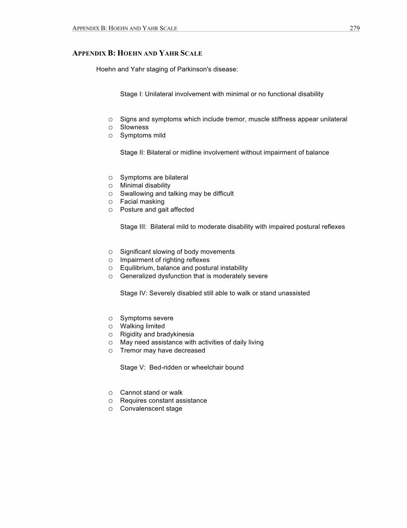
APPENDIX B: HOEHN AND YAHR SCALE 279
APPENDIX B: HOEHN AND YAHR SCALE
Hoehn and Yahr staging of Parkinson's disease:
Stage I: Unilateral involvement with minimal or no functional disability
o Signs and symptoms which include tremor, muscle stiffness appear unilateral o Slowness o Symptoms mild
Stage II: Bilateral or midline involvement without impairment of balance
o Symptoms are bilateral o Minimal disability o Swallowing and talking may be difficult o Facial masking o Posture and gait affected
Stage III: Bilateral mild to moderate disability with impaired postural reflexes
o Significant slowing of body movements o Impairment of righting reflexes o Equilibrium, balance and postural instability o Generalized dysfunction that is moderately severe
Stage IV: Severely disabled still able to walk or stand unassisted
o Symptoms severe o Walking limited o Rigidity and bradykinesia o May need assistance with activities of daily living o Tremor may have decreased
Stage V: Bed-ridden or wheelchair bound
o Cannot stand or walk o Requires constant assistance o Convalenscent stage

APPENDIX C: EXERCISE PROTOCAL 280
APPENDIX C: EXERCISE PROTOCAL

APPENDIX C: EXERCISE PROTOCAL 281

APPENDIX C: EXERCISE PROTOCAL 282

APPENDIX C: EXERCISE PROTOCAL 283

APPENDIX C: EXERCISE PROTOCAL 284

APPENDIX C: EXERCISE PROTOCAL 285

APPENDIX C: EXERCISE PROTOCAL 286

APPENDIX C: EXERCISE PROTOCAL 287

APPENDIX C: EXERCISE PROTOCAL 288

APPENDIX C: EXERCISE PROTOCAL 289
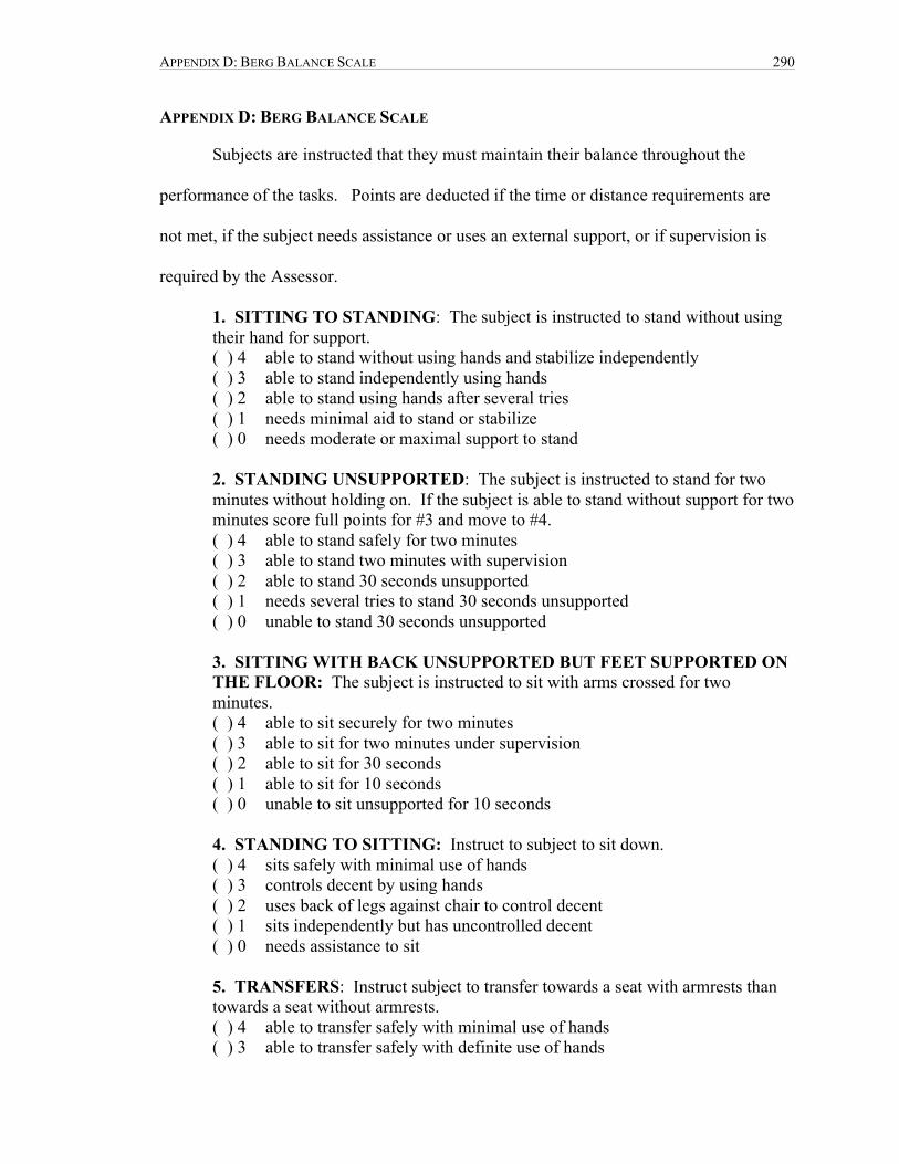
APPENDIX D: BERG BALANCE SCALE 290
APPENDIX D: BERG BALANCE SCALE
Subjects are instructed that they must maintain their balance throughout the
performance of the tasks. Points are deducted if the time or distance requirements are
not met, if the subject needs assistance or uses an external support, or if supervision is
required by the Assessor.
1. SITTING TO STANDING: The subject is instructed to stand without using their hand for support. ( ) 4 able to stand without using hands and stabilize independently ( ) 3 able to stand independently using hands ( ) 2 able to stand using hands after several tries ( ) 1 needs minimal aid to stand or stabilize ( ) 0 needs moderate or maximal support to stand 2. STANDING UNSUPPORTED: The subject is instructed to stand for two minutes without holding on. If the subject is able to stand without support for two minutes score full points for #3 and move to #4. ( ) 4 able to stand safely for two minutes ( ) 3 able to stand two minutes with supervision ( ) 2 able to stand 30 seconds unsupported ( ) 1 needs several tries to stand 30 seconds unsupported ( ) 0 unable to stand 30 seconds unsupported 3. SITTING WITH BACK UNSUPPORTED BUT FEET SUPPORTED ON THE FLOOR: The subject is instructed to sit with arms crossed for two minutes. ( ) 4 able to sit securely for two minutes ( ) 3 able to sit for two minutes under supervision ( ) 2 able to sit for 30 seconds ( ) 1 able to sit for 10 seconds ( ) 0 unable to sit unsupported for 10 seconds 4. STANDING TO SITTING: Instruct to subject to sit down. ( ) 4 sits safely with minimal use of hands ( ) 3 controls decent by using hands ( ) 2 uses back of legs against chair to control decent ( ) 1 sits independently but has uncontrolled decent ( ) 0 needs assistance to sit 5. TRANSFERS: Instruct subject to transfer towards a seat with armrests than towards a seat without armrests. ( ) 4 able to transfer safely with minimal use of hands ( ) 3 able to transfer safely with definite use of hands

APPENDIX D: BERG BALANCE SCALE 291
( ) 2 able to transfer safely with verbal cuing and/or supervision ( ) 1 needs one person to assist ( ) 0 needs two people to assist or supervise to be safe 6. STANDING UNSUPPORTED WITH EYES CLOSED: Instruct the
subject close their eyes and stand still for 10 seconds. ( ) 4 able to stand for 10 seconds safely ( ) 3 able to stand 10 seconds with supervision ( ) 2 able to strand 3 seconds ( ) 1 unable to keep eyes closed for 3 seconds but stays safely ( ) 0 needs help to keep from falling 7. STANDING UNSUPPORTED WITH FEET TOGETHER: Instruct
subject to place feet together and stand without holding on. ( ) 4 able to place feet together independently and stand safely for one minute ( ) 3 able to place feet together independently and stand one minute with
supervision ( ) 2 able to place feet together independently but unable to hold for 30
seconds ( ) 1 needs help to obtain position but able to stand 15 seconds feet together ( ) 0 needs help to attain position and unable to hold to 15 seconds 8. REACHING FORWARD WITH OUTSTRETCHED ARM WHILE
STANDING: Instruct the subject to lift an arm to 90 degrees and to stretch out their fingers and reach forward as far as they can. The Assessor places
a ruler at the end of the fingertips, but not touching. It is up to the individual as to which arm they chose to use. The recorder measure is the distance forward that the fingers reach while the subject is in the most forward lean position. When possible ask the subject to use both arms to minimize trunk rotation. ( ) 4 can reach forward confidently 25cm ( ) 3 can reach forward 12 cm ( ) 2 can reach forward 5 cm ( ) 1 reaches forward but needs supervision ( ) 0 loses balance while trying/requires external support 9. PICK UP OBJECT FROM THE FLOOR FROM A STANDING
POSITION: Instruct subject to pick up the shoe/slipper, which is placed in front of their feet.
( ) 4 able to pick up slipper safely and easily ( ) 3 able to pick up slipper but needs supervision ( ) 2 unable to pick up but reaches 2-5cm from slipper and keeps balance
independently ( ) 1 unable to pick up and needs supervision while trying ( ) 0 unable to try/needs assistance to keep from losing balance or falling
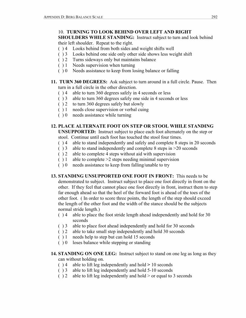
APPENDIX D: BERG BALANCE SCALE 292
10. TURNING TO LOOK BEHIND OVER LEFT AND RIGHT SHOULDERS WHILE STANDING: Instruct subject to turn and look behind their left shoulder. Repeat to the right. ( ) 4 Looks behind from both sides and weight shifts well ( ) 3 Looks behind one side only other side shows less weight shift ( ) 2 Turns sideways only but maintains balance ( ) 1 Needs supervision when turning ( ) 0 Needs assistance to keep from losing balance or falling
11. TURN 360 DEGREES: Ask subject to turn around in a full circle. Pause. Then turn in a full circle in the other direction. ( ) 4 able to turn 360 degrees safely in 4 seconds or less ( ) 3 able to turn 360 degrees safely one side in 4 seconds or less ( ) 2 to turn 360 degrees safely but slowly ( ) 1 needs close supervision or verbal cuing ( ) 0 needs assistance while turning
12. PLACE ALTERNATE FOOT ON STEP OR STOOL WHILE STANDING UNSUPPORTED: Instruct subject to place each foot alternately on the step or stool. Continue until each foot has touched the stool four times. ( ) 4 able to stand independently and safely and complete 8 steps in 20 seconds ( ) 3 able to stand independently and complete 8 steps in >20 seconds ( ) 2 able to complete 4 steps without aid with supervision ( ) 1 able to complete >2 steps needing minimal supervision ( ) 0 needs assistance to keep from falling/unable to try
13. STANDING UNSUPPORTED ONE FOOT IN FRONT: This needs to be demonstrated to subject. Instruct subject to place one foot directly in front on the other. If they feel that cannot place one foot directly in front, instruct them to step far enough ahead so that the heel of the forward foot is ahead of the toes of the other foot. ( In order to score three points, the length of the step should exceed the length of the other foot and the width of the stance should be the subjects normal stride length.) ( ) 4 able to place the foot stride length ahead independently and hold for 30
seconds ( ) 3 able to place foot ahead independently and hold for 30 seconds ( ) 2 able to take small step independently and hold 30 seconds ( ) 1 needs help to step but can hold 15 seconds ( ) 0 loses balance while stepping or standing
14. STANDING ON ONE LEG: Instruct subject to stand on one leg as long as they can without holding on. ( ) 4 able to lift leg independently and hold > 10 seconds ( ) 3 able to lift leg independently and hold 5-10 seconds ( ) 2 able to lift leg independently and hold > or equal to 3 seconds

APPENDIX D: BERG BALANCE SCALE 293
( ) 1 tries to lift leg unable to hold 3 seconds but remains standing independently
( ) 0 unable to try or needs assistance to prevent fall ( ) Total Score (maximum 56)
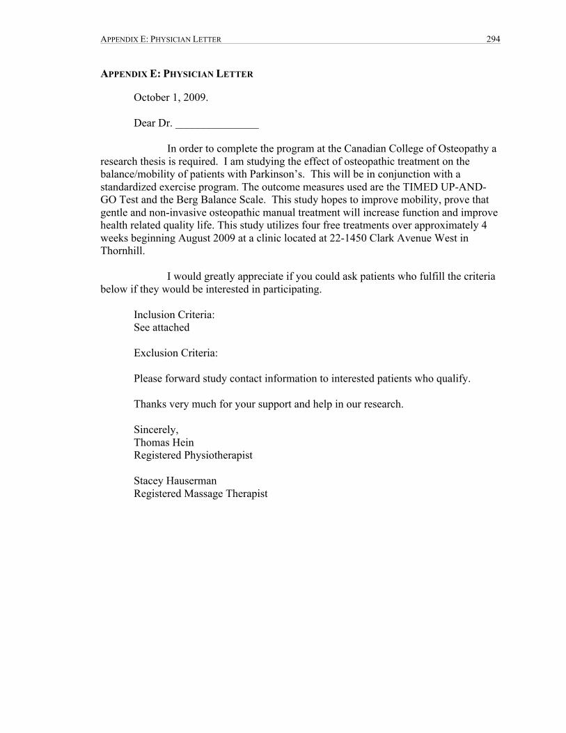
APPENDIX E: PHYSICIAN LETTER 294
APPENDIX E: PHYSICIAN LETTER
October 1, 2009. Dear Dr. _______________ In order to complete the program at the Canadian College of Osteopathy a
research thesis is required. I am studying the effect of osteopathic treatment on the balance/mobility of patients with Parkinson’s. This will be in conjunction with a standardized exercise program. The outcome measures used are the TIMED UP-AND-GO Test and the Berg Balance Scale. This study hopes to improve mobility, prove that gentle and non-invasive osteopathic manual treatment will increase function and improve health related quality life. This study utilizes four free treatments over approximately 4 weeks beginning August 2009 at a clinic located at 22-1450 Clark Avenue West in Thornhill.
I would greatly appreciate if you could ask patients who fulfill the criteria
below if they would be interested in participating. Inclusion Criteria: See attached Exclusion Criteria: Please forward study contact information to interested patients who qualify. Thanks very much for your support and help in our research. Sincerely, Thomas Hein Registered Physiotherapist Stacey Hauserman Registered Massage Therapist

APPENDIX F: BUSINESS CARD 295
APPENDIX F: BUSINESS CARD

APPENDIX G: RECRUITMENT NOTICE 296
APPENDIX G: RECRUITMENT NOTICE
Are you interested in FREE treatment?
Stacey Hauserman and Thomas Hein are currently working on their graduate thesis study from the Canadian College of Osteopathy on:
The Effect of Osteopathic Treatment and Exercise for Mobility and Balance in those
with PARKINSON'S DISEASE.
The purpose of the study is to determine whether osteopathic treatment can improve walking and balance.
Participants will undergo 4 osteopathic treatments and 5 individual and small group exercise classes FREE of charge.
To be included in this research participants are required to:
• Attend 5 sessions of exercise &/or osteopathy (a form of hands on manual therapy)
• Have a primary diagnoses of Parkinson’s disease • Be able to walk with or without an assisted device • Be between the ages of 55 and 90
If you are interested in participating, please call 905-695-0371
Or email: [email protected]

APPENDIX H: TELEPHONE INTERVIEW 297
APPENDIX H: TELEPHONE INTERVIEW
Preliminary Telephone Subject Screening Interview (First Contract)
Subject Name ____________________ Age ________________
Contact Number(s)______________________
Thank you for responding. This study is for a thesis at the Canadian College of
Osteopathy and it is studying Parkinson’s Disease.
1. Can you understand spoken and written English? 2. You will be required to visit a clinic 5 times (over 5 weeks) at 22-1450 Clark
Avenue West in Thornhill. It is called Physioactive. Your first and last appointments may be up to 2 hours long and the others one hour or less. You must be able to attend all these appointments to participate in this study.
3. Parking is free or the clinic is accessible by public transit. 4. To qualify you need to be diagnosed with Parkinson’s Disease by a physician.
You need to have a doctor’s note stating that diagnosis. It can be faxed to 905 695 0833 with ATTN: Parkinson’s disease Study.
5. Osteopathy is a gentle, non-invasive, manual therapy but you still need to be in reasonable physical health. When you are at the clinic you will need to sign a consent sheet, fill out a health history (once) and several questionnaires.
6. You will be required to perform a functional test 7. Your Parkinson’s disease medications will be monitored for the duration of the
study. 8. All personal information is confidential and will be destroyed upon completion of
the study.
To see if you qualify I need to ask you a few questions:
1. How old are you? 2. Do you have a physician diagnosis of Parkinson’s disease? 3. Are you able to walk safely across a room? 4. Is there any reason that you might not be able to stand from sitting and walk three
meters and then sit back down again? 5. Do you have significant back or leg pain that would inhibit you from getting up
out of a chair?
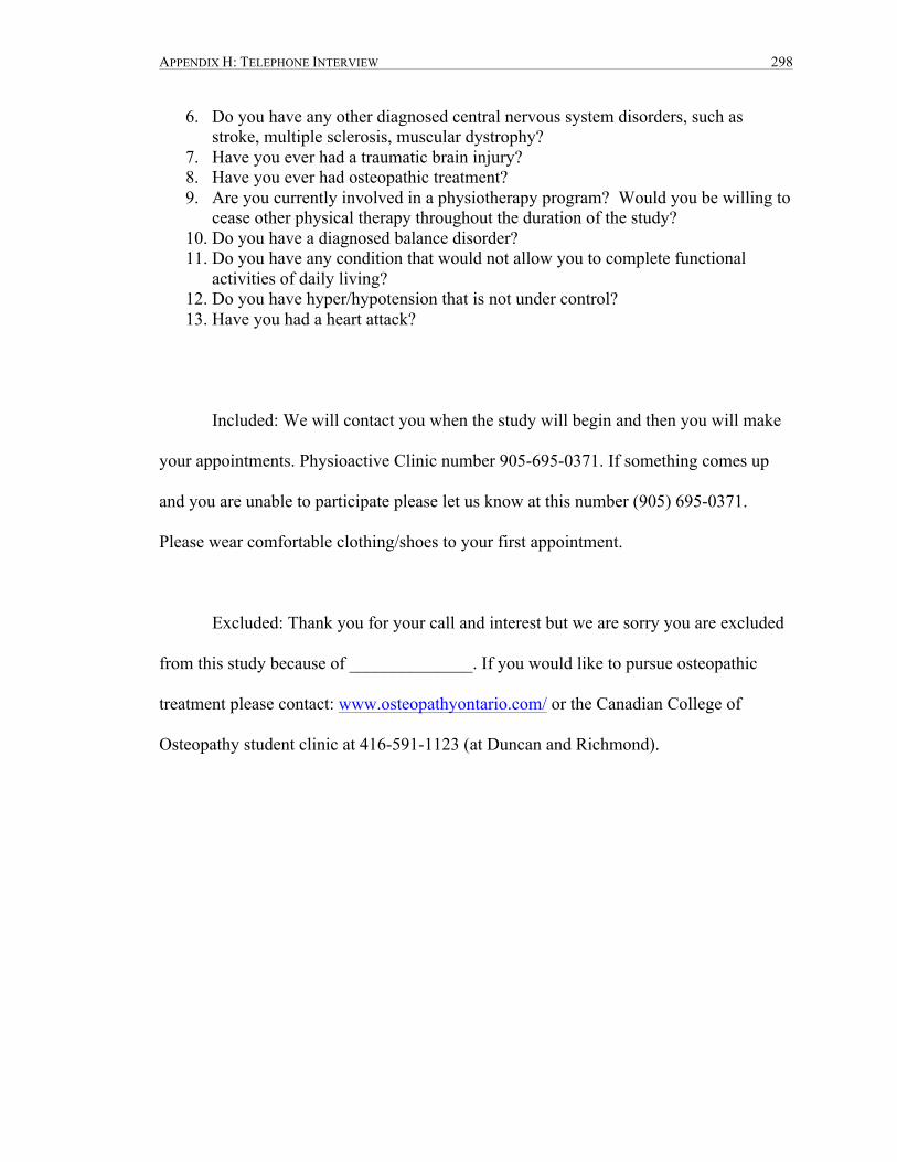
APPENDIX H: TELEPHONE INTERVIEW 298
6. Do you have any other diagnosed central nervous system disorders, such as stroke, multiple sclerosis, muscular dystrophy?
7. Have you ever had a traumatic brain injury? 8. Have you ever had osteopathic treatment? 9. Are you currently involved in a physiotherapy program? Would you be willing to
cease other physical therapy throughout the duration of the study? 10. Do you have a diagnosed balance disorder? 11. Do you have any condition that would not allow you to complete functional
activities of daily living? 12. Do you have hyper/hypotension that is not under control? 13. Have you had a heart attack?
Included: We will contact you when the study will begin and then you will make
your appointments. Physioactive Clinic number 905-695-0371. If something comes up
and you are unable to participate please let us know at this number (905) 695-0371.
Please wear comfortable clothing/shoes to your first appointment.
Excluded: Thank you for your call and interest but we are sorry you are excluded
from this study because of ______________. If you would like to pursue osteopathic
treatment please contact: www.osteopathyontario.com/ or the Canadian College of
Osteopathy student clinic at 416-591-1123 (at Duncan and Richmond).

APPENDIX I: DATA LOG 299
APPENDIX I: DATA LOG
Patient Number Control Experimental
1. X
2. X
3. X
4. X
5. X
6. X
7. X
8. X
9. X X
10. X
11. X
12.
13.
14.
15.
16.
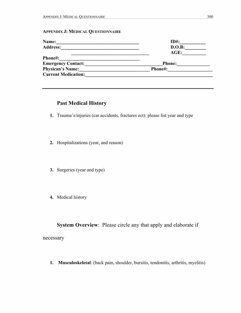
APPENDIX J: MEDICAL QUESTIONNAIRE 300
APPENDIX J: MEDICAL QUESTIONNAIRE
Name:___________________________________ ID#:___________ Address:_________________________________ D.O.B:_________
_________________________________ AGE:__________ Phone#:__________________________________ Emergency Contact:_________________________________Phone:______________ Physican’s Name:______________________________ Phone#:___________________ Current Medication:______________________________________________________
Past Medical History
1. Trauma’s/injuries (car accidents, fractures ect): please list year and type
2. Hospitalizations (year, and reason)
3. Surgeries (year and type)
4. Medical history
System Overview: Please circle any that apply and elaborate if
necessary
1. Musculoskeletal: (back pain, shoulder, bursitis, tendonitis, arthritis, myelitis)

APPENDIX J: MEDICAL QUESTIONNAIRE 301
2. Circulatory: (phlebitis, varicosities, cramps, high/low blood pressure, cholesterol, cardiac problems, numbness, cold extremities)
3. Respiratory: (allergies, cold, cough, asthma, bronchitis, emphysema)
4. Digestive: (gastric reflux, heartburn, ulcer, bowel movements, gas, bloating, nausea, appetite, dysphasia, slow digestion, hemorrhoid, cirrhosis, hepatitis, gallstone, diarrhea, constipation)
5. Urinary: (cystitis, dysuria, polyuria, burning, kidney stone, kidney insufficiency, incontinence, bladder ptosis)
6. Gynecology/Urogenital : (prostatitis, sexual dysfunction, pregnancy, endometrosis)
7. Ear/Nose/Throat: (tinnitus, rhinitis, loss of hearing, glaucoma, transitory loss of vision)

APPENDIX J: MEDICAL QUESTIONNAIRE 302
8. Nervous System: (headaches, migraine)
9. Skin: (psoriasis, parasites, infection, eczema, dermatitis)
10. Endocrine: (thyroid, adrenals, pancreas, gonads)
11. Sleeping habits:
12. Anything else that you feel is relevant:

APPENDIX K: PRIVACY POLICY 303
APPENDIX K: PRIVACY POLICY
Privacy Policy Personal information is regulated federally and may be defined as any information
of a personal nature including personal characteristics, health, activities and views. In Ontario, health specific information is regulated provincially and may be collected either orally or recorded. All information collected and used is for the primary purpose of compiling research to complete a thesis with the Canadian College of Osteopathy.
Paper information and electronic hardware is either under supervision or secured in a locked area at all times. Paper files containing personal information will be destroyed by shredding when they are no longer needed for the purpose of this study. Electronic data will be destroyed by deleting when the information is no longer required for the purpose of this study. In the unlikely event of privacy breech you will be informed immediately.
If you have any questions regarding these privacy practices please feel free to ask for clarification from either Stacey Hauserman or Thomas Hein.
If we are not able to satisfy your concerns please feel free to contact the Canadian College of Osteopathy at 416-
Thank you, Stacey Hauserman, RMT Thomas Hein, PT (osteopathic thesis writers) For general inquires regarding privacy information in Canada: Information and Privacy Commissioner of Canada 12 Kent St, Ottawa, ON K1A 1H3 613-995-8210, 1-800-282-1376 www.privcom.gc.ca
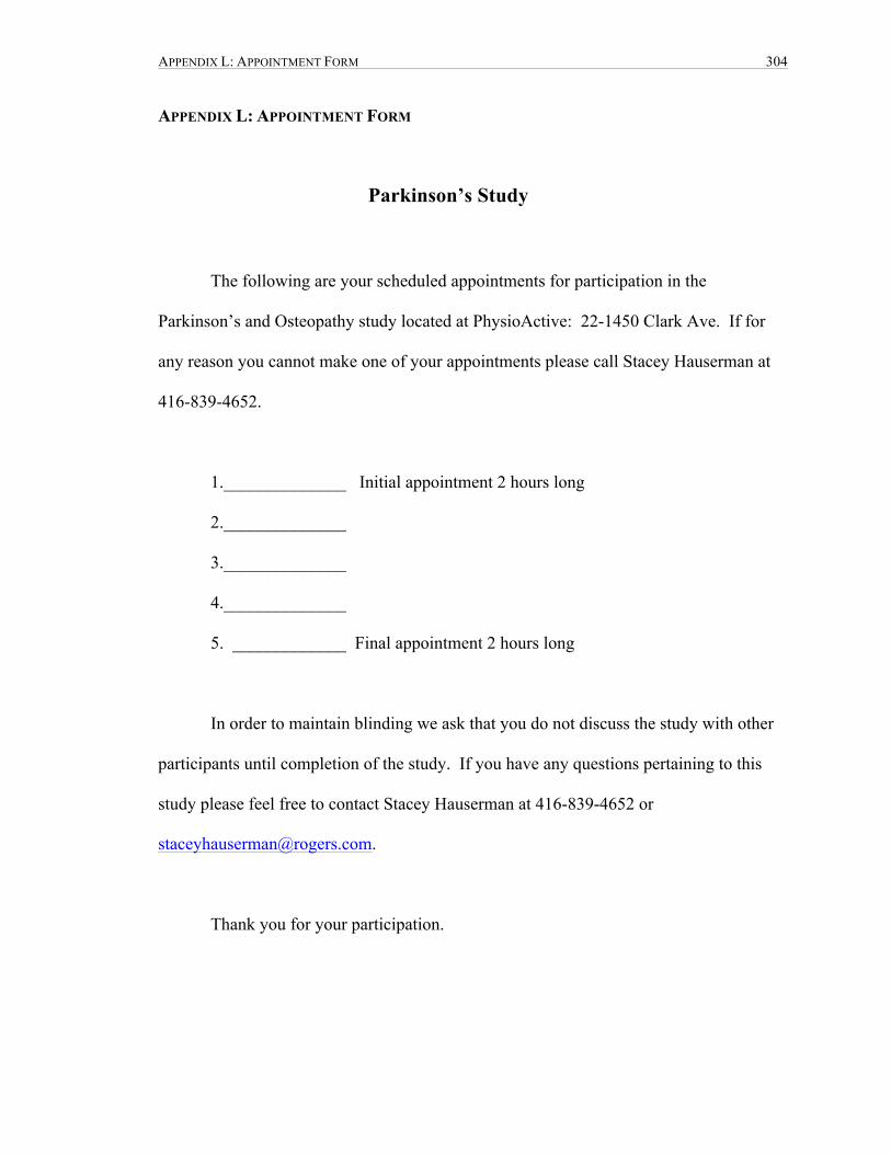
APPENDIX L: APPOINTMENT FORM 304
APPENDIX L: APPOINTMENT FORM
Parkinson’s Study
The following are your scheduled appointments for participation in the
Parkinson’s and Osteopathy study located at PhysioActive: 22-1450 Clark Ave. If for
any reason you cannot make one of your appointments please call Stacey Hauserman at
416-839-4652.
1.______________ Initial appointment 2 hours long
2.______________
3.______________
4.______________
5. _____________ Final appointment 2 hours long
In order to maintain blinding we ask that you do not discuss the study with other
participants until completion of the study. If you have any questions pertaining to this
study please feel free to contact Stacey Hauserman at 416-839-4652 or
Thank you for your participation.

APPENDIX M: OSTEOPATHIC ASSESSMENT FORM 305
APPENDIX M: OSTEOPATHIC ASSESSMENT FORM
Subject #_____________________ Date_________________________
Observations Line of Barre Ascending Descending Neutral Typology Anterior Posterior Neutral Compensation Compensated Decompensated Symmetry Eyes
Ears Shoulders Scapula Iliacs
Cranial Base Structure Specific
Structure Position Classification Vitality
Sphenobasilar Symphysis
Temporal OM Right Left
Petrobasilar Left Right
Occiput
Vitality 0- absent 1-poor 2-fair 3-normal
Classification of Lesions According to CCO Methodology 4- Compaction 3- Non Physiological Without Respect to the Axis 2- Non Physiological With Respect to the Axis 1- Physiological 0-Normal

APPENDIX M: OSTEOPATHIC ASSESSMENT FORM 306
Cranial Vault
Structure Position Classification Vitality
Parietals Left Right
Temporals Left Right
Frontal Left Right
Occipital Squama
Sphenoid Greater Wings
CO/C1 Right Left Bilateral
CO/C1 on C2
Ethmoid
Jugular Foramen Foramen Magnum

APPENDIX M: OSTEOPATHIC ASSESSMENT FORM 307
Facial Bones Structure Position Classification Vitality
Zygoma Left Right
Maxilla Left Right
Mandible
Palatine Left Right
Vomer
Lacrimal Left Right
Nasal Left Right
Sacrum Structure Position Classification Vitality
Sacrum
L5 – S1
Sacroiliac Left Right

APPENDIX M: OSTEOPATHIC ASSESSMENT FORM 308
Cervical Spine Segment Position Classification Vitality
C1
C2
C3
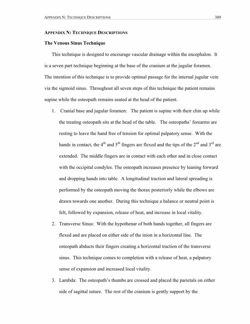
APPENDIX N: TECHNIQUE DESCRIPTIONS 309
APPENDIX N: TECHNIQUE DESCRIPTIONS
The Venous Sinus Technique
This technique is designed to encourage vascular drainage within the encephalon. It
is a seven part technique beginning at the base of the cranium at the jugular foramen.
The intention of this technique is to provide optimal passage for the internal jugular vein
via the sigmoid sinus. Throughout all seven steps of this technique the patient remains
supine while the osteopath remains seated at the head of the patient.
1. Cranial base and jugular foramen: The patient is supine with their chin up while
the treating osteopath sits at the head of the table. The osteopaths’ forearms are
resting to leave the hand free of tension for optimal palpatory sense. With the
hands in contact, the 4th and 5th fingers are flexed and the tips of the 2nd and 3rd are
extended. The middle fingers are in contact with each other and in close contact
with the occipital condyles. The osteopath increases presence by leaning forward
and dropping hands into table. A longitudinal traction and lateral spreading is
performed by the osteopath moving the thorax posteriorly while the elbows are
drawn towards one another. During this technique a balance or neutral point is
felt, followed by expansion, release of heat, and increase in local vitality.
2. Transverse Sinus: With the hypothenar of both hands together, all fingers are
flexed and are placed on either side of the inion in a horizontal line. The
osteopath abducts their fingers creating a horizontal traction of the transverse
sinus. This technique comes to completion with a release of heat, a palpatory
sense of expansion and increased local vitality.
3. Lambda: The osteopath’s thumbs are crossed and placed the parietals on either
side of sagittal suture. The rest of the cranium is gently support by the
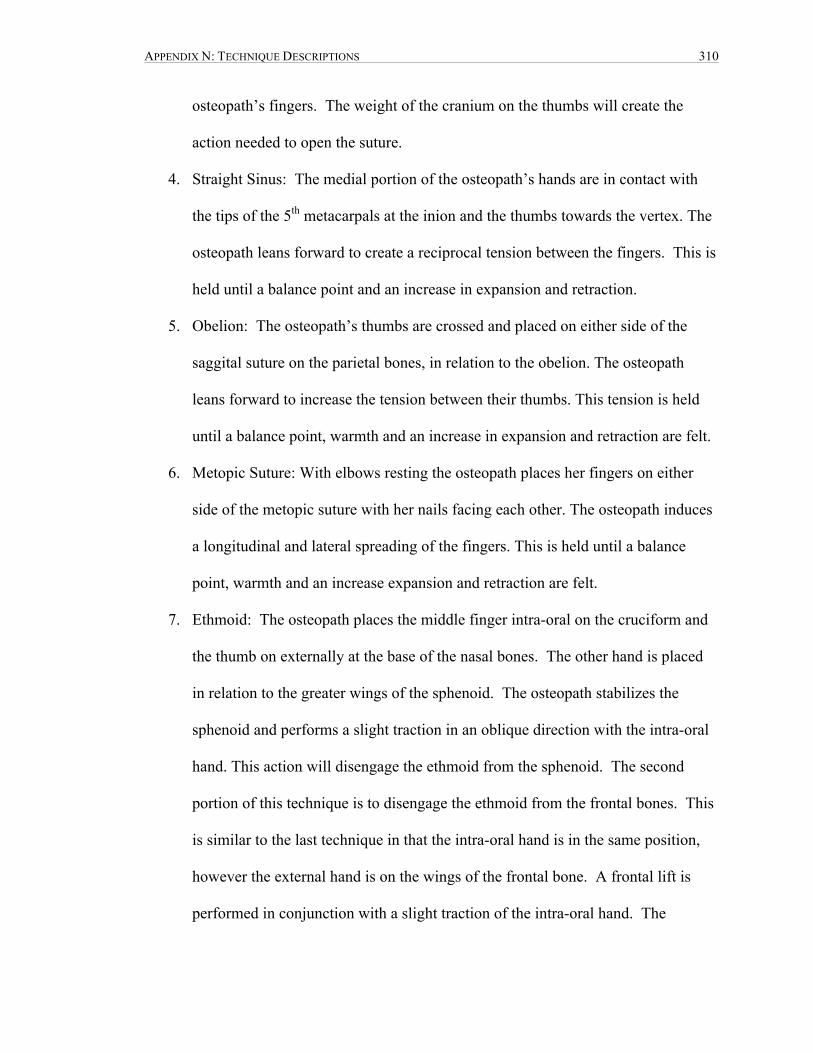
APPENDIX N: TECHNIQUE DESCRIPTIONS 310
osteopath’s fingers. The weight of the cranium on the thumbs will create the
action needed to open the suture.
4. Straight Sinus: The medial portion of the osteopath’s hands are in contact with
the tips of the 5th metacarpals at the inion and the thumbs towards the vertex. The
osteopath leans forward to create a reciprocal tension between the fingers. This is
held until a balance point and an increase in expansion and retraction.
5. Obelion: The osteopath’s thumbs are crossed and placed on either side of the
saggital suture on the parietal bones, in relation to the obelion. The osteopath
leans forward to increase the tension between their thumbs. This tension is held
until a balance point, warmth and an increase in expansion and retraction are felt.
6. Metopic Suture: With elbows resting the osteopath places her fingers on either
side of the metopic suture with her nails facing each other. The osteopath induces
a longitudinal and lateral spreading of the fingers. This is held until a balance
point, warmth and an increase expansion and retraction are felt.
7. Ethmoid: The osteopath places the middle finger intra-oral on the cruciform and
the thumb on externally at the base of the nasal bones. The other hand is placed
in relation to the greater wings of the sphenoid. The osteopath stabilizes the
sphenoid and performs a slight traction in an oblique direction with the intra-oral
hand. This action will disengage the ethmoid from the sphenoid. The second
portion of this technique is to disengage the ethmoid from the frontal bones. This
is similar to the last technique in that the intra-oral hand is in the same position,
however the external hand is on the wings of the frontal bone. A frontal lift is
performed in conjunction with a slight traction of the intra-oral hand. The

APPENDIX N: TECHNIQUE DESCRIPTIONS 311
technique is completed when there is a neutral point, heat and an increase in
expansion and retraction.
Normalization of the Posterior Fossa (EV4)
The EV4 is a reciprocal balance technique that is used to normalize the posterior
fossa.
Patient is positioned supine with his chin slightly elevated. The osteopath sits at
the head of the patient and supports her arms on the table. She places her 5th fingers at
the level of the inion while the rest of her fingers are in relation to the falx cerebella
insertion. The osteopath’s thumbs are on the tips of the mastoids. The osteopath than
spreads the posterior fossa into two compartments by providing a longitudinal and
transverse between the fingers. This reciprocal balance is maintained around a calm
reference point until a balance point, still point, and return of PRM are obtained.
Dorsiflexion can be added to increase overall tension.
Compression of the Fourth Ventricle (CV4)
Patient is positioned in supine with her chin slightly elevated. The osteopath sits
at the head of the patient with his arms supported. The osteopath hypothenar’s and ulnar
border of his hands are connected in a cup like position. The hands are placed below the
tentorium with the thenar eminence of both hands on the external bevels of the occiput.
The osteopath then applies a light medial compression followed by a reciprocal tension
posteriorly with a light cephalic traction. A reciprocal balance is established at the
osseous level followed by a second reciprocal balance corresponding to the ventricular
volume. The osteopathy waits for a balance point, still point and release. After release
an expansion and retraction of the ventricular system should be felt.

APPENDIX N: TECHNIQUE DESCRIPTIONS 312
Parietal Lift
The patient is supine with the osteopath sitting at the head of the patient. The
osteopaths fingers are placed along the external bevels of the parietal bones, with the
thumbs joined above the head. The osteopath disengages the external bevels by creating a
medial compression and then leans back to traction within the spring of the tissue, to
disengage the parietals. This is held until a balance point, still point and return of the
primary respiratory mechanism are achieved.
The second portion of the parietal lift is used to decompress the encephalon from
the base of the cranium. The osteopath maintains the same position but allows palpation
to sink to the second spring. A reciprocal tension is placed on the membranes on the
inspiration phase and held for three to four primary respiratory cycles. This tension is
released at 2/3 of the expiration cycle. This is repeated a few times until more motion is
perceived.
Bilateral Rocking of the Temporal Bones
(W.G Sutherland, adapted by Phillipe Druelle, D.O.)
This technique is used to stimulate the central thalamic region and the basal
ganglia.
The patient lies supine with a pillow under their knees and the chin slightly
elevated. The treating osteopath sits at the head of the patient with their forearms
supported. The osteopath’s hands are cupped under the occiput with the tips of her
thumbs at the mastoid processes. The osteopath leans forward flexing her trunk to bring
the mastoids into a flexion state. This is done at the speed and intensity of the patient’s

APPENDIX N: TECHNIQUE DESCRIPTIONS 313
tissue. This rocking motion is continued until the initial inertia that was perceived has
softened.
Lateral Ventricles
The osteopath sits at the head of the patient and places with both thumbs along the
external bevels of the parietals facing the ceiling. The 4th and 5th fingers are place at the
tips of the mastoid processes with the index fingers along the mandibular condyles and
the fingers along the mandible. The osteopath achieves a reciprocal tension by straighten
their thorax which causes a medial compression of the parietals and a anterior-inferior
disengagement of the mandible.
Often a still point is required in the first spring in order to reach the lateral
ventricles. Once the second spring level is achieved the osteopath establishes a reciprocal
balance of the endocranium in its entirety. Following a still point and release the
osteopath will land onto the lateral ventricles after perceiving an anterior tilt and a deeper
sinking sensation. Once on the lateral ventricles the osteopath will balance each ventricle
around its calm reference point. A third reference point is then established in the middle.
After a still point and release are achieved there should be an increase in the ventricular
motion.

APPENDIX O: CANADIAN COLLEGE OF OSTEOPATHY METHODOLOGY 314
APPENDIX O: CANADIAN COLLEGE OF OSTEOPATHY METHODOLOGY
Classification of Lesions in order of priority based on the clinical methodology of
the Canadian College of Osteopathy
Classification of Lesions
1. Scars
2. Compactions
3. Non-physiological without respect to the axis lesions
4. Non-physiological with respect to the axis lesions
5. Physiology lesions
The priority can be further determined by the vitality of each tissue assessed.

APPENDIX P: STATISTICIAN LETTER 315
APPENDIX P: STATISTICIAN LETTER
June 5, 2011
To Whom It May Concern,
I was the Statistician for the research thesis: ‘Osteopathic Treatment for Balance in
Parkinson’s Disease Patients’ authored by Stacey Hauserman.
The analysis, tables and graphs were done with SPSS Release 16. All work was done in a
professional manner using appropriate statistical techniques.
Regards,
Peter Lewycky
B.Sc., M.Eng., P.Eng.

APPENDIX Q: LESION CLASSIFICATION CORRELATION TABLES 316
APPENDIX Q: LESION CLASSIFICATION CORRELATION TABLES
For Control, there is no statistically significant relationship between pre and post
and the distribution of SBS scores; Exact significance, p= 0.771. For Experimental, there
is a statistically significant difference in the distribution of SBS scores between pre and
post; Exact significance, p= 0.005.

APPENDIX Q: LESION CLASSIFICATION CORRELATION TABLES 317
For Control, there is no statistically significant relationship between pre and post
and the distribution of TB OM-L scores; Exact significance, p= 0.821. For Experimental,
there is a statistically significant difference in the distribution of scores between pre and
post; Exact significance, p= 0.010.

APPENDIX Q: LESION CLASSIFICATION CORRELATION TABLES 318
For Control, there is no statistically significant relationship between pre and post
and the distribution of TB OM-R scores; Exact significance p= 0.771. For Experimental,
there is a statistically significant difference in the distribution of scores between pre and
post; Exact significance, p= 0.001.

APPENDIX Q: LESION CLASSIFICATION CORRELATION TABLES 319
For Control, there is no statistically significant relationship between pre and post
and the distribution of Pet-Bas L scores; Exact significance, p= 1.000. For Experimental,
there is a statistically significant difference in the distribution of scores between pre and
post; Exact significance, p= 0.016.
For Control, there is no statistically significant relationship between pre and post
and the distribution of Pet-Bas R scores, p= 1.000, For Experimental, there is a

APPENDIX Q: LESION CLASSIFICATION CORRELATION TABLES 320
statistically significant difference in the distribution of scores between pre and post; p=
0.005.
For Control, there is no statistically significant relationship between pre and post
and the distribution of C0-C1 L scores; Exact significance, p= 0.829, For Experimental,
there is a statistically significant difference in the distribution of scores between pre and
post; Exact significance, p= 0.005.

APPENDIX Q: LESION CLASSIFICATION CORRELATION TABLES 321
For Control, there is no statistically significant relationship between pre and post
and the distribution of C0-C1 R scores; Exact significance p= 0.743. For Experimental,
there is no statistically significant difference in the distribution of scores between pre and
post; Exact significance, p= 0.462.

APPENDIX Q: LESION CLASSIFICATION CORRELATION TABLES 322
For Control, there is no statistically significant relationship between pre and post
and the distribution of C0-C1/C2 scores; Exact significance, p= 1.000. For Experimental,
there is a statistically significant difference in the distribution of scores between pre and
post; Exact significance, p= 0.037.
For Control, there is no statistically significant relationship between pre and post
and the distribution of Eth scores; Exact significance, p= 0.657. For Experimental, there
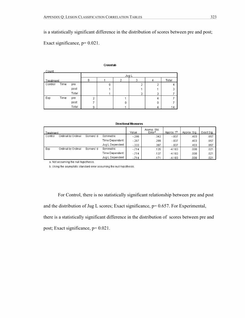
APPENDIX Q: LESION CLASSIFICATION CORRELATION TABLES 323
is a statistically significant difference in the distribution of scores between pre and post;
Exact significance, p= 0.021.
For Control, there is no statistically significant relationship between pre and post
and the distribution of Jug L scores; Exact significance, p= 0.657. For Experimental,
there is a statistically significant difference in the distribution of scores between pre and
post; Exact significance, p= 0.021.

APPENDIX Q: LESION CLASSIFICATION CORRELATION TABLES 324
For Control, there is no statistically significant relationship between pre and post
and the distribution of Jug R scores; Exact significance, p= 1.000. For Experimental,
there is no statistically significant difference in the distribution of scores between pre and
post; Exact significance, p= 1.000.

APPENDIX Q: LESION CLASSIFICATION CORRELATION TABLES 325
For Control, there is no statistically significant relationship between pre and post
and the distribution of For Mag scores; Exact significance, p= 0.743. For Experimental,
there is a statistically significant difference in the distribution of scores between pre and
post; Exact significance, p= 0.002.

APPENDIX Q: LESION CLASSIFICATION CORRELATION TABLES 326
For Control, there is no statistically significant relationship between pre and post
and the distribution of Par L scores; Exact significance, p= 1.000. For Experimental,
there is no statistically significant difference in the distribution of scores between pre and
post; Exact significance, p= 0.119.
For Control, there is no statistically significant relationship between pre and post
and the distribution of Par T scores; Exact significance, p= 0.486. For Experimental,
there is no statistically significant difference in the distribution of scores between pre and
post; Exact significance, p= 0.315.

APPENDIX Q: LESION CLASSIFICATION CORRELATION TABLES 327
For Control, there is no statistically significant relationship between pre and post
and the distribution of TL scores; Exact significance, p= 1.000. For Experimental, there
is no statistically significant difference in the distribution of scores between pre and post;
Exact significance, p= 0.315.
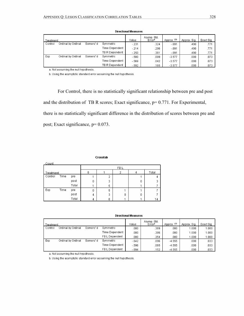
APPENDIX Q: LESION CLASSIFICATION CORRELATION TABLES 328
For Control, there is no statistically significant relationship between pre and post
and the distribution of TB R scores; Exact significance, p= 0.771. For Experimental,
there is no statistically significant difference in the distribution of scores between pre and
post; Exact significance, p= 0.073.
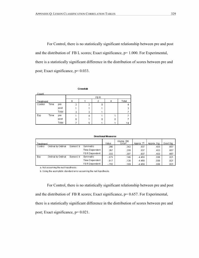
APPENDIX Q: LESION CLASSIFICATION CORRELATION TABLES 329
For Control, there is no statistically significant relationship between pre and post
and the distribution of FB L scores; Exact significance, p= 1.000. For Experimental,
there is a statistically significant difference in the distribution of scores between pre and
post; Exact significance, p= 0.033.
For Control, there is no statistically significant relationship between pre and post
and the distribution of FB R scores; Exact significance, p= 0.657. For Experimental,
there is a statistically significant difference in the distribution of scores between pre and
post; Exact significance, p= 0.021.

APPENDIX Q: LESION CLASSIFICATION CORRELATION TABLES 330
For Control, there is no statistically significant relationship between pre and post
and the distribution of Occ Sq scores; Exact significance, p= 0.771. For Experimental,
there is a statistically significant difference in the distribution of scores between pre and
post; Exact significance, p= 0.002.

APPENDIX Q: LESION CLASSIFICATION CORRELATION TABLES 331
For Control, there is no statistically significant relationship between pre and post
and the distribution of Sph > wing scores; Exact significance, p= 1.000. For
Experimental, there is no statistically significant difference in the distribution of scores
between pre and post; Exact significance, p= 0.510.
For Control, there is no statistically significant relationship between pre and post
and the distribution of Zyg L scores; Exact significance, p= 1.000. For Experimental,

APPENDIX Q: LESION CLASSIFICATION CORRELATION TABLES 332
there is a statistically significant difference in the distribution of scores between pre and
post; Exact significance, p= 0.122.
For Control, there is no statistically significant relationship between pre and post
and the distribution of Zyg R scores; Exact significance, p= 1.000. For Experimental,
there is no statistically significant difference in the distribution of scores between pre and
post; Exact significance, p= 0.070.

APPENDIX Q: LESION CLASSIFICATION CORRELATION TABLES 333
For Control, there is no statistically significant relationship between pre and post
and the distribution of Max L scores; Exact significance, p= 0.200. For Experimental,
there is no statistically significant difference in the distribution of scores between pre and
post; Exact significance, p= 0.478.
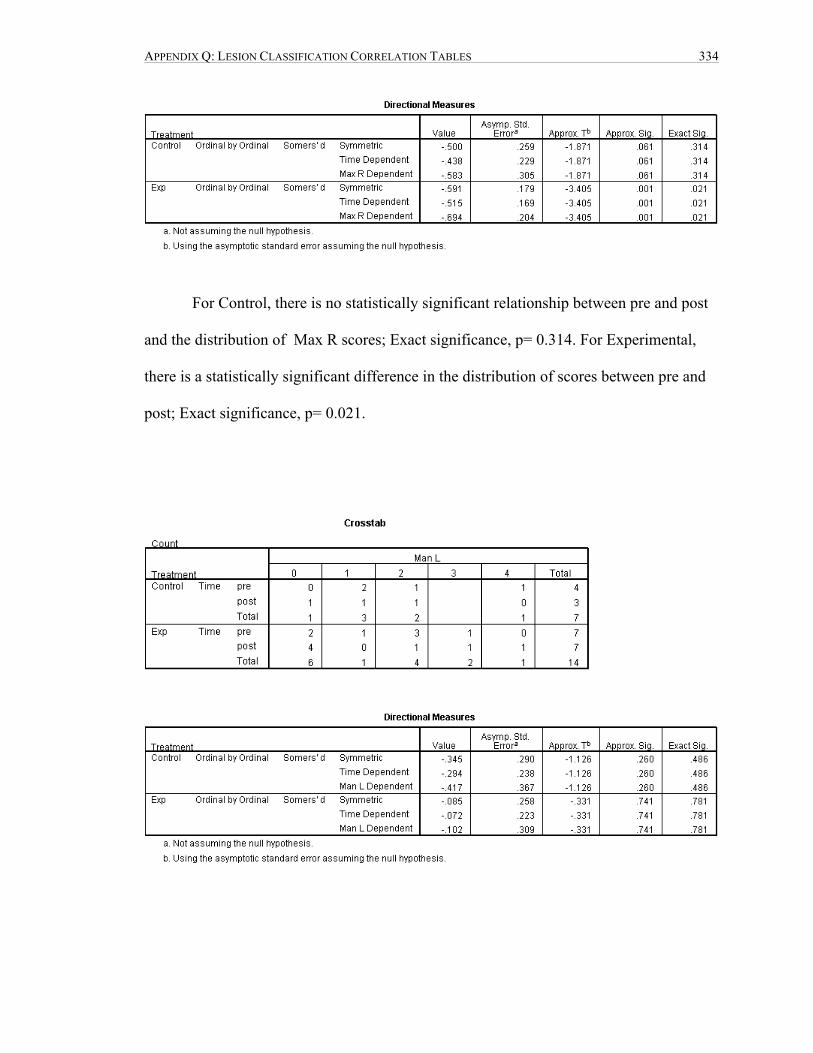
APPENDIX Q: LESION CLASSIFICATION CORRELATION TABLES 334
For Control, there is no statistically significant relationship between pre and post
and the distribution of Max R scores; Exact significance, p= 0.314. For Experimental,
there is a statistically significant difference in the distribution of scores between pre and
post; Exact significance, p= 0.021.

APPENDIX Q: LESION CLASSIFICATION CORRELATION TABLES 335
For Control, there is no statistically significant relationship between pre and post
and the distribution of Man L scores; Exact significance, p= 0.486. For Experimental,
there is no statistically significant difference in the distribution of scores between pre and
post; Exact significance, p= 0.781.
For Control, there is no statistically significant relationship between pre and post
and the distribution of Man R scores; Exact significance, p= 0.829. For Experimental,
there is no statistically significant difference in the distribution of scores between pre and
post; Exact significance, p= 0.780.

APPENDIX Q: LESION CLASSIFICATION CORRELATION TABLES 336
For Control, there is no statistically significant relationship between pre and post
and the distribution of Pal L scores; Exact significance, p= 0.429. For Experimental,
there is no statistically significant difference in the distribution of scores between pre and
post; Exact significance, p= 1.000.

APPENDIX Q: LESION CLASSIFICATION CORRELATION TABLES 337
For Control, there is no statistically significant relationship between pre and post
and the distribution of Pal R scores; Exact significance, p= 1.000. For Experimental,
there is no statistically significant difference in the distribution of scores between pre and
post; Exact significance, p= 0.192.

APPENDIX Q: LESION CLASSIFICATION CORRELATION TABLES 338
For Control, there is no statistically significant relationship between pre and post
and the distribution of Vomer scores; Exact significance, p= 0.571. For Experimental,
there is a statistically significant difference in the distribution of scores between pre and
post; Exact significance, p= 0.013.

APPENDIX Q: LESION CLASSIFICATION CORRELATION TABLES 339
For Control, there is no statistically significant relationship between pre and post
and the distribution of Lac L scores; Exact significance, p= 0.400. For Experimental,
there is no statistically significant difference in the distribution of scores between pre and
post; Exact significance, p= 0.592.
For Control, there is no statistically significant relationship between pre and post
and the distribution of Lac R scores; Exact significance, p= 1.000. For Experimental,
there is no statistically significant difference in the distribution of scores between pre and
post; Exact significance, p= 0.731.

APPENDIX Q: LESION CLASSIFICATION CORRELATION TABLES 340
For Control, there is no statistically significant relationship between pre and post
and the distribution of Nas L scores; Exact significance, p= 0.429. For Experimental,
there is no statistically significant difference in the distribution of scores between pre and
post; Exact significance, p= 0.286.
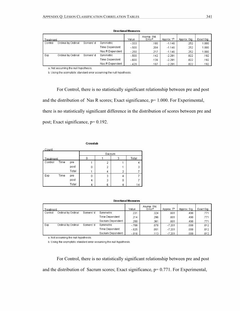
APPENDIX Q: LESION CLASSIFICATION CORRELATION TABLES 341
For Control, there is no statistically significant relationship between pre and post
and the distribution of Nas R scores; Exact significance, p= 1.000. For Experimental,
there is no statistically significant difference in the distribution of scores between pre and
post; Exact significance, p= 0.192.
For Control, there is no statistically significant relationship between pre and post
and the distribution of Sacrum scores; Exact significance, p= 0.771. For Experimental,

APPENDIX Q: LESION CLASSIFICATION CORRELATION TABLES 342
there is no statistically significant difference in the distribution of scores between pre and
post; Exact significance, p= 0.012.
For Control, there is no statistically significant relationship between pre and post
and the distribution of L5-S1 scores; Exact significance, p= 0.971. For Experimental,
there is no statistically significant difference in the distribution of scores between pre and
post; Exact significance, p= 0.202.

APPENDIX Q: LESION CLASSIFICATION CORRELATION TABLES 343
For Control, there is no statistically significant relationship between pre and post
and the distribution of INN L scores; Exact significance, p= 1.000. For Experimental,
there is a statistically significant difference in the distribution of scores between pre and
post; Exact significance, p= 0.315.

APPENDIX Q: LESION CLASSIFICATION CORRELATION TABLES 344
For Control, there is no statistically significant relationship between pre and post
and the distribution of INN R scores; Exact significance, p= 0.686. For Experimental,
there is a statistically significant difference in the distribution of scores between pre and
post; Exact significance, p= 0.031.

APPENDIX Q: LESION CLASSIFICATION CORRELATION TABLES 345
For Control, there is no statistically significant relationship between pre and post
and the distribution of C3 scores; Exact significance, p= 0.571. For Experimental, there
is a statistically significant difference in the distribution of scores between pre and post;
Exact significance, p= 0.021.

APPENDIX R: VITALITY CORRELATION TABLES 346
APPENDIX R: VITALITY CORRELATION TABLES
For Control, there is no statistically significant relationship between pre and post
and the distribution of SBS scores; Exact significance, p= 1.000. For Experimental, there
is a statistically significant difference in the distribution of SBS scores between pre and
post; Exact significance, p= 0.001.

APPENDIX R: VITALITY CORRELATION TABLES 347
For Control, there is no statistically significant relationship between pre and post
and the distribution of TB OM-L scores; Exact significance, p= 0.429. For Experimental,
there is a statistically significant difference in the distribution of scores between pre and
post; Exact significance, p= 0.015.

APPENDIX R: VITALITY CORRELATION TABLES 348
For Control, there is no statistically significant relationship between pre and post
and the distribution of TB OM-R scores; Exact significance p= 0.771. For Experimental,
there is a statistically significant difference in the distribution of scores between pre and
post; Exact significance, p= 0.001.
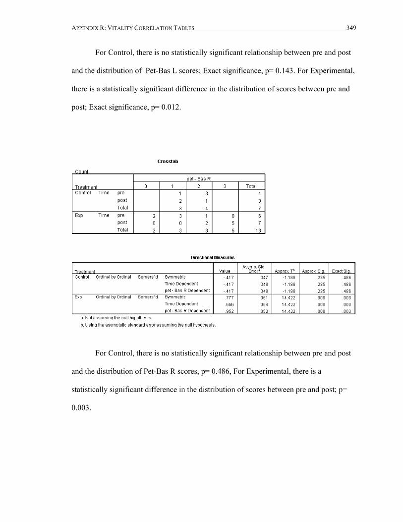
APPENDIX R: VITALITY CORRELATION TABLES 349
For Control, there is no statistically significant relationship between pre and post
and the distribution of Pet-Bas L scores; Exact significance, p= 0.143. For Experimental,
there is a statistically significant difference in the distribution of scores between pre and
post; Exact significance, p= 0.012.
For Control, there is no statistically significant relationship between pre and post
and the distribution of Pet-Bas R scores, p= 0.486, For Experimental, there is a
statistically significant difference in the distribution of scores between pre and post; p=
0.003.

APPENDIX R: VITALITY CORRELATION TABLES 350
For Control, there is no statistically significant relationship between pre and post
and the distribution of C0-C1 L scores; Exact significance, p= 0.829, For Experimental,
there is a statistically significant difference in the distribution of scores between pre and
post; Exact significance, p= 0.003.
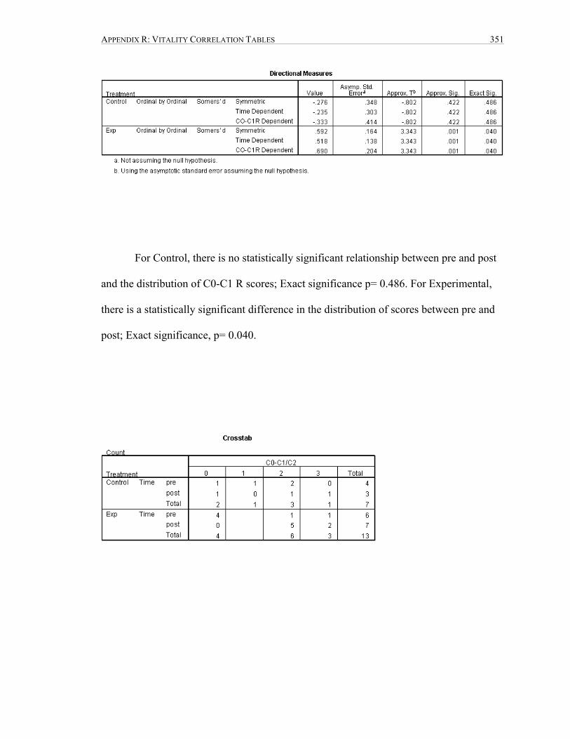
APPENDIX R: VITALITY CORRELATION TABLES 351
For Control, there is no statistically significant relationship between pre and post
and the distribution of C0-C1 R scores; Exact significance p= 0.486. For Experimental,
there is a statistically significant difference in the distribution of scores between pre and
post; Exact significance, p= 0.040.

APPENDIX R: VITALITY CORRELATION TABLES 352
For Control, there is no statistically significant relationship between pre and post
and the distribution of C0-C1/C2 scores; Exact significance, p= 0.686. For Experimental,
there is a statistically significant difference in the distribution of scores between pre and
post; Exact significance, p= 0.031.

APPENDIX R: VITALITY CORRELATION TABLES 353
For Control, there is no statistically significant relationship between pre and post
and the distribution of Eth scores; Exact significance, p= 0.400. For Experimental, there
is a statistically significant difference in the distribution of scores between pre and post;
Exact significance, p= 0.012.

APPENDIX R: VITALITY CORRELATION TABLES 354
For Control, there is no statistically significant relationship between pre and post
and the distribution of Jug L scores; Exact significance, p= 0.429. For Experimental,
there is a statistically significant difference in the distribution of scores between pre and
post; Exact significance, p= 0.009. Post has higher scores.

APPENDIX R: VITALITY CORRELATION TABLES 355
For Control, there is no statistically significant relationship between pre and post
and the distribution of Jug R scores; Exact significance, p= 1.000. For Experimental,
there is a statistically significant difference in the distribution of scores between pre and
post; Exact significance, p= 0.035.

APPENDIX R: VITALITY CORRELATION TABLES 356
For Control, there is no statistically significant relationship between pre and post
and the distribution of For Mag scores; Exact significance, p= 0.257. For Experimental,
there is a statistically significant difference in the distribution of scores between pre and
post; Exact significance, p= 0.001.
For Control, there is no statistically significant relationship between pre and post
and the distribution of Par L scores; Exact significance, p= 0.486. For Experimental,
there is a statistically significant difference in the distribution of scores between pre and
post; Exact significance, p= 0.020.

APPENDIX R: VITALITY CORRELATION TABLES 357
For Control, there is no statistically significant relationship between pre and post
and the distribution of Par T scores; Exact significance, p= 0.486. For Experimental,
there is a statistically significant difference in the distribution of scores between pre and
post; Exact significance, p= 0.020.

APPENDIX R: VITALITY CORRELATION TABLES 358
For Control, there is no statistically significant relationship between pre and post
and the distribution of TL scores; Exact significance, p= 0.143. For Experimental, there
is no statistically significant difference in the distribution of scores between pre and post;
Exact significance, p= 0.269.

APPENDIX R: VITALITY CORRELATION TABLES 359
For Control, there is no statistically significant relationship between pre and post
and the distribution of TB R scores; Exact significance, p= 0.486. For Experimental,
there is a statistically significant difference in the distribution of scores between pre and
post; Exact significance, p= 0.004.

APPENDIX R: VITALITY CORRELATION TABLES 360
For Control, there is no statistically significant relationship between pre and post
and the distribution of FB L scores; Exact significance, p= 0.657. For Experimental,
there is a statistically significant difference in the distribution of scores between pre and
post; Exact significance, p= 0.009.

APPENDIX R: VITALITY CORRELATION TABLES 361
For Control, there is no statistically significant relationship between pre and post
and the distribution of FB R scores; Exact significance, p= 1.000. For Experimental,
there is a statistically significant difference in the distribution of scores between pre and
post; Exact significance, p= 0.002.
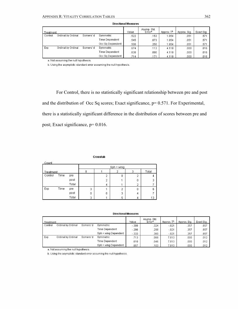
APPENDIX R: VITALITY CORRELATION TABLES 362
For Control, there is no statistically significant relationship between pre and post
and the distribution of Occ Sq scores; Exact significance, p= 0.571. For Experimental,
there is a statistically significant difference in the distribution of scores between pre and
post; Exact significance, p= 0.016.

APPENDIX R: VITALITY CORRELATION TABLES 363
For Control, there is no statistically significant relationship between pre and post
and the distribution of Sph > wing scores; Exact significance, p= 0.657. For
Experimental, there is a statistically significant difference in the distribution of scores
between pre and post; Exact significance, p= 0.012.
For Control, there is no statistically significant relationship between pre and post
and the distribution of Zyg L scores; Exact significance, p= 1.000. For Experimental,
there is a statistically significant difference in the distribution of scores between pre and
post; Exact significance, p= 0.125.

APPENDIX R: VITALITY CORRELATION TABLES 364
For Control, there is no statistically testable relationship between pre and post and
the distribution of Zyg R scores. For Experimental, there is a statistically significant
difference in the distribution of scores between pre and post; Exact significance, p=
0.009.

APPENDIX R: VITALITY CORRELATION TABLES 365
For Control, there is no statistically significant relationship between pre and post
and the distribution of Max L scores; Exact significance, p= 1.000. For Experimental,
there is a statistically significant difference in the distribution of scores between pre and
post; Exact significance, p= 0.003.
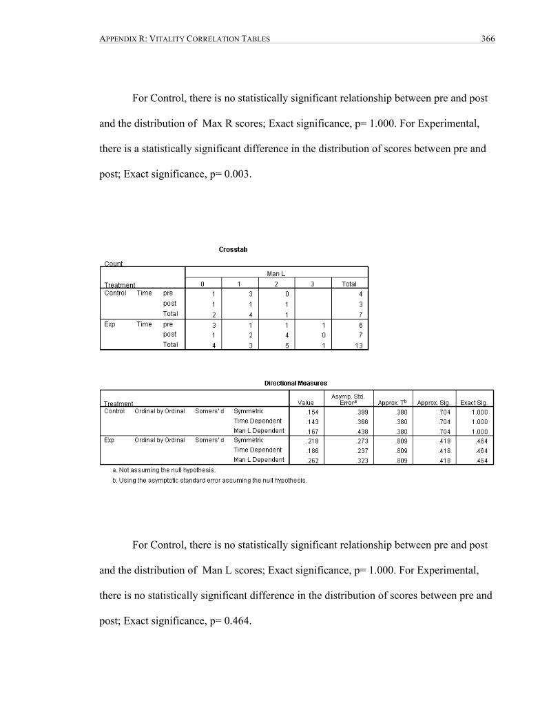
APPENDIX R: VITALITY CORRELATION TABLES 366
For Control, there is no statistically significant relationship between pre and post
and the distribution of Max R scores; Exact significance, p= 1.000. For Experimental,
there is a statistically significant difference in the distribution of scores between pre and
post; Exact significance, p= 0.003.
For Control, there is no statistically significant relationship between pre and post
and the distribution of Man L scores; Exact significance, p= 1.000. For Experimental,
there is no statistically significant difference in the distribution of scores between pre and
post; Exact significance, p= 0.464.
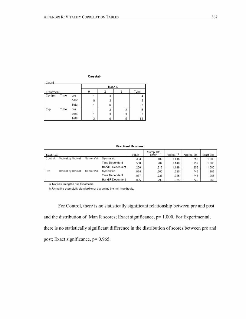
APPENDIX R: VITALITY CORRELATION TABLES 367
For Control, there is no statistically significant relationship between pre and post
and the distribution of Man R scores; Exact significance, p= 1.000. For Experimental,
there is no statistically significant difference in the distribution of scores between pre and
post; Exact significance, p= 0.965.

APPENDIX R: VITALITY CORRELATION TABLES 368
For Control, there is no statistically significant relationship between pre and post
and the distribution of Pal L scores; Exact significance, p= 0.771. For Experimental,
there is no statistically significant difference in the distribution of scores between pre and
post; Exact significance, p= 0.241.

APPENDIX R: VITALITY CORRELATION TABLES 369
For Control, there is no statistically significant relationship between pre and post
and the distribution of Pal R scores; Exact significance, p= 0.771. For Experimental,
there is a statistically significant difference in the distribution of scores between pre and
post; Exact significance, p= 0.049.

APPENDIX R: VITALITY CORRELATION TABLES 370
For Control, there is no statistically significant relationship between pre and post
and the distribution of Vomer scores; Exact significance, p= 0.686. For Experimental,
there is a statistically significant difference in the distribution of scores between pre and
post; Exact significance, p= 0.034.

APPENDIX R: VITALITY CORRELATION TABLES 371
For Control, there is no statistically significant relationship between pre and post
and the distribution of Lac L scores; Exact significance, p= 0.543. For Experimental,
there is no statistically significant difference in the distribution of scores between pre and
post; Exact significance, p= 0.134.
For Control, there is no statistically significant relationship between pre and post
and the distribution of Lac R scores; Exact significance, p= 1.000. For Experimental,
there is a statistically significant difference in the distribution of scores between pre and
post; Exact significance, p= 0.003.
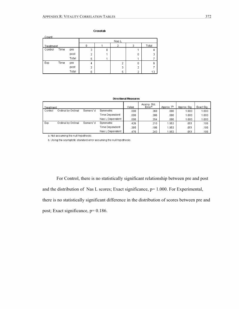
APPENDIX R: VITALITY CORRELATION TABLES 372
For Control, there is no statistically significant relationship between pre and post
and the distribution of Nas L scores; Exact significance, p= 1.000. For Experimental,
there is no statistically significant difference in the distribution of scores between pre and
post; Exact significance, p= 0.186.

APPENDIX R: VITALITY CORRELATION TABLES 373
For Control, there is no statistically significant relationship between pre and post
and the distribution of Nas R scores; Exact significance, p= 0.571. For Experimental,
there is a statistically significant difference in the distribution of scores between pre and
post; Exact significance, p= 0.009.

APPENDIX R: VITALITY CORRELATION TABLES 374
For Control, there is no statistically significant relationship between pre and post
and the distribution of Sacrum scores; Exact significance, p= 1.000. For Experimental,
there is no statistically significant difference in the distribution of scores between pre and
post; Exact significance, p= 0.122.

APPENDIX R: VITALITY CORRELATION TABLES 375
For Control, there is no statistically significant relationship between pre and post
and the distribution of Sacrum scores; Exact significance, p= 0.657. For Experimental,
there is a statistically significant difference in the distribution of scores between pre and
post; Exact significance, p= 0.048.

APPENDIX R: VITALITY CORRELATION TABLES 376
For Control, there is no statistically significant relationship between pre and post
and the distribution of INN L scores; Exact significance, p= 0.371. For Experimental,
there is a statistically significant difference in the distribution of scores between pre and
post; Exact significance, p= 0.026.
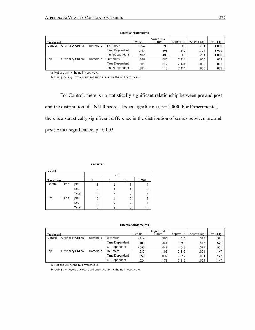
APPENDIX R: VITALITY CORRELATION TABLES 377
For Control, there is no statistically significant relationship between pre and post
and the distribution of INN R scores; Exact significance, p= 1.000. For Experimental,
there is a statistically significant difference in the distribution of scores between pre and
post; Exact significance, p= 0.003.

APPENDIX R: VITALITY CORRELATION TABLES 378
For Control, there is no statistically significant relationship between pre and post
and the distribution of C3 scores; Exact significance, p= 0.571. For Experimental, there
is no statistically significant difference in the distribution of scores between pre and post;
Exact significance, p= 0.147.

APPENDIX S: PILOT STUDY APPROVAL LETTER 379
APPENDIX S: PILOT STUDY APPROVAL LETTER

APPENDIX T: PROPOSAL 380
APPENDIX T: PROPOSAL

APPENDIX T: PROPOSAL 381