stability of enzymes in granular enzyme products for ... · stability of enzymes in granular enzyme...
TRANSCRIPT
General rights Copyright and moral rights for the publications made accessible in the public portal are retained by the authors and/or other copyright owners and it is a condition of accessing publications that users recognise and abide by the legal requirements associated with these rights.
Users may download and print one copy of any publication from the public portal for the purpose of private study or research.
You may not further distribute the material or use it for any profit-making activity or commercial gain
You may freely distribute the URL identifying the publication in the public portal If you believe that this document breaches copyright please contact us providing details, and we will remove access to the work immediately and investigate your claim.
Downloaded from orbit.dtu.dk on: Jul 29, 2019
Stability of Enzymes in Granular Enzyme Products for Laundry Detergents
Biran, Suzan
Publication date:2010
Document VersionPublisher's PDF, also known as Version of record
Link back to DTU Orbit
Citation (APA):Biran, S. (2010). Stability of Enzymes in Granular Enzyme Products for Laundry Detergents. TechnicalUniversity of Denmark (DTU).
STABILITY OF ENZYMES IN GRANULAR ENZYME PRODUCTS FOR LAUNDRY
DETERGENTS
PhD thesis by
Suzan Biran
2010
DEPARTMENT OF CHEMICAL AND BIOCHEMICAL ENGINEERING
TECHNICAL UNIVERSITY OF DENMARK
Building 229 DK-2800 Kgs. Lyngby
Denmark

To my bellowed sister, Canan.
Preface
1
PREFACE This PhD thesis is submitted in accordance with the partial requirements for the PhD degree at Technical University of Denmark. The project was performed in both the Combustion and Harmful Emission Control (CHEC) and Product Design research groups at the Department of Chemical and Biochemical Engineering, Technical University of Denmark, and Novozymes A/S in Bagsvaerd, Denmark. This project was financially supported by Technical University of Denmark and Novozymes Bioprocess Academy.
The PhD project has been supervised by Professor Anker Degn Jensen (main supervisor) and Associate professor Søren Kiil at the Department of Chemical and Biochemical Engineering, and senior science managers Poul Bach and Ole Simonsen at Novozymes A/S.
First of all, I would like to express my deepest gratitude to my supervisor Anker Degn Jensen, who provided me with great support, guidance and motivation, not only scientifically but in every other respect during this mostly difficult period of my personal life. I can’t describe how strongly I appreciate his efforts towards encouraging me and how thankful I am for him not giving up on me. I wish to thank Søren Kiil for his insights and comments during the duration of the study. I am grateful to Poul Bach and Ole Simonsen for their invaluable input on detergents and enzymes, comments on results, guidance and support of my experimental work in Novozymes A/S. Finally, I would like to thank my supervisors for their patience and understanding.
Knowledge is a valuable asset for a human being, experience is even more. Through the study, I had the opportunity to work with experts from various backgrounds at different departments, acquiring invaluable skills. Therefore, I would like to acknowledge those who spared their time to mentor me and share their long-time accumulated experience and knowledge with me. I would like to thank to Christian Isak Jørgensen, Pia Wium, Jan Juul de Jong and Carsten P. Sönksen (Protein Engineering Department - Novozymes A/S, Denmark) for their
Preface
2
assistance and provision of the necessary equipment for peptide mapping studies. Ann Lassen (SPD-Novozymes A/S, Denmark) is acknowledged for performing the adsorption experiments. I am also appreciative to Lilian Holgersen (CHEC), analysis experts in Liquid and Solid Product Development Departments in Novozymes A/S for their friendly attitude, answering patiently my questions and assistance during my experimental work.
I am grateful to my dear friends Ayten, Michelangelo, Lusi, Diego, Ada and many others for creating a warm and comfortable environment and contributing to life-long pleasant memories during my stay in Denmark.
The last but not least, I would like to express my deep love and gratitude to my family and my fiancé for their unconditional love, constant support, inspiring motivation and great patience during this long PhD study.
Suzan Biran
2010
Abstract
3
ABSTRACT Enzymes have long been of interest to the detergent industry due to their ability to improve the cleaning efficiency of synthetic detergents, contribute to shortening washing times, and reduce energy and water consumption, provision of environmentally friendlier wash water effluents and fabric care. However, incorporating enzymes in detergent formulations gives rise to numerous practical problems due to their incompatibility with and stability against various detergent components. In powdered detergent formulations, these issues can be partly overcome by physically isolating the enzymes in separate particles. However, enzymes may loose a significant part of their activity over a time period of several weeks.
Possible causes of inactivation of enzymes in a granule may be related to the release of hydrogen peroxide from the bleaching chemicals in a moisture-containing atmosphere, humidity, autolysis of enzymes, high local pH in granule, oxygen, defects in granulate structure and the effect of other detergent components. However, the actual mechanism of inactivation is not known yet. It is believed that a combination of the factors mentioned above plays a role in the activity loss, and is the focus of this study.
The inactivation kinetics of technical grade enzyme powder was determined in a newly developed experimental setup, which was simple and effective and provided a better control over test conditions and fast sample generation. The method was based on the generation of hydrogen peroxide vapor and humidity by bubbling nitrogen gas through their corresponding solutions. An enzyme column, acting as a plug-flow reactor, was exposed to known concentrations of H2O2 (g) and humidity in a thermally stabilized chamber. Samples were analyzed for adsorptive behavior and residual enzyme activity.
Since the moisture is believed to play an important role in the stability of proteins, the monolayer hydration level of Savinase® was experimentally determined and theoretically calculated. Adsorbed moisture was found to have
Abstract
4
a negative effect on enzyme activity. Below monolayer hydration level, the enzyme stability was significantly conserved, while at multilayer hydration level, especially when samples were exposed to 100% RH, the activity was reduced by 80% in a one week period. Since no auto-proteolytic activity and covalently-bound aggregate formation were detected, humidity possibly induced formation of unfavorable conformational changes, resulting in a decrease in enzyme’s catalytic efficiency.
Exposure to H2O2 (g) and humidity also resulted in significant H2O2 adsorption. The amount of adsorbed H2O2 did not depend on humidity in the gas stream, which implied that water and H2O2 were not competing for the same adsorption sites. In addition, the desorption studies revealed that while moisture was adsorbed by physisorption, H2O2 was adsorbed by either chemisorption or possibly involving formation of strong hydrogen bonds.
Inactivation of the solid-state enzyme was caused by the mutual effect of hydration and H2O2 (g) concentration. A simple mechanism for solid-state enzyme oxidation was proposed and the kinetic parameters in the resulting rate expression were derived. A good agreement between the derived equation and experimental data was obtained. The enzyme inactivation was found to depend on the square of moisture adsorbed by the enzyme at the corresponding temperature. The inverse of the reaction rate constant was also proportional to the inverse of H2O2 in the system.
Activity loss was expected to be caused by the oxidation of the enzyme by H2O2 vapor. The oxidative alterations on Savinase were investigated by peptide mapping. Molecular mass examination of CNBr-cleaved fragments by MALDI TOF mass spectroscopy located the oxidation-labile residue. Due to its relatively accessible position on the exterior of the enzyme structure, only methionine 222 (Met 222) was oxidized; while other 2 Met residues, buried in the peptide backbone, remained unaffected. Being adjacent to the active site of Savinase®, Met 222 oxidation resulted in conformational and electrostatic shift in the catalytic site, causing a significant reduction of enzyme activity. The findings are in agreement with previously reported H2O2-induced oxidation studies of Savinase® in solutions.
Abstract
5
Preliminary formulation studies were conducted and application of the designed setup on stability measurements of commercial granulates was illustrated. Addition of salts resulted in a considerable conservation of enzyme activity. Having an anti-oxidative property, sodium thiosulphate had a better activity-preservation effect compared to sodium carbonate. Due to a possible crack formation on granulate surface and/or deliquescence of sodium thiosulphate at high humidity showed that mixing the antioxidant homogeneously with the enzyme provided better protection than coating the salt as a separate layer. The effect of site-directed mutagenesis on Savinase® stability was illustrated and possible stability enhancing additives for enzyme granulates were proposed.
The present study is the first to report the solid-state inactivation kinetics and mechanism of Savinase®, subjected to controlled concentrations of H2O2 vapor and humidity. It provides practical information on solid-state stability measurements of biocatalysts in oxidative environments.

Resume
7
RESUME by Anker Degn Jensen
Enzymer har længe været brugt i detergentindustrien da de forbedrer vaskeevnen af detergenter, forkorter vasketiden og reducerer energi- og vandforbruget. Anvendelse af enzymer i detergenter skåner således miljøet og forlænger tøjets levetid. Imidlertid er der en række praktiske problemer forbundet med at anvende enzymer i detergenter, hvoraf reduceret stabilitet af enzymerne på grund af de andre ingredienser i detergentet er fremherskende. I pulverformige detergent formuleringer kan dette problem delvist løses ved at enzymerne indkapsles i separate partikler, men ikke desto mindre taber enzymerne i detergentblandingen en stor del af deres aktivitet over nogle måneder.
Tab af enzymaktivitet kan skyldes frigivelse af hydrogenperoxid fra blegemidler i en fugtig atmosfære, høj relativ fugtighed, autolyse af enzymer, høj lokal pH i partiklerne, oxygen, defekter i partiklens struktur (primært coatinglag), og eventuelt indflydelsen fra andre detergent komponenter. Den detaljerede mekanisme for deaktivering af enzymer formodes at skyldes en kombination af de ovenfor nævnte faktorer, og er hovedemnet for nærværende afhandling, med vægten lagt på samtidig indflydelse af vanddamp og H2O2 (g).
Kinetikken for deaktivering af teknisk rent enzympulver er blevet undersøgt i en forsøgsopstilling udviklet som en del af projektet. Opstillingen giver mulighed for at eksponere en enzymprøve for en gas indeholdende hydrogenperoxid og vanddamp i forskellige koncentrationer under kontrollerede forhold. Hydrogen peroxid gas blev doseret ved at boble inert gas (N2) eller luft gennem en koncentreret hydrogenperoxid opløsning, og vanddampindholdet i gassen blev efterfølgende justeret ved at tilføre tør eller fugtig gas for at opnå den ønskede gasblanding. Enzympulveret blev eksponeret for gassen i en lille stempelstrømningsreaktor af glas i et termostateret kammer.
Resume
8
Efterfølgende blev pulveret analyseret for adsorberet hydrogenperoxid og rest-enzymaktivitet.
Fugt spiller en væsentlig rolle i stabiliteten af proteiner, og fugtoptaget af Savinase® pulver, herunder monolags vandmængden, blev derfor undersøgt eksperimentelt og teoretisk.
Stabilitetsforsøg viste at adsorberet fugt alene har en negativ indflydelse på enzym stabiliteten. Ved mængder under ét monolag af vand var enzymstabiliteten god, men ved multilag af vand, og især tæt på 1oo % relativ fugtighed, observeredes betydelig deaktivering, svarende til 80 % aktivitetstab i løbet af 1 uge. Der blev ikke fundet auto-proteolyse eller covalent aggregat dannelse, og indflydelsen af fugt er derfor formentlig på grund af ændringer i enzymets tertiære eller kvaternære struktur.
Eksponering af enzymer for to H2O2 (g) og vanddamp resulterede i en betydelig adsorption af H2O2. Mængden af adsorberet H2O2 afhang ikke af vanddampkoncentrationen hvilket indikerer at H2O2 og vanddamp ikke konkurrerer om de samme pladser på enzymet. Desorptionsstudier viste at mens vand adsorberer ved physisorption, adsorberer H2O2 enten ved kemisorption eller stærke hydrogen bindinger.
Samtidig tilstedeværelse af vanddamp og H2O2 (g) førte til hurtig deaktivering af enzymer i fast form. En simpel mekanisme for reaktionen blev foreslået og på basis heraf blev der udledt et hastighedsudtryk for reaktionen. Parametrene i udtrykket blev fastlagt på basis af eksperimentelle resultater og der blev observeret god overensstemmelse mellem model og data. Deaktiveringen af enzymet var afhængig af mængden af adsorberet vand i anden potens og cirka afhængig af H2O2 (g) koncentrationen i første potens.
Eftersom H2O2 (g) er et kraftigt oxidationsmiddel var det forventet at mekanismen for deaktivering af enzymet var oxidation af oxidationslabile aminosyrer. Dette blev undersøgt på Savinase ved peptid mapping. Molekylær masse bestemmelse af CNBr-kløvede fragmenter ved hjælp af MALDI TOF masse spektroskopi lokaliserede den oxidationslabile amonisyre som værende methionin i position 222 (Met 222). På grund af dens tilgængelighed tæt på
Resume
9
overfladen af enzymet er det kun denne methionin enhed der oxideres, mens to andre methionin enheder, der er begravet i enzymets struktur, forblev intakte. Met 222 sidder tæt på det aktive site i Savinase®, og fører derfor til strukturelle ændringer i enzymet som giver en drastisk reduktion i dets aktivitet. Dette er i overensstemmelse med konklusionen på undersøgelser i litteraturen af indflydelsen af H2O2-induceret oxidation af Savinase® i vandige opløsninger.
Der er blevet udført indledende formuleringsstudier, samt tests på kommercielle granulater i den udviklede opstilling. Tilsætning af salte til enzympulveret førte til en betydelig forbedring af enzymstabiliteten. Natrium thiosulfat havde bedre aktivitetsbevarende egenskaber end natrium karbonat, hvilket også var forventet da førstnævnte er et antioxidant middel, der kan reagere med H2O2. Blanding af enzympulver med natrium thiosulfat gav bedre stabilitet end for granulater hvor saltet lå som et lag rundt om den enzymholdige kerne. Dette skyldes muligvis revnedannelser i granulaternes saltlag ved høj fugtighed. Effekten af site-dirigeret mutagenese på Savinase® stabilitet blev undersøgt og mulige stabilitetsforbedrende additiver til enzymgranulater blev foreslået.
Dette er det første studie i litteraturen af kinetik og mekanismer for deaktivering af Savinase® i fast form, under eksponering for kontrollerede koncentrationer af vanddamp og H2O2 (g). Der er opnået praktisk viden om stabilitet af bio-katalysatorer på fast form i oxidative miljøer.

Contents
11
CONTENTS
Preface .......................................................................................................................................... 1
Abstract ........................................................................................................................................ 3
Resume (abstract in Danish)………………………………………………..………………….7
Table of contents ………………………………………………………………………………….11
1 General introduction ........................................................................................................... 15
1.1 Problem definition ....................................................................................................... 16
1.2 Project objective ............................................................................................................ 16
2 Literature survey .................................................................................................................. 18
2.1 Detergents ........................................................................................................................ 18
2.1.1 Surfactants .............................................................................................................. 19
2.1.2 Builders .................................................................................................................... 20
2.1.3 Bleaching system ................................................................................................. 21
2.1.4 Detergent enzymes ............................................................................................. 26
2.1.5 Soil anti-redeposition polymers .................................................................... 28
2.1.6 Others ....................................................................................................................... 28
2.2 The enzyme ..................................................................................................................... 28
2.2.1 Enzyme structure ................................................................................................ 29
2.2.2 Savinase® ................................................................................................................. 31
2.3 Solid-state proteins ...................................................................................................... 33
2.3.1 Production .............................................................................................................. 33
2.3.2 Structural properties of the solid-state proteins ................................... 34
2.3.3 Stability of solid-state proteins ...................................................................... 36
2.3.4 Stabilization methods ........................................................................................ 45
Contents
12
2.4 Enzyme granules ........................................................................................................... 46
2.4.1 Types of granules ................................................................................................. 47
2.4.2 Factors affecting granule stability in detergents ................................... 50
2.5 Summary .......................................................................................................................... 54
2.6 References ....................................................................................................................... 55
3 Construction of the experimental setup ..................................................................... 62
3.1 Demands for the set-up .............................................................................................. 62
3.2 Description of the conventional technique ....................................................... 63
3.3 Hydrogen peroxide vapor generation and measurement ........................... 64
3.4 Humidity measurement and control .................................................................... 66
3.5 Preliminary setup for enzyme exposure ............................................................ 67
3.6 Enzyme column set-up ............................................................................................... 69
3.7 Summary .......................................................................................................................... 70
3.8 References ....................................................................................................................... 71
4 Methods of analysis ............................................................................................................. 73
4.1 Measurement of H2O2 concentration ................................................................... 73
4.1.1 High concentration.............................................................................................. 73
4.1.2 Low concentration .............................................................................................. 74
4.2 Savinase® activity measurements ......................................................................... 75
4.1.3 Using n,n-dimethyl casein as a substrate .................................................. 75
4.1.4 Using n-succinyl ala-ala-pro-phe-p-nitroanilide as a substrate ...... 76
4.3 SDS PAGE .......................................................................................................................... 76
4.4 CNBr cleavage ................................................................................................................ 77
4.5 MALDI TOF mass spectroscopy .............................................................................. 77
4.6 IES mass spectroscopy ............................................................................................... 78
4.7 Adsorption-desorption studies .............................................................................. 78
Contents
13
4.8 References ....................................................................................................................... 78
5 Investigation of solid-state inactivation of Savinase® .......................................... 79
5.1 Water adsorption-desorption studies on Savinase® powder ................... 79
5.2 Savinase® exposure to H2O2 (g) and humidity ................................................ 85
5.1.1 Adsorption of hydrogen peroxide on Savinase® .................................... 85
5.1.2 Enzyme inactivation ........................................................................................... 88
5.1.3 inactivation dynamics in the enzyme column ......................................... 95
5.1.4 Kinetics of Savinase® inactivation ................................................................ 97
5.3 Summary ....................................................................................................................... 101
5.4 References .................................................................................................................... 102
6 Mechanism of Savinase® inactivation ....................................................................... 106
6.1 Molecular structure of Savinase® ....................................................................... 106
6.2 Inactivation mechanism study ............................................................................. 109
6.1.1 Enzyme activity ................................................................................................. 110
6.1.2 SDS PAGE analysis ............................................................................................ 110
6.1.3 IES mass spectroscopy ................................................................................... 112
6.1.4 CNBr cleavage .................................................................................................... 112
6.1.5 MALDI TOF mass spectroscopy .................................................................. 114
6.3 Summary ....................................................................................................................... 119
6.4 References .................................................................................................................... 120
7 Stability measurements of formulated enzymes ................................................. 123
7.1 Introduction ................................................................................................................. 123
7.2 Effect of salts on enzyme powder stability ..................................................... 124
7.3 Effect of sodium thiosulfate on enzyme granulate stability .................... 127
7.4 Effect of humidity on enzyme granulate .......................................................... 129
7.5 Effect of site-directed mutagenesis on enzyme stability in commercial granulates .................................................................................................................................. 132
Contents
14
7.6 Additives ........................................................................................................................ 133
7.7 Summary ....................................................................................................................... 135
7.8 References .................................................................................................................... 136
8 Conclusions and suggestions for future work ....................................................... 139
Appendix I Derivation of enzyme kinetic expression ..............................................142
Appendix II Inactivation of a solid-state detergent protease by hydrogen
peroxide vapor and humidity………..………..…………..…………………144
Chapter 1. Introduction
15
CHAPTER 1 GENERAL INTRODUCTION
Enzymes are used today in a wide range of industrial processes and in consumer products. The largest application of industrial enzymes is in detergents. The detergent industry absorbs about 45% of enzyme sales in Western Europe and more than 25% of the total worldwide enzyme production. Their presence in laundry detergents results in higher performance at low temperatures. Beside their performance and economic benefits, enzymes are also environmentally friendly due to their biodegradability.
Enzymes have long been of interest to the detergent industry due to their contribution to shortening washing times, reduction of energy and water consumption, provision of environmentally friendlier wash water effluents and fabric care. The first application detergent enzymes dates back to 1913, when Otto Röhm, founder and partner of Röhm and Haas in Germany, used pancreatic proteases and soda in washing detergents. The first detergent containing bacterial enzymes was introduced in 1956 under the trade name Bio-40, developed by Swiss company Schweizerische Ferment AG. The poor effectiveness of the enzymes in alkaline wash conditions lead to investigation for more robust enzymes. In 1958, Novo Industry (Denmark) developed a product containing bacterial protease, Alcalase, exhibiting high stability and activity at pH 8-10. However, detergent proteases faced a set back in the early 1970s, due to unfavorable publicity when some workers developed an allergic reaction during the handling of these enzymes. Like many other proteins foreign to the human body, enzymes are potential inhalation allergens. Inhalation of even small concentrations of a foreign protein in the form of dust or aerosols can stimulate the body's immune system to produce antibodies. To overcome the unwanted dust release and increase the stability of enzyme mixtures, they are encapsulated or coated to form enzyme granulates.
Chapter 1. Introduction
16
In powdered laundry detergents, they are granulated and covered with protective coating layers to prevent dust release and increase the stability of the enzymes.
1.1 PROBLEM DEFINITION
Storage stability of detergent enzymes is an important quality parameter that should be considered in the development of a new product. Laundry detergents typically consist of a mixture of separate granular materials including surfactants, builders, bleaching agents and enzymes, which can lose activity in such environments, where harsh chemicals are present. In practice enzymes loose a significant part of their activity over a time period of several weeks. The deactivation is mainly related to the release of hydrogen peroxide from the bleaching chemicals in a moisture-containing atmosphere. Moreover, humidity, autolysis of enzymes, high local pH in granule, oxygen, defects in granulate structure and other detergent components are some of the factors affecting the granulate stability during storage. However, the actual kinetics and mechanism of inactivation is not known yet. It is believed that a combination of the factors mentioned above plays a role in the activity loss.
Close attention is given to the formulation of both enzyme granulates and the composition of the other constituents of solid laundry detergents. However, testing the effectiveness of different granulate formulations takes considerable amount of time (4-6 weeks). Therefore, there is a need for a method of accelerated stability trials and, more importantly, illustrative description of the kinetics and mechanism of inactivation of solid enzyme products in the detergent.
1.2 PROJECT OBJECTIVE
The main objective of this study is to understand the mechanism(s) and assess the kinetics of inactivation of detergent enzymes during storage. For this purpose, a design of an experimental set up, providing controlled conditions for testing and resembling detergent box environment is required. The set up needs to present fast and reproducible test results. It is also aimed to investigate
Chapter 1. Introduction
17
the effect of different detergent ingredients on the granulated enzyme stability. In light of the results, new stability-enhancing components or coatings will be proposed and tested for their efficiency in reducing enzyme deactivation in powdered detergents.
The objectives of this PhD project have been addressed through theoretical and experimental considerations. The literature survey in chapter 2 provides introductory information on granulated laundry detergent powder, listing the main ingredients and their function in the washing process. A focus on enzyme structure and specifically on the tested detergent protease, Savinase®, is given. Later, the properties of solid-state proteins are illustrated and the reactions that may result in enzyme instability during solid-state storage are described. Analytical techniques that can be used for detection of the instability reactions are depicted. Finally, the structure and properties of enzyme granulates is explained and the factors resulting in enzyme instability in detergent matrix are discussed. In chapter 3, the conventional testing method is illustrated and the design and development of the new experimental setup are described. Chapter 4 provides a list of the analytical techniques and experimental protocols used throughout this study. The results of enzyme exposure to H2O2 (g) and humidity are presented in chapter 5. Calculation of the hydration monolayer of Savinase®, H2O2 and moisture adsorption, and enzyme inactivation as a function of H2O2 partial pressure and humidity are described. The kinetic expression of enzyme inactivation is derived and the relation of proposed equation and experimental results is shown. In chapter 6, the structural modifications of Savinase® resulting in enzyme inactivation are illustrated on a molecular level; thus, mechanism of inactivation is revealed. Chapter 7 includes the results of granulated enzyme tests, focusing on formulation strategies. The thesis ends with chapter 8, in which final conclusions and suggestions for future work are provided.
Chapter 2. Literature survey
18
CHAPTER 2 LITERATURE SURVEY
The following chapter provides a brief introduction on the basic laundry detergent ingredients and their function in the washing process. A focus on enzyme structure and function is given; and further information on solid-state proteins is provided. Their production and structural properties are described. Stability concerns during storage are depicted and possible instability reactions and their measurement techniques are listed. Finally, types of enzyme granules are introduced and the factors affecting their stability in the detergent matrix are described.
2.1 DETERGENTS
The main objective of a detergent is to remove soil (dirt) and other contaminants from the fabrics while keeping their integrity (e.g. mechanical strength and color). Washing process comprises of several distinct steps: 1) hydration of the soil; 2) removal of soil from the fabric through mechanical or chemical action; 3) dispersion of the soil in the wash liquor; 4) prevention of re-deposition of contaminants to the laundry; 5) bleaching of the remaining or re-deposited soil for better end result; and 6) final fabric modification to improve consumers’ satisfaction (Jakobi and Löhr 1987; Ponnusamy et al. 2008). Consequently, today’s detergents are sophisticated products containing a large number of ingredients with a variety of individual functions throughout the cleaning process. Generally, six groups of substances are present: surfactants (ca 30%), builders (ca 40%), bleaching agents (ca 20%) and other low level of additives; such as: enzymes, dispersing agents, fabric softener clay, dry-transfer inhibiting ingredients, and optical brighteners etc. (Carson et al. 2006; Yu et al. 2008). The need for and efficiency of any of these ingredients is a function of soil
Chapter 2. Literature survey
19
amount and type (water soluble soils, pigments, fats, proteins, carbohydrates, bleachable dyes, etc.), water hardness, temperature, and fabric type and color.
2.1.1 SURFACTANTS
Surfactants are surface active chemical substances, which concentrate at interfaces (e.g. water/fabric surface) and thereby lower the surface tension of water and facilitate wetting of surfaces by the aqueous phase. They are the most important ingredient in household cleaning products, comprising 15-40% of the total detergent formulation (Scheibel 2004; Yu et al. 2008). The peculiar properties of these chemicals reside in their amphophilic character, which stems from the fact that each surfactant molecule has both a hydrophilic or solubility-enhancing functional group and a hydrophobic portion (usually a long alkyl chain) (Jakobi and Löhr 1987). In addition to their wetting ability, surfactants may have properties like foaming (suds forming) ability, foam inhibition properties, emulsification power and the ability to lift soil particles from surfaces and carry them away (Novozymes 2002).
Depending on the charge present in the chain-carrying portion of the molecule after dissociation in water, surfactants can be divided into anionic (negatively charged), cationic (positively charged), nonionic (uncharged) and amphoteric (present both positive and negative charges at intermediate pH) classes. Generally, laundry detergents contain a certain mixture of different surfactants to enhance detergent’s washing performance capability (Kume et al. 2008).
Anionics are historically the earliest (soap) and the most commonly used surfactants, due to their ease and low cost of manufacture and high performance in removal of grease and oil. They are usually considered as the “workhorse” of detergent and the largest contributor to the overall cleaning process (Scheibel 2004). Furthermore, they are especially beneficial for their excellent detersive action and particulate removal capability (Novozymes 2002). However, anionic surfactants are sensitive to water hardness and their detergency power significantly diminishes due to sequestration and precipitation by divalent cations in the washing solution (Yu et al. 2008). For this reason, they are used in mixtures with nonionic surfactants to improve detergent performance. Linear
Chapter 2. Literature survey
20
alkylbenzene sulphonates, (LAS or LABS) are the dominant class of anionic surfactants used in today’s detergents (Scheibel 2004).
Nonionics are normally a mixture of homologuos structures composed of alkyl chains of carbons and hydrophilic moieties that differ in the number of ethylene oxide (ethoxylate, EO), propylene oxide (propoxylate, PO), and butylenes oxide (butoxylate, BO) units (Sak-Bosnar et al. 2007). They are especially useful due to their low sensitivity to water hardness compared to anionics. They do not interact significantly with the other detergent compounds; for this reason they are used in a mixture with other surfactants for better performance. By far the most important class for laundry detergents is alcohol ethoxylates (AEO) (Novozymes 2002).
Cationics used in detergent compositions are based on the nitrogen atom carrying positive charge (Yu et al. 2008). The main class of cationics is the “quats”, i.e. quaternary ammonium salts (Guertechin 1999). They are mostly used in rinse aids and are added in the final rinse cycle to soften the garments, to decrease wrinkling and to reduce static electricity afterwards. A number of cationic surfactants have bactericidal activity against wide range of gram-positive and some gram-negative organisms (Effendy and Maibach 1996).
Amphoterics, also known as zwitterionic surfactants, are represented mainly by acyl ethylenediamines and alkyl amino acids (Kume et al. 2008). They are usually used in the combination with other surfactants to obtain desired foam or detergency. Amphoterics are generally mild, with lower skin and eye irritation when compared with the commonly used anionic and nonionic surfactants (Effendy and Maibach 1996); however, they are only employed in specialty detergents due to economic reasons (Jakobi and Löhr 1987).
2.1.2 BUILDERS
Hardness ions – Ca2+ and Mg2+ entering the wash liquor via tap water and washload (Hollingsworth 1978) – diminish the cleaning effectiveness of a laundry detergent. These ions may precipitate the active surfactant by forming insoluble calcium or magnesium salts, increasing the levels of calcium-bound or
Chapter 2. Literature survey
21
calcium-bridged redeposited soils (Nagarajan and Paine 1984), or may catalyze the decomposition of bleaching agents (Coons 1978).
The primary function of builders in a laundry detergent is to reduce the concentration of hardness ions in the wash liquor below 10-4 or 10-5 M (Hollingsworth 1978). Examples of water softening agents commonly used in the detergent industry are: 1) Sequestrant builders like sodium tripolyphosphate (STPP), nitrilotriacetic acid (NTA), citric acid and polyacrylic acid (PAA); 2) Precipitant builder like sodium carbonate, and 3) Ion exchange builder like crystalline sodium aluminosilicate or Zeolite Type A (Nagarajan and Paine 1984). Other functions of builders are provision of alkalinity, dispersion and suspension of soils in the wash liquor, and stabilization of other detergent components (Jakobi and Löhr 1987). All the potential interactions between the ingredients of the detergent formula, oxidation, absorption of moisture from the air, and light are some of the factors likely to shorten the shelf life of a detergent. Since builders are often mineral powders found in large proportions, they act as insulating barriers between the antagonistic components; thus, increasing the shelf life of the product.
2.1.3 BLEACHING SYSTEM
Sometimes, the surfactant/builder system is not able to clean perfectly certain soils, such as blood, fruit, wine, coffee and tea stains. The staining molecules are so strongly attached that they remain on the fabric. Oxidative agents are required to address such stains. Depending on the laundry habits, generally used detergent bleaches are either chlorine (e.g. sodium hypochlorite, NaOCl) or peroxy compounds (e.g. sodium percarbonate, Na2CO3.1.5H2O2; sodium perborate, NaBO3.H2O or NaBO3.4H2O) (Carson et al. 2006). Although hypochlorite shows a good performance at relatively low temperatures, peroxy compounds are more extensively used in detergent formulations, due to the incompatibility of hypochlorite with some detergent ingredients (e.g. enzymes and fluorescent whitening agents), ability to cause fabric damage, and malodor. Studies on washing and bleaching habits show that peroxy compounds dominate Europe, chlorine bleaching is used predominantly in Mediterranean countries (Smulders 2002). The bleach system also acts as a sanitizer in the
Chapter 2. Literature survey
22
washing process at temperatures at which thermal disinfection is not possible or the residence time is not sufficiently long (Coons 1978).
Peroxide bleaching from dry mixed laundry detergents relies on the liberation of hydrogen peroxide, H2O2, from the dissolved solid inorganic carriers. Under alkaline conditions (pH ~ 9), H2O2 deprotonates, forming the bleaching species: perhydroxyl anion (HOO-) (Coons 1978; Skagerlind et al. 1998). Sodium perborate monohydrate is the peroxide preferred by the industry owing to its high rate of dissolution, high percentage of active oxygen (16%) (Carson et al. 2006; Smulders 2002) and stability (Skagerlind et al. 1998). However, an environmental concern has been expressed about contamination of irrigation water by phytotoxic boron from perborates, which led to a search for environmentally-friendly peroxy compounds. Today, sodium percarbonate (SPC) is becoming more popular, especially after its improved detergent stability, which is achieved by coating the SPC particles (Johonsson et al. 2007). Moreover, it is a multifunctional compound, i.e. it carries hydrogen peroxide (bleaching) and soda ash (alkalinity). It also shows a high dissolution rate and solubility (Novozymes 2002). However, it is well-known that peroxide alone is ineffective bleach below 60°C. Therefore, for good bleach performance in colder water, many detergent manufacturers rely on activators in order to transform the peroxide into more effective peracid bleach.
2.1.3.1 Sodium percarbonate (SPC)
Sodium percarbonate is an attractive perhydrate for use in detergent compositions because it dissolves readily in water, is weight efficient and, after giving up its available oxygen, provides a useful source of carbonate ions for detergency purposes. The name “sodium percarbonate” does not reflect the structure or true nature of the material; in fact, the compound is sodium carbonate sesquiperhydrate, Na2CO3.1.5H2O2 (McKillop and Sanderson 1995). In detergents, SPC is in the form of particles having an average particle size in the range from about 500 micrometers to about 1,000 micrometers. About 10% by weight of these particles are smaller than about 200 micrometers and approximately 10% by weight of them are larger than about 1,250 micrometers.
Chapter 2. Literature survey
23
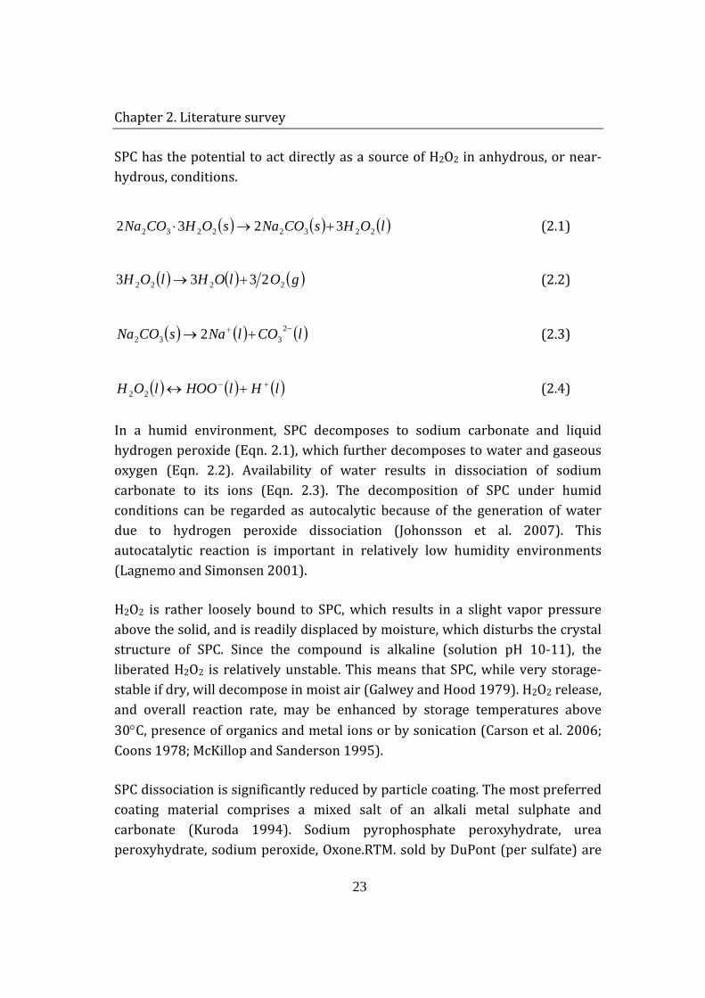
SPC has the potential to act directly as a source of H2O2 in anhydrous, or near-hydrous, conditions.
( ) ( ) ( )lOHsCONasOHCONa 22322232 3232 +→⋅ (2.1)
( ) ( ) ( )gOlOHlOH 2222 2333 +→ (2.2)
( ) ( ) ( )lCOlNasCONa −+ +→ 2332 2 (2.3)
( ) ( ) ( )lHlHOOlOH +− +↔22 (2.4)
In a humid environment, SPC decomposes to sodium carbonate and liquid hydrogen peroxide (Eqn. 2.1), which further decomposes to water and gaseous oxygen (Eqn. 2.2). Availability of water results in dissociation of sodium carbonate to its ions (Eqn. 2.3). The decomposition of SPC under humid conditions can be regarded as autocalytic because of the generation of water due to hydrogen peroxide dissociation (Johonsson et al. 2007). This autocatalytic reaction is important in relatively low humidity environments (Lagnemo and Simonsen 2001).
H2O2 is rather loosely bound to SPC, which results in a slight vapor pressure above the solid, and is readily displaced by moisture, which disturbs the crystal structure of SPC. Since the compound is alkaline (solution pH 10-11), the liberated H2O2 is relatively unstable. This means that SPC, while very storage-stable if dry, will decompose in moist air (Galwey and Hood 1979). H2O2 release, and overall reaction rate, may be enhanced by storage temperatures above 30°C, presence of organics and metal ions or by sonication (Carson et al. 2006; Coons 1978; McKillop and Sanderson 1995).
SPC dissociation is significantly reduced by particle coating. The most preferred coating material comprises a mixed salt of an alkali metal sulphate and carbonate (Kuroda 1994). Sodium pyrophosphate peroxyhydrate, urea peroxyhydrate, sodium peroxide, Oxone.RTM. sold by DuPont (per sulfate) are
Chapter 2. Literature survey
24
further examples of inorganic perhydrate salts suitable for use in the stabilisation of SPC. Furthermore, organic molecules like amino alkylene (polyalkylene phosphonates) and amino alkylene (polyalkylene carboxylates) (Kowalski 1980); metal sequestering agents, like polyethyleneimine; preventing metal ion catalyzed decomposition of peroxygen bleaches, are also utilized to enhance and control bleach stabilization (Gutierrez 1999).
2.1.3.2 Hydrogen peroxide
Hydrogen peroxide (H2O2) is clear, colorless liquid, which is miscible with water in all proportions. It has long been used in industrial applications as a powerful oxidant. It is a stronger oxidant compared to chlorine and permanganate and has the advantage of releasing non-polluting decomposition products. Some of the application processes are oxidation of sulphides with respect to odour control, notably in paper and pulp manufacture and textile plant wastes. It can also be used as an additional oxygen source for overloaded activated sludge plants and controlling filamentous bulking during fermentation (Guwy et al. 2000).
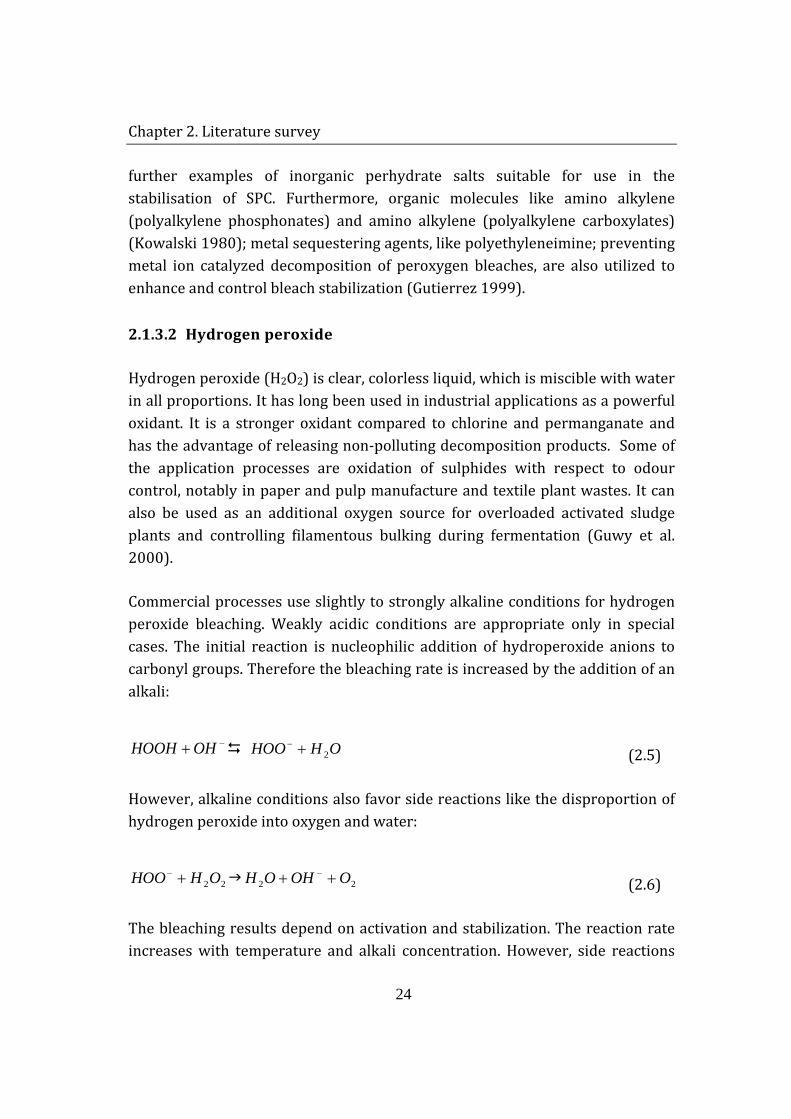
Commercial processes use slightly to strongly alkaline conditions for hydrogen peroxide bleaching. Weakly acidic conditions are appropriate only in special cases. The initial reaction is nucleophilic addition of hydroperoxide anions to carbonyl groups. Therefore the bleaching rate is increased by the addition of an alkali:
−+OHHOOH OHHOO 2+− (2.5)
However, alkaline conditions also favor side reactions like the disproportion of hydrogen peroxide into oxygen and water:
22OHHOO +− 22 OOHOH ++ − (2.6)
The bleaching results depend on activation and stabilization. The reaction rate increases with temperature and alkali concentration. However, side reactions
Chapter 2. Literature survey
25
such as thermal decomposition of hydrogen peroxide into radicals become more likely or even dominant at high temperatures, which also lead to yield losses due to solubilization of the material being bleached.
Hydrogen peroxide bleaching liquor is stabilized, i.e., the rate of decomposition of hydrogen peroxide is lowered without decreasing the bleaching rate, by sodium silicate, chelating agents and magnesium salts. The effect of stabilizers is to chelate transition metal ions or to absorb these ions on colloidal silica or magnesium hydroxide. Sodium silicate also acts as a buffer that decreases the activity losses caused by a high concentration of hydroxyl anions (Galwey and Hood 1979). The transition metal ions are catalysts that tend to increase both the consumption of hydrogen peroxide and the damage to the fibers.
2.1.3.3 Bleach activators
Many modern fabrics and dyes cannot be washed at high temperatures. The bleaches used in detergents are capable of giving rise to satisfactory bleaching only if the wash temperature is in excess of 60°C. In the absence of high temperatures or extended wash times, commercially used bleaches are ineffective when used alone. In order to obtain satisfactory bleaching at low-temperatures, chemicals known as bleach activators are added to the formulations. These compounds, having also antimicrobial effect, are reacting with the hydrogen peroxide anion to form a peroxycarboxylic acid, which is a considerably better bleaching agent than hydrogen peroxide at low temperatures (Novozymes, 2002):
HLOOOCROOHLOCR +−=−→+−=− −− )()( (2.7)
The two most common activators used today are N´N´-tetraacetyl ethylene diamine (TAED) and nonanoyloxybenzene sulphonate (NOBS). In the wash, TAED undergoes a perhydrolysic reaction with the perhydroxyl anion from peroxide in order to generate peracetic acid, which provides satisfactory whitening at temperature range of 40-60°C. NOBS reacts much in the same manner but generates the more hydrophobic pernonanoic acid (Skagerlind et al. 1998).
Chapter 2. Literature survey
26
2.1.3.4 Photobleaching agent
Special types of bleaching agents are photobleaches (generally metal phthalocyanines). They are adsorbed to the fabric during the wash cycle. Then, on exposure to light and air, they catalyze the formation of singlet dioxygen, an electronically excited state of O2 and a strong oxidant (Watson 2006). This, of course, requires laundry to be line-dried; therefore, photobleaches are of interest in countries subject to intense solar radiation(Jakobi and Löhr 1987).
2.1.4 DETERGENT ENZYMES
Enzymes have been used in the detergent industry since the mid 1960's. This is probably the best-known application of industrial enzymes, especially in laundry products - the so-called "biological" washing powders, liquids and tablets. They are minor but important constituents in the laundry detergents due to their contribution to shortening washing times, reduction of energy and water consumption, provision of environmentally friendlier wash water effluents and fabric care. Enzymes themselves are environmentally attractive since they are derived from renewable sources, i.e. microorganisms that are mainly Bacillus species (spp.).
Enzymes have become particularly important in products developed for the presoaking or spot application onto laundry. In these cases, soils are loosened by enzyme action prior to the main wash. Such products result in reduced detergent costs and energy conservation due to the fact that they work at lower washing temperatures. Moreover, detergents became more environmentally-friendly products, containing less bleaching chemicals and phosphates.
The main enzyme activity in biological laundry detergents is protease; however, it has become more common in recent years to include a "cocktail" of enzymes including lipases, amylases and cellulases.
Proteases are the most widely used enzymes. In laundry detergents, protein stains such as grass, blood, egg and human sweat are removed through proteolysis. Proteases are classified according to their source of origin (animal,
Chapter 2. Literature survey
27
plant, microbial), their catalytic action (endo-peptidase or exo-peptidase) and the nature of the catalytic site (active site). They are characterized by common names and trade names, typical pH ranges and preferential specificity. Based on a comparison of active sites, catalytic residues, and three-dimensional structures, four major protease families are recognized: serine, thiol, aspartic and metalloproteases. The serine protease family contains two sub-groups: chymotrypsin-like and subtilisin-like. The latter is the most important group for detergent applications.
The action of proteases improves cleaning of fibers by increasing the solubility of soils, promoting emulsification, foaming properties, reducing surface tension and redeposition of degraded protein material.
Amylases facilitate the removal of “processed” starch-containing stains, e.g. pasta, potato, gravy, chocolate, and baby food. They also prevent swollen starch from adhering to the surface of laundry, which may otherwise act as glue for particulate soiling.
Lipases are effective on stains resulting from fatty products such as oils and fats. Because of their strong hydrophobicity, fats and oils (triglycerides) are difficult to remove from laundry at low temperatures. Lipases hydrolyze triglyceride to more hydrophilic mono- and diglycerides, free fatty acids, and glycerol. These hydrolysis products are all soluble in alkaline conditions. At pH > 8 the hydrolysis reaction may be favoured by small amounts of free Ca ions due to the formation of Ca soap (Olsen and Falholt 1998).
Cellulases cleave ß-1,4-glucosidic bonds in cellulose and operate directly on the natural cotton fibers or cotton/flax blends and on the cellulose portion in synthetic fibers. This enzyme class is divided into endo-cellulases (endo-glucanase = EG) and exo-cellulases (cellobiohydrolase = CBH).
Cellulases are "color clarification” enzymes, which are applied in detergents to make cotton fabrics regain and maintain clear colors, a smooth surface, and softness. They provide these effects by shaving off the fuzz and pills of cotton fibrils that are generated on the fabric by normal wear and washing. However,
Chapter 2. Literature survey
28
extremely high dosages of "color clarification cellulases" can inflict fabric damage in some cotton products after repeated washings. Damage may appear as loss of fabric strength and excessive softening of the mechanically exposed parts of laundry items, such as hems and edges. These effects may be eliminated by balancing the dosage to manage the desired benefits.
2.1.5 SOIL ANTI-REDEPOSITION POLYMERS
Although redeposition of soil in laundry applications can be largely prevented by careful selection of detergent surfactants and builders, addition of special anti-redeposition agents is also helpful. These compounds generally work by becoming adsorbed irreversibly on the textile fibers and soil particles, and sterically interfere with the approach of soil to the fibers. Traditionally, anti-redeposition agents were carboxymethyl cellulose (CMC) derivatives, which worked only with cellulose-containing fibers such as cotton. With the abundance of synthetic fibers for clothing, other non-CMC derived polymers like non-ionic cellulose ethers were developed.
2.1.6 OTHERS
Fragrance and color are usually considered as “minor ingredients” in formulations. However, they are the first contacts with the consumer; and even if they do not contribute to the technical aspects of performance, they are playing an active role in the commercial success of the detergent. In addition to perfume and color, anti-corrosion agents, foam regulators, bactericides, etc. are some of the other ingredients contributing to the wash process (Jakobi and Löhr 1987).
2.2 THE ENZYME
This study is focused on a detergent protease called Savinase®. The main objective is to investigate the conservation of catalytic activity of this enzyme in the detergent matrix. Therefore, it is essential firstly to understand the basic
Chapter 2. Literature survey
29
structural and functional properties of enzymes and then focus on Savinase® characteristics. In this section, a general introduction to enzymes and later a specifical description of Savinase® are provided.
2.2.1 ENZYME STRUCTURE
Enzymes are protein molecules that catalyze a specific chemical reaction by lowering its activation energy and increasing its rate. These biomolecules are made up of α-amino acids, which are linked together in different configurations and sequences to create different enzymes. The α-amino acids in peptides and proteins consist of a carboxylic acid (-COOH) and an amino (-NH2) functional group attached to the same tetrahedral carbon atom. This carbon is the α-carbon. Each of the 20 α-amino acids found in proteins can be distinguished by the R-group substitution on the α-carbon atom. Depending on their R-group, amino acids are classified as hydrophobic or hydrophilic. The hydrophobic amino acids tend to repel the aqueous environment and, therefore, reside predominantly in the interior of proteins. This class of amino acids does not ionize nor participate in the formation of H-bonds. The hydrophilic amino acids, on the other hand, tend to interact with the aqueous environment, forming H-bonds, and are predominantly found on the exterior surfaces of proteins or in the active centers of enzymes.
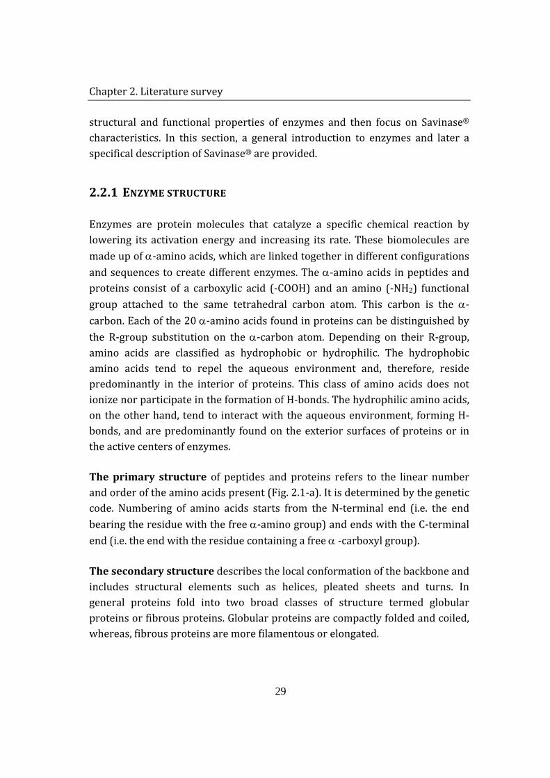
The primary structure of peptides and proteins refers to the linear number and order of the amino acids present (Fig. 2.1-a). It is determined by the genetic code. Numbering of amino acids starts from the N-terminal end (i.e. the end bearing the residue with the free α-amino group) and ends with the C-terminal end (i.e. the end with the residue containing a free α -carboxyl group).
The secondary structure describes the local conformation of the backbone and includes structural elements such as helices, pleated sheets and turns. In general proteins fold into two broad classes of structure termed globular proteins or fibrous proteins. Globular proteins are compactly folded and coiled, whereas, fibrous proteins are more filamentous or elongated.
Chapter 2. Literature survey
30
The α-helix is a common secondary structure encountered in proteins of the globular class. The formation of the α-helix is spontaneous and is stabilized by H-bonding between amide nitrogens and carbonyl carbons of peptide bonds spaced four residues apart. This orientation of H-bonding produces a helical coiling of the peptide backbone such that the R-groups lie on the exterior of the helix and perpendicular to its axis (Fig. 2.1-b-α helix).
The β pleated sheet differs markedly from the rod-like α-helix. β-sheets are composed of 2 or more different regions of stretches of at least 5-10 amino acids. They are almost fully extended rather than being tightly coiled as in the α helix (Fig. 2.1-b-pleated sheet). The folding and alignment of stretches of the polypeptide backbone aside one another to form β-sheets is stabilized by H-bonding between amide nitrogens and carbonyl carbons.
The tertiary structure describes the three-dimensional arrangement of the secondary structural elements together with the spatial arrangement of the side chains (Fig. 2.1-c). Secondary structures of proteins often constitute distinct domains, which interact with each other by several forces; such as: hydrogen bonding, covalent disulfide bonds, hydrophobic interactions, electrostatic interactions and van der Waals forces.
The quaternary structure refers to the number and interaction of polymer chains constituting a protein. Many proteins contain 2 or more different polypeptide chains that are held in association by the same non-covalent forces that stabilize their tertiary structure. Proteins with multiple polypetide chains are termed oligomeric proteins (Fig. 2.1-d).
Enzymes are highly specific and sophisticated catalysts. They attach to the participants of a reaction, i.e. substrate(s), holding the corresponding groups in close proximity while catalyzing. These binding and catalytic sites comprise enzyme’s active site. Two models have been proposed to describe their mode of action. Based on the specificity of catalysis, lock-and-key relation was attained between an enzymes and its corresponding substrate(s). The enzymes’ active site was depicted as a rigid structure complementary to substrate’s attachment location.
Chapter 2. Literature survey
31
Figure 2.1 Protein structure
Induced-fit hypothesis, on the other hand, states that a conformational change takes place in the enzyme during binding to the substrate, which results in the required matching of structures. This implies that at least some active sites are flexible; possessing a structure complementary to that of a substrate only when the later is bound to the enzyme. Therefore, conservation of catalytic activity is strongly dependent on the structural conformation of the enzyme (Palmer 1991).
2.2.2 SAVINASE®
Proteases used in the commercial detergents are mainly obtained from Bacillus amyloliquefaciens (subtilisin BPN’), B. licheniformis (subtilisin Carlsberg), and from highly alkalophilic bacilli such as B. lentus (Savinase® and Esperase®). Nowadays, detergent enzymes account for 89% of the total protease sales in the
Chapter 2. Literature survey
32
world. Significant share of the market is captured by subtilisins and/or alkaline proteases from various Bacillus species (Gupta et al. 2002).
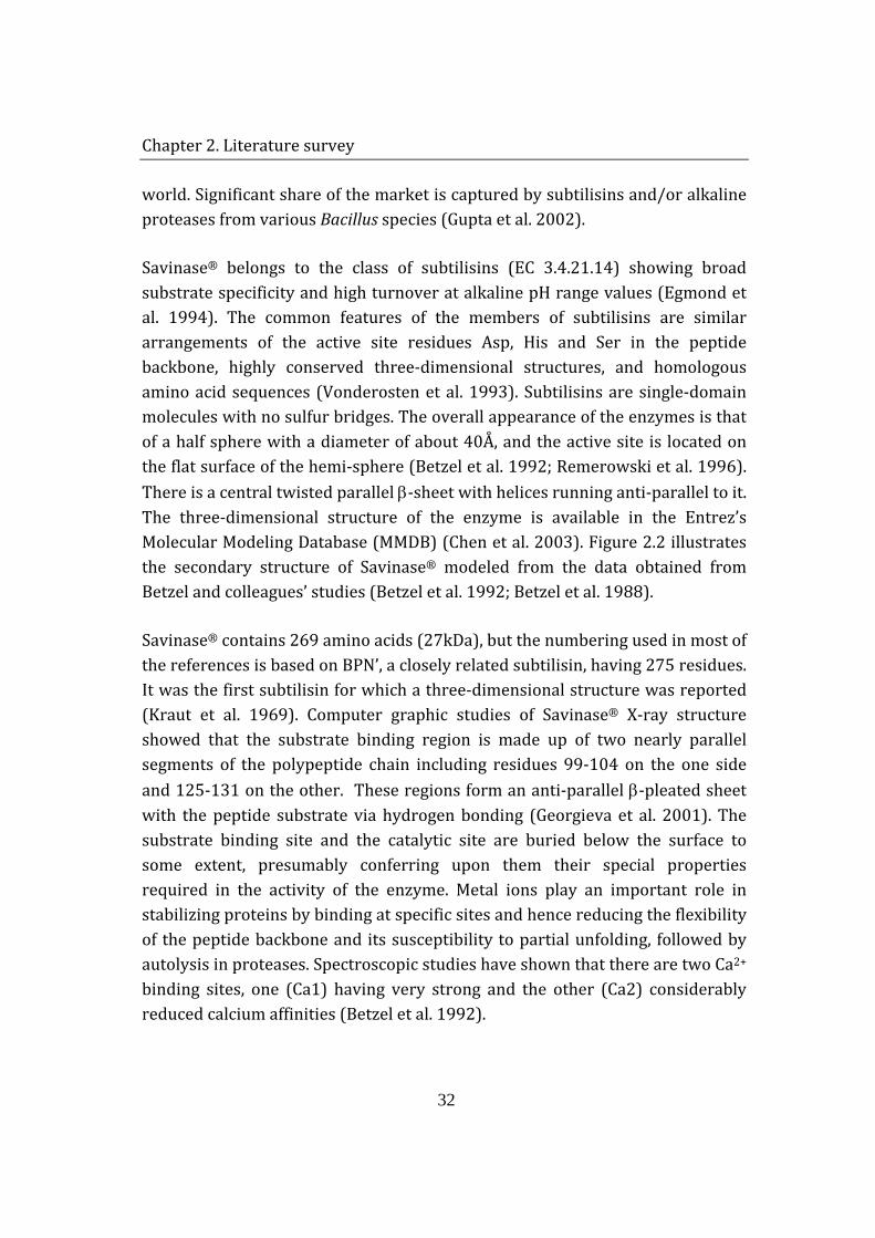
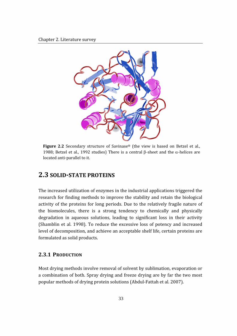
Savinase® belongs to the class of subtilisins (EC 3.4.21.14) showing broad substrate specificity and high turnover at alkaline pH range values (Egmond et al. 1994). The common features of the members of subtilisins are similar arrangements of the active site residues Asp, His and Ser in the peptide backbone, highly conserved three-dimensional structures, and homologous amino acid sequences (Vonderosten et al. 1993). Subtilisins are single-domain molecules with no sulfur bridges. The overall appearance of the enzymes is that of a half sphere with a diameter of about 40Å, and the active site is located on the flat surface of the hemi-sphere (Betzel et al. 1992; Remerowski et al. 1996). There is a central twisted parallel β-sheet with helices running anti-parallel to it. The three-dimensional structure of the enzyme is available in the Entrez’s Molecular Modeling Database (MMDB) (Chen et al. 2003). Figure 2.2 illustrates the secondary structure of Savinase® modeled from the data obtained from Betzel and colleagues’ studies (Betzel et al. 1992; Betzel et al. 1988).
Savinase® contains 269 amino acids (27kDa), but the numbering used in most of the references is based on BPN’, a closely related subtilisin, having 275 residues. It was the first subtilisin for which a three-dimensional structure was reported (Kraut et al. 1969). Computer graphic studies of Savinase® X-ray structure showed that the substrate binding region is made up of two nearly parallel segments of the polypeptide chain including residues 99-104 on the one side and 125-131 on the other. These regions form an anti-parallel β-pleated sheet with the peptide substrate via hydrogen bonding (Georgieva et al. 2001). The substrate binding site and the catalytic site are buried below the surface to some extent, presumably conferring upon them their special properties required in the activity of the enzyme. Metal ions play an important role in stabilizing proteins by binding at specific sites and hence reducing the flexibility of the peptide backbone and its susceptibility to partial unfolding, followed by autolysis in proteases. Spectroscopic studies have shown that there are two Ca2+ binding sites, one (Ca1) having very strong and the other (Ca2) considerably reduced calcium affinities (Betzel et al. 1992).
Chapter 2. Literature survey
33
Figure 2.2 Secondary structure of Savinase® (the view is based on Betzel et al., 1988; Betzel et al., 1992 studies) There is a central β-sheet and the α-helices are located anti-parallel to it.
2.3 SOLID-STATE PROTEINS
The increased utilization of enzymes in the industrial applications triggered the research for finding methods to improve the stability and retain the biological activity of the proteins for long periods. Due to the relatively fragile nature of the biomolecules, there is a strong tendency to chemically and physically degradation in aqueous solutions, leading to significant loss in their activity (Shamblin et al. 1998). To reduce the excessive loss of potency and increased level of decomposition, and achieve an acceptable shelf life, certain proteins are formulated as solid products.
2.3.1 PRODUCTION
Most drying methods involve removal of solvent by sublimation, evaporation or a combination of both. Spray drying and freeze drying are by far the two most popular methods of drying protein solutions (Abdul-Fattah et al. 2007).
Chapter 2. Literature survey
34
Freeze-drying (lyophilization) employs the principle of ice sublimation at reduced pressure. This provides moderate temperature conditions for the drying of heat-labile proteins (Towns 1995). Freeze-drying is accomplished in two major steps: freezing of the protein solution, and drying of the frozen solid under vacuum. The drying step is subdivided into two more phases: primary and secondary drying. In the primary drying phase, the bulk water present in the ice matrix of the frozen solid sublimes; then, in the secondary drying phase, the multilayer water surrounding the protein, i.e. the non-frozen ‘bound’ water, is removed, leaving residual moisture content in the product (Towns 1995; Wang 2000). Levels of residual moisture vary for different products. Generally, it is less than 1-5 % (Towns 1995) and constitutes a small portion of strongly bound water molecules.
Spray drying, on the other hand, is a directly particle generating method, in which a liquid product is atomized in a hot gas current to instantaneously obtain a very fine powder (10-50 µm) or large-size particles (2-3 mm) (Gharsallaoui et al. 2007). The drying process in a spray dryer comprises of three phases. The solution is fed through the atomizer nozzle at a controlled rate and as the liquid emerges from the nozzle orifice, due to large liquid-air interfacial expansion, the stream breaks up into small fine droplets (atomization) in the drying chamber. Aided by the large specific surface area of the droplets and the hot air in the chamber, evaporation takes place in the atomized droplets (drying) and the formed dried particles are passed through a cyclone separator into a collecting tube (recovery) (Abdul-Fattah et al. 2007). Powders produced by spray-drying are wetter than those prepared by freeze-drying, 4-10% vs. 1-4% (Ameri and Maa 2006).
2.3.2 STRUCTURAL PROPERTIES OF THE SOLID-STATE PROTEINS
During the formation of solid-state product of small molecules, big effort is made to obtain a highly crystalline product, in which molecules have regular and well-defined molecular packing, since amorphous solids are generally less stable than the corresponding crystals (Yu 2001). However, for macromolecules like proteins the situation is quite different. Unlike traditional small molecules, proteins possess higher order structures, which are required for biological
Chapter 2. Literature survey
35
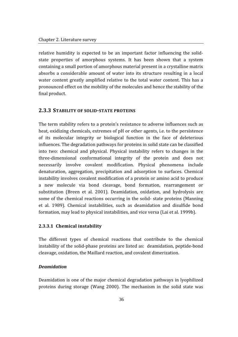
activity. Every protein molecule can be considered to be a disordered array of structures some of which are more locally ordered than others. Disordered regions (e.g. random coils, turns, loops) and more ordered structures (e.g. lamellar sheets helices) give proteins a character that is analogous to partially-crystalline synthetic macromolecules with respect to having regions of order or disorder (Shamblin et al. 1998). The conventional drying methods (spray-drying or lyophilization) applied for production of solid-state proteins may result in formation of partially or fully amorphous materials. Moreover, proteinous solids rarely exist in 100% crystalline or 100% amorphous structure (Hancock and Zografi 1997). The three-dimensional long-range order that normally exists in a crystalline material does not exist in the amorphous state. Amorphous solids are not random at the molecular level, but may possess short-range order, residual crystallinity, polymorphic states and regions of different density (Yu, 2001) (Fig. 2.3).
a) crystalline solid b) amorphous solid c) heterogeneity in an
amorphous solid
Figure 2.3 Schematic representation of the structure of a) crystalline, b) and c) amorphous solid. The molecular arrangement in an amorphous solid is not totally random, but features short-range molecular order similar to that in a crystalline solid. However, unlike crystals, an amorphous solid lacks the long-range order of molecular packing. An amorphous solid may have distinct regions (e.g., α and β) which have different densities and relaxation behaviours (Yu 2001).
Since molecules in the amorphous state exist in a higher energy state than in the crystalline state, it would be expected that properties requiring certain levels of molecular mobility would be influenced by the presence of amorphous structure. Small amount of adsorbed water can ‘plasticize’ amorphous solids, so
Chapter 2. Literature survey
36
relative humidity is expected to be an important factor influencing the solid-state properties of amorphous systems. It has been shown that a system containing a small portion of amorphous material present in a crystalline matrix absorbs a considerable amount of water into its structure resulting in a local water content greatly amplified relative to the total water content. This has a pronounced effect on the mobility of the molecules and hence the stability of the final product.
2.3.3 STABILITY OF SOLID-STATE PROTEINS
The term stability refers to a protein’s resistance to adverse influences such as heat, oxidizing chemicals, extremes of pH or other agents, i.e. to the persistence of its molecular integrity or biological function in the face of deleterious influences. The degradation pathways for proteins in solid state can be classified into two: chemical and physical. Physical instability refers to changes in the three-dimensional conformational integrity of the protein and does not necessarily involve covalent modification. Physical phenomena include denaturation, aggregation, precipitation and adsorption to surfaces. Chemical instability involves covalent modification of a protein or amino acid to produce a new molecule via bond cleavage, bond formation, rearrangement or substitution (Breen et al. 2001). Deamidation, oxidation, and hydrolysis are some of the chemical reactions occurring in the solid- state proteins (Manning et al. 1989). Chemical instabilities, such as deamidation and disulfide bond formation, may lead to physical instabilities, and vice versa (Lai et al. 1999b).
2.3.3.1 Chemical instability
The different types of chemical reactions that contribute to the chemical instability of the solid-phase proteins are listed as: deamidation, peptide-bond cleavage, oxidation, the Maillard reaction, and covalent dimerization.
Deamidation
Deamidation is one of the major chemical degradation pathways in lyophilized proteins during storage (Wang 2000). The mechanism in the solid state was
Chapter 2. Literature survey
37
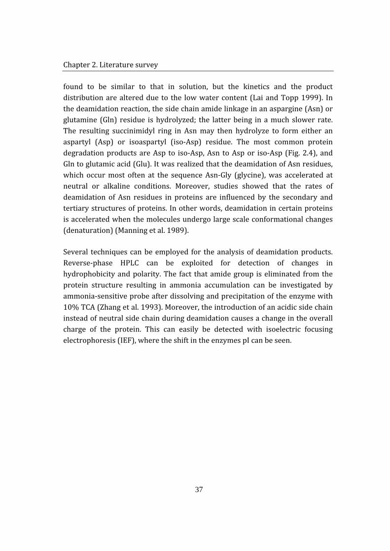
found to be similar to that in solution, but the kinetics and the product distribution are altered due to the low water content (Lai and Topp 1999). In the deamidation reaction, the side chain amide linkage in an aspargine (Asn) or glutamine (Gln) residue is hydrolyzed; the latter being in a much slower rate. The resulting succinimidyl ring in Asn may then hydrolyze to form either an aspartyl (Asp) or isoaspartyl (iso-Asp) residue. The most common protein degradation products are Asp to iso-Asp, Asn to Asp or iso-Asp (Fig. 2.4), and Gln to glutamic acid (Glu). It was realized that the deamidation of Asn residues, which occur most often at the sequence Asn-Gly (glycine), was accelerated at neutral or alkaline conditions. Moreover, studies showed that the rates of deamidation of Asn residues in proteins are influenced by the secondary and tertiary structures of proteins. In other words, deamidation in certain proteins is accelerated when the molecules undergo large scale conformational changes (denaturation) (Manning et al. 1989).
Several techniques can be employed for the analysis of deamidation products. Reverse-phase HPLC can be exploited for detection of changes in hydrophobicity and polarity. The fact that amide group is eliminated from the protein structure resulting in ammonia accumulation can be investigated by ammonia-sensitive probe after dissolving and precipitation of the enzyme with 10% TCA (Zhang et al. 1993). Moreover, the introduction of an acidic side chain instead of neutral side chain during deamidation causes a change in the overall charge of the protein. This can easily be detected with isoelectric focusing electrophoresis (IEF), where the shift in the enzymes pI can be seen.
Chapter 2. Literature survey
38
Figure 2.4 Deamidation mechanism of Asn to Asp and iso-Asp (Lai et al. 1999a).
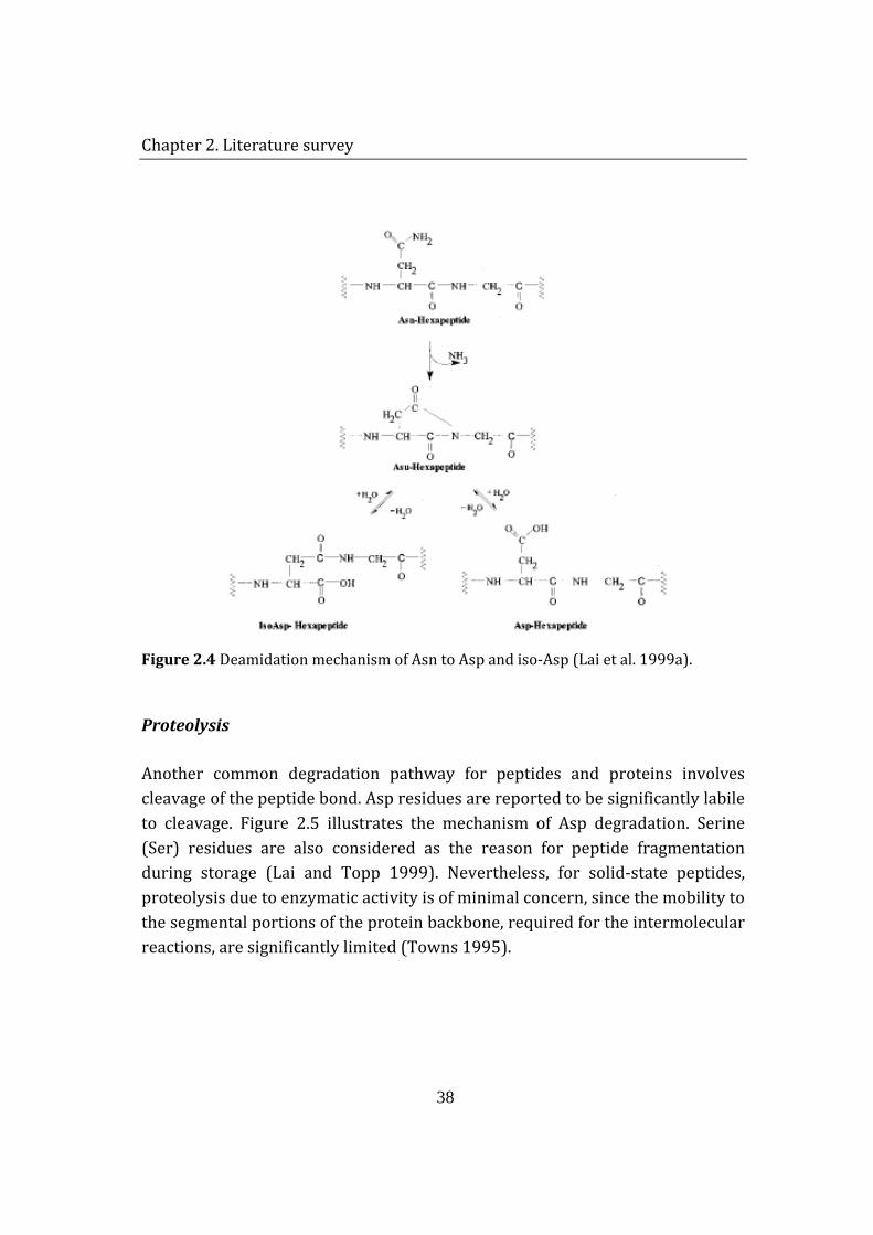
Proteolysis
Another common degradation pathway for peptides and proteins involves cleavage of the peptide bond. Asp residues are reported to be significantly labile to cleavage. Figure 2.5 illustrates the mechanism of Asp degradation. Serine (Ser) residues are also considered as the reason for peptide fragmentation during storage (Lai and Topp 1999). Nevertheless, for solid-state peptides, proteolysis due to enzymatic activity is of minimal concern, since the mobility to the segmental portions of the protein backbone, required for the intermolecular reactions, are significantly limited (Towns 1995).
Chapter 2. Literature survey
39
Figure 2.5 Mechanism of aspartyl (Asp) degradation (Manning et. al., 1989).
Peptide bond fragmentation is generally detected by following the molecular mass and size changes in the protein. Cleavage of the peptide bond results in major conformational changes, which may lead in alteration of hydrophobicity, polarity, and fluorescence characteristic of the protein. SDS-PAGE, followed by Comassie blue staining is the widely applied analytical method for monitoring hydrolysis. Another qualitative method is gel permeation chromatography (GPC), where, in contrast to SDS-PAGE, the hydrolysis products have lower migration velocity and longer retention time. Quantitative methods for analysis
Chapter 2. Literature survey
40
of peptide bond fragmentation can also be employed. Isocratic and gradient HPLC are very useful to monitor hydrolysis. Generally, detection is carried out UV spectrophotometrically at wavelength 214 nm. The most reliable and precise mass values can be obtained by using mass spectrometry (MS). Especially when the primary structure is known, it is possible to determine the site of hydrolysis of protein.
Oxidation
Among all amino acid residues, those containing a sulphur atom (methionine (Met) or cycteine (Cys)) or an aromatic ring (histidine (His), tryptophan (Trp), and tyrosine (Tyr)) are most susceptible to oxidation. Oxidation during processing and storage can be induced by contaminating oxidants, catalyzed by the presence of transition metal ions and induced by light (Li et al. 1995).
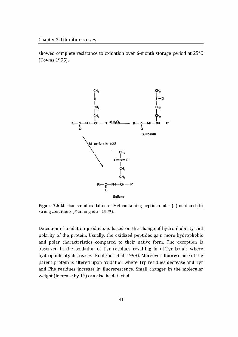
Oxidation of Met to Met sulfoxide has been frequently observed in protein products. Hydrogen peroxide is an effective oxidant for Met sulfoxide formation. The transfer of oxygen from H2O2 to sulphur in Met is catalyzed under mild acidic conditions. Transformation of Met to Met-sulfone, on the other hand, requires more drastic conditions and reagents, e.g. 95% performic acid (Fig. 2.6).
Oxidation of Met residues to Met-sulfoxide is associated with alteration of protein’s biological activity. Modification of single Met residue to Met-sulfoxide in subtilisin at pH 8.8, did not terminate the catalytic efficiency of the enzyme completely but resulted in changes in the kinetic parameters of the enzyme activity (Stauffer and Etson 1969). The same reaction caused loss of activity in ribosomal protein L12 in E.coli, but could be restored by incubation of the protein with high concentrations of β-mercaptoethanol. Regain of biological activity was found to coincide with the reduction of Met-sulfoxide to Met (Manning et al. 1989). It was also shown that within a given protein, the susceptibility of Met residues to oxidation may be different depending upon their position. For instance, human growth hormone (hGH) has three Met residues in its structure. While Met 14 and Met 125 were oxidized, Met 171
Chapter 2. Literature survey
41
showed complete resistance to oxidation over 6-month storage period at 25°C (Towns 1995).
Figure 2.6 Mechanism of oxidation of Met-containing peptide under (a) mild and (b) strong conditions (Manning et al. 1989).
Detection of oxidation products is based on the change of hydrophobicity and polarity of the protein. Usually, the oxidized peptides gain more hydrophobic and polar characteristics compared to their native form. The exception is observed in the oxidation of Tyr residues resulting in di-Tyr bonds where hydrophobicity decreases (Reubsaet et al. 1998). Moreover, fluorescence of the parent protein is altered upon oxidation where Trp residues decrease and Tyr and Phe residues increase in fluorerescence. Small changes in the molecular weight (increase by 16) can also be detected.
Chapter 2. Literature survey
42
Maillard reactions
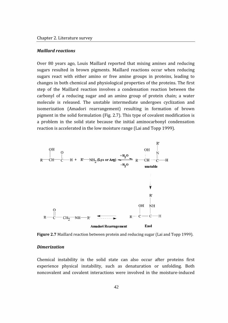
Over 80 years ago, Louis Maillard reported that mixing amines and reducing sugars resulted in brown pigments. Maillard reactions occur when reducing sugars react with either amino or free amine groups in proteins, leading to changes in both chemical and physiological properties of the proteins. The first step of the Maillard reaction involves a condensation reaction between the carbonyl of a reducing sugar and an amino group of protein chain; a water molecule is released. The unstable intermediate undergoes cyclization and isomerization (Amadori rearrangement) resulting in formation of brown pigment in the solid formulation (Fig. 2.7). This type of covalent modification is a problem in the solid state because the initial aminocarbonyl condensation reaction is accelerated in the low moisture range (Lai and Topp 1999).
Figure 2.7 Maillard reaction between protein and reducing sugar (Lai and Topp 1999).
Dimerization
Chemical instability in the solid state can also occur after proteins first experience physical instability, such as denaturation or unfolding. Both noncovalent and covalent interactions were involved in the moisture-induced
Chapter 2. Literature survey
43
aggregation of bovine insulin (Costantino et al. 1997). The covalent interactions were due to intermolecular disulfide bonds. Lyophilized bovine serum albumin, ovalbumin, glucose oxidase, and β-lactoglobulin were also observed to form covalent intermolecular disulfide linkages via a thiol-disulfide interchange reaction (Liu et al. 1991). Liu et al. (1991) postulated that the intermolecular thiol-disulfide interchange results from the ionized thiol on one albumin molecule attacking the disulfide linkage of another albumin molecule:
P1-S- + P2-S-S-P2 → P1-S-S-P2 + P2-S- , (2.8)
where P1 and P2 are the first and second protein molecules.
2.3.3.2 Physical instability
Denaturation
In molecular terms, denaturation is usually attributed to a disorientation of the structure of the protein molecule, renaturation to reorganization of the structure to its original form (Klotz 1958). Caused by a variety of conditions (increase in temperature, extreme pH, addition of organic solvents or other denaturants), this process can be envisioned as reversible or irreversible. Reversible denaturation is defined as unfolding caused by increase in temperature which can be reversed by subsequent lowering of the temperature. Irreversible denaturation or inactivation is caused by a variety of chemical or physical processes. Such proteins may be ‘misfolded’ in a conformation which does not allow the protein to regain its natural 3D structure. Irreversible denaturation is distinguished from chemical inactivation by unfolding of the wrongly oriented protein molecules by addition of a denaturant (e.g. urea). Then, the samples are dialysed to remove the denaturating agent and allowed to renaturate to their 3D structure at optimal conditions. Recovery of activity is an evidence of physical instability, i.e. absence of covalent bond formation, chemical instability (Manning et al. 1989).
Chapter 2. Literature survey
44
Techniques like solid-state NMR and attenuated total reflectance (ATR) can be used for assessing the 2° and 3° structures of the enzyme. Especially, ATR is very suitable for monitoring the differences in the conformational arrangement of hydrated proteins. Comparing the absorption spectra of reference and exposed proteins it would be possible to identify any structural changes in the enzyme. It is also possible to study the denaturation of the enzyme after dissolving the solid sample. Due to alterations in the peptide backbone positions, Tyr and Trp residues may become more exposed compared to the compact native structure. The shift in the fluorescence properties of the enzyme may give valuable information about the structural changes during unfolding process. Size exclusion chromatography (SEC) and sodium dodecyl sulfate poly acrylamide gel electrophoresis (SDS-PAGE) may also reveal evidence of alteration of the conformational arrangement. However, they are not much in use in case of reversible denaturation, where the conversion rate is fast.
Aggregation
There is a direct correlation between the extent of unfolding in the solid state and the amount of soluble aggregate present after dissolution. However, mechanism and the thermodynamics of protein aggregation, as well as the relation between aggregation and folding pathways still remain largely unclear. It is suggested that protein aggregation occurs when main chain interactions overrule specific side-chain contacts in a folded protein. The presence of stable tertiary contacts is a distinct feature of folded globular proteins, discriminating them from the “regular” polymers. When these tertiary contacts are loosened, the protein molecules become more prone to aggregation (Dzwolak et al. 2004). Hydrophobic interactions are probably the major driving force for aggregation. Once the protein is unfolded, these sites may come together changing the native conformation of the protein or forming clusters with other unfolded peptides. This results in alteration in the protein volume and formation of insoluble precipitates. This process occurs in both soluble and solid formulations but at very different rates (Shenoy et al. 2001). A study (Lin et al. 2000) showed that although most of the protein refolds back to its native-like conformation upon rehydration, a small fraction of the protein molecules forms soluble aggregates that can be
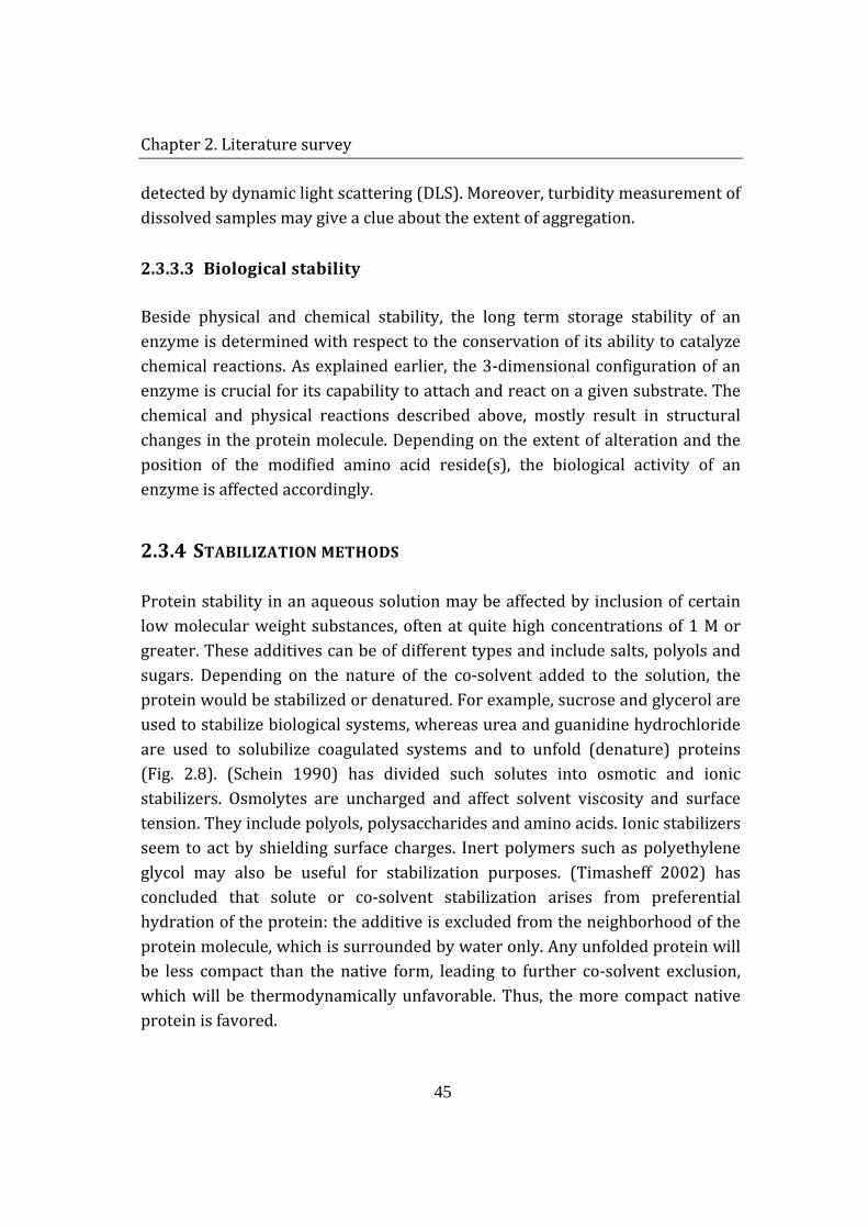
Chapter 2. Literature survey
45
detected by dynamic light scattering (DLS). Moreover, turbidity measurement of dissolved samples may give a clue about the extent of aggregation.
2.3.3.3 Biological stability
Beside physical and chemical stability, the long term storage stability of an enzyme is determined with respect to the conservation of its ability to catalyze chemical reactions. As explained earlier, the 3-dimensional configuration of an enzyme is crucial for its capability to attach and react on a given substrate. The chemical and physical reactions described above, mostly result in structural changes in the protein molecule. Depending on the extent of alteration and the position of the modified amino acid reside(s), the biological activity of an enzyme is affected accordingly.
2.3.4 STABILIZATION METHODS
Protein stability in an aqueous solution may be affected by inclusion of certain low molecular weight substances, often at quite high concentrations of 1 M or greater. These additives can be of different types and include salts, polyols and sugars. Depending on the nature of the co-solvent added to the solution, the protein would be stabilized or denatured. For example, sucrose and glycerol are used to stabilize biological systems, whereas urea and guanidine hydrochloride are used to solubilize coagulated systems and to unfold (denature) proteins (Fig. 2.8). (Schein 1990) has divided such solutes into osmotic and ionic stabilizers. Osmolytes are uncharged and affect solvent viscosity and surface tension. They include polyols, polysaccharides and amino acids. Ionic stabilizers seem to act by shielding surface charges. Inert polymers such as polyethylene glycol may also be useful for stabilization purposes. (Timasheff 2002) has concluded that solute or co-solvent stabilization arises from preferential hydration of the protein: the additive is excluded from the neighborhood of the protein molecule, which is surrounded by water only. Any unfolded protein will be less compact than the native form, leading to further co-solvent exclusion, which will be thermodynamically unfavorable. Thus, the more compact native protein is favored.
Chapter 2. Literature survey
46
Figure 2.8 Effect of additives on protein structure (Timasheff 2002).
There are alternative means of stabilizing proteins in the dry state. Glass has many useful properties but is in fact not solid; rather it is a highly viscous liquid which has undergone a ‘glass transition’ to yield a brittle, amorphous solid. Franks (Franks and Hatley 1992) has used stabilizing carbohydrate-based formulations, which undergo a similar glass transition. These protect proteins during drying operations and the resulting solid effectively preserves the protein in a fully active form. Since the glass formulation is water-soluble, addition of water allows one to re-dissolve the protein.
Additives can greatly enhance stability without chemical or genetic manipulation of the target protein. However, it should be noted that, operations such as dilution, dialysis, buffer exchange, or rehydration of a dry mixture containing enzymes would remove the stabilizing influences. The mentioned drawbacks can be overcome by chemical modification of the target protein. For example, covalent attachment of PEG to a serine protease subtilisin was studied by (Yang et al. 1996). As a result of the study, PEG-modified enzymes were significantly more stable against both increased temperature and pH. Stabilizing the enzymes is also possible by purposeful manipulation of the protein structure by genetic engineering. There are many successfully ‘designed’ subtilisins. However, such work is out of the scope of this study.
2.4 ENZYME GRANULES
Like many other proteins foreign to the human body, enzymes are potential inhalation allergens. Inhalation of even small concentrations of a foreign protein in the form of dust or aerosols can stimulate the body's immune system to produce antibodies. In some individuals, increased concentrations of
Chapter 2. Literature survey
47
antibody-enzyme protein complexes can trigger increased concentrations of histamine. The latter compounds can cause hay fever-like symptoms such as watery eyes, running nose, and a sore throat. To overcome the unwanted dust release and increase the stability of enzyme mixtures, they are encapsulated or coated to form enzyme granulates.
2.4.1 TYPE OF GRANULES
The mixing of the different components in order to encapsulate the enzyme is very important to control. The typical procedure is first to add all solid materials to the granulator, and add the binder solution hereafter. The type of granulator used for this granulation process is usually a high-shear mixer; but also a fluid bed coating granulator can be used. Herein layers of enzyme and protective coating are sprayed onto a core. The addition order and application method of the ingredients varies depending on the type of the produced granulate. The granules produced by Novozymes for detergent applications are mainly categorized in two groups: T-type and TK-type. They are formed by a core and different coating layers (Novozymes 2002).
In T-type granules (T for tough), enzymes are homogeneously mixed with stabilizing agents, like sugars and salts, to create a core. Then, this core is coated with a protective layer consisting of polymers like polyethylene glycol, salts, anti-dust agents like kaolin or calcium carbonate and colors (Fig. 2.9-a).
The enzyme-containing core is manufactured by mixing the stabilizer and enzymes. Generally, one the following methods are employed: a) a solution containing an enzyme and a stabilizer is dried, followed by granulation; b) a solution containing enzyme and a stabilizer is subjected to a wet granulation process; and c) powders of enzyme and stabilizer are blended uniformly, followed by granulation. The first method is more utilized (US 5,858,952).
The particle size of T-type granulates ranges between 300-1200µm. It is important to have particle size similar to that of the rest of the detergent components in order to prevent segregation during transportation. The average
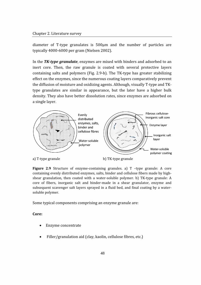
Chapter 2. Literature survey
48
diameter of T-type granulates is 500µm and the number of particles are typically 4000-6000 per gram (Nielsen 2002).
In the TK-type granulate, enzymes are mixed with binders and adsorbed to an inert core. Then, the raw granule is coated with several protective layers containing salts and polymers (Fig. 2.9-b). The TK-type has greater stabilizing effect on the enzymes, since the numerous coating layers comparatively prevent the diffusion of moisture and oxidizing agents. Although, visually T-type and TK-type granulates are similar in appearance, but the later have a higher bulk density. They also have better dissolution rates, since enzymes are adsorbed on a single layer.
a) T-type granule b) TK-type granule
Figure 2.9 Structure of enzyme-containing granules. a) T –type granule: A core containing evenly distributed enzymes, salts, binder and cellulose fibers made by high-shear granulation, then coated with a water-soluble polymer. b) TK-type granule: A core of fibers, inorganic salt and binder-made in a shear granulator, enzyme and subsequent scavenger salt layers sprayed in a fluid bed, and final coating by a water-soluble polymer.
Some typical components comprising an enzyme granule are:
Core:
• Enzyme concentrate
• Filler/granulation aid (clay, kaolin, cellulose fibres, etc.)
Chapter 2. Literature survey
49
• Binder solution (dextrin, polyvinyl pyrrolidone (PVP), )
• Matrix material (sugar + starch, Na2SO4, NaCl, Na2CO3, etc.,)
Coating:
• Inorganic salt as protection and/or scavenger layer (Na2S2O3, Na2SO4, MgSO4, etc.)
• Polyethylene glycol (PEG), Polyvinyl alcohol (PVA), stearic acid, latex polymers, methylhydroxypropylcellulose (MHPC)
• Colouring agents (TiO2, CaCO3, etc.)
The enzyme concentrate purified from the fermentation broth is usually either a constituent of the actual core, or it is sprayed onto the core after it has been formed (TK-type) or directly mixed with the core ingredients (T-type). The addition of cellulose fibres to the core material can have multiple effects. First of all the fibres can act as an anti-lumping agent, i.e. they make the granulation much easier to control. Secondly, they act as a reinforcement of the granule core, i.e. they tie the components together also in stressed situations. A third advantage of cellulose fibres is that they can reduce crack propagation in the granule. The cracks can only propagate until they reach a fibre which distributes the forces into the surrounding material. This is often enough in order for the crack propagation to be terminated. Salts are primarily added as matrix builders, since they are cheap and easily dissolve in water. However, they can also be added as a protective or scavenger layer outside the enzyme layer. The kaolin and clay are added as cheap fillers in order to reach the required density. As binder material several polymers can be chosen. One of the most common is polyvinyl pyrrolidone (PVP), which also helps stabilising the enzyme. Another common binder is dextrin. In the coating process a protective layer of about 10-15µm in thickness is applied to the granule core. The coating protects the enzyme granules from degradation by the sometimes harsh environment and prevents the enzyme from escaping the granule. The most common coating material is polyethylene glycol, PEG 900-4000. In order to get the desired
Chapter 2. Literature survey
50
appearance of the granule, a coloring agent is often added to the final coating. A very common choice is TiO2 (white). Depending on the customer desires other colors are also employed (blue, green, red, etc.) All components, except cellulose, comprising the detergent enzyme granule are water-soluble.
The major quality parameters of granulates are listed below (Nielsen 2002):
• Low dust release • Enzymatic stability in detergent • Uniform particle size distribution • Bulk density • High enzyme release • Total solubility • Cake strength (stickiness) • Low odor • Low production price
2.4.2 FACTORS AFFECTING GRANULE STABILITY IN DETERGENTS
Enzymes are fragile molecules. In the harsh environments like detergent powders, they are easily denaturated. For this reason, granulates are formed and incorporated in detergents as separately formulated particles rather than using free powder of enzymes. As was mentioned before, granulates; besides the prevention of undesired biological dust release, serve as protective structures for the enzymes. However, there are some factors that still have the ability to inactivate granulated detergent enzymes, even in appropriate storage conditions. In fact, instability is not caused by a single factor but a combination of number of events taking place in the detergent matrix.
2.4.2.1 Humidity
How should we understand water in the detergent matrix? In a controlled atmosphere with a fixed water activity, the hydration of a material continues until the local water activity matches that of the surroundings. The humidity in a
Chapter 2. Literature survey
51
detergent box mainly is as a result of free water present in the ingredients of detergent matrix. High humidity has destructive effects not only to the enzymes in granulates but also to the other components like bleaches.
The effects of water content on the stability of proteins in solid state can be summarized as follows: 1) changes in the dynamic flexibility of the protein, 2) participation of water as a reactant or inhibitor, or 3) participation of water as a medium for mobilization of reactants (Li et al. 1995). On their own, granulates may remain fairly dry in a humid atmosphere; however, embedded in a matrix that is hygroscopic, the water content may have serious impacts to the stability of enzymes. This is due to the fact that high humidity is necessary for the deteriorating factors to act on enzymes; in other words, harmful compounds rely on transport via a water phase. Actually, the granule as a whole is not completely destroyed, but the microenvironments generated due to increased humidity may have such an impact. Beside the facilitation of the transport of inactivating compounds, humidity may have significant impact on the morphology of granulates by causing a formation of cracks on the coatings, which results in an accelerated deactivation of the enzymes due to facilitated penetration of destructive components through these cracks.
The studies done in Novozymes showed that at RH less than 80%, enzymes can maintain their activity for several weeks; therefore, humidity does not cause auto-proteolysis. However, higher RH values resulted in approximately 30% loss of activity of Savinase® at the end of 4-week period when the granulates were incubated alone. Incubation with a detergent matrix resulted in 20% reduction at 35°C/55%RH and 70% at 37°C/70%RH after 12 weeks.
2.4.2.2 Hydrogen peroxide
The bleaching agents are the source of hydrogen peroxide in the detergent box. The release rate of H2O2 is considerably increased by water present in the surrounding.
Na2CO3 x 1½H2O2(s) + H2O(aq/g) 1½H2O2(aq) + CO32-(aq) + 2 Na+(aq)←
→(2.9)
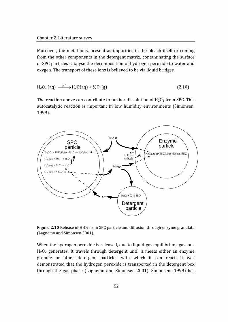
Chapter 2. Literature survey
52
Moreover, the metal ions, present as impurities in the bleach itself or coming from the other components in the detergent matrix, contaminating the surface of SPC particles catalyse the decomposition of hydrogen peroxide to water and oxygen. The transport of these ions is believed to be via liquid bridges.
H2O2 (aq) →+M H2O(aq) + ½O2(g) (2.10)
The reaction above can contribute to further dissolution of H2O2 from SPC. This autocatalytic reaction is important in low humidity environments (Simonsen, 1999).
Na 2 CO 3 x 1½H 2 O 2 (s) + H 2 O → H 2 O 2 ( aq) H 2 O 2 ( aq) + OH -
→ H 2 O H 2 O 2 ( aq) + M n+
→ H 2 O H 2 O 2 ( aq) ↔ H 2 O 2 (g)
} H 2 O 2 → radicals
H 2 O 2 (g)
H 2 O(g) SPC
particle
Detergent particle
Enzyme particle
H 2 O 2 ( aq/g)+ENZ(s/ aq) → Deact. ENZ M n+
H 2 O 2 + X → H 2 O M n+
Figure 2.10 Release of H2O2 from SPC particle and diffusion through enzyme granulate (Lagnemo and Simonsen 2001).
When the hydrogen peroxide is released, due to liquid-gas equilibrium, gaseous H2O2 generates. It travels through detergent until it meets either an enzyme granule or other detergent particles with which it can react. It was demonstrated that the hydrogen peroxide is transported in the detergent box through the gas phase (Lagnemo and Simonsen 2001). Simonsen (1999) has
Chapter 2. Literature survey
53
found that the maximum travel length of H2O2(g) does not exceed 5mm. The experiments done with increased ratio of detergent : enzyme+SPC, this is the distance between enzymes and bleaching agent is increased, proves that detergent matrix prevents hydrogen peroxide to reach granulates. Therefore, enzyme particles, near-by SPC particles, are only affected by peroxide oxidation.
Hydrogen peroxide inactivates Subtilisin with the concomitant formation of methionine sulfoxide at position 222 (Met222). Oxidized enzyme activity towards synthetic substrates decreases relative to native enzyme activity due to Met222’s proximity to the active site, which employs a common catalytic triad. The newly introduced carbonyl oxygen evidently alters the electronic configuration of the active site residues, thus affecting the rate of catalytic bond breaking and formation. Replacing Met222 with other amino acids does not result in mutant enzymes with comparable catalytic efficiency as the wild-type enzyme; thus, the goal is to preserve an unoxidized methionine at position 222 for maximum performance (DePaz et al. 2000).
The sulfoxide in the oxidized enzyme, further reacts with hydrogen peroxide to form sulfone. The reactions are as follows:
-C-S-CH3(s/aq)+ H2O2(aq/g) → -C-SO-CH3(s/aq)+ H2O(aq/g) (2.11)
-C-SO-CH3(s/aq)+ H2O2(aq/g) → -C-SO2-CH3(s/aq)+ H2O(aq/g) (2.12)
However, it is not clear which step of peroxide oxidation causes a complete inactivation of the enzyme.
Enzyme granulates are stabilized by mixing the biomolecules with inert ingredients like sugars. This provides conformational stability to the proteins by preferential exclusion mechanism. In other words, due to decreasing local water availability in the presence of sugars, a protein’s structure favour the most compact conformations, hence decreasing the available surface area. This non-specific mechanism leads to stabilization of the enzymes by limiting the active site’s accessibility to oxidative agents. In the previous experiments in
Chapter 2. Literature survey
54
Novozymes, organic molecules like potato protein and rice or corn starch were tested for improving the granulate stability. However, a conclusion about their direct effect cannot be derived from the results since they were used in the combination with other coating ingredients.
2.5 SUMMARY
An introduction to laundry detergents has been given. The diversity of ingredients was illustrated and their role in the washing process was described. The variety of chemicals present in the detergent matrix implies the possibility of complex interaction mechanism among them. Especially, for biomolecules like enzymes this mixture comprises a fairly harsh environment. Therefore, it is essential to determine the main destructive reactions and correspondingly protect and stabilize individual components in the matrix.
The scope of this study is focused on the stability of a detergent protease, Savinase®. A brief introduction on enzyme structure and mode of action was provided. Detailed information on the properties of solid-state proteins revealed that, despite of limited water availability, biomolecules are still susceptible to reactions resulting in physical or chemical modification. These alterations may also significantly diminish the catalytic activity of enzymes.
Special attention is required in the selection of analytical methods for detection of studied reactions. Analytical method choice usually depends on availability of equipment and expert personal to use it, cost of the analysis and the time required to establish the experimental protocol. Moreover, the method itself should be specific for the analysis. For instance, using fluorescence spectroscopy would give an idea about the conformational alterations in the protein structure, but would be difficult to assess whether the shift in the spectra is related to denaturation or major structural changes due to chemical reactions like deamidation or proteolysis. Therefore, it would be appropriate to confirm a result obtained from one method with the result of another analytical technique.
Chapter 2. Literature survey
55
For handling purpose and stability considerations, enzymes are added to the laundry detergents in formulated particles. The structure of these enzyme granulates was described. The main inactivation cause is addressed to the oxidation of enzymes by hydrogen peroxide released from the bleaching agent. Dissociation of the bleaching agent is enhanced by availability of water vapor. Humidity also increases the reactivity of solid-state proteins. Focusing on these factors, the kinetics and mechanism of enzyme inactivation can be reviled. Accordingly, stabilization strategies can be developed.
2.6 REFERENCES
Abdul-Fattah AM, Kalcinia DS, Pikal MI. 2007. The challenge of drying method selection for protein pharmaceuticals: Product quality implications. Journal of Pharmaceutical Sciences 96(8):1886-1916.
Ameri M, Maa YF. 2006. Spray drying of biopharmaceuticals: Stability and process considerations. Drying Technology 24(6):763-768.
Betzel C, Klupsch S, Papendorf G, Hastrup S, Branner S, Wilson KS. 1992. Crystal-Structure of the Alkaline Proteinase Savinase from Bacillus-Lentus at 1.4-a Resolution. Journal of Molecular Biology 223(2):427-445.
Betzel C, Pal GP, Saenger W. 1988. 3-Dimensional Structure of Proteinase-K at 0.15-Nm Resolution. European Journal of Biochemistry 178(1):155-171.
Breen ED, Curley JG, Overcashier DE, Hsu CC, Shire SJ. 2001. Effect of moisture on the stability of a lyophilized humanized monoclonal antibody formulation. Pharmaceutical Research 18(9):1345-1353.
Carson PA, Fairclough CS, Mauduit C, Colsell M. 2006. Peroxy bleaches - Part 1. Background and techniques for hazard evaluation. Journal of Hazardous Materials 136(3):438-445.
Chen J, Anderson JB, DeWeese-Scott C, Fedorova ND, Geer LY, He SQ, Hurwitz DI, Jackson JD, Jacobs AR, Lanczycki CJ and others. 2003. MMDB: Entrez's 3D-structure database. Nucleic Acids Research 31(1):474-477.
Chapter 2. Literature survey
56
Coons DM. 1978. Bleach - Facts, Fantasy, and Fundamentals. Journal of the American Oil Chemists Society 55(1):104-108.
Costantino HR, Curley JG, Hsu CC. 1997. Determining the water sorption monolayer of lyophilized pharmaceutical proteins. Journal of Pharmaceutical Sciences 86(12):1390-1393.
DePaz RA, Barnett CC, Dale DA, Carpenter JF, Gaertner AL, Randolph TW. 2000. The excluding effects of sucrose on a protein chemical degradation pathway: Methionine oxidation in subtilisin. Archives of Biochemistry and Biophysics 384(1):123-132.
Dzwolak W, Ravindra R, Winter R. 2004. Hydration and structure - the two sides of the insulin aggregation process. Physical Chemistry Chemical Physics 6(8):1938-1943.
Effendy I, Maibach HI. 1996. Detergent and skin irritation. Clinics in Dermatology 14(1):15-21.
Egmond MR, Antheunisse WP, Vanbemmel CJ, Ravestein P, Devlieg J, Peters H, Branner S. 1994. Engineering Surface-Charges in a Subtilisin - the Effects on Electrophoretic and Ion-Exchange Behavior. Protein Engineering 7(6):793-800.
Franks F, Hatley RMH; 1992. Storage of materials. U.S. patent 5,098,893.
Galwey AK, Hood WJ. 1979. Thermal-Decomposition of Sodium-Carbonate Perhydrate in the Solid-State. Journal of Physical Chemistry 83(14):1810-1815.
Georgieva DN, Stoeva S, Voelter W, Genov N, Betzel C. 2001. Differences in the specificities of the highly alkalophilic proteinases Savinase and Esperase imposed by changes in the rigidity and geometry of the substrate binding sites. Archives of Biochemistry and Biophysics 387(2):197-201.
Gharsallaoui A, Roudaut G, Chambin O, Voilley A, Saurel R. 2007. Applications of spray-drying in microencapsulation of food ingredients: An overview. Food Research International 40(9):1107-1121.
Guertechin LO. 1999. Handbook of detergents. Broze G, editor. New York: Marcel Dekker. Bd. p.
Chapter 2. Literature survey
57
Gupta R, Beg QK, Lorenz P. 2002. Bacterial alkaline proteases: molecular approaches and industrial applications. Applied Microbiology and Biotechnology 59(1):15-32.
Gutierrez EMP, NJ), Wu, Shang-ren (Shanghai, CN), Racherla, Uday (Fort Lee, NJ), Vermeer, Robert (Nutley, NJ); Lever Brothers Company, Division of Conopco, Inc., assignee. 1999. Detergent compositions containing polyethyleneimines for enhanced peroxygen bleach stability. United States.
Guwy AJ, Hawkes FR, Martin SR, Hawkes DL, Cunnah P. 2000. A technique for monitoring hydrogen peroxide concentration off-line and on-line. Water Research 34(8):2191-2198.
Hancock B, Zografi G. 1997. Characteristics and significance of the amorphous
state in pharmaceutical systems. Journal of Pharmaceutical Sciences 86(1):1-12.
Hollingsworth MW. 1978. Role of Detergent Builders in Fabric Washing Formulations. Journal of the American Oil Chemists Society 55(1):49-51.
Jakobi G, Löhr A. 1987. Detergents and Textile Washing: principles and practice. New York: VCH.
Johonsson C, Pekonen P, Forsstrom D. 2007. The long-term stability of sodium percarbonate in presence of zeolite as measured by heat flow calorimetry. Tenside Surfactants Detergents 44(4):210-+.
Klotz IM. 1958. Protein Hydration and Behavior. Science 128(3328):815-822.
Kowalski XSL, MO); Monsanto Company (St. Louis, MO), assignee. 1980. Peroxide stabilization. United States.
Kraut J, Alden RA, Wright CS, Westall FC. 1969. X-Ray Crystallographic Study of Active Site of Subtilisin Bpn'. Abstracts of Papers of the American Chemical Society(Sep):Bi63-&.
Kume G, Gallotti M, Nunes G. 2008. Review on anionic/cationic surfactant mixtures. Journal of Surfactants and Detergents 11(1):1-11.
Chapter 2. Literature survey
58
Kuroda MT, JP), Suzuki, Akira (Tochigi, JP), Kikuchi, Hideo (Fukushima, JP), Saito, Masahiro (Fukushima, JP), Yamaguchi, Nobuyoshi (Wakayama, JP); Kao Corporation (Tokyo, JP),Nippon Peroxide Co., Ltd. (Tokyo, JP), assignee. 1994. Stable slurry-coated sodium percarbonate, process for producing the same and bleach detergent composition containing the same. United States.
Lagnemo H, Simonsen O. Mechanism of deactivation of enzyme and sodium perbonate in HDP; 2001; Strasbourg, France. p 228-231.
Lai MC, Hageman MJ, Schowen RL, Borchardt RT, Laird BB, Topp EM. 1999a. Chemical stability of peptides in polymers. 2. Discriminating between solvent and plasticizing effects of water on peptide deamidation in poly(vinylpyrrolidone). Journal of Pharmaceutical Sciences 88(10):1081-1089.
Lai MC, Hageman MJ, Schowen RL, Borchardt RT, Topp EM. 1999b. Chemical stability of peptides in polymers. 1. Effect of water on peptide deamidation in poly(vinyl alcohol) and poly(vinyl pyrrolidone) matrixes. Journal of Pharmaceutical Sciences 88(10):1073-1080.
Lai MC, Topp EM. 1999. Solid-state chemical stability of proteins and peptides. Journal of Pharmaceutical Sciences 88(5):489-500.
Li SH, Schoneich C, Borchardt RT. 1995. Chemical-Instability of Protein Pharmaceuticals - Mechanisms of Oxidation and Strategies for Stabilization. Biotechnology and Bioengineering 48(5):490-500.
Lin JJ, Meyer JD, Carpenter JF, Manning MC. 2000. Stability of human serum albumin during bioprocessing: Denaturation and aggregation during processing of albumin paste. Pharmaceutical Research 17(4):391-396.
Liu WR, Langer R, Klibanov AM. 1991. Moisture-Induced Aggregation of Lyophilized Proteins in the Solid-State. Biotechnology and Bioengineering 37(2):177-184.
Manning MC, Patel K, Borchardt RT. 1989. Stability of Protein Pharmaceuticals. Pharmaceutical Research 6(11):903-918.
Chapter 2. Literature survey
59
McKillop A, Sanderson WR. 1995. Sodium Perborate and Sodium Percarbonate - Cheap, Safe and Versatile Oxidizing-Agents for Organic-Synthesis. Tetrahedron 51(22):6145-6166.
Nagarajan MK, Paine HL. 1984. Water Hardness Control by Detergent Builders. Journal of the American Oil Chemists Society 61(9):1475-1478.
Nielsen H. 2002. Granulate quality. Novozymes A/S.
Novozymes. 2002. Internal Detergent Enzyme Handbook. Novozymes.
Olsen H, Falholt P. 1998. The role of enzymes in detergency. Bagsvaerd: Novo Nordisk A/S.
Palmer T. 1991. Understanding enzymes. Chichester: Wiley, John & Sons, Inc.
Ponnusamy T, Dubal SA, Momin SA. 2008. Studies in Detergency: Influence of Different Factors for Removing Motor Oil Stain from the Cotton Fabric. Journal of Dispersion Science and Technology 29:1123–1128.
Remerowski ML, Pepermans HAM, Hilbers CW, VandeVen FJM. 1996. Backbone dynamics of the 269-residue protease Savinase determined from N-15-NMR relaxation measurements. European Journal of Biochemistry 235(3):629-640.
Reubsaet JLE, Beijnen JH, Bult A, van Maanen RJ, Marchal JAD, Underberg WJM. 1998. Analytical techniques used to study the degradation of proteins and peptides: chemical instability. Journal of Pharmaceutical and Biomedical Analysis 17(6-7):955-978.
Sak-Bosnar M, Madunic-Cacic D, Matesic-Puac R, Grabaric Z. 2007. Nonionic surfactant-selective electrode and its application for determination in real solutions. Analytica Chimica Acta 581(2):355-363.
Scheibel JJ. 2004. The evolution of anionic surfactant technology to meet the requirements of the laundry detergent industry. Journal of Surfactants and Detergents 7(4):319-328.
Schein CH. 1990. Solubility as a Function of Protein-Structure and Solvent Components. Bio-Technology 8(4):308-315.
Chapter 2. Literature survey
60
Shamblin SL, Hancock BC, Zografi G. 1998. Water vapor sorption by peptides, proteins and their formulations. European Journal of Pharmaceutics and Biopharmaceutics 45(3):239-247.
Shenoy B, Wang Y, Shan WZ, Margolin AL. 2001. Stability of crystalline proteins. Biotechnology and Bioengineering 73(5):358-369.
Skagerlind P, Lindahl G, Rasmusson B. 1998. Performance and stability of the bleaching system, sodium percarbonate, compared to the traditional bleach system, sodium perborate. SÖFW-Journal 124:680-689.
Smulders E. 2002. Laundry detergents. Weinheim: Wiley-VCH.
Stauffer CE, Etson D. 1969. Effect of Subtilisin Activity of Oxidizing a Methionine Residue. Journal of Biological Chemistry 244(19):5333-&.
Timasheff SN. 2002. Protein-solvent preferential interactions, protein hydration, and the modulation of biochemical reactions by solvent components. Proceedings of the National Academy of Sciences of the United States of America 99(15):9721-9726.
Towns JK. 1995. Moisture-Content in Proteins - Its Effects and Measurement. Journal of Chromatography A 705(1):115-127.
Vonderosten C, Branner S, Hastrup S, Hedegaard L, Rasmussen MD, Bisgardfrantzen H, Carlsen S, Mikkelsen JM. 1993. Protein Engineering of Subtilisins to Improve Stability in Detergent Formulations. Journal of Biotechnology 28(1):55-68.
Wang W. 2000. Lyophilization and development of solid protein pharmaceuticals. International Journal of Pharmaceutics 203(1-2):1-60.
Watson RA. 2006. Laundry detergent formulations. In: Showell MS, editor. Handbook of detergents: Formulation. Boca Raton, FL: CRC Press. p 63.
Yang Z, Domach M, Auger R, Yang FX, Russell AJ. 1996. Polyethylene glycol-induced stabilization of subtilisin. Enzyme and Microbial Technology 18(2):82-89.
Yu L. 2001. Amorphous pharmaceutical solids: preparation, characterization and stabilization. Advanced Drug Delivery Reviews 48(1):27-42.
Chapter 2. Literature survey
61
Yu YX, Zhao J, Bayly AE. 2008. Development of surfactants and builders in detergent formulations. Chinese Journal of Chemical Engineering 16(4):517-527.
Zhang J, Lee TC, Ho CT. 1993. Thermal Deamidation of Proteins in a Restricted Water Environment. Journal of Agricultural and Food Chemistry 41(11):1840-1843.
Chapter 3. Construction of the experimental setup
62
CHAPTER 3 CONSTRUCTION OF THE EXPERIMENTAL
SETUP The experimental setup development was a fundamental part of this project. The main objective was to create a controlled environment for testing enzyme stability in detergent-like conditions. The following chapter gives detailed description of the experimental design, equipment and procedure used for the stability testing of solid state enzymes under oxidizing conditions.
3.1 DEMANDS FOR THE SET-UP
Conserved cleaning power during storage is an important quality parameter for a detergent. Customers are expecting to be able to store open detergent boxes for half a year and still get a satisfying result. Therefore, enzyme manufacturers have to get a realistic picture of the activity of the enzyme after 6 months.
Testing stability of enzymes in detergent powders is a slow process due to the fact that the degradation happens over months. There is a continuous demand for faster testing methods, especially when developing new products. The tests applied for that purpose generally last for 4-8 weeks, where granulates are stored at elevated temperatures (30-37°C) and humidities (55-70%RH).
Stability of enzymes in granulated detergent powders depends on numerous factors; such as enzyme granulate composition, detergent matrix ingredients, storage conditions, etc. Hydrogen peroxide is considered to be the major cause of enzyme inactivation in powdered laundry detergents (Lagnemo and Simonsen 2001). It is released by the bleaching agent (mainly sodium percarbonate), which is decomposed in the presence of water vapor during long-term storage of the detergent. Moreover, it is known that humidity alone
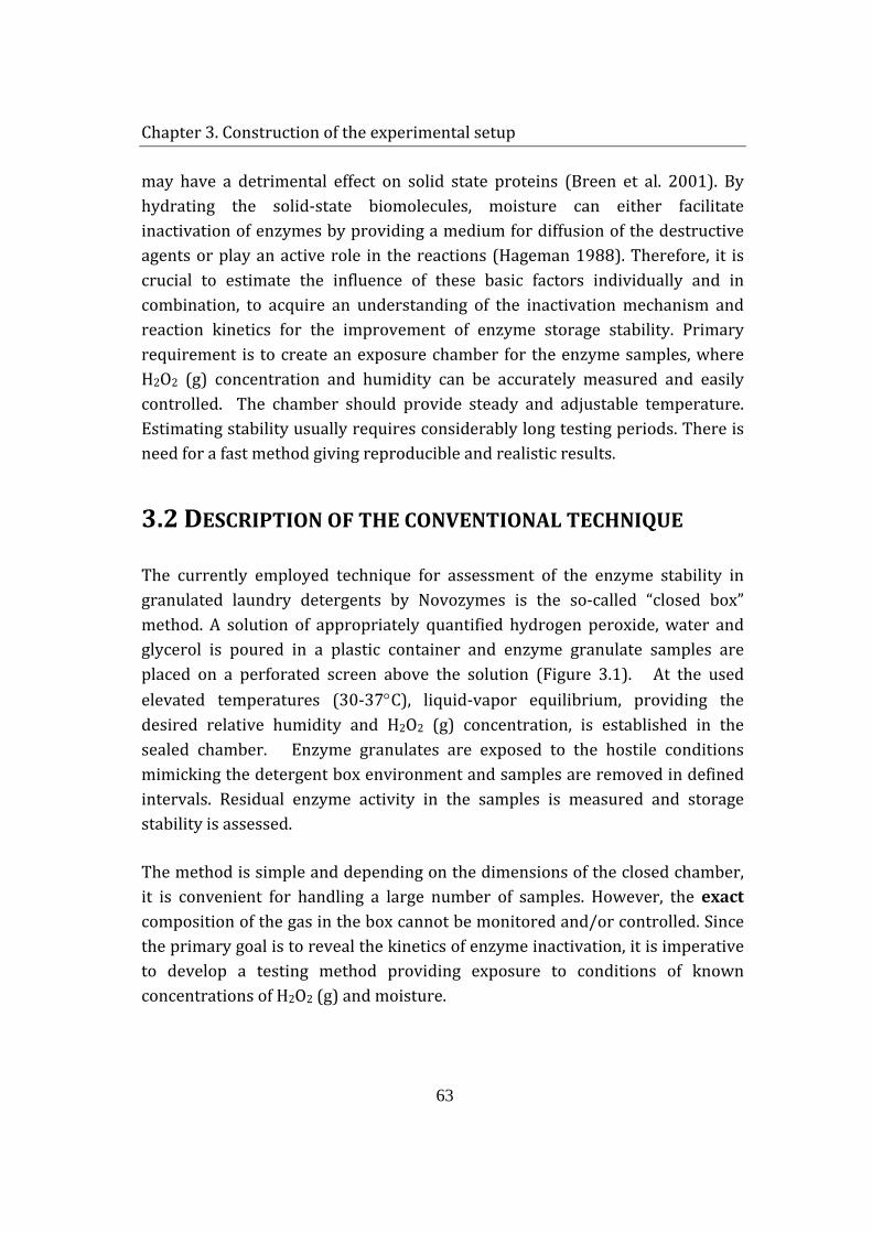
Chapter 3. Construction of the experimental setup
63
may have a detrimental effect on solid state proteins (Breen et al. 2001). By hydrating the solid-state biomolecules, moisture can either facilitate inactivation of enzymes by providing a medium for diffusion of the destructive agents or play an active role in the reactions (Hageman 1988). Therefore, it is crucial to estimate the influence of these basic factors individually and in combination, to acquire an understanding of the inactivation mechanism and reaction kinetics for the improvement of enzyme storage stability. Primary requirement is to create an exposure chamber for the enzyme samples, where H2O2 (g) concentration and humidity can be accurately measured and easily controlled. The chamber should provide steady and adjustable temperature. Estimating stability usually requires considerably long testing periods. There is need for a fast method giving reproducible and realistic results.
3.2 DESCRIPTION OF THE CONVENTIONAL TECHNIQUE
The currently employed technique for assessment of the enzyme stability in granulated laundry detergents by Novozymes is the so-called “closed box” method. A solution of appropriately quantified hydrogen peroxide, water and glycerol is poured in a plastic container and enzyme granulate samples are placed on a perforated screen above the solution (Figure 3.1). At the used elevated temperatures (30-37°C), liquid-vapor equilibrium, providing the desired relative humidity and H2O2 (g) concentration, is established in the sealed chamber. Enzyme granulates are exposed to the hostile conditions mimicking the detergent box environment and samples are removed in defined intervals. Residual enzyme activity in the samples is measured and storage stability is assessed.
The method is simple and depending on the dimensions of the closed chamber, it is convenient for handling a large number of samples. However, the exact composition of the gas in the box cannot be monitored and/or controlled. Since the primary goal is to reveal the kinetics of enzyme inactivation, it is imperative to develop a testing method providing exposure to conditions of known concentrations of H2O2 (g) and moisture.
Chapter 3. Construction of the experimental setup
64
Figure 3.1 Closed box method for measuring enzyme stability under oxidizing conditions.
3.3 HYDROGEN PEROXIDE VAPOR GENERATION AND
MEASUREMENT
A simple and effective system for the generation and measurement of H2O2 vapor was established. Firstly, H2O2 solutions of different concentrations were prepared and before each set of experiments, their concentrations were determined by titration with potassium permanganate (KMnO4) (section 4.1.1). Production of gaseous H2O2 was achieved by bubbling inert gas through a concentrated solution of H2O2 in a bubble-flask (impinger) at room temperature (21°C). N2 was used as a carrier gas and its flow rate was controlled with Bronkhorst Hi-tech (Holland) mass flow controller and measured with a primary flow calibrator, bubble generator (Gilian® GilibratorTM 2, USA). It was assumed that equilibrium between the liquid and gas in the bubble-flask was established. Hence, the gas stream leaving the impinger, contained gaseous H2O2 as the vapor pressure of the corresponding solution at room temperature. To confirm that, partial pressure of H2O2 (g) in the releasing gas stream was determined by measuring the absorption rate of H2O2 (g) in ultra-pure water, produced in an Elgastat Maxima Analytical (Germany). An effective absorption was achieved by insertion of the collection bottle in an ice-water bath. 1-ml samples were withdrawn from the solution in the collection bottle with 3-min
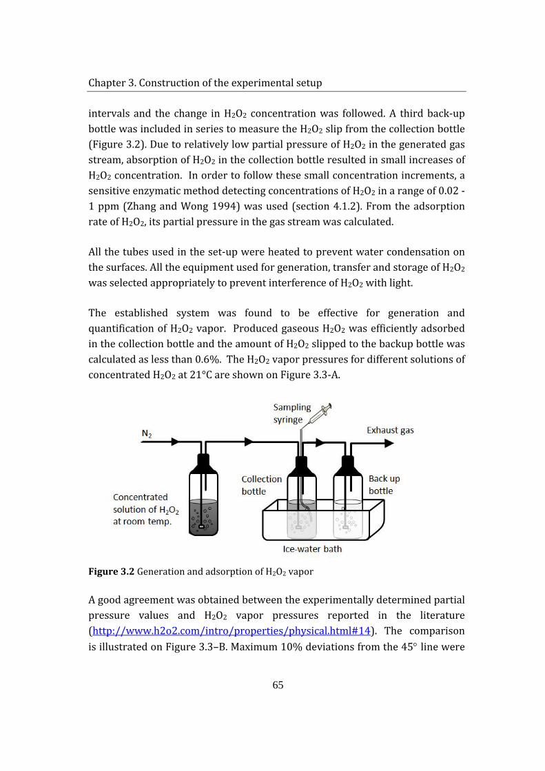
Chapter 3. Construction of the experimental setup
65
intervals and the change in H2O2 concentration was followed. A third back-up bottle was included in series to measure the H2O2 slip from the collection bottle (Figure 3.2). Due to relatively low partial pressure of H2O2 in the generated gas stream, absorption of H2O2 in the collection bottle resulted in small increases of H2O2 concentration. In order to follow these small concentration increments, a sensitive enzymatic method detecting concentrations of H2O2 in a range of 0.02 - 1 ppm (Zhang and Wong 1994) was used (section 4.1.2). From the adsorption rate of H2O2, its partial pressure in the gas stream was calculated.
All the tubes used in the set-up were heated to prevent water condensation on the surfaces. All the equipment used for generation, transfer and storage of H2O2 was selected appropriately to prevent interference of H2O2 with light.
The established system was found to be effective for generation and quantification of H2O2 vapor. Produced gaseous H2O2 was efficiently adsorbed in the collection bottle and the amount of H2O2 slipped to the backup bottle was calculated as less than 0.6%. The H2O2 vapor pressures for different solutions of concentrated H2O2 at 21°C are shown on Figure 3.3-A.
Figure 3.2 Generation and adsorption of H2O2 vapor
A good agreement was obtained between the experimentally determined partial pressure values and H2O2 vapor pressures reported in the literature (http://www.h2o2.com/intro/properties/physical.html#1 ). The comparison is illustrated on Figure 3.3–B. Maximum 10% deviations from the 45° line were
4
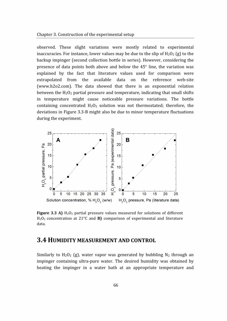
Chapter 3. Construction of the experimental setup
66
observed. These slight variations were mostly related to experimental inaccuracies. For instance, lower values may be due to the slip of H2O2 (g) to the backup impinger (second collection bottle in series). However, considering the presence of data points both above and below the 45° line, the variation was explained by the fact that literature values used for comparison were extrapolated from the available data on the reference web-site (www.h2o2.com). The data showed that there is an exponential relation between the H2O2 partial pressure and temperature, indicating that small shifts in temperature might cause noticeable pressure variations. The bottle containing concentrated H2O2 solution was not thermostated; therefore, the deviations in Figure 3.3-B might also be due to minor temperature fluctuations during the experiment.
Figure 3.3 A) H2O2 partial pressure values measured for solutions of different H2O2 concentration at 21°C and B) comparison of experimental and literature data.
3.4 HUMIDITY MEASUREMENT AND CONTROL
Similarly to H2O2 (g), water vapor was generated by bubbling N2 through an impinger containing ultra-pure water. The desired humidity was obtained by heating the impinger in a water bath at an appropriate temperature and
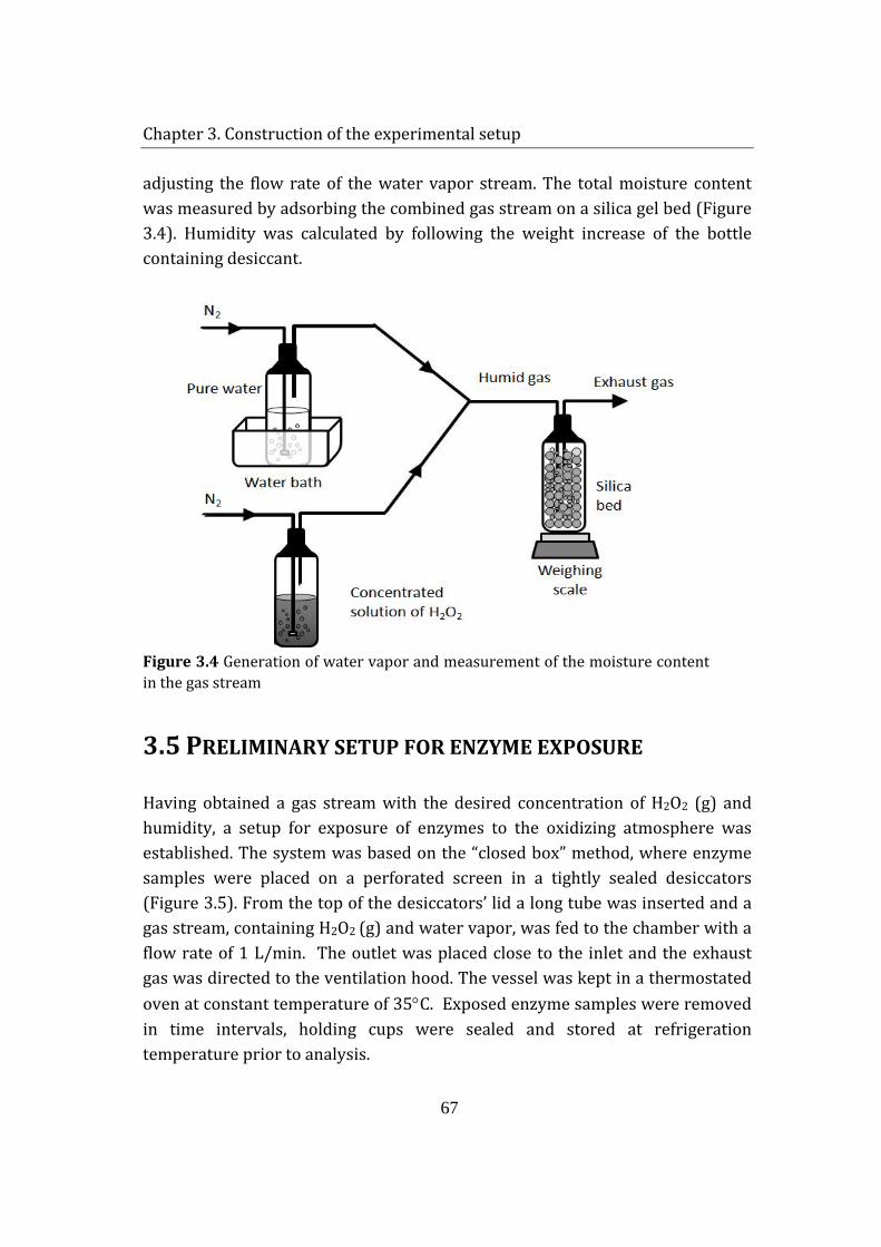
Chapter 3. Construction of the experimental setup
67
adjusting the flow rate of the water vapor stream. The total moisture content was measured by adsorbing the combined gas stream on a silica gel bed (Figure 3.4). Humidity was calculated by following the weight increase of the bottle containing desiccant.
Figure 3.4 Generation of water vapor and measurement of the moisture content in the gas stream
3.5 PRELIMINARY SETUP FOR ENZYME EXPOSURE
Having obtained a gas stream with the desired concentration of H2O2 (g) and humidity, a setup for exposure of enzymes to the oxidizing atmosphere was established. The system was based on the “closed box” method, where enzyme samples were placed on a perforated screen in a tightly sealed desiccators (Figure 3.5). From the top of the desiccators’ lid a long tube was inserted and a gas stream, containing H2O2 (g) and water vapor, was fed to the chamber with a flow rate of 1 L/min. The outlet was placed close to the inlet and the exhaust gas was directed to the ventilation hood. The vessel was kept in a thermostated oven at constant temperature of 35°C. Exposed enzyme samples were removed in time intervals, holding cups were sealed and stored at refrigeration temperature prior to analysis.
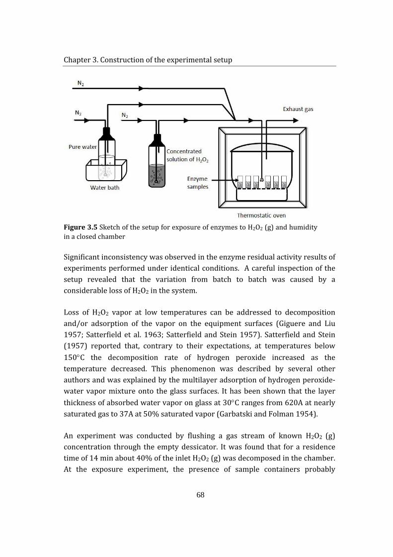
Chapter 3. Construction of the experimental setup
68
Figure 3.5 Sketch of the setup for exposure of enzymes to H2O2 (g) and humidity in a closed chamber
Significant inconsistency was observed in the enzyme residual activity results of experiments performed under identical conditions. A careful inspection of the setup revealed that the variation from batch to batch was caused by a considerable loss of H2O2 in the system.
Loss of H2O2 vapor at low temperatures can be addressed to decomposition and/or adsorption of the vapor on the equipment surfaces (Giguere and Liu 1957; Satterfield et al. 1963; Satterfield and Stein 1957). Satterfield and Stein (1957) reported that, contrary to their expectations, at temperatures below 150°C the decomposition rate of hydrogen peroxide increased as the temperature decreased. This phenomenon was described by several other authors and was explained by the multilayer adsorption of hydrogen peroxide-water vapor mixture onto the glass surfaces. It has been shown that the layer thickness of absorbed water vapor on glass at 30°C ranges from 620A at nearly saturated gas to 37A at 50% saturated vapor (Garbatski and Folman 1954).
An experiment was conducted by flushing a gas stream of known H2O2 (g) concentration through the empty dessicator. It was found that for a residence time of 14 min about 40% of the inlet H2O2 (g) was decomposed in the chamber. At the exposure experiment, the presence of sample containers probably
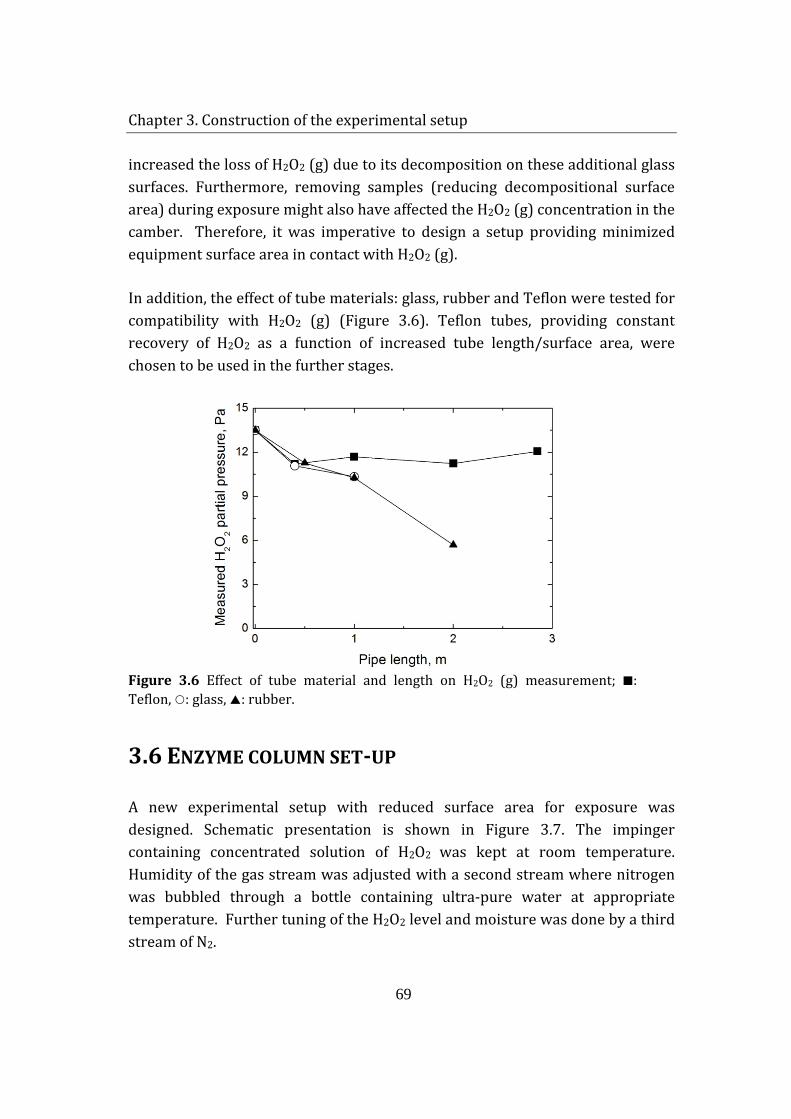
Chapter 3. Construction of the experimental setup
69
increased the loss of H2O2 (g) due to its decomposition on these additional glass surfaces. Furthermore, removing samples (reducing decompositional surface area) during exposure might also have affected the H2O2 (g) concentration in the camber. Therefore, it was imperative to design a setup providing minimized equipment surface area in contact with H2O2 (g).
In addition, the effect of tube materials: glass, rubber and Teflon were tested for compatibility with H2O2 (g) (Figure 3.6). Teflon tubes, providing constant recovery of H2O2 as a function of increased tube length/surface area, were chosen to be used in the further stages.
Figure 3.6 Effect of tube material and length on H2O2 (g) measurement; : Teflon, : glass, : rubber.
3.6 ENZYME COLUMN SET-UP
A new experimental setup with reduced surface area for exposure was designed. Schematic presentation is shown in Figure 3.7. The impinger containing concentrated solution of H2O2 was kept at room temperature. Humidity of the gas stream was adjusted with a second stream where nitrogen was bubbled through a bottle containing ultra-pure water at appropriate temperature. Further tuning of the H2O2 level and moisture was done by a third stream of N2.
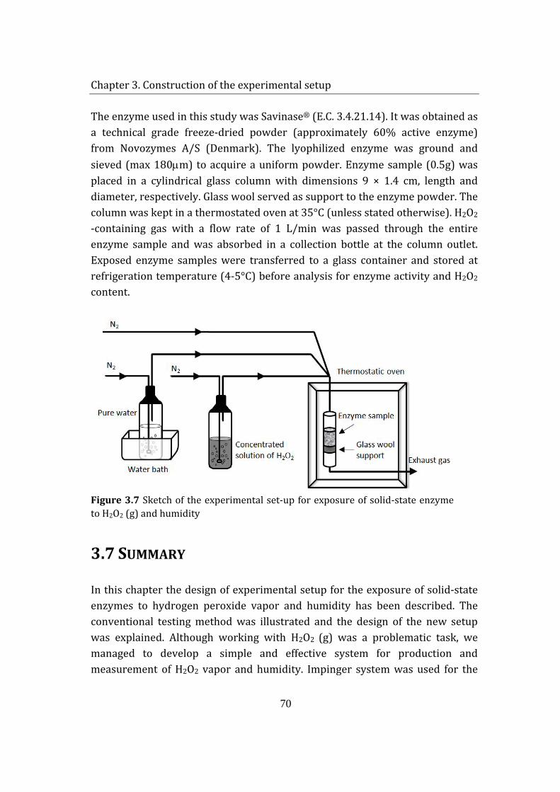
Chapter 3. Construction of the experimental setup
70
The enzyme used in this study was Savinase® (E.C. 3.4.21.14). It was obtained as a technical grade freeze-dried powder (approximately 60% active enzyme) from Novozymes A/S (Denmark). The lyophilized enzyme was ground and sieved (max 180µm) to acquire a uniform powder. Enzyme sample (0.5g) was placed in a cylindrical glass column with dimensions 9 × 1.4 cm, length and diameter, respectively. Glass wool served as support to the enzyme powder. The column was kept in a thermostated oven at 35°C (unless stated otherwise). H2O2 -containing gas with a flow rate of 1 L/min was passed through the entire enzyme sample and was absorbed in a collection bottle at the column outlet. Exposed enzyme samples were transferred to a glass container and stored at refrigeration temperature (4-5°C) before analysis for enzyme activity and H2O2 content.
Figure 3.7 Sketch of the experimental set-up for exposure of solid-state enzyme to H2O2 (g) and humidity
3.7 SUMMARY
In this chapter the design of experimental setup for the exposure of solid-state enzymes to hydrogen peroxide vapor and humidity has been described. The conventional testing method was illustrated and the design of the new setup was explained. Although working with H2O2 (g) was a problematic task, we managed to develop a simple and effective system for production and measurement of H2O2 vapor and humidity. Impinger system was used for the
Chapter 3. Construction of the experimental setup
71
production of H2O2 (g) and humidity by bubbling inert gas through a corresponding solution at an appropriate temperature. Powder state enzyme samples were placed in a reaction column and exposed to the wet oxidant gas. The major problem of H2O2 loss due to decomposition and/or adsorption on equipment surfaces was significantly overcome by designing a setup with minimized contact surface area. The generated H2O2 vapor was successfully adsorbed for quantified with a sensitive analytical method for low concentrations of H2O2. Calculations showed that measured partial vapor pressure was in a good agreement with the data reported in the literature.
The enzyme column setup enabled exposure of solid enzymes to different concentrations of H2O2 (g) and humidity for revealing the inactivation kinetics. Even though a single sample could be handled at a time, the method still provided fast stability testing, where samples were obtained in a few hours instead of several weeks.
3.8 REFERENCES
Breen ED, Curley JG, Overcashier DE, Hsu CC, Shire SJ. 2001. Effect of moisture on the stability of a lyophilized humanized monoclonal antibody formulation. Pharmaceutical Research 18(9):1345-1353.
Garbatski U, Folman M. 1954. Multilayer Adsorption of Water near Saturation Pressure on Plane Glass Surfaces. Journal of Chemical Physics 22(12):2086-2086.
Giguere PA, Liu ID. 1957. Kinetics of the Thermal Decomposition of Hydrogen Peroxide Vapor. Canadian Journal of Chemistry-Revue Canadienne De Chimie 35(4):283-293.
Hageman MJ. 1988. The Role of Moisture in Protein Stability. Drug Development and Industrial Pharmacy 14(14):2047-2070.
Lagnemo H, Simonsen O. 2001. Mechanism of deactivation of enzyme and sodium perbonate in HDP. 40th International Detergency Conference. Strasbourg, France.
Chapter 3. Construction of the experimental setup
72
Satterfield CN, Reid RC, Wechsler AE. 1963. Chemical Kinetics in Porous Walled Reactors - the Decomposition of Hydrogen Peroxide Vapor. Aiche Journal 9(2):168-175.
Satterfield CN, Stein TW. 1957. Decomposition of Hydrogen Peroxide Vapor on Relatively Inert Surfaces. Industrial and Engineering Chemistry 49(7):1173-1180.
Zhang LS, Wong GTF. 1994. Spectrophotometric Determination of H2o2 in Marine Waters with Leuco Crystal Violet. Talanta 41(12):2137-2145.
Chapter 4. Methods of analysis
73
CHAPTER 4 METHODS OF ANALYSIS The following chapter provides detailed information on the analytical methods used in the study. In the sections, a brief introduction to the method, purpose of the application and thorough protocol are given.
4.1 MEASUREMENT OF H2O2 CONCENTRATION
4.1.1 HIGH CONCENTRATION
Determination of high concentrations of H2O2 (30%-70% (w/w)) was done by potassium permanganate (KMnO4) titration. In the presence of sulphuric acid (H2SO4), the following reaction takes place:
5𝐻𝐻2𝑂𝑂2(𝑎𝑎𝑎𝑎) + 2𝐾𝐾𝐾𝐾𝐾𝐾𝑂𝑂4(𝑎𝑎𝑎𝑎) + 3𝐻𝐻2𝑆𝑆𝑂𝑂4 → 5𝑂𝑂2(𝑔𝑔) + 2𝐾𝐾𝐾𝐾𝑆𝑆𝑂𝑂4(𝑎𝑎𝑎𝑎) + 𝐾𝐾2𝑆𝑆𝑂𝑂4(𝑎𝑎𝑎𝑎) + 8𝐻𝐻2𝑂𝑂(𝑙𝑙)
5 ml (or 5 g) of H2O2 solution was transferred to a 250 ml baker and mixed with 5 ml 2 M H2SO4 (aq). The titration was performed with 0.02 N standardized KMnO4 solution. The reaction products are colorless. Therefore, the titration was conducted until slight pink color was observed in the solution indicating excess of KMnO4. Concentration of H2O2 solution was calculated according to the following formula:
%𝐻𝐻2𝑂𝑂2(𝑤𝑤 𝑤𝑤⁄ ) =𝑁𝑁𝑁𝑁𝑁𝑁𝑁𝑁𝑎𝑎𝑙𝑙𝑁𝑁𝑁𝑁𝑁𝑁 𝑁𝑁𝑜𝑜 𝐾𝐾𝐾𝐾𝐾𝐾𝑂𝑂4 × 𝑉𝑉𝑁𝑁𝑙𝑙𝑉𝑉𝑁𝑁𝑉𝑉 𝑁𝑁𝑜𝑜 𝐾𝐾𝐾𝐾𝐾𝐾𝑂𝑂4(𝑁𝑁𝑚𝑚) × 1.701
𝑊𝑊𝑉𝑉𝑁𝑁𝑔𝑔ℎ𝑁𝑁 𝑁𝑁𝑜𝑜 𝐻𝐻2𝑂𝑂2 𝑠𝑠𝑎𝑎𝑁𝑁𝑠𝑠𝑙𝑙𝑉𝑉 (𝑔𝑔) (4.1)
Chapter 4. Methods of analysis
74
Standardization of KMnO4 solution:
Approximately 0.2 g sodium oxalate (Na2C2O4)-exact amount was noted- were weighed in 250ml baker. 50 ml distilled water and 5ml 1M H2SO4 were added to the flask. The solution was heated to 80°C and -while it was hot- the solution was titrated with KMnO4 until light pink color was observed. The calculation was performed according to the following formula:
𝑁𝑁𝑁𝑁𝑁𝑁𝑁𝑁𝑎𝑎𝑙𝑙𝑁𝑁𝑁𝑁𝑁𝑁 𝑁𝑁𝑜𝑜 𝐾𝐾𝐾𝐾𝐾𝐾𝑂𝑂4 =𝑊𝑊𝑉𝑉𝑁𝑁𝑔𝑔ℎ𝑁𝑁 𝑁𝑁𝑜𝑜 𝑁𝑁𝑎𝑎2𝐶𝐶2𝑂𝑂4(𝑔𝑔)
134.0× 2 ×
1000𝑉𝑉𝑁𝑁𝑙𝑙𝑉𝑉𝑁𝑁𝑉𝑉 𝑁𝑁𝑜𝑜 𝐾𝐾𝐾𝐾𝐾𝐾𝑂𝑂4(𝑁𝑁𝑚𝑚) (4.2)
4.1.2 LOW CONCENTRATION
A sensitive method for measuring low concentrations of H2O2 (0.6-30 µM) was adapted from the study described by Zhang and Wong (1994). This assay involves oxidation of leuco crystal violet (LCV) [4,4’4”-methylidynetris (N,N-dimethylaniline)] by H2O2 in the presence of horseradish peroxidase (HRP) at pH 4 to form crystal violet (CV+) [{4-{bis[p-dimethylamino)phenyl] methylene-2,5-cyclohexadien- 1 –ylidine} dimethylammonium ion] (Zhang and Wong 1994). The concentration of oxidized crystal violet (CV+), which strongly absorbed at 592 nm, was measured with UV/visible spectrophotometer – Ultrospec 3000 (Pharmacia Biotech).
Reagents: 1 M KH2PO4 buffer was prepared and its pH was adjusted to 4.1 with concentrated H3PO4. 1 mM leuco crystal violet (LCV) was prepared by dissolving the chemical in 0.06 M HCl and the solution was kept in dark at refrigeration temperature. 200 purpurogallin units horseradish peroxidase (HRP)/ml solution was prepared in deionized water and stored at refrigeration temperature.
Procedure: 140µl H2O2 sample was mixed with the following reagents in the given order: 400µl buffer, 50ml LCV and 10µl HRP. The mixture was diluted with distilled water to 5ml final volume and its absorbance was measured at 592nm.
Chapter 4. Methods of analysis
75
4.2 SAVINASE® ACTIVITY MEASUREMENTS
Enzyme activity of Savinase® samples was determined using two different analysis methods, which are based on the spectrophotometric measurement of concentration increase rate of a specific product upon the catalytic activity of the enzyme.
4.2.1 USING N,N-DIMETHYL CASEIN AS A SUBSTRATE
Savinase activity was determined by Konelab 30 Analyser (Thermo Electron Corporation, Finland). N,N-Dimethyl casein (DMC) was used as a substrate. The absorbance change at 405 nm due to the reaction between formed primary amino groups and 2,4,6-trinitrobenzene sulphonic acid (TNBS) was monitored. The increase in absorbance was proportional to the reaction rate and thus to the enzyme activity. Substrate solution contained 0.32% (w/w) DMC, 2.59% (w/w) Na2B4O7.10H2O, 1.33% (w/w) NaH2PO4.H2O and 0.018% (w/w) Brij 35; the pH was adjusted to 8.00 ± 0.05. The solution was stored at refrigeration temperature for 24 h and then transferred in small vials kept in a freezer for subsequent use. TNBS solution was prepared daily in a concentration of 0.1% (w/w) in water and the pH was adjusted to 2.5 ± 0.3. Solid enzyme samples (0.5 g) were dissolved in 250 ml of freshly prepared 0.05M H3BO3, 2% Na2SO3, 40% urea (CH4N2O) and 0.0225% Brij 35 by stirring on a magnetic stirrer for 20 min at room temperature. Further dilutions were done by addition of solution containing 2% Na2SO3 and 0.0225% Brij 35. The activity analysis was carried out by Konelab 30 Analyzer and proceeded as follows: 180μl DMC was transferred to the cuvettes. The substrate was incubated for 480 sec to equilibrate at 50ºC. Then, 36μl TNBS was added and the mixture was incubated for 60 sec. Finally, 18μl Savinase solution was pipetted to the cuvette and 360 sec after the addition of the enzyme the first absorbance measurement was taken. 6 consecutive absorbance data were taken with an interval of 18 sec. Sample activities were determined relative to the standard curve obtained for reference Savinase with known activity (Novozymes 2005).
Chapter 4. Methods of analysis
76
4.2.2 USING N-SUCCINYL ALA-ALA-PRO-PHE-P-NITROANILIDE AS A
SUBSTRATE
The following method was used for quick estimation of Savinase® activity. Stock substrate solution was prepared by dissolving 25 mg N-Succinyl-Ala-Ala-Pro-Phe-pNA (Calbiochem, Germany) in 400µl dimethyl sulfoxide (DMSO) and kept in the freezer. Prior to use 400µl stock solution was diluted in 20 ml 1 M tris [hydroxymethyl] aminomethane (TRIS) buffer at pH 8.6. 650µl appropriately diluted Savinase® was mixed with 650 µl substrate solution and the absorbance of the sample was measured at 410 nm. Sample activities were determined relative to the standard curve obtained for reference Savinase with known activity.
4.3 SDS PAGE
Sodium Dodecyl Sulfate PolyAcrylamide Gel Electrophoresis (SDS PAGE), is a technique used to separate proteins according to their size (or molecular weight). The first step involves denaturating the protein (or reducing it to its primary structure) and coating the linearised protein backbone with negative charges. Then the protein sample is loaded on a ployacrylamide gel and subjected to an electric field, which allows the protein fragments with the same size to migrate identically through the gel and therefore separate the protein mixture according to their molecular weights.
SDS PAGE was carried out on NuPAGE 10% Bis-Tris gels (Invitrogen, USA). To prevent auto-proteolytic activity of Savinase, 100µl sample solution (10 mg/ml) was added to a mixture of 10µl 6.0 % (w/w) phenyl methyl sulfonyl fluoride (PMSF) and 1.0 % (w/w) EDTA, and kept at room temperature for 10 min. Then, 50µl NuPAGE sample buffer and 20µl 0.5 M dithiothreitol (DTT) was added and the samples were boiled for 2 min.10µl of the samples were loaded onto the gel. Electrophoresis was performed at 200 volts for 30 min, and then at 50 volts for 50 min using Pharmacia Electrophoresis power supply EPS 3500. The gel was stained with comassie blue overnight and destained until the background was clear. A low molecular weight calibration kit for SDS
Chapter 4. Methods of analysis
77
electrophoresis (Amersham Biosciences) containing proteins from 14.4 kDa (α-lactalbumin) to 97 kDa (phosphorylase b) was used. The gel was visually inspected to evaluate intact enzyme and its fragments.
4.4 CNBR CLEAVAGE
A specific cleavage of peptide bond at carboxyl site of methionine (Met) residues can be achieved by incubation of a protein in an acidic solution of CNBr, provided Met is not oxidized. The reaction is highly specific and effective.
Enzyme powder (0.5g), previously exposed to H2O2 (g) and moisture, was dissolved in 5ml 0.05 M borate buffer (pH=9) containing 2% Na2SO3 and 1.1% KCl to decompose adsorbed H2O2. Enzyme solutions were further diluted to 1 mg/ml final protein concentration with addition of ultra-pure water. CNBr solution was prepared by addition of 2 CNBr crystals to 900µl 0.1M HCl. 90µl of CNBr and 10µl enzyme solution were mixed in an eppendorf tube. The tubes were placed in an incubator at 37°C and covered with aluminum folio to prevent interaction with light. Mild shaking was applied and the incubation was carried out for 21h. These samples were used for further analysis without other processing. All the steps involving usage of CNBr were performed under the hood with care, applying safety requirements of the chemical. All the waste of CNBr-involving experiments was inserted in 4 M NaOH for a few hours and then discharged according to the environmental regulations.
4.5 MALDI-TOF MASS SPECTROSCOPY
MALDI mass spectra of fragmented enzyme samples were acquired on a time-of-flight (TOF) mass spectrometer Voyager-DE STR (PE Biosystems, USA). The instrument was operated in linear, positive ion mode. 0.5µl CNBr-cleavage solution was mixed with 0.5µl sinapinic acid matrix (10 mg/ml in 30% CH3CN and 0.1% TFA) on a 100-well plate. The mixture was applied to the target well, dried at room temperature and analyzed without further treatment. Spectra from about 400 laser shots were averaged to improve the signal-to-noise level. Mass calibration was done using SequazymeTM peptide mass standards kit (PE
Chapter 4. Methods of analysis
78
Biosystems, USA): des-Arg1-Bradykinin (905.05 Da), Angiotensin I (1297.51 Da), Glu1-Fibrinopeptide B (1571.61 Da), Neutrotensin (1673.96 Da), ACTH (clip 1-17) (2097.51 Da), ACTH (clip 18-39) (2466.72 Da), ACTH (clip 7-38) (3660.19 Da) and Insulin (bovine) (5734.59 Da).
4.6 IES MASS SPECTROSCOPY
Mass spectra of intact enzyme were obtained in MicrOTOF focus ESI MS (Bruker Daltonics, Germany). The instrument was operated in a positive ion mode with ESI orthogonal electrospray source. The mass spectra were recorded for a range of m/z 400 – 3500. The sample was applied with a 100µl-syringe (Hamiltom micro syringe) placed in Cole permer pump. The solvent/sample solution was 50% Acetonitrile and 0.1% TFA.
4.7 ADSORPTION-DESORPTION STUDIES
Adsorption-desorption experiments were carried out in IGAsorp Gravimetric Vapor Sorption Analyser (Hiden Analytical, UK). At a given temperature, the same batch of enzyme was used for the entire sorption-desorption isotherm determination (∼92 h). Approximately 50 mg of sample, spread as a thin layer, was used. The experiments were initiated by firstly drying the sample and then gradually increasing the relative humidity. Nitrogen was used as a carrier and the flow rate of the gas was adjusted to 250 ml/min.
4.8 REFERENCES
Novozymes. 2005. Savinase activity analysis by Konelab (KNPU(S)). Novozymes A/S.
Zhang LS, Wong GTF. 1994. Spectrophotometric Determination of H2O2 in Marine Waters with Leuco Crystal Violet. Talanta 41(12):2137-2145.
Chapter 5. Investigation of solid-state inactivation of Savinase®
79
CHAPTER 5 INVESTIGATION OF SOLID-STATE
INACTIVATION OF SAVINASE®
The results in the following sections include the moisture adsorption and desorption data for the calculation of the monolayer hydration layer of Savinase. The simultaneous adsorption of H2O2 and water on solid-state enzyme as a function of humidity, H2O2 (g) concentration and temperature are discussed. The findings of the inactivation study of freeze-dried Savinase as a result of exposure to humidity and H2O2 (g) are presented and a kinetic model of enzyme oxidation as a function of H2O2 (g) concentration and water hydration is derived. Finally, the oxidative alterations on enzyme’s primary structure are reported.
5.1 WATER ADSORPTION-DESORPTION STUDIES ON
SAVINASE® POWDER
The amount of water present in solid-state protein has a significant impact on its chemical and physical stability (Ahlneck and Zografi 1990). Proteinuous materials generally adsorb considerable amount of water under ambient conditions (Shamblin et al. 1998). It is stated that moisture availability above the monolayer level of hydration results in increased rates of decomposition due to the increased conformational flexibility of the protein and enhanced mobility of the reactants participating in the degradation reactions (Costantino et al. 1997; Towns 1995). Therefore, it is imperative to assess the “safe” residual moisture content in solid-state proteins for improved shelf-stability.
Chapter 5. Investigation of solid-state inactivation of Savinase®
80
Monolayer water content is the first adsorbed layer around the protein molecule. It is not necessarily defined as a uniform layer surrounding the particle but rather as the amount of water required to cover the highly active sorption sites of the protein molecule (Hageman 1988). Typically, monolayer coverage of a protein is about 4-9% (or g water/100g protein). At these levels, it is usually unavailable as a solvent, but may be available for certain reactions (Chou and Morr 1979).
Monolayer hydration level of protein can be calculated from the water sorption isotherms, which are used for the determination of equilibrium amount of moisture for varying humidities at a specific temperature. The independent variable is usually given as water activity, aw, which can be defined as:
𝑎𝑎𝑤𝑤 = 𝑝𝑝 𝑝𝑝0⁄ = 𝑟𝑟𝑟𝑟𝑟𝑟𝑎𝑎𝑟𝑟𝑟𝑟𝑟𝑟𝑟𝑟 ℎ𝑢𝑢𝑢𝑢𝑟𝑟𝑢𝑢𝑟𝑟𝑟𝑟𝑢𝑢100 , (5.1)
where p is the partial pressure of water in the protein (atm) and p0 is the vapor pressure of pure water at the same temperature (atm).
A gravimetric method was used for the moisture sorption-desorption studies of Savinase®, in which the enzyme sample was exposed to different himidities and its weight change was monitored (section 4.9). The sorption isotherm of Savinase was constructed for two temperatures, 15°C and 35°C. Both isotherms showed a sigmoidal behavior as a function of water activity, type II isotherm (Figure 5.1). The isotherms can be roughly separated into three regions. The first region is between 0 - 0.3 aw, where moisture content of the sample rapidly increases with the incrementally raised humidity. It represents the adsorption of water to highly active sites, such as charged and polar groups. It is the region in which monolayer coverage of the protein is achieved. Generally, water molecules adsorbed in that region are not available for chemical reactions or as plasticizers (Al-Muhtaseb et al. 2002). The second region is between 0.3 – 0.7 aw and represents a transition region, where additional water binding occurs via clustering at/or near highly polar sites on the protein and filling of voids formed due to swelling of the enzyme (Hageman 1988).
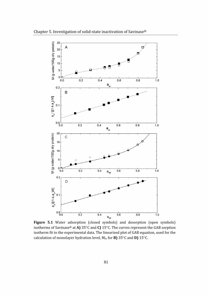
Chapter 5. Investigation of solid-state inactivation of Savinase®
81
Figure 5.1 Water adsorption (closed symbols) and desorption (open symbols) isotherms of Savinase® at A) 35°C and C) 15°C. The curves represent the GAB sorption isotherm fit to the experimental data. The linearized plot of GAB equation, used for the calculation of monolayer hydration level, M0, for B) 35°C and D) 15°C.
Chapter 5. Investigation of solid-state inactivation of Savinase®
82
The water here is available as a solvent for low-molecular solutes and for some biochemical reactions (Al-Muhtaseb et al. 2002). The last region, or multilayer region, is from 0.7 to 1.0 aw. It occurs with condensation of water at very weak binding sites and layering of loosely held water. It exhibits nearly all the properties of bulk water and is capable of acting as a solvent. Therefore, major decomposition reactions for proteins occur in this region.
In general, if water activity is maintained constant, an increase in temperature causes a decrease in the amount of sorbed water (Iglesias et al. 1986; Quirijns et al. 2005). This implies that, at higher temperatures, some water molecules are activated to energy levels that allow them to break away from their sorption sites, thus decreasing the equilibrium moisture content. No significant difference, however, was observed for the isotherms of Savinase® at 15°C and 35°C (Figure 5.1 - A and C). In fact, at the 0-0.6 aw region, the isotherms showed similar adsorption values. Contrary to expectations, at high relative humidity region, moisture adsorption increased as the temperature increased. This phenomena was previously reported for certain sugars (glucose) and salts, which become more hygroscopic at higher temperatures due to their ability to dissolve in water (Al-Muhtaseb et al. 2002). This provides a possible behavior explanation to the deviation in water sorption isotherm of Savinase® at a higher temperature. The Savinase® powder used in the study was a technical grade (60% active protein) freeze-dried product of a fermentation broth. Since no thorough purification was performed on the enzyme slurry prior to freeze-drying step, some impurities, such as salts, sugars and other small peptides are likely to be present in the final product. In such mixtures, at low aw values, the sorption of water is mainly due to the biopolymers; however, as aw is raised beyond the transition (intermediate) region, sugars and low molecular weight constituents begin to dissolve in the formed multilayers of water, which have properties of bulk water (described above). Hence, as the temperature increases, the solubility of some impurities increases and the equilibrium moisture content of the mixture increases.
A small hysteresis was observed in the low aw region (0-0.5 aw) of the adsorption-desorption isotherm at 15°C (Figure 5.1-C). As the temperature was raised to 35°C, hysteresis disappeared (Figure 5.1-A). A similar trend was observed for a number of dehydrated food products (Tsami et al. 1990) and the
Chapter 5. Investigation of solid-state inactivation of Savinase®
83
absence of hysteresis at high temperatures was related to the dissolution of sugar and other low molecular ingredients (Roman et al. 1982).
Numerous mathematical models exist to describe water sorption isotherms of biological materials, like foods (Chirife and Iglesias 1978). A theoretically based equation for describing sorption isotherms of proteins (type II isotherms) is the Brunauer – Emmett - Teller (BET) model. Although the model provides a good estimate for the monolayer value of adsorbed moisture, the applicability of the BET equation is limited to the low humidity region (0.05<aw<0.35). The reason of this limitation is related to the theory behind the BET equation, which permits the sorption of a second layer before the first is completed (Mclaren and Rowen 1951) and assumes that the binding energy of the higher layers is equal to those of the pure adsorbate (Al-Muhtaseb et al. 2002). Timmerman et al. (2001) showed that the Guggenheim – Andreson – de Boer (GAB) model is more versatile, has better physical meaning and represents more accurately the water adsorption isotherms for proteins at a wide range of water activity (0.05<aw<0.95) compared to the BET model. The GAB model represents a refined extension of the BET theory, postulating that the state of sorbate molecules in the second and higher layers is equal, but different from that in the liquid-like state. The linearized form of the GAB equation is written as:
( )( ) ,11
1 00 CMCa
CkMMaka w
w
w −+=
−
(5.2)
where M is the moisture content of the sample (in g water/100g dry protein), M0 is the monolayer water coverage capacity, aw is the water activity (or RH/100), C is an energy constant related to the difference between chemical potential of sorbate molecules in the first sorption layer and the upper layers, while k is related to the difference in the sorbate’s pure liquid state and the upper layers (Timmermann et al. 2001).
The plot of GAB model curve and experimental data points were in a close agreement for both temperatures (Figure 5.1- A and C). The plot of aw/ (1-kaw)M as a function of aw, for k values giving the minimum of the sum of the
Chapter 5. Investigation of solid-state inactivation of Savinase®
84
least squares of the linear regression, was used for the calculation of monolayer coverage capacity, M0. The k-values giving the best fit were found as 0.9 and 0.82 for Savinase adsorption isotherm at 35°C and 15°C, respectively (Figure 5.1-B and D). The monolayer water level of the enzyme was calculated as 5.11 and 5.14 (g water/ 100g dry protein) for 35°C and 15°C, respectively. Monolayer coverage of Savinase® was achieved at equilibrium when enzyme was exposed to 30% RH and 37%RH at 35°C and 15°C, respectively (calculated from the GAB model). The findings are summarized in Table 5.1.
Table 5.1 Adsorption-desorption values of Savinase® calculated by GAB equation
Temperature 35°C 15°C k value 0.9 0.82 M0, g water/ 100g dry protein 5.11 5.14 C energy constant 7.81 5.28 Monolayer equilibrium RH, % 30 37
To the authors’ knowledge, hydration monolayer values for Savinase® are not previously reported in the literature. A theoretical estimation, proposed by Pauling (1945), was used for the comparison to experimentally obtained value. The method is based on the assumption that monolayer coverage is achieved by adsorption of one molecule of water to each polar group of the side-chains of the amino acid residues in the protein. The polar groups in proteins which can bind water include serine (34), threonine (17), tyrosine (7), hydroxyproline (0), tryptophan (3), histidine (7), lysine (5), arginine (8), aspartic acid (5), glutamic acid (5), and hydroxyglutamic acid (0) (Pauling 1945). The numbers in the parenthesizes indicate the number of the corresponding amino acid residues in the Savinase® structure (the complete amino acid sequence is shown in the next chapter, Fig. 6.2). The theoretical monolayer was calculated as 6.1 g water/100g enzyme. Being higher than the experimentally measured 5.1 g water/100g enzyme, the outcome was in agreement with the results listed by Timmermann et al. (2001), reporting that the experimentally determined monolayer coverage values are slightly lower than the polar groups in the protein, but in the same order of magnitude. It is postulated that the local adsorption sites do not necessarily have to be located in the surface layer of the protein; however, their
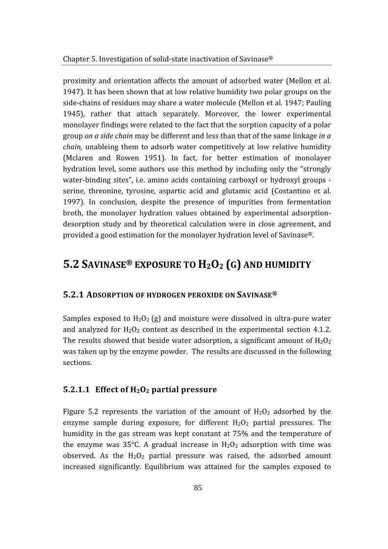
Chapter 5. Investigation of solid-state inactivation of Savinase®
85
proximity and orientation affects the amount of adsorbed water (Mellon et al. 1947). It has been shown that at low relative humidity two polar groups on the side-chains of residues may share a water molecule (Mellon et al. 1947; Pauling 1945), rather that attach separately. Moreover, the lower experimental monolayer findings were related to the fact that the sorption capacity of a polar group on a side chain may be different and less than that of the same linkage in a chain, unableing them to adsorb water competitively at low relative humidity (Mclaren and Rowen 1951). In fact, for better estimation of monolayer hydration level, some authors use this method by including only the “strongly water-binding sites”, i.e. amino acids containing carboxyl or hydroxyl groups - serine, threonine, tyrosine, aspartic acid and glutamic acid (Costantino et al. 1997). In conclusion, despite the presence of impurities from fermentation broth, the monolayer hydration values obtained by experimental adsorption-desorption study and by theoretical calculation were in close agreement, and provided a good estimation for the monolayer hydration level of Savinase®.
5.2 SAVINASE® EXPOSURE TO H2O2 (G) AND HUMIDITY
5.2.1 ADSORPTION OF HYDROGEN PEROXIDE ON SAVINASE®
Samples exposed to H2O2 (g) and moisture were dissolved in ultra-pure water and analyzed for H2O2 content as described in the experimental section 4.1.2. The results showed that beside water adsorption, a significant amount of H2O2 was taken up by the enzyme powder. The results are discussed in the following sections.
5.2.1.1 Effect of H2O2 partial pressure
Figure 5.2 represents the variation of the amount of H2O2 adsorbed by the enzyme sample during exposure, for different H2O2 partial pressures. The humidity in the gas stream was kept constant at 75% and the temperature of the enzyme was 35°C. A gradual increase in H2O2 adsorption with time was observed. As the H2O2 partial pressure was raised, the adsorbed amount increased significantly. Equilibrium was attained for the samples exposed to
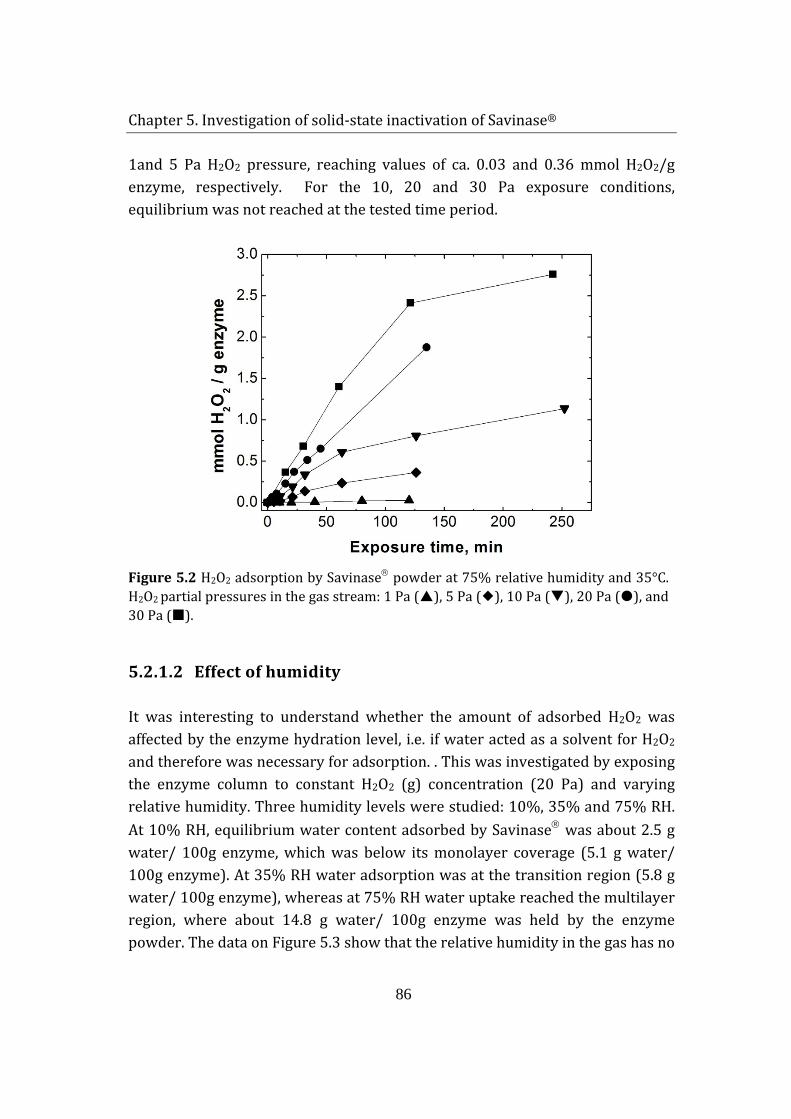
Chapter 5. Investigation of solid-state inactivation of Savinase®
86
1and 5 Pa H2O2 pressure, reaching values of ca. 0.03 and 0.36 mmol H2O2/g enzyme, respectively. For the 10, 20 and 30 Pa exposure conditions, equilibrium was not reached at the tested time period.
Figure 5.2 H2O2 adsorption by Savinase powder at 75% relative humidity and 35°C. H2O2 partial pressures in the gas stream: 1 Pa (), 5 Pa (), 10 Pa (), 20 Pa (), and 30 Pa ().
5.2.1.2 Effect of humidity
It was interesting to understand whether the amount of adsorbed H2O2 was affected by the enzyme hydration level, i.e. if water acted as a solvent for H2O2 and therefore was necessary for adsorption. . This was investigated by exposing the enzyme column to constant H2O2 (g) concentration (20 Pa) and varying relative humidity. Three humidity levels were studied: 10%, 35% and 75% RH. At 10% RH, equilibrium water content adsorbed by Savinase was about 2.5 g water/ 100g enzyme, which was below its monolayer coverage (5.1 g water/ 100g enzyme). At 35% RH water adsorption was at the transition region (5.8 g water/ 100g enzyme), whereas at 75% RH water uptake reached the multilayer region, where about 14.8 g water/ 100g enzyme was held by the enzyme powder. The data on Figure 5.3 show that the relative humidity in the gas has no
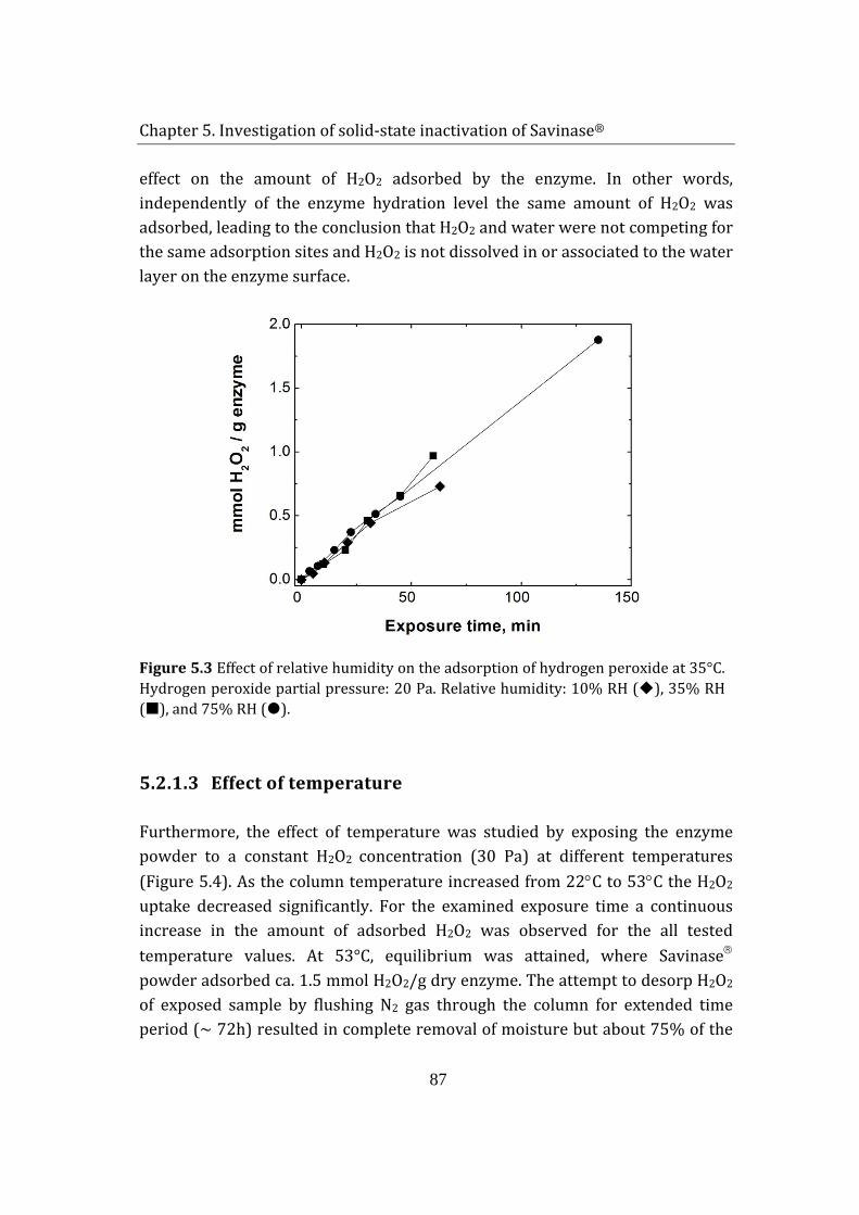
Chapter 5. Investigation of solid-state inactivation of Savinase®
87
effect on the amount of H2O2 adsorbed by the enzyme. In other words, independently of the enzyme hydration level the same amount of H2O2 was adsorbed, leading to the conclusion that H2O2 and water were not competing for the same adsorption sites and H2O2 is not dissolved in or associated to the water layer on the enzyme surface.
Figure 5.3 Effect of relative humidity on the adsorption of hydrogen peroxide at 35°C. Hydrogen peroxide partial pressure: 20 Pa. Relative humidity: 10% RH (), 35% RH (), and 75% RH ().
5.2.1.3 Effect of temperature
Furthermore, the effect of temperature was studied by exposing the enzyme powder to a constant H2O2 concentration (30 Pa) at different temperatures (Figure 5.4). As the column temperature increased from 22°C to 53°C the H2O2 uptake decreased significantly. For the examined exposure time a continuous increase in the amount of adsorbed H2O2 was observed for the all tested temperature values. At 53°C, equilibrium was attained, where Savinase powder adsorbed ca. 1.5 mmol H2O2/g dry enzyme. The attempt to desorp H2O2 of exposed sample by flushing N2 gas through the column for extended time period (~ 72h) resulted in complete removal of moisture but about 75% of the
Chapter 5. Investigation of solid-state inactivation of Savinase®
88
initial H2O2 content remained in the sample. It is, therefore, concluded that H2O2 adsorption by Savinase powder was achieved by combined physisorption and chemisorption, possibly involving formation of strong hydrogen bonds.
Figure 5.4 Effect of temperature on hydrogen peroxide adsorbed by Savinase exposed to 30 Pa H2O2. Temperatures: 22°C (), 35°C (), 45°C () and 53°C ().
5.2.2 ENZYME INACTIVATION
5.2.2.1 Effect of humidity in the absence of H2O2
The detrimental effect of moisture on storage stability is often interpreted in terms of mobility in the solid and reactivity of the protein. Above monolayer levels of water, a protein’s conformational flexibility is increased and the additional water has ability to mobilize the potential reactants in the amorphous phase, where both effects increase the rate of protein degradation (Prestrelski et al. 1994).
To determine the effect of protein hydration on enzyme storage stability at moderate temperature (35°C), enzyme samples were exposed to moist gas with 10% RH and 75% RH. From the sorption isotherm of Savinase, it was seen that
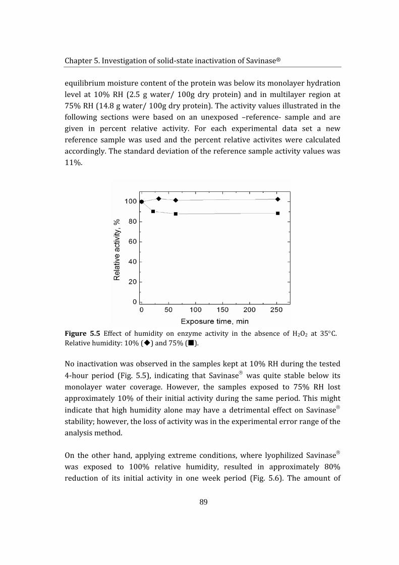
Chapter 5. Investigation of solid-state inactivation of Savinase®
89
equilibrium moisture content of the protein was below its monolayer hydration level at 10% RH (2.5 g water/ 100g dry protein) and in multilayer region at 75% RH (14.8 g water/ 100g dry protein). The activity values illustrated in the following sections were based on an unexposed –reference- sample and are given in percent relative activity. For each experimental data set a new reference sample was used and the percent relative activites were calculated accordingly. The standard deviation of the reference sample activity values was 11%.
Figure 5.5 Effect of humidity on enzyme activity in the absence of H2O2 at 35°C. Relative humidity: 10% () and 75% ().
No inactivation was observed in the samples kept at 10% RH during the tested 4-hour period (Fig. 5.5), indicating that Savinase was quite stable below its monolayer water coverage. However, the samples exposed to 75% RH lost approximately 10% of their initial activity during the same period. This might indicate that high humidity alone may have a detrimental effect on Savinase stability; however, the loss of activity was in the experimental error range of the analysis method.
On the other hand, applying extreme conditions, where lyophilized Savinase was exposed to 100% relative humidity, resulted in approximately 80% reduction of its initial activity in one week period (Fig. 5.6). The amount of
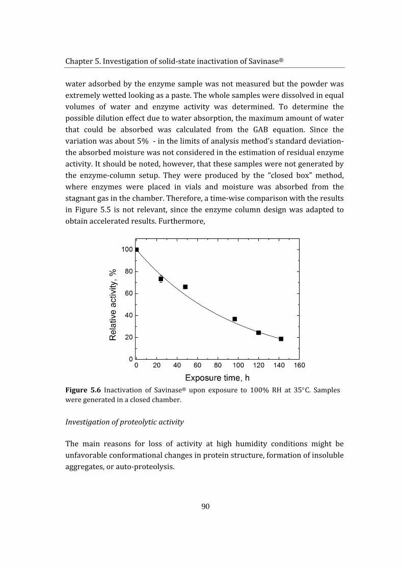
Chapter 5. Investigation of solid-state inactivation of Savinase®
90
water adsorbed by the enzyme sample was not measured but the powder was extremely wetted looking as a paste. The whole samples were dissolved in equal volumes of water and enzyme activity was determined. To determine the possible dilution effect due to water absorption, the maximum amount of water that could be absorbed was calculated from the GAB equation. Since the variation was about 5% - in the limits of analysis method’s standard deviation- the absorbed moisture was not considered in the estimation of residual enzyme activity. It should be noted, however, that these samples were not generated by the enzyme-column setup. They were produced by the “closed box” method, where enzymes were placed in vials and moisture was absorbed from the stagnant gas in the chamber. Therefore, a time-wise comparison with the results in Figure 5.5 is not relevant, since the enzyme column design was adapted to obtain accelerated results. Furthermore,
Figure 5.6 Inactivation of Savinase® upon exposure to 100% RH at 35°C. Samples were generated in a closed chamber.
Investigation of proteolytic activity
The main reasons for loss of activity at high humidity conditions might be unfavorable conformational changes in protein structure, formation of insoluble aggregates, or auto-proteolysis.
Chapter 5. Investigation of solid-state inactivation of Savinase®
91
The solid-state enzyme samples exposed to 100% RH were examined for proteolytic activity by SDS-PAGE analysis (Fig. 5.7). This method involves unfolding (linearizing) of the protein backbone and separation of the peptides according to their molecular weight. However, it is well documented that Savinase, in a liquid medium, is susceptible to auto-hydrolysis in the presence of its denaturated conformations (Stoner et al. 2004). In order to prevent autolysis during sample preparation, the enzyme powder was dissolved in solution containing protease inhibitor-PMSF (phenylmethylsulfonyl fluoride) and sequestring agent EDTA (ethylenediaminetetraacetic acid). PMSF binds to the enzyme and prevents any catalytic activity, while EDTA removes the two Ca ions in the Savinase® structure, which are important for its stability. Then the reducing agent was added and the samples were boiled to achieve complete linearization of the enzymes. Finally, the samples were loaded on the gel, and the separation according to peptides' molecular weight was done.
The main Savinase band was detected at approximately 26.7 kDa. The absence of higher molecular weight peptides indicated that loss of activity was not caused by formation of covalently bound aggregates. In fact, studies show that moisture-induced aggregation of solid proteins result in covalently bound aggregates mainly in the presence of disulfide bonds in the protein structure and free thiol (-HS) groups (Costantino et al. 1994; Klibanov and Schefiliti 2004; Liu et al. 1991). Therefore, the result was not surprising, because Savinase has neither disulfide bonds nor cysteine in its structure (Betzel et al. 1992).
A careful inspection of the gel revealed that the main Savinase® band intensified gradually as the exposure time increased (line 29). The lowest intensity was observed for 0-h sample (line 1), which was not subjected to humidity and therefore no proteolytic activity was expected during storage. Moreover, below the intact form of the enzyme (26.7kDa band), small peptide fragments were observed in the analyzed samples. The low molecular bands may indicate that these small peptides already existed as impurities in the freeze-dried enzyme powder; and/or may imply that auto-proteolytic activity took place. Since the intensities of small peptides slightly decreased as exposure time increased; i.e. as enzyme has lost its activity (lines 8 and 9), the proteolytic activity most likely occurred during sample preparation rather than during storage at high humidity. In fact, gel electrophoresis studies report that despite the addition of
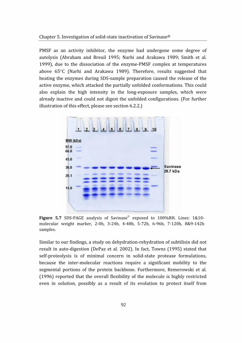
Chapter 5. Investigation of solid-state inactivation of Savinase®
92
PMSF as an activity inhibitor, the enzyme had undergone some degree of autolysis (Abraham and Breuil 1995; Narhi and Arakawa 1989; Smith et al. 1999), due to the dissociation of the enzyme-PMSF complex at temperatures above 65°C (Narhi and Arakawa 1989). Therefore, results suggested that heating the enzymes during SDS-sample preparation caused the release of the active enzyme, which attacked the partially unfolded conformations. This could also explain the high intensity in the long-exposure samples, which were already inactive and could not digest the unfolded configurations. (For further illustration of this effect, please see section 6.2.2.)
Figure 5.7 SDS-PAGE analysis of Savinase exposed to 100%RH. Lines: 1&10- molecular weight marker, 2-0h, 3-24h, 4-48h, 5-72h, 6-96h, 7-120h, 8&9-142h samples.
Similar to our findings, a study on dehydration-rehydration of subtilisin did not result in auto-digestion (DePaz et al. 2002). In fact, Towns (1995) stated that self-proteolysis is of minimal concern in solid-state protease formulations, because the inter-molecular reactions require a significant mobility to the segmental portions of the protein backbone. Furthermore, Remerowski et al. (1996) reported that the overall flexibility of the molecule is highly restricted even in solution, possibly as a result of its evolution to protect itself from
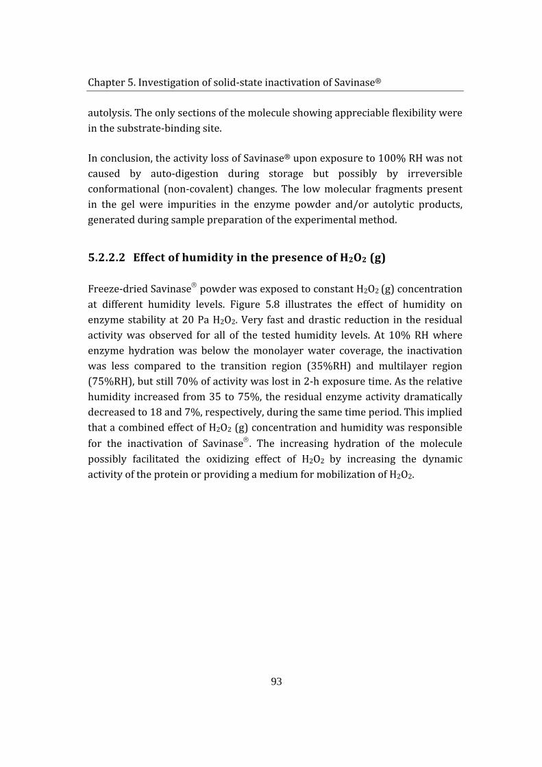
Chapter 5. Investigation of solid-state inactivation of Savinase®
93
autolysis. The only sections of the molecule showing appreciable flexibility were in the substrate-binding site.
In conclusion, the activity loss of Savinase® upon exposure to 100% RH was not caused by auto-digestion during storage but possibly by irreversible conformational (non-covalent) changes. The low molecular fragments present in the gel were impurities in the enzyme powder and/or autolytic products, generated during sample preparation of the experimental method.
5.2.2.2 Effect of humidity in the presence of H2O2 (g)
Freeze-dried Savinase powder was exposed to constant H2O2 (g) concentration at different humidity levels. Figure 5.8 illustrates the effect of humidity on enzyme stability at 20 Pa H2O2. Very fast and drastic reduction in the residual activity was observed for all of the tested humidity levels. At 10% RH where enzyme hydration was below the monolayer water coverage, the inactivation was less compared to the transition region (35%RH) and multilayer region (75%RH), but still 70% of activity was lost in 2-h exposure time. As the relative humidity increased from 35 to 75%, the residual enzyme activity dramatically decreased to 18 and 7%, respectively, during the same time period. This implied that a combined effect of H2O2 (g) concentration and humidity was responsible for the inactivation of Savinase. The increasing hydration of the molecule possibly facilitated the oxidizing effect of H2O2 by increasing the dynamic activity of the protein or providing a medium for mobilization of H2O2.
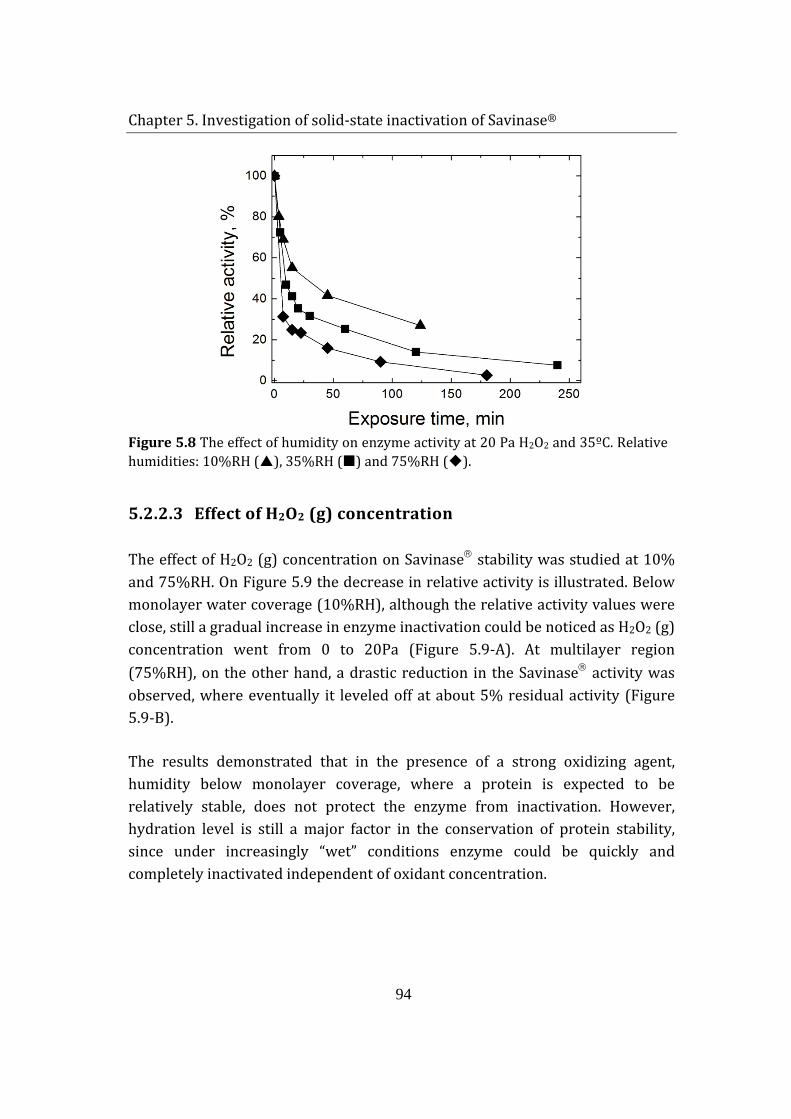
Chapter 5. Investigation of solid-state inactivation of Savinase®
94
Figure 5.8 The effect of humidity on enzyme activity at 20 Pa H2O2 and 35ºC. Relative humidities: 10%RH (), 35%RH () and 75%RH ().
5.2.2.3 Effect of H2O2 (g) concentration
The effect of H2O2 (g) concentration on Savinase stability was studied at 10% and 75%RH. On Figure 5.9 the decrease in relative activity is illustrated. Below monolayer water coverage (10%RH), although the relative activity values were close, still a gradual increase in enzyme inactivation could be noticed as H2O2 (g) concentration went from 0 to 20Pa (Figure 5.9-A). At multilayer region (75%RH), on the other hand, a drastic reduction in the Savinase activity was observed, where eventually it leveled off at about 5% residual activity (Figure 5.9-B).
The results demonstrated that in the presence of a strong oxidizing agent, humidity below monolayer coverage, where a protein is expected to be relatively stable, does not protect the enzyme from inactivation. However, hydration level is still a major factor in the conservation of protein stability, since under increasingly “wet” conditions enzyme could be quickly and completely inactivated independent of oxidant concentration.
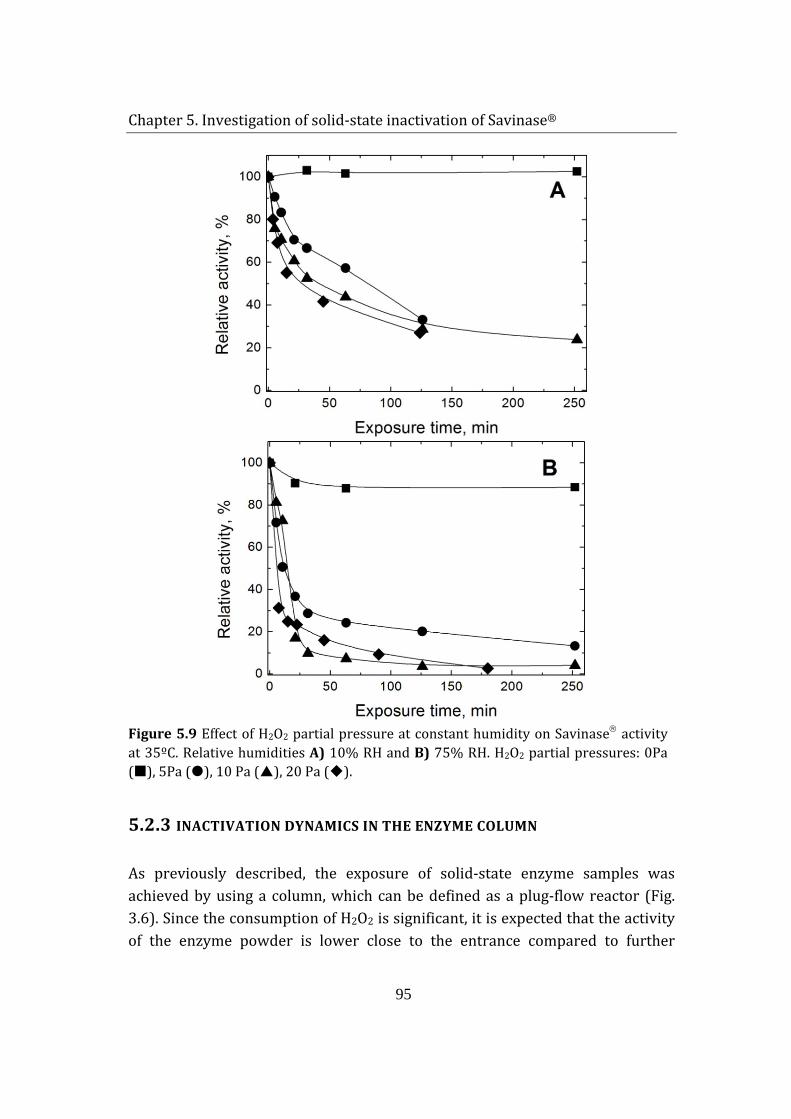
Chapter 5. Investigation of solid-state inactivation of Savinase®
95
Figure 5.9 Effect of H2O2 partial pressure at constant humidity on Savinase activity at 35ºC. Relative humidities A) 10% RH and B) 75% RH. H2O2 partial pressures: 0Pa (), 5Pa (), 10 Pa (), 20 Pa ().
5.2.3 INACTIVATION DYNAMICS IN THE ENZYME COLUMN
As previously described, the exposure of solid-state enzyme samples was achieved by using a column, which can be defined as a plug-flow reactor (Fig. 3.6). Since the consumption of H2O2 is significant, it is expected that the activity of the enzyme powder is lower close to the entrance compared to further
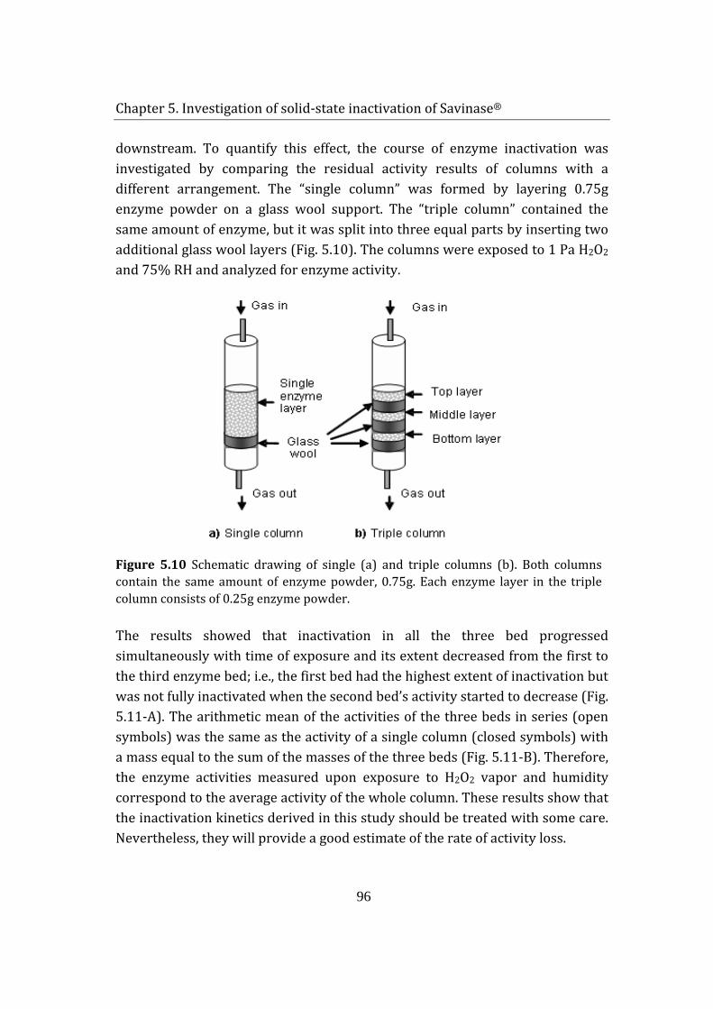
Chapter 5. Investigation of solid-state inactivation of Savinase®
96
downstream. To quantify this effect, the course of enzyme inactivation was investigated by comparing the residual activity results of columns with a different arrangement. The “single column” was formed by layering 0.75g enzyme powder on a glass wool support. The “triple column” contained the same amount of enzyme, but it was split into three equal parts by inserting two additional glass wool layers (Fig. 5.10). The columns were exposed to 1 Pa H2O2 and 75% RH and analyzed for enzyme activity.
Figure 5.10 Schematic drawing of single (a) and triple columns (b). Both columns contain the same amount of enzyme powder, 0.75g. Each enzyme layer in the triple column consists of 0.25g enzyme powder.
The results showed that inactivation in all the three bed progressed simultaneously with time of exposure and its extent decreased from the first to the third enzyme bed; i.e., the first bed had the highest extent of inactivation but was not fully inactivated when the second bed’s activity started to decrease (Fig. 5.11-A). The arithmetic mean of the activities of the three beds in series (open symbols) was the same as the activity of a single column (closed symbols) with a mass equal to the sum of the masses of the three beds (Fig. 5.11-B). Therefore, the enzyme activities measured upon exposure to H2O2 vapor and humidity correspond to the average activity of the whole column. These results show that the inactivation kinetics derived in this study should be treated with some care. Nevertheless, they will provide a good estimate of the rate of activity loss.
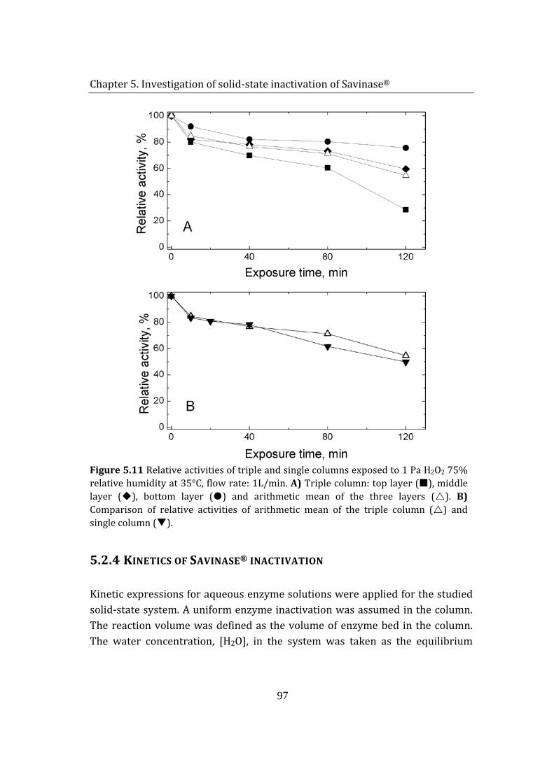
Chapter 5. Investigation of solid-state inactivation of Savinase®
97
Figure 5.11 Relative activities of triple and single columns exposed to 1 Pa H2O2 75% relative humidity at 35°C, flow rate: 1L/min. A) Triple column: top layer (), middle layer (), bottom layer () and arithmetic mean of the three layers (). B) Comparison of relative activities of arithmetic mean of the triple column () and single column ().
5.2.4 KINETICS OF SAVINASE® INACTIVATION
Kinetic expressions for aqueous enzyme solutions were applied for the studied solid-state system. A uniform enzyme inactivation was assumed in the column. The reaction volume was defined as the volume of enzyme bed in the column. The water concentration, [H2O], in the system was taken as the equilibrium
Chapter 5. Investigation of solid-state inactivation of Savinase®
98
amount of moisture adsorbed by enzyme powder in the reaction volume at the corresponding % RH, calculated by equation (5.2).
Assuming that only wetted enzymes were affected and only one molecule of H2O2 was necessary for the formation of an oxidized enzyme (OxE) (See Chapter 6) the following expressions were used for the derivation of inactivation kinetics (for detailed derivation see Appx. I).
1
12 2*k
kE nH O E H O
−
→+ ←
(5.3)
2 3
22 2 2 2 2 2* * *k k
kE H O H O E H O H O OxE
−
→+ →← (5.4)
The enzyme oxidation rate was expressed as:
[ ] [ ]3 2 2 2* *d E dt k E H O H O− = (5.5)
Derivation and substitution of [E*H2O*H2O2] resulted in the following equation:
[ ] [ ] [ ][ ] [ ]E
OHCBOHOHA
dtEd n
22
222
+=− (5.6)
where 321 kkkA = , )( 321 kkkB += −− and ( ) 22322 −− −+= kkkkkC . Since there was a
continuous feed of humidity and H2O2 (g) at constant concentration, enzyme inactivation could be assumed as a pseudo-first order reaction in terms of enzyme concentration, [E]. Then the equation (5.6) was rewritten with a reaction rate constant, kapp, which was dependent on [H2O] and [H2O2]:
[ ] [ ]appd E dt k E− = (5.7)
where [ ] [ ]
[ ]2 2 2
2 2
n
app
A H O H Ok
B C H O=
+ (5.8)
Chapter 5. Investigation of solid-state inactivation of Savinase®
99
The oxidation of the enzyme was measured by the residual activity, A/A0, that remained after the exposure experiments; A being the activity at time t and A0 the initial activity of unoxidized enzyme. As discussed previously, upon extensive oxidation of the enzyme, Savinase® retained approximately 5% residual activity, α, when DMC was used as a substrate. Therefore, equation (5.7) was transformed in the following form (DePaz et al. 2000; Hausdorf et al. 1988):
( ) tkappeAA −−=− .10 αα (5.9)
The plot of equation (5.9) as ln((A/A0-α)/(1-α)) versus time, t, gave the values of rate constant, kapp, listed in Table 5.2 . The experimental points for the first 30 min were used, since the affinity constant of the oxidized enzyme to the substrate differs significantly from the native form (Christianson and Paech 1994) and this affects the linearity of the plot.
Table 5.2 Pseudo first-order rate constants of Savinase oxidation by H2O2 (g) at different humidity levels.
Experimental conditions kapp(min-1)×100a H2O2 partial pressure (Pa) % Relative humidity 0 10 0 5 10 1.53 ± 0.10 10 10 2.45 ± 0.27 20 10 4.45 ± 0.16 20 35 6.30 ± 0.41 1 75 1.25 ± 0.06 5 75 4.86 ± 0.37 10 75 8.34 ± 1.59 20 75 11.77 ± 1.89 a ± value is the standard error reported by the linear regression routine
The expression for kapp, equation (5.8), reveals that there should be a linear relation between 1/kapp and 1/[H2O2]. A good agreement with equation (5.8) was obtained for the experimental points at 10% and 75% RH, indicating that the proposed mechanism described the oxidation reaction of Savinase (Fig. 5.12-A). Furthermore, an effort to relate the water hydration dependency of the
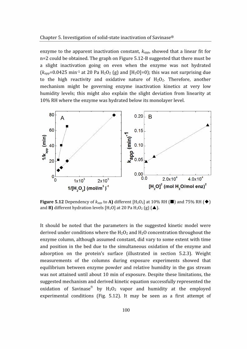
Chapter 5. Investigation of solid-state inactivation of Savinase®
100
enzyme to the apparent inactivation constant, kapp, showed that a linear fit for n=2 could be obtained. The graph on Figure 5.12-B suggested that there must be a slight inactivation going on even when the enzyme was not hydrated (kapp=0.0425 min-1 at 20 Pa H2O2 (g) and [H2O]=0); this was not surprising due to the high reactivity and oxidative nature of H2O2. Therefore, another mechanism might be governing enzyme inactivation kinetics at very low humidity levels; this might also explain the slight deviation from linearity at 10% RH where the enzyme was hydrated below its monolayer level.
Figure 5.12 Dependency of kapp to A) different [H2O2] at 10% RH () and 75% RH () and B) different hydration levels [H2O] at 20 Pa H2O2 (g) ().
It should be noted that the parameters in the suggested kinetic model were derived under conditions where the H2O2 and H2O concentration throughout the enzyme column, although assumed constant, did vary to some extent with time and position in the bed due to the simultaneous oxidation of the enzyme and adsorption on the protein’s surface (illustrated in section 5.2.3). Weight measurements of the columns during exposure experiments showed that equilibrium between enzyme powder and relative humidity in the gas stream was not attained until about 10 min of exposure. Despite these limitations, the suggested mechanism and derived kinetic equation successfully represented the oxidation of Savinase by H2O2 vapor and humidity at the employed experimental conditions (Fig. 5.12). It may be seen as a first attempt of
Chapter 5. Investigation of solid-state inactivation of Savinase®
101
quantifying the rate of decomposition of Savinase® by the combined action of H2O2 and H2O.
5.3 SUMMARY
Water adsorption-desorption studies on freeze-dried Savinase® powder revealed that, the enzyme adsorbs significant amounts of moisture at elevated temperatures (15°C and 35°C). The adsorption-desorption graphs showed sigmoidal behavior with respect to water activity, corresponding to type II isotherms. Contrary to samples examined at 35°C, a small hysteresis in the 0- 0.3 aw region was detected for the sample studied at 15 °C. The lack of hysteresis effect at the higher temperature is possibly related to dissolution of water in the impurities present in the enzyme powder. Isotherm data were fitted to GAB equation and the monolayer coverage of Savinase® was calculated as. The experimentally determined and theoretically estimated values for hydration monolayer were in a reasonable agreement, and were found as 5.1 g water/100g dry protein and 6.1 g water/100g dry protein, respectively.
Exposure of Savinase® to H2O2 (g) and humidity resulted in significant adsorption of H2O2 and moisture. It was found that H2O2 and water might not be competing for the same adsorption sites, since the amount of adsorbed H2O2 was not affected by the variation of humidity in the gas stream. Moreover, desorption studies revealed that while water could be completely removed, 75% of H2O2 remained in the sample, indicating that H2O2 adsorption was possibly achieved by combined physisorption and chemisorption.
The results showed that both oxidant concentration and humidity in the gas stream influenced the inactivation of the enzyme. For instance, exposure to 5 Pa H2O2 for 30 min resulted in activity loss by 30% at 10% RH and 50% at 75% RH. Increasing the H2O2 concentration to 20 Pa caused 50% activity reduction at 10% RH and 80% loss at 75% RH during the same exposure time. In addition, moisture alone had a negative influence on the solid-state enzyme by decreasing its activity by 80% after exposure to 100% RH for one week. Hydration of the enzyme possibly increased the mobility of protein backbone leading to formation of unfavorable conformational changes or non-covalently bound
Chapter 5. Investigation of solid-state inactivation of Savinase®
102
aggregates. Nevertheless, these conformational modifications were not substantial to initiate an auto-digestive reaction.
A kinetic model for the inactivation of Savinase® under the combined effect of H2O2 (g) and humidity was derived. The kinetic expression for activity loss was based on the oxidation of one amino acid residue. Solid-state inactivation of Savinase® column was assumed to prevail solution kinetics behavior and constant oxidant and water concentration values were considered for simplicity. The expression for reaction rate constant was derived and the experimental results were used to confirm the validity of the model. According to the obtained equation, the pseudo-first order reaction rate constant was dependent on the square of moisture adsorbed by the enzyme at the corresponding temperature. Moreover, the inverse of the reaction rate constant was proportional to the inverse of H2O2 (g) concentration. Despite the limitation of the model and assumptions made, a good agreement was obtained between the experimental findings and the mathematical expression. This study is the first attempt at quantifying the combined effect of H2O2 (g) and H2O (g) on the inactivation of solid-state Savinase®.
5.4 REFERENCES
Abraham LD, Breuil C. 1995. Factors Affecting Autolysis of a Subtilisin-Like Serine Proteinase Secreted by Ophiostoma-Piceae and Identification of the Cleavage Site. Biochimica Et Biophysica Acta-General Subjects 1245(1):76-84.
Ahlneck C, Zografi G. 1990. The Molecular-Basis of Moisture Effects on the Physical and Chemical-Stability of Drugs in the Solid-State. International Journal of Pharmaceutics 62(2-3):87-95.
Al-Muhtaseb AH, McMinn WAM, Magee TRA. 2002. Moisture sorption isotherm characteristics of food products: A review. Food and Bioproducts Processing 80(C2):118-128.
Betzel C, Klupsch S, Papendorf G, Hastrup S, Branner S, Wilson KS. 1992. Crystal-Structure of the Alkaline Proteinase Savinase from Bacillus-Lentus at 1.4-a Resolution. Journal of Molecular Biology 223(2):427-445.
Chapter 5. Investigation of solid-state inactivation of Savinase®
103
Chirife J, Iglesias HA. 1978. Equations for Fitting Water Sorption Isotherms of Foods .1. Review. Journal of Food Technology 13(3):159-174.
Chou DH, Morr CV. 1979. Protein-Water Interactions and Functional Properties. Journal of the American Oil Chemists Society 56(1):A53-A62.
Christianson T, Paech C. 1994. Peptide-Mapping of Subtilisins as a Practical Tool for Locating Protein-Sequence Errors during Extensive Protein Engineering Projects. Analytical Biochemistry 223(1):119-129.
Costantino HR, Curley JG, Hsu CC. 1997. Determining the water sorption monolayer of lyophilized pharmaceutical proteins. Journal of Pharmaceutical Sciences 86(12):1390-1393.
Costantino HR, Langer R, Klibanov AM. 1994. Moisture-Induced Aggregation of Lyophilized Insulin. Pharmaceutical Research 11(1):21-29.
DePaz RA, Barnett CC, Dale DA, Carpenter JF, Gaertner AL, Randolph TW. 2000. The excluding effects of sucrose on a protein chemical degradation pathway: Methionine oxidation in subtilisin. Archives of Biochemistry and Biophysics 384(1):123-132.
DePaz RA, Dale DA, Barnett CC, Carpenter JF, Gaertner AL, Randolph TW. 2002. Effects of drying methods and additives on the structure, function, and storage stability of subtilisin: role of protein conformation and molecular mobility. Enzyme and Microbial Technology 31(6):765-774.
Hageman MJ. 1988. The Role of Moisture in Protein Stability. Drug Development and Industrial Pharmacy 14(14):2047-2070.
Hausdorf G, Kruger K, Kuttner G, Holzhutter HG, Frommel C, Hohne WE. 1988. Oxidation of a Methionine Residue in Subtilisin-Type Proteinases by the Hydrogen-Peroxide Borate System - an Active Site-Directed Reaction. Biochimica Et Biophysica Acta 952(1):20-26.
Iglesias HA, Chirife J, Fontan CF. 1986. Temperature-Dependence of Water Sorption Isotherms of Some Foods. Journal of Food Science 51(3):551-553.
Chapter 5. Investigation of solid-state inactivation of Savinase®
104
Klibanov AM, Schefiliti JA. 2004. On the relationship between conformation and stability in solid pharmaceutical protein formulations. Biotechnology Letters 26(14):1103-1106.
Liu WR, Langer R, Klibanov AM. 1991. Moisture-Induced Aggregation of Lyophilized Proteins in the Solid-State. Biotechnology and Bioengineering 37(2):177-184.
Mclaren AD, Rowen JW. 1951. Sorption of Water Vapor by Proteins and Polymers - a Review. Journal of Polymer Science 7(2-3):289-324.
Mellon EF, Korn AH, Hoover SR. 1947. Water Absorption of Proteins .1. The Effect of Free Amino Groups in Casein. Journal of the American Chemical Society 69(4):827-831.
Narhi LO, Arakawa T. 1989. Sodium Dodecyl-Sulfate Polyacrylamide-Gel Electrophoresis as a Method for Studying the Stability of Subtilisin. Biochimica Et Biophysica Acta 990(2):144-149.
Pauling L. 1945. The Adsorption of Water by Proteins. Journal of the American Chemical Society 67(4):555-557.
Prestrelski SJ, Pikal KA, Arakawa T. 1994. The Effect of Dried-State Protein Conformation of the Storage Stability of Lyophilized Proteins. Abstracts of Papers of the American Chemical Society 207:95-Biot.
Quirijns EJ, van Boxtel AJB, van Loon WKP, van Straten G. 2005. Sorption isotherms, GAB parameters and isosteric heat of sorption. Journal of the Science of Food and Agriculture 85(11):1805-1814.
Roman GN, Urbicain MJ, Rotstein E. 1982. Moisture Equilibrium in Apples at Several Temperatures - Experimental-Data and Theoretical Considerations. Journal of Food Science 47(5):1484-1488.
Shamblin SL, Hancock BC, Zografi G. 1998. Water vapor sorption by peptides, proteins and their formulations. European Journal of Pharmaceutics and Biopharmaceutics 45(3):239-247.
Smith CA, Toogood HS, Baker HM, Daniel RM, Baker EN. 1999. Calcium-mediated thermostability in the subtilisin superfamily: The crystal
Chapter 5. Investigation of solid-state inactivation of Savinase®
105
structure of Bacillus Ak.1 protease at 1.8 angstrom resolution. Journal of Molecular Biology 294(4):1027-1040.
Stoner MR, Dale DA, Gualfetti PJ, Becker T, Manning MC, Carpenter JF, Randolph TW. 2004. Protease autolysis in heavy-duty liquid detergent formulations: effects of thermodynamic stabilizers and protease inhibitors. Enzyme and Microbial Technology 34(2):114-125.
Timmermann EO, Chirife J, Iglesias HA. 2001. Water sorption isotherms of foods and foodstuffs: BET or GAB parameters? Journal of Food Engineering 48(1):19-31.
Towns JK. 1995. Moisture-Content in Proteins - Its Effects and Measurement. Journal of Chromatography A 705(1):115-127.
Tsami E, Marinoskouris D, Maroulis ZB. 1990. Water Sorption Isotherms of Raisins, Currants, Figs, Prunes and Apricots. Journal of Food Science 55(6):1594-1597.
Chapter 6. Mechanism of Savinase® inactivation
106
CHAPTER 6 MECHANISM OF SAVINASE®
INACTIVATION
The following chapter provides information about the modifications on Savinase®’s structure resulting in enzyme inactivation. The inactivation results of enzyme oxidation in the presence of H2O2 (g) and humidity, reported in Chapter 5, are further investigated on a molecular level. The used techniques are briefly introduced and the findings are presented. As a result, the mechanism of Savinase® inactivation is revealed.
6.1 MOLECULAR STRUCTURE OF SAVINASE®
Savinase® (EC 3.4.21.14) is a protease secreted by the alkalophilic bacterium Bacillus lentus. It is a representative of subtilisin enzymes having broad substrate specificity, high thermostability at alkaline pHs and pH-optimum for protease activity in the region 8-12 (Betzel et al. 1992; Georgieva et al. 2001). For this reason it creates a great interest as protein-degrading additive to detergents in the washing powder industry.
The enzyme itself has a hemi-spherical shape with a diameter of 40Å. The active site (substrate binding and catalytic domain) of the enzyme is slightly buried on the flat surface of the half-sphere (Georgieva et al. 2001; Remerowski et al. 1996). Remerowski et al.’s (1996) study revealed that the overall flexibility of the molecule is highly restricted, possibly as a result of its evolution to protect itself from autolysis. The only sections of the molecule showing appreciable flexibility are in the substrate-binding site; a small, solvent-exposed loop region (G258-S259); and one residue (V81), which is a calcium ligand in the high
Chapter 6. Mechanism of Savinase® inactivation
107
affinity calcium-binding site. Cn3D application (available at http://www.ncbi.nlm.nih.gov/Structure/CN3D/cn3d.shtml ) was used to view the three-dimensional structure of the enzyme. The structure information is available in National Center for Biotecnology Information’s (NCBI) molecular modeling data base (MMDB) (Chen et al. 2003). Tubes representation of Savinase® structure was used to focus on the substrate binding and the catalytic center of the enzyme (Fig. 6.1).
Figure 6.1 Schematics of Savinase® structure using tubes representation. The substrate binding site (blue) and the catalytic triad (red) of Savinase® partially buried inside the molecule are shown (Betzel et al., 1988; Betzel et al., 1992).
The amino acid sequence of Savinase® is previously published (Betzel et al. 1992; Vonderosten et al. 1993). It contains 269 amino acids, comprising 26 698 Da molecular weight of the enzyme. The numbering of the residues is based on subtilisin BPN’ (bio-protein novo), (having 275 a.a.) , which was the firstly sequenced subtilisin (Markland and Smith 1967). The primary structure of the enzyme is given in Figure 6.2, where deletions with respect to BPN’ are indicated by asterisk (Egmond et al. 1994).
Chapter 6. Mechanism of Savinase® inactivation
108
Figure 6.2 Amino acid sequence of Savinase® using subtilisin BPN’ numbering (Egmond et al. 1994).
Considering the detergent matrix, the primary cause of enzyme inactivation during storage would be oxidation by H2O2 released from the bleaching agent. Susceptibility to oxidation is more profound for proteins containing aromatic amino acids like histidine (His), tyrosine (Tyr) and tryptophan (Trp), and/or sulfur-containing residues like methionine (Met) and cysteine (Cys). Beside these amino acids, arginine (Arg) lysine (Lys) and proline (Pro) are affected by metal-catalysed oxidation (Reubsaet et al. 1998). Savinase® has 48 of these oxidation-labile residues in its structure: 7 His (H), 3 Met (M), 7 Tyr (Y), 3 Trp (W), 13 Pro (P), 5 Lys (L) and 8 Arg (R).
The numerous studies on oxidation of subtilisins by H2O2 in aqueous solution revealed that generally Met residues are affected (Bott et al. 1988; Christianson and Paech 1994; DePaz et al. 2000; DePaz et al. 2002; Estell et al. 1988; Nonaka et al. 2004; Stauffer and Etson 1969) showing that they can be oxidized to methionine sulfoxide (Fig. 6.3). Some of the studies on H2O2-induced oxidation of subtilisins in solution revealed that, independently on the number of methionines (Met) present in the subtilisin structure, activity decrease of enzymes was accompanied by the oxidation of just 1 Met at position 222 (Met 222 – BPN’ numbering) (Christianson and Paech, 1994; DePaz et al., 2000; Staufer and Etson, 1969). However, Bott et al. (1988) and Nonaka et al. (2004)
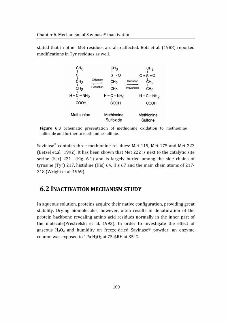
Chapter 6. Mechanism of Savinase® inactivation
109
stated that in other Met residues are also affected. Bott et al. (1988) reported modifications in Tyr residues as well.
Figure 6.3 Schematic presentation of methionine oxidation to methionine sulfoxide and further to methionine sulfone.
Savinase contains three methionine residues: Met 119, Met 175 and Met 222 (Betzel et.al., 1992). It has been shown that Met 222 is next to the catalytic site serine (Ser) 221 (Fig. 6.1) and is largely buried among the side chains of tyrosine (Tyr) 217, histidine (His) 64, His 67 and the main chain atoms of 217-218 (Wright et al. 1969).
6.2 INACTIVATION MECHANISM STUDY
In aqueous solution, proteins acquire their native configuration, providing great stability. Drying biomolecules, however, often results in denaturation of the protein backbone revealing amino acid residues normally in the inner part of the molecule(Prestrelski et al. 1993). In order to investigate the effect of gaseous H2O2 and humidity on freeze-dried Savinase® powder, an enzyme column was exposed to 1Pa H2O2 at 75%RH at 35°C.
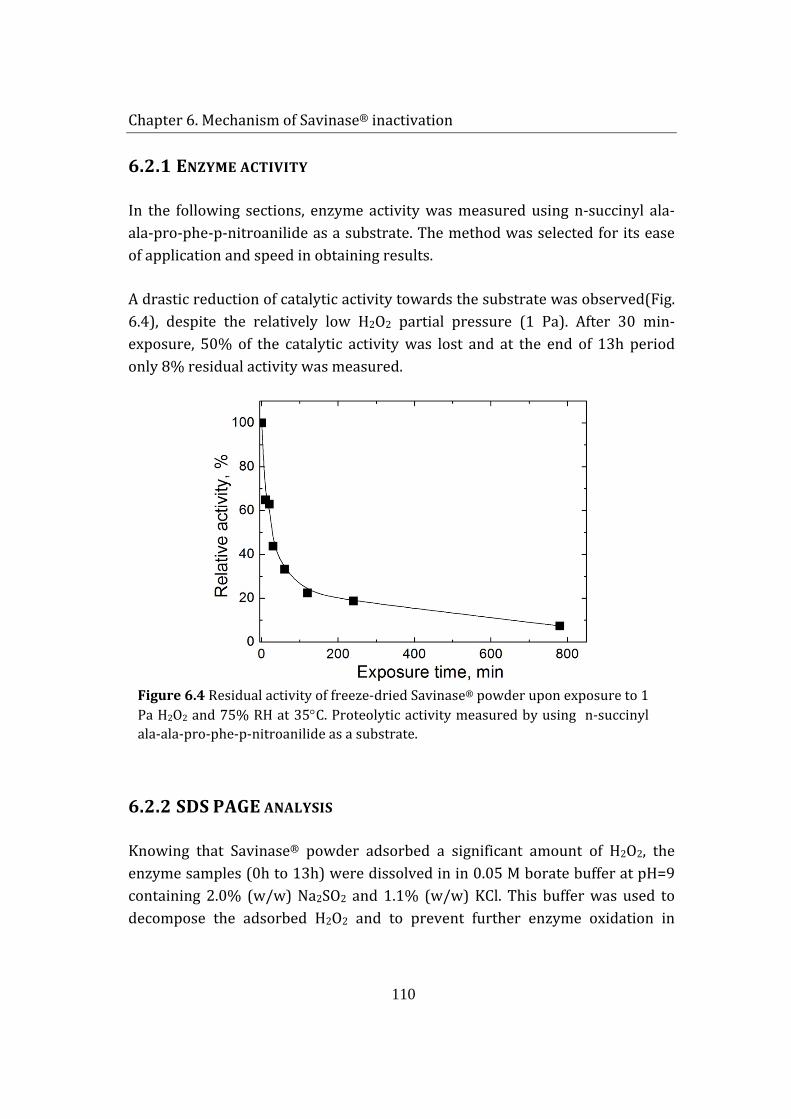
Chapter 6. Mechanism of Savinase® inactivation
110
6.2.1 ENZYME ACTIVITY
In the following sections, enzyme activity was measured using n-succinyl ala-ala-pro-phe-p-nitroanilide as a substrate. The method was selected for its ease of application and speed in obtaining results.
A drastic reduction of catalytic activity towards the substrate was observed(Fig. 6.4), despite the relatively low H2O2 partial pressure (1 Pa). After 30 min-exposure, 50% of the catalytic activity was lost and at the end of 13h period only 8% residual activity was measured.
Figure 6.4 Residual activity of freeze-dried Savinase® powder upon exposure to 1 Pa H2O2 and 75% RH at 35°C. Proteolytic activity measured by using n-succinyl ala-ala-pro-phe-p-nitroanilide as a substrate.
6.2.2 SDS PAGE ANALYSIS
Knowing that Savinase® powder adsorbed a significant amount of H2O2, the enzyme samples (0h to 13h) were dissolved in in 0.05 M borate buffer at pH=9 containing 2.0% (w/w) Na2SO2 and 1.1% (w/w) KCl. This buffer was used to decompose the adsorbed H2O2 and to prevent further enzyme oxidation in
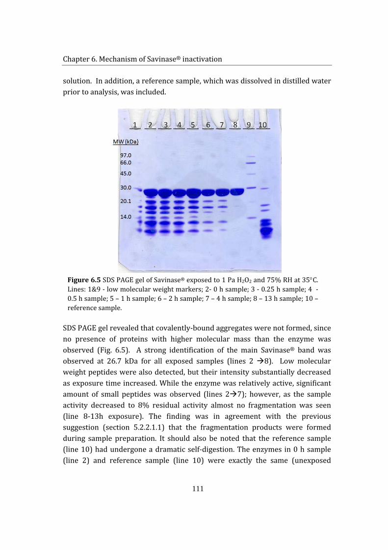
Chapter 6. Mechanism of Savinase® inactivation
111
solution. In addition, a reference sample, which was dissolved in distilled water prior to analysis, was included.
Figure 6.5 SDS PAGE gel of Savinase® exposed to 1 Pa H2O2 and 75% RH at 35°C. Lines: 1&9 - low molecular weight markers; 2- 0 h sample; 3 - 0.25 h sample; 4 - 0.5 h sample; 5 – 1 h sample; 6 – 2 h sample; 7 – 4 h sample; 8 – 13 h sample; 10 – reference sample.
SDS PAGE gel revealed that covalently-bound aggregates were not formed, since no presence of proteins with higher molecular mass than the enzyme was observed (Fig. 6.5). A strong identification of the main Savinase® band was observed at 26.7 kDa for all exposed samples (lines 2 8). Low molecular weight peptides were also detected, but their intensity substantially decreased as exposure time increased. While the enzyme was relatively active, significant amount of small peptides was observed (lines 27); however, as the sample activity decreased to 8% residual activity almost no fragmentation was seen (line 8-13h exposure). The finding was in agreement with the previous suggestion (section 5.2.2.1.1) that the fragmentation products were formed during sample preparation. It should also be noted that the reference sample (line 10) had undergone a dramatic self-digestion. The enzymes in 0 h sample (line 2) and reference sample (line 10) were exactly the same (unexposed
Chapter 6. Mechanism of Savinase® inactivation
112
enzyme powder). The difference between them was that the 0 h sample was dissolved in a borate buffer, whereas the reference sample was dissolved in distilled water. The autocatalytic activity was relatively inhibited when the sample was prepared in borate buffer. This suggested that high pH probably facilitated the denaturation effect of the reducing agent, limiting the reaction time for self-digestion.
6.2.3 IES MASS SPECTROSCOPY
Alterations in Savinase®’s structure were investigated on a molecular basis by mass spectroscopy. Samples, exposed to 1 Pa H2O2 and 75%RH, were dissolved in 0.05 M borate buffer at pH=9 containing 2.0% (w/w) Na2SO2 and 1.1% (w/w) KCl. This buffer was used to decompose H2O2 adsorbed by the sample and to prevent further enzyme oxidation in solution. Four samples were selected for analysis: 0h, 0.5h, 4h and 13h.
Firstly, solutions were run in ESI MS device to determine the molecular mass changes in the oxidized samples. The reference sample showed a clear peak at 26 698 Da, which corresponds to the native enzyme (Fig. 6.6-0 h). As a result of half an hour exposure to H2O2 (g), the peak of intact enzyme decreased and a product with additional mass of ∼16 Da was formed (Figure 6.6 -0.5 h). This corresponded to a single-oxidized variant of Savinase. As the exposure time increased, the oxidized product signal amplified and eventually, after 13 h, the native form of Savinase disappeared and only its single-oxidized form was detected (Figure 6.6 -13 h).
6.2.4 CNBR CLEAVAGE
For the determination of the position of oxidized residue, the enzyme was fragmented by CNBr cleavage. A specific cleavage of peptide bond at carboxyl site of Met residues can be achieved by incubation of a protein in an acidic solution of CNBr (Gross and Witkop 1962). Fragmentation of the protein backbone is achieved only if Met is not oxidized. The reaction is highly specific and effective. During CNBr digestion, due to the acidity of incubation solution, Met may be converted to homoserine and further to homoserine lactone.
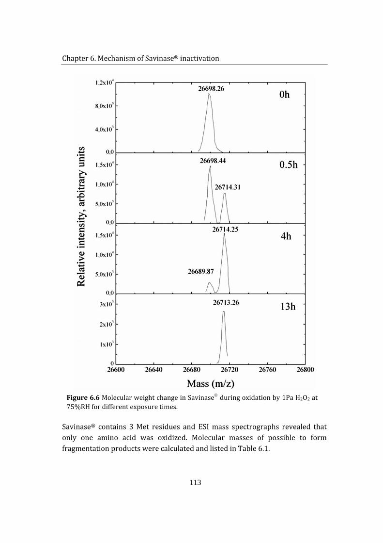
Chapter 6. Mechanism of Savinase® inactivation
113
Figure 6.6 Molecular weight change in Savinase during oxidation by 1Pa H2O2 at 75%RH for different exposure times.
Savinase® contains 3 Met residues and ESI mass spectrographs revealed that only one amino acid was oxidized. Molecular masses of possible to form fragmentation products were calculated and listed in Table 6.1.
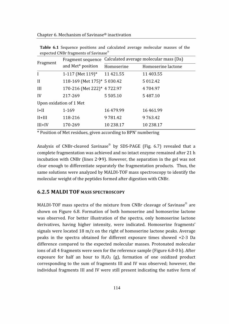
Chapter 6. Mechanism of Savinase® inactivation
114
Table 6.1 Sequence positions and calculated average molecular masses of the expected CNBr fragments of Savinase
Fragment Fragment sequence and Met* position
Calculated average molecular mass (Da) Homoserine Homoserine lactone
I 1-117 (Met 119)* 11 421.55 11 403.55 II 118-169 (Met 175)* 5 030.42 5 012.42 III 170-216 (Met 222)* 4 722.97 4 704.97 IV 217-269 5 505.10 5 487.10 Upon oxidation of 1 Met I+II 1-169 16 479.99 16 461.99 II+III 118-216 9 781.42 9 763.42 III+IV 170-269 10 238.17 10 238.17 * Position of Met residues, given according to BPN’ numbering
Analysis of CNBr-cleaved Savinase by SDS-PAGE (Fig. 6.7) revealed that a complete fragmentation was achieved and no intact enzyme remained after 21 h incubation with CNBr (lines 29). However, the separation in the gel was not clear enough to differentiate separately the fragmentation products. Thus, the same solutions were analyzed by MALDI-TOF mass spectroscopy to identify the molecular weight of the peptides formed after digestion with CNBr.
6.2.5 MALDI TOF MASS SPECTROSCOPY
MALDI-TOF mass spectra of the mixture from CNBr cleavage of Savinase are shown on Figure 6.8. Formation of both homoserine and homoserine lactone was observed. For better illustration of the spectra, only homoserine lactone derivatives, having higher intensity, were indicated. Homoserine fragments’ signals were located 18 m/z on the right of homoserine lactone peaks. Average peaks in the spectra obtained for different exposure times showed +2-3 Da difference compared to the expected molecular masses. Protonated molecular ions of all 4 fragments were seen for the reference sample (Figure 6.8-0 h). After exposure for half an hour to H2O2 (g), formation of one oxidized product corresponding to the sum of fragments III and IV was observed; however, the individual fragments III and IV were still present indicating the native form of
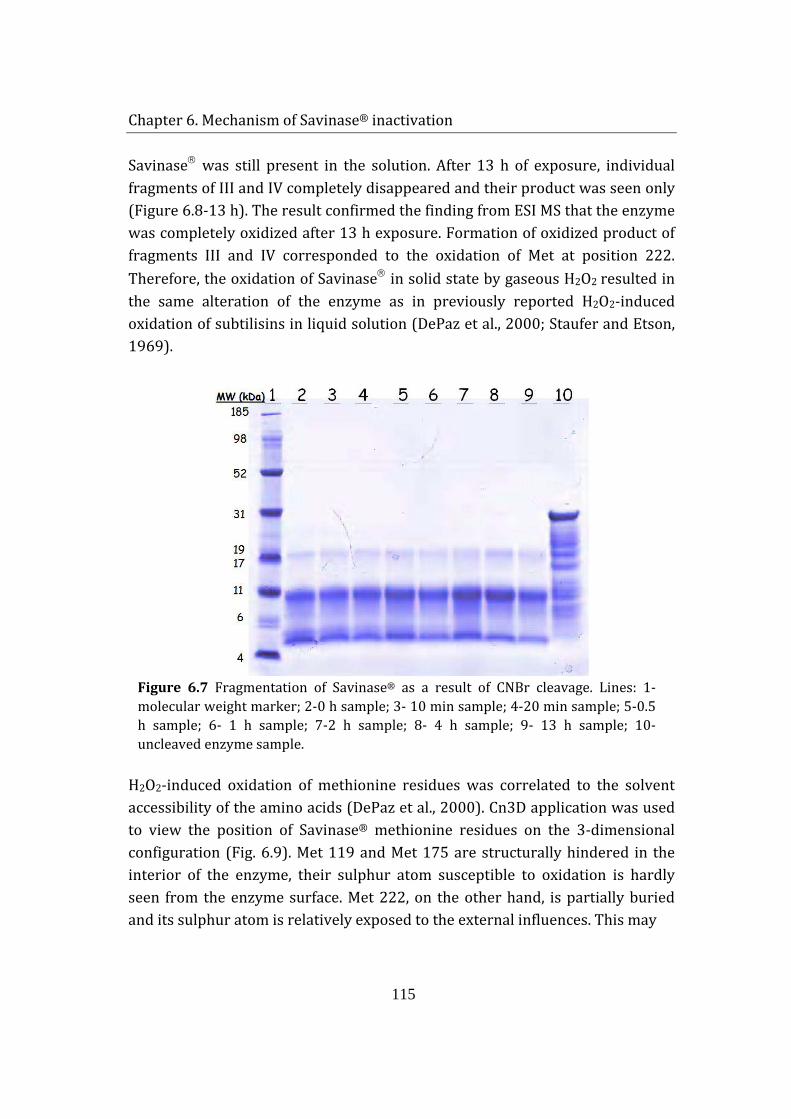
Chapter 6. Mechanism of Savinase® inactivation
115
Savinase was still present in the solution. After 13 h of exposure, individual fragments of III and IV completely disappeared and their product was seen only (Figure 6.8-13 h). The result confirmed the finding from ESI MS that the enzyme was completely oxidized after 13 h exposure. Formation of oxidized product of fragments III and IV corresponded to the oxidation of Met at position 222. Therefore, the oxidation of Savinase in solid state by gaseous H2O2 resulted in the same alteration of the enzyme as in previously reported H2O2-induced oxidation of subtilisins in liquid solution (DePaz et al., 2000; Staufer and Etson, 1969).
Figure 6.7 Fragmentation of Savinase® as a result of CNBr cleavage. Lines: 1-molecular weight marker; 2-0 h sample; 3- 10 min sample; 4-20 min sample; 5-0.5 h sample; 6- 1 h sample; 7-2 h sample; 8- 4 h sample; 9- 13 h sample; 10- uncleaved enzyme sample.
H2O2-induced oxidation of methionine residues was correlated to the solvent accessibility of the amino acids (DePaz et al., 2000). Cn3D application was used to view the position of Savinase® methionine residues on the 3-dimensional configuration (Fig. 6.9). Met 119 and Met 175 are structurally hindered in the interior of the enzyme, their sulphur atom susceptible to oxidation is hardly seen from the enzyme surface. Met 222, on the other hand, is partially buried and its sulphur atom is relatively exposed to the external influences. This may
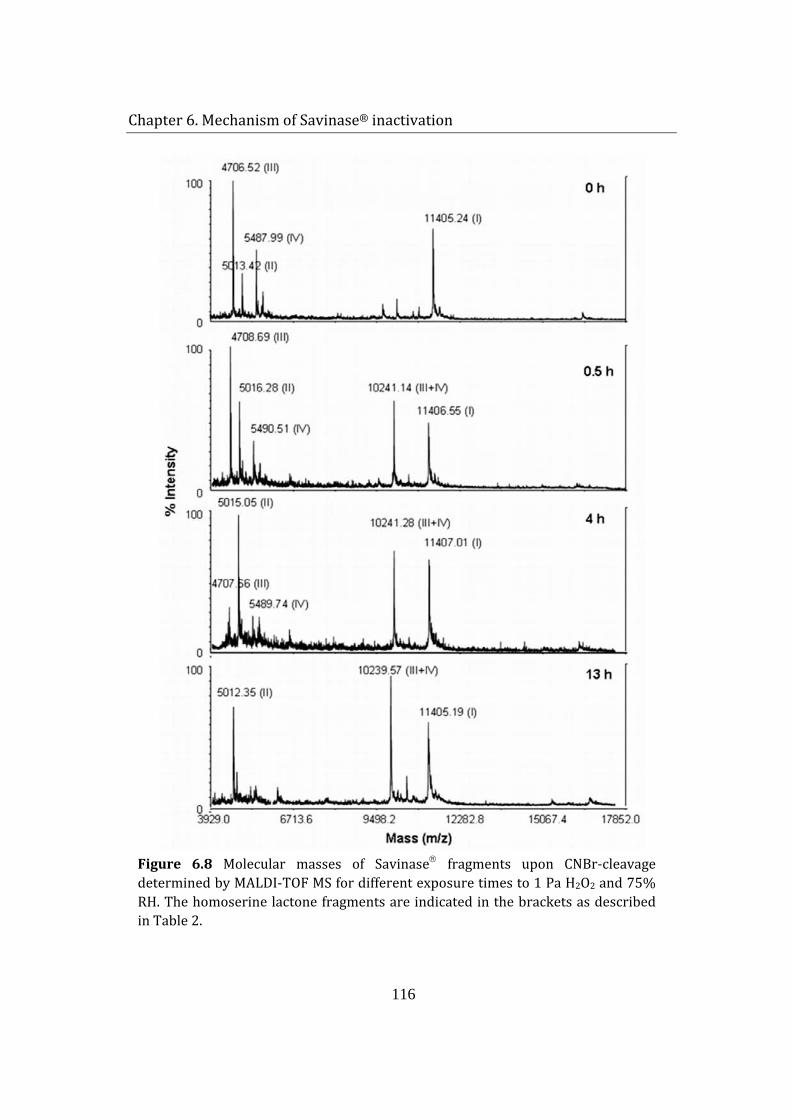
Chapter 6. Mechanism of Savinase® inactivation
116
Figure 6.8 Molecular masses of Savinase fragments upon CNBr-cleavage determined by MALDI-TOF MS for different exposure times to 1 Pa H2O2 and 75% RH. The homoserine lactone fragments are indicated in the brackets as described in Table 2.
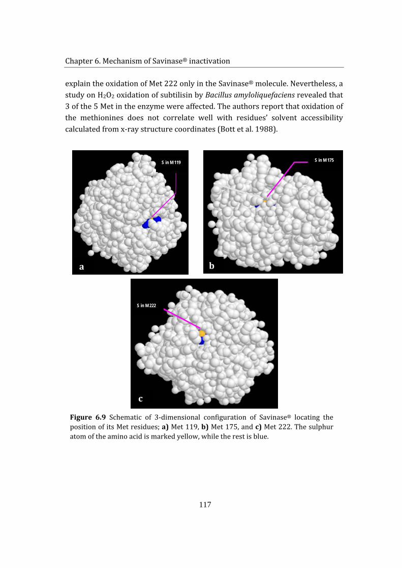
Chapter 6. Mechanism of Savinase® inactivation
117
explain the oxidation of Met 222 only in the Savinase® molecule. Nevertheless, a study on H2O2 oxidation of subtilisin by Bacillus amyloliquefaciens revealed that 3 of the 5 Met in the enzyme were affected. The authors report that oxidation of the methionines does not correlate well with residues’ solvent accessibility calculated from x-ray structure coordinates (Bott et al. 1988).
Figure 6.9 Schematic of 3-dimensional configuration of Savinase® locating the position of its Met residues; a) Met 119, b) Met 175, and c) Met 222. The sulphur atom of the amino acid is marked yellow, while the rest is blue.
S in M 119
a
S in M 175
b
S in M 222
c
Chapter 6. Mechanism of Savinase® inactivation
118
The activity results of the exposed samples have shown that significant loss of activity was attained as the enzyme was oxidized at 1 Pa H2O2 (g) and 75% RH (Fig. 6.4). Moreover, it was noticed that even though Savinase was completely oxidized after 13 h exposure, there was still about 8% residual activity left. Hence, oxidation of Met 222 substantially reduced the catalytic activity of the enzyme but did not completely inactivate it. The dramatic decrease in activity by oxidation of a single residue is related to the fact that Met 222 is adjacent to the catalytic triad (Ser 221, His 64 and Asp 32) of Savinase. Therefore, the oxidative modification of Met 222 probably results in a minor but significant conformational change in the protein’s tertiary structure (Staufer and Etson, 1969); the addition of one oxygen atom alters the electronic configuration of the active site by introducing a repulsive electrostatic term, which affects the optimum interaction between substrate and enzyme molecules (Bott et al., 1988; DePaz et al., 2000; Staufer and Etson, 1969).
Estell at al. (1985) tried to improve the stability of subtilisin by substituting the Met222 residue by site-directed mutagenesis. They substituted Met 222 with all remaining 19 amino acids and compared their activities and resistances to oxidation. The specific activity of mutants varied between 0.3 to 138% of the wild type enzyme, when examined in the presence of succinyl-L-Ala-L-Ala-L-Pro-p-nitroanalide as a substrate. In general, small amino acids were the most active toward this substrate followed by the amino acids with amides and aliphatic side chains. Bulky aromatic and charged amino acid substitutions were less active. More specifically, both sulfur-containing amino acids were most active, and the cysteine mutant had greater specific activity (138%) compared to the wild type enzyme at the specified conditions. The following highest values were obtained by replacing Met222 with alanin (Ala) and serine (Ser), which resulted in 53% and 35% relative specific activity, respectively. In terms of kinetic constants, however, the wild-type subtilisin remained the best performer. Comparing the kcat/Km values of the enzymes, the highest ratio was obtained for Met222 subtilisin, followed by Cys, Ser, Ala and Leu- substituted mutants. kcat/Km ratio illustrates the catalytic performance of the enzyme towards a specific substrate. Having a high kcat/Km value indicates that an enzyme has a tight binding capacity with the substrate and high turnover number, i.e. more substrate is converted to product per enzyme per unit time.
Chapter 6. Mechanism of Savinase® inactivation
119
Stability studies against hydrogen peroxide oxidation, on the other hand, showed that mutant enzymes were more resistant. Indeed, the mutants containing Ala222 and Ser222 were almost unaffected, retaining more than 90% of their activity, when exposed to 0.1M and 1M H2O2-borate solution. Comparing the Cys222 and Met222-type subtilisins at 0.1M H2O2 solution, the former showed significant resistance against oxidation by conserving ca. 90% of its activity, while the latter enzyme had less than 20% residual activity. At high H2O2 concentration (1M), the Cys-mutant was inactivated (t½ ∼ 12min), but not as rapidly as the wild-type enzyme (t½ ∼ 1min). Moreover, the inactivated wild type enzyme (Met222) maintained residual activity of 12% after elongated exposure in 0.1M and 1M H2O2 solutions. Similarly, the Cys222 mutant retained 7% residual activity. Similarly, DePaz et al. (2000) found out that 8% residual activity was retained in fully oxidized subtilisin towards N-succinyl-Ala-Ala-Pro-Phe-p-nitroanilide. The authors concluded that substitutions or chemical modification at position 222 of subtilisin resulting in bulky or charged residues have a deleterious effect on enzyme function (Estell et al., 1985).
6.3 SUMMARY
The preceding chapter was focused on revealing the mechanism of Savinase® inactivation as a result of exposure to H2O2 and humidity. Initially, the enzyme structure was described and amino acid sequence was listed. Modifications on Savinase® on the molecular level were studied on a freeze-dried sample exposed to 1 Pa H2O2 and 75% RH humidity at 35°C. A drastic reduction in enzyme activity was observed. At the end of 13h of exposure ca. 90% of the initial activity was lost.
SDS PAGE analysis showed that no covalently bound aggregates were formed, nor did proteolytic activity take place during exposure.
IES mass spectroscopy revealed that as exposure time proceeds a single-oxidation product of Savinase® is formed. At the end of 13h-period, the native enzyme signal disappeared and only oxidized Savinase® was present in the column.
Chapter 6. Mechanism of Savinase® inactivation
120
Location of the oxidized residue was performed by selective digestion of Savinase® by CNBr followed by MALDI TOF mass spectroscopy analysis of the fragmented peptides. The spectrographs showed that Met 222 was the residue oxidized by H2O2. The susceptibility of this amino acid to oxidation was related to its solvent accessibility.
The investigation revealed that Savinase® inactivation is caused solely by oxidation of Met 222 residue. The importance of this amino acid arises due to its vicinity to the catalytic triad if the enzyme, specifically Ser 221. Although all the enzyme were oxidized, still ca 10% residual activity was measured. This implied that modification in Met 222 affects the configuration and electron environment around the active site resulting in repulsive interaction between the enzyme and substrate.
6.4 REFERENCES
Betzel C, Klupsch S, Papendorf G, Hastrup S, Branner S, Wilson KS. 1992. Crystal-Structure of the Alkaline Proteinase Savinase from Bacillus-Lentus at 1.4-a Resolution. Journal of Molecular Biology 223(2):427-445.
Bott R, Ultsch M, Kossiakoff A, Graycar T, Katz B, Power S. 1988. The 3-Dimensional Structure of Bacillus-Amyloliquefaciens Subtilisin at 1.8-a and an Analysis of the Structural Consequences of Peroxide Inactivation. Journal of Biological Chemistry 263(16):7895-7906.
Chen J, Anderson JB, DeWeese-Scott C, Fedorova ND, Geer LY, He SQ, Hurwitz DI, Jackson JD, Jacobs AR, Lanczycki CJ and others. 2003. MMDB: Entrez's 3D-structure database. Nucleic Acids Research 31(1):474-477.
Christianson T, Paech C. 1994. Peptide-Mapping of Subtilisins as a Practical Tool for Locating Protein-Sequence Errors during Extensive Protein Engineering Projects. Analytical Biochemistry 223(1):119-129.
DePaz RA, Barnett CC, Dale DA, Carpenter JF, Gaertner AL, Randolph TW. 2000. The excluding effects of sucrose on a protein chemical degradation pathway: Methionine oxidation in subtilisin. Archives of Biochemistry and Biophysics 384(1):123-132.
Chapter 6. Mechanism of Savinase® inactivation
121
DePaz RA, Dale DA, Barnett CC, Carpenter JF, Gaertner AL, Randolph TW. 2002. Effects of drying methods and additives on the structure, function, and storage stability of subtilisin: role of protein conformation and molecular mobility. Enzyme and Microbial Technology 31(6):765-774.
Egmond MR, Antheunisse WP, Vanbemmel CJ, Ravestein P, Devlieg J, Peters H, Branner S. 1994. Engineering Surface-Charges in a Subtilisin - the Effects on Electrophoretic and Ion-Exchange Behavior. Protein Engineering 7(6):793-800.
Estell DA, Graycar TP, Adams R, Power SD, Ulltsch M, Bott RR, Cunningham BC, Carter P, Wells JA. 1988. Structure-Function Relationships in Subtilisin. Biochemistry 27(8):3078-3078.
Georgieva DN, Stoeva S, Voelter W, Genov N, Betzel C. 2001. Differences in the specificities of the highly alkalophilic proteinases Savinase and Esperase imposed by changes in the rigidity and geometry of the substrate binding sites. Archives of Biochemistry and Biophysics 387(2):197-201.
Gross E, Witkop B. 1962. Nonenzymatic Cleavage of Peptide Bonds - Methionine Residues in Bovine Pancreatic Ribonuclease. Journal of Biological Chemistry 237(6):1856-1860.
Markland FS, Smith EL. 1967. Subtilisin Bpn' .7. Isolation of Cyanogen Bromide Peptides and Complete Amino Acid Sequence. Journal of Biological Chemistry 242(22):5198-5211.
Nonaka T, Fujihashi M, Kita A, Saeki K, Ito S, Horikoshi K, Miki K. 2004. The crystal structure of an oxidatively stable subtilisin-like alkaline serine protease, KP-43, with a C-terminal beta-barrel domain. Journal of Biological Chemistry 279(45):47344-47351.
Prestrelski SJ, Tedeschi N, Arakawa T, Carpenter JF. 1993. Dehydration-Induced Conformational Transitions in Proteins and Their Inhibition by Stabilizers. Biophysical Journal 65(2):661-671.
Remerowski ML, Pepermans HAM, Hilbers CW, VandeVen FJM. 1996. Backbone dynamics of the 269-residue protease Savinase determined from N-15-NMR relaxation measurements. European Journal of Biochemistry 235(3):629-640.
Chapter 6. Mechanism of Savinase® inactivation
122
Reubsaet JLE, Beijnen JH, Bult A, van Maanen RJ, Marchal JAD, Underberg WJM. 1998. Analytical techniques used to study the degradation of proteins and peptides: chemical instability. Journal of Pharmaceutical and Biomedical Analysis 17(6-7):955-978.
Stauffer CE, Etson D. 1969. Effect of Subtilisin Activity of Oxidizing a Methionine Residue. Journal of Biological Chemistry 244(19):5333-5338.
Vonderosten C, Branner S, Hastrup S, Hedegaard L, Rasmussen MD, Bisgardfrantzen H, Carlsen S, Mikkelsen JM. 1993. Protein Engineering of Subtilisins to Improve Stability in Detergent Formulations. Journal of Biotechnology 28(1):55-68.
Wright CS, Alden RA, Kraut J. 1969. Structure of Subtilisin Bpns' at 2.5 a Resolution. Nature 221(5177):235-242.
Chapter 7. Stability measurements of formulated enzymes
123
CHAPTER 7 STABILITY MEASUREMENTS OF
FORMULATED ENZYMES
7.1 INTRODUCTION
Incorporating enzymes in detergent formulations gives rise to numerous practical problems due to their incompatibility with and lack of stability against various detergent components (Joo and Chang 2006; Lalonde et al. 1995; Stoner et al. 2004). Moreover, exposure to enzymes during their processing, packaging, distribution and use may result in serious allergic responses, skin rashes and/or asthmatic reactions (Vanhanen et al. 2000). In powdered detergent formulations, these issues can be partly overcome by physically isolating the enzymes in separate particles.
Enzyme particles are mainly produced as granules by high-shear granulation, which results in homogeneous granules or via fluid-bed granulation, which produces layer-structured granules. The structure, constituents and functions of the ingredients in an enzyme granulate were described in section 2.4.1. A typical enzyme granule has a diameter of 300 to 1200 µm with a coating layer of approximately 10-15µm (Jørgensen et al. 2005). The enzyme content of a detergent granulate varies between 1 to 30 w/w % and the quantity of the binder is between 1 and 50 w/w %. Salts, talc, starch, cellulose and starch derivatives are used as fillers. Different types of materials can be employed as binders or as coating materials, such as polyethyleneglycol (Harkonen et al. 1993). Using the enzymes in a granulated form provides good flowability, ease of measurement, no sticking to the walls of containers, improved appearance by control of size and color to match the detergent matrix particles, high stability, and controlled enzyme release during washing (Chan et al. 2006; Harkonen et al. 1993; Liu et al. 2005).
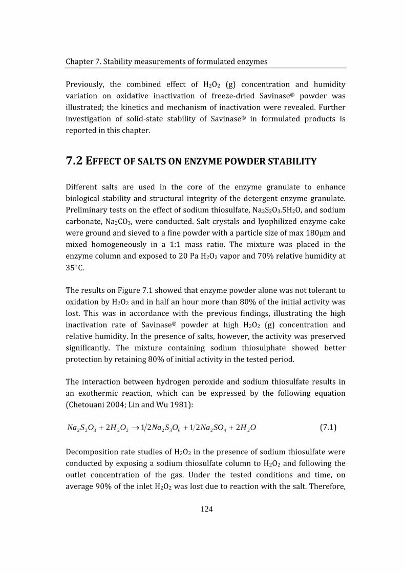
Chapter 7. Stability measurements of formulated enzymes
124
Previously, the combined effect of H2O2 (g) concentration and humidity variation on oxidative inactivation of freeze-dried Savinase® powder was illustrated; the kinetics and mechanism of inactivation were revealed. Further investigation of solid-state stability of Savinase® in formulated products is reported in this chapter.
7.2 EFFECT OF SALTS ON ENZYME POWDER STABILITY
Different salts are used in the core of the enzyme granulate to enhance biological stability and structural integrity of the detergent enzyme granulate. Preliminary tests on the effect of sodium thiosulfate, Na2S2O3.5H2O, and sodium carbonate, Na2CO3, were conducted. Salt crystals and lyophilized enzyme cake were ground and sieved to a fine powder with a particle size of max 180μm and mixed homogeneously in a 1:1 mass ratio. The mixture was placed in the enzyme column and exposed to 20 Pa H2O2 vapor and 70% relative humidity at 35°C.
The results on Figure 7.1 showed that enzyme powder alone was not tolerant to oxidation by H2O2 and in half an hour more than 80% of the initial activity was lost. This was in accordance with the previous findings, illustrating the high inactivation rate of Savinase® powder at high H2O2 (g) concentration and relative humidity. In the presence of salts, however, the activity was preserved significantly. The mixture containing sodium thiosulphate showed better protection by retaining 80% of initial activity in the tested period.
The interaction between hydrogen peroxide and sodium thiosulfate results in an exothermic reaction, which can be expressed by the following equation (Chetouani 2004; Lin and Wu 1981):
OHSONaOSNaOHOSNa 24263222322 221212 ++→+ (7.1)
Decomposition rate studies of H2O2 in the presence of sodium thiosulfate were conducted by exposing a sodium thiosulfate column to H2O2 and following the outlet concentration of the gas. Under the tested conditions and time, on average 90% of the inlet H2O2 was lost due to reaction with the salt. Therefore,
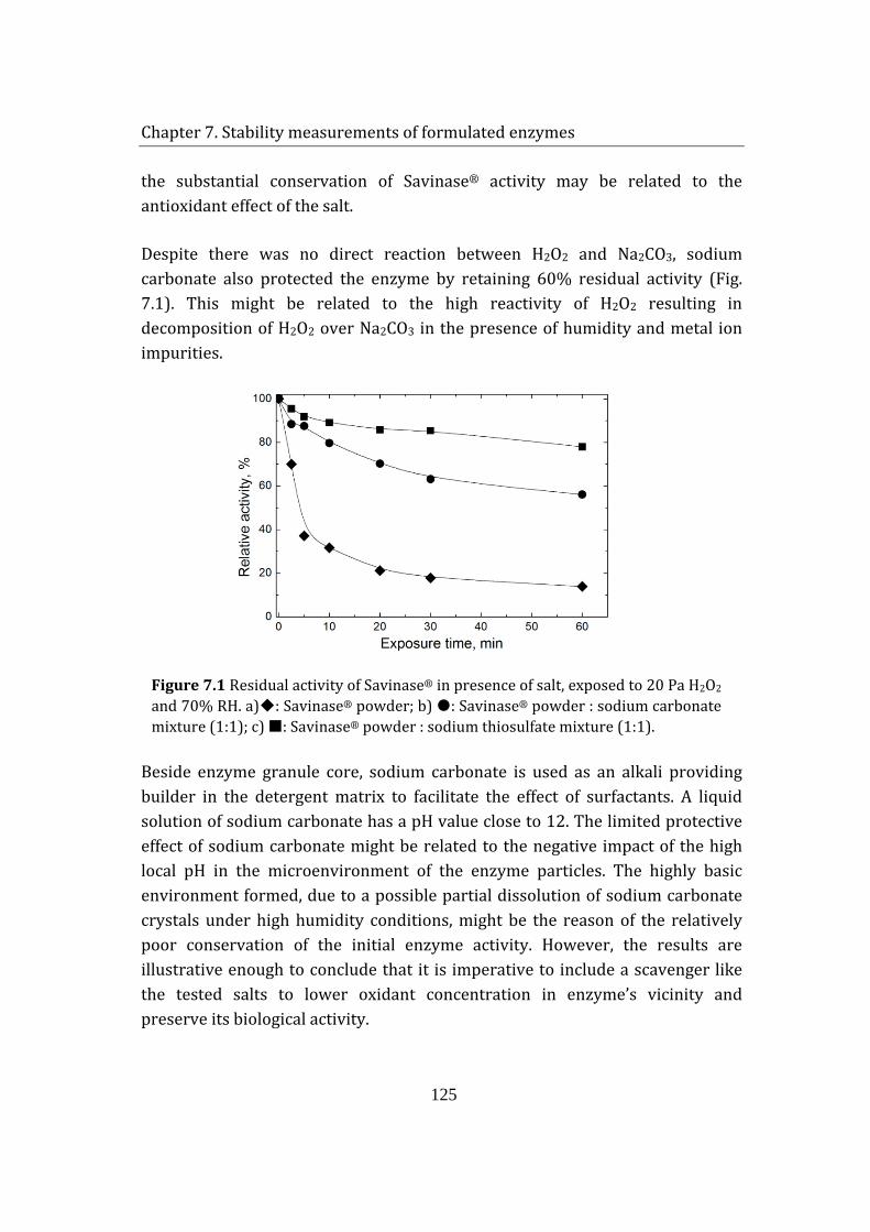
Chapter 7. Stability measurements of formulated enzymes
125
the substantial conservation of Savinase® activity may be related to the antioxidant effect of the salt.
Despite there was no direct reaction between H2O2 and Na2CO3, sodium carbonate also protected the enzyme by retaining 60% residual activity (Fig. 7.1). This might be related to the high reactivity of H2O2 resulting in decomposition of H2O2 over Na2CO3 in the presence of humidity and metal ion impurities.
Figure 7.1 Residual activity of Savinase® in presence of salt, exposed to 20 Pa H2O2 and 70% RH. a): Savinase® powder; b) : Savinase® powder : sodium carbonate mixture (1:1); c) : Savinase® powder : sodium thiosulfate mixture (1:1).
Beside enzyme granule core, sodium carbonate is used as an alkali providing builder in the detergent matrix to facilitate the effect of surfactants. A liquid solution of sodium carbonate has a pH value close to 12. The limited protective effect of sodium carbonate might be related to the negative impact of the high local pH in the microenvironment of the enzyme particles. The highly basic environment formed, due to a possible partial dissolution of sodium carbonate crystals under high humidity conditions, might be the reason of the relatively poor conservation of the initial enzyme activity. However, the results are illustrative enough to conclude that it is imperative to include a scavenger like the tested salts to lower oxidant concentration in enzyme’s vicinity and preserve its biological activity.
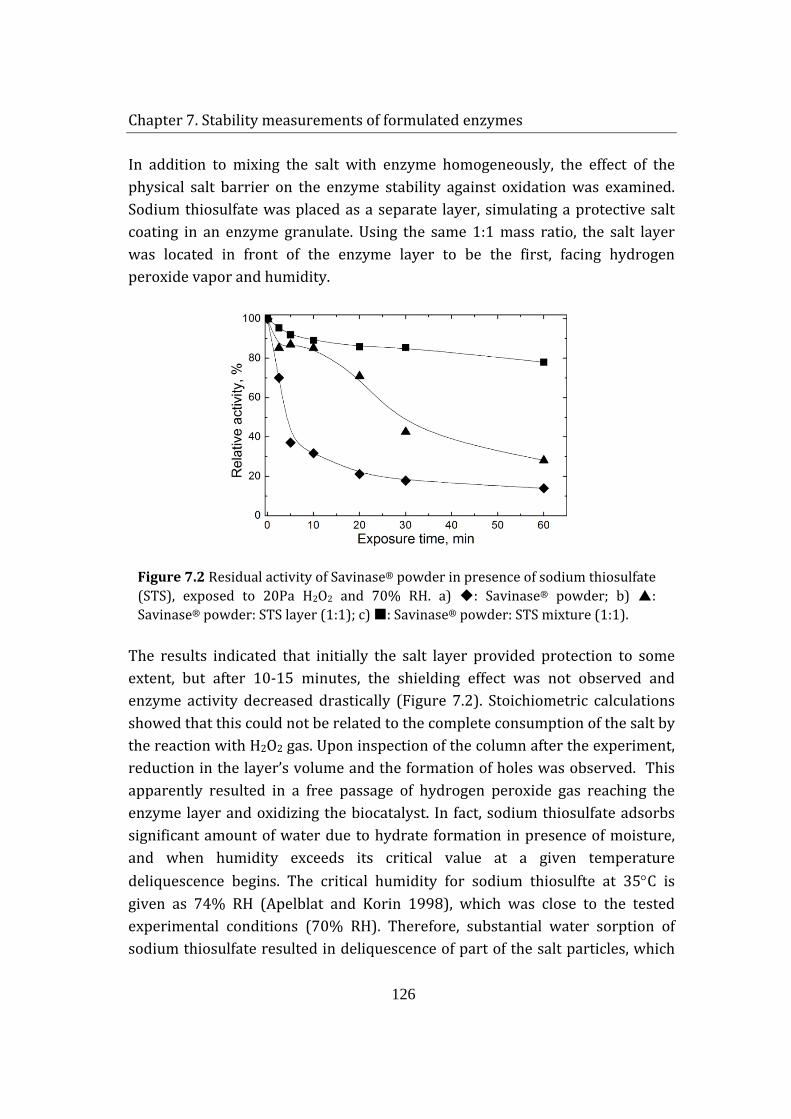
Chapter 7. Stability measurements of formulated enzymes
126
In addition to mixing the salt with enzyme homogeneously, the effect of the physical salt barrier on the enzyme stability against oxidation was examined. Sodium thiosulfate was placed as a separate layer, simulating a protective salt coating in an enzyme granulate. Using the same 1:1 mass ratio, the salt layer was located in front of the enzyme layer to be the first, facing hydrogen peroxide vapor and humidity.
Figure 7.2 Residual activity of Savinase® powder in presence of sodium thiosulfate (STS), exposed to 20Pa H2O2 and 70% RH. a) : Savinase® powder; b) : Savinase® powder: STS layer (1:1); c) : Savinase® powder: STS mixture (1:1).
The results indicated that initially the salt layer provided protection to some extent, but after 10-15 minutes, the shielding effect was not observed and enzyme activity decreased drastically (Figure 7.2). Stoichiometric calculations showed that this could not be related to the complete consumption of the salt by the reaction with H2O2 gas. Upon inspection of the column after the experiment, reduction in the layer’s volume and the formation of holes was observed. This apparently resulted in a free passage of hydrogen peroxide gas reaching the enzyme layer and oxidizing the biocatalyst. In fact, sodium thiosulfate adsorbs significant amount of water due to hydrate formation in presence of moisture, and when humidity exceeds its critical value at a given temperature deliquescence begins. The critical humidity for sodium thiosulfte at 35°C is given as 74% RH (Apelblat and Korin 1998), which was close to the tested experimental conditions (70% RH). Therefore, substantial water sorption of sodium thiosulfate resulted in deliquescence of part of the salt particles, which
Chapter 7. Stability measurements of formulated enzymes
127
in turn destroyed salt’s physical barrier. Consequently, better preservation of enzyme activity against oxidation might be achieved by mixing sodium thiosulfate with enzyme in the granulate core, instead of coating it as a separate layer.
7.3 EFFECT OF SODIUM THIOSULFATE ON ENZYME
GRANULATE STABILITY
Due to the low enzyme content in granulates, there was a need to adjust the concentration of H2O2 to obtain a reasonable illustration of the inactivation kinetics. It was important to estimate the release rate of H2O2 from sodium percarbonate (SPC) at the tested conditions. However, the conventional methods for measuring the stability of SPC involved long-term temperature ageing in a climate chamber and subsequent analysis of residual H2O2 content as a function of time (Johonsson et al. 2007). The problem of time-consuming testing was overcome by utilization of the designed experimental setup. A 1g-bed of commercial sodium percarbonate (SPC) was exposed to a gas stream with 70% RH and 1L/min flow rate, and the exhaust gas from the column was absorbed in a collection bottle, where the increase in H2O2 concentration was monitored. The release rate of H2O2 was measured as 0.2 Pa H2O2/g SPC.min at 35°C. Generally, a detergent matrix contains about 10% bleaching agents; therefore, if a typical detergent is placed in the used experimental setup the enzyme particles will encounter H2O2 concentration not higher than 2 Pa under the examined conditions.
The basic structure of the formulated enzyme granulates used in the study is illustrated on figure 7.3. The enzyme granules were T- type, high-shear granulated particles containing finely ground cellulose fibers (100-300 µm long), binders, salts (Jørgensen et al., 2005) and homogeneously distributed Savinase®. The coating contained PEG 4000, kaolin and TiO2. The mean particle size of enzyme granulates was 500μm.
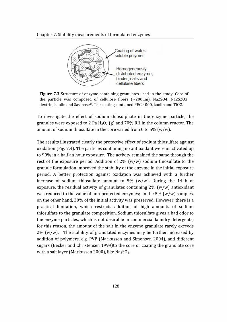
Chapter 7. Stability measurements of formulated enzymes
128
Figure 7.3 Structure of enzyme-containing granulates used in the study. Core of the particle was composed of cellulose fibers (~200μm), Na2SO4, Na2S2O3, dextrin, kaolin and Savinase®. The coating contained PEG 4000, kaolin and TiO2.
To investigate the effect of sodium thiosulphate in the enzyme particle, the granules were exposed to 2 Pa H2O2 (g) and 70% RH in the column reactor. The amount of sodium thiosulfate in the core varied from 0 to 5% (w/w).
The results illustrated clearly the protective effect of sodium thiosulfate against oxidation (Fig. 7.4). The particles containing no antioxidant were inactivated up to 90% in a half an hour exposure. The activity remained the same through the rest of the exposure period. Addition of 2% (w/w) sodium thiosulfate to the granule formulation improved the stability of the enzyme in the initial exposure period. A better protection against oxidation was achieved with a further increase of sodium thiosulfate amount to 5% (w/w). During the 14 h of exposure, the residual activity of granulates containing 2% (w/w) antioxidant was reduced to the value of non-protected enzymes; in the 5% (w/w) samples, on the other hand, 30% of the initial activity was preserved. However, there is a practical limitation, which restricts addition of high amounts of sodium thiosulfate to the granulate composition. Sodium thiosulfate gives a bad odor to the enzyme particles, which is not desirable in commercial laundry detergents; for this reason, the amount of the salt in the enzyme granulate rarely exceeds 2% (w/w). The stability of granulated enzymes may be further increased by addition of polymers, e.g. PVP (Markussen and Simonsen 2004), and different sugars (Becker and Christensen 1999)to the core or coating the granulate core with a salt layer (Markussen 2000), like Na2SO4.
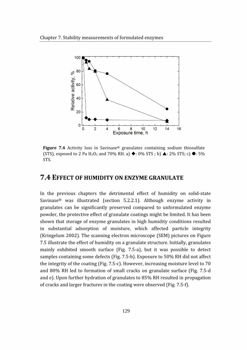
Chapter 7. Stability measurements of formulated enzymes
129
Figure 7.4 Activity loss in Savinase® granulates containing sodium thiosulfate (STS), exposed to 2 Pa H2O2 and 70% RH. a) : 0% STS ; b) : 2% STS; c) : 5% STS.
7.4 EFFECT OF HUMIDITY ON ENZYME GRANULATE
In the previous chapters the detrimental effect of humidity on solid-state Savinase® was illustrated (section 5.2.2.1). Although enzyme activity in granulates can be significantly preserved compared to unformulated enzyme powder, the protective effect of granulate coatings might be limited. It has been shown that storage of enzyme granulates in high humidity conditions resulted in substantial adsorption of moisture, which affected particle integrity (Kringelum 2002). The scanning electron microscope (SEM) pictures on Figure 7.5 illustrate the effect of humidity on a granulate structure. Initially, granulates mainly exhibited smooth surface (Fig. 7.5-a), but it was possible to detect samples containing some defects (Fig. 7.5-b). Exposure to 50% RH did not affect the integrity of the coating (Fig. 7.5-c). However, increasing moisture level to 70 and 80% RH led to formation of small cracks on granulate surface (Fig. 7.5-d and e). Upon further hydration of granulates to 85% RH resulted in propagation of cracks and larger fractures in the coating were observed (Fig. 7.5-f).
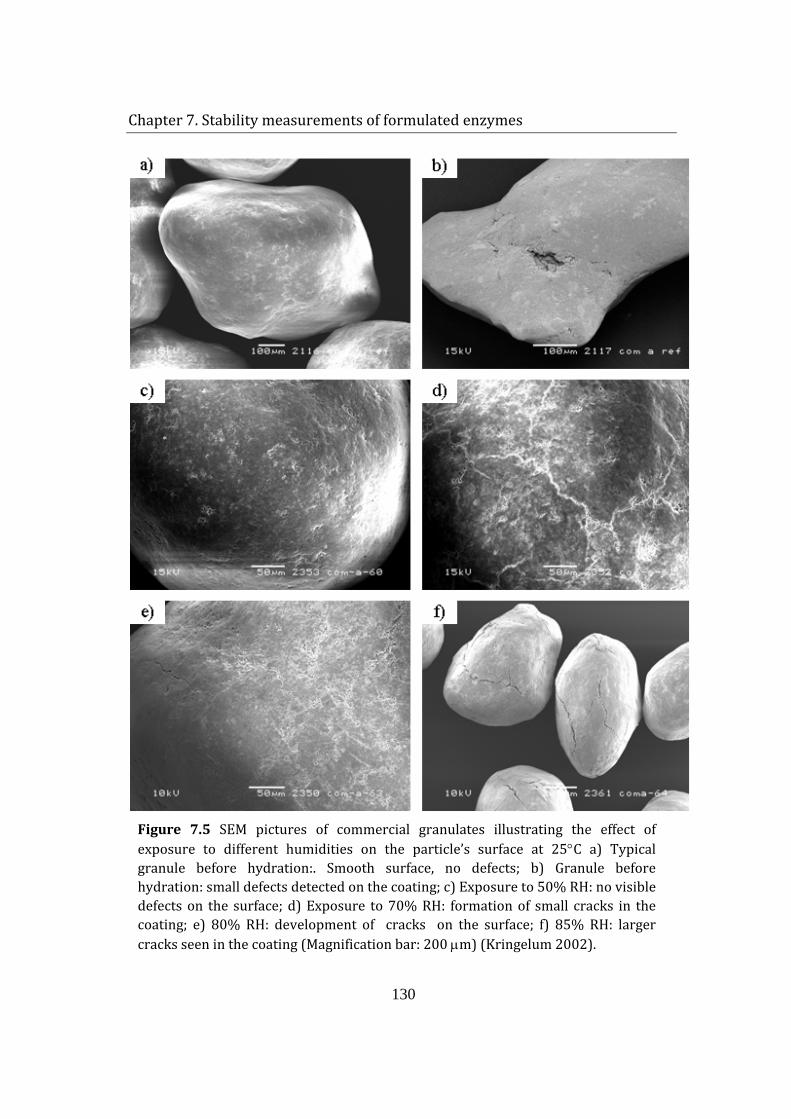
Chapter 7. Stability measurements of formulated enzymes
130
Figure 7.5 SEM pictures of commercial granulates illustrating the effect of exposure to different humidities on the particle’s surface at 25°C a) Typical granule before hydration:. Smooth surface, no defects; b) Granule before hydration: small defects detected on the coating; c) Exposure to 50% RH: no visible defects on the surface; d) Exposure to 70% RH: formation of small cracks in the coating; e) 80% RH: development of cracks on the surface; f) 85% RH: larger cracks seen in the coating (Magnification bar: 200 µm) (Kringelum 2002).
Chapter 7. Stability measurements of formulated enzymes
131
The gradual development of the cracks was related to water absorption, which possibly resulted in swelling of granulate components, e.g. cellulose fibers (Kringelum 2002). Fractures on the granulate’s surface enhance water diffusion through the particle and facilitate the penetration of destructive agents like H2O2 (g) to enzymes, which subsequently reduce granulate’s biological activity.
The effect of humidity on granulated enzyme activity was investigated at 70% RH (Fig. 7.6). The composition of the enzyme particles was as depicted on Figure 7.3; no anti-oxidant was used but a green dye was added to the coating to obtain colored granulates.
Firstly, enzyme particles were exposed to humidity alone and complete conservation of enzyme activity was observed during the 15 h exposure period. Next, the granulates were exposed to 2 Pa H2O2 (g) and 70% RH. Due to lack of antioxidant effect of sodium thiosulfate, a drastic loss of 90% of enzyme activity was seen. Finally, the enzyme particles were mixed with a bleach containing detergent in a 1:9 weight ratio. The detergent matrix was placed in the column and exposed to 70% RH. To eliminate a possible interference of the detergent ingredients with the analytical method, a sample of the colored enzyme granulates was `harvested` from the detergent matrix after exposure to humidity. Gradual decrease in residual enzyme activity was observed. The activity loss was related to release of H2O2 (g) from the bleach (sodium percarbonate). A comparison of enzyme inactivation curves showed that the enzymes loose their activity slower in a detergent matrix, indicating that H2O2
(g) concentration in the granulate vicinity might be lower than 2 Pa when mixed with a detergent. Probably, there is equilibrium between the release rate of H2O2 from sodium percarbonate and the decomposition rate of H2O2 over other detergent particles and surfaces, which decreases the total concentration of H2O2 in the surrounding.
An attempt to measure the amount of H2O2 releasing from the bleach in the presence of other detergent ingredients resulted in an intensefoaming in the collection bottle. This indicated that humidity also triggered the release of other components to the gas phase, suggesting that not only oxidation but other mechanisms might be involved in the inactivation of Savinase® during storage in detergent matrix.
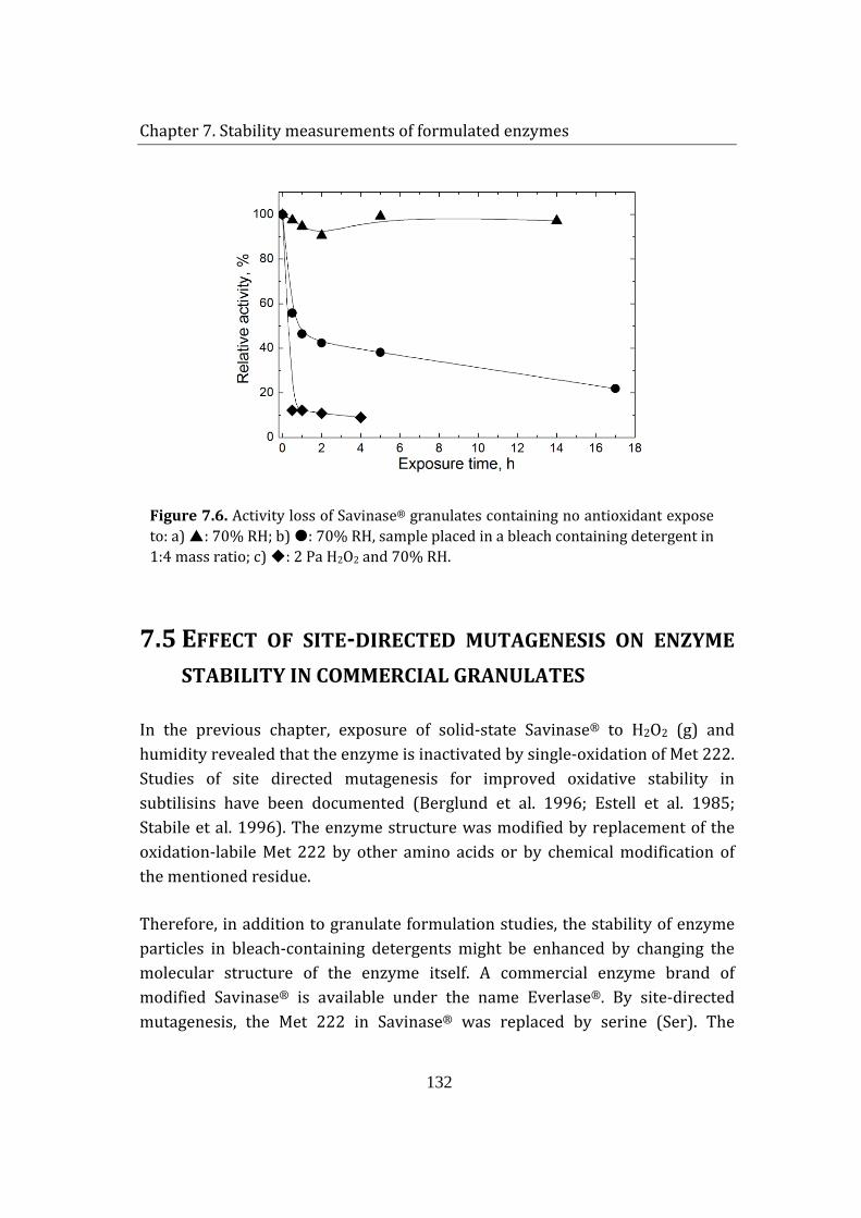
Chapter 7. Stability measurements of formulated enzymes
132
Figure 7.6. Activity loss of Savinase® granulates containing no antioxidant expose to: a) : 70% RH; b) : 70% RH, sample placed in a bleach containing detergent in 1:4 mass ratio; c) : 2 Pa H2O2 and 70% RH.
7.5 EFFECT OF SITE-DIRECTED MUTAGENESIS ON ENZYME
STABILITY IN COMMERCIAL GRANULATES
In the previous chapter, exposure of solid-state Savinase® to H2O2 (g) and humidity revealed that the enzyme is inactivated by single-oxidation of Met 222. Studies of site directed mutagenesis for improved oxidative stability in subtilisins have been documented (Berglund et al. 1996; Estell et al. 1985; Stabile et al. 1996). The enzyme structure was modified by replacement of the oxidation-labile Met 222 by other amino acids or by chemical modification of the mentioned residue.
Therefore, in addition to granulate formulation studies, the stability of enzyme particles in bleach-containing detergents might be enhanced by changing the molecular structure of the enzyme itself. A commercial enzyme brand of modified Savinase® is available under the name Everlase®. By site-directed mutagenesis, the Met 222 in Savinase® was replaced by serine (Ser). The
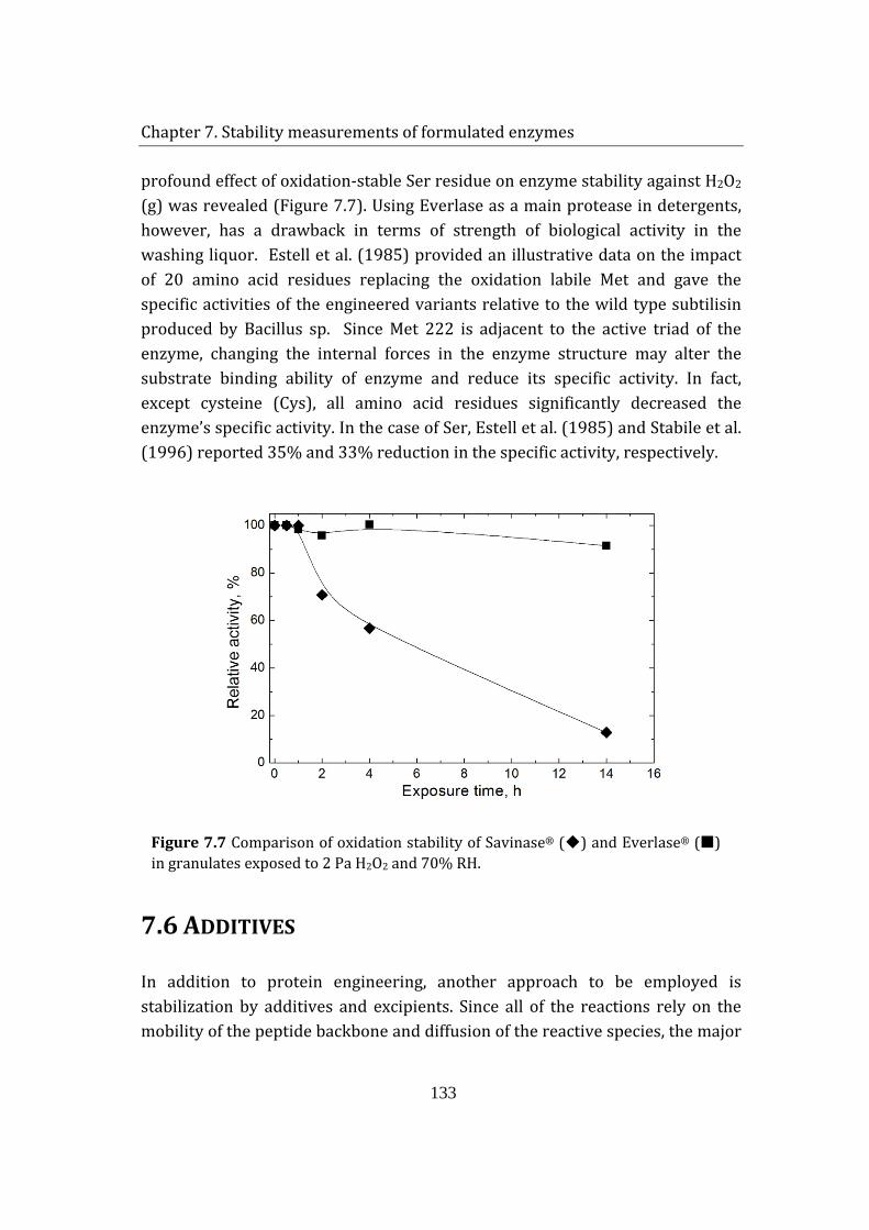
Chapter 7. Stability measurements of formulated enzymes
133
profound effect of oxidation-stable Ser residue on enzyme stability against H2O2 (g) was revealed (Figure 7.7). Using Everlase as a main protease in detergents, however, has a drawback in terms of strength of biological activity in the washing liquor. Estell et al. (1985) provided an illustrative data on the impact of 20 amino acid residues replacing the oxidation labile Met and gave the specific activities of the engineered variants relative to the wild type subtilisin produced by Bacillus sp. Since Met 222 is adjacent to the active triad of the enzyme, changing the internal forces in the enzyme structure may alter the substrate binding ability of enzyme and reduce its specific activity. In fact, except cysteine (Cys), all amino acid residues significantly decreased the enzyme’s specific activity. In the case of Ser, Estell et al. (1985) and Stabile et al. (1996) reported 35% and 33% reduction in the specific activity, respectively.
Figure 7.7 Comparison of oxidation stability of Savinase® () and Everlase® () in granulates exposed to 2 Pa H2O2 and 70% RH.
7.6 ADDITIVES
In addition to protein engineering, another approach to be employed is stabilization by additives and excipients. Since all of the reactions rely on the mobility of the peptide backbone and diffusion of the reactive species, the major
Chapter 7. Stability measurements of formulated enzymes
134
factor resulting in loss of activity during storage is the moisture content in the detergent box. Once the box is opened the humidity level increases and the rate of enzyme inactivation rises.
One strategy to reduce the moisture effect is to include water-absorbing agents in the detergent matrix. Having high water absorption properties, beside their ion-exchange function, zeolites like aluminum sodium silicates can be used for this purpose(Fruijtier-Polloth 2009). More direct precaution against water diffusion through enzyme granulate can taken by using hydrophobic coating on the outer layer of the granulate. Currently, wax coatings serve as a barrier to the moisture uptake. However, due to physical stresses, the coatings may be broken and the cracks on the granule surface may facilitate water diffusion through the protective layers.
Furthermore, excipients added to the enzyme slurry before granulation may be used to limit the flexibility of the enzyme, by helping to maintain compact protein structure during drying via hydrogen bonding. Various sugars like sucrose and trehalose, polyols, like sorbitol, and polymers, like dextran and maltotextrin, can be added in the formulations (DePaz et al. 2002; Yin et al. 2005). The sugars should be carefully selected among the non-reducing ones to prevent the possibility of Maillard reactions between the reducing group of the sugar and the free amine group of the protein. It was shown that disaccarides preserve the conformation of the protein and prevent degradation caused by protein unfolding (Byrn et al. 2001). Dextran, on the other hand, does not affect protein structure but prevent aggregate formation. It is postulated that the dextran may spatially separate the dried protein molecule and prevent formation of intramolecular contacts during rehydration or the dextran molecules may force unfolded protein into more compact form and prevent intermolecular interactions that would lead to aggregation (Bryn et al., 2001). DePaz et al.’s (2002) work showed that excipients like sucrose and trehalose and polymers can preserve the native structure of the enzyme upon drying; hence, inhibiting unfolding. However, the oxidation studies revealed that these additives cannot provide enough protection to limit the exposure of Met 222.
It has been previously discussed that trace amounts of heavy metal ions substantially speed up the decomposition rate of hydrogen peroxide (Coons
Chapter 7. Stability measurements of formulated enzymes
135
1978). Thus, in addition to anti-oxidants like sodium thiosulphate, certain salts, e.g. MnSO4 and FeSO4, can be included in the enzyme granulate composition. In the humid environment, when the granulate integrity is compromised, the Mn2+ and Fe2+ ions in the vicinity of enzymes may decompose the diffusing oxidant and protect the biocatalysts. Beside acting as a H2O2-scavenger, MnSO4 can be used due to its stabilizing effect on alkaline proteases (Banerjee et al. 1999). Upon inspection of the effect of 12 different salts, Banerjee et al. (1999) found that MnSO4 improved protease’s thermostability by increasing enzyme activity by 24% at 60°C . Moreover, CaCl2, glycine and Ca-acetate were found to have a stabilizing effect on proteases; lyoprotectants like sorbitol and PEG were reported to increase the activity of subtilisin powder (Bovara et al. 1997).
7.7 SUMMARY
The designed experimental setup was further used for basic formulation studies and granulate stability tests. The method was proven to be suitable for acquiring fast results. This is an important criteria for developing a new product especially when a large number of ingredients are tested and fast stability assessments are needed.
Initially, the effect of the presence of salts was investigated. Both sodium carbonate and sodium thiosulphate had protective effect against oxidation. Sodium thiosulphate was found to be more efficient, retaining 80% activity after exposure to 20 Pa H2O2 and 70% RH at 35°C for 1h, compared to 60% residual activity for sodium carbonate. Moreover, the results revealed that mixing the salt with the enzyme, rather than applying as a separate coating, should be considered in granulate formulation. Examination of a layered column configuration showed that the protective effect of sodium thiosulphate is compromised due to deliquence of the salt. If applied separately as a coating, humidity may significantly destroy the integrity of the salt layer. Formation of cracks on granulate surface would result in water uptake, which may facilitate the diffusion of oxidizing agents and other destructive components into the core and destroy the enzyme.
Chapter 7. Stability measurements of formulated enzymes
136
Finally, the inactivation trend of native (Savinase®) and oxidation resistant enzyme (Everlase®) was illustrated. Examples of possible stability enhancing additives were listed.
7.8 REFERENCES
Apelblat A, Korin E. 1998. Vapour pressures of saturated aqueous solutions of ammonium iodide, potassium iodide, potassium nitrate, strontium chloride, lithium sulphate, sodium thiosulphate, magnesium nitrate, and uranyl nitrate from T = (278 to 323) K. Journal of Chemical Thermodynamics 30(4):459-471.
Banerjee UC, Sani RK, Azmi W, Soni R. 1999. Thermostable alkaline protease from Bacillus brevis and its characterization as a laundry detergent additive. Process Biochemistry 35(1-2):213-219.
Becker N, Christensen R; 1999. Matrix granule. WO patent 9932613.
Berglund P, Stabile MR, Gold M, Jones JB, Mitchinson C, Bott RR, Graycar TP. 1996. Altering the specificity of subtilisin B-lentus by combining site-directed mutagenesis and chemical modification. Bioorganic & Medicinal Chemistry Letters 6(21):2507-2512.
Bovara R, Carrea G, Gioacchini AM, Riva S, Secundo F. 1997. Activity, stability, and conformation of methoxypoly(ethylene glycol)-subtilisin at different concentrations of water in dioxane. Biotechnology and Bioengineering 54(1):50-57.
Byrn SR, Xu W, Newman AW. 2001. Chemical reactivity in solid-state pharmaceuticals: formulation implications. Advanced Drug Delivery Reviews 48(1):115-136.
Chan AWJ, Mazeaud I, Becker T, Neufeld RJ. 2006. Granulation of subtilisin by internal gelation of alginate microspheres for application in detergent formulation. Enzyme and Microbial Technology 38(1-2):265-272.
Chetouani Y. 2004. Fault detection by using the innovation signal: application to an exothermic reaction. Chemical Engineering and Processing 43(12):1579-1585.

Chapter 7. Stability measurements of formulated enzymes
137
Coons DM. 1978. Bleach - Facts, Fantasy, and Fundamentals. Journal of the American Oil Chemists Society 55(1):104-108.
DePaz RA, Dale DA, Barnett CC, Carpenter JF, Gaertner AL, Randolph TW. 2002. Effects of drying methods and additives on the structure, function, and storage stability of subtilisin: role of protein conformation and molecular mobility. Enzyme and Microbial Technology 31(6):765-774.
Estell DA, Graycar TP, Wells JA. 1985. Engineering an Enzyme by Site-Directed Mutagenesis to Be Resistant to Chemical Oxidation. Journal of Biological Chemistry 260(11):6518-6521.
Fruijtier-Polloth C. 2009. The safety of synthetic zeolites used in detergents. Archives of Toxicology 83(1):23-35.
Harkonen H, Koskinen M, Linko P, Siikaaho M, Poutanen K. 1993. Granulation of Enzyme Powders in a Fluidized-Bed Spray Granulator. Food Science and Technology-Lebensmittel-Wissenschaft & Technologie 26(3):235-241.
Johonsson C, Pekonen P, Forsstrom D. 2007. The long-term stability of sodium percarbonate in presence of zeolite as measured by heat flow calorimetry. Tenside Surfactants Detergents 44(4):210-+.
Joo HS, Chang CS. 2006. Production of an oxidant and SDS-stable alkaline protease from an alkaophilic Bacillus clausii I-52 by submerged fermentation: Feasibility as a laundry detergent additive. Enzyme and Microbial Technology 38(1-2):176-183.
Jørgensen K, Bach P, Jensen AD. 2005. Impact and attrition shear breakage of enzyme granules and placebo particles-application to particle design and formulation. Powder Technology 149(2-3):157-167.
Kringelum J. 2002. Measurement and modeling of water sorption kinetics of enzyme granulates. Master thesis: Technical University of Denmark, Kgs. Lyngby, Denmark.
Lalonde J, Witte EJ, Oconnell ML, Holliday L. 1995. Protease Stabilization by Highly Concentrated Anionic Surfactant Mixtures. Journal of the American Oil Chemists Society 72(1):53-59.
Chapter 7. Stability measurements of formulated enzymes
138
Lin KF, Wu LL. 1981. Performance of an Adiabatic Controlled Cycled Stirred Tank Reactor. Chemical Engineering Science 36(2):435-444.
Liu ZM, Becker T, Neufeld RJ. 2005. Spherical alginate granules formulated for quick-release active subtilisin. Biotechnology Progress 21(2):568-574.
Markussen EK; 2000. A new improved enzyme containing granule. WO patent WO0001793 (A1)
Markussen EK, Simonsen O; 2004. Stabilization of granules. WO patent 2004003188.
Stabile MR, Lai WG, DeSantis G, Gold M, Jones JB. 1996. Probing the specificity of the S-1 binding site of M222 mutants of subtilisin B-lentus with boronic acid inhibitors. Bioorganic & Medicinal Chemistry Letters 6(21):2501-2506.
Stoner MR, Dale DA, Gualfetti PJ, Becker T, Manning MC, Carpenter JF, Randolph TW. 2004. Protease autolysis in heavy-duty liquid detergent formulations: effects of thermodynamic stabilizers and protease inhibitors. Enzyme and Microbial Technology 34(2):114-125.
Vanhanen M, Tuomi T, Tiikkainen U, Tupasela O, Voutilainen R, Nordman H. 2000. Risk of enzyme allergy in the detergent industry. Occupational and Environmental Medicine 57(2):121-125.
Yin J, Chu JW, Ricci MS, Brems DN, Wang DIC, Trout BL. 2005. Effects of excipients on the hydrogen peroxide-induced oxidation of methionine residues in granulocyte colony-stimulating factor. Pharmaceutical Research 22(1):141-147.
Chapter 8. Conclusions
139
CHAPTER 8 CONCLUSIONS AND SUGGESTIONS FOR
FUTURE WORK In this PhD project, the inactivation kinetics and mechanism of solid-state Savinase® has been investigated. Due to time-consuming conventional testing methods, a new experimental setup has been designed. The new method has provided a better control over test conditions and fast sample generation. The employed enzyme column setup enabled exposure of solid enzymes to different concentrations of H2O2 (g) and humidity for revealing the inactivation kinetics. Even though only a single sample could be handled at a time, the method still provided fast stability testing, where samples were obtained in a few hours instead of several weeks.
Technical grade freeze-dried Savinase® powder was used as a model detergent enzyme. Firstly, the moisture adsorption desorption isotherms of solid-state protein were constructed. The sigmoidal, type II, isotherm was fitted to GAB equation, which enabled calculation of the monolayer hydration level of the enzyme. The experimentally determined monolayer hydration was found to be 5.1 g water/100g dry enzyme. Then the effect of adsorbed moisture on enzyme activity and structure was examined. At humidity levels below monohydration level, Savinase® had high stability. In the multilayer region, however, 10% activity loss was detected. Exposure to 100% RH revealed that enzyme activity can be reduced as much as 80% in a one-week period. The results illustrated that even moisture alone may have detrimental effects on solid-state Savinase®. It was confirmed that auto-digestion was not the cause of inactivation; formation of unfavorable conformational changes of non-covalently bound aggregates were considered; however, further investigation was not conducted.
It is known that the bleaching agents in the detergent matrix may decompose during storage and release H2O2 (g), which may potentially harm the
Chapter 8. Conclusions
140
biocatalysts in the detergent. To assess the effect of releasing gas, enzymes were exposed to known concentrations of H2O2 (g). The results stated that significant amount of H2O2 was adsorbed by the solid-state enzyme. As the adsorption increased with the increasing oxidant concentration, it was independent of the extent of enzyme hydration. This implied that H2O2 and water were not competing for the same adsorption sites, and that H2O2 was not dissolved or dissociated in the multilayered water on the enzyme surface.
Exposure of the enzyme to gaseous H2O2 led to a drastic reduction in enzyme activity. It was also shown that the increased hydration of Savinase® molecules not only amplified the flexibility of the protein backbone resulting in both oxidative and non-oxidative malfunctions, but also enhanced the reactivity of H2O2 with the enzyme. In other words, in the absence of oxidant, one week-exposure to high humidity (100% RH at 35°C) resulted in 80% activity loss, which was related to unfavorable conformational changes since no auto-digestion or aggregate formation was detected. Furthermore, in the presence of constant H2O2 concentration (20 Pa), increasing Savinase® hydration from monolayer to multilayer resulted in led to activity loss from ca 60 to 85%.
A kinetic model of enzyme inactivation was proposed and the kinetic expression of the reaction was derived. Fitting the experimental findings to the kinetic expression, the enzyme inactivation was found to depend on the square of moisture adsorbed by the enzyme at the corresponding temperature. The inverse of the reaction rate constant was also proportional to the inverse of H2O2 in the system. Peptide mapping studies revealed that enzyme inactivation was caused by the oxidation of Met 222 residue, which is adjacent to the catalytic triad of the enzyme. Consequently, solid-state inactivation of Savinase® by gaseous H2O2 resulted in the same alterations on molecular level as the oxidation of the enzyme in aqueous solution.
Preliminary formulation studies indicated that H2O2 oxidation of the enzyme can be substantially reduced by addition of salt to the solid mixture. The salt acted either as a decomposition front to the H2O2 (g) or had a dilution effect in the powder and surrounding the enzyme molecule. Using sodium thiosulphate as an antioxidant was found to be effective when the salt was mixed with the protein rather than being layered as a protective coating.
Chapter 8. Conclusions
141
Finally, the enzyme-column was used for the assessment of enzyme stability in granulated products and was found to be a useful tool for formulation studies. The effect of sodium thiosulphate content in particles on enzyme activity was shown and the result of site-directed mutagenesis on enzyme stability was illustrated.
The designed setup proved to be satisfactory in approximating the inactivation kinetics of enzymes under oxidizing conditions. Despite the relatively accelerated testing it provides, the setup could benefit from further improvement. A multi-column reactor system could be developed for simultaneous testing of numerous samples at identical conditions. Moreover, the setup may serve as a useful tool for the estimation of enzymes’ storage stability in the detergent matrix, by establishing an extrapolative relation between the accelerated test results and real-time storage stability data.
The investigation of the effect of humidity on detergent matrix revealed that, in addition to H2O2 (g), moisture triggered the release other gaseous detergent components. By analyzing the releasing gas stream by gas chromatography and mass spectroscopy, the composition of the gas phase may be (partially) indentified. This would give some insight into the complex chemistry involved in the inactivation of detergent enzymes.
It has been found that exposure of solid-state Savinase® to high humidity resulted in substantial activity loss. However, the actual mechanism responsible for enzyme inactivation was not clear. Further investigation on possible structural changes is advised. The findings may provide information of strategies on stabilization of peptide backbone by excipients.
Having established a fast testing method, extensive formulation studies may be conducted. The protective effect of cost-feasible granulate ingredients could be tested for improving the shelf-life of detergent enzymes. Granulate additives like MnSO4 can be studied as a possible H2O2-scavenger and enzyme stabilizer. Acetate salts of Ca2+ and Na+, glycine and sorbitol may be investigated.
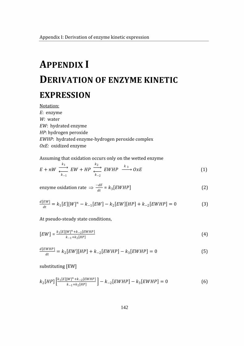
Appendix I: Derivation of enzyme kinetic expression
142
APPENDIX I DERIVATION OF ENZYME KINETIC
EXPRESSION E: enzyme Notation:
W: water EW: hydrated enzyme HP: hydrogen peroxide EWHP: hydrated enzyme-hydrogen peroxide complex OxE: oxidized enzyme Assuming that oxidation occurs only on the wetted enzyme
𝐸𝐸 + 𝑛𝑛𝑛𝑛 𝑘𝑘1 �⎯⎯� 𝑘𝑘−1 �⎯⎯� 𝐸𝐸𝑛𝑛 + 𝐻𝐻𝐻𝐻
𝑘𝑘2 �⎯⎯� 𝑘𝑘−2 �⎯⎯� 𝐸𝐸𝑛𝑛𝐻𝐻𝐻𝐻
𝑘𝑘 3 �⎯⎯� 𝑂𝑂𝑂𝑂𝐸𝐸 (1)
enzyme oxidation rate ⇒ −𝑑𝑑𝐸𝐸𝑑𝑑𝑑𝑑
= 𝑘𝑘3[𝐸𝐸𝑛𝑛𝐻𝐻𝐻𝐻] (2)
𝑑𝑑[𝐸𝐸�𝑛𝑛]𝑑𝑑𝑑𝑑
= 𝑘𝑘1[𝐸𝐸][𝑛𝑛]𝑛𝑛 − 𝑘𝑘−1[𝐸𝐸𝑛𝑛] − 𝑘𝑘2[𝐸𝐸𝑛𝑛][𝐻𝐻𝐻𝐻] + 𝑘𝑘−2[𝐸𝐸𝑛𝑛𝐻𝐻𝐻𝐻] = 0 (3)
At pseudo-steady state conditions,
[𝐸𝐸𝑛𝑛] = 𝑘𝑘1[𝐸𝐸][𝑛𝑛]𝑛𝑛+𝑘𝑘−2[𝐸𝐸𝑛𝑛𝐻𝐻𝐻𝐻 ]𝑘𝑘−1+𝑘𝑘2[𝐻𝐻𝐻𝐻] (4)
𝑑𝑑[𝐸𝐸�𝑛𝑛𝐻𝐻𝐻𝐻]𝑑𝑑𝑑𝑑
= 𝑘𝑘2[𝐸𝐸𝑛𝑛][𝐻𝐻𝐻𝐻] + 𝑘𝑘−2[𝐸𝐸𝑛𝑛𝐻𝐻𝐻𝐻] − 𝑘𝑘3[𝐸𝐸𝑛𝑛𝐻𝐻𝐻𝐻] = 0 (5)
substituting [EW]
𝑘𝑘2[𝐻𝐻𝐻𝐻] �𝑘𝑘1[𝐸𝐸][𝑛𝑛]𝑛𝑛+𝑘𝑘−2[𝐸𝐸𝑛𝑛𝐻𝐻𝐻𝐻 ]𝑘𝑘−1+𝑘𝑘2[𝐻𝐻𝐻𝐻] � − 𝑘𝑘−2[𝐸𝐸𝑛𝑛𝐻𝐻𝐻𝐻] − 𝑘𝑘3[𝐸𝐸𝑛𝑛𝐻𝐻𝐻𝐻] = 0 (6)
Appendix I: Derivation of enzyme kinetic expression
143
𝑘𝑘1𝑘𝑘2[𝐻𝐻𝐻𝐻][𝐸𝐸][𝑛𝑛]𝑛𝑛+𝑘𝑘−2[𝐸𝐸𝑛𝑛𝐻𝐻𝐻𝐻 ]𝑘𝑘−1+𝑘𝑘2[𝐻𝐻𝐻𝐻] + 𝑘𝑘2𝑘𝑘−2[𝐻𝐻𝐻𝐻][𝐸𝐸𝑛𝑛𝐻𝐻𝐻𝐻 ]
𝑘𝑘−1+𝑘𝑘2[𝐻𝐻𝐻𝐻] − 𝑘𝑘−2[𝐸𝐸𝑛𝑛𝐻𝐻𝐻𝐻] − 𝑘𝑘3[𝐸𝐸𝑛𝑛𝐻𝐻𝐻𝐻] = 0 (7)
[𝐸𝐸𝑛𝑛𝐻𝐻𝐻𝐻] =𝑘𝑘1𝑘𝑘2[𝐻𝐻𝐻𝐻 ][𝐸𝐸][𝑛𝑛 ]𝑛𝑛
𝑘𝑘−1+𝑘𝑘2[𝐻𝐻𝐻𝐻 ]𝑘𝑘2𝑘𝑘−2[𝐻𝐻𝐻𝐻 ]𝑘𝑘−1+𝑘𝑘2[𝐻𝐻𝐻𝐻 ]− 𝑘𝑘−2+𝑘𝑘3
= 𝑘𝑘1𝑘𝑘2[𝐻𝐻𝐻𝐻][𝐸𝐸][𝑛𝑛]𝑛𝑛
( 𝑘𝑘−2+𝑘𝑘3)𝑘𝑘−1+(𝑘𝑘−2+𝑘𝑘3)𝑘𝑘2[𝐻𝐻𝐻𝐻]−𝑘𝑘2𝑘𝑘−2[𝐻𝐻𝐻𝐻]
(8)
[𝐸𝐸𝑛𝑛𝐻𝐻𝐻𝐻] = 𝑘𝑘1𝑘𝑘2[𝐻𝐻𝐻𝐻][𝐸𝐸][𝑛𝑛]𝑛𝑛
( 𝑘𝑘−2+𝑘𝑘3)𝑘𝑘−1+{(𝑘𝑘−2+𝑘𝑘3)𝑘𝑘2−𝑘𝑘2𝑘𝑘−2}𝐻𝐻𝐻𝐻 (9)
Inserting the expression in the rate equation
−𝑑𝑑𝐸𝐸𝑑𝑑𝑑𝑑
= 𝑘𝑘3𝐴𝐴[𝐻𝐻𝐻𝐻][𝐸𝐸][𝑛𝑛]𝑛𝑛
𝐵𝐵+𝐶𝐶[𝐻𝐻𝐻𝐻]× [𝐸𝐸] (10)
Since there is a continuous feed of [W] and [HP] at constant concentration, enzyme inactivation can be expressed as a pseudo-first order reaction in terms of enzyme concentration [E]
−𝑑𝑑𝐸𝐸𝑑𝑑𝑑𝑑
= 𝑘𝑘𝑎𝑎𝑎𝑎𝑎𝑎 [𝐸𝐸] (11)
The oxidation reaction is measured by the residual activity of the enzyme A/A0.
Completely oxidized enzyme retains 5% residual activity, α
−𝑑𝑑� 𝐴𝐴𝐴𝐴0−𝛼𝛼�
𝑑𝑑𝑑𝑑= −𝑘𝑘𝑎𝑎𝑎𝑎𝑎𝑎 (𝐴𝐴 − 𝛼𝛼) (12)
𝐴𝐴𝐴𝐴0− 𝛼𝛼 = (1 − 𝛼𝛼) × 𝑒𝑒−𝑘𝑘𝑎𝑎𝑎𝑎𝑎𝑎 𝑑𝑑 (13)
The plot of ln �𝐴𝐴𝐴𝐴0−𝛼𝛼
1−𝛼𝛼� vs. time t gives 𝑘𝑘𝑎𝑎𝑎𝑎𝑎𝑎
𝑘𝑘𝑎𝑎𝑎𝑎𝑎𝑎 = 𝑘𝑘3𝐴𝐴[𝐻𝐻𝐻𝐻][𝐸𝐸][𝑛𝑛]𝑛𝑛
𝐵𝐵+𝐶𝐶[𝐻𝐻𝐻𝐻] (14)
Appendix II
144
APPENDIX II
Inactivation of a solid-state detergent protease by hydrogen peroxide vapor and humidity
Suzan Birana, Anker Degn Jensena , Søren Kiila, Poul Bachb, Ole Simonsenb
a CHEC Research Centre, Department of Chemical and Biochemical Engineering, Technical University of Denmark, Building 229, DK-2800 Kgs. Lyngby, Denmark
b Solid Products Development, Novozymes A/S, Krogshøjvej 36, DK-2880 Bagsværd, Denmark
Published in Journal of Biotechnology 141 (2009) 73–79
Appendix II
145
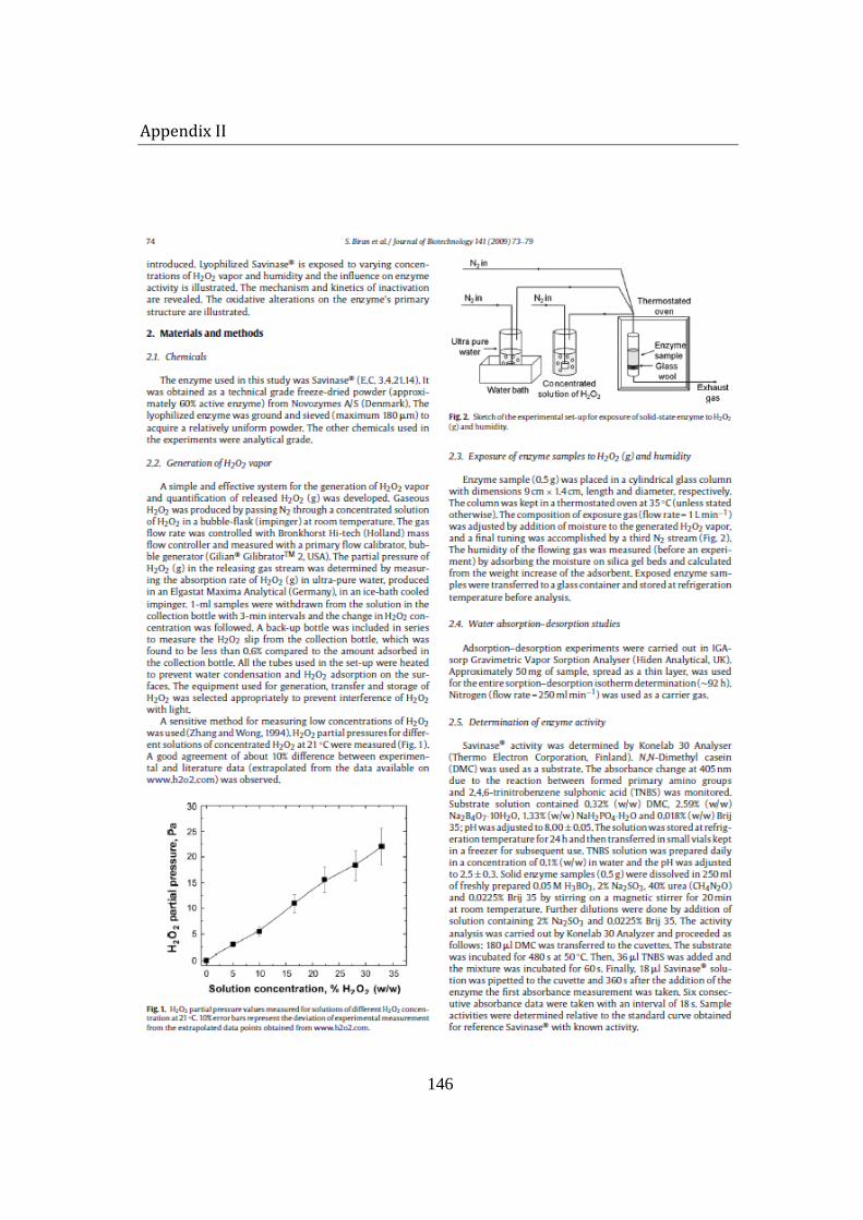
Appendix II
146
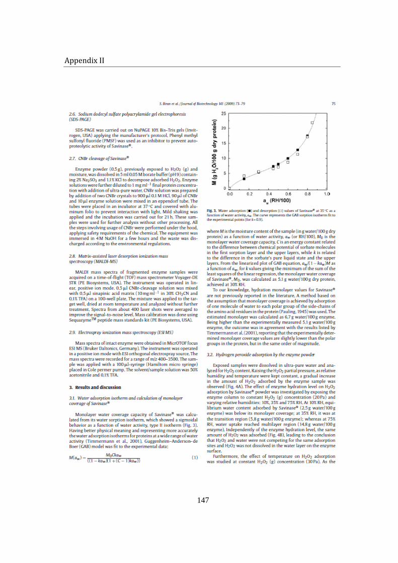
Appendix II
147
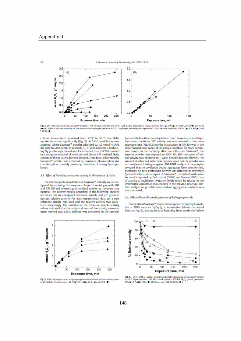
Appendix II
148
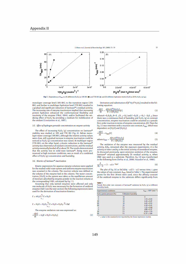
Appendix II
149
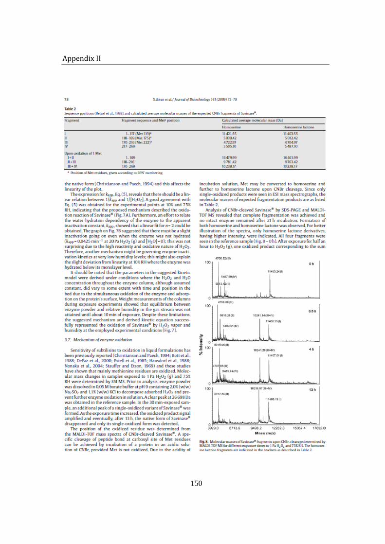
Appendix II
150
Appendix II
151