spontaneous pneumothorax
TRANSCRIPT
BTS guidelines for the management of spontaneouspneumothoraxM Henry, T Arnold, J Harvey, on behalf of the BTS Pleural Disease Group, a subgroupof the BTS Standards of Care Committee. . . . . . . . . . . . . . . . . . . . . . . . . . . . . . . . . . . . . . . . . . . . . . . . . . . . . . . . . . . . . . . . . . . . . . . . . . . . . . . . . . . . . . . . . . . . . . . . . . . . . . . . . . . . . . . . . . . . . . . . . . . . .
Thorax 2003;58(Suppl II):ii39–ii52
1 INTRODUCTIONPneumothorax is defined as air in the pleural
space—that is, between the lung and the chest
wall.1 Primary pneumothoraces arise in otherwise
healthy people without any lung disease. Second-
ary pneumothoraces arise in subjects with under-
lying lung disease. The term pneumothorax was
first coined by Itard, a student of Laennec, in
1803,2 and Laennec himself described the clinical
picture of pneumothorax in 1819.2 He described
most pneumothoraces as occurring in patients
with pulmonary tuberculosis, although he recog-
nised that pneumothoraces also occurred in
otherwise healthy lungs, a condition he described
as “pneumothorax simple”. The modern descrip-
tion of primary spontaneous pneumothorax
occurring in otherwise healthy people was pro-
vided by Kjaergard in 1932.3 Primary pneumotho-
rax remains a significant global problem, occur-
ring in healthy subjects with a reported incidence
of 18–28/100 000 per year for men and 1.2–6/
100 000 per year for women.4 5 Secondary pneu-
mothorax is associated with underlying lung dis-
ease, whereas primary pneumothorax is not. By
definition, there is no apparent precipitating
event in either. Hospital admission rates for com-
bined primary and secondary pneumothorax are
reported in the UK at between 5.8/100 000 per
year for women and 16.7/100 000 per year for
men. Mortality rates in the UK were 0.62/million
per year for women and 1.26/million per year for
men between 1991 and 1995.6 This guideline
describes the management of spontaneous pri-
mary and secondary pneumothorax. It excludes
the management of trauma. Algorithms for the
management of spontaneous primary and sec-
ondary pneumothorax are shown in figs 1 and 2.
• Strong emphasis should be placed on therelationship between the recurrence ofpneumothorax and smoking in an effortto encourage patients to stop smoking.[B]
Despite the absence of underlying pulmonary
disease in patients with primary pneumothorax,
subpleural blebs and bullae are likely to play a role
in the pathogenesis since they are found in up to
90% of cases of primary pneumothorax at thora-
coscopy or thoracotomy and in up to 80% of cases
on CT scanning of the thorax.7 8 The aetiology of
such bullous changes in otherwise apparently
healthy lungs is unclear. Undoubtedly, smoking
plays a role9–11; the lifetime risk of developing a
pneumothorax in healthy smoking men may be
as much as 12% compared with 0.1% in non-
smoking men.11 This trend is also present, though
to a lesser extent, in women.11 There does not
appear to be any relationship between the onset
of pneumothorax and physical activity.5 Small
airway obstruction, mediated by an influx of
inflammatory cells, often characterises pneumo-
thorax and may become manifest in the smaller
airways of men at an earlier stage.12 Patients with
primary pneumothoraces tend to be taller than
control patients.13 14 The gradient in pleural
pressure increases from the lung base to the
apex,1 thus alveoli at the lung apex in tall
individuals are subject to significantly greater
distending pressure than those at the base of the
lung and, theoretically, are more predisposed to
the development of subpleural blebs.15 Strong
emphasis should be placed on the relationship
between smoking and pneumothorax in an effort
to deter those smokers who have developed a
pneumothorax from smoking. Despite the appar-
ent relationship between smoking and pneumo-
thorax, 80–86% of young patients continue to
smoke after their first primary pneumothorax.16
The risk of recurrence of primary pneumothorax
is 54% within the first 4 years, with isolated risk
factors including smoking, height in male
patients,14 17 and age over 60 years.17 Risk factors
for secondary pneumothorax recurrence include
age, pulmonary fibrosis, and emphysema.17 18
In an effort to standardise treatment ofprimary and secondary pneumothoraces, theBritish Thoracic Society (BTS) published guide-lines for the treatment of both in 1993.19 Severalstudies suggest that compliance with the 1993guidelines, though improving, remains at only20–40% among non-respiratory and A&Estaff.20–22 Clinical guidelines have been shown toimprove clinical practice23 24; compliance withguidelines is related to the complexity of practicalprocedures that are described25 and is strength-
ened by an evidence base.26 Steps in the 1993
guidelines which may need clarification include:
(1) simple aspiration (thoracocentesis) versus
intercostal tube drainage (tube thoracostomy) as
the first step in the management of primary and
secondary pneumothoraces, (2) treatment of eld-
erly pneumothorax patients or patients with
underlying lung disease, (3) when to refer
patients with a difficult pneumothorax to a chest
physician or thoracic surgeon for persistent air
leak or failure of re-expansion of the lung, and (4)
treatment of the recurrent pneumothorax. This
guideline addresses these issues. Its purpose is to
provide a comprehensive evidence based review
of the epidemiology, aetiology, and treatment of
pneumothorax to complement the summary
guidelines for diagnosis and treatment.
2 CLINICAL EVALUATION AND IMAGING• Expiratory chest radiographs are not rec-
ommended for the routine diagnosis ofpneumothorax. [B]
See end of article forauthors’ affiliations. . . . . . . . . . . . . . . . . . . . . . .
Correspondence to:Dr M Henry, The GeneralInfirmary at Leeds, GreatGeorge Street, LeedsLS1 3EX, UK;[email protected]. . . . . . . . . . . . . . . . . . . . . . .
ii39
www.thoraxjnl.com
group.bmj.com on December 12, 2011 - Published by thorax.bmj.comDownloaded from
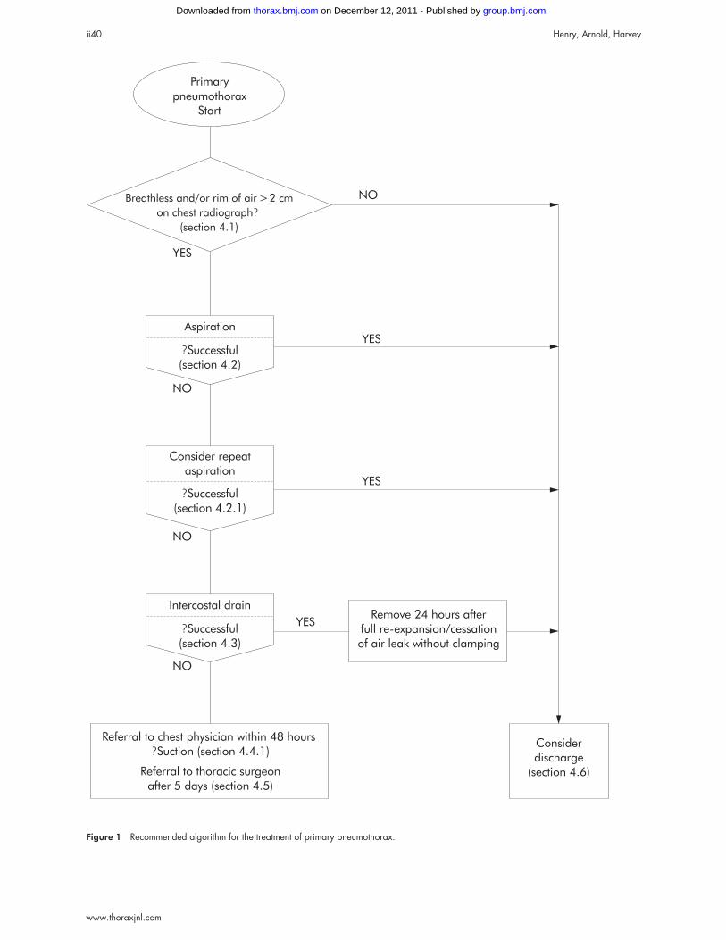
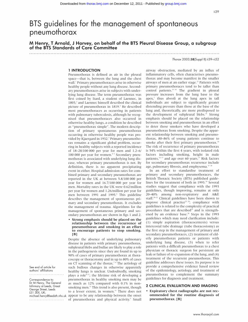
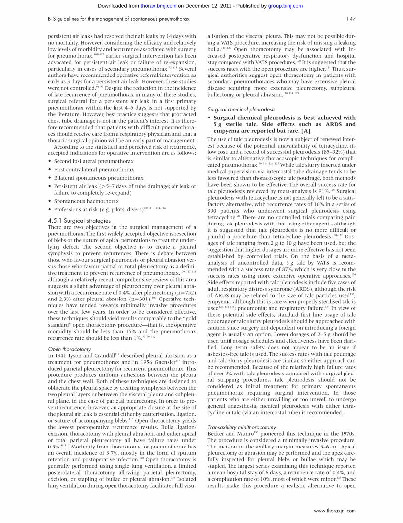
Figure 1 Recommended algorithm for the treatment of primary pneumothorax.
Primary
pneumothorax
Start
Breathless and/or rim of air >2 cm
on chest radiograph?(section 4.1)
Aspiration
Consider repeat
aspiration
?Successful(section 4.2)
Intercostal drain
?Successful(section 4.3)
?Successful(section 4.2.1)
Referral to chest physician within 48 hours
?Suction (section 4.4.1)
Referral to thoracic surgeon
after 5 days (section 4.5)
Consider
discharge
(section 4.6)
Remove 24 hours after
full re-expansion/cessation
of air leak without clamping
NO
YES
YES
YES
YES
NO
NO
NO
ii40 Henry, Arnold, Harvey
www.thoraxjnl.com
group.bmj.com on December 12, 2011 - Published by thorax.bmj.comDownloaded from
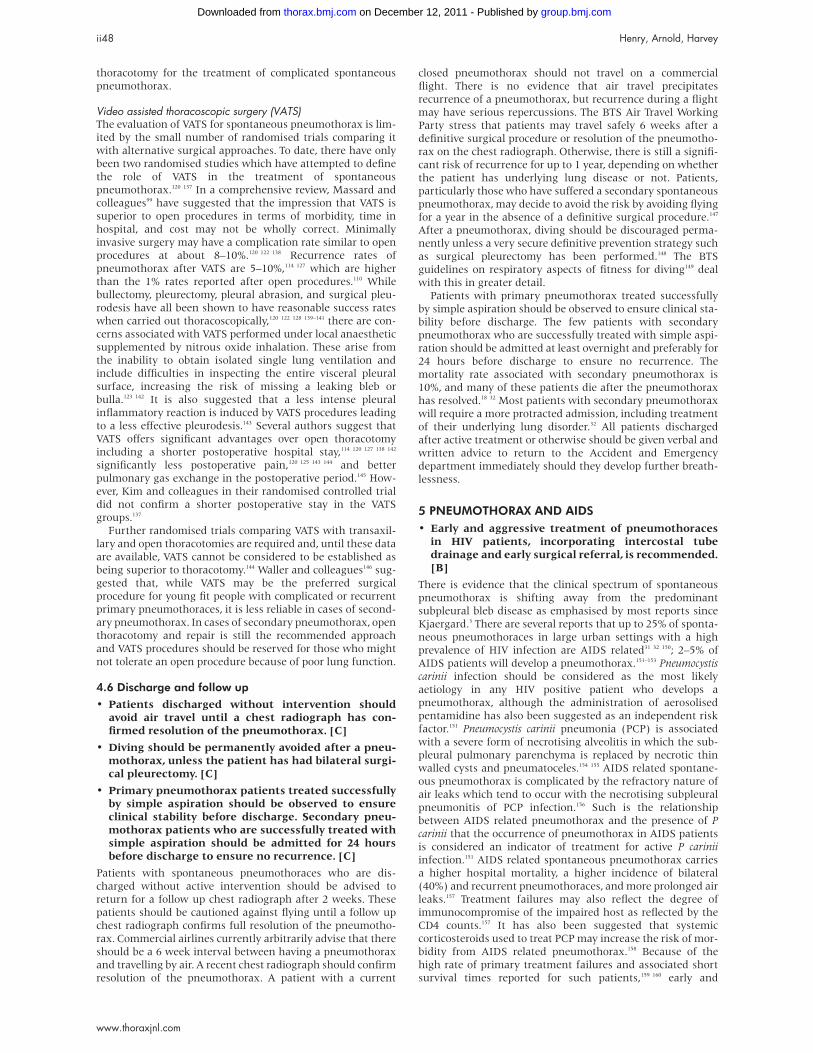
Secondary
pneumothorax
Start
Breathless + age > 50 years
+ rim of air >2 cm
on chest radiograph?(section 4.1)
Intercostal drain
Referral to chest
physician after 48 hours
?Suction (section 4.4.1)
?Successful(section 4.3)
?Successful
Early discussion with
surgeon after 3 days
(section 4.5)
Consider
discharge
(section 4.6)
Admit to
hospital for
24 hours
Aspiration
?Successful(section 4.2)
Remove 24 hours after
full re-expansion/cessation
of air leak
YES
YES
NO
NO
NO
NO
YES YES
Figure 2 Recommended algorithm for the treatment of secondary pneumothorax.
BTS guidelines for the management of spontaneous pneumothorax ii41
www.thoraxjnl.com
group.bmj.com on December 12, 2011 - Published by thorax.bmj.comDownloaded from
• A lateral chest or lateral decubitus radiographshould be performed if the clinical suspicion ofpneumothorax is high, but a PA radiograph isnormal. [B]
• CT scanning is recommended when differentiating apneumothorax from complex bullous lung disease,when aberrant tube placement is suspected, andwhen the plain chest radiograph is obscured bysurgical emphysema. [C]
• The clinical history is not a reliable indicator ofpneumothorax size. [C]
Clinical history and physical examination usually suggest the
presence of a pneumothorax, although clinical manifestations
are not reliable indicators of size.29 30 In general, the clinical
symptoms associated with secondary pneumothoraces are
more severe than those associated with primary pneumotho-
races, and most patients with a secondary pneumothorax
complain of breathlessness which is out of proportion to the
size of the pneumothorax.31 32 Many patients, particularly
those with primary pneumothoraces, do not seek medical
advice for several days, 46% waiting more than 2 days with
symptoms.9 This feature is important because the occurrence
of re-expansion pulmonary oedema (RPO) after re-inflation
may be related to the length of time the lung has been
collapsed (see section 4.4.1).33 34
Arterial blood gas measurements are frequently abnormal
in patients with pneumothorax with the arterial oxygen ten-
sion (PaO2) being less than 10.9 kPa (80 mm Hg) in 75% of
patients.35 The presence of underlying lung disease along with
the size of pneumothorax predicts the degree of
hypoxaemia.35 Arterial PaO2 was below 7.5 kPa (55 mm Hg)
and PaCO2 above 6.9 kPa (50 mm Hg) in 16% of cases of
secondary pneumothorax in the largest reported series.36 Pul-
monary function tests are weakly sensitive measures of the
presence or size of pneumothorax and are not
recommended.8
In both primary and secondary spontaneous pneumothorax
the diagnosis is normally established by plain chest radio-
graphy. In general, expiratory radiographs add little and are
not indicated as a routine investigation, even in the case of a
suspected small apical pneumothorax.37 38 When a pneumo-
thorax is suspected but not confirmed by standard postero-
anterior (PA) chest radiographs, lateral radiographs provide
added information in up to 14% of cases.39 The lateral decubi-
tus radiograph is superior to the erect or supine chest
radiograph and is felt to be as sensitive as CT scanning in
pneumothorax detection.40 The upright lateral or lateral decu-
bitus radiograph is clinically helpful where findings on the
upright PA radiograph are unclear. While such small
pneumothoraces may not have much clinical relevance in
patients without underlying lung disease, in patients with
suspected secondary pneumothoraces, even small pneumo-
thoraces may have significant implications and here lateral or
lateral decubitus radiographs are probably valuable. In
patients with severe bullous lung disease CT scanning will dif-
ferentiate emphysematous bullae from pneumothoraces and
save the patient an unnecessary and potentially dangerous
aspiration.41
3 SIZE OF PNEUMOTHORAX• The previous classification of the size of a pneumo-
thorax tends to underestimate its volume. In thesenew guidelines the size of a pneumothorax is dividedinto “small” or “large” depending on the presence ofa visible rim of <2 cm or >2 cm between the lungmargin and the chest wall.
The plain PA radiograph is a poor method of quantifying the
size of a pneumothorax as it usually underestimates it. Exact
methods of estimating size from PA chest radiographs are
cumbersome and generally used only as a research tool.42 The
size of a pneumothorax, in terms of volume, is difficult to
assess accurately from a chest radiograph which is a two
dimensional image. In the 1993 guidelines19 pneumothoraces
were classified into three groups:
• “small”: defined as a “small rim of air around the lung”;
• “moderate”: defined as lung “collapsed halfway towards
the heart border”; and
• “complete”: defined as “airless lung, separate from the dia-
phragm”.
This attempt to quantify a pneumothorax tends to underesti-
mate the volume of anything greater than the smallest of
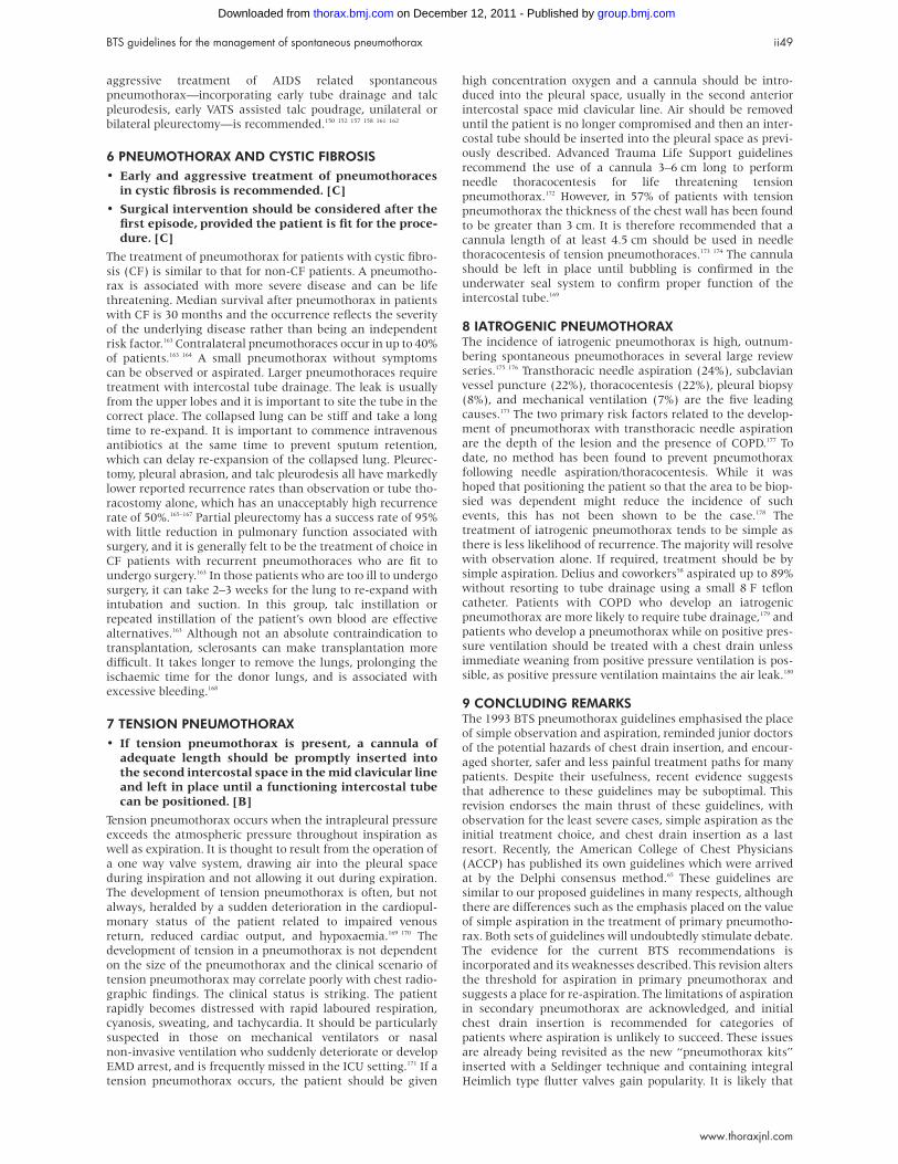
pneumothoraces.1 Since the volume of a pneumothorax
approximates to the ratio of the cube of the of the lung diam-
eter to the hemithorax diameter, a pneumothorax of 1 cm on
the PA chest radiograph occupies about 27% of the hemithorax
volume if the lung is 9 cm in diameter and the hemithorax
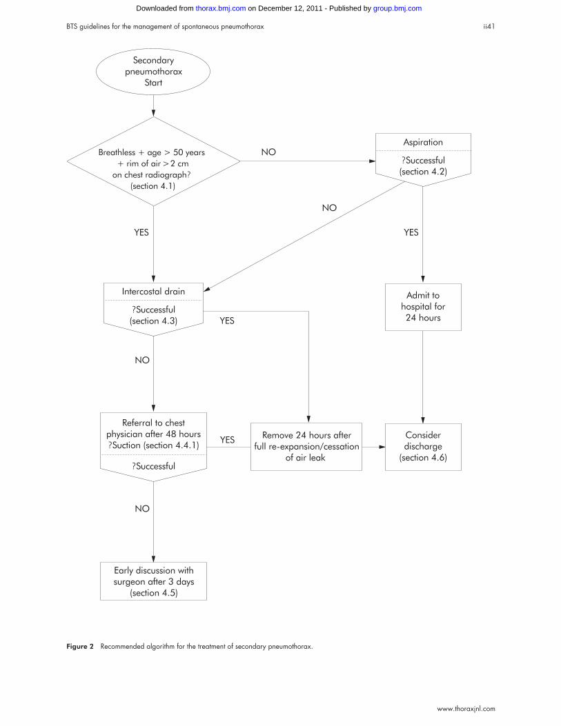
10 cm: (103 – 93)/103 = 27%. Similarly, a 2 cm radiographic
pneumothorax occupies 49% of the hemithorax on the same
basis (fig 3).
In view of the proximity of the lung surface to the chest wall
in a pneumothorax of <1 cm, aspiration using a sharp needle
may not be advisable. However, given that the actual volume of
a 2 cm pneumothorax approximates to a 50% pneumothorax,
this should be considered large in size and can be treated
safely by aspiration when circumstances dictate. For the pur-
poses of these new guidelines, “small” is therefore regarded as
a pneumothorax of <2 cm and “large” as a pneumothorax of
>2 cm.
If accurate size estimates are required, CT scanning is the
most robust approach.43 Otherwise, it is only recommended for
difficult cases such as patients in whom the lungs are obscured
by overlying surgical emphysema, or to differentiate a
pneumothorax from a suspected bulla in complex cystic lung
disease.44 The routine use of CT scans preoperatively in
patients with pneumothorax and suspected emphysema or
isolated bullae adds little to the plain PA chest radiograph
from the point of view of management of the patient.45
4 TREATMENT OPTIONS FOR SPONTANEOUSPNEUMOTHORAX4.1 Observation• Observation should be the treatment of choice for
small closed pneumothoraces without significantbreathlessness. [B]
• Patients with small (<2 cm) primary pneumothora-ces not associated with breathlessness should beconsidered for discharge with early outpatientreview. These patients should receive clear writtenadvice to return in the event of worsening breathless-ness. [B]
1993:Small secondary pneumothoraxTx suggested: simple
aspiration
2003:Volume of pneumothorax
(123_9.53) /123 = 50%Tx suggested: intercostal tube
drainage
9.5 cm
12 cm
Figure 3 Quantitation of size of pneumothorax: 1993 versus 2003guidelines.
ii42 Henry, Arnold, Harvey
www.thoraxjnl.com
group.bmj.com on December 12, 2011 - Published by thorax.bmj.comDownloaded from
• If a patient with a pneumothorax is admittedovernight for observation, high flow (10 l/min) oxy-gen should be administered, with appropriate cau-tion in patients with COPD who may be sensitive tohigher concentrations of oxygen. [B]
• Breathless patients should not be left without inter-vention regardless of the size of the pneumothoraxon a chest radiograph. [C]
4.1.1 Primary pneumothoraces, minimal symptomsObservation alone is advised for small, closed, mildly sympto-
matic spontaneous pneumothoraces.21 30 46–48 70–80% of pneu-
mothoraces estimated at smaller than 15% have no persistent
air leak and recurrence in those managed with observation
alone is less than in patients treated with intercostal tube
drainage.48 Patients with small primary pneumothoraces and
minimal symptoms do not require hospital admission, but it
should be stressed before discharge that they should return
directly to hospital in the event of developing breathlessness.
Most patients in this group who fail this “treatment” and
require intercostal tube drainage have secondary
pneumothoraces.48
4.1.2 Secondary pneumothoraces, minimal symptomsObservation alone is only recommended in patients with small
secondary pneumothoraces of less than 1 cm depth or isolated
apical pneumothoraces in asymptomatic patients. Hospitalisa-
tion is recommended in these cases. All other cases will
require active intervention (aspiration or chest drain inser-
tion, see later sections).
4.1.3 Symptomatic pneumothoraces, primary orsecondaryObservation alone is inappropriate and active intervention is
required. Marked breathlessness in a patient with a small
(<2 cm) primary pneumothorax may herald tension
pneumothorax.48 If a patient is hospitalised for observation,
supplemental high flow (10 l/min) oxygen should be given
where feasible.49 Inhalation of high concentrations of oxygen
may reduce the total pressure of gases in pleural capillaries by
reducing the partial pressure of nitrogen. This should increase
the pressure gradient between the pleural capillaries and the
pleural cavity, thereby increasing absorption of air from the
pleural cavity. The rate of resolution/reabsorption of spontane-
ous pneumothoraces is 1.25–1.8% of the volume of hemi-
thorax every 24 hours.47 50 In a group of 11 patients with
pneumothoraces ranging in size from 16% to 100%, the mean
rate of re-expansion was 1.8% per day and full re-expansion
occurred at a mean of 3.2 weeks.47 A 15% pneumothorax
would therefore take 8–12 days to resolve fully. The addition of
high flow oxygen therapy has been shown to result in a four-
fold increase in the rate of pneumothorax reabsorption during
periods of oxygen supplementation.49
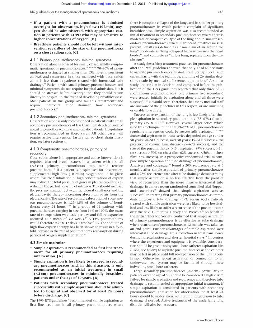
4.2 Simple aspiration• Simple aspiration is recommended as first line treat-
ment for all primary pneumothoraces requiringintervention. [A]
• Simple aspiration is less likely to succeed in second-ary pneumothoraces and, in this situation, is onlyrecommended as an initial treatment in small(<2 cm) pneumothoraces in minimally breathlesspatients under the age of 50 years. [B]
• Patients with secondary pneumothoraces treatedsuccessfully with simple aspiration should be admit-ted to hospital and observed for at least 24 hoursbefore discharge. [C]
The 1993 BTS guidelines19 recommended simple aspiration as
first line treatment in all primary pneumothoraces where
there is complete collapse of the lung, and in smaller primary
pneumothoraces in which patients complain of significant
breathlessness. Simple aspiration was also recommended as
initial treatment in secondary pneumothoraces where there is
moderate or complete collapse of the lung and in smaller sec-
ondary pneumothoraces where significant breathlessness is
present. Small was defined as a “small rim of air around the
lung”, moderate as “lung collapsed halfway towards the heart
border”, and complete as “airless lung, separate from the dia-
phragm”.
A study describing treatment practices for pneumothoraces
after the 1993 guidelines showed that only 17 of 43 decisions
to aspirate pneumothoraces by A&E staff, perhaps because of
unfamiliarity with the technique, and nine of 26 similar deci-
sions made by medical staff seemed appropriate.20 A similar
study undertaken in Scotland and completed before the pub-
lication of the 1993 guidelines reported that only three of 38
spontaneous pneumothoraces (one primary, two secondary)
were treated initially by aspiration alone and all three were
successful.21 It would seem, therefore, that many medical staff
are unaware of the guidelines in this respect, or are unwilling
or unable to aspirate.
Successful re-expansion of the lung is less likely after sim-
ple aspiration in secondary pneumothorax (33–67%) than in
primary (59–83%).51–53 However, several larger series which
used this technique found that 59–73% of all pneumothoraces
requiring intervention could be successfully aspirated.51 52 54–56
Successful aspiration in these series depended on age (under
50 years: 70–81% success, over 50 years: 19–31% success), the
presence of chronic lung disease (27–67% success), and the
size of the pneumothorax (<3 l aspirated: 89% success, >3 l:
no success; >50% on chest film: 62% success, <50% on chest
film: 77% success). In a prospective randomised trial to com-
pare simple aspiration and tube drainage of pneumothoraces,
Andrivert and colleagues55 found a 20% recurrence rate at 3
months after simple aspiration of primary pneumothoraces
and a 28% recurrence rate after tube drainage demonstrating
that simple aspiration is no less effective from the point of
view of recurrence than the more invasive intercostal tube
drainage. In a more recent randomised controlled trial Noppen
and coworkers53 showed that simple aspiration was as
successful in treating first primary pneumothoraces as imme-
diate intercostal tube drainage (59% versus 63%). Patients
treated with simple aspiration were less likely to be hospital-
ised and less likely to suffer a recurrence of the pneumothorax
over the next 12 months. Harvey and Prescott,56 on behalf of
the British Thoracic Society, confirmed that simple aspiration
of primary pneumothoraces is as effective as tube drainage
when recurrence of pneumothorax at 12 months was taken as
an end point. Further advantages of simple aspiration over
intercostal tube drainage are a reduction in total pain scores
during hospitalisation and shorter hospital stays.56 In centres
where the experience and equipment is available, considera-
tion should be give to using small bore catheter aspiration kits
(CASP, see below) to aspirate pneumothoraces as the catheter
may be left in place until full re-expansion of the lung is con-
firmed. Otherwise, repeat aspiration or connection to an
underwater seal system may be facilitated through these
indwelling small bore catheters.
Large secondary pneumothoraces (>2 cm), particularly in
patients over the age of 50, should be considered a high risk of
failure for simple aspiration and recurrence and therefore tube
drainage is recommended as appropriate initial treatment. If
simple aspiration is considered in patients with secondary
pneumothoraces, admission for observation for at least 24
hours should be undertaken, with prompt progression to tube
drainage if needed. Active treatment of the underlying lung
disorder will also be necessary.
BTS guidelines for the management of spontaneous pneumothorax ii43
www.thoraxjnl.com
group.bmj.com on December 12, 2011 - Published by thorax.bmj.comDownloaded from
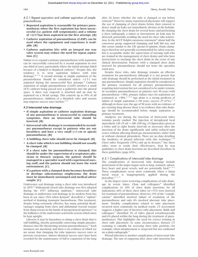
4.2.1 Repeat aspiration and catheter aspiration of simplepneumothorax• Repeated aspiration is reasonable for primary pneu-
mothorax when the first aspiration has been unsuc-cessful (i.e. patient still symptomatic) and a volumeof <2.5 l has been aspirated on the first attempt. [B]
• Catheter aspiration of pneumothorax (CASP) can beused where the equipment and experience is avail-able. [B]
• Catheter aspiration kits with an integral one wayvalve system may reduce the need for repeat aspira-tion. [C]
Failure to re-expand a primary pneumothorax with aspiration
can be successfully corrected by a second aspiration in over
one third of cases, particularly where the initial attempt failed
because of a kinked or displaced catheter.51 Despite this, the
tendency is to treat aspiration failures with tube
drainage.20 51 52 A second attempt at simple aspiration of the
pneumothorax should be considered unless >2.5 l was
aspirated during the unsuccessful first attempt.56 Catheter
aspiration of simple pneumothorax (CASP) involves a small
(8 F) catheter being passed over a guidewire into the pleural
space. A three way stopcock is attached and air may be
aspirated via a 50 ml syringe. This controls up to 59% of all
pneumothoraces. Addition of a Heimlich valve and suction
may improve success rates further.57–59
4.3 Intercostal tube drainage• If simple aspiration or catheter aspiration drainage
of any pneumothorax is unsuccessful in controllingsymptoms, then an intercostal tube should beinserted. [B]
• Intercostal tube drainage is recommended in second-ary pneumothorax except in patients who are notbreathless and have a very small (<1 cm or apical)pneumothorax. [B]
• A bubbling chest tube should never be clamped. [B]
• A chest tube which is not bubbling should not usuallybe clamped. [B]
• If a chest tube for pneumothorax is clamped, thisshould be under the supervision of a respiratory phy-sician or thoracic surgeon, the patient should bemanaged in a specialist ward with experienced nurs-ing staff, and the patient should not leave the wardenvironment. [C]
• If a patient with a clamped drain becomes breathlessor develops subcutaneous emphysema, the drainmust be immediately unclamped and medical advicesought. [C]
Underwater seal drainage using a chest tube was introduced
in 1875.60 Widespread closed tube drainage was first adopted
during the 1917 influenza epidemic.61 Intercostal tube
drainage or underwater seal drainage in its modern form has
been in use since 1916 when Kenyon62 described a “siphon”
method of draining traumatic haemothorax. This treatment,
despite being extremely effective, has many potential disad-
vantages ranging from chest and abdominal visceral trauma
from sharp trocars in the hands of inexperienced operators63 to
the bulkiness of the underwater seal bottle system which must
be kept upright.
Likewise it may be hazardous to clamp a chest drain that is
still bubbling, thereby potentially converting simple pneumo-
thoraces into life threatening tension pneumothoraces.64 Such
instances are anecdotal, and there is no evidence of which we
are aware that clamping the tube improves success rates or
prevents recurrence. Almost identical success rates have been
recorded for the maintenance of full re-expansion of the lung
after 24 hours whether the tube is clamped or not before
removal.80 However, many experienced physicians still support
the use of clamping of chest drains before their removal to
detect small air leaks not immediately obvious at the bedside.
By clamping the chest drain for several hours and performing
a chest radiograph, a minor or intermittent air leak may be
detected, potentially avoiding the need for chest tube reinser-
tion. In the ACCP Delphi consensus statement65 about half the
consensus group supported clamping and half did not, and
this seems similar to the UK spread of opinion. Drain clamp-
ing is therefore not generally recommended for safety reasons,
but is acceptable under the supervision of nursing staff who
are trained in the management of chest drains and who have
instructions to unclamp the chest drain in the event of any
clinical deterioration. Patients with a clamped chest drain
inserted for pneumothorax should not leave the specialist
ward area.
Despite these risks, tube drainage remains an effective
treatment for pneumothorax although it is not proven that
tube drainage should be performed as the initial treatment in
any pneumothorax. Simple aspiration should be considered as
the primary treatment for all primary pneumothoraces
requiring intervention but not considered to be under tension.
In secondary pneumothoraces in patients over 50 years with
pneumothorax >50%, primary failure rates for aspiration are
estimated at >50%.51 52 66 Age alone is a strong predictor of
failure of simple aspiration (>50 years, success 27–67%),51 52
although in those over the age of 50 years with no evidence of
pre-existing lung disease there is no evidence that tube drain-
age should be recommended before simple aspiration as
primary treatment.
Analgesic use during the insertion of intercostal tubes
remains poorly studied. The injection of intrapleural local
anaesthetic (20–25 ml = 200–250 mg, 1% lignocaine) given as
a bolus and at eight hourly intervals as necessary after the
insertion of the drain significantly and safely reduced pain
scores without affecting blood gas measurements, either with
or without chemical pleurodesis. There are no data detailing
the incidence of pleural infection with this technique.67 68
Chest tubes frequently (59%) end up in fissures,69 but these
tubes seem to retain their effectiveness. Step by step
guidelines to chest drain insertion are described elsewhere in
these guidelines (page ii53).
4.3.1 Complications of intercostal tube drainageThe complications of intercostal tube drainage include
penetration of the major organs such as lung, stomach, spleen,
liver, heart and great vessels, and are potentially fatal.63 70–73
These complications occur more commonly when a sharp
metal trocar is inappropriately applied during the
procedure.63 72 73
In the largest series reviewing complications of tube drain-
age in recent times, Chan and colleagues74 identified
complications in 18% of chest drain insertions for all
indications; 64% of these chest tubes (n=373) were inserted
for treatment of pneumothorax. However, 15% of the “compli-
cations” identified involved failure of resolution of the
pneumothorax and only 4% involved aberrant tube place-
ment. Notably, complications related to tube placement
occurred most commonly on medical wards. CT assessment
suggests a higher rate of incorrect tube placement. Baldt and
colleagues75 identified 3% of tubes placed extrathoracically
and 6% placed within the lung during the treatment of pneu-
mothoraces. This highlights the need for correct training in
chest tube placement. In some circumstances, thoracic CT
scanning is valuable for assessing chest tube position—for
example, when misplacement is suspected but not confirmed
on a plain radiograph.75
Pleural infection is another complication of intercostal tube
drainage. The rate of empyema after chest tube insertion has
ii44 Henry, Arnold, Harvey
www.thoraxjnl.com
group.bmj.com on December 12, 2011 - Published by thorax.bmj.comDownloaded from
been estimated as 1%.74 Other series have reported anincidence of up to 6% of chest tube related empyema intrauma cases and suggested that the administration ofprophylactic antibiotics should be considered, particularlywhere a prolonged period of chest tube drainage might beanticipated.76 77 This highlights the need for full aseptictechnique in the insertion or manipulation of any chest drain-age system.
Finally, surgical emphysema is a well recognised complica-tion of intercostal tube drainage.78 This is generally of cosmeticimportance only, subsiding after a few days. The developmentof surgical emphysema associated with pneumothorax in-volves an air filled space, not formerly in communication withthe subcutaneous tissue, being brought into communicationwith the subcutaneous tissues. This may occur in the presenceof a malpositioned, kinked, blocked, or clamped tube.Likewise, a small tube in the presence of a very large leak maypotentially cause surgical emphysema. Occasionally, theresulting acute airway obstruction or thoracic compressionmay lead to respiratory compromise.78 79 The treatment isusually conservative but, in life threatening situations,tracheostomy, skin incision decompression, and insertion oflarge bore modified subcutaneous chest drains have all beenused.78
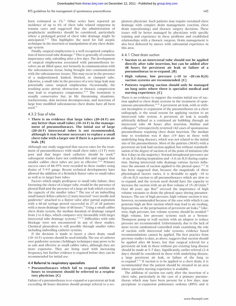
4.3.2 Size of tube• There is no evidence that large tubes (20–24 F) are
any better than small tubes (10–14 F) in the manage-ment of pneumothoraces. The initial use of large(20–24 F) intercostal tubes is not recommended,although it may become necessary to replace a smallchest tube with a larger one if there is a persistent airleak. [B]
Although one study suggested that success rates for the treat-
ment of pneumothoraces with small chest tubes (13 F) were
poor and that larger sized catheters should be used,80
subsequent studies have not confirmed this and suggest that
smaller calibre chest tubes are just as effective.81–84 Primary
success rates of 84–97% were recorded in these studies using
drains of 7–9 F gauge. Recent technical developments have
allowed the addition of a Heimlich flutter valve to small tubes
as well as to larger bore tubes.Factors which might predispose to small tube failure, thus
favouring the choice of a larger tube, would be the presence ofpleural fluid and the presence of a large air leak which exceedsthe capacity of the smaller tubes.82 The use of an indwellingsmall lumen Teflon catheter (2 mm) inserted “over needle andguidewire” attached to a flutter valve after partial aspirationwith a 60 ml syringe proved successful in 27 of 28 patientswith a mean drainage time of 48 hours.59 Using a small calibrechest drain system, the median duration of drainage rangedfrom 2 to 4 days, which compares very favourably with largerintercostal tube drainage systems.59 82 83 Difficulties with tubeblockage were not encountered in any of these studies.Chemical pleurodesis is still possible through smaller tubesincluding indwelling catheter systems.
If the decision is made to insert a chest drain, small(10–14 F) systems should be used initially. The use of catheterover guidewire systems (Seldinger technique) may prove to beas safe and effective as small calibre tubes, although they aremore expensive. They are being used with increasingfrequency, but further evidence is required before they can berecommended for initial use.
4.4 Referral to respiratory specialists• Pneumothoraces which fail to respond within 48
hours to treatment should be referred to a respira-tory physician. [C]
Failure of a pneumothorax to re-expand or a persistent air leak
exceeding 48 hours duration should prompt referral to a res-
piratory physician. Such patients may require sustained chest
drainage with complex drain management (suction, chest
drain repositioning) and thoracic surgery decisions. These
issues will be better managed by physicians with specific
training and experience in these problems and established
relationships with a thoracic surgeon. Drain management is
also best delivered by nurses with substantial experience in
this area.
4.4.1 Chest drain suction• Suction to an intercostal tube should not be applied
directly after tube insertion, but can be added after48 hours for persistent air leak or failure of apneumothorax to re-expand. [B]
• High volume, low pressure (–10 to –20 cm H2O)suction systems are recommended. [C]
• Patients requiring suction should only be managedon lung units where there is specialist medical andnursing experience. [C]
There is no evidence to support the routine initial use of suc-
tion applied to chest drain systems in the treatment of spon-
taneous pneumothorax.80 85 A persistent air leak, with or with-
out incomplete re-expansion of the pneumothorax on a chest
radiograph, is the usual reason for applying suction to an
intercostal tube system. A persistent air leak is usually
arbitrarily defined as a continued air bubbling through an
intercostal tube 48 hours after insertion. Mathur and
colleagues86 retrospectively reviewed 142 cases of spontaneous
pneumothorax requiring chest drain insertion. The median
time to resolution was 8 days (19 days in those with
underlying lung disease), which was not related to the initial
size of the pneumothorax. Most of the patients (30/43) with a
persistent air leak had suction applied, but without standardi-
sation of the degree of suction or of the point of initiation (the
first 4 days in the majority). Normal intrapleural pressures are
–8 cm H2O during inspiration and –3.4 cm H2O during expira-
tion. During intercostal tube drainage various factors influ-
ence the amount of suction applied to the pleural space.63 87 It
has been suggested that, because the magnitude of these
physiological factors varies, it is desirable to apply –10 to
–20 cm H2O suction to all pneumothoraces which are slow to
re-expand, and the system used should have the capacity to
increase the suction with an air flow volume of 15–20 l/min.88
Over 40 years ago Roe89 stressed the importance of high
volume vacuums to drain the pleural space during pneumot-
horaces. The use of high pressure, high volume suction is not,
however, recommended because of the ease with which it can
generate high air flow suction which may lead to air stealing,
hypoxaemia, or the perpetuation of persistent air leaks.90 Like-
wise, high pressure, low volume systems should be avoided.64
High volume, low pressure systems such as a Vernon-
Thompson pump or wall suction with an adaptor to reduce
pressure are recommended. Unfortunately, due to the lack of
more recent randomised controlled trials examining the role
of suction with intercostal tube systems, evidence based
recommendations cannot be applied. The best practice from
previous studies to date, as above, suggests that suction should
be applied after 48 hours, but that surgical referral for a
persistent air leak in those without pre-existing lung disease
should be made at 5–7 days. Significantly, earlier referral (2–4
days) should be considered in those with underlying disease,
a large persistent air leak, or failure of the lung to
re-expand.91–93 If suction is to be applied to a chest drain, it is
recommended that the patient should be situated in an area
where specialist nursing experience is available.
The addition of suction too early after the insertion of a
chest tube, particularly in the case of a primary pneumo-
thorax which may have been present for a few days, may
precipitate re-expansion pulmonary oedema (RPO) and is
BTS guidelines for the management of spontaneous pneumothorax ii45
www.thoraxjnl.com
group.bmj.com on December 12, 2011 - Published by thorax.bmj.comDownloaded from
contraindicated. RPO is probably caused by the increased per-
meability of capillaries damaged during a pneumothorax. This
becomes manifest as oedema during re-expansion due to fur-
ther mechanical stresses applied to the already “leaky”
capillaries.94 Clinically, these patients manifest symptoms of
coughing and breathlessness or chest tightness after insertion
of the chest tube. In those whose symptoms persist, a repeat
chest radiograph after 24 hours will often show pulmonary
oedema in the treated lung, although pulmonary oedema may
also develop in the contralateral lung.95 The incidence of RPO
may be up to 14% and is higher in those with larger primary
pneumothoraces and in younger patients (<30 years),
although in most cases RPO does not progress beyond a radio-
logical phenomenon.96 However, the clinical relevance of RPO
must not be understated as the outcome has been reported as
fatal in 20% of 53 reported cases who developed a clinical
deterioration as part of an RPO syndrome in one series.95 Par-
ticular caution should therefore be exercised in treating young
patients with large pneumothoraces and suction should not be
used immediately in the treatment of a spontaneous
pneumothorax. Even when employed later and it is suspected
that the pneumothorax has been present for a considerable
period of time, the potential development of RPO should be
considered.96
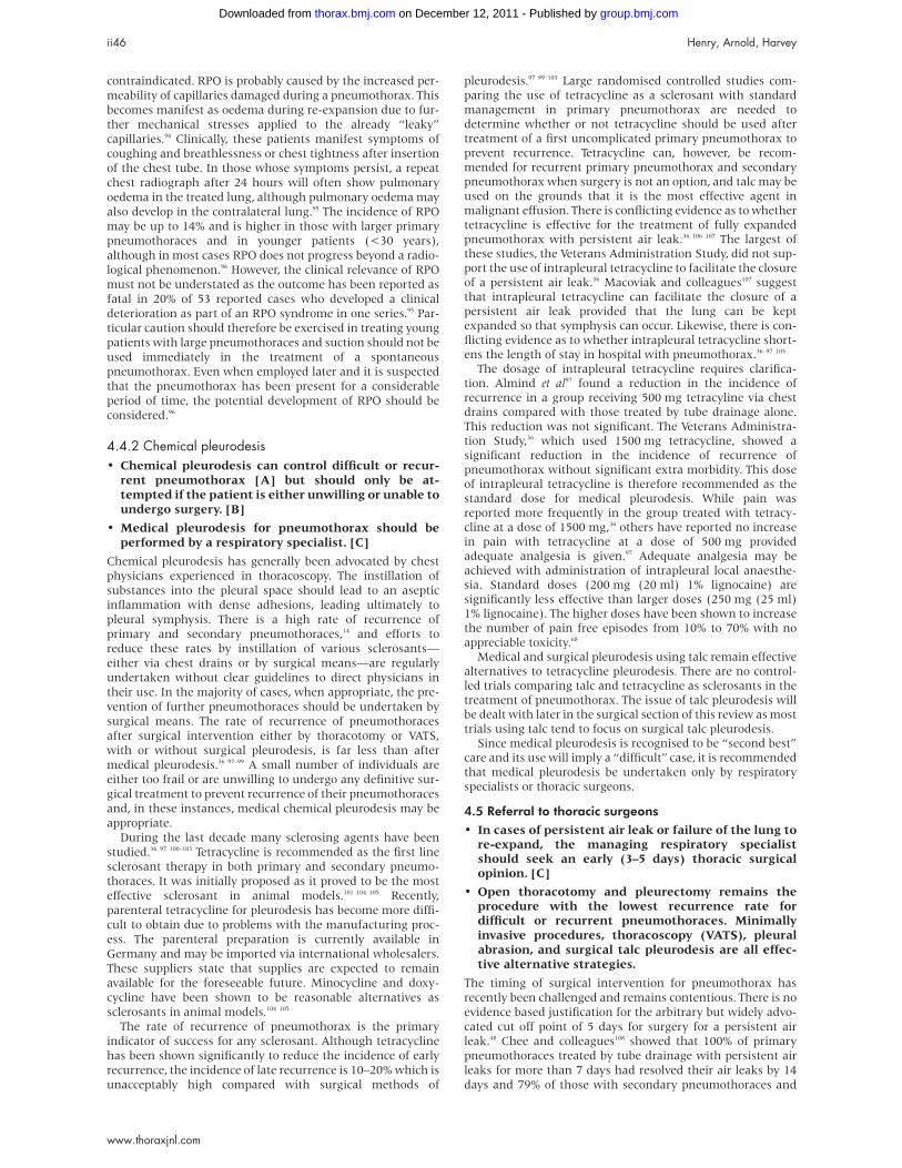
4.4.2 Chemical pleurodesis• Chemical pleurodesis can control difficult or recur-
rent pneumothorax [A] but should only be at-tempted if the patient is either unwilling or unable toundergo surgery. [B]
• Medical pleurodesis for pneumothorax should beperformed by a respiratory specialist. [C]
Chemical pleurodesis has generally been advocated by chest
physicians experienced in thoracoscopy. The instillation of
substances into the pleural space should lead to an aseptic
inflammation with dense adhesions, leading ultimately to
pleural symphysis. There is a high rate of recurrence of
primary and secondary pneumothoraces,14 and efforts to
reduce these rates by instillation of various sclerosants—
either via chest drains or by surgical means—are regularly
undertaken without clear guidelines to direct physicians in
their use. In the majority of cases, when appropriate, the pre-
vention of further pneumothoraces should be undertaken by
surgical means. The rate of recurrence of pneumothoraces
after surgical intervention either by thoracotomy or VATS,
with or without surgical pleurodesis, is far less than after
medical pleurodesis.36 97–99 A small number of individuals are
either too frail or are unwilling to undergo any definitive sur-
gical treatment to prevent recurrence of their pneumothoraces
and, in these instances, medical chemical pleurodesis may be
appropriate.
During the last decade many sclerosing agents have been
studied.36 97 100–103 Tetracycline is recommended as the first line
sclerosant therapy in both primary and secondary pneumo-
thoraces. It was initially proposed as it proved to be the most
effective sclerosant in animal models.101 104 105 Recently,
parenteral tetracycline for pleurodesis has become more diffi-
cult to obtain due to problems with the manufacturing proc-
ess. The parenteral preparation is currently available in
Germany and may be imported via international wholesalers.
These suppliers state that supplies are expected to remain
available for the foreseeable future. Minocycline and doxy-
cycline have been shown to be reasonable alternatives as
sclerosants in animal models.104 105
The rate of recurrence of pneumothorax is the primary
indicator of success for any sclerosant. Although tetracycline
has been shown significantly to reduce the incidence of early
recurrence, the incidence of late recurrence is 10–20% which is
unacceptably high compared with surgical methods of
pleurodesis.97 99 103 Large randomised controlled studies com-paring the use of tetracycline as a sclerosant with standardmanagement in primary pneumothorax are needed todetermine whether or not tetracycline should be used aftertreatment of a first uncomplicated primary pneumothorax toprevent recurrence. Tetracycline can, however, be recom-mended for recurrent primary pneumothorax and secondarypneumothorax when surgery is not an option, and talc may beused on the grounds that it is the most effective agent inmalignant effusion. There is conflicting evidence as to whethertetracycline is effective for the treatment of fully expandedpneumothorax with persistent air leak.36 106 107 The largest ofthese studies, the Veterans Administration Study, did not sup-port the use of intrapleural tetracycline to facilitate the closureof a persistent air leak.36 Macoviak and colleagues107 suggestthat intrapleural tetracycline can facilitate the closure of apersistent air leak provided that the lung can be keptexpanded so that symphysis can occur. Likewise, there is con-flicting evidence as to whether intrapleural tetracycline short-ens the length of stay in hospital with pneumothorax.36 97 103
The dosage of intrapleural tetracycline requires clarifica-tion. Almind et al97 found a reduction in the incidence ofrecurrence in a group receiving 500 mg tetracyline via chestdrains compared with those treated by tube drainage alone.This reduction was not significant. The Veterans Administra-tion Study,36 which used 1500 mg tetracycline, showed asignificant reduction in the incidence of recurrence ofpneumothorax without significant extra morbidity. This doseof intrapleural tetracycline is therefore recommended as thestandard dose for medical pleurodesis. While pain wasreported more frequently in the group treated with tetracy-cline at a dose of 1500 mg,36 others have reported no increasein pain with tetracycline at a dose of 500 mg providedadequate analgesia is given.97 Adequate analgesia may beachieved with administration of intrapleural local anaesthe-sia. Standard doses (200 mg (20 ml) 1% lignocaine) aresignificantly less effective than larger doses (250 mg (25 ml)1% lignocaine). The higher doses have been shown to increasethe number of pain free episodes from 10% to 70% with noappreciable toxicity.68
Medical and surgical pleurodesis using talc remain effectivealternatives to tetracycline pleurodesis. There are no control-led trials comparing talc and tetracycline as sclerosants in thetreatment of pneumothorax. The issue of talc pleurodesis willbe dealt with later in the surgical section of this review as mosttrials using talc tend to focus on surgical talc pleurodesis.
Since medical pleurodesis is recognised to be “second best”care and its use will imply a “difficult” case, it is recommendedthat medical pleurodesis be undertaken only by respiratoryspecialists or thoracic surgeons.
4.5 Referral to thoracic surgeons• In cases of persistent air leak or failure of the lung to
re-expand, the managing respiratory specialistshould seek an early (3–5 days) thoracic surgicalopinion. [C]
• Open thoracotomy and pleurectomy remains theprocedure with the lowest recurrence rate fordifficult or recurrent pneumothoraces. Minimallyinvasive procedures, thoracoscopy (VATS), pleuralabrasion, and surgical talc pleurodesis are all effec-tive alternative strategies.
The timing of surgical intervention for pneumothorax has
recently been challenged and remains contentious. There is no
evidence based justification for the arbitrary but widely advo-
cated cut off point of 5 days for surgery for a persistent air
leak.48 Chee and colleagues108 showed that 100% of primary
pneumothoraces treated by tube drainage with persistent air
leaks for more than 7 days had resolved their air leaks by 14
days and 79% of those with secondary pneumothoraces and
ii46 Henry, Arnold, Harvey
www.thoraxjnl.com
group.bmj.com on December 12, 2011 - Published by thorax.bmj.comDownloaded from
persistent air leaks had resolved their air leaks by 14 days with
no mortality. However, considering the efficacy and relatively
low levels of morbidity and recurrence associated with surgery
for pneumothorax,109–112 earlier surgical intervention has been
advocated for persistent air leak or failure of re-expansion,
particularly in cases of secondary pneumothorax.92 113 Several
authors have recommended operative referral/intervention as
early as 3 days for a persistent air leak. However, these studies
were not controlled.91 93 Despite the reduction in the incidence
of late recurrence of pneumothorax in many of these studies,
surgical referral for a persistent air leak in a first primary
pneumothorax within the first 4–5 days is not supported by
the literature. However, best practice suggests that protracted
chest tube drainage is not in the patient’s interest. It is there-
fore recommended that patients with difficult pneumothora-
ces should receive care from a respiratory physician and that a
thoracic surgical opinion will be an early part of management.According to the statistical and perceived risk of recurrence,
accepted indications for operative intervention are as follows:
• Second ipsilateral pneumothorax
• First contralateral pneumothorax
• Bilateral spontaneous pneumothorax
• Persistent air leak (>5–7 days of tube drainage; air leak orfailure to completely re-expand)
• Spontaneous haemothorax
• Professions at risk (e.g. pilots, divers)108 110 114–116
4.5.1 Surgical strategiesThere are two objectives in the surgical management of a
pneumothorax. The first widely accepted objective is resection
of blebs or the suture of apical perforations to treat the under-
lying defect. The second objective is to create a pleural
symphysis to prevent recurrences. There is debate between
those who favour surgical pleurodesis or pleural abrasion ver-
sus those who favour partial or total pleurectomy as a defini-
tive treatment to prevent recurrence of pneumothorax,109 117 118
although a relatively recent comprehensive review of this area
suggests a slight advantage of pleurectomy over pleural abra-
sion with a recurrence rate of 0.4% after pleurectomy (n=752)
and 2.3% after pleural abrasion (n=301).109 Operative tech-
niques have tended towards minimally invasive procedures
over the last few years. In order to be considered effective,
these techniques should yield results comparable to the “gold
standard” open thoracotomy procedure—that is, the operative
morbidity should be less than 15% and the pneumothorax
recurrence rate should be less than 1%.37 99 112
Open thoracotomyIn 1941 Tyson and Crandall119 described pleural abrasion as a
treatment for pneumothorax and in 1956 Gaensler117 intro-
duced parietal pleurectomy for recurrent pneumothorax. This
procedure produces uniform adhesions between the pleura
and the chest wall. Both of these techniques are designed to
obliterate the pleural space by creating symphysis between the
two pleural layers or between the visceral pleura and subpleu-
ral plane, in the case of parietal pleurectomy. In order to pre-
vent recurrence, however, an appropriate closure at the site of
the pleural air leak is essential either by cauterisation, ligation,
or suture of accompanying blebs.116 Open thoracotomy yields
the lowest postoperative recurrence results. Bulla ligation/
excision, thoracotomy with pleural abrasion, and either apical
or total parietal pleurectomy all have failure rates under
0.5%.48 110 Morbidity from thoracotomy for pneumothorax has
an overall incidence of 3.7%, mostly in the form of sputum
retention and postoperative infection.110 Open thoracotomy is
generally performed using single lung ventilation, a limited
posterolateral thoracotomy allowing parietal pleurectomy,
excision, or stapling of bullae or pleural abrasion.120 Isolated
lung ventilation during open thoracotomy facilitates full visu-
alisation of the visceral pleura. This may not be possible dur-
ing a VATS procedure, increasing the risk of missing a leaking
bulla.121–123 Open thoracotomy may be associated with in-
creased postoperative respiratory dysfunction and hospital
stay compared with VATS procedures.120 It is suggested that the
success rates with the open procedure are higher.124 Thus, sur-
gical authorities suggest open thoracotomy in patients with
secondary pneumothoraces who may have extensive pleural
disease requiring more extensive pleurectomy, subpleural
bullectomy, or pleural abrasion.110 114 125
Surgical chemical pleurodesis• Surgical chemical pleurodesis is best achieved with
5 g sterile talc. Side effects such as ARDS andempyema are reported but rare. [A]
The use of talc pleurodesis is now a subject of renewed inter-
est because of the potential unavailability of tetracycline, its
low cost, and a record of successful pleurodesis (85–92%) that
is similar to alternative thoracoscopic techniques for compli-
cated pneumothorax.99 114 126 127 While talc slurry inserted under
medical supervision via intercostal tube drainage tends to be
less favoured than thoracoscopic talc poudrage, both methods
have been shown to be effective. The overall success rate for
talc pleurodesis reviewed by meta-analysis is 91%.126 Surgical
pleurodesis with tetracycline is not generally felt to be a satis-
factory alternative, with recurrence rates of 16% in a series of
390 patients who underwent surgical pleurodesis using
tetracycline.98 There are no controlled trials comparing pain
during talc pleurodesis with that using other agents, although
it is suggested that talc pleurodesis is no more difficult or
painful a procedure than tetracycline pleurodesis.128–132 Dos-
ages of talc ranging from 2 g to 10 g have been used, but the
suggestion that higher dosages are more effective has not been
established by controlled trials. On the basis of a meta-
analysis of uncontrolled data, 5 g talc by VATS is recom-
mended with a success rate of 87%, which is very close to the
success rates using more extensive operative approaches.126
Side effects reported with talc pleurodesis include five cases of
adult respiratory distress syndrome (ARDS), although the risk
of ARDS may be related to the size of talc particles used133;
empyema, although this is rare when properly sterilised talc is
used126 134 135; pneumonia; and respiratory failure.134 In view of
these potential side effects, standard first line usage of talc
poudrage or talc slurry pleurodesis should be approached with
caution since surgery not dependent on introducing a foreign
agent is usually an option. Lower dosages of 2–5 g should be
used until dosage schedules and effectiveness have been clari-
fied. Long term safety does not appear to be an issue if
asbestos-free talc is used. The success rates with talc poudrage
and talc slurry pleurodesis are similar, so either approach can
be recommended. Because of the relatively high failure rates
of over 9% with talc pleurodesis compared with surgical pleu-
ral stripping procedures, talc pleurodesis should not be
considered as initial treatment for primary spontaneous
pneumothorax requiring surgical intervention. In those
patients who are either unwilling or too unwell to undergo
general anaesthesia, medical pleurodesis with either tetra-
cycline or talc (via an intercostal tube) is recommended.
Transaxillary minithoracotomyBecker and Munro136 pioneered this technique in the 1970s.
The procedure is considered a minimally invasive procedure.
The incision in the axillary margin measures 5–6 cm. Apical
pleurectomy or abrasion may be performed and the apex care-
fully inspected for pleural blebs or bullae which may be
stapled. The largest series examining this technique reported
a mean hospital stay of 6 days, a recurrence rate of 0.4%, and
a complication rate of 10%, most of which were minor.125 These
results make this procedure a realistic alternative to open
BTS guidelines for the management of spontaneous pneumothorax ii47
www.thoraxjnl.com
group.bmj.com on December 12, 2011 - Published by thorax.bmj.comDownloaded from
thoracotomy for the treatment of complicated spontaneous
pneumothorax.
Video assisted thoracoscopic surgery (VATS)The evaluation of VATS for spontaneous pneumothorax is lim-
ited by the small number of randomised trials comparing it
with alternative surgical approaches. To date, there have only
been two randomised studies which have attempted to define
the role of VATS in the treatment of spontaneous
pneumothorax.120 137 In a comprehensive review, Massard and
colleagues99 have suggested that the impression that VATS is
superior to open procedures in terms of morbidity, time in
hospital, and cost may not be wholly correct. Minimally
invasive surgery may have a complication rate similar to open
procedures at about 8–10%.120 122 138 Recurrence rates of
pneumothorax after VATS are 5–10%,114 127 which are higher
than the 1% rates reported after open procedures.110 While
bullectomy, pleurectomy, pleural abrasion, and surgical pleu-
rodesis have all been shown to have reasonable success rates
when carried out thoracoscopically,120 122 128 139–141 there are con-
cerns associated with VATS performed under local anaesthetic
supplemented by nitrous oxide inhalation. These arise from
the inability to obtain isolated single lung ventilation and
include difficulties in inspecting the entire visceral pleural
surface, increasing the risk of missing a leaking bleb or
bulla.123 142 It is also suggested that a less intense pleural
inflammatory reaction is induced by VATS procedures leading
to a less effective pleurodesis.143 Several authors suggest that
VATS offers significant advantages over open thoracotomy
including a shorter postoperative hospital stay,114 120 127 138 142
significantly less postoperative pain,120 125 143 144 and better
pulmonary gas exchange in the postoperative period.145 How-
ever, Kim and colleagues in their randomised controlled trial
did not confirm a shorter postoperative stay in the VATS
groups.137
Further randomised trials comparing VATS with transaxil-
lary and open thoracotomies are required and, until these data
are available, VATS cannot be considered to be established as
being superior to thoracotomy.144 Waller and colleagues146 sug-
gested that, while VATS may be the preferred surgical
procedure for young fit people with complicated or recurrent
primary pneumothoraces, it is less reliable in cases of second-
ary pneumothorax. In cases of secondary pneumothorax, open
thoracotomy and repair is still the recommended approach
and VATS procedures should be reserved for those who might
not tolerate an open procedure because of poor lung function.
4.6 Discharge and follow up• Patients discharged without intervention should
avoid air travel until a chest radiograph has con-firmed resolution of the pneumothorax. [C]
• Diving should be permanently avoided after a pneu-mothorax, unless the patient has had bilateral surgi-cal pleurectomy. [C]
• Primary pneumothorax patients treated successfullyby simple aspiration should be observed to ensureclinical stability before discharge. Secondary pneu-mothorax patients who are successfully treated withsimple aspiration should be admitted for 24 hoursbefore discharge to ensure no recurrence. [C]
Patients with spontaneous pneumothoraces who are dis-
charged without active intervention should be advised to
return for a follow up chest radiograph after 2 weeks. These
patients should be cautioned against flying until a follow up
chest radiograph confirms full resolution of the pneumotho-
rax. Commercial airlines currently arbitrarily advise that there
should be a 6 week interval between having a pneumothorax
and travelling by air. A recent chest radiograph should confirm
resolution of the pneumothorax. A patient with a current
closed pneumothorax should not travel on a commercial
flight. There is no evidence that air travel precipitates
recurrence of a pneumothorax, but recurrence during a flight
may have serious repercussions. The BTS Air Travel Working
Party stress that patients may travel safely 6 weeks after a
definitive surgical procedure or resolution of the pneumotho-
rax on the chest radiograph. Otherwise, there is still a signifi-
cant risk of recurrence for up to 1 year, depending on whether
the patient has underlying lung disease or not. Patients,
particularly those who have suffered a secondary spontaneous
pneumothorax, may decide to avoid the risk by avoiding flying
for a year in the absence of a definitive surgical procedure.147
After a pneumothorax, diving should be discouraged perma-
nently unless a very secure definitive prevention strategy such
as surgical pleurectomy has been performed.148 The BTS
guidelines on respiratory aspects of fitness for diving149 deal
with this in greater detail.
Patients with primary pneumothorax treated successfully
by simple aspiration should be observed to ensure clinical sta-
bility before discharge. The few patients with secondary
pneumothorax who are successfully treated with simple aspi-
ration should be admitted at least overnight and preferably for
24 hours before discharge to ensure no recurrence. The
mortality rate associated with secondary pneumothorax is
10%, and many of these patients die after the pneumothorax
has resolved.18 32 Most patients with secondary pneumothorax
will require a more protracted admission, including treatment
of their underlying lung disorder.32 All patients discharged
after active treatment or otherwise should be given verbal and
written advice to return to the Accident and Emergency
department immediately should they develop further breath-
lessness.
5 PNEUMOTHORAX AND AIDS• Early and aggressive treatment of pneumothoraces
in HIV patients, incorporating intercostal tubedrainage and early surgical referral, is recommended.[B]
There is evidence that the clinical spectrum of spontaneous
pneumothorax is shifting away from the predominant
subpleural bleb disease as emphasised by most reports since
Kjaergard.3 There are several reports that up to 25% of sponta-
neous pneumothoraces in large urban settings with a high
prevalence of HIV infection are AIDS related31 32 150; 2–5% of
AIDS patients will develop a pneumothorax.151–153 Pneumocystiscarinii infection should be considered as the most likely
aetiology in any HIV positive patient who develops a
pneumothorax, although the administration of aerosolised
pentamidine has also been suggested as an independent risk
factor.151 Pneumocystis carinii pneumonia (PCP) is associated
with a severe form of necrotising alveolitis in which the sub-
pleural pulmonary parenchyma is replaced by necrotic thin
walled cysts and pneumatoceles.154 155 AIDS related spontane-
ous pneumothorax is complicated by the refractory nature of
air leaks which tend to occur with the necrotising subpleural
pneumonitis of PCP infection.156 Such is the relationship
between AIDS related pneumothorax and the presence of Pcarinii that the occurrence of pneumothorax in AIDS patients
is considered an indicator of treatment for active P cariniiinfection.151 AIDS related spontaneous pneumothorax carries
a higher hospital mortality, a higher incidence of bilateral
(40%) and recurrent pneumothoraces, and more prolonged air
leaks.157 Treatment failures may also reflect the degree of
immunocompromise of the impaired host as reflected by the
CD4 counts.157 It has also been suggested that systemic
corticosteroids used to treat PCP may increase the risk of mor-
bidity from AIDS related pneumothorax.158 Because of the
high rate of primary treatment failures and associated short
survival times reported for such patients,159 160 early and
ii48 Henry, Arnold, Harvey
www.thoraxjnl.com
group.bmj.com on December 12, 2011 - Published by thorax.bmj.comDownloaded from
aggressive treatment of AIDS related spontaneous
pneumothorax—incorporating early tube drainage and talc
pleurodesis, early VATS assisted talc poudrage, unilateral or
bilateral pleurectomy—is recommended.150 152 157 158 161 162
6 PNEUMOTHORAX AND CYSTIC FIBROSIS• Early and aggressive treatment of pneumothoraces
in cystic fibrosis is recommended. [C]
• Surgical intervention should be considered after thefirst episode, provided the patient is fit for the proce-dure. [C]
The treatment of pneumothorax for patients with cystic fibro-
sis (CF) is similar to that for non-CF patients. A pneumotho-
rax is associated with more severe disease and can be life
threatening. Median survival after pneumothorax in patients
with CF is 30 months and the occurrence reflects the severity
of the underlying disease rather than being an independent
risk factor.163 Contralateral pneumothoraces occur in up to 40%
of patients.163 164 A small pneumothorax without symptoms
can be observed or aspirated. Larger pneumothoraces require
treatment with intercostal tube drainage. The leak is usually
from the upper lobes and it is important to site the tube in the
correct place. The collapsed lung can be stiff and take a long
time to re-expand. It is important to commence intravenous
antibiotics at the same time to prevent sputum retention,
which can delay re-expansion of the collapsed lung. Pleurec-
tomy, pleural abrasion, and talc pleurodesis all have markedly
lower reported recurrence rates than observation or tube tho-
racostomy alone, which has an unacceptably high recurrence
rate of 50%.165–167 Partial pleurectomy has a success rate of 95%
with little reduction in pulmonary function associated with
surgery, and it is generally felt to be the treatment of choice in
CF patients with recurrent pneumothoraces who are fit to
undergo surgery.163 In those patients who are too ill to undergo
surgery, it can take 2–3 weeks for the lung to re-expand with
intubation and suction. In this group, talc instillation or
repeated instillation of the patient’s own blood are effective
alternatives.163 Although not an absolute contraindication to
transplantation, sclerosants can make transplantation more
difficult. It takes longer to remove the lungs, prolonging the
ischaemic time for the donor lungs, and is associated with
excessive bleeding.168
7 TENSION PNEUMOTHORAX• If tension pneumothorax is present, a cannula of
adequate length should be promptly inserted intothe second intercostal space in the mid clavicular lineand left in place until a functioning intercostal tubecan be positioned. [B]
Tension pneumothorax occurs when the intrapleural pressure
exceeds the atmospheric pressure throughout inspiration as
well as expiration. It is thought to result from the operation of
a one way valve system, drawing air into the pleural space
during inspiration and not allowing it out during expiration.
The development of tension pneumothorax is often, but not
always, heralded by a sudden deterioration in the cardiopul-
monary status of the patient related to impaired venous
return, reduced cardiac output, and hypoxaemia.169 170 The
development of tension in a pneumothorax is not dependent
on the size of the pneumothorax and the clinical scenario of
tension pneumothorax may correlate poorly with chest radio-
graphic findings. The clinical status is striking. The patient
rapidly becomes distressed with rapid laboured respiration,
cyanosis, sweating, and tachycardia. It should be particularly
suspected in those on mechanical ventilators or nasal
non-invasive ventilation who suddenly deteriorate or develop
EMD arrest, and is frequently missed in the ICU setting.171 If a
tension pneumothorax occurs, the patient should be given
high concentration oxygen and a cannula should be intro-
duced into the pleural space, usually in the second anterior
intercostal space mid clavicular line. Air should be removed
until the patient is no longer compromised and then an inter-
costal tube should be inserted into the pleural space as previ-
ously described. Advanced Trauma Life Support guidelines
recommend the use of a cannula 3–6 cm long to perform
needle thoracocentesis for life threatening tension
pneumothorax.172 However, in 57% of patients with tension
pneumothorax the thickness of the chest wall has been found
to be greater than 3 cm. It is therefore recommended that a
cannula length of at least 4.5 cm should be used in needle
thoracocentesis of tension pneumothoraces.173 174 The cannula
should be left in place until bubbling is confirmed in the
underwater seal system to confirm proper function of the
intercostal tube.169
8 IATROGENIC PNEUMOTHORAXThe incidence of iatrogenic pneumothorax is high, outnum-
bering spontaneous pneumothoraces in several large review
series.175 176 Transthoracic needle aspiration (24%), subclavian
vessel puncture (22%), thoracocentesis (22%), pleural biopsy
(8%), and mechanical ventilation (7%) are the five leading
causes.173 The two primary risk factors related to the develop-
ment of pneumothorax with transthoracic needle aspiration
are the depth of the lesion and the presence of COPD.177 To
date, no method has been found to prevent pneumothorax
following needle aspiration/thoracocentesis. While it was
hoped that positioning the patient so that the area to be biop-
sied was dependent might reduce the incidence of such
events, this has not been shown to be the case.178 The
treatment of iatrogenic pneumothorax tends to be simple as
there is less likelihood of recurrence. The majority will resolve
with observation alone. If required, treatment should be by
simple aspiration. Delius and coworkers58 aspirated up to 89%
without resorting to tube drainage using a small 8 F teflon
catheter. Patients with COPD who develop an iatrogenic
pneumothorax are more likely to require tube drainage,179 and
patients who develop a pneumothorax while on positive pres-
sure ventilation should be treated with a chest drain unless
immediate weaning from positive pressure ventilation is pos-
sible, as positive pressure ventilation maintains the air leak.180
9 CONCLUDING REMARKSThe 1993 BTS pneumothorax guidelines emphasised the place
of simple observation and aspiration, reminded junior doctors
of the potential hazards of chest drain insertion, and encour-
aged shorter, safer and less painful treatment paths for many
patients. Despite their usefulness, recent evidence suggests
that adherence to these guidelines may be suboptimal. This
revision endorses the main thrust of these guidelines, with
observation for the least severe cases, simple aspiration as the
initial treatment choice, and chest drain insertion as a last
resort. Recently, the American College of Chest Physicians
(ACCP) has published its own guidelines which were arrived
at by the Delphi consensus method.65 These guidelines are
similar to our proposed guidelines in many respects, although
there are differences such as the emphasis placed on the value
of simple aspiration in the treatment of primary pneumotho-
rax. Both sets of guidelines will undoubtedly stimulate debate.
The evidence for the current BTS recommendations is
incorporated and its weaknesses described. This revision alters
the threshold for aspiration in primary pneumothorax and
suggests a place for re-aspiration. The limitations of aspiration
in secondary pneumothorax are acknowledged, and initial
chest drain insertion is recommended for categories of
patients where aspiration is unlikely to succeed. These issues
are already being revisited as the new “pneumothorax kits”
inserted with a Seldinger technique and containing integral
Heimlich type flutter valves gain popularity. It is likely that
BTS guidelines for the management of spontaneous pneumothorax ii49
www.thoraxjnl.com
group.bmj.com on December 12, 2011 - Published by thorax.bmj.comDownloaded from
they will prove to be as successful and possibly replace simple
aspiration followed by immediate removal of the catheter as
recommended in these guidelines.
Several aspects of management that were not previously
covered are now included. These include the place of CT scan-
ning in diagnosis, which patients to refer for surgery, a discus-
sion of surgical techniques, and issues such as intercostal tube
size and the place of suction and pleurodesis. Complex
scenarios including tension pneumothorax, subcutaneous
emphysema, pneumothorax in HIV disease and adult CF are
also discussed. It is hoped that these changes build on the
clinical benefits produced by the first set of guidelines
which—if adhered to—should, we calculate, prevent approxi-
mately 7000 unnecessary chest drain insertions every year in
the UK.
. . . . . . . . . . . . . . . . . . . . .Authors’ affiliationsM Henry, Department of Respiratory Medicine, The General Infirmary atLeeds, Great George Street, Leeds LS1 3EX, UKT Arnold, Medical Chest Unit, Castle Hill Hospital, Cottingham, NorthHumberside HU16 5JQ, UKJ Harvey, Department of Respiratory Medicine, Southmead Hospital,Bristol BS10 5NB, UK
REFERENCES1 Light RW. Pneumothorax. In: Pleural diseases. 3rd ed. Baltimore:
Williams and Wilkins, 1995: 242–77.2 Laennec RTH. Trait de l’auscultation mediate et des maladies des
poumons et du coeur. Tome Second. Paris, 1819.3 Kjaergard H. Spontaneous pneumothorax in the apparently healthy.
Acta Med Scand (Suppl) 1932;43:1–159.4 Melton LJ, Hepper NCG, Offord KP. Incidence of spontaneous
pneumothorax in Olmsted County, Minnesota: 1950–1974. Am RevRespir Dis 1979;29:1379–82. [III]
5 Bense L, Wiman LG, Hedenstierna G. Onset of symptoms inspontaneous pneumothorax: correlations to physical activity. Eur J RespirDis 1987;71:181–6. [III]
6 Gupta D, Hansell A, Nichols T, et al. Epidemiology of pneumothorax inEngland. Thorax 2000;55:666–71. [III]
7 Donahue DM, Wright CD, Viale G, et al. Resection of pulmonary blebsand pleurodesis for spontaneous pneumothorax. Chest1993;104:1767–9. [IIb]
8 Lesur O, Delorme N, Frogamet JM, et al. Computed tomography in theaetiological assessment of idiopathic spontaneous pneumothorax. Chest1990;98:341–7. [IIa]
9 O’Hara VS. Spontaneous pneumothorax. Milit Med 1978;143:32–5.[III]
10 Jansveld CA, Dijkman JH. Primary spontaneous pneumothorax andsmoking. BMJ 1975;4:559–60. [IIa]
11 Bense L, Eklund G, Odont D, et al. Smoking and the increased risk ofcontracting pneumothorax. Chest 1987;92:1009–12. [IIa]
12 Schramel FM, Meyer CJ, Postmus PE. Inflammation as a cause ofspontaneous pneumothorax and emphysematous-like changes: results ofbronchoalveolar lavage. Eur Respir J 1995;8:397s. [IIb]
13 Withers JN, Fishback M.E, Kiehl PV, et al. Spontaneous pneumothorax.Am J Surg 1964;108:772–6. [IV]
14 Sadikot RT, Greene T, Meadows K, et al. Recurrence of primarypneumothorax. Thorax 1997;52:805–9. [III]
15 West JB. Distribution of mechanical stress in the lung, a possible factor inlocalisation of pulmonary disease. Lancet 1971;i:839–41. [IIb]
16 Smit HJM, Chatrou M, Postmus PE. The impact of spontaneouspneumothorax and its treatment on the smoking behaviour of young adultsmokers. Respir Med 1998;92:1132–6. [III]
17 Lippert HL, Lund O, Blegrad S, et al. Independent risk factors forcumulative recurrence rate after first spontaneous pneumothorax. EurRespir J 1991;4:324–31. [III]
18 Videm V, Pillgram-Larsen J, Ellingsen Ø, et al. Spontaneouspneumothorax in chronic obstructive pulmonary disease: complications,treatment and recurrences. Eur J Respir Dis 1987;71:365–71. [III]
19 Miller AC, Harvey JE. Guidelines for the management of spontaneouspneumothorax. BMJ. 1993;307:114–6. [IV]
20 Soulsby T. British Thoracic Society guidelines for the management ofspontaneous pneumothorax: do we comply with them and do they work?J Accid Emerg Med 1998;15:317–21. [III]
21 Selby CD, Sudlow MF. Deficiencies in the management of spontaneouspneumothoraces. Scot Med J 1994;39:75–6. [III]
22 Yeoh JH, Ansari S, Campbell IA. Management of spontaneouspneumothorax : a Welsh survey. Postgrad Med J 2000;76:496–9. [III]
23 Grimshaw JM, Russell IT. Effect of clinical guidelines on medicalpractice: a systematic review of rigorous evaluation. Lancet1993;342:1317–21. [IIb]
24 Bero LA, Grilli R, Grimshaw JM, et al. Closing the gap between researchand practice: an overview of systematic reviews of interventions topromote the implementation of research findings. BMJ 1998;317:465–8.[Review]
25 Grilli R, Lomas J. Evaluating the message: the relationship betweencompliance rate and the subject of a practice guideline. Med Care1994;32:202–13. [IV]
26 Woolf SH, Grol R, Hutchinson A, et al. Potential benefits, limitations andharms of clinical guidelines. BMJ 1999;318:527–30.
27 Agency for Health Care Policy and Research. Acute painmanagement, operative or medical procedures and trauma. Clinicalpractice guidelines. Publication no. 92-0032. Rockville, Maryland, USA:Agency for Healthcare Policy and Research Publications, 1992.
28 Petrie GJ, Barnwell E, Grimshaw J, on behalf of the ScottishIntercollegiate Guidelines Network. Clinical guidelines criteria forappraisal for national use. Edinburgh: Royal College of Physicians,1995.
29 Vail WJ, Alway AE, England NJ. Spontaneous pneumothorax. Dis Chest1963;38:512–5. [III]
30 Serementis MG. The management of spontaneous pneumothorax. Chest1970;57:65–8. [III]
31 Wait MA, Estrera A. Changing clinical spectrum of spontaneouspneumothorax. Am J Surg 1992;164:528–31. [III]
32 Tanaka F, Itoh M, Esaki H, et al. Secondary spontaneous pneumothorax.Ann Thorac Surg 1993;55:372–6. [III]
33 Pavlin DJ, Cheney FW Jr. Unilateral pulmonary edema in rabbits afterre-expansion of collapsed lung. J Appl Physiol 1979;46:31–5. [IIb]
34 Miller WC, Toon R, Palat H, et al. Experimental pulmonary edemafollowing re-expansion of pneumothorax. Am Rev Respir Dis1973;108:664–6. [Ib]
35 Norris RM, Jones JG, Bishop JM. Respiratory gas exchange in patientswith spontaneous pneumothorax. Thorax 1968;23:427–33. [IIb]
36 Light RW, O’Hara VS, Moritz TE, et al. Intrapleural tetracycline for theprevention of recurrent spontaneous pneumothorax: results of aDepartment of Veterans Affairs Co-operative Study. JAMA1990;264:2224–30. [Ib]
37 Schramel FM, Wagenaar M, Sutedja TA, et al. Diagnosis ofpneumothorax not improved by additional roentgen pictures of the thoraxin the expiratory phase. Ned Tijdschr Geneesk 1995;139:131–3. [IIb]
38 Schramel FM, Golding RP, Haakman CD, et al. Expiratory chestradiographs do not improve visibility of small apical pneumothoraces byenhanced contrast. Eur Resp J 1996;9:406–9. [IIb]
39 Glazer H, Anderson DJ, Wilson BS, et al. Pneumothorax: appearanceson lateral chest radiographs. Radiology 1989;173:707–11. [III]
40 Carr JJ, Reed JC, Choplin RH, et al. Plain and computed radiographyfor detecting experimentally induced pneumothorax in cadavers:implications for detection in patients. Radiololgy 1992;183:193–9. [IIb]
41 Bourgouin P, Cousineau G, Lemire P, et al. Computed tomography usedto exclude pneumothorax in bullous lung disease. J Can Ass Radiol1985;36:341–2. [IV]
42 Collins CD, Lopez A, Mathie A, et al. Quantification of pneumothoraxsize on chest radiographs using intrapleural distances: regressionanalysis based on volume measurements from helical CT. AJR1995;165:1127–30. [III]
Audit points
• Proportion of patients treated by (a) simple observation, (b)aspiration, and (c) chest drains and their appropriateness(relative to the guidelines) and outcome (in terms ofrecurrence rates, complications, and lengths of stay in hos-pital)
• Number of chest drains clamped and the reasons for this.• Referral rates to physicians and surgeons and the timing of
such referrals.• Use of analgesics and local anaesthetics.• Follow up rates
Future potential areas for research
• Prospective randomised controlled trials comparing:• simple observation versus aspiration ± tube drainage
for primary pneumothoraces larger than 2 cm on thechest radiograph;
• use of small catheter/Heimlich valve kits versus inter-costal tube drainage following failed aspiration inprimary pneumothoraces;
• small catheter aspiration (CASP) versus conventionalaspiration or tube drainage;
• VATS versus open thoracotomy for the difficult pneu-mothorax.
• Use of suction with regard to its timing and optimal mode.• Comparison of “clamping” and “ non-clamping” strategies
after cessation of air leak.
ii50 Henry, Arnold, Harvey
www.thoraxjnl.com
group.bmj.com on December 12, 2011 - Published by thorax.bmj.comDownloaded from
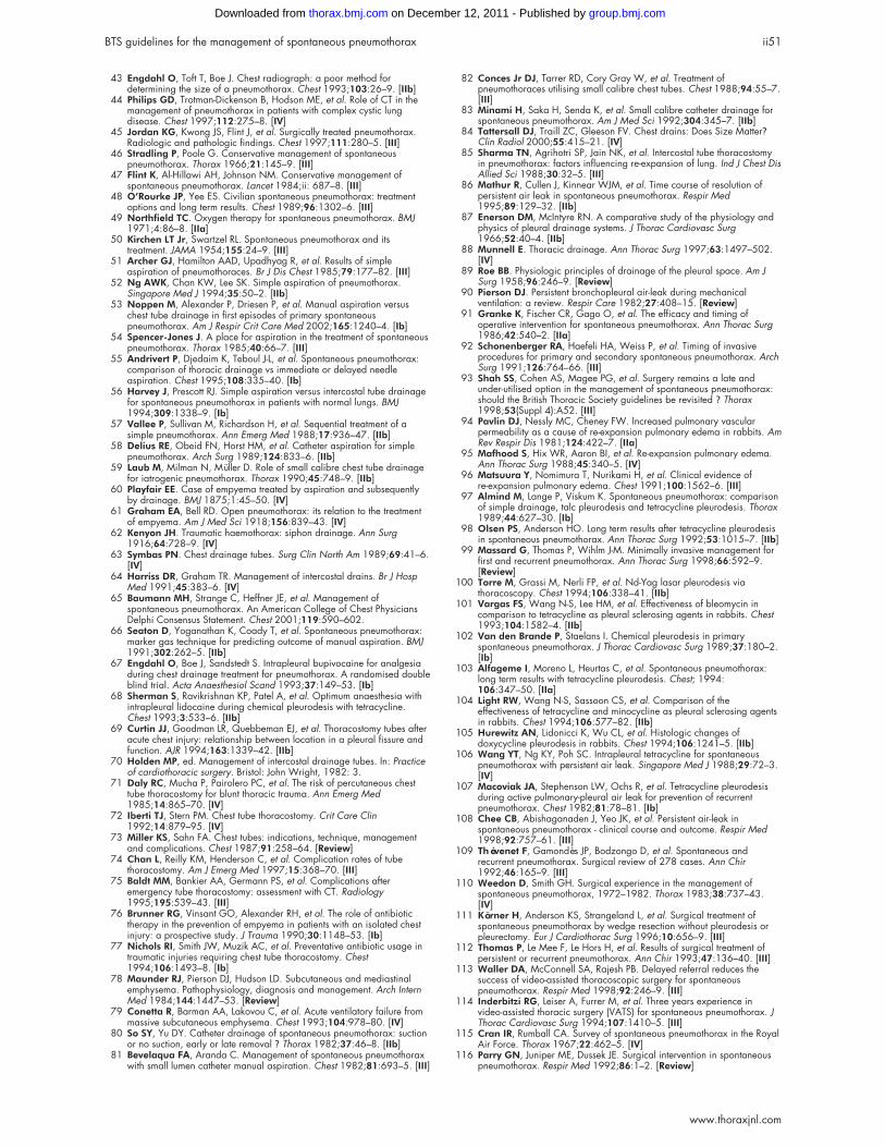
43 Engdahl O, Toft T, Boe J. Chest radiograph: a poor method fordetermining the size of a pneumothorax. Chest 1993;103:26–9. [IIb]
44 Philips GD, Trotman-Dickenson B, Hodson ME, et al. Role of CT in themanagement of pneumothorax in patients with complex cystic lungdisease. Chest 1997;112:275–8. [IV]
45 Jordan KG, Kwong JS, Flint J, et al. Surgically treated pneumothorax.Radiologic and pathologic findings. Chest 1997;111:280–5. [III]
46 Stradling P, Poole G. Conservative management of spontaneouspneumothorax. Thorax 1966;21:145–9. [III]
47 Flint K, Al-Hillawi AH, Johnson NM. Conservative management ofspontaneous pneumothorax. Lancet 1984;ii: 687–8. [III]
48 O’Rourke JP, Yee ES. Civilian spontaneous pneumothorax: treatmentoptions and long term results. Chest 1989;96:1302–6. [III]
49 Northfield TC. Oxygen therapy for spontaneous pneumothorax. BMJ1971;4:86–8. [IIa]
50 Kirchen LT Jr, Swartzel RL. Spontaneous pneumothorax and itstreatment. JAMA 1954;155:24–9. [III]
51 Archer GJ, Hamilton AAD, Upadhyag R, et al. Results of simpleaspiration of pneumothoraces. Br J Dis Chest 1985;79:177–82. [III]
52 Ng AWK, Chan KW, Lee SK. Simple aspiration of pneumothorax.Singapore Med J 1994;35:50–2. [IIb]
53 Noppen M, Alexander P, Driesen P, et al. Manual aspiration versuschest tube drainage in first episodes of primary spontaneouspneumothorax. Am J Respir Crit Care Med 2002;165:1240–4. [Ib]
54 Spencer-Jones J. A place for aspiration in the treatment of spontaneouspneumothorax. Thorax 1985;40:66–7. [III]
55 Andrivert P, Djedaim K, Teboul J-L, et al. Spontaneous pneumothorax:comparison of thoracic drainage vs immediate or delayed needleaspiration. Chest 1995;108:335–40. [Ib]
56 Harvey J, Prescott RJ. Simple aspiration versus intercostal tube drainagefor spontaneous pneumothorax in patients with normal lungs. BMJ1994;309:1338–9. [Ib]
57 Vallee P, Sullivan M, Richardson H, et al. Sequential treatment of asimple pneumothorax. Ann Emerg Med 1988;17:936–47. [IIb]
58 Delius RE, Obeid FN, Horst HM, et al. Catheter aspiration for simplepneumothorax. Arch Surg 1989;124:833–6. [IIb]
59 Laub M, Milman N, Müller D. Role of small calibre chest tube drainagefor iatrogenic pneumothorax. Thorax 1990;45:748–9. [IIb]
60 Playfair EE. Case of empyema treated by aspiration and subsequentlyby drainage. BMJ 1875;1:45–50. [IV]
61 Graham EA, Bell RD. Open pneumothorax: its relation to the treatmentof empyema. Am J Med Sci 1918;156:839–43. [IV]
62 Kenyon JH. Traumatic haemothorax: siphon drainage. Ann Surg1916;64:728–9. [IV]
63 Symbas PN. Chest drainage tubes. Surg Clin North Am 1989;69:41–6.[IV]
64 Harriss DR, Graham TR. Management of intercostal drains. Br J HospMed 1991;45:383–6. [IV]
65 Baumann MH, Strange C, Heffner JE, et al. Management ofspontaneous pneumothorax. An American College of Chest PhysiciansDelphi Consensus Statement. Chest 2001;119:590–602.
66 Seaton D, Yoganathan K, Coady T, et al. Spontaneous pneumothorax:marker gas technique for predicting outcome of manual aspiration. BMJ1991;302:262–5. [IIb]
67 Engdahl O, Boe J, Sandstedt S. Intrapleural bupivocaine for analgesiaduring chest drainage treatment for pneumothorax. A randomised doubleblind trial. Acta Anaesthesiol Scand 1993;37:149–53. [Ib]
68 Sherman S, Ravikrishnan KP, Patel A, et al. Optimum anaesthesia withintrapleural lidocaine during chemical pleurodesis with tetracycline.Chest 1993;3:533–6. [IIb]
69 Curtin JJ, Goodman LR, Quebbeman EJ, et al. Thoracostomy tubes afteracute chest injury: relationship between location in a pleural fissure andfunction. AJR 1994;163:1339–42. [IIb]
70 Holden MP, ed. Management of intercostal drainage tubes. In: Practiceof cardiothoracic surgery. Bristol: John Wright, 1982: 3.
71 Daly RC, Mucha P, Pairolero PC, et al. The risk of percutaneous chesttube thoracostomy for blunt thoracic trauma. Ann Emerg Med1985;14:865–70. [IV]
72 Iberti TJ, Stern PM. Chest tube thoracostomy. Crit Care Clin1992;14:879–95. [IV]
73 Miller KS, Sahn FA. Chest tubes: indications, technique, managementand complications. Chest 1987;91:258–64. [Review]
74 Chan L, Reilly KM, Henderson C, et al. Complication rates of tubethoracostomy. Am J Emerg Med 1997;15:368–70. [III]
75 Baldt MM, Bankier AA, Germann PS, et al. Complications afteremergency tube thoracostomy: assessment with CT. Radiology1995;195:539–43. [III]
76 Brunner RG, Vinsant GO, Alexander RH, et al. The role of antibiotictherapy in the prevention of empyema in patients with an isolated chestinjury: a prospective study. J Trauma 1990;30:1148–53. [Ib]
77 Nichols RI, Smith JW, Muzik AC, et al. Preventative antibiotic usage intraumatic injuries requiring chest tube thoracostomy. Chest1994;106:1493–8. [Ib]
78 Maunder RJ, Pierson DJ, Hudson LD. Subcutaneous and mediastinalemphysema. Pathophysiology, diagnosis and management. Arch InternMed 1984;144:1447–53. [Review]
79 Conetta R, Barman AA, Lakovou C, et al. Acute ventilatory failure frommassive subcutaneous emphysema. Chest 1993;104:978–80. [IV]
80 So SY, Yu DY. Catheter drainage of spontaneous pneumothorax: suctionor no suction, early or late removal ? Thorax 1982;37:46–8. [IIb]
81 Bevelaqua FA, Aranda C. Management of spontaneous pneumothoraxwith small lumen catheter manual aspiration. Chest 1982;81:693–5. [III]
82 Conces Jr DJ, Tarrer RD, Cory Gray W, et al. Treatment ofpneumothoraces utilising small calibre chest tubes. Chest 1988;94:55–7.[III]
83 Minami H, Saka H, Senda K, et al. Small calibre catheter drainage forspontaneous pneumothorax. Am J Med Sci 1992;304:345–7. [IIb]
84 Tattersall DJ, Traill ZC, Gleeson FV. Chest drains: Does Size Matter?Clin Radiol 2000;55:415–21. [IV]
85 Sharma TN, Agrihotri SP, Jain NK, et al. Intercostal tube thoracostomyin pneumothorax: factors influencing re-expansion of lung. Ind J Chest DisAllied Sci 1988;30:32–5. [III]
86 Mathur R, Cullen J, Kinnear WJM, et al. Time course of resolution ofpersistent air leak in spontaneous pneumothorax. Respir Med1995;89:129–32. [IIb]
87 Enerson DM, McIntyre RN. A comparative study of the physiology andphysics of pleural drainage systems. J Thorac Cardiovasc Surg1966;52:40–4. [IIb]
88 Munnell E. Thoracic drainage. Ann Thorac Surg 1997;63:1497–502.[IV]
89 Roe BB. Physiologic principles of drainage of the pleural space. Am JSurg 1958;96:246–9. [Review]
90 Pierson DJ. Persistent bronchopleural air-leak during mechanicalventilation: a review. Respir Care 1982;27:408–15. [Review]
91 Granke K, Fischer CR, Gago O, et al. The efficacy and timing ofoperative intervention for spontaneous pneumothorax. Ann Thorac Surg1986;42:540–2. [IIa]
92 Schonenberger RA, Haefeli HA, Weiss P, et al. Timing of invasiveprocedures for primary and secondary spontaneous pneumothorax. ArchSurg 1991;126:764–66. [III]
93 Shah SS, Cohen AS, Magee PG, et al. Surgery remains a late andunder-utilised option in the management of spontaneous pneumothorax:should the British Thoracic Society guidelines be revisited ? Thorax1998;53(Suppl 4):A52. [III]
94 Pavlin DJ, Nessly MC, Cheney FW. Increased pulmonary vascularpermeability as a cause of re-expansion pulmonary edema in rabbits. AmRev Respir Dis 1981;124:422–7. [IIa]
95 Mafhood S, Hix WR, Aaron BI, et al. Re-expansion pulmonary edema.Ann Thorac Surg 1988;45:340–5. [IV]
96 Matsuura Y, Nomimura T, Nurikami H, et al. Clinical evidence ofre-expansion pulmonary edema. Chest 1991;100:1562–6. [III]
97 Almind M, Lange P, Viskum K. Spontaneous pneumothorax: comparisonof simple drainage, talc pleurodesis and tetracycline pleurodesis. Thorax1989;44:627–30. [Ib]
98 Olsen PS, Anderson HO. Long term results after tetracycline pleurodesisin spontaneous pneumothorax. Ann Thorac Surg 1992;53:1015–7. [IIb]
99 Massard G, Thomas P, Wihlm J-M. Minimally invasive management forfirst and recurrent pneumothorax. Ann Thorac Surg 1998;66:592–9.[Review]
100 Torre M, Grassi M, Nerli FP, et al. Nd-Yag lasar pleurodesis viathoracoscopy. Chest 1994;106:338–41. [IIb]
101 Vargas FS, Wang N-S, Lee HM, et al. Effectiveness of bleomycin incomparison to tetracycline as pleural sclerosing agents in rabbits. Chest1993;104:1582–4. [IIb]
102 Van den Brande P, Staelans I. Chemical pleurodesis in primaryspontaneous pneumothorax. J Thorac Cardiovasc Surg 1989;37:180–2.[Ib]
103 Alfageme I, Moreno L, Heurtas C, et al. Spontaneous pneumothorax:long term results with tetracycline pleurodesis. Chest; 1994:106:347–50. [IIa]
104 Light RW, Wang N-S, Sassoon CS, et al. Comparison of theeffectiveness of tetracycline and minocycline as pleural sclerosing agentsin rabbits. Chest 1994;106:577–82. [IIb]
105 Hurewitz AN, Lidonicci K, Wu CL, et al. Histologic changes ofdoxycycline pleurodesis in rabbits. Chest 1994;106:1241–5. [IIb]
106 Wang YT, Ng KY, Poh SC. Intrapleural tetracycline for spontaneouspneumothorax with persistent air leak. Singapore Med J 1988;29:72–3.[IV]
107 Macoviak JA, Stephenson LW, Ochs R, et al. Tetracycline pleurodesisduring active pulmonary-pleural air leak for prevention of recurrentpneumothorax. Chest 1982;81:78–81. [Ib]
108 Chee CB, Abishaganaden J, Yeo JK, et al. Persistent air-leak inspontaneous pneumothorax - clinical course and outcome. Respir Med1998;92:757–61. [III]
109 Thévenet F, Gamondès JP, Bodzongo D, et al. Spontaneous andrecurrent pneumothorax. Surgical review of 278 cases. Ann Chir1992;46:165–9. [III]
110 Weedon D, Smith GH. Surgical experience in the management ofspontaneous pneumothorax, 1972–1982. Thorax 1983;38:737–43.[IV]
111 Körner H, Anderson KS, Strangeland L, et al. Surgical treatment ofspontaneous pneumothorax by wedge resection without pleurodesis orpleurectomy. Eur J Cardiothorac Surg 1996;10:656–9. [III]
112 Thomas P, Le Mee F, Le Hors H, et al. Results of surgical treatment ofpersistent or recurrent pneumothorax. Ann Chir 1993;47:136–40. [III]
113 Waller DA, McConnell SA, Rajesh PB. Delayed referral reduces thesuccess of video-assisted thoracoscopic surgery for spontaneouspneumothorax. Respir Med 1998;92:246–9. [III]
114 Inderbitzi RG, Leiser A, Furrer M, et al. Three years experience invideo-assisted thoracic surgery (VATS) for spontaneous pneumothorax. JThorac Cardiovasc Surg 1994;107:1410–5. [III]
115 Cran IR, Rumball CA. Survey of spontaneous pneumothorax in the RoyalAir Force. Thorax 1967;22:462–5. [IV]
116 Parry GN, Juniper ME, Dussek JE. Surgical intervention in spontaneouspneumothorax. Respir Med 1992;86:1–2. [Review]
BTS guidelines for the management of spontaneous pneumothorax ii51
www.thoraxjnl.com
group.bmj.com on December 12, 2011 - Published by thorax.bmj.comDownloaded from
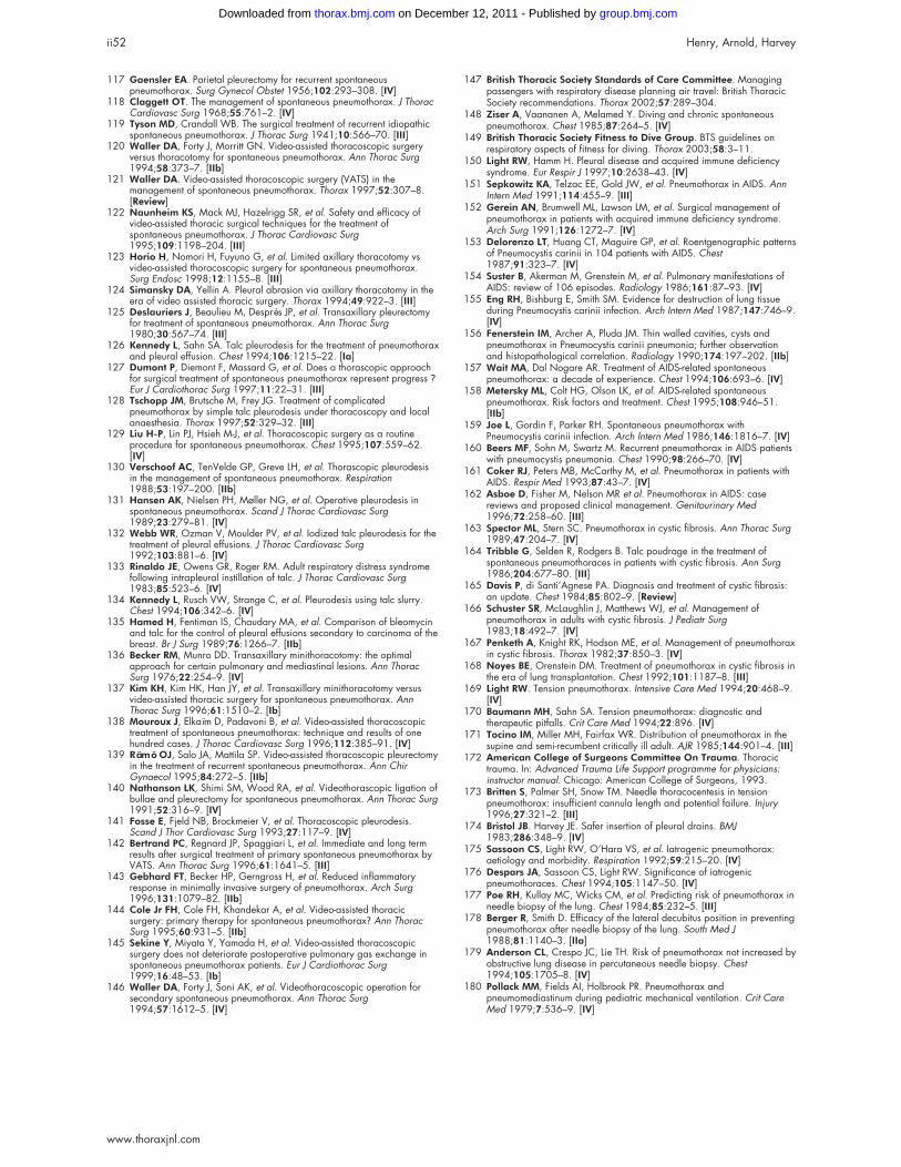
117 Gaensler EA. Parietal pleurectomy for recurrent spontaneouspneumothorax. Surg Gynecol Obstet 1956;102:293–308. [IV]
118 Claggett OT. The management of spontaneous pneumothorax. J ThoracCardiovasc Surg 1968;55:761–2. [IV]
119 Tyson MD, Crandall WB. The surgical treatment of recurrent idiopathicspontaneous pneumothorax. J Thorac Surg 1941;10:566–70. [III]
120 Waller DA, Forty J, Morritt GN. Video-assisted thoracoscopic surgeryversus thoracotomy for spontaneous pneumothorax. Ann Thorac Surg1994;58:373–7. [IIb]
121 Waller DA. Video-assisted thoracoscopic surgery (VATS) in themanagement of spontaneous pneumothorax. Thorax 1997;52:307–8.[Review]
122 Naunheim KS, Mack MJ, Hazelrigg SR, et al. Safety and efficacy ofvideo-assisted thoracic surgical techniques for the treatment ofspontaneous pneumothorax. J Thorac Cardiovasc Surg1995;109:1198–204. [III]
123 Horio H, Nomori H, Fuyuno G, et al. Limited axillary thoracotomy vsvideo-assisted thoracoscopic surgery for spontaneous pneumothorax.Surg Endosc 1998;12:1155–8. [III]
124 Simansky DA, Yellin A. Pleural abrasion via axillary thoracotomy in theera of video assisted thoracic surgery. Thorax 1994;49:922–3. [III]
125 Deslauriers J, Beaulieu M, Després JP, et al. Transaxillary pleurectomyfor treatment of spontaneous pneumothorax. Ann Thorac Surg1980;30:567–74. [III]
126 Kennedy L, Sahn SA. Talc pleurodesis for the treatment of pneumothoraxand pleural effusion. Chest 1994;106:1215–22. [Ia]
127 Dumont P, Diemont F, Massard G, et al. Does a thorascopic approachfor surgical treatment of spontaneous pneumothorax represent progress ?Eur J Cardiothorac Surg 1997;11:22–31. [III]
128 Tschopp JM, Brutsche M, Frey JG. Treatment of complicatedpneumothorax by simple talc pleurodesis under thoracoscopy and localanaesthesia. Thorax 1997;52:329–32. [III]
129 Liu H-P, Lin PJ, Hsieh M-J, et al. Thoracoscopic surgery as a routineprocedure for spontaneous pneumothorax. Chest 1995;107:559–62.[IV]
130 Verschoof AC, TenVelde GP, Greve LH, et al. Thorascopic pleurodesisin the management of spontaneous pneumothorax. Respiration1988;53:197–200. [IIb]
131 Hansen AK, Nielsen PH, Møller NG, et al. Operative pleurodesis inspontaneous pneumothorax. Scand J Thorac Cardiovasc Surg1989;23:279–81. [IV]
132 Webb WR, Ozman V, Moulder PV, et al. Iodized talc pleurodesis for thetreatment of pleural effusions. J Thorac Cardiovasc Surg1992;103:881–6. [IV]
133 Rinaldo JE, Owens GR, Roger RM. Adult respiratory distress syndromefollowing intrapleural instillation of talc. J Thorac Cardiovasc Surg1983;85:523–6. [IV]
134 Kennedy L, Rusch VW, Strange C, et al. Pleurodesis using talc slurry.Chest 1994;106:342–6. [IV]
135 Hamed H, Fentiman IS, Chaudary MA, et al. Comparison of bleomycinand talc for the control of pleural effusions secondary to carcinoma of thebreast. Br J Surg 1989;76:1266–7. [IIb]
136 Becker RM, Munro DD. Transaxillary minithoracotomy: the optimalapproach for certain pulmonary and mediastinal lesions. Ann ThoracSurg 1976;22:254–9. [IV]
137 Kim KH, Kim HK, Han JY, et al. Transaxillary minithoracotomy versusvideo-assisted thoracic surgery for spontaneous pneumothorax. AnnThorac Surg 1996;61:1510–2. [Ib]
138 Mouroux J, Elkaïm D, Padavoni B, et al. Video-assisted thoracoscopictreatment of spontaneous pneumothorax: technique and results of onehundred cases. J Thorac Cardiovasc Surg 1996;112:385–91. [IV]
139 Rämö OJ, Salo JA, Mattila SP. Video-assisted thoracoscopic pleurectomyin the treatment of recurrent spontaneous pneumothorax. Ann ChirGynaecol 1995;84:272–5. [IIb]
140 Nathanson LK, Shimi SM, Wood RA, et al. Videothorascopic ligation ofbullae and pleurectomy for spontaneous pneumothorax. Ann Thorac Surg1991;52:316–9. [IV]
141 Fosse E, Fjeld NB, Brockmeier V, et al. Thoracoscopic pleurodesis.Scand J Thor Cardiovasc Surg 1993;27:117–9. [IV]
142 Bertrand PC, Regnard JP, Spaggiari L, et al. Immediate and long termresults after surgical treatment of primary spontaneous pneumothorax byVATS. Ann Thorac Surg 1996;61:1641–5. [III]
143 Gebhard FT, Becker HP, Gerngross H, et al. Reduced inflammatoryresponse in minimally invasive surgery of pneumothorax. Arch Surg1996;131:1079–82. [IIb]
144 Cole Jr FH, Cole FH, Khandekar A, et al. Video-assisted thoracicsurgery: primary therapy for spontaneous pneumothorax? Ann ThoracSurg 1995;60:931–5. [IIb]
145 Sekine Y, Miyata Y, Yamada H, et al. Video-assisted thoracoscopicsurgery does not deteriorate postoperative pulmonary gas exchange inspontaneous pneumothorax patients. Eur J Cardiothorac Surg1999;16:48–53. [Ib]
146 Waller DA, Forty J, Soni AK, et al. Videothoracoscopic operation forsecondary spontaneous pneumothorax. Ann Thorac Surg1994;57:1612–5. [IV]
147 British Thoracic Society Standards of Care Committee. Managingpassengers with respiratory disease planning air travel: British ThoracicSociety recommendations. Thorax 2002;57:289–304.
148 Ziser A, Vaananen A, Melamed Y. Diving and chronic spontaneouspneumothorax. Chest 1985;87:264–5. [IV]
149 British Thoracic Society Fitness to Dive Group. BTS guidelines onrespiratory aspects of fitness for diving. Thorax 2003;58:3–11.
150 Light RW, Hamm H. Pleural disease and acquired immune deficiencysyndrome. Eur Respir J 1997;10:2638–43. [IV]
151 Sepkowitz KA, Telzac EE, Gold JW, et al. Pneumothorax in AIDS. AnnIntern Med 1991;114:455–9. [III]
152 Gerein AN, Brumwell ML, Lawson LM, et al. Surgical management ofpneumothorax in patients with acquired immune deficiency syndrome.Arch Surg 1991;126:1272–7. [IV]
153 Delorenzo LT, Huang CT, Maguire GP, et al. Roentgenographic patternsof Pneumocystis carinii in 104 patients with AIDS. Chest1987;91:323–7. [IV]
154 Suster B, Akerman M, Grenstein M, et al. Pulmonary manifestations ofAIDS: review of 106 episodes. Radiology 1986;161:87–93. [IV]
155 Eng RH, Bishburg E, Smith SM. Evidence for destruction of lung tissueduring Pneumocystis carinii infection. Arch Intern Med 1987;147:746–9.[IV]
156 Fenerstein IM, Archer A, Pluda JM. Thin walled cavities, cysts andpneumothorax in Pneumocystis carinii pneumonia; further observationand histopathological correlation. Radiology 1990;174:197–202. [IIb]
157 Wait MA, Dal Nogare AR. Treatment of AIDS-related spontaneouspneumothorax: a decade of experience. Chest 1994;106:693–6. [IV]
158 Metersky ML, Colt HG, Olson LK, et al. AIDS-related spontaneouspneumothorax. Risk factors and treatment. Chest 1995;108:946–51.[IIb]
159 Joe L, Gordin F, Parker RH. Spontaneous pneumothorax withPneumocystis carinii infection. Arch Intern Med 1986;146:1816–7. [IV]
160 Beers MF, Sohn M, Swartz M. Recurrent pneumothorax in AIDS patientswith pneumocystis pneumonia. Chest 1990;98:266–70. [IV]
161 Coker RJ, Peters MB, McCarthy M, et al. Pneumothorax in patients withAIDS. Respir Med 1993;87:43–7. [IV]
162 Asboe D, Fisher M, Nelson MR et al. Pneumothorax in AIDS: casereviews and proposed clinical management. Genitourinary Med1996;72:258–60. [III]
163 Spector ML, Stern SC. Pneumothorax in cystic fibrosis. Ann Thorac Surg1989;47:204–7. [IV]
164 Tribble G, Selden R, Rodgers B. Talc poudrage in the treatment ofspontaneous pneumothoraces in patients with cystic fibrosis. Ann Surg1986;204:677–80. [III]
165 Davis P, di Santi’Agnese PA. Diagnosis and treatment of cystic fibrosis:an update. Chest 1984;85:802–9. [Review]
166 Schuster SR, McLaughlin J, Matthews WJ, et al. Management ofpneumothorax in adults with cystic fibrosis. J Pediatr Surg1983;18:492–7. [IV]
167 Penketh A, Knight RK, Hodson ME, et al. Management of pneumothoraxin cystic fibrosis. Thorax 1982;37:850–3. [IV]
168 Noyes BE, Orenstein DM. Treatment of pneumothorax in cystic fibrosis inthe era of lung transplantation. Chest 1992;101:1187–8. [III]
169 Light RW. Tension pneumothorax. Intensive Care Med 1994;20:468–9.[IV]
170 Baumann MH, Sahn SA. Tension pneumothorax: diagnostic andtherapeutic pitfalls. Crit Care Med 1994;22:896. [IV]
171 Tocino IM, Miller MH, Fairfax WR. Distribution of pneumothorax in thesupine and semi-recumbent critically ill adult. AJR 1985;144:901–4. [III]
172 American College of Surgeons Committee On Trauma. Thoracictrauma. In: Advanced Trauma Life Support programme for physicians:instructor manual. Chicago: American College of Surgeons, 1993.
173 Britten S, Palmer SH, Snow TM. Needle thoracocentesis in tensionpneumothorax: insufficient cannula length and potential failure. Injury1996;27:321–2. [III]
174 Bristol JB. Harvey JE. Safer insertion of pleural drains. BMJ1983;286:348–9. [IV]
175 Sassoon CS, Light RW, O’Hara VS, et al. Iatrogenic pneumothorax:aetiology and morbidity. Respiration 1992;59:215–20. [IV]
176 Despars JA, Sassoon CS, Light RW. Significance of iatrogenicpneumothoraces. Chest 1994;105:1147–50. [IV]
177 Poe RH, Kullay MC, Wicks CM, et al. Predicting risk of pneumothorax inneedle biopsy of the lung. Chest 1984;85:232–5. [III]
178 Berger R, Smith D. Efficacy of the lateral decubitus position in preventingpneumothorax after needle biopsy of the lung. South Med J1988;81:1140–3. [IIa]
179 Anderson CL, Crespo JC, Lie TH. Risk of pneumothorax not increased byobstructive lung disease in percutaneous needle biopsy. Chest1994;105:1705–8. [IV]
180 Pollack MM, Fields AI, Holbrook PR. Pneumothorax andpneumomediastinum during pediatric mechanical ventilation. Crit CareMed 1979;7:536–9. [IV]
ii52 Henry, Arnold, Harvey
www.thoraxjnl.com
group.bmj.com on December 12, 2011 - Published by thorax.bmj.comDownloaded from
doi: 10.1136/thorax.58.suppl_2.ii39 2003 58: ii39-ii52Thorax
M Henry, T Arnold and J Harvey spontaneous pneumothoraxBTS guidelines for the management of
http://thorax.bmj.com/content/58/suppl_2/ii39.full.htmlUpdated information and services can be found at:
These include:
References
http://thorax.bmj.com/content/58/suppl_2/ii39.full.html#related-urlsArticle cited in:
http://thorax.bmj.com/content/58/suppl_2/ii39.full.html#ref-list-1This article cites 164 articles, 96 of which can be accessed free at:
serviceEmail alerting
box at the top right corner of the online article.Receive free email alerts when new articles cite this article. Sign up in the
Notes
http://group.bmj.com/group/rights-licensing/permissionsTo request permissions go to:
http://journals.bmj.com/cgi/reprintformTo order reprints go to:
http://group.bmj.com/subscribe/To subscribe to BMJ go to:
group.bmj.com on December 12, 2011 - Published by thorax.bmj.comDownloaded from