spontaneous dural tear leading to intracranial hypotension and tonsillar herniation in marfan...
TRANSCRIPT
Pabaney et al. BMC Neurology 2010, 10:54http://www.biomedcentral.com/1471-2377/10/54
Open AccessC A S E R E P O R T
Case reportSpontaneous dural tear leading to intracranial hypotension and tonsillar herniation in Marfan syndrome: a case reportAqueel H Pabaney1, Farhan A Mirza*2, Nadir A Syed1 and Humera Ahsan3
AbstractBackground: We describe the case of a 38 year old male with Marfan syndrome who presented with orthostatic headaches and seizures.
Case Presentation: The patient was diagnosed with Spontaneous Intracranial Hypotension secondary to CSF leaks, objectively demonstrated by MR Myelogram with intrathecal contrast. Epidural autologus blood patch was administered at the leakage site leading to significant improvement.
Conclusion: Our literature search shows that this is the second reported case of a Marfan patient presenting with symptomatic spontaneous CSF leaks along with tonsillar herniation.
BackgroundSpontaneous intracranial hypotension (SIH) is an underdiagnosed entity that was first described by Schalten-brand in 1938 [1]; however, a more objective descriptionof SIH was later proposed as a "decrease in CSF pressureto less than 60 mm H2O associated with occipital head-aches [2,3]". An emergency department based study esti-mated the annual incidence of SIH to be 5 per 100,000[4]; this condition is twice as common in females [5].
A traumatic event and generalized connective tissuedisorders (CTDs) are amongst the commonest etiologiesfor SIH. Among the CTDs, Marfan,[6-8] Ehlers-Danlostype II [9] and autosomal dominant polycystic kidney dis-ease [10] are associated with spontaneous CSF leaks lead-ing to SIH.
SIH can present in a variety of ways including orthos-tatic headache, diplopia, tinnitus, photophobia, hypera-cusis and vomiting [11-13]. On clinical examination,bradycardia, nystagmus, Abducens nerve palsy and neckstiffness are common findings [14,15]. Radiologically,there are 5 characteristic features of SIH on MR imaging:1. Subdural fluid collections; 2.Enhancement of pachy-meninges; 3. Engorgement of venous structures; 4. Pitu-
itary hyperemia; 5. Sagging of the brain (acronym:SEEPS) [16]. MR Myelography with intrathecal contrastis considered the imaging modality of choice to accu-rately locate the CSF leak [3].
We present a case of intracranial hypotension second-ary to a spontaneous dural tear in an adult patient withMarfan syndrome.
Case PresentationOur patient, a 38 year old male, diagnosed case of Mar-fan's syndrome, presented with complaints of orthostaticheadaches and one episode of seizure. His current illnessstarted 15 days ago with bilateral neck pain, which pro-gressed to a holocranial headache which would get mark-edly worse on sitting, standing or bending forward andrelived by lying down. These symptoms progressed overthe past 2 weeks and on the day of presentation he suf-fered a generalized tonic - clonic seizure which promptedadmission. There was no history of trauma.
On examination, our patient was hemodynamically sta-ble and afebrile. He displayed characteristic marfanoidfeatures including micrognathia, tall and lean stature, dis-proportionately long limbs and joint hypermobility. Ondetailed neurological examination, he was drowsy butarousable to vocal commands and had no signs of menin-geal irritation. Cranial nerves, motor, sensory and cere-
* Correspondence: [email protected] Medical College, Aga Khan University Hospital, Stadium Road, P.O. Box 3500, Karachi, PakistanFull list of author information is available at the end of the article
© 2010 Pabaney et al; licensee BioMed Central Ltd. This is an Open Access article distributed under the terms of the Creative CommonsAttribution License (http://creativecommons.org/licenses/by/2.0), which permits unrestricted use, distribution, and reproduction inany medium, provided the original work is properly cited.
Pabaney et al. BMC Neurology 2010, 10:54http://www.biomedcentral.com/1471-2377/10/54
Page 2 of 4
bellar examination was unremarkable except forbilaterally up going plantars.
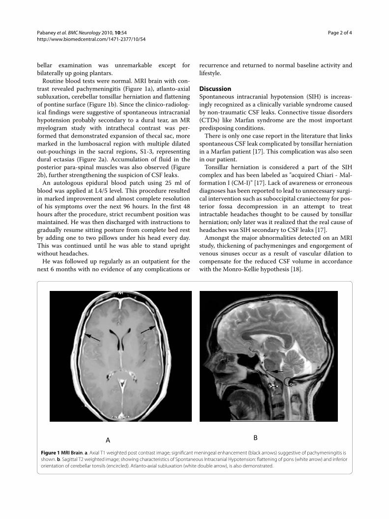
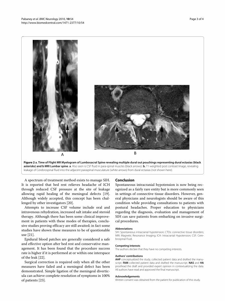
Routine blood tests were normal. MRI brain with con-trast revealed pachymeningitis (Figure 1a), atlanto-axialsubluxation, cerebellar tonsillar herniation and flatteningof pontine surface (Figure 1b). Since the clinico-radiolog-ical findings were suggestive of spontaneous intracranialhypotension probably secondary to a dural tear, an MRmyelogram study with intrathecal contrast was per-formed that demonstrated expansion of thecal sac, moremarked in the lumbosacral region with multiple dilatedout-pouchings in the sacral regions, S1-3, representingdural ectasias (Figure 2a). Accumulation of fluid in theposterior para-spinal muscles was also observed (Figure2b), further strengthening the suspicion of CSF leaks.
An autologous epidural blood patch using 25 ml ofblood was applied at L4/5 level. This procedure resultedin marked improvement and almost complete resolutionof his symptoms over the next 96 hours. In the first 48hours after the procedure, strict recumbent position wasmaintained. He was then discharged with instructions togradually resume sitting posture from complete bed restby adding one to two pillows under his head every day.This was continued until he was able to stand uprightwithout headaches.
He was followed up regularly as an outpatient for thenext 6 months with no evidence of any complications or
recurrence and returned to normal baseline activity andlifestyle.
DiscussionSpontaneous intracranial hypotension (SIH) is increas-ingly recognized as a clinically variable syndrome causedby non-traumatic CSF leaks. Connective tissue disorders(CTDs) like Marfan syndrome are the most importantpredisposing conditions.
There is only one case report in the literature that linksspontaneous CSF leak complicated by tonsillar herniationin a Marfan patient [17]. This complication was also seenin our patient.
Tonsillar herniation is considered a part of the SIHcomplex and has been labeled as "acquired Chiari - Mal-formation I (CM-I)" [17]. Lack of awareness or erroneousdiagnoses has been reported to lead to unnecessary surgi-cal intervention such as suboccipital craniectomy for pos-terior fossa decompression in an attempt to treatintractable headaches thought to be caused by tonsillarherniation; only later was it realized that the real cause ofheadaches was SIH secondary to CSF leaks [17].
Amongst the major abnormalities detected on an MRIstudy, thickening of pachymeninges and engorgement ofvenous sinuses occur as a result of vascular dilation tocompensate for the reduced CSF volume in accordancewith the Monro-Kellie hypothesis [18].
Figure 1 MRI Brain. a. Axial T1 weighted post contrast image; significant meningeal enhancement (black arrows) suggestive of pachymeningitis is shown. b. Sagittal T2 weighted image; showing characteristics of Spontaneous Intracranial Hypotension: flattening of pons (white arrow) and inferior orientation of cerebellar tonsils (encircled). Atlanto-axial subluxation (white double arrow), is also demonstrated.
A B
Pabaney et al. BMC Neurology 2010, 10:54http://www.biomedcentral.com/1471-2377/10/54
Page 3 of 4
A spectrum of treatment method exists to manage SIH.It is reported that bed rest relieves headache of ICHthrough reduced CSF pressure at the site of leakageallowing rapid healing of the meningeal defects [19].Although widely accepted, this concept has been chal-lenged by other investigators [20].
Attempts to increase CSF volume include oral andintravenous rehydration, increased salt intake and steroidtherapy. Although there has been some clinical improve-ment in patients with these modes of therapies, conclu-sive studies proving efficacy are still awaited; in fact somestudies have shown these measures to be of questionableuse [21].
Epidural blood patches are generally considered a safeand effective option after bed rest and conservative man-agement. It has been found that the procedure successrate is higher if it is performed at or within one interspaceof the leak [22].
Surgical correction is required only when all the othermeasures have failed and a meningeal defect has beendemonstrated. Simple ligation of the meningeal divertic-ula can achieve complete resolution of symptoms in 100%of patients [23].
ConclusionSpontaneous intracranial hypotension is now being rec-ognized as a fairly rare entity but is more commonly seenin settings of connective tissue disorders. However, gen-eral physicians and neurologists should be aware of thiscondition while providing consultations to patients withpostural headaches. Proper education to physiciansregarding the diagnosis, evaluation and management ofSIH can save patients from embarking on invasive surgi-cal procedures.
AbbreviationsSIH: Spontaneous intracranial hypotension; CTDs: connective tissue disorders;MRI: Magnetic Resonance Imaging; ICH: Intracranial Hypotension; CSF: Cere-brospinal Fluid.
Competing interestsThe authors declare that they have no competing interests.
Authors' contributionsAHP conceptualized the study, collected patient data and drafted the manu-script. FAM collected patient data and drafted the manuscript. NAS and HAproofread the draft and provided expert opinion in contextualizing the data.All authors have read and approved the final manuscript.
AcknowledgementsWritten consent was obtained from the patient for publication of this study.
Figure 2 a. Time of Flight MR Myelogram of Lumbosacral Spine revealing multiple dural out pouchings representing dural ectasias (black asterisks) and b MRI Lumbar spine. a. Also seen is CSF fluid in para-spinal muscles (black arrows). b. T1 weighted post contrast Image, revealing leakage of Cerebrospinal fluid into the adjacent paraspinal musculature (white arrows) from dural ectasias (not shown here).
A B
Pabaney et al. BMC Neurology 2010, 10:54http://www.biomedcentral.com/1471-2377/10/54
Page 4 of 4
Author Details1Department of Neurology, Aga Khan University Hospital, Stadium Road, P.O. Box 3500, Karachi, Pakistan, 2Medical College, Aga Khan University Hospital, Stadium Road, P.O. Box 3500, Karachi, Pakistan and 3Department of Neuroradiology, Aga Khan University Hospital, Stadium Road, P.O. Box 3500, Karachi, Pakistan
References1. Schaltenbrand V: Neuere Anschauungen zur Pathophysiologie der
Liquorzirkulation. Zbl Neurochir 1938, 3:290-295.2. Rando TA, Fishman RA: Spontaneous intracranial hypotension: report of
two cases and review of the literature. Neurology 1992, 42:481-487.3. Schievink WI, Meyer FB, Atkinson JLD: Spontaneous spinal cerebrospinal
fluid leaks and intracranial hypotension. J Neurosurg 1996, 84:598-605.4. Schievink WI, Roiter V: Epidemiology of cervical artery dissection. In
Handbook of Cerebral Artery Dissection Edited by: Baumgartner RW, Bogouss-lavsky J, Caso V, et al. Basel, Germany: Karger; 2005:12-15.
5. Schievink WI: Misdiagnosis of spontaneous intracranial hypotension. Arch Neurol 2003, 60:1713-1718.
6. Fukutake T, Sakakibara R, Mori M, Araki M, Hattori T: Chronic intractable headache in a patient with Marfan's syndrome. Headache 1997, 37:291-305.
7. Rosser T, Finkel J, Vezina G, Majid M: Postural headache in a child with Marfan syndrome: case report and review of the literature. J Child Neurol 2005, 20:153-155.
8. Milledge JT, Ades LC, Cooper MG, Jaumees A, Onikul E: Severe spontaneous intracranial hypotension and Marfan syndrome in an adolescent. J Paediatr Child Health 2005, 41:68-71.
9. Schievink WI, Gordon OK, Tourje J: Connective tissue disorders with spontaneous spinal cerebrospinal fluid leaks and intracranial hypotension: a prospective study. Neurosurgery 2004, 54:65-70.
10. Schievink WI, Torres VE: Spinal meningeal diverticula in autosomal dominant polycystic kidney disease. Lancet 1997, 349:1223-1224.
11. Capobianco DJ, Kuczler FJ Jr: Case report: primary intracranial hypotension. Milit Med 1990, 155:64-66.
12. Lipman IJ: Primary intracranial hypotension. The syndrome of spontaneous low cerebrospinal fluid pressure with traction headache. Dis Nerv Syst 1977, 38:212-213.
13. Teng P, Papatheodorou C: Primary cerebrospinal fluid hypotension. Bull LA Neurol Soc 1968, 33:121-128.
14. Garcia-Albea E, Cabrera F, Tejeiro J: Delayed postexertional headache, intracranial hypotension and racket sports. J Neurol Neurosurg Psychiatry 1992, 55:975. Letter
15. Horton JC, Fishman RA: Neurovisual findings in the syndrome of spontaneous intracranial hypotension from dural cerebrospinal fluid leak. Ophthalmology 1994, 101:244-251.
16. Atkinson JL, Weinshenker BG, Miller GM, Piepgras DG, Mokri B: Acquired Chiari I malformation secondary to spontaneous spinal cerebrospinal fluid leakage and chronic intracranial hypotension syndrome in seven cases. J Neurosurg 1998, 88:237-242.
17. Puget S, Kondageski C, Wray A, Boddaert N, Roujeau T, Di Rocco F, Zerah M, Sainte-Rose C: Chiari-like tonsillar herniation associated with intracranial hypotension in Marfan syndrome. J Neurosurg 2007, 106(1 Suppl):48-52. Case report
18. Fishman RA, Dillon WP: Dural enhancement and cerebral displacement secondary to intracranial hypotension. Neurology 1993, 43:609-611.
19. Nosik WA: Intracranial hypotension secondary to lumbar nerve sleeve tear. JAMA 1955, 157:1110-1111.
20. Teece S, Crawford I: Bed rest after lumbar puncture. Emerg Med J 2002, 19:432-3.
21. Mokri B, Piepgras DG, Miller GM: Syndrome of orthostatic headaches and diffuse pachymeningeal gadolinium enhancement. Mayo Clin Proc 1997, 72:400-413.
22. Davenport RJ, Chataway SJ, Warlow CP: Spontaneous intracranial hypotension from a CSF leak in a patient with Marfan's syndrome. J Neurol Neurosurg Psychiatry 1995, 59:516-519.
23. Lay C, Campbell K, Mokri B: Low cerebrospinal fluid headache. In Headache Edited by: Goadsby PJ, Silberstein SD. Boston: Butterworth-Heinemann; 1997:355-367.
Pre-publication historyThe pre-publication history for this paper can be accessed here:http://www.biomedcentral.com/1471-2377/10/54/prepub
doi: 10.1186/1471-2377-10-54Cite this article as: Pabaney et al., Spontaneous dural tear leading to intrac-ranial hypotension and tonsillar herniation in Marfan syndrome: a case report BMC Neurology 2010, 10:54Received: 4 November 2009 Accepted: 28 June 2010
Published: 28 June 2010This article is available from: http://www.biomedcentral.com/1471-2377/10/54© 2010 Pabaney et al; licensee BioMed Central Ltd. This is an Open Access article distributed under the terms of the Creative Commons Attribution License (http://creativecommons.org/licenses/by/2.0), which permits unrestricted use, distribution, and reproduction in any medium, provided the original work is properly cited.BMC Neurology 2010, 10:54