spontaneous bacterial peritonitis … dirk jan.pdfspontaneous bacterial peritonitis pathogenesis,...
TRANSCRIPT
SPONTANEOUS BACTERIAL PERITONITIS
pathogenesis. diagnosis and management

SPONTANEOUS BACTERIAL PERITONITIS
PATHOGENESIS, DIAGNOSIS AND MANAGEMENT
SPONTANE BACTERIELE PERITONITIS
PATHOGEN ESE, DIAGNOSTIEK EN BELEID
PROEFSCHRIFT
Ter verkrijging van de graad van doctor
aan de Erasmus Universiteit Rotterdam
op gezag van de Rector Magnificus
Prof. Dr. P.W.C. Akkermans M.A.
en volgens besluit van het College voor Promoties.
De open bare verdediging zal plaatsvinden op
woensdag 21 december 1994 om 13.45 uur
door
Dirk Jan Bac
geboren te Moerkapelie
Promotiecommissie
Promotor:
Overige leden:
Prof. J.H.P. Wilson
Prof. Dr. H.A. Bruining
Prof. Dr. S.W. Sch.lm
Prof. Dr. H.A. Verbrugh
This study was performed at the Department of lnterna] Medicine II of the University Hospital Dijkzigt Rotterdam,
The Netherlands. Financial SUpport for this thesis was kindly given by Glaxo BV, Duphar BV, Tramedico BV,
Sandoz BV, Merck Sharp & Dohme BV and Yamanouchi Pharma BV.
'Het hart heeft zijn redenen die de rede niet kent'
Blaise Pascal, natuurkundige, 11623-1662},
Vaor Drieka'
Johanna en 'Jacob
Marien en Simon
Table of contents
Chapter 1
1 .1 Introduction
1.2 Definitions
1.3 Pathogenesis
1.4 Prevalence, recurrence and survival
1 .5 Prevention
1.6 Antibiotic treatment
1.7 Aims of this thesis
Chapter 2
Paracentesis; The importance of optimal ascitic fluid analysis.
Chapter 3
Optimal analysis of total protein, albumin, white cell count and
differential in ascitic fluid.
Chapter 4
Blood culture bottles are superior to Iysis·centrifugation tubes for
bacteriological diagnosis of spontaneous bacterial peritonitis.
Chapter 5
High interleukin·6 production within the peritoneal cavity in
decompensated cirrhosis and malignancy· related ascites.
Chapter 6
Small bowel wall function in patients with advanced cirrhosis and portal
9
10
11
13
15
16
19
25
43
55
65
hypertension: studies on permeability and luminal bacterial overgrowth. 77
Chapter 7
Spontaneous bacterial peritonitis: outcome and predictive factors. 89
Chapter 8
Bacterial peritonitis following esophageal sclerotherapy:
a complication of sclerotherapy or of variceal bleeding?
Chapter 9
Spontaneous bacterial peritonitis complicating malignancy-related
ascites. Two case reports and a review of the literature.
Chapter 10
Summary and conclusion
Samenvatting en conclusie
list of publications related to this thesis
Dankwoord
Curriculum Vitae
105
117
125
129
133
135
137

9
CHAPTER 1
1.1 Introduction
On the 26 of March AD 1827 Ludwig van Beethoven died. The cause of Beethoven's
death, liver failure due to cirrhosis, was confirmed by autopsy. From 1821 Beethoven had
suffered regular attacks of jaundice and slowly progressivB hepatic failure. In November
1826 he suffered from pneumonia. This infection probably triggered the terminal phase of
Beethoven's illness. He was treated with bed rest and his health gradually improved.
However on the seventh day of his illness he developed vomiting and diarrhoea. The
jaundice deepened and his abdomen became distended due to the accumulation of fluid.
He became oliguric and developed paroxysmal nocturnal dyspnoea. In the third week a
paracentesis was carried out draining 11 litres of straw coloured fluid. Three subsequent
paracentesis were performed, the second yielding 22 litres of fluid. However the
paracentesis wound did not close, and continuously leaked ascitic fluid. Over the next
three months, Beethoven slowly deteriorated. He became increasingly anorectic and
emaciated and died in coma on the 26 of March 1827 at the age of 57 years. At the
autopsy it was noted that the abdominal cavity contained 8 litres of greyish-brown turbid
fluid, representing infected ascites (1).
Although medical science made revolutionary progress during the last 150 years, a medical
history similar to the above mentioned is familiar to many physicians even today. Although
diuretics and antibiotics relieve some of the symptoms described above, end-stage liver
disease remains one of the most difficult entities to treat. Spontaneous bacterial peritonitis
was recognized as a specific entity in the early 1960s and clusters of cases were
published from Paris (2), London (3) and West Haven, Connecticut (4). The first patients
published by Harold O. Conn presented a discrete homogeneous syndrome. Each patient
had been admitted to hospital with decompensated alcoholic cirrhosis and ascites. After
one week in hospital they developed abdominal pain, fever and encephalopathy.
Paracentesis showed cloudy ascitic fluid that contained large numbers of
polymorphonuclear leukocytes and coliform bacteria were cultured from the ascitic fluid.
All patients died (4).
Since then the spectrum of disease has broadened considerably. Initially it was almost
exclusively described in alcoholic cirrhosis, but now it has been recognized in all types of
10
cirrhosis, but very rarely in non-cirrhotic ascites. Clinical signs and symptoms may be
absent and deterioration of laboratory values may be the only sign that an underlying
bacterial infection of ascitic fluid is present. At first it was thought to be a nosocomial
infection with a prevalence of 10-25% of all patients with cirrhosis and ascites admitted
to a hospital (5,6). However prospective studies learned that the infection is often
community-acquired, with a 12% prevalence in patients with cirrhosis and ascites seeking
admission for various reasons (7). Due to increased awareness of the disease, earlier
diagnosis and better treatment, in-hospital mortality rates have improved during the last
20 years from an initial mortality rate of 95% to about 40% in more recent series (8~ 13).
A high recurrence rate after a first episode of spontaneous bacterial peritonitis and
progression of the underlying liver disease results in a poor 1 ~year survival of about 20~
30% (9,11).
1.2 Definitions
spontaneous bacterial peritonitis (SBP) is defined as the bacterial infection of the ascitic
fluid without any identifiable intra·abdominal source of the infection (14·16). The most
widely accepted diagnostic criteria of SSP include:
1) Increased polymorphonuclear leucocyte (PMN) count in the ascitic fluid. A cut-off level
above 0.25xl0' cellsll is most frequently used.
2) A positive ascitic fluid culture.
"Spontaneous" infections are distinquished from secondary bacterial peritonitis in that
spontaneous infections do not have a surgically·treatable source. Although many patients
with SSP have a focus of infection, e.g. urinary tract infection or pneumonia, they are
diagnosed as having SSP.
There are two variants of SSP that require further clarification:
Culture-negative neutrocytic ascites, in which the the PMN count of ascitic fluid is
elevated, but cultures remain negative. Obviously the culture technique determines which
percentage of suspected episiodes of SSP will remain culture-negative. Cut-off levels for
the PMN counts in the ascites are above 0.25 or 0.50 x 10' cellsll. Although there have
been some discussions in the literature suggesting that culture-negative neutrocytic ascites
is a less severe variant of SSP, usually they are regarded as similar diseases with identical
prognosis (17-19).
Monobacter ascites, or monomicrobial non neutrocytic bacterascites, is a controversial
11
entity and defined as a positive ascitic fluid culture with a single organism (excluding
Staph. epidermidis) with an ascitic fluid PMN count < 0.25x1 0' cellsll (20,21). This
transient residence of bacteria in the ascitic fluid without neutrocytic response may
progress to real SSP, sometimes in a few hours time, or may resolve without treatment
(7,20,21). Antibiotics are not routinely recommended for this subgroup of patients, but
close monitoring of symptoms and early retapping of ascites is advisable.
1.3 Pathogenesis
H.D. Conn, who first used the term SSP (4), thought that bacteremia-induced infection of
preexistent ascites was the most likely way of developing SSP. Another possibility he
raised was the direct transmural migration from the gut to the peritoneal cavity, and he
also suggested that paracentesis by itself could lead to SSP. Over the ensuing years the
pathogenesis of spontaneous ascitic fluid infections has become a bit clearer.
It is possible that enteric bacteria regularly will enter the ascitic fluid, but that in the
majority of cases the bacteria are cleared by local host defense mechanisms (7,20,21).
SBP will occur if there is an increase of invading bacteria or impaired host defenses.
Assuming that the normally present equilibrium between host-environ me nt-microorganism
becomes disturbed, it seems worthwhile to have a look at the pathogenesis of SSP using
this model.
Host factors; Most patients who develop SSP have severe liver disease with diminished
function of the hepatic reticuloendothelial system (RES) and increased porto-systemic
shunting. These changes increase the chance that viable bacteria will not be cleared from
the blood stream. Rimola et al. (22) using an elimination constant for 99~echnetium-sulfur
colloid as a marker of RES phagocytic activity in the liver demonstrated a consistently
lower elimination rate in patients with cirrhosis and bacteremia occurred more frequently
in the patients with the lowest elimination rates. Other deficits in cirrhosis, not limited to
the portal circulation, include decreased intracellular killing properties, impairment of
neutrophil chemotaxis, reduced serum complement ~evels and reduced opsonization
unrelated to hypocomplementemia (23,24).
Factors present in ascites; Recent studies (25-28) have shown that patients who develop
SBP have low chemoattractant and opsonic activities of the ascitic fluid, thus indicating
that the ascitic fluid itself plays an important role in preventing colonization by pathogenic
bacteria. Mal et a!. (25) studied prospectively a number of patients with ascites due to
12
different etiologies and found that diminished complement C3 levels in ascites had an
independent predictive value for the occurrence of S8P, which correlated with impaired
chemoattractant and opsonic activity. This was confirmed in a number of other studies
(26,27). By establishing a correlation between the total protein content and C3
concentration in the ascitic fluid, a number of studies found the total protein content of
ascitic fluid to be a good parameter for predicting which patients arB most at risk to
develop SBP (28,29).
In summary I ascitic fluid opsonic activity correlates with ascitic fluid C3 concentration and
total protein concentration. Patients whose ascitic fluid is dilute and deficient in these
complement factors are predisposed to ascitic fluid infection. Runyon (28) found that
patients with an ascitic fluid protein concentration < 10911. were ten times more likely to
develop SSP during hospitalization than patients with higher protein ascitic fluid levels.
This does not however completely explain why some patients are more susceptible than
others. A very interesting French study (30) described an improved PMN function within
cirrhotic ascitic fluid after serial dilutions or when adding ascitic fluid from malignant
peritonitis. This suggests the presence of a suppressive factor in cirrhotic fluid which
suppresses PMN function.
The bacteriological flora, responsible for the spontaneous infections of the ascitic fluid is
mainly gut derived (5,8,31-35). Gram-negative aerobic rods such as E. coli and Klebsiella
pneumoniae are responsible for about two-third of all cases of SSP. If organisms could
easily traverse the gut wall and directly enter the ascitic fluid, polymicrobial infections
would be the rule and not the exception. Additionally, the aerobic Gram-negative
microorganisms in the gut are outnumbered by 2-4 orders of magnitude by anaerobes and
enterococci (14), which seldom cause SBP. Apparently there is some kind of a "filter"
between the gut lumen and the fluid.
Studies in rodents have demonstrated that under ceTtain circumstances bacteria can
"translocate" from the gut lumen across the mucosa into submucosal lymphatics and be
detected in mesenteric lymph nodes (36). From the mesenteric lymph nodes bacteria may
spread to the spleen, liver, or bloodstream. Interestingly, it has been confirmed that Gram
negative rods translocate in greatest numbers through the bowel wall, Gram-positive cocci
moderately and anaerobes seldom (37). Circumstances which promote translocation
include (1) bacterial overgrowth in the gut, (2) disruption of the gut mucosal barrier and
(3) abnormal host defenses. It still a matter of debate if patients with cirrhosis have altered
gut flora, but there is some evidence supporting this (38-40). Some authors describe a
13
contamination of the jejunum and duodenum in up to 75% of patients with cirrhosis (38).
Biopsies of the upper small bowel in patients with cirrhosis often demonstrates venous
stasis, edema of villi and a degeneration of epithelium and basement membrane. It might
well be that the gut mucosa is abnormally permeable in patients with cirrhosis, promoting
translocation of bacteria from the gut to mesenteric lymph nodes and on to the peripheral
blood.
A report of Runyon et al. (41) is the first to describe a rodent model of SBP with
experimental cirrhosis induced by the oral administration of phenobarbital and the
intra gastric installation of carbon tetrachloride. More than 50% of the rats who survived
the cirrhosis-induction program developed spontaneous infections of the ascitic fluid
caused by Gram~negative bacilli of enteric origin. However, the effect of the carbon
tetrachloride on the mucosal gut permeability is unknown, which makes these results
difficult to interpret. The ideal animal model to study the mechanisms of pathogenesis of
SBP has not yet been developed.
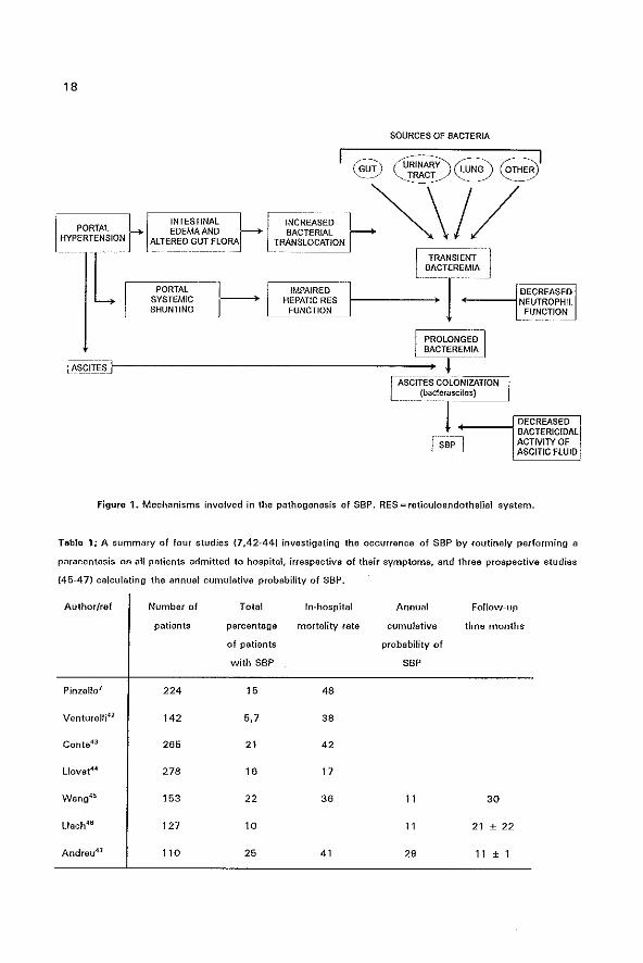
In summary, general host defenses, factors present in the ascites, and the microorganisms
in the gut flora, act together to facilitate the development of spontaneous ascitic fluid
infections (figure 1). Most bacteria causing SBP are gut~derived and translocate through
the mucosal wall to mesenteric Iymphnodes, on to the thoracic duct and bloodstream and
leak across the sinusoids of the liver to the ascites.
In some patients bacteria colonizing the urinary tract or the respiratory tract may cause
bacteremia which subsequently leads to ascitic fluid infection. Invasive procedures such
as paracentesis, endoscopy or sclerotherapy do not seem to playa major role in most
cases, and SSP is not necessarily a nosocomial infection.
1.4 Prevalence, recurrence and survival
There is some variance in data as to how often SBP occurs in patients with cirrhosis and
ascites, most articles quoting a figure between 10% and 25% (5~8). A great deal depends
on the index of suspicion and the threshold to perform a paracentesis in hospitalized
patients. Some studies prospectively looked at the prevalence of SSP when paracentesis
was routinely performed at admission (7,42·47). Seven prospective studies are
summarized in Table 1. SBP and culture-negative neutrocytic ascites are grouped together.
Monobacter ascites has been excluded from these data. There is no great difference in the

14
prevalence of SBP between alcoholic and nonalcoholic cirrhosis. In 5,7% -21 % of patients
with cirrhosis and ascites, S8P was diagnosed when paracentesis was routinely performed
at admission. The reason for admission and presenting symptoms varied in the different
studies. Also the Child-Pugh classification, which is an important predictive factor for the
development of SBP, differs in the different studies. Venturelli et al. (42) found a low
prevalence of 5,7%, but most patients had a relatively mild cirrhosis (Child-Pugh class B)
and this was a multi centre study involving general medical departments, and not referral
centres. Three studies (45-47) did a follow-up of their patients with cirrhosis and ascites
and calculated an annual cumulative probability of developing S8P varying between 11 %
and 29% within their patient groups. The latter figure probably being higher because more
of these patients had Child-Pugh category C cirrhosis, compared to the other two studies.
When in a multivariate analysis clinical and laboratory variables were analyzed to establish
predictive factors, the total protein content of ascitic fluid and the bilirubin serum level
were the most important predictors for the develoment of a first episode of S8P (9,12,46).
Llach et al. (46) calculated a probability for a first episode of SBP within 3 years of follow
up of 24% when the total protein content in ascitic fluid was lower than 10 gIl and of 4%
when the total protein content was greater than 10 gIl.
If a patient had an episode of S8P the recurrence rate is much higher, between 35% and
69% at 1 year (9,43,48). Wang et al. (45) found an 11 % annual occurrence rate for a first
episode of S8P and a 47% annual recurrence rate in patients with a previous episode of
S8P. The only predictive factor for this high recurrence rate was an ascitic total protein
level below 7.5 gIl, other clinical and laboratory data were not statistically different
between the two groups, although Tito et al. (9) showed that the serum bilirubin and a
delayed prothrombin time were also associated with a higher risk of recurrence.
S8P is a serious complication with a high in-hospital mortality and a poor l-year survival.
Most studies report a direct mortality related to the S8P of about 40-50%, older series
have a higher mortality (70%-95%) than more recent published studies. When a more
aggressive approach is followed and every patient undergoes a paracentesis on admission,
irrespective of symptoms, the mortality rate may be as low as 17% (44). This again
emphasizes the need to perform a paracentesis routinely in every patient with cirrhosis and
ascites who is admitted in hospital, as early diagnosis results in a much better prognosis.
However, long-term survival after a first episode of S8P is possible and has been reported
by Hoefs (4 out of 13 patients survived more than 3 years,(49) and Tito (8 out of 59
patients survived more than 3 years (9).
15
1.5 Prevention
Because enteric bacteria are the most common causative agents of SSP, it has been
suggested that selective intestinal decontamination, which largely eliminates the aerobic
Gram-negative intestinal flora and preserves the remaining aerobic and anaerobic flofa
could be useful in its prophylaxis. Two recent placebo-controlled studies have demon
strated the efficacy of oral norfloxacin in the prevention of SSP episodes without attaining
a reduction in mortality (50,51). The first study (50) used antibiotic prophylaxis in a group
of patients recovering from a first episode of SSP and found a decreased risk of recurrence
at 1-year of follow·up of 20% compared to 68% in the placebo group. The SSP
recurrences in the norfloxacin treated group were mainly due to Gram-positive cocci, which
are not sensitive to norfloxacin. There was no significant difference between both groups
with respect to the probability of SBP recurrence caused by Gram-positive cocci or culture
negative SBP (17 % in the norfloxacin group and 20% in the placebo group at 1-year of
follow-up). In the second study (51) 63 patients with cirrhosis and with a total protein
content in the ascitic fluid < 1 Og/l were randomized either to receive placebo or norfloxacin
during hospitalization. Also in this study there was a lower incidence of SSP, but also in
total number of infections, during hospitalization in the patients receiving selective
intestinal decontamination, without effect on the overall mortality.
Norfloxacin was chosen in both studies because it is incompletely absorbed by the
intestine, is highly effective against Gram-negative bacilli, has low effectivity against
anaerobic bacteria and has a low incidence of side effects when administered chronically
(52). At least two mechanisms may contribute to the efficacy of long-term norfloxacin
administration in preventing SSP. The first and most attractive mechanism is the selective
intestinal decontamination caused by this drug. Its effect on the fecal flora confirms that
there is a marked reduction of the aerobic Gram-negative bacilli with no significant effects
on Gram-positive cocci and anaerobic bacteria (50). On the other hand, in patients with
cirrhosis and ascites, bactericidal levels are obtained in serum, urine and ascites, which
could be an additional mechanism explaining the efficacy in the prevention of SBP. A
possible third mechanism was pointed out by a study (53) which described increasing
serum and ascitic fluid C31evels during selective intestinal decontamination by norfloxacin.
It is not clear from this study if the increase in serum C3 and ascitic fluid C3 is due to
decreased consumption of complement or improved hepatic complement synthesis. In this
way norfloxacin may indirectly increase the bactericida1 and opsonic activity of ascitic
16
fluid.
Long·term prophylaxis with norfloxacin remains a controversial matter (54,55). What arB
the indications for prophylaxis? How long is long~term prophylaxis? What are the side
effects, especially concerning infections with norfloxacin·resistant strains and yeasts.
Finally the issue of cost and cost·effectiveness should be considered, comparing long-term
prophylaxis (which did not show a decrease in hospital admissions) compared to no
prophylaxis with a "diagnose and treat" strategy.
Yet another group of patients who might benefit from antibiotic prophylaxis should be
discussed in this setting. It has been noted that cirrhotics with a gastrointestinal
hemorrhage are at an increased risk to develop bacterial infections, especially caused by
enteric bacteria (56,57). In the most recent study (56) norfloxacin 400mg twice daily was
given during 7 days, immediately starting after emergency gastroscopy. There was a lower
incidence of infections in the treated group, mainly attributable to a decrease of
bacteremia's and urinary tract infections, which did not result in a significantly decreased
mortality in the treated group.
In conclusion, norfloxacin has been shown to be an effective agent in the prevention of
SSP in patients with quiescent chronic liver disease, but the number of hospitalisations and
the survival is similar to prospective detection and treatment of SBP. More studies are
needed to define subgroups of patients with liver disease to determine which patients
benefit most from proper antibiotic prophylaxis.
1.6 Antibiotic treatment of Spontaneous Bacterial Peritonitis
Survival of the patient with SSP can be improved by the early institution of antibiotic
therapy. Empiric intravenous antibiotic treatment (before culture results are avaliable)
should be given to patients with an elevated PMN cell count in the ascitic fluid. A PMN
count> 0.5x1 09 cells/l.. independent of the presence or absence of symptoms, or a PMN
count> O.25x1 09cells/l in the presence of a compatible clinical picture should be sufficient
reason to start antibiotic therapy immediately. Because more than 90% of cases of SSP
are caused by enteric Gram-negative aerobes and Gram-positive cocci, the combination of
ampicillin with an aminoglycoside provides good coverage and has been shown to be
effective in the treatment of SSP (58). However the nephrotoxicity of aminoglycosides in
patients with cirrhosis limits its usefulness.
Aztreonam, a monobactam, has been compared with cefotaxim in the treatment of SSP
17
(59); its lack of activity against Gram·positive organisms and a higher frequency of Gram·
positive superinfections makes it suboptimal as a first choice antibiotic.
Cefotaxim, a third generation cephalosporin, is most widely used as a first·choice antibiotic
in the empiric treatment of S8P. A cure rate of 85% was described in a recent study (12).
At least 10% of the infections did not respond satisfactorily to this first-choice treatment
and the antibiotic regimen had to be changed. Interestingly, the in vitro resistance of the
isolated bacteria to cefotaxim did not always correspond to infection resolution in vivo.
The efficient and rapid ascitic fluid penetration may account for this finding. The
combination of amoxicillin and clavulanic acid has recently been shown to be effective in
85% of acute episodes of S8P in a French study (60) with minimal side effects and low
frequency of resistant organisms. Also for these drugs a good distribution in the ascitic
fluid and prolonged serum half-lives due to slow return from the ascitic compartment was
proven (61). Unfortunately, this combination has never been compared to cefotaxim, but
one of these drugs or drug combinations are suitable as a first-choice treatment for acute
S8P. Once culture results are available, optimal antibiotic treatment obviously depends on
the organism identified. Monitoring the ascitic fluid PMN count during the first days of
treatment is useful to detect at an early stage resistance to the antibiotic regimen or to
discover secondary peritonitis, which does not respond as well as SBP to antibiotic
treatment (62).
Because S8P is a severe infection with a high mortality, it is common practice to treat for
10 to 14 days with antibiotics. Two studies compared long and short-term antibiotic
regimens. Fang et al. (63) stopped the antibiotic treatment when the PMN count in the
ascites was below O.25x10'cells/l and they did not find a difference in survival or infection
recurrence between patients who received conventional treatment and the patients whose
antibiotic therapy was discontinued when the ascitic fluid PMN count was < O.25x1 0 9_
cellsll. Runyon et al (64) compared 10 days cefotaxim with 5 days treatment and found
similar results in both groups concerning hospital mortality and infection recurrence. Both
studies are not very large, thus a type II error can not be excluded.
18
L INTESTINAL EDEMA AND
ALTERED GUT FLORA
PORTAL ---1 SYSTEMIC. 1-----+ SHUNTING
--------
f]_ ..
INCREAseD BACTERIAL
TRAN~_L_OCATION
SOURCES OF BACTERIA
l TRANSIENT J H~~~~:~~S 1-_____ .. _-~-_+~~TIER~~----~8f~~~~
L--"F"U"NC"T"IO",N"---" - L FUNCTION
IA8WE"JI--------------------=~r===~ IAsciTEs COLONIZATION L (baC!eras':~ita~'~) __ ~ .. -.--.--.- 1
[SBP-] DECREAseD BACTERICIDAL ACTNITYOF ASCITIC flUID
Figure 1. Mechanisms involved in the pathogenesis of SSP. RES=reticuloandothelial system.
Table 1; A summary of four studies (7.42-44) investigating the occurrence of SSP by routinely performing a
paracentesis on all patients admitted to hospital, irrespective of their symptoms, and three prospective studies
145-47) calculating the annual cumulative probability of SSP.
Author/ref Number of Total In-hospital Annual Follow-up
patients percentage mortality rate cumulative time months
of patients probability of
with SSP S8P
PinzeUo7 224 15 48
VentureJli41 142 5,7 38
Conta43 265 21 42
Llovet44 278 16 17
Wang45 153 22 36 11 30
Uach46 127 10 11 21 ± 22
Andreu41 110 25 41 29 11 ± 1
19
1.7 Aims of this thesis
1 . To investigate and evaluate the occurrence of spontaneous bacterial peritonitis in
a tertiary referral centre in the Netherlands.
2. To improve the diagnostic yield of ascitic fluid investigations and to develop
standardization in diagnostic tests with respect to the bacteriological as well as to
the haematological and biochemical analysis.
3. To clarify the pathogenesis of spontaneous bacterial peritonitis. especially
concerning the presence of bacterial overgrowth and small bowel wall permeability
in patients with portal hypertension.
4. To determine which subgroups of patients with decompensated cirrhosis might
benefit from selective intestinal decontamination.
5. To augment knowledge and awareness of the existense of spontaneous bacterial
peritonitis in order to improve early diagnosis and to lower the mortality due to this
serious complication.
20
O'Shea J. In: Music and Medicina, medical profiles of great composers. Dent and ,Sons LTD, London
1990 pp 39-65,
2 Caroli J, Platteborse R. Septicemia portocave du faie at septicemia a colibaciUi. Sam Hop Paris
1958;34:472-487.
3 Kerr DRS, Pearson DT, Read AE. Infection of ascitic fluid in patients with hepatic cirrhosis. Gut
1963;4:394AOO.
4 Conn HO. Spontaneous bacterial peritonitis and bacteremia in cirrhotic patients caused by enteric
bacteria. Ann tntern Mad 1964;60:668-580.
5 Hoefs Je, Runyon BA. Spontaneous bacterial peritonitis. Dis Mon 1985;31: 1-48.
6 Almdal TP, Skinhoy P. Spontaneous bacterial peritonitis in cirrhosis. Scand J Gastroanterol
1987;22:295-300.
7 Pinzallo G, Simonetti RG, Craxi A, Piazza SO, Spano C, Pagliaro l. Spontaneous bacterial peritonitis: A
prospective investigation in predominantly nonalcoholic cirrhotic patients. Hepatology 1983;3:545-549.
8 Wilcox CM, Dismukes WE. Spontaneous bacterial peritonitis. Medicine 1987;66:447-456.
9 Tito L, Rimola A, Gines P, Llach J, Arroyo V, Rodes J. Recurrence of Spontaneous Bacterial
Peritonitis: Frequency and Predictive Factors. Hepatology 1988;8:27-31.
10 Ariza J, Gudiol F, Dolz J, et al. Evaluation of Aztreonam in the treatment of spontaneous bacterial
peritonitis in patients with cirrhosis. Hepatology 1986;6:906-910.
11 Ink 0, Pelletier G, Salmon 0, Attali P, Pessione F, Hannoun S, Buffet C, Etienne JP. Prognostic de
I'infection spontanee d'ascite chez Ie cirrhotique. Gastroenterol·Clin-Biol 1989;13:566-561.
12 Toledo C, Salmeron JM, Rimola A, et al. Spontaneous bacterial peritonitis in cirrhosis: Predictive factors
of infection resolution and survival in patients treated with cefotaxim. Hepatology 1993;17:251-267.
13 Runyon BA, Mchutchison JG, Antillon MR, Akrivadis ER, Montano AA. Short-course versus long-course
antibiotic treatment of spontaneous bacterial peritonitis. Gastroenterology 1991;100:1737-1742.
14 Runyon BA. Pathogenesis and diagnosis of spontaneous bacterial peritonitis in cirrhosis. In: Rodes J,
Arroyo V. Therapy in liver disease. Ediciones Doyma, Barcelona. 1992;388-396.
16 Hoefs JC. Diagnostic Paracentesis: A potent clinical tool. Gastroenterology 1990;98:230-236.
16 Gines P. Spontaneous bacterial peritonitis. CUrr Opinion GastfO 1992;8:403-408.
17 Runyon BA, Hoefs JC. Culture-Negative Neutrocytic Ascites: A variant of spontaneous bacterial
peritonitis. Hepatology 1984;4:1209-1211.
18 Pelletier G, Salmon 0, Ink 0, Hannoun S, Attali P, Buffet C, Etienne JP. Culture·negative neutrocytic
ascites: a less severe variant of spontaneous bacterial peritonitis. J HepatoJ 1990;10:327-331.
19 Terg R. Levi 0, lopez D, et aI, Analysis of clinical course and prognosis of culture-positive
spontaneous bacterial peritonitis and neutrocytic ascites. Dig Dis and Sci 1992;37: 1499-1502.
20 Runyon BA. Monomicrobial nonneutrocytic bacterascites: A variant of spontaneous bacterial
peritonitis. HepatoJogy 1990;12:710-716.
21 Pelletier G, lesur G, Ink ° et a!. Asymptomatic bacterascitas: Is it spontaneous bacterial peritonitis?
Hepatology 1991;14:112-115.
22 Rimola A, Soto R, Bory F, ot a!. Reticuloendothelial system phagocytic activity in cirrhosis and its relation
to bacterial infections and prognosis. Hepatology 1984;4:53-68.
23 Rajkovic lA, Williams R. Abnormalities of neutrophil phagocytosis, intracelluler killing and metabolic

activity in alcoholic cirrhosis and hepatitis. Hepatology 1988;6:262-262.
24 Yousif-Kadura AGM, Rajkovic R, Wyke RJ, Williama R. Defects in serum attractant activity in
different types of chronic liver disease. Gut 1984;25:79-84.
21
25 Mal F, Pham Huu T, Bendahou M, at af. Chemoattractant and opsonic activity in ascitic lIuid. J Hepatol
1991 ;12:45-49.
26 Rabinovitz M, Gavaler JS, KUmar 5, Kejllni M, van Thiel OH. Role of serum complement,
immunoglobulins, and cell mediated immune system in the pathogenesis of spontaneous bacterial
peritonitis. Dig Dis Sci 1989;34:1647-1652.
27 Such J, Guerner C, Enriguez J, Rodriquez Jl, Seres I, Vilardell F. low C3 in cirrhotic ascites
predisposes to spontaneous bacterial peritonitis. J Hapatol 1988;6:80·84.
28 Runyon BA. low protein concentration ascitic fluid is predisposed to spontaneous bacterial
peritonitis. Gastroenterology 1986;91: 1343-1346.
29 L1ach J, Rimola A, NavasH M, et al. Incidence and predictive factors of first episode of spontaneous
bacterial peritonitis in cirrhosis with ascites: Relevance of ascitic fluid protein concentration. Hepatology
1992;16:724-727.
30 Lebrun l, Pelletier G, Sriantais MJ, Galanaud P, Etienne JP. Impaired functions of normal peripheral
polymorphonuclear leucocytes in cirrhotic ascitic fluid. J Hepatol 1992;16:98-101.
31 Hoefs JC, Jonas GM. Diagnostic paracentesis. Adv Int Med 1992;391-409.
32 Runyon BA, Umland ET, Merlin T. Inoculation of blood culture boWes with ascitic fluid. Arch Intern Med
1987;147:73~75.
33 Runyon SA, Canawati HC, Akriviadis EA. Optimization of ascitic fluid culture technique.
Gastroenterology 1988;95:1351-1366.
34 Bobedilla M, SifUentes J, Garcia-Tsao G. Improved method for bacteriological diagnosis of
spontaneous bacterial peritonitis. J Clin Microbiol 1989;27:2146-2147.
36 Runyon BA, Antillon MR, Akrivadis A, McHutchison JG. Bedside inoculation of blood culture bottles with
ascitic fluid is superior to delayed inoculation in the detection of spontaneous bacterial
peritonitis. J Clin Microbiol 1990;28:2811-2812.
36 Sorell WT, Quigley EMM, Jin G, Johnson T J, Rikkers l T. Bacterial translocation in the portar~
hypertensive rat: Studies in basal conditions and on exposure to hemorrhagic shock. Gastroenterology
1993;104:1722-1726.
37 Steffen EK, Berg RD, Djetch EA. Comparison of translocation rates of various indigenous baoteria from
the gastrointestinal traot to the mesenterio lymph node. J Infeot Dis 1988;167:1032-1038.
38 Gorbaoh Cl, laiD, Levitan R: Intestinal microtlora in Laennec's cirrhosis. J Clin Invest 1970;49: 36 H.
39 Martini GA, Phear EA: The baoterial content of the small intestine in normal and cirrhotic patients. Clin
Sci 1967;16:35·61.
40 Simon Gl, Gorbach Sl; Intestinal microflora.
Med Clin of North Am 1982;66:No 3.pp 667-673.
41 Runyon BA, Sugano S, Kanel G, Mellencarnp MA. A rodent model of cirrhosis, ascites, and bacterial
peritonitis. Gastroenterology 1991;100:489-493.
42 Venturelli R, Bonzi G, Bortoli A, et at. Spontaneous baoterial peritonitis: a prospective multicentre study.
Eur J Gastroenterol Hepatol 1993;3:149-152.
22
43 Conte 0, Bolzoni P, Bodin; P, at 81. Frequenoy of spontaneous bacterial peritonitis in 265 cirrhotics with
ascites. Eur J Gastroanterol and Hepatol 1993;5:41-46.
44 Llovat JM, Planas A, MaTHias R, at al. Short-term prognosis of cirrhotics with spontaneous bacterial
peritonitis! Multivariate study. Am J Gastroenterol 1993;88:388-392.
45 Wang 55, Tsal YT, Lee SO, at ai, Spontaneous bacterial peritonitis in patients with hepatitis-B related
cirrhosis and hepatocellular carcinoma. Gastroenterology 1991;101 :1656-1662.
46 Uach J, Rimala A, Navasa M, at a!. Incidence and predicNve factors of first episode of spontaneous
bacteria! peritonitis in cirrhosis with ascites: relevance of ascitic fluid protein concentration.
Hepatology 1992;16:724-727.
47 Andreu M, Sola R, Sitges-Serra A, et at. Risk factors for spontaneous bacterial peritonitis in cirrhotic
patients with ascites. Gastroenterology 1993;104:1133-1138.
48 Silvain e, Mannant PA, Ingrand P, Fort E, Besson I, Beauchant M. Rl;'icidive de I'infection spontanee du
fiquide d'ascite au cours de la cirrhose. Gastroenterol Clin Bioi 1991,16:106-109.
49 Hoefs Je, Canawati HN, Sapieo Fl, Hopkins RR, Wiener J, Montgomerie JZ. Spontaneous bacterial
peritonitis. Hepatology 1982;2:399-407.
60 Gines P, Rimola A, Planas R. et at. Norlloxacin prevents spontaneous bacterial peritonitis recurrence in
cirrhosis: results of a double blind, placebo controlled trial. Hepatology 1990;12:716-724.
61 Soriano G, GUarner e, Teixido M, et al. Selective intestinal decontamination prevents spontaneous
bacterial peritonitis. Gastroenterology 1991;100:477-481.
62 Wolfson JS, Hooper DC. Norfloxacin: a new tergeted fluoroquinolone antimicrobial agent. Ann Intern
Med 1988;108:238-261.
63 Such J, Guarner C, Soriano G, et al. Selective intestinal decontamination incraasas sarum and ascitic
fluid C3 levels in cirrhosis. Hepato!ogy 1990;12:1176-1178.
64 Schubert Ml, Sanyal AJ, Wong ES. Antibiotic prophylaxis for prevention of spontaneous bacterial
peritonitis? Gastroenterology 1991 ;101 :660-662.
66 Hoefs JC. Spontaneous Bacterial Peritonitis: Prevention and Therapy. Hepatology 1990;12:776-780.
66 Soriano G, Guarner C, Tomas A, et al. Norfloxacin prevents bacteria! infection in cirrhotics with
gastrointestinal hemorrhage. Gastroenterology 1992;103:1267-1272.
67 Rimola A, Bory F, Teres J, et a1. Orel, nonabsorbable antibiotics prevent infection in cirrhotics with
gastrointestinal hemorrhage. Hepatology 1985;5:463-467.
68 FeJisart J, Rimola A, Arroyo V et al. Cefotaxim is more effective than is ampicillin-tobramycin in
cirrhotics with severe infection. Hepatology 1986;6:457-462.
69 Ariza J, Xiol X, Esteve M. et al. Aztreonam vs cefota)(im in the treatment of Gram-negative
spontaneous peritonitis in cirrhotic patients. Hepatology 1991;14:91-98.
60 Grange JO, Amiot X, Grange V,et al. Amoxicillin-clavulanic acid therapy of spontaneous bacterial
peritonitis: A prospective study of twenty-seven cases in cirrhotic patients. Hepatology
1990;11 :360-364.
61 Grange JD, Gouyette A, Gutmann l, et al. Pharmacokinetics of amoxycillin/clavulanio acid in serum and
ascitic fluid in cirrhotic patients. J Antimicrob Chemother 1989;23:605-611.
62 Runyon BA, Hoefs JC. Spontaneous vs secondary bacterial peritonitis: Differentiation by reSponse of
ascitic fluid neutrophil count to antimicrobial tharapy. Arch Intern Med 1986;146:1583-1666.
23
63 Fong TL, Akrivadis EA, Runyon BA, Reynolds TB. Polymorphonuc!ear cell count response and duration
of antibiotic therapy in spontaneous bacterial peritonitis. Hepatology 1989;9:423-426.
64 Runyon SA, McHutchison JG, Antillon MR, Akriviadis EA, Montano AA. Short-course versus long-course
antibiotic treatment of Spontaneous Bacterial Peritonitis. Gastroenterology 1991,100: 1737-1742.

25
CHAPTER 2
PARACENTESIS: THE IMPORTANCE OF OPTIMAL ASCITIC FLUID ANALYSIS.
This chapter is a modified and updated version of a review article published under the
same title in Neth J Med 1993; 43: 147-155 by the following authors D.J. Bac. P.O.
Siersema and J.H.P.Wilson.
26
Summary
An accumulation of peritoneal fluid can result from a variety of conditions, cirrhosis of the
liver being responsible for about 75% of all patients with ascites. Malignancy accounts
for 10-12% and cardiac failure for about 5%. The remaining 8-10% of ascites cases have
a variety of causes, including tuberculosis, pancreatic disease and kidney disease. An early
and accurate diagnosis often depends on an appropriate ascitic fluid analysis. Patients with
known liver cirrhosis and clinical deterioration also need to have a paracentesis, with a
determination of the ascitic fluid leucocyte and neutrophil count and adequate bacteriol
ogical cultures of their ascitic fluid.
The diagnostic value of different ascitic fluid parameters and their ability to separate
between the several etiologies and their complications, is discussed.

27
Introduction
Diagnostic paracentesis has become increasingly important as the key initial test in the
assessment of a patient with ascites. During the last two decades the value of
paracentesis has been demonstrated for the rapid diagnosis of bacterial peritonitis {1 ~4],
and for the differentiation of spontaneous from secondary peritonitis using chemical
parameters (5) and bacteriological analysis [6,7). The albumin gradient between serum and
ascites has been· used as an index of portal hypertension. This gradient is helpful to
distinguish between different causes of ascites [2.4,8,91. In addition, parameters such as
cholesterol [10-13), fibronectin [11, 13, 14), ferritin [15,16), pH [17,18), and lactate (17)
and the various tumor markers [151 in ascites have been evaluated in recent years. In every
patient with new-onset ascites a paracentesis should be performed. This applies especially
to patients with known pre-existent cirrhosis as this may be complicated by superimposed
spontaneous bacterial peritonitis (SSP)' hepatocellular carcinoma or peritoneal car
cinomatosis. Even in cirrhotic patients with severe coagulopathy a lower abdominal
paracentesis is unlikely to be harmful. On the other hand, the risk of undetected peritonitis
is significant being a potential reversible cause of clinical deterioration, especially in
patients with advanced liver disease.
Pathophysiology of ascites
The different causes of accumulation of fluid within the peritoneal cavity are summarized
in Table 1. The pathogenesis of ascites formation in patients with hepatic disorders has
not been fully elucidated. Traditionally, two theories have explained the excessive sodium
retention in cirrhosis. The "underfill" theory proposes that the combination of an hepatic
venous block in the liver and portal hypertension lead to fluid transudation into the
abdominal cavity (19). This results in a decreased effective plasma flow ("underfill"),
leading to secondary sodium and water retention as a compensatory mechanism. The
"overflow" theory proposes that ascites formation is a secondary phenomenon that results
from primary renal sodium and water retention, probably due to a hepatorenal reflex [20]'
which predominates over the normal volume regulatory mechanism.
A third hypothesis, integrating the above mentioned pathogenetiC mechanisms has been
suggested by Schrier et al (21]. This theory is that cirrhosis leads to periferal arterial
28
vasodilatation as the initial event, resulting in a decreased "effective" plasma volume and
the activation of compensating hormonal systems causing renal sodium retention {22]. This
is supported by findings that demonstrate systemic haemodynamic changes which are
characterized by primary peripheral vasodilatation and a secondary increase in cardiac
output prior to ascites formation [23,24J. Patients with higher plasma renin activities tend
to have more advanced liver disease, as evidenced by more ascites, a lower glomerular
filtration rate and diminished sodium excretion and a shorter survival [25]. Further evidence
for impaired renal sodium excretion is found by a blunted response to Atrial Natriuretic
Factor (ANF) in patients with cirrhosis [26] and increased circulating plasma ANF levels in
patients with cirrhosis and ascites (27].
Primary peritoneal processes, most importantly infection and malignancy causes usually
high protein ascites due to increased fluid and lymph production from the peritoneum and
increased permeability of the peritoneal capillaries, at the site of the inflamed peritoneum.
Extravasation of pancreatic fluid, bile or lipid rich Iymphe into the peritoneal cavity may
also lead to chronic peritoneal fluid accumulation. Finally ascites may occur in patients
with myxoedema or benign ovarian tumours (Meig's syndrome) in which the mechanism
is unknown.
Initial evaluation of a patient with ascites
In many cases the history and physical examination will yield valuable clues to the likely
cause of the ascites. For instance liver cirrhosis is suggested by stigmata such as spider
angiomata and splenomegaly. Distended jugular veins may accompany congestive heart
failure or constrictive pericarditis. The peritoneal cavity normally contains less than 50 ml
of fluid. Accumulation of over 1 liter of ascitic fluid is required for detection during routine
physical examination, using the signs of shifting dullness and positive fluid wave (28]. The
reliability of these physical findings has been questioned and several studies have shown
that the clinical diagnosis of ascites is incorrect in ± 50% of cases [29]. A plain abdominal
X ray may suggest the presence of fluid through blurring of the psoas shadow, however,
this requires over 2.5 liters of fluid to be accumulated in the abdominal cavity (30],
Ultrasonography is the most sensitive technique to detect ascites, since as little as 150
ml of fluid can be detected in the right lateral decubitus position [31 [. Abdominal
ultrasound can also detect potential causes of ascites. such as liver masses, pelvic and
abdominal masses, or the presence of retroperitoneal lymphadenopathy.
29
The paracentesis
Diagnostic paracentesis can be performed with low morbidity if the ascitic fluid is
accurately localized, an aseptic technique is followed, and a small needle (21-22 gauge)
is used (321. The patient is placed in a semi-recumbent position and a site caudal to the
umbilicus is selected, either the avascular linea alba (midline) or the iliac fossae. The area
selected should be away from visible vessels and surgical scars. If multiple scars are
present. or obesity precludes reliable percussion of ascites an ultrasound guided
paracentesis should be performed. Approximately 50 ml of fluid is withdrawn for
diagnostic purposes. Paracentesis is not contraindicated in the presence of coagulopathy.
Prophylactic transfusion of fresh frozen plasma should not be administered as the risk of
hepatitis exceeds the risk of bleeding from the paracentesis, which has been estimated to
be 1% [33[.
Ascitic fluid analysis
The ascitic fluid sample should be analyzed using the tests listed in Table 2. The gross
appearance of the fluid may help in the differential diagnosis. Bloody ascitic fluid is most
often found in patients with malignant ascites, primary liver cancer, or as a result of a
ruptured mesenteric varix [34]. Milky fluid suggests chylous ascites. This can be confirmed
by determining its triglyceride content [35J. Turbid or cloudy ascites may indicate infection,
though infected ascites is usually clear.
White blood cell count and differential
The white blood cell count (WBC) and the percentage of polymorphonuclear (PMN) cells
is the most important determinator in ascitic fluid. As half of the patients with sterile
ascites may have slightly increased WBC counts (usually < 300 cells/mm') in the ascites,
mostly consisting of lymphocytes and only about 25% of PMN cells [361. the use of the
PMN count in ascites has a higher specificty than the WBC count [2,4]. An ascitic fluid
PMN count of > 250 cellslmm' (0.25x10' cellslll suggests bacterial infection [1,3,36,37).
If the ascites is blood-tinged due to a traumatic tap one should correct for the contamina
tion of ascitic fluid with blood by SUbtracting 1 PMN per 250 red cells from the absolute
PMN count [1). Eight studies have been published comparing the PMN count in ascites
with the ascitic fluid pH andlor lactate level, as a means of making a rapid presumptive
30
diagnosis of SBP, using bacterial culture as the gold standard [371. The PMN count (with
a discriminant level above 500 ceJ/s/mml) remains the single best test with a sensitivity
of 90%, a specificity of 98% and a positive predictive value of 86% (4}. The only
exception to this rule is patients with malignancy-related ascites with a high WBC count
with a low PMN percentage « 75%) in the ascites which may be due to tumor cell
infiltration in peritoneal carcinomatosis, and not related to infection (38].
In our hospital we use an EDTA hematology tube to collect ascites for the determination
of the white cell count, which is counted in a Sysmex NE 8000" analyser (TOA Medical
Electronics Co, Kobe Japan). If leucocytes are above 4000 cells/mm 3 a differential count
by hand can be performed for the determination of the percentage of PMN cells. If there
are between 300 and 4000 leucocytes/mm3 the ascites is centrifuged at 2000 rpm for 5
min. and the pellet stained by Giemsa stain. Below 300 leucocytes/mm3 a differential cell
count is not reliable. This is described in more detail in chapter 3 of this thesis. A
lymphocyte predominance in ascitic fluid is found in tuberculosis, fungal infections and
peritoneal lymphoma [2,41.
pH and lactate
The ascitic fluid pH is decreased only if the ascitic fluid PMN count is > 250 cells/mm 3 ;
therefore this test has little impact on clinical decision making regarding the use of
antibiotics [2]. Lactate levels in ascites correlate inversely with ascitic fluid pH in most
studies [17,181. Ascitic fluid lactate is higher and pH lower in secondary peritonitis [5,7],
than in SBP but add little to direct patient management.
Glucose and lactate dehydrogenase (LDH)
Ascitic fluid glucose levels do not differentiate between SSP, malignant ascites or
uncomplicated ascites [161, although decreased glucose levels « 3.2 mmol/l; < 50 mg/dl)
arB more frequent found in secondary peritonitis and in peritoneal carcinomatosis [7,11].
LDH in ascites is derived from disintegration of the PMNs or malignant cells. Thus the
severest forms of infectious or malignant peritonitis can lead to low glucose and high LDH
levels. The LDH ratio (ascites LDH/serum LDHI is usually above 0.6 in malignant ascites
[ 161.
Total protein
The traditional concept of exudate (total protein> 25 g/l) and transudate « 25g/l) is of
31
questionable value in the differential diagnosis of ascites. The normal peritoneal fluid
protein concentration is > 409/1 [2], the ascitic fluid protein concentration increases in
cirrhotic patients during diuresis and albumin infusions [39]' and some "transudative"
etiologies such as ascites due to cardiac [40J and renal disease (41] have high protein
concentrations, while some "exudative" etiologies including malignancy may have low
concentrations [42). The ascitic fluid protein concentration is the result of (a) the serum
ancotic pressure, which is closely related to the serum albumin concentration, and (b) the
hydrostatic pressure gradient, which is determined primarily by the portal pressure gradient
(43). In other words, the total protein concentration in ascitic fluid is influenced by serum
protein concentration as well as by portal pressure. The total protein concentration does
not directly influence clinical decision making, although in patients with liver cirrhosis a low
ascitic fluid total protein concentration « 1 Og/l) might be predictive of the development
of SSP (44]. A low protein concentration is associated with decreased opsonic activity and
diminished complement concentrations, especially complement C3 concentration (45).
Three mechanisms are responsible for the low C3 levels in ascitic fluid; low hepatic
synthesis, dilution with greater ascitic fluid volumes and high consumption due to classical
pathway activation [461.
Albumin gradient (serum albumin minus ascites albumin concentration)
A close relationship between the portal pressure gradient and the albumin gradient has
been established [431. The albumin gradient correlates with only one physiologic factor,
the portal pressure. This raised the possibility that the albumin gradient could be used
clinically to separate etiologies of ascites based on the presence or absence of portal
hypertension (Table 31. A wide gradient (> 11 g/l) could be described as portal hyperten
sive, and a narrow gradient « 11g/l) as non-portal hypertensive [2,8,47,50,51). The
diagnostic discrimination in distinquishing chronic liver disease from peritoneal carcino·
matosis by the albumin gradient was established to be 95% and superior to the exudate
transudate concept in the differential diagnosis of ascites (48,51 J. Moreover the albumin
gradient can differentiate between ascites due to peritoneal carcinomatosis (non-portal
hypertensive) and massive liver metastases (portal hypertensive) [49·51).
Fibronectin, cholesterol and triglycerides
These parameters have been used to distinquish malignant from non-malignant ascites (11-
14). Fibronectin is a glycoprotein with a molecular weight of 440.kD and is composed of
32
two identical subunits. Its insoluble form is associated with the extracellular matrix of
many celis, and increased concentrations in body fluids are presumed to be due to
shedding of the matrix of malignant cells 1141. Fibronectin, with cut-off levels above 50
or 75 pg/ml in ascitic fluid had a diagnostic accuracy of respectively 95 and 100% [11,141
in differentiating between malignant and non-malignant ascites.
Elevations of ascitic cholesterol levels have been described in peritoneal carcinomatosis.
This is mainly caused by the increased movement of plasma HDL and LDL into the
peritoneal cavity [101. Cut-off levels of 46 and 48 mgldl [respectively 1.20 and 1.25
mmolJl) had a diagnostic accuracy of 97 and 92 % in differentiating between malignant
and non-malignant causes of ascites [11, 12J.
Triglycerides are only useful when chylous ascites is suspected. In cases of chylous
leakage, ascitic concentrations are above serum values (35,52]. Chylous ascites is present
in about 1 % of patients with liver cirrhosis and ascites, [35) but it is usually found in
patients with malignancies especially lymphomas (52].
Amylase
The determination of ascitic fluid amylase is performed when ascites due to pancreatitis
is suspected. Ascitic levels of amylase are often above the serum values [15,54], and
indicate leakage of pancreatic secretions to the peritoneal cavity. In these situations,
endoscopic retrograde cholangio pancreatography (ERCP) will demonstrate passage of
contrast material from a pancreatic duct or a pseudocyst into the peritoneal cavity.
Ferritin and tumor markers
The determination of ascitic fluid ferritin levels has not been widely investigated but some
authors describe a 97% sensitivity in patients with malignant ascites. A cut-off level
between 1 70 and 200 nglml has been suggested 116]. The usefulness of other tumor
markers such as carcinoembryonic antigen, CA 125, CA 19-9, a-foetoprotein and prostate
specific antigen has been disappointing 115,16,55-57].
Cytology
Cytological investigation, despite its high specificity, has been found unreliable in many
cases of malignant ascites due to the.high percentage (40-70%) of false-negative results
(49,58). Cytology is more likely to be positive with advanced ovarian carcinoma and with
33
peritoneal carcinomatosis than with gastro-intestinal tumors (58J. Examination of
centrifuged larger volumes (> 200 ml) may increase the diagnostic yield to 60-90%. Up
to now, the USB of flow cytometry does not seem to add to the diagnostic accuracy
[59,60[.
Bacteriological examination
spontaneous or primary bacterial peritonitis (S8P) occurs in 10-25% of hospitalized
patients with cirrhosis (2,7,61.621. S8P is rarely reported in patients with a non-cirrhotic
cause of ascites (63,64, chapter 9 of this thesis). This reflects probably the deficient
opsonic activity of ascitic fluid in patients with liver cirrhosis [44,45). Conventional culture
results are negative in 40 to 60% of episodes with suspected S8P due to the low number
of microorganisms (usually 1 microorganism/ml) in ascitic fluid [651. Several prospective
studies have documented the superiority of the use of blood culture bottles as medium for
the ascitic fluid [65-691. Moreover, the inoculation of ascites at the bedside is superior to
delayed inoculation in the laboratory [70]. A sensitivity of about 80% in isolating
microorganisms causing S8P can be expected, when 5-10 ml of ascites is inoculated at
the bedside in blood culture bottles [65,68,69]. Aerobic Gram-negative bacilli from the
intestinal flora are responsible for more than two thirds of the episodes of S8P in most
series [6,65-68]. The remaining episodes are caused by Gram-positive cocci and rarely by
anaerobes (6,65-691. When an intra-abdominal source for the ascitic fluid infection is
present it is called secondary peritonitis. In this situation usually several microorganisms
can be isolated [5,7].
Infected versus sterile ascites
Several types of infected ascites can be distinquished, depending on the culture results,
the PMN cell count and the biochemistry of ascites.
Spontaneous bacterial peritonitis: This is defined as a positive ascitic fluid culture and a
PMN count of > 250 cells/mm3 in the absence of an intra-abdominal source of the
infection. The total protein, albumin, and glucose levels of ascitic fluid do not add to the
diagnosis. Lactate is usually elevated (47-89%) above 32 mg/dl and the pH is usually
< 7.32 however, only if the PMN count is above 250 cells/mm 3 (5,7J.
Culture Negative Neutrocytic Ascites' This is identical to S8P except that culture results
34
remain negative, but the PMN cell count is elevated. (cutoff levels 250 or 500 PMN
cells/mm3)[70, 71). Obviously the culture technique determines which percentage of
suspected episodes of S8P will remain culture negative. With bedside inoculation in blood
culture bottles this will be between 15 to 20%. The patient should be treated as having
an episode of SBP (70,71].
Monobacter ascites: This is a controversial entity and defined as a positive ascitic fluid
culture with a single organism (excluding Staph. epidermidis) and an ascitic fluid PMN
count of < 250 cells/mm3, The spontaneous evolution of asymptomatic monobacter
ascites has been evaluated in several studies and progression to S8P has been noted in
14% to 38% of cases, usually in a short time, sometimes within a few hours (62J. In other
cases however, the ascitic fluid spontaneously became sterile (62,72,73). Early retapping
of patients with asymptomatic monobacter ascites is recommended to assess the
necessity of a specific treatment.
Secondary bacterial peritonitis: In ± 15% of infected patients an intra~abdominal source
is responsible for the ascitic fluid infection [5,7). Peritonitis associated with perforation
may be identifiable by the following criteria: ascitic fluid total protein > 10gll. glucose
<3.2 mmol/l and LDH greater than the upper limit of normal for serum [5,7]. It has been
described that the ascitic fluid PMN count frequently increases despite therapy (75].
Tuberculous peritonitis: This is characterized by a lymphocyte predominance, a high total
protein content and a narrow albumin gradient. The Ziehl-Neelsen staining is nearly always
negative and positive culture results are reported to vary between 0 and 69% [76]. A
laparoscopy with peritoneal biopsies is advised for establishing the diagnosis during the
early clinical course (77).
Future developments
Concentrations in ascitic fluid of the cytokines Intedeukin-l, Interleukin-6 and Tumor
Necrosis Factor-a have been studied in patients with and without S8P (78-811, and a
tremendous increase in concentrations of especially IL-S and to a lesser extent of TNF-a
within the peritoneal cavity was noted during infection, compared to only a slight increase
of plasma values [78-81, chapter 5], The high ratio of ascites to plasma values suggests
that IL·S is continuously produced within the peritoneal cavity. Following antibiotic
treatment the cytokines levels returned to normal within a few days [79}. This suggests
that IL-6 might become a use full marker both for the diagnosis of SBP and the monitoring
35
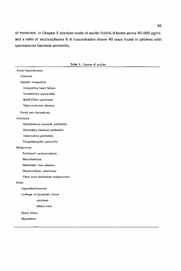
of treatment. In Chapter 5 absolute levels of ascitic fluid IL-6 levels above 60.000 pglml.
and a ratio of ascites/plasma IL-6 concentration above 40 were found in patients with
spontaneous bacterial peritonitis.
Portal hypertension
Cirrhosis
Hepatic congestion
Congestive heart failure
Constrictive pericarditis
Budd-Chiari syndrome
Vena-occlusive disease
Portal vein thrombosis
Infections
Spontaneous bacterial peritonitis
Secondary bacterial peritonitis
Tuberculous peritonitis
FUngal/parasitic peritonitis
Malignancy
Peritoneal carcinomatosis
Mesothelioma
Metastatic liver disease
Hepatocellular carcinoma
Other intra-abdominal malignancies
Other
Hypoalbuminaemia
leakage of lymphatic tissue
pancreas
biliary tract
Renal failure
Myxedema
Table 1. Causes of ascites
36
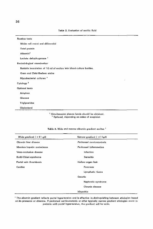
Table 2. Evaluation of ascitic fluid
Routine tests
White cell count and differential
Total protein
Albumin1
Lactate dehydrogenase 1
Bacteriological examination"
Bedside inoculation of 10 ml of ascites into blood culture bottles.
Gram and Ziehf-Neelsen stains
Mycobacterial cultures "2
Cytology 2
Optional tests
Amylase
Glucose
Triglycerides
Cholesterol
1 Simultaneous plasma levels should be obtained. 2 Optional. depending on index of suspicion
Tabla 3, Wide and narrow albumin gradient ascites 1
Wide gradient { > 11 gill Narrow gradient { < 11g/ll
Chronic liver disease Peritoneal carcinomatosis
Massive hepatic metastases Peritoneal inflammation
Vena-occlusive disease Infection
Budd-Chiari syndrome Serositis
Portal vein thrombosis Hollow organ leak
Cardiac Pancraas
Lymphatic tissue
Dncotic
Nephrotic syndrome
Chronic disease
Idiopathic
1 The albumin gradient reflects portal hypertension and is effective in distinquishing between etiologies based on its presence or absence. If peritoneal carcinomatosis or other typically narrow gradiant etiologies occur in
patients with porta! hypertension, the gradient will be wide.
37
Runyon SA. Pathogenesis and diagnosis of spontaneous bacterial peritonitis in cirrhosis. In:Rodes J,
Arroyo V. Therapy in liver disease. Ediciones Doyma, Barcelona. 1992;388-396.'
2 Hoefs JC. Diagnostic Paracentesis: A potent clinical tool. Gastroenterology 1990;98:230-236.
3 Gines P. Spontaneous bacterial peritonitis. CUH opinion Gastro 1992; 8:403-408.
4 AlbillosA, Cuervas-Mons V, Millen I. Canton T, Montes J, 8arfios C, Garrodo A, Escertin P. Ascitic lIuid
polymorphonuclear count and serum to ascites albumin gradient in the diagnosis of bacterial peritonitis.
Gastroenterology 1990;98:134-140,
5 Akt1vadis EA, Runyon SA. Utility of an algoritm in differentiating spontaneous from secondary bacterial
peritonitis. Gastroenterology 1990;98:127~ 133.
6 Wilcox CM, Dismukes WE. Sponteneous bacterial peritonitis. Medicine 1987;66:447~456.
7 Savli H, Pritautz H, Zach G, Eherer A, Schreiber F, Krejs GJ. Spontaneous bacterial peritonitis: a serious
problem in patients with ascites. Eur J Gastroenterol Hepato! 1991 ;4:165-171.
8 Runyon 8A, Montano AA, Akriviadis EA, Antillon MR, Irving MA, Me Hutchison JG. The serum-ascites
albumin gradient is superior to the exudate-transudate concept in the differential diagnosis of ascites.
Ann Int Med 1992;117:216~220.
9 Kajani MA, Yoo YK, Alexander JA et al. Serum-ascites albumin gradients in nonalcoholic liver disease.
Dig Dis Sci 1990;35:33-37.
10 JOngst 0, Xie Y, Gerbes AL Pathofysiology of elevated ascites ftuid cholesterol in malignant ascites.
J Hepatol 1992;14:244-248.
11 Prieto M, Gomez-Lechon MJ, Hoyos M, Castell J, Carrasco M, Berenguer J. Diagnosis of malignant
ascites. Dig Dis Sci 1988;33:833-838.
12 JGngst 0, Gerbes AL, Martin R, Paumgartner G. Velue of ascitic lipids in the differentiation between
cirrhotic and malignant ascites. Hepatology 1986;6:239-243.
13 Gerbes Al, Xie Y, Mezger J, Jangst D. Ascitic fluid concentrations of fibronectin and cholesterol:
comparison of differential diagnostic value with the conventional protein determination. liver
1990;10:152-157.
14 Sch61merich J, Volk BA, Kollgen E, Ehlers 5, Gerok W. Fibronectin concentration in ascites
differentiates between malignant and nonmalignant ascites. Gastroenterology 1984;87:1160-1164.
15 Satz N, Joller-Jemelka HI, Grob PJ, Hofer Ch, Schmid E, Knoblauch M. Tumormarker und immun
modulatorische substanzan im aszites- Ihre wertigkeit als screening und diagnoseparameter. Schweiz
Med Wschr.1989;119:762-765.
16 Satz N. laborchemische untersuchungen im aszites. Schweiz Med Wschr 1991 ;121 :636-647.
17 Yang CH, Liaw YF, Chu CM, Sheen IS. White count, pH and lactate in ascites in the diagnosis of
spontaneous bacterial peritonitis, Hepatotogy 1986;6:85-90.
18 Storgaard JS, Svendsen JH, Hegnhuj, Krintel JJ, Nielsen PB. Incidence of spontaneous bacteria!
peritonits in patients with ascites. Diagnostic value of white blood cett count and pH measurement in
ascitic fluid. liver 1991 ;11:248-262.
19 Atkinson M, losowsky MS. The mechanism of ascites formation in chronic liver disease, Quart J Med
1961 ;30: 163-166.
20 Lieberman Fl, Denison EK. Reynolds TB. The relationship of plasma volume, portal hypertension, ascites
and renal sodium retention in cirrhosis: The ~overflow~ theory of ascites formation. Ann NY Acad Sci

38
1970;170:202-208.
21 Schrier RW, Arroyo V, Bernardi M. Epstein M, Henriksen JH, Rodes J, Peripheral arterial vasodilatation
hypothesis: A proposal for the initiation of renal sodium and water retention in cirrhosis. Hepatology
1988;8:1151-1167.
22 Henriksen JH, Brondtsen F, Sorensen T, Stadeager C, Ring-Larsen H. Reduced central blood volume in
cirrhosis. Gastroenterology 1989;97:1606-1613.
23 Vorobioff J, Bredtfeldt JE, Groszmen Ai. Increased blood flow through the portal system in cirrhotic rats.
Gastroenterology 1984;87:1120·1126.
24 Bosch J,Arroyo V, Betriu A, at 01. Hepatic haemodynamics and the renin-angiotensin-aldosterone system
in ciuhosis. Gastroenterology 1908;78:92-99.
26 Arroyo V, Bosch J, Gaya-Beltran J et al. Plasma renin activity and urinary sodium excretion as
prognostic indicators in nonazotemic cirrhosis with ascites. Ann Inter Med 1981 ;94:194-199.
26 Bender MD, Ockner OK. Ascites,in Fordtran JS, Sieisinger MH, (eds): Gastrointestinal Disease.
Philadelphia, WB Saunders, Co.,1989,pp.428-454.
27 Beutler JJ, Koomans HA, Rabelink T J, at al. Blunted natriuretic response and low blood pressure after
atrial natriuretic factor in early cirrhosis. Hepatology 1989;10:148-163.
28 Salerno F, Badlamenti S, Moser P, Lorenzano E, Incerti P, Dioguardi N. Atrial natriuretic factor in cirrhotic
patients with tense ascites. Gastroenterologv 1990;98:1063-1070.
29 Cattau El, Benjamin S8, Knuff TE, Castell DO. The accuracy of the physical examination in the diagnosis
of suspected ascites. JAMA 1982;247:1164-1166.
30 Jorulf H, Roentgen diagnosis of intraperitoneal fluid. A physical, anatomic and clinical investigation. Acta
Radiol 1976;343{suppl):1-124.
31 Slack M, Friedman AC. Ultrasound examination in the patient with ascites. Ann Inter Med
1989;110:253-255.
32 Runyon SA. Paracentesis of ascitic fluid: a safe procedUre. Arch Intern Med 1986;146:2259-2261.
33 Dienstag JL, Alter HJ. Non·A,Non-S hepatitis: EVolving epidemiologic and cli[lical perspective. Sem liver
Dis 1986;6:67-78.
34 De Sitter l, Rector WG. The significance of bloody ascites in patients with cirrhosis. Am J Gastroenterol
1984;79:136-138.
35 Sultan S, Pauwels A, Poupon R, levy VG. Ascite chyleusa du cirrhotique. Gastroenterol Clin Bioi
1990;14:842-847.
36 Bar Meir S, lerner E, Conn HO. Analysis of ascitic fluid in cirrhosis. Dig Dis Sci 1979;24:136-144.
37 Runyon BA, Spontaneous bacterial peritonitis: an explosion of information. Hepatology 1988;8:171-175.
38 Wang SS, Lu CW, Chao Y at a1. Malignancv-related ascites:a diagnostic pitfall of spontaneous bacterial
paritonitis by ascitic fluid polymorphonuclear cell count. J Hapatol 1994;20:79-84.
39 Hoefs JC. Increasa in ascitas white blood cell and protein concentrations dUring diuresis in patients with
chronic liver disease. Hepatology 1981;1:249.
40 Runyon SA. Cardiac ascites: a characterization. J Clin Gastroentero/ 1988;10:410-412.
41 Mauk PM, Schwartz JT, Lowe JE, Smith JC, Graham JY. Diagnosis and course of nephrogeniC ascites.
Arch Intern Mad 1988;148:1677-1679.
42 Boyer TO, Kahn AM, Raynolds TS. Diagnostic value of ascitic fluid lactic dehydrogenase, protein, and

39
WBC levels. Arch Intern Mad 1978;138:1103-1106.
43 Hoefs JC. Serum protein concentration and portal pressure determine the ascitic fluid protein
concentration in patients with chronic liver disease. J lab Clin Mad 1983;102:260-273.
44 Runyon SA. low protein concentration ascitic fluid is predisposed to spontaneous bacterial
peritonitis. Gastroenterology 1986;91 :1343-1346.
45 Such J, Guarner C, Enriguez J, Rodriquez Jl, Seres I, Vilardell F. Low C3 in cirrhotic ascites
predisposes to spontaneous bacterial peritonitis. J Hepatol 1988;6:80·84.
46 Bird G, Senaldi G, Panos M at al. Activation of the classical complement pathway in spontaneous
bacterial peritonitis. Gut 1992;33:307-311.
47 Rector WG, Reynolds TB. Superiority of the serum-ascites albumin difference over the ascites totel
protein concentration in preparation of transudativa and exudative ascites. Am J Med 1984;77:83-86.
48 Pare P, Talbot J, Hoefs JC. Serum-ascites albumin concentration gradient: a physiological approach to
the differentia! diagnosis of ascites. Gastroenterology 1983;86:240-244.
49 Runyon BA, Hoefs JC, Morgan T. Ascitic fluid analysis in malignancy-related ascites. Hepatology
1988;8:1104-1109.
60 Mauer K, Manzione N. Usefullness of serum-ascites albumin difference in separating transudative from
exudative ascites-another look. Dig Dis Sci 1988;33:1208-1213.
61 Runyon BA, Montano AA, Akriviadis EA, AntH!on MR, Irving MA, McHutchison JG. The serum-ascites
albumin gradient is superior to the exudate-transudate concept in the differential diagnosis of ascites.
Ann Int Med 1992;117:216-220.
62 Varga J, Palmer RC, Koff RS. Chylous ascites in adults. South Mad J 1986;78:1240-1248.
63 Press OW, OUman-Press N, Kaufman SO. Evaluation and management of chylous ascites. Ann Int Med
1982;96:368-364.
64 Uchiyama T, Yamamato T, Mizuta E, Suzuki T. Pancreatic ascites-a collected review of 37 cases in
Japan. Hepato-Gastroenterol. 1989;36:242+249.
66 Bergmann JF, Bidart JM, George M, Beaugrand M, levy VG, Bohuon C. Elevation of CA 126 in patients
with benign end malignent ascites. Cancer 1987;69:213-217.
66 Mezger J, Wilmanns W, Lamerz R. Elevated serum CA 126 levels in patients with benign ascitic or
pleural effusions. Tumor Bioi 1988;9:47-62.
67 Mezger J, PermaneUer W, Gerbes Al, Wilmanns W, Lamerz R. Tumour associated antigens in
diagnosis of serous effusions. J Clin Pathol 1988;41 :833-843.
68 Tomb JA, A cytopathological study on serous fluid in cancer. Lab Med J 1974;27:61-58.
69 Ghilain JM, Henrion J, Shapiro M, Majois F, Beauduin M, Hellar FR.Le liquide d'ascite: interet de divers
tests biologiques dans Ie diagnostic differential entre ascite cirrhotique et neoplastique. Acta
GastroEnterol Belg 1990;63:168-179.
60 Croonen AM, Valk van der P, Chester JH, Lindeman J. Cytology, Immunopathology and flow
cytometry in the diagnosis of pleural and peritoneal effusions. Laboratory Investigation 1988;68:726-
732.
61 Liach J, Rimola A, Navasa M et 81. Incidence and predictive factors of first episode of spontaneous
bacteria! peritonitis in cirrhosis with ascites: Ralevance of ascitic fluid protein concentration.
Hepatology 1992;16:724-727.
40
62 Pinzello. G, Simonetti RG, Craxi A, Piazza SO, Spano C, Pagliaro L. Spontaneous bacterial paritonitis:A
prospective investigation in predominantly nonalcoholic cirrhotic patients. Hapetology 1983;3:645-549.
63 Kurtz CR, Bronza Rl. Does spontaneous bacterial peritonitis occur in malignant ascites? Am J
Gastroenterol 1982;77:146·148.
64 Runyon SA. Spontaneous bacterial peritonitis associated with cardiac ascites. Am J Gastroenterol
1984;79:796.
65 Siersema PO, de Marie S, van Zeijl JH, Bae OJ, Wilson JHP. Blood culture bottles are superior to lysis
centrifugation tubes for bacteriological diagnosis of spontaneous bacterial peritonitis. J Clio Microbial
1992;30:667-669.
66 Runyon SA. Umland El. Merlin T. Inoculation of blood culture bottles with ascitic fluid. Arch Intern Med
1987;147:73-75.
67 Runyon SA, Canawati HC, Akriviadis EA. Optimization of ascitic fluid culture technique.
Gastroenterology 1988;96:1361-1366.
68 Bobedilla M, Sifuentes J, Garcia-Tsao G. Improved method for bacteriological diagnosis of spontaneous
bacterial peritonitis. J Clin MicrobioI1989;27:2146-2147.
69 Runyon SA, AntiUon MR, Akrivadis A, McHutchison JG. Bedside inoculation of blood culture bottles with
ascitic fluid is superior to delayed inoculation in the detection of spontaneous bacterial peritonitis. J Clin
Microbiol 1990;28:2811-2812.
70 Runyon BA, Hoefs JC. CUlture-Negative Neutrocytic Ascites: A variant of spontaneous bacterial
peritonitis. Hepatology 1984;4:1209-1211.
71 Pelletier G, Salmon 0, Ink 0, Hannoun S, Attal1 P, Buffet C, Etienne JP. Culture·negative neutrocytic
ascites: a less severe variant of spontaneous bacterial peritonitis. J Hepatol 1990;10:327·331.
72 Runyon BA. Monomicrobial nonneutrocytic bacterascites: A variant of spontaneous bacterial
peritonitis. Hepatology 1990:12:710·716
73 Pelletier G, Lesur G, Ink 0 et al. Asymptomatic bacterascites: Is it spontaneous bacterial peritonitis?
Hepatology 1991 ;14:112-115.
74 Hoefs JC, Jonas GM. Diagnostic paracentesis. Adv Int Med 1992;391-409.
76 Runyon BA, Hoefs JC. Spontaneous vs Bacterial peritonitis: Differentiation by response of ascitic fhid
neutrophil count to antimicrobial herapy. Arch Intern med 1986;146:1663·1565.
76 Manohar A, Simjee AE. Haffejee AA, Pettengell KE. Symptoms and investigatiVe findings in 146 patients
with tuberculous peritonitis diagnosed by peritoneoscopy and biopsy over a five year paTiod. Gut
1990;31 :1130-1132.
77 Reddy KA, DiPrima RE, Raskin JB et at. Tuberculous Peritonitis: Laparoscopic diagnosis of an
uncommon disease in the United States. Gastrointestinal Endoscopy 1988;34:422-426.
78 Zeni F, Tardy B, Vindimian M et a1. Local synthesis of tumor necrosis factor~a and lnterleukin·l in the
peritoneal cavity during spontaneous bacteria! peritonitis. J Infect Dis 1991 ;164:1241-1243.
79 Deviere J, Content J, Crusiaux A, Dupont E. lL-6 and TNF-a iii ascitic fluid during spontaneous bacterial
peritonitis. Dig Dis Sci 1991 ;36:123~ 125.
80 Pelletier G, Briantais MJ, Seta N, Lebrun l, Durand G, Galanaud P. Effects of spontaneous bacterial
peritonitis on ascitic fluid interleukin-6 and al-acid glycoprotein levels in cirrhotic patients. Eur J
Gastroenterol Hepatol 1992;4:295-300.
41
81 Andus T, Gross A, Hoistege M at ai, Evidence for the production of high amounts of interleukin-6 in
the peritoneal cavity of patients with ascites. J Hepatol 1992;15:378-381.

CHAPTER 3
OPTIMAL ANALYSIS OF TOTAL PROTEIN. ALBUMIN. WHITE CEll COUNT
AND DIFFERENTIAL IN ASCITIC FLUID
43
The contents of this chapter have been submitted for publication to Ann elin Biochem
under the same title with the following authors: H. Engel. D.J. Bac. R. Brouwer. B. G.
Blijenberg. and J. Lindemans.
44
Summary
Accurate and precise procedures are described for the determination of total protein,
albumin, absolute white cell count and differential count in ascitic fluid. The total protein
method (biuret) on our routine serum chemistry analyzer, the Bayer-Technicon Chem-1™,
was calibrated in the measuring ranges covering serum as well as ascitic fluid values (1 -
1 00 g/l) against the biuret reference method. The albumin method (BeG: bromcresol
green) on the Chem-1 ™ was calibrated for these measurement ranges (1 - 50 g/l) against
new human plasma protein international reference preparations and compared to the
nephelometric method on the Beckman ArrayTM, which was also calibrated against these
reference preparations. A good correlation was obtained for total protein between the
Ghem-l ™ (y) and the biuret reference method (x) in 58 ascitic fluids (y = 1.02x - 0.3; r
= 1.00) and can be measured down to 1 gIl. Also, a good correlation between the Chem~
1 ™ (y) and the ArrayTM (x) was obtained for albumin (r = 0.99), however the BCG
method, which is not fully specific, results in significantly higher results (y = 1.32x ~ 1.3).
Therefore, the immunochemical procedure will be more accurate for the determination of
albumin in ascitic fluid.
The white cell count is carried out on a haematology analyzer. Depending on the absolute
count, a reliable and convenient procedure was established for the manual differential. The
ascitic fluid leucocytes in smears are equally distributed among artificial red and white
blood cells from a quality control sample, the artificial white cells are clearly distinguisable
from ascitic fluid white cells. The ascitic fluid is either directly mixed with this blood when
the absolute count is above 4000 cells/mm 3, or the pellet is mixed with this blood after
centrifugation of the ascitic fluid when the count is between 300 and 4000 cells/mm3•
This type of analysis is preferred above the cytospin procedure, and is currently also
applied for other biological fluids, i.e. pleural fluid, and synovial fluid.
45
Introduction
A variety of conditions have been described to be responsible for the accumulation of
peritoneal fluid. The major causes are cirrhosis of the liver (75%). malignancy (12%). and
cardiac failure (5%). A more comprehensive list of causes is given in Table 1 (page 35).
The diagnostic value of differences in ascitic fluid composition for the differentiation
between the various aetiologies have been reported earlier (1,2). Traditionally, ascitic fluid
was analyzed for absolute white cell count, total protein concentration, and for bacterial
growth. The ascitic fluid total protein concentration was used to classify the fluids in
categories like exudate (> 25 gIl) or transudate « 25 9/1) in order to distinguish
aetiologies like bacterial peritonitis, tuberculous peritonitis, pancreatitis, and peritoneal
carcinomatosis from cirrhosis, heart failure, and nephrotic syndrome respectively. This
exudate-transudate concept has resulted in many problems and exceptions (3A). Since the
serum oncotic pressure, which is closely related to the serum albumin concentration, and
the hydrostatic pressure gradient, which is determined primarily by the portal pressure
gradient, are both responsible for ascitic fluid accumulation, the serum-ascitic fluid albumin
gradient is superior to the total protein concentration when used to distinguish between
different causes of ascites (5). An albumin gradient wider than 11 gIl is indicative of the
presence of portal hypertension, and a narrow gradient « 11 g/l) of the absence of portal
hypertension (6).
It might be clear in this respect that an accurate and precise determination of albumin in
ascitic fluid is of great importance. Procedures are available for the determination of total
protein and albumin in urine and cerebrospinal fluid in mg amounts per liter, however these
methods are not directly applicable to ascitic fluid because the values range for total
protein from 1 - 45 gl) and for albumin from 1-30 gil. Also, the methods on the most
common routine serum chemistry analyzers for total protein and albumin can not be
directly applied for ascitic fluid analysis, the lower limits, as far as linearities are
concerned, being approximately 25 gIl and 15 gIl respectively.
The ascitic fluid white cell count, especially the percentage of polymorphonuclear (PMN)
cells, is the most important determinator for bacterial infection (7,S). The diagnosis of
spontaneous bacterial peritonitis (SSP) in a patient with advanced cirrhosis is often difficult
to make and easily missed, as clinical signs and symptoms are often lacking. A low
threshold to perform a paracentesis followed by an accurate white cell count and
differential of the ascitic fluid is essential to exclude the presence of peritonitis in any
46
patient with a deteriorating condition. A PMN count greater than 250 cells/mm3 with signs
or symptoms of SSP, or a PMN count> 500 cells/mm 3 without signs or symptoms of S8P
are an indication to start broad-spectrum antibiotics, even before culture results are
available. An early diagnosis of S8P is important because of several reasons. First of all,
awareness and early diagnosis of S8P improves the mortality and secondly the presence
of S8P precludes the possiblity of liver transplantation until the infection has been properly
treated. A lymphocyte predominance in ascitic fluid is found in tuberculosis, fungal
infections and peritoneal lymphoma. These considerations emphasize the importance of a
reliable white cell count and differential but, again, established reliable techniques are
lacking.
Therefore, we describe methods for the accurate and precise assessment of on the one
hand total protein and albumin and on the other for the performance of an accurate white
cell count and cell differentiation in ascitic fluid.
Materials and Methods
Materials
Haematology Quality Control blood was purchased from J.T. Baker (Philipsburg, PA, USA).
Human plasma protein international reference preparations (OSAU 07 and ORDT 07), also
containing albumin, were obtained from Behringwerke AG (Marburg, Germany).
Procedure for the accurate determination of total protein and albumin
Total protein and albumin were determined on a Chem~1TM routine serum chemistry
analyzer (Bayer-Technicon, Tarrytown, NY, USA). The analytical ranges were verified using
serum pools with high analyte concentrations and dilutions with saline according to NeClS
EP6-P recommendations (9). The total protein method on the analyzer was calibrated
against the biuret reference method in a measuring range covering serum as well as ascitic
fluid (1 - 100 g/l) with neat and saline·diluted patient sera (10). The albumin method (BCG:
bromcresol green) on the Chem-1™ was calibrated for the combined measuring range for
serum and ascitic fluid (1 - 50 g/l) using two human plasma protein international reference
preparations and their dilutions with saline. The reference preparations had assigned values
for albumin established by radial immunodiffusion, nephelometry, and turbidimetry,
47
performed by 8ehringwerke AG (Marburg, Germany) and were calibrated against the new
International Reference Material CRM No.4 70 (Bureau Communitaire de Reference of the
European Economic Community, Brussels, Belgium). The Chem-1™ BeG method was
compared with nephelometry using a Beckman ArrayTM Protein System nephelometer (Brea,
CA, USA), also calibrated with these two reference preparations.
Procedure for assessment of haematology parameters
Ascitic fluid was collected in an EDT A Vacutainer tube for the determination of the
absolute white cell count on a Sysmex NE-8000n.t analyzer (TOA Medical Electronics Co.,
Kobe, Japan). If leucocytes were above 4000 cells/mm3, a 5-part manual 100-cell
microscopic differential count was performed. For that purpose, 1 volume ascitic fluid was
mixed with 1 volume Haematology Quality Control blood and a wedge smear was
prepared. With this procedure, the ascitic fluid white cells are equally distributed among
red cells which results in a better assessment of the differential count. Direct manual
analysis of ascitic fluid white cell differential results often in clumped leucocytes near the
edge of the smear which, consequently, leads to an unreliable differential count. The
artificial white cells in the control blood appear as filled blue spheres and can be clearly
distinguished from ascitic fluid white cells.
If there are between 300 and 4000 leucocytes!mm', the ascitic fluid is centrifuged for 3
minutes at 2000 rpm, the supernatant is discarded and 50 pi of the pellet is mixed with
50 pi Baker blood. A manual 100-cell microscopic 5-part differential is carried out as
described above. Below 300 leucocytes/mm 3, no differential is carried out.
Imprecision
Imprecision of the total protein and albumin methods was evaluated following the NeClS
EP5-T2 guidelines (11). The within-run and between-run imprecisions were determined
using serum pools and dilutions thereof with low, normal and high concentration of total
protein and albumin with respect to the ascitic fluid measuring ranges and were analyzed
in single assays or on 10 different days, respectively.

48
Patient samples
A total number of 58 patient ascitic fluids were collected and analyzed for total protein on
the Chem-1 ™ and with the reference biuret method, and for albumin on the Chem-1 ™ and
the ArrayTM,
Method comparison
The manual biuret reference procedure for the determination of total protein was performed
on a Philips spectrophotometer (Eindhoven, The Netherlands), Regression analysis was
performed according to the procedure described by Passing & Bablok (12).
Results and Discussion
The determination of total protein and albumin in ascitic fluid on a routine serum chemistry
analyzer is described. The linearity ranges were expanded to lower regions for both
parameters considering the ascitic fluid measuring ranges. The total protein and albumin
methods, on our currently in USB routine serum chemistry analyzer, were standardized over
the complete ascitic fluid and serum measuring ranges against either a reference method
for total protein or human plasma protein international reference preparation for albumin
(13, 14). These new international reference preparations for albumin have been prepared
recently under auspicies of the International Federation of Clinical Chemistry (IFCC) in
collaboration with the Bureau Communitaire de Reference of the European Economic
Community (Brussels, Belgium), and the College of American Pathologists (CAP, Northfield,
USA).
A total number of 58 ascitic fluids were analyzed for total protein and albumin on the
Chem-1 ™ as well as total protein with the biuret reference method and albumin on the
ArrayTM nephelometer (Table 2). Total protein could be measured down to 1 g/l on the
Chem-1 ™ with respect to the lower limit of the linearities and a good correlation was
obtained between the Chem-1™ and the biuret reference method (y (Chem-1) = 1.02x
(biuret) - 0.3; r :::: 1.00; fig. 1 A). In addition, a good correlation was found for albumin
between the Chem-1 1M and the ArrayTM (r :::: 0.99), however, above 7 gIl the Chern-1 1M
gives significantly higher results (y (Chem·1) = 1.32x (Array) - 1.3; fig. 1 B). Since the
BCG method is not fully specific for albumin and since albumin values of the reference
49
preparations were assigned by radial immunodiffusion, turbidimetry, and nephelometry, we
conclude that albumin in ascitic fluid should be determined nephelometrically. This type of
analysis might be difficult to perform when ascitic fluid, concerning total protein and
albumin analysis, must be analyzed immediately 24 h a day. However, this is not the case
in our hospital. When necessary, only total protein is analyzed immediately and albumin
afterwards.
Total protein is accurately and precisely analyzed in the ascitic fluid measuring ranges. The
between-run coefficients of variation are smaller than 3 % over the complete measuring
range (Table 3).
The absolute white cell count in ascitic fluid with our haematology analyzer, a Sysmex NE-
8000H,\ showed a good correlation with the manual count (Table 2). However, the result
of white cell differential count by the analyzer is unreliable due to the abundance of
degenerated leucocytes. Traditionally, a smear is prepared and manual white cell
differential count is carried out, this leads however either to almost no leucocytes in the
smear when the absolute count is low, or the leucocytes are clustered near the edge of
the smear when the absolute count is high. In both cases, these manual differential counts
are not reliable. Our current procedure, as described in the Materials and Methods section,
results in smears with the ascitic fluid leucocytes equally distributed among artificial red
and white blood cells from the haematology quality control material (fig. 2). This procedure
is preferred above the cytospin procedure which is performed in order to differentiate cells
present in cerebrospinal fluid. The cytospin procedure, applied to ascitic fluids with high
absolute white cell counts, results in smears of highly concentrated leucocytes which are
difficult to differentiate.
Depending on the absolute count, the ascitic fluid is mixed either directly with an equal
volume of Baker blood (when the white cell count is above 4000 cells/mm3) or the pellet
is mixed after centrifugation of the ascitic fluid (when the white cell count is between 300
and 4000 cells/mm3). This procedure is very convenient, gives reproducible results and is
currently also applied for pleural and synovial fluids.
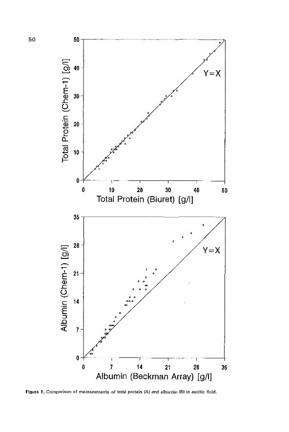
50 50
~
-=:. 40 OJ y=x ~
~
~ , E OJ 30 ..c 0 ~
c OJ ...- 20 0 ~
a... <1l 10 ...-~
0 0 10 20 30 40 50
Total Protein (Biuret) [g/I)
35
~ 28 y=x -=:. ~ ~
~ , 21 E
OJ .. ..c 0
. . ~
c 14 -
E ::J .0
« 7
o o 7 14 21 28 35
Albumin (Beckman Array) [g/I)
Figura 1. Comparison of measurements of total protein (AI and albumin (B) in ascitic fluid.
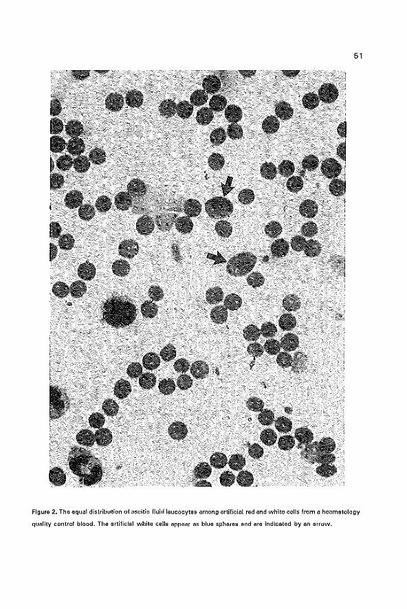
51
Figure 2. The aqua! distribution of ascitic fluid leucocytes among artificial red and white cells from a heamatology
quality control blood. The artificial white cells appear as blue spheres and are indicated by an arrow.
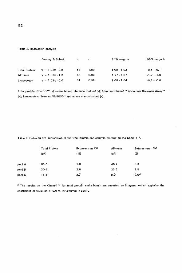
52
Table 2. Regression analysis
Passing & BabIak n 96% range a 96% range b
Total Protein y 1.02x - 0.3 58 1.00 1.00 - 1.03 -0.6 - -0.1
Albumin y 1.32x - 1.3 68 0.99 1.27 - 1.37 -1.7 - -1.0
Leucocytes y 1.02x - 0.0 31 0,98 1.00 - 1.04 -0.1 - 0.0
Total protein: Chern_1 1M (y) versus biuret reference method (xl; Albumin: Chem-' 1M (y) versus Beckman Array'M
(x); Leucocytes: Sysmex NE_8000lM (y) versus manual count (x).
Table 3. Between-run imprecision of the total protein and albumin method on the Chem-1 fM.
Total Protein Between-run CV Albumin Between-fun CV
(gill (%) (gill (%)
pool A 68,8 1.8 46.2 0.9
pool B 30.8 2.6 23.9 2.9
pool C 16.8 2.7 9.0 0.0.1
-) The results on the Chem-1 TM for total protein and albumin are reported as integers, which explains the
coefficient of variation of 0.0 % for albumin in pool c.
53
1. Bac OJ, Siersema PO, Wilson JHP. Paracentesis; The importance of optimal ascitic fluid analysis. Neth
J Mad 1993; 43: 147-166.
2. Runyon SA. Current concepts: Care of patients with ascites. New Engl J Mad 1994; 330: 337-343.
3. Rector WG, Reynolds TB. Superiority of the serum-ascites albumin difference over the ascites total
protein concentration in separation of transudativa and exudative ascites. Am J Mad 1984; 77: 83·85.
4. Runyon BA, Montano AA, Akrivadis EA, Antillan MR, Irving MA, Me Hutchison JG. The serum-ascites
albumin gradient is superior to the exudate-transudate concept in the differential diagnosis of ascites.
Ann Intern Mad 1992; 117: 215·220.
5. Hoefs JC. Serum protein concentration and portal pressure determine the ascitic fluid protein
concentration in patients with chronic liver disease. J Lab Clin Med 1983; 102: 260-273.
6. Hoefs JC. Diagnostic paracentesis: a potent clinical tool. Gastroenterology. 1992; 8: 230-236.
7. Runyon BA. Spontaneous bacterial peritonitis: an explosion of information. Hepatology. 1988; 8: 171-175.
8. Wang SS, Lu CW, Chao Y, Lee MY, Lin HC, Lee SD, Tsai YT, Chen CC, Lo Kl. Malignancy-related
ascites: a diagnostic pitfall of spontaneous bacterial peritonitis by ascitic f1uid polymorphonuclear cell count.
J Hepatol 1994; 20: 79-84.
9. Passey RB, Bee BE, Caffo A, Erikson JM. Evaluation of the linearity of quantitative analytical methods;
proposed guidline. 1986. NCCLS Document EP6-P. Villanova, PAj National Committee for the Clinical
Laboratory Standards.
10. Reinhold JG. TotaJ protein, albumin, and globuline. Stand Methods Clin Chem 1953; 1: 88-97.
II. Kennedy JW, Carey RN, Coolen RB, Garber CC, Hartmann AE, Lee HT, Levine lE, Osberg IM:.
Evaluation of precision performance of clinical chemistry devices. 1992. NCCLS Document EP5-T2.
Villanova, PA; National Committee for Clinical Laboratory Standards.
12. Passing H, Babiak W. A new biomedical procedure for testing the equality of measurements from two
different analytical methods. J Clin Chem Clin Biochem 1983; 21: 709-720.
13. Bandner S. Why a new International reference preparation ORP) for human plasma proteins. J Clin ub
Analysis 1993; 7: 273-277.
14. Johnson ML A new international reference preparation for proteins in human serum. Arch Palho! Lab
Med 1993; ll7: 29-31.

CHAPTER 4
BLOOD CULTURE BOTTLES ARE SUPERIOR TO LYSIS-CENTRIFUGATION
TUBES FOR BACTERIOLOGICAL DIAGNOSIS OF
SPONTANEOUS BACTERIAL PERITONITIS
55
The contents of this chapter have been published under the same title in J Clin Microbial
1992; 30, 667-669 by the following authors: P.O. Siersema, S.de Marie, J.H. van Zeijl,
D.J. Sac and J.H.P. Wilson.
56
Summary
The conventional method of ascitic fluid culture was compared with the bedside
inoculation of ascites into blood culture bottles and into lysis-centrifugation tubes. The
conventional culture method was compared with the blood culture bottle method in 31
episodes of spontaneous bacterial peritonitis (S8P). Cultures were positive with the
conventional method in 11 (35%1 and with the blood culture bottle method in 26 (84%)
episodes (p<O.OOl). The lysis-centrifugation tube method could be compared with the
blood culture bottle method in 24 episodes of SBP. Cultures were positive with the Iysis
centrifugation tube method in 11 (46%) and with the blood culture bottle method in 19
(79%) episodes (p<O.05). Moreover, the blood culture bottle method also shortened the
time for the detection of bacterial growth. In conclusion. bedside inoculation of ascites into
blood culture bottles should be used routinely in patients with suspected S8P. Culturing
of ascites in lysis-centrifugation tubes is more laborious than and inferior to that in blood
culture bottles.

57
Introduction
Spontaneous bacterial peritonitis (SSP) is a frequent and severe complication of cirrhosis
with ascites (4). With conventional ascitic fluid culture methods, consisting of inoculation
of ascites into thioglycolate broth and onto agar plates, cultures are positive in only 42 to
57% of episodes of suspected SSP 11,10-12). In 1987, Runyon et a!. reported that
sensitivity of ascites cultures could be improved by inoculation of ascites into blood culture
bottles (11). Recently this finding was confirmed in three prospective studies 11.10,12).
When we started our study in 1987 , the purpose was to prospectively compare the
conventional method with the blood culture bottle method of ascitic fluid culturing.
Meanwhile, it was demonstrated that even with blood culture bottles, ascites cultures
were negative in up to 19% of cases of suspected S8P (1,10·12). Therefore, in an effort
to further improve sensitivity of ascites cultures, starting in 1988, we also inoculated
ascites into Iysis·centrifugation tubes (8). Lysis-centrifugation tubes have been reported
to be as good or even more effective than blood culture bottles for the detection of Gram
negative bacilli (5), which account for 69% of isolates in S8P (14). On the other hand, the
detection rate of Gram-positive bacteria has been demonstrated to be lower with Iysis
centrifugation tubes (5). Since streptococci account for 26% of isolates of S8P (14). this
could cause a lower detection rate of these microorganisms in cases of suspected S8P.
Materials and methods
Between July 1987 and April 1991 all patients with liver cirrhosis and ascites, who were
suspected of having S8P, were included in the study. A diagnosis of S8P was suspected
if: a) patients had clinical signs and symptoms compatible with a diagnosis of S8P
(abdominal pain andlor fever andlor worsening of hepatic encephalopathy without another
explanation), b) the ascitic fluid polymorphonuclear IPMN) count was" 500/mm3 (3) and
c) there was no intra-abdominal source of infection. In all episodes of suspected S8P an
alternative explanation for an elevated PMN count, e.g. pancreatitis, cholecystitis,
peritoneal carcinomatosis, hemorrhage into ascites, or tuberculosis was exluded. Patients
were also excluded if antibiotic therapy had been given within 14 days of presentation.
During the period from June 1987 to April 1991, both the conventional culture method
and the blood culture bottle method were simultaneously used to test ascitic fluid samples.
In addition, from November 1988 to April 1991. the lysis-centrifugation tube method was
58
used to test all ascitic fluid samples. Ascites was obtained under sterile conditions by
paracentesis with or without ultrasound-guided control.
For the conventional method, 10 mt of ascites was transported immediately to the
Microbiology Laboratory. The specimen was centrifuged at 3000g for 5 min. Gram staining
was performed on all centrifuged sediments. The sediments were inoculated on blood agar,
MacConkey agar I chocolate agar and GC agar plates for aerobic cultures and incubated for
48 h in a 5% CO 2 atmosphere at 3rC. In addition, ascites was inoculated on Brucella
blood agar, Brucella blood agar with 100 pg/ml neomycin, and Brucella blood agar plates
with 100 pg/mt kanamycin and 7.5 pg/mt vancomycin, and incubated under anaerobic
conditions for 5 days at 37°C. Thioglycolate medium was inoculated and subcultured onto
Brucella blood agar plates after 2 days for aerobic culture and after 5 days for anaerobic
culturing; these agar plates were subsequently incubated for 2 days at 37'e.
For the blood culture bottle method, 5 ml of ascitic fluid was inoculated at the bedside in
45 ml of Columbia broth (with sodium polyanetholesulfonate and increased cysteine)
(Becton Dickinson vacutainer systems, Becton Dickinson and Company, Rutherford, New
Jersey, USA) for aerobic incubation and 5 ml was inoculated in 45 ml of Columbia broth
for anaerobic incubation at 3rc. Subculturing was performed as soon as growth was
visible or blindly after 2 and 7 days from the aerobic cultures, and after 7 days for the
anaerobic cultures.
For the lysis-centrifugation tube method, 10 ml of ascites was inoculated at the bedside
into a lysis-centrifugation tube (du Pont Isolator 10-tube, E.I. du Pont de Nemours & Co.,
Inc., Wilmington, Delaware, USA). After centifugation at 3000g for 30 min, the pellet was
inoculated onto the culture media of the conventional culture method.
The times at which cultures became positive were recorded and microorganisms were
identified by standard laboratory methods as described in the Manual of Clinical Microbio
logy (7).
The ascitic fluid PMN count was calculated as the percentage of PMN's per total amount
of leucocytes in ascites samples. The total amount of leucocytes was counted in a Sysmex
NE 8000· (TOA Medical Electronics, Kobe Japan). For the calculation of the percentage
of PMN's, 10·20 ml of ascites was centrifuged at 2000 rpm for 10 minutes. The pellet
was stained with Giemsa-stain. At least 50 cells were counted to determine the
percentage of PMN's.
For the statistical analysis of the difference between the results of the three culture
methods, the McNemar chi-square test (3) was used. For the statistical analysiS of the
59
difference in the median time interval for detection of bacterial growth between the three
culture methods, the SAS lifetest procedure was used (6). A P value of < 0.05 was
considered significant.
Results
During the study, 31 episodes of suspected SSP were diagnosed in 28 cirrhotic patients
with ascites. In the 31 episodes of suspected SBP, the mean ± SO PMN count in ascites
was 4,900 ± 2,700/mm' (range, 600 to 9,400/mm'). In 2 episodes of SBP, the Gram
stain of ascitic fluid revealed Gram-negative rods, whereas in the other 29 episodes the
Gram stain did not detect bacteria. The conventional method was compared with the blood
culture bottle method in 31 episodes of suspected SBP. Ascites cultures were positive by
the conventional method in 11 of 31 (35%) episodes and by the blood culture bottle
method in 26 of 31 (84%) episodes (p<O.OOl). Conventional cultures did not grow
bacteria that were not also detected by the blood culture bottle method. The Iysis
centrifugation tube method was compared with the conventional method and with the
blood culture bottle method in 24 episodes of suspected SSP. Ascitic fluid samples were
positive by the conventional method in 8 of 24 (33%) episodes, by the lysis-centrifugation
tube method in 11 of 24 (46%) episodes (p, not significant) and by the blood culture bottle
method in 19 of 24 (79%) episodes (blood culture bottle method vs. lysis-centrifugation
tube method: p < 0.05; blood culture bottle method vs. conventional method: p <0.001).
All isolates detected by the lysis-centrifugation tube method were also detected by the
blood culture bottle method. In Table 1 the microorganisms isolated by the conventional
culture and the blood culture bottle methods were compared. For the 26 CUlture-positive
episodes, Gram-negative bacilli were detected in 17 episodes (65%) and Gram-positive
cocci in 9 episodes (35%). Multiple microorganisms in a single ascites specimen or
anaerobes were not isolated. The median time intervals for detection of bacterial growth
was 48 h (range, 24 h to 120 h) for the blood culture bottle method (26 episodes) and 72
h (range, 24 h to 120 h) for the conventional method (11 episodes) (p<0.001).
In Table 2 the microorganisms isolated by the lysis-centrifugation tube, the conventional
culture and the blood culture bottle methods were compared. The median time intervals
for detection of bacterial growth was 48 h (range, 24 h to 120 h) with the blood culture
bottle method (19 episodes), 48 h (range, 24 h to 120 h) with the lysis-centrifugation tube
method (11 episodes) (p < 0.01), and 72 h (range, 48 to 120 h) for the conventional
60
culture method (8 episodes). (blood culture bottle method versus lysis-centrifugation tube
method, p < 0.01; blood culture method versus conventional culture method, p< 0.001).
Blood cultures were done for 24 patients and were positive in 10 (42%) episodes (in 9
episodes, the same microorganism was isolated from blood as from ascites; in 1 episode,
Klebsiella pneumoniae was isolated only from blood).
Discussion
In accordance with the findings of others (1,10-12), our study demonstrates that the
bedside inoculation of ascites into blood culture bottles is superior to conventional methods
of ascitic fluid culturing in the detection of bacterial growth in SBP (84% versus 35%;
p<O.OOl).
Among the isolated pathogens, staphylococci were detected in 2 episodes (1 with
StaphyJococcus aureus and 1 with Staph. epidermidis). Strikingly, both these patients were
treated with esophageal injection sclerotherapy for variceal hemorrhage, 24 hrs. and 36
hrs. respectively before these microorganisms were isolated. Three cases of SBP, following
injection sclerotherapy, have also been reported by Tam et al. (13). In one of these cases
S.aureus was also isolated.
In cases of S8P very low concentrations of bacteria are detected in ascites (10). This fact
is demonstrated by the finding that only 2 (6%) of the 31 Gram stains of centrifuged
ascites, revealed microorganisms, in accordance with findings of Sainz et al. (12), who
also found Gram stains of ascites to be positive in only 6% of cases of SSP.
In our study, ascitic fluid cultures remained negative in 5 (16%) of the 31 episodes of
suspected SSP, comparable to the results of other studies (7 to 19% negative cultures)
(1,10,11). There are two possible explanations for the failure rate of blood culture bottles
in our study. In the blood culture bottle system that we used, only 5 ml of ascites is
inoculated. Runyon et a!. reported that inoculation of larger amounts of ascites (10 or 20
ml) resulted in more CUlture-positive episodes than the inoculation of smaller amounts of
ascites (10). Moreover, blood culture bottles containing Thiol have been found to be more
sensitive than bottles containing tryptic soy broth (10). Since we used Columbia broth
bottles, it remains to be established whether these bottles are less sensitive than blood
culture culture bottles containing Thiol for the detection of microorganisms from ascites.
Blood cultures with lysis-centrifugation tubes have been described to increase the isolation
rate of both Gram-negative bacilli, and fastidious and intracellularly growing micro-
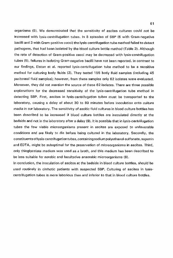
61
organisms (5). We demonstrated that the sensitivity of ascites cultures could not be
increased with lysis-centrifugation tubes. In 8 episodes of SSP (5 with Gram-negative
bacilli and 3 with Gram-positive cocci) the lysis-centrifugation tube method failed to detect
pathogens, that had been isolated by the blood culture bottle method (Table 2). Although
the rate of detection of Gram-positive cocci may be decreased with lysis-centrifugation
tubes (5). failures in isolating Gram-negative bacilli have not been reported. In contrast to
our findings, Elston at al. reported lysis-centrifugation tube method to be a sensitive
method for culturing body fluids (2). They tested 155 body fluid samples (including 45
peritoneal fluid samples); however, from these samples only 62 isolates were evaluated.
Moreover, they did not mention the source of these 62 isolates. There are three possible
explanations for the decreased sensitivity of the lysis-centrifugation tube method in
detecting S8P. Firsc ascites in lYSis-centrifugation tubes must be transported to the
laboratory, causing a delay of about 30 to 60 minutes before inoculation onto culture
media in our laboratory. The sensitivity of ascitic fluid cultures in blood culture bottles has
been described to be increased if blood culture bottles are inoculated directly at the
bedside and not in the laboratory after a delay (9). It is possible that in Iysis·centrifugation
tubes the few viable microorganisms present in ascites are exposed to unfavorable
conditions and are likely to die before being cultured in the laboratory. Secondly, the
constituents of Iysis-centri fugation tubes, containing sodium polyethanol-sulfonate, saponin
and EDTA, might be suboptimal for the preservation of microorganisms in ascites. Third,
only thioglycolate medium was used as a broth, and this medium has been described to
be less suitable for aerobic and facultative anaerobic microorganisms (8),
In conclusion, the inoculation of ascites at the bedside in blood culture bottles, should be
used routinely in cirrhotic patients with suspected S8P. Culturing of ascites in Iysis
centrifugation tubes is more laborious than and inferior to that in blood culture bottles.
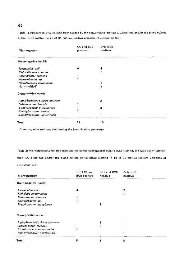
62
Table 1: Microorganisms isolated from ascites by the conventional culture {CCI method and/or the blood culture
bottle (8CB) method in 26 of 31 culture-positive episodes of suspected SSP.
Microorganism
Gram-negative bacilli:
Eschedchia coli Klebsiella pneumoniae Enterobacter cloacae Acinetobacter sp. Pseudomonas aeruginosB Not specified'
Gram-positive coocl:
Alpha-hemolytic Streptococcus Enterococcus fn8ca/is Streptococcus pneumon;ae Staphylococcus aureus Staphylococcus epidermidis
Total
CC and BCB positive
6
11
Only BCB positive
4 2
2 1
2 1 2
15
1 Gram-negative rod that died during the identification procedure
Tabla 2: Microorganisms isolated from ascites by the conventional culture (CC) method, the lysis-centrifUgation
tube (LCT) method andlor tho blood culture bottle (BCS) method in 19 of 24 CUlture-positive episodes of
suspected SBP_
Microorganism
Gram-negative bacilli:
Escherichill coli Klebsiella pneumonille Enterob8cter cloacae Acinetob8cter sp_ Pseudomonas aeruginosa
Gram-positive cocci:
Alphe-hemolytic Streptococcus Enterococcus faecalis Streptococcus pneumoniae Staphylococcus epidermidis
Total
CC, LCT and BCS positiVe
4
8
LCT and BCB positive
3
Only BCB positiVe
3 2
8

63
(1) Bobadilla M., J. Sifuentes, and G. Garcia-Tsaa. 1989, Improved method for bacteriological diagnosis of
spontaneous bacterial peritonitis. J. Clin. Microbial. 27:2145-2147.
(2) Elston H.R., M. Wang, and A. Philip. 1990. Evaluation of isolator system and large-volume centrifugation
method for culturing body fluids. J, Clin. Microbial. 28: 124-126.
(3) Fleiss J.l. (ed.). 1981. Statistical methods for retes and proportions, 2nd ed. John Wiley & Sons, Inc.,
New York.
(4) Hallak A. 1989. Spontaneous bacterial peritonitis. Am. J. Gastroenterol. 84:345-350.
(5) Kelly M.T., G.E. Buck, and M.F. Fojtasek. 1983. Evaluation of a lysis-centrifugation and biphasic blood
culture system dUring routine use. J. Crin. Microbial. 18:664-657.
(6) lee E. T. (ed.). 1980. Statistical methods for survival data analysis. lifetime learning pUblications,
Belmont, Ca.
(7) lennette E.H., A. Balows, W.J. Hausler, Jr., and H.J. Shadomy (ed.). 1985. Manual of clinical microbiol
ogy, 4th ed. American Society for Microbiology, Washington, D.C.
(8) Reller l.B. 1983. Recent and innovative methods for detection of bacteremia and fungemia. Am. J.Med.
76 (Suppl 1 B):26-30.
(9) Runyon B.A., M.R. Antillon, E.A. Akriviadis, and J.G. McHutchison. 1990. Bedside inoculation of blood
culture bottles with ascitic fluid is superior to delayed inoculation in the detection of spontaneous
bacterial peritonitis. J. Clin. MicrobioI.28:2811-2812.
(10) Runyon B.A., H.N. Canawati, and E.A. Akriviadis. 19BB. Optimalization of ascitic fluid culture technique.
Gastroentarology 95:1351-1366.
(11) Runyon B.A., E.T. Umland, and T. Merlin. 1987. Inoculation of blood culture bottles with ascitic fluid:
improved detection of spontaneous bacterial peritonitis. Arch. Intern. Med. 147:73-75.
(12) Sainz S., G. Soriano, P. Coli P, M. Teixid6, C. Alonso, J. Such, and C. Y. Guarner. 1990. Peritonitis
bacteriana espomtanea: estudio comparativo de dos m~hodos de cultivo dellfquido escftico. Rev. Esp.
Enf. Digest. 78:76-78.
(13) Tam F., H. Chow, T. Prindiville, D. Cornish, T. Haulk, W. Trudeau, and P. Hoeprich. 1990. Bacterial
peritonitis following esophageal injection sclerotherapy for variceal hemorrhage. Gastrointest. Endosc.
36:131-133.
(14) Wilcox C.M., and W.E. Dismukes. 1987. Spontaneous bacterial peritonitis: a review of pathogenesis,
diagnosis and treatment. Medicine 66:447-466.

CHAPTER 5
HIGH INTERLEUKIN-6 PRODUCTION WITHIN THE PERITONEAL CAVITY IN
DECOMPENSATED CIRRHOSIS AND MALIGNANCY-RELATED ASCITES.
65
The contents of this chapter have been submitted for publication in Liver under the same
title with the following authors: D.J. Bac, W.M. Pruimboom, P.G.H. Mulder, F.J. Zijlstra
and J.H.P. Wilson.
66
Summary
To assess the diagnostic and prognostic value of interleukin-6 (lL·6), interleukin 1P (lL-1PL
and tumor necrosis factor-a (TNF-a) assays in plasma and ascites, we measured these
cytokines in 8 patients with malignancy-related ascites and in 32 patients with decompen
sated cirrhosis. Five patients had an episode of bacterial peritonitis (BP) during which
period one or more ascitic fluid samples were analyzed. IL-6 and TNF-a were not
significantly different between the cirrhotic and malignant two groups: ascitic IL-6 13816
± 15314 vs 28138 ± 23403 pg/ml, plasma IL-6 542 ± 719 vs 559 ± 604 pg/ml; ascitic
TNF-a 19 ± 50 vs 12 ± 31 pg/ml, plasma TNF-a 3,4 ± 8,2 vs 6,1 ± 13,8 pg/ml. During
an episode of BP there was a significant increase only in ascitic IL-6, (133268 ± 99743
pg/ml) which declined after antibiotic treatment. None of the parameters was associated
with the 6 months survival (11 of the 40 patients died within 6 months). There was a
correlation (r=0.675; p=0.002) between plasma IL-6levels and the Child-Pugh score in
patients with liver cirrhosis, but not with the etiology of the liver disorder. Plasma IL-G
levels correlated with Ig A levels (r=0.649; p=0.004) but not with CRP, ESR, fibrinogen,
Ig M or Ig G.
U-6 is produced within the peritoneal cavity in hepatic and malignant ascites. There is a
sharp increase in the local production of IL-6 during an episode of bacterial peritonitis.
This increase was not detectable for TNF-a. It remains to be assessed what the
physiological meaning is for this over-production of locally generated cytokines and if this
relates to the poor prognosis of patients with cirrhosis and infectious disorders.
67
Introduction
Interleukin·6 (lL-6L also called interferon P2' or hepatocyte-stimulating growth factor, is
produced by several cell types such as monocytes, fibroblasts and endothelial cells.
Monocytes seem to be the most important in this process (1,2). IL-6 acts in concert with
other cytokines such as IL-l and TNF-a and leads to a variety of different actions including
maturation of B cells for immunoglobulin synthesis, and induction of the synthesis of
acute-phase proteins by hepatocytes (3). IL-6 appears to be a key member of the IL family,
however many of the interactions with other Iymphokines are poorly understood. Elevated
serum levels of different cytokines in chronic liver disease have been reported (4,5). The
pattern of cytokine elevation is stage dependant and is only moderately affected by the
type of liver disease. There are some preliminary reports, suggesting high IL-6 production
in ascitic fluid during spontaneous bacterial peritonitis {6-8}, and without peritonitis (9). A
correlation between survival and elevated TNF-a (10) and high serum IL-6 levels (11,12)
has been established for patients with alcoholic hepatitis. A profound increase of IL-6 and
TNF-a after the onset of septicaemia which lasted much longer in patients with cirrhosis
compared to controls may be related to the high mortality of infectious complications in
cirrhotic patients (13,14).
In order to assess the diagnostic and prognostic value of these cytokines, we measured
plasma as well as ascitic fluid IL-6, TNF-a and IL-1P in patients with ascites of different
etiology, including carcinomatous peritonitis and different causes of decompensated
cirrhosis and portal hypertension. In some patients with an episode of sap cytokines were
measured serially in ascitic fluid. Plasma and ascitic fluid IL-6, TNF-a and IL-1P levels were
compared to clinical data, biochemical measurements, diagnosis and prognosis.
Material and methods.
Patients
Thirty-two patients with hepatic ascites and 8 patients with malignancy-related ascites
were prospectively studied from June to December 1993. Sixteen patients had alcoholic
cirrhosis, 7 patients had hepatitis a or C virus induced cirrhosis, 5 patients had primary
biliary cirrhosis or sclerosing cholangitis and 4 patients had other another liver disease
(auto-immune disease 2x, cryptogenic 1x and M Wilson lx). Of the malignancy-related
ascites group all patients had metastasized disease (2 patients had pancreatic adenocar-

68
cinoma, 2 peritoneal adenocarcinomatosis with unknown primary tumor, 1 gastric
carcinoma, 1 peritoneal mesothelioma, 1 coloncarcinoma and 1 leiomyosarcoma). Five
patients developed SBP during the course of the disease, four patients with cirrhosis and
one patient with a peritoneal adenocarcinomatosis. For further analysis patients are divided
in three groups;cirrhosis (n = 28), malignancy·related ascites (n = 7) and bacterial peritonitis
(n = 5). In 23 out of the 40 patients, plasma samples were available, taken within 24 hours
after paracentesis. In those 40 patients a total of 56 ascitic fluid samples were available
for biochemical analysis, cultures and cytokines assays. If more than one sample was
analyzed in a patient, the patient's average was used in the statistical analysis. Patient
characteristics are given in Table 1. Survival was assessed during a period of six months.
Cytokine assay
Ascites and blood were obtained under sterile conditions and immediately cooled to OOC
and centrifuged. Aliquots of cellefree samples were stored at -70°C till assay. 11-6 was
determined in ascites and plasma by an ELISA (High cult, Uden, The Netherlands) and TNF
a (Eurogenetics, Tessenderlo, Belgium) and IL·1p(Eurogenetics, Tessenderlo, Belgium}. The
determination threshold for IL-6 was 2 pg/ml., for TNF-a 10 pg/ml. and for IL-1P 2 pg/ml.
Blood and ascitic fluid sampling
Blood samples were taken within 24 h after the paracentesis was performed. In addition
to routine biochemical assessment, plasma C reactive protein, IgG, IgM, IgA, and
fibrinogen levels were measured. All ascitic fluid samples were cultured with bedside
inoculation in blood culture bottles. Total protein, lactate dehydrogenase, cholesterol.
triglycerides and leucocyte count with differential counting was measured in each ascitic
fluid sample. In all patients with cirrhosis the severity was graded by the Child-Pugh score.
Spontaneous bacterial peritonitis was defined as a positive ascitic fluid culture with more
than 0.25x1 0' polymorphonuclear cellsll.
Statistical analysis
Data are summarized as the mean ± standard deviation. The Mann-Whitney U-test was
used to compare data between two groups. The Kruskal-Wallis test was used to compare
data between more than 2 groups. Correlations were estimated by the Spearman rank
correlation coefficient. p-Values (two-tailed) of less than 0.05 were considered statistically
significant.
69
Results
Ascitic fluid IL-6levels were higher in patients with malignancy-related ascites compared
to hepatic ascites ( 28138 ± 23403 pg/ml vs 13816 ± 15314 pg/ml), but due to the
wide scatter of values this did not reach statistical significancy (p = O. 21). However during
an episode of SSP there was a marked increase in ascitic fluid IL-6 levels (133268 ±
99743 pg/ml, p =0.002 when compared to hepatic and malignant ascites) while plasma
values (1637 ± 907 pg/ml vs 542 ± 719 pg/ml, p =0.07) were barely influenced by this
sharp rise in ascitic fluid levels (fig 1). In 2 patients ascitic fluid levels were assessed after
antibiotic treatment and declining values were obtained, finally similar to other patients
with hepatic ascites without infection. The type of microorganism causing the ascitic fluid
infection (Escherichia coli 2x, Klebsiella pneumoniae 1 x, Staph. Epidermidis 1 x, and
culture-negative 1 x) did not seem to influence the IL-6 levels. TNF-a levels were not
statisticaly different between the patients with hepatic ascites and those with malignant
ascites. Ascitic TNF-a 19 ± 50 pg/ml vs 12 ± 31 pg/ml and plasma TNF-a 3.4 ± 8.2 vs
6.1 ± 13.8 pg/ml. As TNF~a was only detectable in plasma in 7 patients and in ascites in
8 patients, no reliable statistical analysis was possible. Even in the patients with bacterial
peritonitis and in some terminally ill patients with malignant ascites TNF~a could not be
detected in ascites and plasma. IL~1P was not detected in any ascites or plasma samples.
None of the parameters could differentiate between patients who survived more than six
months and the eleven patients who died during this period. There was a correlation
between plasma IL-6 and the Child'Pugh score (r=0.675; p=0.002, fig 2). No relation
could be established between the diagnosis of the underlying liver disorder (alcoholic, viral
or P8C/PSC) and the cytokine profile (fig 3). Plasma IL-6 levels correlated with serum Ig
A levels (r = 0.649; P =0.004, Fig 4) and slightly with C reactive protein (r =0.49; p =0.04)
but not with Ig M (r = 0.30; p =0.22), Ig G (r =0.34; p =0.15), and fibrinogen (r =-0.17;
p=0.53).
Discussion
In this study we found evidence for a high production of IL-6 locally produced within the
peritoneal cavity. The ascitic fluid levels were 30 times higher than plasma levels for
hepatic as well as for malignant disease. Apparently, even the presence of sterile ascites
generates enough inflammation of the peritoneum with an increase of locally active
70
monocytes and macrophages to cause these high levels of cytokines. Hepatic clearances
of locally generated cytokines may be diminished in patients with cirrhosis, however this
mechanism does not seem to be of much importance, as malignant ascites with an intact
liver function and normal hepatic clearance generates the same high ascites/plasma ratio's.
More evidence for the local production within the peritoneal cavity comes also from the
fact that during bacterial inflammation of the ascitic fluid ascites levels afe about ten times
higher, with an ascites/plasma ratio of about 60, compared to ascitic fluid levels without
infection. Ascites dynamics in patients with cirrhosis suggests that there is a continual
shift between ascitic fluid and the circulation with peritoneal free-water clearances of 4-5
IIhour (15) and a high Iymphe flow within the thoracic duct rich in protein and albumin
content (16,17). Therefore the Il-6 is quickly cleared from the circulation as plasma levels
are low and not very much influenced by the presence of bacterial peritonitis. Indeed the
half-life of Il-6 has been determined to be only a few minutes (3).
High Il-6 synovial fluid levels of patients with rheumatoid arthritis with little activity in
serum have been demonstrated (18) as well as high cerebrospinal fluid levels in patients
with acute meningitis, but not in those with chronic or non-infectious neural disease (3).
Local production of IL-6 in acute and chronic liver disease has also been demonstrated by
endothelial, Kuppfer. and infiltrating mononuclear celJs in liver tissue, expression being
more pronounced in areas with most inflammation (19,20). Many of the activities of TNF
a, IL-1, and Il-6 indicate a local rather than a systemic function for these cytokines. IL-6
shares many of its biological activities with TNF-a and IL-1, and probably all three
cytokines act in concert directing inflammatory and immunological reaction. For example,
Il-1 and TNF-a can stimulate Il-6 production, but Il-6 suppresses TNF-a and Il-1P
production (21). The fact that we did not find elevated TNF-a levels in plasma and ascites
of most patients does-not seem to concur with other studies. Most studies describe an
increase in ascitic fluid levels during bacterial inflammation of both IL-6 and TNF-a (6-8).
Several explanations could be given for this discrepancy. First of all there might be a
difference in the methodology measuring TNF-a (22). For example Propst et a!. (6) used
an enzyme amplified immunoassay (EASIA) which might detect lower levels of TNF-a than
the assay we used. Zeni et al. (7) used a competitive inhibition radioimmunoassay with the
use of coated monoclonal antibodies to TNf-a, fixed to the inner plastic surface of the
tubes. Also Deviere et al. (8) made use of an immunoradiometric assay to measure TNF-a.
Another explanation for the undetectable levels of TNF-a levels might be sought in the
presence of high concentration" of soluble TNf receptors in ascites, which might be a
71
potential source of interference for immunoassays (23,24). Van Zee 8t al. (25) found a
reduction of TNF-a immunoactivity, as determined by ELISA, when soluble type I and type
II TNF-a receptors were added to plasma. The low concentration of IL-1 in ascites, even
during periods of acute bacterial inflammation, was also confirmed in other studies (6,7).
Plasma IL-6 levels correlated with the severity of the underlying liver disease, indicated by
the Child-Pugh score, but not with the etiology of the cirrhosis. This stage dependency
suggests that porto-systemic shunting or impaired hepatic clearance may also contribute
to the sustained elevation of endogenous cytokines. Bacterial lipopolysaccharide-induced
macrophage stimulation due to prolonged endotoxaemia in patients with chronic liver
disease could be responsible for activation of the cytokine cascade. A correlation between
serum Ig A levels and IL-6 concentration has been described before (26) and seems to
represent increased B cell maturation directly caused by IL-6. Why this correlation has
repeatedly been found only for Ig A levels and not for Ig M and Ig G levels is not entirely
clear. It might be that B-cells producing immunoglobulin Ig A are simply more responsive
to the stimulatory effect of IL-G. In our findings there is no evidence that this effect was
restricted to patients with alcoholic liver disease.
What is the potential pathogenetic role of the cytokine imbalance in patients with
cirrhosis? Although locally produced IL-6 might provide stimulatory signals for the
hepatocytes, resulting in liver regeneration, excess of cytokines could lead to tissue
damage and fibrosis (27). Prolonged increased levels of IL-G and TNF-a after septicaemia
have been found in cirrhotics and there is some evidence suggesting that increased
cytokine levels makes the host more susceptable to endotoxaemia (13). This may be
related to the poor prognosis of infectious complications during cirrhosis (14). Furthermore,
it does seem possible that the very high concentrations of IL-6 locally produced within the
peritoneal cavity during bacterial peritonitis, is contributing to the decompensation of liver
disease often found in patients with SSP.
Finally, we found evidence for high IL-6 production in the ascites in patients with and
without cirrhosis. Plasma IL-6 levels in patients with cirrhosis are stage-dependant but do
not correlate with the etiology. Due to the broad scatter of values in plasma as well in
ascitic fluid, IL-6 can not be used to differentiate between different etiologies of ascites
in the individual patient. The exact effects of IL-G, whether beneficial or harmful to the
patient are at present not well understood, therefore it is too early to speculate on the
therapeutic use of IL-6 antagonists, for example in patients with recurrent bacterial
infection of the ascitic fluid.
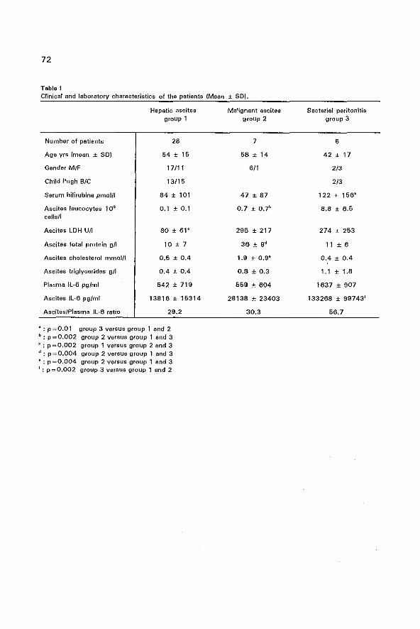
72
Table I Clinical and laboratory characteristics of the patients (Mean ± SO).
Number of patiants
Age yrs {mean ± SO)
Gender M/F
Child-Pugh BIC
Serum bifirubine pmol/l
Ascites leucocytes 109
cells!!
Ascites LDH U/l
Ascites total protein gIl
Ascites cholesterol O1mol/l
Ascites triglycerides gIl
Plasma IL-6 pgfml
Ascites IL-6 pg/ml
Ascites/Plasma 1l-6 ratio
Hepatic ascites group 1
28
54 ± 16
17/11
13/15
84 ± 101
0.1 ± 0.1
80 ± 6l e
10 ± 7
0,6 ± 0.4
0.4 ± 0.4
542 ± 719
13816 ± 15314
29.2
.: p=O.Ol group 3 versus group 1 and 2 b: p=O,002 group 2 versus group 1 end 3 c: p=O.002 group 1 versus group 2 and 3 d: p=O.004 group 2 versus group 1 and 3 .: p=O.004 group 2 versus group 1 and 3 f: p=O.002 group 3 versus group 1 and 2
Malignant ascites group 2
7
58 ± 14
6/1
47 ± 87
0.7 ± O.7b
295 ± 217
1.9 ± O.9~
0.8 ± 0.3
559 ± 604
28138 ± 23403
30,3
Bacterial peritonitis group 3
5
42 ± 17
2/3
2/3
122 ± 156'
B.8 ± 6.5
274 ± 263
11 ± 6
0.4 ± 0.4
1.1 ± loB
1637 ± 907
133268 ± 99743 1
66.7
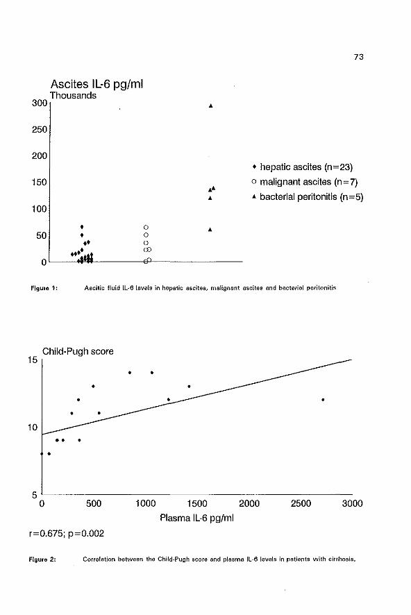
300
250
200
150
100
50
Ascites IL-6 pg/ml Thousands
• • •• .... o o o CD
..
.... ..
73
• hepatic ascites (n=23)
o malignant ascites (n = 7)
.. bacterial peritonitis (n=5)
Figure 1: Ascitic fluid IL-a levels in hepatic ascites, malignant ascites and bacterial peritonitis
Child-Pugh score 15
• •
• 10
• • • •
• • •
•
5L----------------------------------------------o 500 1000
r=0.675; p=0.002
1500
Plasma IL-6 pg/ml
2000 2500 3000
Figure 2: Correlation between the Child-Pugh score and plasma Il-6 levels in patients with cirrhosis.
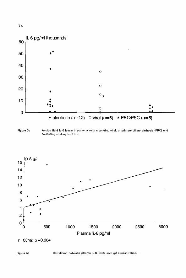
74
IL-6 pg/ml thousands 60
50 ••
40 •
30
20 • 10 t
•••
o
o
o ~ ~~ o ~ ______ -L~ ______________ -t~ ______________ ~~~~~ ____ __
Figure 3:
16 Ig Ag/I
14
12
10
8 • •
6
4 • • 2 •
0 0
• alcoholic (n=12) 0 viral (n=6) ~ PBC/PSC (n=5)
Ascitic fluid Il-B levels in patients with alcoholic. viral, or primary biliary cirrhosis (PSC) and sclerosing cholangitis (PSC)
•
•
500 1000 1500 2000 2500 3000
Plasma IL-6 pg/ml
r=0649; p=0.004
Figure 4: Correlation between plasma fl-6 levels and IgA concentration.
75
1. Tilg H, Wilmer At Vogel W, at al. Serum levels of cytokines in chronic liver disease. Gastroenterology
1992: 103: 264·274,
2, Rehermann Bt Trautwein C, Boker KHW, Manns MP. Interleukin-6 in liver disease. J Hapetol 1992: 16:
277·280.
3. Wolvekamp MCJ, Marquet RL. Interleukin-6: historical background, genetics and biological significance.
Immunol Letters 1990: 24: 1-10.
4. Kakuma S, Shinagawa Tt Ishikawa Tt at a!. Serum inter/aukin 61evals in patients with chronic hepatitis
B. Am J Gastroenterol 1991: 86: 1804-1808.
5, Khoruts A, Stahnke l, McClain CJ, logan G, Allen JI. Circulating tumor necrosis factor, interleukin-l
and interleukin-6 concentrations in chronic alcoholic patients. Hapetology 1991: 13: 267-276.
6. Propst T, Propst A, Herold M, et a!. Spontaneous bacterial peritonitis is associated with high levels of
interleukin-6 and its secondary mediators in ascitic fluid. Eur J Clin Invest 1993: 23: 832-836.
7. Zeni F, Tardy B, Vindimian M, et a!. High levels of tumor necrosis factor-a and interleukin·6 in the ascitic
fluid of cirrhotic patients with spontaneous bacterial peritonitis. Clin Infect Dis 1993: 17: 218-223.
8. Devillre J, Content J, Crusiaux A, Dupont E. Il·6 and TNFa in ascitic fluid during spontaneous bacterial
peritonitis. Dig Dis Sci 1991: 36: 123-124.
9. Andus T, Gross V, Holstege A, et el. Evidence for the prodUction of high amounts of interleukin-6 in the
peritoneal cavity of patients with ascites. J Hepatol 1992: 15: 378-381.
10. Felver ME, Mezey M, McGuire M, et al. Plasma tumor necrosis factor a predicts decreased long-term
survival in severe alcoholic hepatitis. Alcohol Clin Exp Res 1990: 14: 255-259.
11. She ron N, Bird G, Goka J, Williams R. Elevated plasma intcrleukin·6 and increased severity and mortality
in alcoholic hepatitis. Clin Exp Immunol1991: 84: 449·463.
12. Hilt DB, Marsano l, Cohen 0, Allan J, Shedlovsky S, Mclain CJ. Increased plasma interleukin-6
concentrations in alcoholic hepatitis. J lab Clin Med 1992: 119: 647-552.
13. Yoshimoto T, Nakanishi K, Hirose S, et al. High serum IL-6level reflects susceptible status of the host
to endotoxin and Il·1/tumor necrosis factor. J Immunol 1992: 248: 3696·3603.
14. Byl B, Roucloux I, Crusiaux A, Dupont E, Devillre J. Tumor necrosis factor a and interleukin 6 plasma
levels in infected cirrhotic patients. Gastroenterology 1993: 104: 1492-1497.
15. Milani A, Ciammelli AM, Degen C, Siciliano M, Rossi L. Ascites dynamics in cirrhosis. J Hepatol 1992:
16: 369·375.
16. Witte MH, Witte Cl, Dumont AE. Estimated net transcapillary water and proteinflux in the liver and
intestine of patients with portal hypertension from hepatic cirrhosis. Gastroenterology 1981: 80: 265-
270.
17. Rector WG, Ibarra F. Observations on the mechanism and loc.otion of ascites reabsorption in man. Am
J Gastroenterol 1987: 82: 342-346.
18. Houssiau FA, Devogelaer JP, Van Damme J, de Deuxchaisnes CN, van Snick J.
Interleukin-6 in synovial fluid and serum of patients with rheumatoid arthritis and other inflammatory
arthritides. Arthr Rheum 1988: 31: 784·788.
19. Kakuma S, Fukatsu A, Shlnagawa T, Kurokawa S, Kusakabe A. localisation of intrahepatic interleukin
6 in patients with acute and chronic liver disease. J Clin Pathol 1992: 45: 408·411.
20, MOiler C, Zielinski CC. Interleukin-6 production by peripheral blood monocytes in patients with chronic
76
liver disease and acute viral hepatitis. J Hepatoi 1992: 16: 372-377.
21. Schindler A. Mancilla J, Endres S, at a1. Correlations and interactions in the prodUction of Il-6 ,ll-1, and
TNF in human blood mononuclerar cells: IL-B supprass Il-1 and TNF. Blood 1990: 75: 40+47.
22. Engelberts I, Stephens 5, Francot GJM, Van der Unden CJ, Buurman WA. Evidence of different effects
of soluble TNF-receptors on various TNF measurements in human biological fluids. Lancet 1991: 338:
616-516.
23. TUg H, Vogel W, Wiedermann CJ, at at. Circulating interleukin-' and tumor necrosis factor antagonists
in liver disease. Hepato[ogy 1993: 18: 1132-1138.
24. Andus T, Gross V, Hoistega A, at at. High concentrations of soluble tumor necrosis factor receptors in
ascites. Hepato[ogy 1992: 16: 749-755.
26. Ven Zee KJ, Kohno T, Fischer E, Pock CS, Mo!dawer ll, lowry SF. Tumor necrosis factor soluble
receptors circulate during experimental and clinical inflammation and can protect against excessive
tumor necrosis factor a in vitro and in vivo. Proc Natl Acad Sci 1992: 89: 4846-4849.
26. Deviare J, Content J Denys C, et 01. High interleukin·6 serum levels and increased production by
leucocytas in alcoholic liver cirrhosis. Correlation with [g A serum levels and Iymphokines production.
Clin Exp Immunol1989: 77: 221-225.
27. Ramadori G, Meyer zum Buschenfelde KH. liver cells and cytokines. Curr Opin Gastroenterol 1993: 9:
359-366.
CHAPTER 6
SMALL BOWEL WALL FUNCTION IN PATIENTS WITH
ADVANCED LIVER CIRRHOSIS AND PORTAL HYPERTENSION:
Studies on permeability and luminal bacterial overgrowth.
The contents of this chapter have been published under the same title in
Eur J Gastroenterol Hepatol 1993; 5: 383·387 by the following authors:
D.J. Bac, G.R. Swart, J.W.O. van den Berg and J.H.P. Wilson.
77
78
Summary
Objective: Changes in small bowel wall function could contribute to the complications of
cirrhosis including malnutrition, infection and encephalopathy. To evaluate small bowel
function, we studied intestinal permeability and luminal bacterial overgrowth in patients
with cirrhosis and portal hypertension.
Design: The 14C-glycocholic acid breath test was used to evaluate the presence of bacterial
overgrowth and urine excretion of orally ingested 51Cr-EDTA was used to measure
intestinal permeability. Intestinal clearance of a-1-antitrypsine and faecal blood loss were
also measured.
Setting: Gastroenterology and hepatology unit of a university hospital.
Patients: A total of 18 patients were studied.
Results: No evidence of small intestinal bacterial overgrowth or increased permeability of
the small bowel was found. However, 28% of the patients had increased faecal blood loss.
Conclusions: Although the number of patients investigated was small, our data suggest
that small bowel function is maintained to a large extent in patients with advanced liver
cirrhosis and portal hypertension.
79
Introduction
Little information is available on small bowel function in patients with cirrhosis of the liver
and portal hypertension. Indirect evidence suggests that portal hypertension might lead to
changes in motility and in permeability of the small intestine. Bacterial overgrowth has
been reported in liver cirrhosis [1 ~4L and it is possible that this might be related to changes
in motor function (5,61. The increased prevalence of endotoxaemia in alcoholic cirrhois has
been suggested to be due to an increased gut permeability [7,8]; increased permeability
may be an important factor in spontaneous bacterial peritonitis of cirrhosis (9). Damage
to the epithelial barrier could also result in leakage of plasma, and some conflicting reports
on protein losing enteropathy in cirrhosis and ascites have been presented [10,11]. As
changes in small bowel function could be a contributing factor to the complications of
cirrhosis including malnutrition, infection and encephalopathy, we performed a small study
in patients with cirrhosis and evidence of chronic portal hypertension to examine the
following aspects of small bowel function:
{1} the occurrence of small intestinal bacterial overgrowth
(2) the permeability of the mucosal barrier documented by the transport of 51Cr-EDTA from
lumen to blood
(3) the permeability of the mucosal barrier as judged from the leakage of a-1-antitrypsin
from the plasma into the lumen.

80
Subjects and methods
Patients
Eigtheen patients were included for the various tests, (5 women and 13 men). Eight
patients (Table 1) gave informed consent for the 14·C glycocholic acid breath test and the
151Cr~EDTA permeability study. From 10 other patients only faeces and serum analysis was
done as described below. The diagnoses and the Child-Pugh score is given in Table 2
(class A, n = 2; class S, n = 12; class C, n =4). All patients had histologically confirmed
cirrhosis of the liver with chronic portal hypertension and oesophageal varices,
splenomegaly, andlor ascites. They had no history of any other disease which could cause
small bowel bacterial overgrowth such as gastrointestinal surgery J diabetes, intestinal
motility disorders, inflammatory bowel disease or coeliac disease. Patients were excluded
if they had used antibiotics, non-steroidal anti-inflammatory drugs or alcohol in the 2
weeks previous to the investigation, as these factors have been reported to influence small
bowel permeability or bacterial overgrowth or both. The use of other medications such as
diuretics and H2 antagonists was continued.
Methods
Bacterial overgrowth was tested indirectly by the 14C-glycocholic acid breath test, by
screening for steatorrhoea and by measuring vitamine B12 1evels in blood. Fasting patients
were given 5pCi 14C-glycocholic acid with a light breakfast. Breath samples were taken
every 30 min. for 5 hours and each time 2 mmol of CO 2 was collected in hyamine for
analysis of 14C02 . A 14C02 concentration above 1,5xl0·5 of the administered dose/mmol
CO2, is considered evidence for bacterial overgrowth [12].
Small bowel permeability was measured using 51Cr-EDTA as an indicator. For this test,
fasting subjects were given 10 ml of 51Cr-EDTA solution containing approximately 100 pCi
(3.7 MSq), followed by 300 ml of water at 8.00 am. They fasted for a further 2 hand
then they were permitted normal food and fluid intake.
Urine was collected every 3 h for the first 12 h, followed by a further 12-h collection.
Urine volumes from each collection period were measured and the urine was subsequently
counted for 51Cr in a multichannel analyser with a solid state probe (Canberra instruments,
Meriden, Connecticutt, USA). The urine portion~ were normalised to the same volume to
81
circumvent geometric problems during counting. The urinary recoveries of the orally
administered 51Cr-EDTA were calculated as a percentage of the dose given. Cumulative
urine recoveries of 51Cr -labeled EDTA exceeding 2,5% of the orally ingested dose were
considered abnormal [13-18). Intestinal clearance of serum a-l-antitrypsin (A1AT) was
used as a measure of plasma protein loss into the gut. Alpha-l-antitrypsin was measured
in 24-h stool collection and in a serum specimen collected during this time. The stool was
weighed (wet weight) and homogenized, and an aliquot was centrifuged at 1500 x g for
30 min. Alpha-l-antitrypsin was measured in the supernatant by radial immunodiffusion
(RIDJ (Behringwerke,Marburg/Lahn, Germany) and the clearance calculated.
The upper limit of normal is 13 ml/day in subjects without diarrhea and without fecal blood
loss into the gastrointestinal tract [19-23J. Stools were collected for 3 consecutive days
and in each collection A 1 AT was determined; mean va1ues are presented as the A 1 AT
concentration per 24 h. On the same stool samples fecal blood loss was assessed by using
a modified Haemoquant assay [24 J. Results (the mean of 3 days) are expressed in pi
blood/g dry faeces (upper limit for the normal population is 83 pi blood/g dry faeces) [241.
Fat excretion in stools was assessed by a quantitative assay performed on a sample of a
24 h stool collection.
In addition to routine blood examination, vitamin B12, folate, immunoglobulin G, cholesterol
and triglycerides levels were measured.
Statistical analysis of data was performed using the computer program "Stata"
(statistical/data analysis, Computing Resource Centre, LA, USA).
Results
None of the 8 patients under going 14C bile acid breath test had a level exceeding the upper
level of normal in the first 4 h, indicating the absence of small bowel bacterial
overgrowth . The mean excretion was 0.38xl0·5 of the dose/mmol CO 2; range 0.06-
1.50x10·'. (upper level of normal: 1 ,5x1 0.5 of the dose/mmol CO,)
In the same 8 patients the 51 Cr-EDTA permeability study showed a cumulative 24·h urine
excretion of 1.48% ; SD ±0.49% (range 0.78·2.16%) of the orally administrated dose
51Cr-EDTA (normal < 2.5%). The excretion was highest in the urine collection in the time
period following 6·9 h after the intake. This is somewhat later than that found by other
authors who described a maximal excretion in the period 3-6 h after ingestion of the 51Cr_
EDTA 114, 16J. In three of these patients urine was collected untill 72 h after 51Cr·EDTA
82
ingestion; no further excess urinary excretion of oler was seen after the first 24 h of
observation, making temporary loss of the oler label into the ascites compartment
unlikelv. The intestinal clearance of A1AT could be determined in 16 patients (Table 1)
(Two patients were excluded because of incomplete stoat collection). The mean A1AT
clearance was 10.2 ml plasma per dav (range 1,9-61,8 ml/dav). Onlv two patients were
above the normal upper limit of 13,0 mlldav (no. 2 and 11), Patient 11 was found to have
intestinal blood loss due to angiodysplastic lesions in the stomach. When omitting this
patient the mean clearance is 6,8 mlldav with a range of 1,9-15,6 ml/dav.
Fecal blood loss measurements showed that five patients (28%) had blood loss above the
normal level (Table 1). Four of these were just above the normal level and 1 patient
(number 11) with known vascular anomalies of the stomach mucosa had a strongly
positive test (692 pi blood/g drv faeces). The mean fecal blood loss excluding this last
patient was 56 pi bloodlg drv faeces with a range from 6·98 pi bloodlg drv faeces (normal
< 83 pi bloodlg drv faeces).
Fat excretion in a 24~h stool sample was above normal in three out of 16 patients in whom
a proper stool collection was performed (upper level of normal 10 g fat/24 h). Mean fat
excretion was 6,25 g/24 h with a range from 1,1 -21,0 g/24 h. Vitamin B" levels were
normal in all patients including those with steatorrhoea. IgG levels were raised (normal
< 18 gil) in 13 out of 18 patients, (mean 21.6 gil; range 8,9 - 34,2 9/1). Folic acid levels
were below normal values (7 ng/ml) in five out of 18 patients (Table 2). Statistical analvsis
did not show any significant correlation between the Child-Pugh score and the A 1 AT
clearance (r = 0.0070, P =0.74). the Child-Pugh score and steatorrhoea (r =0.022, -
p=0.55). and steatorrhoea and A1AT clearance (r=0.0013, p=0.88).
Discussion
Although changes in the gastric mucosa in patients with chronic portal hypertension have
received much attention, little is known about the effects of portal hypertension on the
more distal part of the intestinal tract. Some authors have found an increased prevalence
of endotoxemia in cirrhotics with ascites and/or varices and suggested that this could be
due to an enhanced permeability to macromolecules of the mucosa in the upper small
intestine 17,81, possibly in combination with the reported small intestinal bacterial
overgrowth in patients with liver cirrhosis and chronic alcohol abuse [2,4,25],
,Therefore, we studied small, bowel bacterial overgrowth in patients with liver cirrhosis and
83
chronic portal hypertension, together with changes in the intestinal wall permeability to
macromolecules shown by 51Cr-EDTA excretion.
We used an indirect method, the bile acid breath test, to demonstrate bacterial overgrowth
[26]. The sensitivity of the "C-bile acid breath test to detect bacterial overgrowth is about
70% 127] and it seems unlikely that lack of sensitivity is the cause of the negative test
results in all eight patients. The absence of bacterial overgrowth is also supported by the
normal serum levels of vitamin B'2' as malabsorption of vitamine B'2 due to binding of the
vitamin to the bacteria in the small bowel has been described [26]. The use of 61Cr-EDTA
(MW 358Da) as a relatively large (radius =0.63 nm) hydrophilic probe to test intestinal
integrity has proven to be useful in several other conditions. The permeation pathway is
thougt to be through larger intercellular "channels". This is different from the "aqu~ous
pores" through which molecules of low molecular weight such as monosaccharides and
mannitol are thought to be absorbed by diffusion. Using this hydrophilic probe we were
unable to demonstrate an increased permeability in the patients. We were also unable to
demonstrate a significant "leak" from blood to intestinal lumen using A 1AT clearance.
Because the molecular weight of A1AT (54.000 daltons) is comparable to the molecular
weight of albumin (61.000 daltons), excretion of A1AT in stool can serve as a measure
of plasma protein leakage into the intestinal lumen [19,23). Due to its anti proteolytic
activity, A 1 AT that leaks into the intestinal lumen is minimally digested by intestinal
proteases and therefore, mainly excreted as an intact molecule in stool. The intestinal
clearance of A 1AT is closely correlated to the intestinal clearance of serum albumin [20-
22).
We were unable to demonstrate an increased plasma protein loss into the gut lumen in our
patients. Our findings are in line with those of Iber [11) who could not demonstrate
significant protein-losing enteropathy in cirrhotics using 1131 labeled serum albumin.
A remarkable finding in this study is that 28% of the patients with liver cirrhosis and portal
hypertension had increased faecal blood loss.
Increased blood loss from the stomach has been described before in those patients with
portal hypertension and can probably be attributed to abnormal mucosal capillaries and
venous ectasia as seen in congestive gastropathy.
Using indirect methods we have failed to find evidence of small intestinal bacterial
overgrowth or increased permeability of the small bowel. Although the number of patients
investigated is small, our data suggest that small bowel function is maintained to a large
extent in patients with advanced liver cirrhosis and chronic portal hypertension.
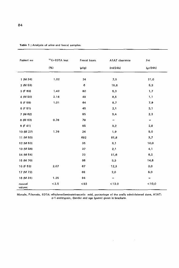
84
Table 1 ; Analvsis of urine and faecal samples.
Patient no 5ICr-EDTA tast Faecal haem A1AT clearance Fat
1%) WI/g) (m1/24hl (gr/24hl
1 (M34) 1.02 24 7,0 21,0
21M 69) 6 16,6 6,3
3 (F 62) 1.42 80 6,3 1,7
4 1M 60) 2.18 49 8,6 1,1
5 IF 69) 1.01 64 8,7 7,9
6 (F 61) 45 2,1 3,1
7 (M 621 85 9,4 2,3
81M 83) 0.78 79
9 (F 41) 86 3,2 2,8
101M 27) 1.76 24 1,9 6,0
11 (M 53) 692 61,8 3,7
121M 50) 36 9,1 10,0
131M 66) 37 2,1 4,1
14 (M 64) 23 11,6 6,2
161M 70) 98 3,3 14,8
16 (F 63) 2.07 67 12,3 3,0
171M 72) 68 2,0 8,0
18(M31) 1.25 94
normal <2,5 <83 <13.0 <10,0 values:
M:male, F:female, EDTA: ethylenediaminet6traacetic acid. percentage of the orally administered dose, A1AT: a-l-antitrypsin, Gender and age (years) given 1n brackets.

85
Table 2; Clinical and biochemical characteristics of the patients studied.
Patient no Diagnosis Child-Pugh Ascites Albumin tg G Serum folic scora acid lavols
(g{l) 10m (ng/mll
1 1M 34) AC C 10 22 30,6 10,0
21M 69) AC C 14 + 20 31,6 12,8
3 (F 621 AC B 8 + 46 24,6 10,1
4 (M50) AC A 6 + 38 12,9 6,1
6 IF 59) AC B 8 30 29,9 6,8
6 IF 61) AC B 8 + 34 19,6 10,2
7 1M 62) AC C 10 + 29 17.0 12,~
81M 63) AC C11 + 34 26,0 2,6
9 IF 41) CC B 8 + 38 8,9 11,3
101M 27) CC A 6 42 20,1 10,4
11 (M 53) CC B 7 + 33 19,8 10,8
121M 60) PVC B 8 + 33 34,2 20,0
13 (M 56) PVC B 9 + 37 20,0 12,8
14 (M 64) PVC B 8 + 34 27,9 16,6
161M 70) PVC B 9 + 32 8,9 11,2
16 IF 53} PBC B 9 + 28 18,0 7,0
17(M72) PBC B 9 + 24 20,0 6,8
181M 31) PSC B 8 + 31 20,4 2,3
AC: alcoholic cirrhosis. CC: cryptogenic cirrhosis. PVC: post viral cirfhosis. PSC: primary sclerosing cholangitis. PBC: primary biliary cirrhosis. F:famate, M:male, Ig: immuno-globulins, Gender and ago (years) given in brackets.
86
1. Gorbach el, Lal D, Levitan R: Intestinal microflara in Laennao's cirrhosis.
J elin Invest 1970,49: 36 B.
2. Martini GA, Phear EA: The bacterial content of the small intestine in normal and cirrhotic patients.
Clin Sci 1967,)6:36·61.
3. Simon Gl, Gorbach Sl: Intestinal mlcroflora. Mad eUn of North Am 1982,86:No 3 pp 667-573.
4. Bode JC, Bode C, Heidelbach R, Durr H-K, Martini GA: Jejunal microllora in patients with chronic
alcohol abuse. Hepato-gastroenterol 1984,31 :30-34.
6. Rielly JA, Forst eF, Neilson E, Quigley EMM, Rikkers LF: The effect of portal hypertension on gastric
emptying of liquids. Current Surg 1989.46:33-35.
6, Reilly JA, Quigley EMM, Forst CF, Rikkers IF: Small intestinal transit in the portal hypertensive rat.
Gastroenterology 1991,100:670-674.
7. Bode C, Kugler V, Bode JC: Endotoxemia in patients with elcoholic and non-alcoholic cirrhosis and in
subjects with no evidence of chronic livar disease following acute alcoholic excess. J Hapatol 19B7.4:B~
14.
B. van Daventer SJH, Knepper A, landsmen J, tan Cate JW, Butter HR, Sturk A, Pauw W: Endotoxins
in portal blood. Hepato-gastroenterol 1988,36:223-226.
9. Runyon BA, Pathogenesis and diagnosis of spontanaous bacterial peritonitis in cirrhosis. In Rhodes
J, Arroyo V. Therapy in liver disease edited by Ediciones Doyma, Barcelona. 1992 pp 388-396.
10. Schwartz MJ: Steatorrhoe and hypoalbuminemia in cirrhotics with ascites. Am J Dig Dis 1964,9:1
28-137.
11. Iber FL: Protein loss into the gastrointestinal tract in cirrhosis of the liver. Am J Clin Nutr 1966,19:2
19-222.
12. From H, Hofmann AF; Breath test for altered bile acid metabolism. lancet 1971 ,ii:621-626.
13. Bjameson I, Wind K, Peters T J: The leaky gut of alcoholism: possible route of entry for toxic
compounds. The lancet 1984,i:179-182.
14. Sjarnason j, O'Morain C, Lavi AJ, Peters TJ: Absorption of 51Cr_Jabelod ethylenadiaminetetraacetate
in inflammatory bowel disease. Gastroenterology 1986,86:318·322.
16. Maxton DG, Sjamason I, Reynolds AP, Catt SO, Paters T J: lactulose, SICr labelled ethylene
diaminetetra-acetate, l-rhemnose and polyothyloneglycol 600 as probe markers for assosment in vivo
of human intestinal permeability. Clin Sci 1986,71:71-80.
16. Sjamason I, Peters T J, Veall N: A persistent defect in intastinal permaebility in coeliac disease
demonstrated by a 51Cr_labeled EDTA absorption test. The lancet 1983,i:323-325.
17. Jenkins RJ, Jones DB, Goodecre Rl, Collins SM, Coates G, Hunt RH, Bienenstock J: Reversibility of
increased intestinal permeability to 51Cr_EDTA in patients with gastrointestinal inflemmatory diseases.
Am J Gastroenterol 1987,82:1169-1164.
18. Fotherby KJ, Wraight EP, Neale G: 51Cr-EDTA/14C-Mannitol intestinal permeability test. Scand J
Gastroenterol 1988,23:171-177.
19. Florent C, l'Hirondel C, Desmazures C, Aymes C, Bernier J: Intestinal clearance of alpha 1 antitryp
sin. Gastroanterology 1981,81 :777-780.
20. Weisdorf SA, SalaH lM, longsdorf JA, Ramsay NK, Sharp H: Graft-Versus-Host disease of the intes
tine: A protein losing enteropathy characterized by feacel alpha1 antitrypsin. Gastroenterology
87
1983,86:1076-1081.
21. Perrault J: Protein·losing gastroentoropathy and the intestinal clearance of serum a ,-antitrypsin.
Mayo Clin Proc 1984,59:278-279.
22. Meyers S, Wolke At Field SP, Feuer EJ, Johnson JW, Janowitz HD: Fecal aI-antitrypsin measure
ment: an indicator of Crahn,s discase activity. Gastroenterology 1985,89:13-18.
23. Strygler Bt Nicer MJ, Santangegelo we, Porter Jl, Fordtren JS: 0", antitrypsin excretion in stool in
normal subjects and in patients with gastrointestinal disorders. Gastroenterology 1990,99:1380·1387.
24. van den Berg JWO, Edixhoven-Bosdijk A, Koole-Lesuis R, Wilson JHP: Faecal haem assay- some
practical modifications of the haemoquant assay for haemoglobin in faeces. Clin Chim Acta 1987, 16~
9:319-322.
26. Lal D, Gorbach SL, Levitan R: Intestinal microtlora in patients with alcoholic cirrhosis: urea splitting
bacteria and Neomycine resistance. Gastroenterology 1972,62:275-279.
26. King CE, PP Toskes: Bacterial Overgrowth Syndromes. In Gastroenterology. Edited by Berk JE: Phila
delphia: W.B. Saunders Company; 1986 pp 1781·1791.
27. King CE, Toskes PP, Guilarte TR, Lorenz E, Welkos SL: Comparison of the one gram d.l 4C Xylose
breath test to the 14C bile acid breath test in patients with small intestine bacterial overgrowth.
Dig Dis Sci 1980,26:63-68.

89
CHAPTER 7
SPONTANEOUS BACTERIAL PERITONITIS; OUTCOME AND PREDICTIVE FACTORS.
The contents of this chapter have been published under the same title in
Eur J Gastroenterol Hepatol 1993; 5: 635-640 by the following authors:
D.J. Bac, P.D. Siersema, P.G.H. Mulder, S. de Marie and J.H.P. Wilson.
90
Summary
Objective: To identify patients who were most at risk of developing spontaneous bacterial
peritonitis and who might benefit from selective intestinal decontamination.
Design: prospective analysis of all patients admitted with liver cirrhosis during a 4·year
period who had ascitic fluid analysis and appropriate bed·side inoculation of ascites in
blood culture bottles.
Setting: Liver transplantation centre in a university hospital.
Patients: Thirty-one positive episodes and 92 negative episodes for spontaneous bacterial
peritonitis were analyzed and compared in 83 patients.
Results: The 1-year survival after spontaneous bacterial peritonitis was 14% compared
to 43% in the group without spontaneous bacterial peritonitis. The calculated recurrence
rate was 20%. The Child-Pugh score and coagulation parameters were identified as
predictive factors for the development of spontaneous bacterial peritonitis, as was a
deterioration in values of liver function tests. The total ascitic fluid protein content was not
predictive. Sclerotherapy for oesophageal varices may be a precipitating factor, but this
requires further investigation.
Conclusions: On the basis of our results we recommend the use of selective intestinal
decontamination for patients with liver cirrhosis Child-Pugh class C and ascites who
develop rapidly progressive deterioration of liver function tests, patients who survived a
first episode of spontaneous bacterial peritonitis and those awaiting liver transplantation.
91
Introduction
Spontaneous bacterial peritonitis (SSP) is a serious complication in patients with cirrhosis
and ascites with an incidence of about 7~23% in patients admitted to hospital [1-31 and
a reported mortality between 37% and 78%, older studies reporting a higher mortality than
the more rBcent ones (2,4,5]. The l-year survival following SSP has been estimated to be
about 30% and the frequency of a recurrence within 1 year after a first episode of SSP
varies between 11 % and 69% (2,4-7].Spontaneous bacterial peritonitis is diagnosed when
(a) pathogenic bacteria are cultured from ascites. (b) the ascitic polymorphonuclear (PMNI
count is >O.25xl09 cells/I and (0) there is no evident intra-abdominal source of infection
[8]. Because of the low number of microorganisms in the ascitic fluid, ascites culture may
remain negative although the clinical picture is compatible with SSP and there is an
elevated PMN count. This entity is recognized as culture-negative neutrocytic ascites
(CNNA) (9J and is defined as: (a) an ascitic fluid PMN count greater than 0.50x1 0' celis/I,
(b) a negative ascitic fluid culture, (c) no administration of antibiotics within 30 days (d)
no other explanation for an elevated PMN count in ascitic fluid, such as pancreatitis,
intraperitoneal hemorrhage, peritoneal tuberculosis or carcinomatosis. Culture-negative
neutrocytic ascites is regarded as a variant of SBP which should be treated with antibiotics
(9,10]. Recently it has become clearthat the proportion of CNNA in patients suspected of
having S8P depends on the culture method used. Bedside inoculation of ascitic fluid into
blood culture bottles has been found to be superior to conventional culture methods (11-
14].
As most microorganisms causing S8P are gut-derived aerobic Gram-negative bacilli,
selective intestinal decontamination, eliminating Gram-negative aerobic intestinal flora has
been proposed for the prophylaxis of SSP (5, 15J. The aim of this study was to identify in
a population of cirrhotic patients, those patients most at risk of developing S8P who might
benefit from selective intestinal decontamination. Predictive factors for SSP were analyzed,
including therapeutic procedures in the period before the development of S8P. In addition
the survival rate after an episode of S8P, the long term prognosis and recurrence rate after
an episode of SBP were investigated.
Patients and methods
Setween July 1987 and April 1991, 123 consecutive abdominal paracenteses were
92
performed in 83 patients with a diagnosis of liver cirrhosis, ascites and portal hypertension.
The indications to perform a paracentesis were (a) suspicion of SBP (b) routine paracen
tesis on admission or (c) large volume therapeutic paracentesis. Patients were included in
this study when ascitic fluid samples were cultured in blood culture bottles as described
elsewhere {14]. Ascites was obtained under sterile conditions with or without ultrasound
guidance.
The totalleucocytes count in ascitic fluid was done by a Sysmex NE 8000' analyzer (TOA
Medical Electronics, Kobe Japan). In addition, 10·20 ml of ascites was centrifuged at 2000
rpm for 5 min, the pellet stained with Giemsa stain, and at least 50 cells were examined
to determine the percentage of PMN cells.
Total protein, albumin, lactate dehydrogenenase (LDH) and amylase levels were determined
in ascitic fluid. Serum liver enzymes, renal function tests, leucocyte count and differentj~
ation and the coagulation parameters antithrombin III activity and Normotest were
determined by routine methods. The Normotest (Nyegard, Oslo) is a modified prothrombin
time in which factor V and fibrinogen are added to the reagent. Thus, the test is only
dependent on coagulation factors II,VII and X and has been reported to be a sensitive
marker of hepatic synthesis [1 6]. The serum~ascites albumin gradient (serum albumin
minus ascites albumin concentration) was calculated . Bilirubin, albumin, Normotest and
creatinine values were determined at the time of the ascitic fluid paracentesis and
compared with values obtained 2~4 months previously. If blood or urine cultures were
performed on the same day, these results were also recorded.
All patients were classified according to the Child·Pugh score [17). In most patients, a liver
biopsy was performed. The performance of ascitic fluid drainage, fibre·endoscopy or
sclerotherapy for oesophageal varices within 2 weeks before paracentesis was recorded.
At the time of the paracentesis the clinical picture was assessed including the presence
and duration of fever, the presence of abdominal pain, progression of jaundice and the
presence and progression of encephalopathy. The severity of hepatic encephalopathy was
graded on a scale of O~4 on the basis of clinical criteria and quantitative EEG analysis (18].
The use of diuretics, antacids and antibiotics was recorded. The follow~up and the number
of recurrences were recorded in all patients. Causes of death were classified as either due
to liver failure or due to another cause.
Since more than one paracentesis was performed in some patients (123 in 83 patients
with at least 14 days between the different episodes), the statistical analysis had to take
account of these repeated observations. Testing of the mean difference of various
93
variables between the group with and without SSP was carried out by univariate logistic
regression analysis with random effects. In the analysis, each patient has his own (random)
effect, around which the repeated observations within each patient were clustered. In this
way, the interdependency in the data was taken into account so that all data could be
used while errors of the mean difference between the group with and without SSP are
prevented from being underestimated. When comparing mortality between these two
groups, the patient is considered the experimental unit and the most recent outcome in
each of the 83 patients was taken as the start of the follow-up.
Results
Patient characteristics:
Table 1 summarizes the clinical characteristics of the patients in 123 episodes of SSP
when a paracentesis was performed. Ninety-two paracenteses in 65 patients were
negative for a diagnosis of SSP because the PMN count in ascitic fluid was < 0.25x10⁹
cells/! and the ascitic fluid culture was negative (SSP negative). Thirty-one paracenteses,
in 29 patients were positive with an ascitic fluid PMN count of > 0.50x10⁹ cellsll and/or
a positive ascitic fluid culture (SSP positve). There were no patients with an ascitic fluid
PMN count between 0.25-0.50x1 0' cellsll. It is noteworthy that of the patients who used
antibiotics at the time of the paracentesis, none were diagnosed as having SSP. Of the
patients in both groups, 10% (three out of 31 in the group with SSP and nine out of 92
in the group without SSP) used immunosuppressive drugs, which did not influence the
clinical presentation. Eleven patients initially had a negative ascitic fluid culture with a low
PMN count (SSP negative) and developed SSP during follow-up (SSP positive).
Biochemical analysis:
The biochemical parameters are summarized in Table 2. Bifirubin, albumin, creatinine and
Normotest values were compared with values taken 2-4 months before the paracentesis.
This information was available in 101 of 123 (82%) episodes (SSP negative; 71, SSP
positive; 30) . There was a statistically (Wilcoxon test) significant increase in bilirubin
(p=0.005) and creatinine (p=0.009) values and a statistically significant decrease in
albumin (p=O.OOl) and Normotest (p=0.002) values in both the SSP-negative and the
SSP-positive groups. However, the difference was significantly greater in the SSP-positive
group. Comparing the SSP-positive group with the SSP-negative group, this reached
94
statistical significance for the albumin decrease only (p =0.011).
Ascitic fluid analysis
Within the group of positive episodes, there was a difference in leucocyte count and PMN
count between cUlture-positive ascitic fluid and CNNA. The PMN count was higher in
cUlture-positive ascites than in those with CNNA, but this did not reach statistical
significance. (PMN count ± SO in ascitic fluid in SSP vs. CNNA: 4.2x1 0' ± 6.9 cellsll and
1.2x10' ± 0.7 cells/l;Mann-Whitney test; p=0.91. Lactate dehydrogenase levels were
significantly higher in the SSP-positive group compared with the SSP-negative group. Total
protein and albumin concentrations in ascites were similar in the SSP-positive and in the
SSP-negative group (Table 31.
Bacteri%gy
These results have also been described in another study comparing different culture
methods (14). In 25 of the 31 (81 %) episodes classified as SSP, bacteria were cultured.
Gram-negative bacilli were detected in 15 of the 25 (60%) episodes
(Escherichia coliin eight. Klebsiella pneumoniae in two, Pseudomonas aeruginosa in two,
Bacteroides sp. in two, Acinetobacter sp. in one) and Gram-positive cocci in 10 of the 25
(40%) episodes (alpha-hemolytic streptococci in three, Enterococcus faecalis in four,
Streptococcuspneumoniae in one, Streptococcus bovis in one, Staphylococcus epidermidis
in one).
In only two of the positive episodes did the Gram stain reveal bacteria (6%). Blood
cultures were obtained during 25 episodes and were positive in nine episodes (36%),
culturing the same microorganisms as from the ascitic fluid. In the patient without SBP,
blood cultures were taken during 33 episodes and were positive in eight episodes (24%),
while ascitic fluid cultures remained negative and the ascitic fluid PMN count was <
0.25x10' cells/l.
Recurrence and survival analysis
The mean follow up time in the SSP-positive group was 194 days (range 1-1278 days).
Twenty-three of the 29 patients in the SBP-positive group died (79%). Nine of these
patients died within 2 weeks, most likely due to SBP, although they were treated with
antibiotics. Only two patients from the SBP- positive group died due to a cause not related
95
to their liver disease (8%). Of the six surviving patients, one received a liver transplant and
is still alive, two were lost to follow-up and the remaining three are still alive and survived
22, 30 and 42 months, respectively. The 1-year survival in the S8P· positive group was
14%. Of the 65 patients in the SSP- negative group, the mean follow-up was 300 days
(range 2-1170 days) and the 1-year survival rate was 43%. Forty out of the 65 patients
in the SSP-negative group died, seven were lost to follow up and 18 arB still alive. By
means of Cox's proportional hazards survival analysis, a hazard ratio of 1.94 (p =0.017)
was estimated for SSP-positive group with respect to SSP-negative group (fig.1).
Within the SBP-positive group there was no significant difference in survival between those
with culture-positive (n = 25) and those with CNNA fluid (six patients). In four of the 20
patients (20%) in the SSP-positive group who survived the initial period, S8P recurred
during follow-up, after 2, 6, 11 and 14 months, respectively.
Discussion
In 29 out of the 31 episodes, a diagnosis of SSP was suspected on clinical grounds
because of fever (oral temperature> 37 .50C), abdominal tenderness, worsening of hepatic
encephalopathy andlor deterioration of liver function tests (Table 1). A diagnosis of SSP
in patients without any symptoms suggesting SSP, was made in only 6% of patients,
which contrasts with a reported frequency of up to 33% of cases when a routine
diagnostic paracentesis was performed on admission irrespective of clinical features [19].
As in other studies [11-14), ascitic fluid culture results were only positve in 81 % of cases
with suspected SSP, therefore, the best diagnostic test to discriminate between those with
and those without SSP and to decide whom to treat, is the total amount of PMN cells in
the ascitic fluid [8J. The Gram stain is rarely helpful and culture results will follow only
after some days. A diagnosis of CNNA is usually defined when the PMN count in ascitic
fluid is somewhere between 0.25-0.50x10' cellsll [8,9]. We confirmed the findings of
Pelletier et al.(9) who found a lower PMN count in patients with culture-negative suspected
bacterial peritonitis compared with those with cUlture-positive bacterial peritonitis. These
authors suggested that this supports the idea that CNNA is a less severe variant of SSP.
in our study, however, mortality rates were not statistically significantly different between
those with culture-positive and those with culture-negative neutrocytic ascitic fluid,
although numbers are small.
Invasive procedures such as fibreoptic endoscopy and ascitic fluid drainage within 2 weeks
96
did not show a significant relationship with the occurrence of S8P, although sclerotherapy
for oesophageal varices within 2 weeks might be a risk factor (p = 0.07). There are a
number of case reports suggesting a relationship between previous sclerotherapy and S8P
(20,21], but larger series are required to confirm this.
In evaluating predictive factors to identify patients at risk of developing S8P we found the
Child-Pugh classification and the coagulation parameters Normotest and antithrombin III
activity to be significantly different between the two groups. The importance of
coagulation parameters in predicting those patients at risk has also been described by
other authors [4,22, 23]. We found no difference in the total protein and albumin content
of ascitic fluid and the blood-ascitic albumin gradient between patients who developed SBP
and those who did not, which is in agreement with findings of Silvain et al. with regard to
recurrent S8P (24). Even in the patients with alcoholic liver disease, no such difference
could be detected. By contrast other studies have indicated that patients with a low
ascitic fluid total protein concentration, reflecting a low opsonic activity. are more likely
to develop SBP [22,23,25). Runyon reported that cirrhotic patients with ascitic fluid total
protein content of less than 10 gil were 10 times more at risk to develop SBP [26),
However, the specificity of the findings was low (60%), and the predictive value was only
15% for patients with a total protein level < 10 gil in ascites. In a recent study, the total
protein content of ascitic fluid was an inclusion criterium to select patients for a study
evaluating the benefit of selective intestinal decontamination in preventing SSP (15).
According to our findings, the ascitic protein content is not a predictive marker for patients
at an increased risk for developing S8P.
Over 2-4 months there was a significant deterioration of liver and renal function in all
cirrhotic patients admitted to our hospital. The drop in albumin levels was greater in
patients who developed SBP in comparison with those who did not. This may reflect a
deterioration in hepatic function mediated by endotoxaemia and increased cytokine
production in those with SBP [27,28). Another possibilitY is that patients with a rapidly
progressive hepatic insufficiency, as suggested by decreased coagulation and protein
synthesis, are more prone to develop SBP. The prognosis of patients who developed SBP
was poor; 30% of patients died within 14 days and the l·year survival rate was only 14%.
Because of the high death rate, despite recovery from a first episode of SSP, a liver
transplant should be considered in all patients recovering from SSP. Liver transplantation
should be performed within 2 months of an episode of SBP to save more than 50% of the
patients, but figures may be different in other centres. We could not confirm the high rate
97
of recurrence of SSP as described by Tito et al141 (43% at 6 months and 69% at 1 year).
In our population only four of the 20 patients who survived the initial event had a
recurrence. An explanation for this difference could be that in our study, cirrhotic patients
with more advanced liver disease were included. In patients with end·stage liver disease,
complications other than SSP contribute substantially to mortality. This might also explain
the higher overall mortality rate in our study.
Because enteric bacteria are the most common causative agents of SSP, it has been
suggested that selective intestinal decontamination eliminating the aerobic Gram-negative
intestinal flora and preserving the remaining aerobic and anaerobic flora could be useful in
its prophylaxis. Two r8cent placebo-controlled studies have demonstrated the efficacy of
oral norfloxacin in the prevention of SSP episodes without attaining a reduction in mortality
[5,151. However, in the study of Gines et al. 151 patients with advanced liver disease
defined by bilirubin (> 170pmol/!), creatinine (> 200 pmol/!) and prothrombin time « 25%)
were excluded from the study [51. Also in the study of Soriano et al. 115), 50% of the
patients included had liver cirrhosis Child-Pugh class A or 8 and apparently had less hepatic
insufficiency than the patients in our study. It is possible that selective intestinal
decontamination with norfloxacin could lead to a decreased mortality in certain sub-groups
of patients who are at high risk of developing SSP. According to our results subgroups
which have the highest incidence of SSP and, therefore, who might benefit from selective
intestinal decontamination, include (1) patients with Child-Pugh class C liver cirrhosis and
rapidly progressive deterioration in liver function tests, (2) patients who have survived a
first episode of SSP and (3) patients awaiting liver transplantation with Child-Pugh class
C liver cirrhosis and ascites. Another group of patients who might benefit from antibiotic
prophylaxis are patients with liver cirrhosis and ascites undergoing oesophageal
sclerotherapy, but data are lacking at present. Because of the mainly Gram-positive flora
in the upper gastrointestinal tract and the potential risk of bacteraemia during manipulation
(29,30)' antibiotic prophylaxis covering both Gram-positive and Gram-negative bacteria
may be more appropriate during sclerotherapy. A category of patients who also might
benefit from antibiotic prophylaxis are cirrhotics who present with an upper gastrointestinal
hemorrhage (31]. Soriano et al. (151 showed in a prospective randomized study, no
reduction of mortality, but a lower incidence of infections when norfloxacin was given
during 7 days, mainly due to less bacteraemia and urinary infections in the treated group.
More studies are needed in the above defined risk groups to assess the benefits and risks
of antibiotic prophylaxis. One should be cautious of the complications of long-term
98
selective intestinal decontamination, such as bacterial overgrowth in the gut and secondary
infection with Gram-positive and norfloxacin-resistant Gram-negative bacteria and with
yeasts [32).
Liver transplantation remain to be considered as a definitive solution in patients who were
already candidates for transplantation as soon as they survived an episode of SSP.
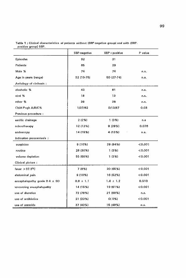
99
Table 1 ; Clinical characteristics of patients without (SSP-negative group) and with (SBP-positive group) SBP.
SSP-negative S8P + positive P value
Episodes 92 31
Patients 65 29
Male % 74 74 n.s.
Aga in years hang e) 52 (19~75) 60 (2n4) n.s.
Aetiology of cirrhosis:
alcoholic % 43 61 n.s.
viral % 16 13 n.s.
othor % 39 26 n.s,
Child-Pugh A/B/C% 1/37/62 0/13/87 0,03
Previous procedure:
ascitic drainage 2 (2%) 1 (3%) n,'
sclerotherapy 12 (13%) 9 (29%) 0.076
endoscopy 14 (16%) 4 (13%) n.s.
Indication paracentesis
suspicion 9 (10%) 29 (94%) <0.001
routine 28 (30%) 1 (3%) <0.001
volume depletion 66 (60%) 1 (3%) <0.001
Clinical pioture :
fever> 37 .5DC 7 t8%} 20 (65%) <0,001
abdominal pain 9 (10%) 16 (62%) <0.001
encephalopathy grade 0-4 ± SD 0,9 ± 1.1 1.8 ± 1.2 0.010
worsening encephalopathy 14 (16%) 19{61%) <0.001
use of diuretics 72 {78%} 21 (68%) n.s.
use of antibiotics 21 (23%) 0(0%) <0.001
use of antacids 37 (40%) 15 (48%) n.s.
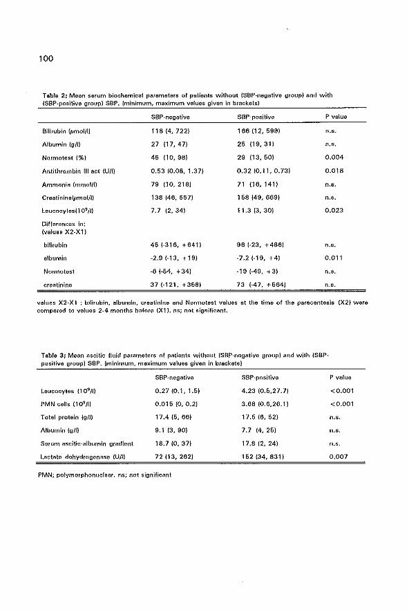
100
Tabla 2; Mean serum biochemical parameters of patients without (SSP-negative group) and with (SSP-positive group) S8P. (minimum, maximum values given in brackets)
Bitirubin {pmolllJ
Albumin (gill
Normotest (%)
Antithrombin til act (UJI)
Ammonia Immol/I)
Craatinine(pmol/IJ
leucocytes(109/1l
Differences in: (values X2-Xl)
bilirubin
albumin
Normotest
creatinine
SSP-negative
118/4,722)
27 117,471
46 110,98)
0,53 (0.08, 1.37)
79 (10,218)
138 (46, 557)
7.7 {2,34J
45 (-316, + 641)
-2.9 (-13, +19)
-6 1-64, + 34)
37 (-121, +358)
SSP-positive
166 (12, 699)
26 119,311
29 (13,50)
0.32 (0.11, 0.73)
71 116,1411
158 (49, 669)
11.3 (3, 30)
96 (-23, +488)
-7.2 (-19, +4)
-19 (A9, +3)
73 (-47, +664)
P value
n.s.
n.5,
0.004
0.018
n.s,
n.s.
0,023
n.s,
0.011
n.s,
n.s.
values X2-Xl : bilirubin, albumin, creatinine and Normotest values at the time of the paracentesis (X2) were compared to values 2-4 months before (Xl). ns; not significant.
Tabla 3; Mean ascitic fluid parameters 01 patients without (SSP-negative group) and with (SSPpositive group) SSP. (minimum, maximum values given in brackets)
leucocytes (109m PMN calls (109m Total protein (glJ)
Albumin (gIl)
Serum ascitic-albumin gradient
lactate dehydrogenase (Un)
SSP-negative
0.27 (0.1, 1.5)
0.016 to, 0.2)
17.4 (5, 66)
9.1 (3,90)
18.7 (0, 37)
72 113, 262}
PMN; polymorphonuclear. ns; not significant
SSP-positive
4.23 (0.6,27.7)
3.68 (0.6,26.1 J
17.5 (6, 52)
7.7 (4,25)
17.8(2,24)
152 (34, 831)
P value
<0.001
<0.001
n.s.
n.s.
n.s.
0.007
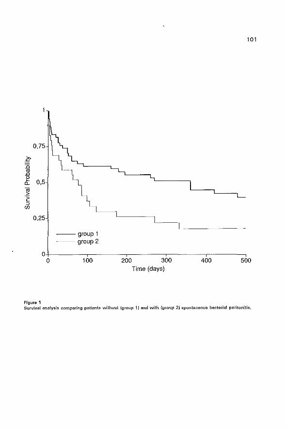
101
0,75
g :0
'" .0 e 0,5 a. (ij > .~
::> en
0,25
--group 1 --- group 2
0 0 100 200 300 400 500
Time (days)
Figure 1 Survival analysis comparing patients without (group 11 and with (group 2) spontaneous bacterial peritonitis.
102
Rima!a A. Treatment snd profyJaxis of Spontaneous Bacterial Peritonitis. In: Rodes J, Arroyo V. Therapy
in liver disease,Ediciones Dayma, Barcelona. 1992,397-406.
2 Hoefs Je, Canaweti HN, Sapieo FL, Hopkins RR, Weiner J, Montgomerie JZ. Spontaneous Bacterial
Peritonitis. Hepato!ogy 1982,2:399·407,
3 Hoefs JC. Spontaneous Bacterial Periton.itis: Prevention Bnd Therapy. Hepatology 1990,12:776·780.
4 lito L. Rimala A, Gines P, Llach J, Arroyo V, Rodes J. Recurrence of Spontaneous Bacterial Paritoni
tis: Frequency and Predictive Factors, Hepatotogy 1988,8:27-31.
5 Gines P, Rimola At Planas R. at at. Norfloxacin prevants spontaneous bacterial peritonitis recurrence
in cirrhosis: results of a double blind, placebo controlled trial. Hepatology 1990,12:716-724.
6 Ink 0, Pelletier G, Salmon D, Attali P, Pessione F, Hannoun S, Buffet C, Etienne JP. Pronostio de I'in
feotion spontanee d'ascite ohez Ie cirrhotique. Gastroenterol-Clin-Biol 1989,13:666-561.
7 Runyon BA, MoHutohison JG, Antillon MR, Akriviadis EA, Montano AA. Short-course versus long
course antibiotic treatment of Sponteneous Bacterial Peritonitis. Gastroenterology 1991, 1 00:1737-
1742.
8 Runyon BA. Pathogenesis and diagnosis of spontaneous bacterial peritonitis in cirrhosis. In: Rodes J,
Arroyo V. Therapy in liver disease. Ediciones Doyma, Barcolona. 1992,388-396.
9 Runyon BA, Hoefs JC. Culture-Negative Neutrocytio Ascites: A variant of spontaneous bacterial
peritonitis. Hepatology 1984,4:1209-1211.
10 Pelletier G, Salmon 0, Ink 0, Hannoun S, Attali P, Buffet C, Etienne JP. Culture-negative nautrocytic
ascites: a less severe variant of spontaneous bacterial peritonitis. J Hepatol 1990,10:327-331.
11 Runyon BA, Umland ET, Marlin T. Inoculation of blood culture bottles with ascitic fluid. Arch Intern
Med 1987,147:73-76.
12 Runyon BA, Canawati HC, Akriviadis EA. Optimization of ascitic fluid culture technique. Gastroen
terology 1988,95:1351-1355.
13 Bobadilla M, Sifuentes J, Garcia-Tsao G. Improved method for bactariological diagnosis of
spontaneous becterial peritonitis. J Clin Microbial 1989,27:2145-2147.
14 Siersema PD, de Marie S, van Zeijl JH, Bac DJ, Wilson JHP. Blood culture bottles are superior to
lysis centrifUgation tubes for bacteriological diagnosis of spontaneous bacterial peritonitis. J Clin
Microbiol 1992,30:667-669.
15 Soriano G, Guarner C, Taixido M, at a1. Selective intestinal decontamination prevents spontaneous
bacterial peritonitis. Gastroenterology 1991,100:477-481.
16 Hillenbrand P, Sheila Sherlock. Use of Normotest and Thrombotest, Coaguletion tests in hepatocel
/ular disease. Scand J Gastroent 1973,8:125-131.
17 PUgh RNH, Murray-lyon 1M, Dawson Jl, at at. Transection of the
oesophagus for bleeding oesophageal varices. Br J Surg 1973,60:646·649.
18 Zieve L. Hepatic encephalopathy. In: Schiff L, Schiff ER. Diseases of the liver JB. Lippincott Compa
ny, Philadelphia-Toronto 1985.
19 Pinzello G, Simonetti RG, Craxi A, Piazza SD, Spano C, Pagliaro L. Spontaneous bacterial peritonitis:
A prospective investigation in predominantly non-alcoholic cirrhotic patients. Hepatology 1983,3:545-
549.
20 Tam F, Chow H, Prindiville T, at at. Bacterial peritonitis following esophageal injection sclerotherapy
103
for variceal hemorrhage. Gastrointest Endose 1990,36:131 ~ 133.
21 Shembrs D, Bjorkman OJ. Post-Sclerotherapy bacterial peritonitis. Am J Gastroenterol. 1991,86:4
81-486.
22 Rabinovitz M, Gavaler JS, Kumar S, Kajani M, van Thiel DH. Role of serum complement, immuno
globulins, and cell mediated immune system in the pathogenesis of spontaneous bacterial peritonitis.
Dig Dis Sci 1989,34:1547-1552.
23 Such J, Guarner C, Enriguez J, Rodriquez JL, Seres I, Vilardell F. Low C3 in cirrhotic ascites predis
poses to spontaneous bacterial peritonitis. J Hepatol 1988,6:80-84.
24 Silvain C, Mannant PR, [ngrand P, Fort E, Besson I, Beauchant M. Recidive de I'infection spontanaa
du liquide d'aseits au COUfS de Ie cirrhose. Gastroenterol Clin Bioi 1991,15:106-109.
26 Mal F, Ph am Huu T, Bendahou M, et 131. Chemoattractant and opsonic activity in ascitic fluid.
J Hepatol 1991,12:45·49.
26 Runyon SR. low protein concentration ascitic fluid is predisposed to spontaneous bacterial peritoni
tis. Gastroenterology 1986,91 :1343·1346.
27 Deviere J, Content J, Denys C, et 131. Excessive in vitro bacterial lipopolysaccharide induced produc
tion of monokines in cirrhosis. Hepatology 1990,11:628-634.
28 Deviere J, Content J, Crusiaux A, Dupont E. IL-6 and TNF·(1 in ascitic fluid during spontaneous bac
terial peritonitis. Dig Dis Sci 1991,36:123-125.
29 Botoman VA, Surawicz CM. Bacteremia with gastrointestinal endoscopic procedures. Gastrointest
Endosc 1986,32:342-346.
30 Ho H, Zuckerman MJ, Wassem C. A prospective controlled study of the risk of bacteremia in erner
gency sclerotherapy of esophageal varices. Gastroenterology 1991,101 :1642-1648.
31 Soriano G, Guarner C, Tomas A, et 131. Norfloxacin prevents bacterial infection in cirrhotics with
gastrointestinal hemorrhage. Gastroenterology 1992,103:1267-1272.
32 Schubert ML, Sanyal AJ, Wong ES. Antibiotic prophylaxis for prevention of spontaneous bactarial
peritonitis? Gastroenterology 1991,101 :550-552.

CHAPTER 8
POST-SCLEROTHERAPY BACTERIAL PERITONITIS: A COMPLICATION
OF SCLEROTHERAPY OR OF VARICEAL BLEEDING?
The contents of this chapter have been published under the same title in
Am J Gastroenterol 1994; 89: 859-862 by the following authors:
O.J. Bac, S. de Marie, P.O. Siersema, J. Snob! and H.R. van Buuren.
105
106
Summary
To assess the risk of bacterial peritonitis following endoscopic variceal sclerotherapy (EVS),
we recorded the incidence of this complication within 2 weeks of the procedure. in all
patients (n = 216) undergoing 1092 sclerotherapy sessions in our hospital during a 5-year
period (1987-1992). The sclerotherapy sessions were separated in prophylactic EVS
(without a previous bleeding, n = 172 sessions), elective EVS (following a previous variceal
bleeding, n = 720) and emergency EVS (within 24 hours of a variceal bleeding, n = 200).
During the study period, 60 patients with spontaneous bacterial peritonitis were recorded.
In 10 patients, peritonitis was diagnosed within 14 days after EVS. Six patients received
emergency EVS and 4 elective EVS. In 7 patients Gram-negative aerobic and anaerobic mi
croorganisms were cultured from the ascitic fluid and in 3 patients cultures were negative;
however, an elevated ascitic fluid polymorphonuclear cell count of > 0.5 x 1 09cells/L was
present. The mean period between EVS and the diagnosis of peritonitis was 3.5 days. On
average, the patients had been febrile during 2.1 days before the diagnosis was
established. None of the patients who had received prophylactic EVS developed peritonitis.
The calculated risk to develop peritonitis following elective EVS was 0.5% (41742 sessi
ons) and following emergency EVS 3% (6/200 sessionsH p=0.019, Fisher's exact test).
Gram-negative gut-derived microorganisms were the most common pathogenic bacteria
cultured from the ascites, which is different from the microbial flora causing bacteremia
after EVS. This suggests that the risk for bacterial peritonitis is determined primarily by
factors associated with bleeding, such as shock with increased bowel wall translocation
of bacteria. These results indicate that standard antibiotic prophylaxis before EVS is not
indicated, but could be considered in patients with liver cirrhosis and ascites receiving
emergency EVS.
107
Introduction
Endoscopic variceal sclerotherapy (EVS) has an established role in the management of
active variceal bleeding and in reducing the frequency of rebleeding (1).
Among a large variety of complications of EVS (2·6). a number of infectious complications,
including bacteremia (7-14), meninigitis (15). subdural empyema (16). perinephric and
cerebral abcesses (16,17), endocarditis (18). and bacterial peritonitis (19-22). have been
reported. Although bacteremia, usually asymptomatic and involving commensals from the
skin and oropharynx, has been reported to occur in up to 54% after EVS (18), most
infectious complications are rare.
EVS was found to be a possible predictive factor for the development of spontaneous
bacterial peritonitis by us in a previous study (23); however, the risk of bacterial peritonitis
is poorly defined, as only one long-term study has been published (21), reporting a 3 %
incidence of bacterial peritonitis within 72 hours of EVS. In nearly all reported patients,
peritonitis developed within 48-72 hours of a variceal bleeding episode and acute EVS.
This suggests that bacterial peritonitis after EVS may, at least in part, be related not
merely to EVS but to the bleeding itself.
The purpose of the current study was to assess the incidence and the causative agents
of bacterial peritonitis after EVS in a large group of patients, and to study the risk of this
complication in relation to the indication for sclerotherapy (prophylactic and elective versus
emergency treatment) and to establish the need for prophylactic measures.
Patients and methods
The records of all patients who underwent EVS from 1987 until 1992 and of all patients
in whom bacterial peritonitis was diagnosed during the same period were reviewed.
Bacterial peritonitis was diagnosed when bacteria were cultured from the ascitic fluid and
the polymorphonuclear white blood cell count was more than 0.25xl0' cellslL. Patients
with another intra-abdominal source of infection were excluded. Patients with an elevated
white blood cell count (> 0.5xl0' cells/L) and a negative ascitic fluid culture result were
classified as culture-negative-neutrocytic-ascites. During the study period, the clinicians
were attentive to the diagnosis of spontaneous bacterial peritonitis, mainly because this
was a subject of research in our department. Thus, in all patients with cirrhosis and ascites
and signs of possible peritonitis (e.g. fever, abdominal pain, clinical deterioration) there was
108
in general a low threshold for performing diagnostic paracentesis and ascitic fluid cultures.
Bacterial peritonitis diagnosed within 2 weeks of EVS was classified as post-sclerotherapy
bacterial peritonitis. An arbitrary period of 2 weeks was chosen because of the likeliness
that esophageal ulcerations due to EVS could be a source of bacterial entry and
subsequent peritonitis. Details of the endoscopic procedure were recorded, especially as
to whether prophylactic (in the context of a clinical triaU, elective {long-term prevention
of rebleeding} or emergency EVS was performed. Emergency EVS was defined as EVS
performed within 24 hours of a variceal bleeding episode. Patients were excluded from the
analysis when they had received antibiotics prior to EVS.
Endoscopic sclerotherapy
During the study period, EVS was the standard treatment in our hospital both for active
variceal bleeding and for the prevention of rebleeding. Patients received premedication with
midazolam (0.075 mg/kg) intravenously and atropine 0.5 mg intramuscular.
Endoscopy was performed with Olympus GIF IT, 010 or 020 endoscopes, 25-gauge 4-mm
disposable flexible injectors, and aethoxysclerol 2% or ethanolamin oleate 5 % as sclerosing
agents. Injections were intentionally intravariceal, maximal 2 ml linjection and 40 ml
Isession. Occasionally, 1 ml boluses of bovine thrombin 1000 NIH unitslml were injected
when the bleeding did not respond to injection of sclerosing agents (24).
Ascitic fluid analysis
In 62% of the spontaneous bacterial peritonitis episodes, ascitic fluid was cultured using
blood culture bottles with bedside inoculation of ascites into the blood culture bottles (251.
In the remaining episodes, conventional culture methods were used. The total leucocyte
count in ascitic fluid was counted by a Sysmex NE8000R analyzer. In addition, 1 0~20 ml
of ascites were centrifuged at 2000 rpm for 5 min, the pellet was stained with Giemsa
stain, and at least 50 cells were examined to calculate the number of polymorphonuclears.
Also, the total protein and albumin content was measured in the ascitic fluid.
Results
During the 5-year study period, 216 patients underwent a total of 1092 treatment
sessions. Of these, 200 sessions were performed on an emergency basis and 720 sessions
were performed electively. Prophylactic EVS was performed in 33 patients, who received
109
a total of 172 treatments. During the study period, bacterial peritonitis was diagnosed in
60 patients with liver cirrhosis admitted to our department. In 50 patients, there was no
relation with EVS, and the peritonitis was classified as spontaneous bacterial peritonitis.
In 10 of the 60 patients, peritonitis developed within 2 weeks of EVS (Table 1), in 6 after
emergency EVS, and in 4 after elective EVS. No case of peritonitis was seen after
prophylactic EVS. The calculated risk to develop bacterial peritonitis after emergency EVS
was 3% (6/200 sessions) and after elective EVS 0.5% (4/720 sessions). The difference
between elective and emergency EVS was statistically significant (p = 0.0 19, two sided
Fisher's exact test).
The mean period between EVS and the diagnosis of bacterial peritonitis was 3.5 days
(range 1-8 days). On average, the patients were febrile (temperature> 37.50 C) during 2.1
days (range 1-5 days) before the diagnosis was made. In each of these 10 patients,
diagnostic paracentesis was performed because of symptoms compatible with bacterial
peritonitis, such as fever, abdominal pain, and worsening of encephalopathy. Nine patients
were classified as Child·Pugh class C patients and 1 patient as class B. All patients had
ascites at the time of the EVS. The results of the biochemical and bacteriological
investigations of the ascitic fluid and the blood cultures are summarized in Table 2. In 3
patients, cultures remained negative despite immediate bedside inoculation of ascitic fluid
into blood culture bottles. Based on an elevated polymorphonuclear cell count in the ascitic
fluid, culture·negative-neutrocytic peritonitis was diagnosed in these patients.
Of the 10 patients with bacterial peritonitis after EVS, 7 died and 3 survived. The survival
rate after 30 days and 1 year was 70% and 40 %, respectively. The causes of death of
the patients who died within 30 days after bacterial peritonitis was diagnosed, were
hepatic insufficiency (1), respiratory insufficiency with ARDS (1), and septicemia (1). The
survival rate and the Child-Pugh score did not differ statistically between the groups of
patients with post·EVS (n = 1 0) and spontaneous bacterial peritonitis (n = 50).
Discussion
Our study shows that patients receiving EVS on an emergency basis carry a significantly
higher risk for developing bacterial peritonitis than patients who are being treated electively
or prophylactically. This outcome strongly suggests that other factors, such as the clinical
condition of the patient at the time of EVS, largely determine the risk of this complication.
It is remarkable that the microorganisms cultured from the ascitic fluid in our patients were
110
different from the bacteria that have been isolated in studies evaluating post-sclerotherapy
bacteremia. These bacteria, usually a·hemolytic streptococci, Staphylococcusepidermidis,
St. aureus and diphteroids represent commensals of the skin and oropharynx (Table 3).
Most likely, these microorganisms are introduced into the circulation via contamination of
the endoscope and disruption of the mucosal barrier with the injection needle. The mainly
Gram-negative pathogens isolated in our patients suggest that peritonitis following EVS
frequently originates from an intestinal source, which is in agreement with some of the
previously reported cases with post-sclerotherapy peritonitis (Table 4). Spontaneous
bacterial peritonitis is caused in more than two-thirds of the episodes by Gram-negative
aerobic bacteria originating from the gut (26). Therefore, the microorganisms identified in
patients with post-sclerotherapy bacterial peritonitis do not seem to be different from the
pathogens causing spontaneous peritonitis. It has been recognized that cirrhotic patients
with a gastrointestinal hemorrhage carry an increased risk of infections, mainly from
aerobic Gram-negative bacteria of intestinal origin (27-29). In addition, hypotensive periods
during EVS, caused by sedatives or bleeding episodes, might promote bacterial
translocation through the bowel wall and could increase the risk of spontaneous bacterial
peritonitis. A recently published paper supports the hypothesis that acute hemorrhage
promotes bacterial translocation in portal hypertensive rats (30).
The incidence of bacterial peritonitis following sclerotherapy in our study was comparable
with that reported in other studies (3,20,31) and indicates that this complication can be
expected to occur in 2-3 % of the patients undergoing non-elective EVS. In fact, the
incidence of bacterial peritonitis is surprisingly low, given the immunocompromized state
with decreased opsonisation in serum and ascitic fluid documented in most cirrhotics (32).
Our data do not support the use of antibiotic prophylaxis in all patients undergoing EVS.
In patients with active or recent gastrointestinal bleeding, especially in patients with
decompensated cirrhosis, the use of prophylactic antibiotics could be considered before
emergency EVS. Moreover, it has been argued that irrespective of diagnostic and
therapeutic procedures, such patients may benefit from antibiotic prophylaxis eliminating
the Gram-negative aerobic intestinal flora (26,28).
We conclude that bacterial peritonitis after EVS in some patients should be regarded as a
complication of the procedure, when commensals of the skin and oropharynx are isolated
from the ascitic fluid. However, in most patients reported with post-sclerotherapy
peritonitis, the bacteria have an intestinal source, and probably did trans locate through the
bowel wall. Therapeutic procedures such as EVS could enhance this bacterial migration,
111
but hypotension and tissue hypoxia due to gastrointestinal blood loss probably contribute
more than sclerotherapy itself. The pathogenesis of post-sclerotherapy peritonitis does not
seem to be different from that of true spontaneous bacterial peritonitis. Patients with
decompensated liver cirrhosis presenting with variceal bleeding are most at risk for
developing this serious complication. If antibiotic prophylaxis is considered in this subgroup
of patients, it is more important to cover the Gram·negative pathogens than the
commensals of the skin and oropharynx, which are usually responsible for the transient
bacteremia associated with the sclerotherapy.
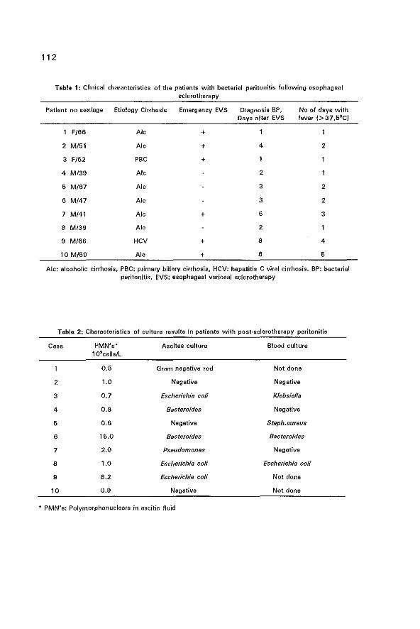
112
Table 1: Clinical characteristics of the patients with bacterial peritonitis following esophageal sclerotherapy
Patient no sex/age Etiology Cirrhosis Emergency EVS magnosia SP, No of days with Days after EVS fever 1>37,5OC)
F/66 AI, +
2 M/51 AI, + 4 2
3 F/62 pac +
4 M/39 AI, 2
6 M/67 AI, 3 2
6 M/47 AI, 3 2
7 M/41 AI, + 5 3
a M/3S AI, 2
9 M/66 HCV + 8 4
10 M/6S AI, + 8 6
Ale: alcoholic cirrhosis, PSC: primary biliary cirrhosis, HCV: hepatitis C viral cirrhosis. BP: bacterial peritonitis. EVS: esophageal variceal sclerotherapy
Table 2: Characteristics of culture results in patients with post-sclerotherapy peritonitis
Case PMN's~ Ascites culture Blood culture 109ce lls /L
0.6 Gram negative rod Not done
2 1.0 Negative Negative
3 0.7 Escherichia coli Kfebsielfa
4 0.8 Bacteroides Negative
6 0.6 Negative Staph.l1ureus
8 15,0 Bacteroides Bacteroides
7 2.0 Pseudomonas NegatiVe
8 1.0 Escherichia coli Escherichia coli
9 8.2 Escherichia coli Not done
10 0.9 Negative Not done
* PMN's: Polymorphonuclears in ascitic fluid
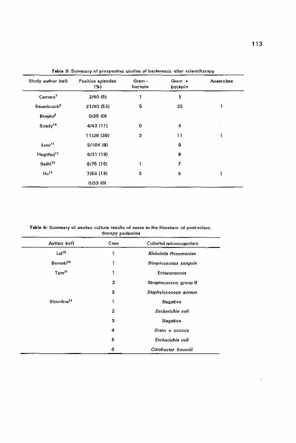
Table 3: Summary of prospective studies of bacteremia after sclerotherapy
Study author (ref) Positive episodes Gram + Gram + Anaerobes (%) bacteria bacteria
Camara l 2/40 (6)
Sauerbruch8 21/40 (63) 6 26
BraykoS 0/26 (0)
Snedyl0 4/43(11) 0 4
11/28 (39) 3 11
Lowll 9/104 (9) 9
Hegnhoj12 6/31 (9) 6
Sethi1l 8/76 (16) 7
HoB 7/66 (13) 3 3
0/33 (0)
Table 4: Summary of ascites culture results of casos in the literature of post-sclerotherapy poritonitis
Author {refl Case Cultured microorganism
Loi1s Klebsiella Pneumoni88
Barnett20 Streptococcus sanguis
Tom21 Enterococcus
2 Streptococcus group B
3 Staphylococcus 8ureus
Shembre2l Negative
2 Escherichia coli
3 Negative
4 Gram + coccus
6 Escherichia coli
6 Citrobacter freundii
113
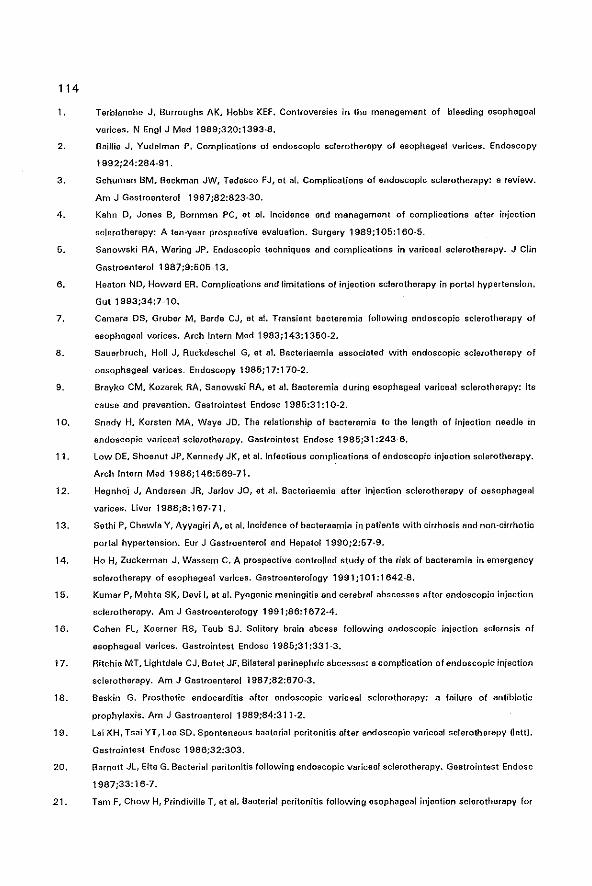
114
1, Terblanche J, Burroughs AK, Hobbs KEF. Controversies in the management of bleeding esophageal
varices. N Engl J Mad 1989;320:1393-8.
2. Baillie J, Yudelman P. Complications of endoscopic sclerotherapy of esophageal varices. Endoscopy
1992;24:284-91.
3. Schuman 8M, Beckman JW, Tedesco FJ, at al. Complications of endoscopic sclerotherapy: a review.
Am J Gastroenterol 1987;82:823-30.
4. Kahn D, Jones B, Bornman PC, at 81. Incidence and management of complications after injection
sclerotherapy: A ten-year prospective evaluation. Surgery 1989;105:160-5.
6. Sanowski RA, Waring JP. Endoscopic techniques and complications in variceal scterotherapy. J Clin
Gastroenterol 1987;9:505-13.
6, Heaton ND, Howard EA. Complications and limitations of injection sclerotherapy in portal hypertension,
Gut 1993;34:7-10.
7. Camara OS, Gruber M, Barde CJ, et al. Transient bacteremia following endoscopic sclerotherapy of
esophageal varices. Arch Intern Med 1983;143:1360·2.
8. Sauerbruch, Holl J, Ruckdeschel G, et al. Bacteriaemia associated with endoscopic sclerotherapy of
oesophageal varices. Endoscopy 1986;17:170-2.
9. Brayko eM, K02arek RA, Sanowski RA, et al. Bacteremia during esophageal variceal sclerotherapy: its
cause and preVention. Gaslrointesl Endosc 1986:31:10-2.
10. Snady H, Korsten MA, Waye JD. The relationship of bacteremia to the length of injection needle in
endoscopic variceal sclerotherapy. Gastrointest Endose 1986;31 :243~6.
11. Low DE, Shoenut JP, Kennedy JK, et at. Infectious complications of endoscopic injection sclerotherapy.
Arch Intern Med 1986;146:669~71.
12. Hegnhoj J, Andersen JR, Jarlov JO, et al. Bacteriaemia after injection sclerotherapy of oesophageal
varices. Liver 1988;8: 187+71.
13. Sethi P, Chawla V, Ayyagiri A, et al. Incidence of bacteraemia in patients with cirrhosis and non-cirrhotic
portal hypertension. Eur J Gastroenterol and Hepatol 1990;2:67-9.
14. Ho H, Zuckerman J, Wassem C. A prospective controlled study of the risk of bacteremia in emergency
sclerotherapy of esophageal varices. Gastroenterology 1991 ;101 :1642-8.
16. Kumar P, Mehta SK, Devi I, et al. Pyogenic meningitis and co-rebral abscesses after endoscopic injection
sclerotherapy. Am J Gastroenterology 1991;86:1672+4.
16. Cohen FL, Koerner RS, Taub SJ. Solitery brain abcess following endoscopic injection sclerosis of
esophageal varices. Gastrointest Endosc 1985;31 :331 ~3.
17. Ritchie MT, Lightdale CJ, Botet JF. Bilateral perinephric abcesses: a complication of endoscopic injection
sclerotherapy. Am J Gastroenterol 1987;82:670-3.
18. Beskin G. Prosthetic endocarditis after endoscopic variceal sclerotherapy: a failure of antibiotic
prophylaxis. Am J Gastroenterol 1989;84:311 +2.
19. Lei KH, Tsai VT I Lea SO. Spontaneous baoterial peritonitis after endoscopic variceal sclerotherapy (lett).
Gastrointest Endosc 1986;32:303.
20. Barnett JL, Elta G. Bacterial peritonitis following endoscopic variceal sclerotherapy. Gestrointest Endosc
1987;33:16~7.
21. Tam F, Chow H, Prindiville T, et a!. Bacterial peritonitis following esophageal injection sclerotherapy for
115
variceal hemorrhage. Gastrointest Endose 1990:36:131-3.
22. Shamb,e 0, Bjorkman OJ, Post-sclerotherapy bacterial peritonitis. Am J Gastrenterol 1991 ;86:481-6.
23. Bee OJ, Sietsema PO, Mulder PGH, at a1. Spontaneous bacterial peritonitis: Outcome and predictive
factors. Eur J Gastroontorol Hepatol 1993;5:636-40.
24. Snobl J, van Buuren HR, van Blenkenstein M. Endoscopic injection therapy using thrombin: an effective
and safa method for controlling esophagastric variceal bleeding. Gastroenterology 1992;102: A 89L
26. Siers em a PO, de Marie S, van Zeijl J, at al. Blood culture bottles are superior to lysis-centrifugation tubas
for bacteriological diagnosis of spontaneous bacterial peritonitis. J Clin Microbial 1992;30:667-9.
26. Wilcox eM, Dismukes WE. Spontaneous bacteriel peritonitis; A review of pathogenesis, diagnosis and
treatment. Medicine 1987;447-456_
27. Rimola A, Bory F, Teres J, et al. Oral, nonabsorbable antibiotics prevent infection in cirrhotics with
gastrointestinal hemorrhage. Hepatology 1986;6:463-7.
28. Bleichner G, Boulanger R, Squara P, et 81. Frequency of infections in cirrhotic patients presenting with
acute gastrointestinal hemorrhage. 8r J Surg 1986;73:724-6.
29. Soriano G, Guerner e, Tomas A, et al. Norfloxacin prevents bacterial infection in cirrhotics with
gastrointestinal hemorrhage. Gastroenterology 1992;1 03:1267~72.
30_ Sorell WT, Quigley EMM, Jin G, et al. Bacterial translocation in the portal-hypertensive rat: Studies in
basal conditions and on exposure to hemorrhagic shock. Gastroenterology 1993;104:1722-6_
31. Stiegman VG, Goff JS, Michaletz-Onody PA, et al. Endoscopic sclerotherapy as compared with
endocopic ligation for bleeding esophageal varices. N Engl J Mad 1992;326:1627-32.
32. Runyon BA. Patients with deficient ascitic fluid opsonic activity are predisposed to spontaneous bacterial
peritonitis. Hepatology 1988;8:632-5.


CHAPTER 9
SPONTANEOUS BACTERIAL PERITONITIS COMPLICATING
MALIGNANCY·RELATEO ASCITES.
TWO CASE REPORTS AND A REVIEW OF THE LITERATURE.
117
The contents of this chapter have been submitted for publication to J Gastroenterol and
Hepatol under the same title with the following authors: D.J. Bac, S. de Marie and M. van
Blankenstein.
118
Summary
Objective: To study the occurrence of spontaneous bacterial peritonitis complicating
ascites due to another etiology than cirrhosis of the liver.
Methods: During a five-year period (1987-1992) all cases with a diagnosis of spontaneous
bacterial peritonitis were prospectively identified and analyzed.
Setting: Gastroenterology and hepatology unit in a tertiary referral centre.
Results: In 2 out of the 60 identified cases the spontaneous bacterial peritonitis occurred
in malignancy-related ascites.
Both patients developed spontaneous bacterial peritonitis (S8P) in pre-existing ascites. The
first patient with diffuse livermetastases of an adenocarcinoma and ascites developed an
episode of spontaneous peritonitis, with Escherichia coli cultured from the blood and
ascitic fluid. The second patient, with peritoneal adenocarcinomatosis and portal vein
thrombosis had peritonitis due to Klebsiella pneumoniae cultured from the blood and ascitic
fluid.
Conclusion: A summary is presented of the rarely reported cases of spontaneous bacterial
peritonitis in malignancy-related ascites without cirrhosis of the liver. The presence of
portal hypertension is recognized as an important predisposing factor.

119
Introduction
spontaneous bacterial peritonitis (S8P) has been well recognized as a severe and common
complication in patients with decompensated cirrhosis of the liver, resulting in a high
mortality [1,21. Knowledge about SSP has been accumulating over the last 20 years,
leading to earlier diagnosis and treatment [3]. However, most studies on S8P only deal
with patients suffering from decompensated cirrhosis, and S8P complicating preexistent
ascites due to other etiologies has rarely been reported. We report 2 episodes of S8P in
2 patients without cirrhosis of the liver. A review of the literature is presented of all cases
diagnosed as S8P without cirrhosis of the liver and its rarity in malignancy· related ascites
is discussed.
Case reports
Patient 1
A 69-year-old Caucasian male was admitted because of poorly regulated non-insulin
dependent diabetes mellitus, weight loss and anorexia. On physical examination the patient
was cachetic and dehydrated. There was hepatomegaly without splenomegaly and no
physically detectable ascites. Laboratory examination showed a raised ESR (66mm/h), an
elevated glucose (26' mmolll) and raised bilirubine 58 pmolll (n: 2-12 umol/l), gamma
glutamyl transpeptidase 332 UII (n:25·75 utI], lactate dehydrogenase 556 UII (n:160·320
UII). Serum albumin was decreased, 28 gil (n:36·48 gil)' and the albumin gradient (serum
minus ascites) was 22 gIl indicating the presence of portal hypertension. There were
impaired coagulation parameters: Prothrombin Time 15,1 sec. (n:9,3-12,3 sec), Normotest
44% (n:65·110%), antithrombine III activity 0,48 Ell (n:0.85·1.20 Ell).
Ultrasonography of the liver showed massive hepatic metastases and ascites. Gastroscopy
revealed mild esophagitis without esophageal varices. Histology of an ultrasound guided
liver biopsy showed a well-differentiated adenocarcinoma. The surrounding liver tissue
showed no histological signs of cirrhosis. No malignant cells were detected on cytological
examination of the ascitic fluid.
Three weeks after admission the patient developed rigors with fever and abdominal
tenderness. Ascitic fluid and blood cultures grew Escherichia co/i. Haematological and
chemical analysis of the ascitic fluid is shown in Table 1. The patient was treated
with antibiotics but died 8 days later. An autopsy was not performed.
120
Patient 2
A 59-year-old male patient was seen in another hospital because of abdominal pains and
weight loss. The patient had insulin-dependent diabetes mellitus since two years.
Laboratory examination revealed chalastatic liver functions and an ultrasonography
demonstrated an enlarged pancreatic head with calcifications. An ERep showed a
strictured common bile duct and an en do prosthesis was placed. The diagnosis was thought
to be chronic pancreatitis with calcifications and an inflammatory reaction in the head of
the pancreas.
Four months later the patient presented with increasing weight loss and abdominal pains.
Physical examination revealed ascites, without abdominal tenderness. Laboratory
examination showed increased cholestasis; bilirubin 11 pmol/l (n:2-12 umol/l). alkaline
phosphatase 623 U/I (n: 40-85 U/II, with normal transaminases. Clotting parameters were
normal with a decreased albumin level of 30 gil (n:36-48 gil). The endoprosthesis was
exchanged but found not to be occluded. Ultrasonography showed an increasing mass
within the pancreatic head, suggesting malignancy, and evidence of portal vein thrombosis.
Thin needle biopsy of the mass in the head of the pancreas failed to confirm malignancy.
Two weeks after admission the patient developed fever and a tender abdomen. Klebsiella
pneumoniae was cultured from the blood and ascitic fluid. The ascitic fluid characteristics
are shown in Table 1. The patient was treated for bacterial peritonitis with amoxycillin
clavulanic acid. Liver functions had remained stable during this time and there was no
evidence of an occluded endoprosthesis. Because of the progressive ascites, without proof
of malignancy, a laparoscopy was performed. The liver appeared normal but there were
many small white dots on the peritoneal surface, biopsy of which confirmed peritoneal
carcinomatosis due to an adenocarcinoma, most probably from the pancreas. Liver
histology was normal. Within six weeks the patient developed progressive esophageal
varices, and a bleeding episode which was treated by sclerotherapy. In spite of this the
patient died from recurrent variceal bleeding 8 weeks later. An autopsy was not performed.
Discussion
Malignancy-related ascites is a heterogeneous group of conditions with wide variations in
ascitic fluid analysis. Within this group peritoneal carcinomatosis, characterized by positive
ascitic fluid cytology, is the most frequently reported cause of ascites formation [4].
Massive liver metastases with concomitant portal hypertension is the second most
121
important group. Two patients are described with a bacterial infection of the malignancy*
related ascites. A review of the literature confirms that SSP complicating malignancy
related ascites has only been described in patients who had evidence of portal hyperten
sion. Isner at al [51 described one patient with a gastric adenocarcinoma who developed
SSP after chemotherapy. At autopsy 75% of this patient's liver parenchyma had been
replaced by tumor. Kurtz et al (61 reviewed more than 100 patients with cytology positive
ascitic fluid and found only three patients with bacterial peritonitis. The authors stated that
these cases do not represent true SSP because invasive procedures in two patients
(indwelling peritoneal catheter and bowel puncture at initial paracentesis) and a nadir
sepsis (leucocyte count < 0,3x10⁹ cellsJl) in one patient clearly predisposed them to
bacterial peritonitis. Runyon (71 described one patient with S8P and cardiac ascites. But
also in this patient there was evidence of portal hypertension with splenomegaly and
esophageal varices due to liver fibrosis.
Our cases and those reported above do suggest that the presence of portal hypertension,
either due to massive hepatic metastases or portal vein thrombosis is a prerequisite for
malignancy-related ascites to become infected. Intact antimicrobial activity of the
peritoneal fluid and functioning hepatic tissue are important determinants in clearing
microorganisms from the ascitic fluid (8J. Comparing cirrhotic ascites with malignant
ascites (without portal hypertension). complement concentrations C3 and C4 are about
four times higher in the latter group with better opsonic and chemo-attractant activity {9].
The hepatic venous pressure gradient in patients with massive liver metastases has been
found to be similar to that quantified in cirrhotic patients [10], This is confirmed by the
presence of a wide (> 11 g/l) serum-ascites albumin gradient (as a manifestation of
oncotic-hydrostatic balance) which has been found in patients with malignancy-related
ascites and massive hepatic metastases [11,12). The presence of portal hypertension
gives rise to a significantly lower total protein concentration in the ascitic fluid, compared
to patients with peritoneal carcinomatosis without portal hypertension [9,11,12). A low
protein concentration in the ascitic fluid reflects decreased complement concentrations and
decreased opsonic and bactericidal activity (9,13,14]. This would explain the susceptibility
of malignant ascites to the development of S8P in the presence of portal hypertension.
However, also other factors might be involved. Lebrun et al. recently described decreased
polymorphonuclear (PMN) function in cirrhotic ascitic fluid [15]. The PMN function
improved with serial dilutions or by adding heat-treated malignant ascitic fluid. This
suggests that phagocytosis is deficient independantly of the C3 level, and that other
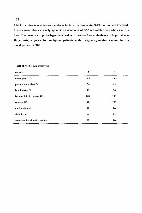
122
inhibitory intracellular and extracellular factors that modulate PMN function are involved.
In conclusion there are only sporadic case reports of SBP not related to cirrhosis of the
liver. The presence of portal hypertension due to massive liver metastases or to portal vein
thrombosis, appears to predispose patients with malignancy-related ascites to the
development of SBP.
Table 1: Ascitic fluid parameters
patient: 2
leucocytosx109 /1 2.a 29,8
polymorphonuclear % aa 90
lymphocytes % 12 10
lactate dehydrogenase U/I 257 246
amylase U/I 46 220
total protein g/l 19 20
albumin gil 6 12
serum-ascites albumin gradient 22 18
123
1. Hoefs JC. Spontaneous Bacterial Peritonitis: Prevention end Therapy. Hopetalogy 1990, 12:776-779.
2. Bae OJ, Siers em a PO, Mulder PGH, de Marie S, Wilson JHP. Spontaneous bacterial paritanitis: outcome
and predictive factors. Eur J Gastroenterol Hepatol 1993, 5:636-640.
3, Runyon SA. Spontaneous Bacterial Peritonitis: An explosion of information. Hapatology 1988, 'S:171·
176.
4. Runyon SA, Hoefs JC, Morgan TR. Ascitic fluid analysis in malignancy-related ascites. Hepatology 1988,
8:1104-1109.
5. Isner J], Macdonald JS, Schein PS. Spontaneous streptococcus pneumonia peritonitis in a patient with
metastatic gastric cancer. Cancer 1977,39:2306-2309.
6. Kurtz Re, Bronza Rl. Does spontaneous bacterial peritonitis occur in malignant ascites? Am J
Gastroenterol 1982, 77:146-148.
7. Runyon BA. Spontaneous bacterial peritonitis associated with cardiac ascites. Am J Gastroenterol 1984,
79:796.
8. Akafin EH, Laleli Y, Teletar H. Bactericidal and opsonic activity of ascitic fluid from cirrhotic and non
cirrhotic patients. J Infect Dis 1983, 147:1011-1017.
9. Mal F, Pham Huu T, Bendahou M, et aI, Chemoattractant and opsonic activity in ascitic fluid. J Hepatol
1991,12:45·49.
10. Lee S, Koshy A, Hadenque A, et al. Hemodynamic derangements of primary and metastatic hepatic
carcinomas (abstr). Hepatology 1986,6:1109.
11. Albillos A, Cuervas-Mons V, Millan I, et al. Ascitic fluid polymorphonuclear cell count and serum to
ascites albumin gradient in the diagnosis of bacterial paritonitis. Gastroenterology 1990, 98:134-140.
12. Runyon BA, Montano AA, Akrivadis EA, et at The serum-ascites albumin gradient is superior to the
exudate-transudate concept in the differential diagnosis of ascites. Ann lnt Med 1992, 117:215-220.
13, Such J, GUa(Oer C, Enriquez J, et al. low C3 in cirrhotic patients predisposes to spontaneous bacterial
peritonitis. J. Hepato! 1988, 6:80-84.
14. Runyon BA, Morrissey Rl, Hoefs JC, Wy!e FA. Opsonic activity of human ascitic fluid: A potentially
important protective mechanism against spontaneous bacterial peritonitis. Hepatology 1985,5:634-637.
15, Lebrun L, Pelletier G, Briantais MJ, Galanaud P, Etienne JP. Impaired functions of normal polymor
phonuclear leukocytas in cirrhotic ascitic fluid. J Hepatol 1992, 16:98-101.


125
Summary
This thesis describes several aspects of spontaneous bacterial peritonitis in patients with
cirrhosis of the liver and ascites. Most of the data discussed and submitted in this thesis
are based on clinical studies during the years 1987-1992 at the Department of Internal
Medicine II, University Hospital Rotterdam-Dijkzigt.
Chapter 1. In this chapter definitions are provided for the diagnosis of spontaneous
bacterial peritonitis which arB to be used in this thesis. A historical background is given
of spontaneous bacterial peritonitis, and a review of the literature concerning its prevalence
and incidence in the different centers is provided. The pathogenesis of spontaneous
bacterial peritonitis is discussed, with special emphasis on host factors, environmental
factors and the microorganisms responsible for the peritonitis. Appropriate antibiotic
treatment is discussed and the possibility of selective intestinal decontamJilation to prevent
recurrences of spontaneous bacterial peritonitis.
Chapter 2. This is a review article concerning the appropriate diagnostic tests to be done
on the ascitic fluid when a paracentesis is performed. The superiority of the albumin
gradient {serum albumin~minus ascites albumin concentration} compared to the traditional
"exudate-transudate" concept in clarifying the etiology of the ascites is discussed. A white
blood cell count with an accurate differentiation is the single most important test to be
done on ascitic fluid.
Chapter 3. Because of the low values of total protein and albumin and the low white cell
count in ascitic fluid special reference values are needed in the laboratory for an accurate
and precise determination of these values. A reliable and convenient procedure is presented
to perform a manual differential when low numbers of white cells are present. The reliablity
of the determination of total protein and albumin in ascitic fluid on a routine serum
chemistry analyzer is described.
Chapter 4. Three different culture techniques of ascitic fluid are described and compared,
to test sensitivity and convenience of each culture method. Inoculation of ascites into
blood culture bottles at the bedside of the patient gives the highest diagnostic yield (84%
positive cultures, compared to 35% positive cultures when using the conventional culture
method and 46% positive cultures when using the lysis-centrifugation tubes). Moreover,
the blood culture bottle method also shortened the time needed for the detection of
bacterial growth. Gram~negative microorganisms were detected in 65% of the culture-
126
positive episodes of spontaneous bacterial peritonitis. Gram-positive cocci were cultured
in the remaining 35% of the episodes.
Chapter 5. The measurements of cytokines in plasma and ascites could be of diagnostic
and prognostic importance, assisting in early diagnosis of infectious complications and in
the differential diagnosis of the etiology of ascites. To test this hypothesis, interleukin-6
(lL-6). interleukin 1P (lL-1P), and tumor necrosis factor-a ITNF-a) were measured in ascites
and plasma in 8 patients with malignancy-related ascites and in 32 patients with
decompensated cirrhosis. Five patients had an episode of bacterial peritonitis during which
period one or more ascitic fluid samples were analyzed. It was found that ascitic fluid IL-6
levels are about 30 times higher compared to plasma values, indicating that IL~6 is
produced in great quantities within the peritoneal cavity of patients with ascites,
irrespective of the etiology of the ascites. During a period of bacterial peritonitis the IL~6
levels, but not the other cytokines, were significantly raised in the ascites, which declined
after antibiotic treatment. The importance of locally generated cytokines and the possibility
that this relates to the poor prognosis of patients with cirrhosis and infectious complica~
tions is discussed.
Chapter 6. In this study the presence of increased intestinal permeability and luminal
bacterial overgrowth in the small bowel of patients with cirrhosis and portal hypertension
is evaluated. Changes in small bowel function in patients with cirrhosis could contribute
to complications, such as malabsorption and infections e.g. spontaneous bacterial
peritonitis. Using the 14C~glycocholic acid breath test and measuring the urinary excretion
of orally ingested 51Cr~EDT A, and by assessing the leakage of alpha-1-antitrypsine into the
intestinal lumen, no evidence was found of small intestinal bacterial overgrowth or of
increased permeability of the small bowel in a small number of patients. It is concluded
that small bowel function is maintained to a large extent in patients with advanced liver
cirrhosis and portal hypertension.
Chapter 7. A comparison is made between 31 positive ascitic fluid samples with 92
negative samples for spontaneous bacterial peritonitis (SSP) in 83 patients. Predictive
factors for the development of SSP are the severity of the cirrhosis, as indicated by the
Child-Pugh score and coagulation parameters. The prognosis for patients who had an
episode of SBP is significantly worse compared to those who had sterile ascites (1 year
survival respectively 14% and 43%; p =0.017). The recurrence rate for those who had an
episode of S8P was 20%. Because enteric microorganisms are the most common
causative agents of SBP, suggestions are made for the use of selective intestinal
127
decontamination in certain subgroups of patients to prevent the occurrence of SSP.
Chapter 8. In this chapter the relationship between endoscopic variceal sclerotherapy (EVS)
and "spontaneous" bacterial peritonitis is elaborated on. It was established that the risk
to develop infected ascites following sclerotherapy is significantly higher after emergency
EVS (sclerotherapy within 24 hours of a variceal bleeding) compared to elective EVS
(respectively 3% vs. 0.5%). Gram-negative gut derived microorganisms were the most
common bacteria cultured from the ascites, which is identicle to the bacteria causing SSP
unrelated to sclerotherapy, but different from studies reporting on the incidence of
bacteremia during sclerotherapy. It is suggested that translocation of Gram-negative
microorganisms during active gastrointestinal bleeding is a more important mechanism
causing bacterial peritonitis than the act of sclerotherapy itself. Antibiotic prophylaxis could
be considered in patients with severe cirrhosis and ascites, presenting with a gastrointes
tinal bleeding.
Chapter 9. Spontaneous bacterial peritonitis complicating ascites due to another etiology
than cirrhosis of the liver has rarely been reported. In 2 of the 60 episodes of SBP
identified during a five-year period, the diagnosis of SSP was made in malignancy-related
ascites without cirrhosis. One patient had massive liver metastases causing portal
hypertension and ascites, and the other patient had peritoneal carcinomatosis with portal
vein thrombosis. It is discussed why SSP is rarely described in patients without cirrhosis
of the liver. The presence of portal hypertension in both these patients is recognized as an
important predisposing factor for the development of SBP.
128
Conclusion
During the period 1987-1992 sixty episodes of spontaneous bacterial peritonitis were
diagnosed in 52 patients. S8P was found to be a serious complication in patients with
cirrhosis and ascites with a l-year survival of 23% (Confidence Interval 17%-35%). Three
of the 1 2 patients who survived more than 1 year had a livertransplant.
Clinical awareness, a lower threshold to perform paracentesis, earlier diagnosis and
treatment might have resulted in a slightly improved survival compared to the results
described in chapter 7.
Improved culture techniques of the ascitic fluid and more adequate leucocyte count with
cell differential will add to a better diagnosis. There is evidence in the literature that the
use of selective bowel decontamination in patients at high risk to develop SBP will
decrease its incidence, as most causative microorganisms are of enteric origin.
However, an episode of SBP should be regarded as a symptom of hepatic insufficiency and
patients who developed an episode of SBP and who are regarded as suitable candidates
for a liver transplantation should have this procedure as soon as possible. As such, an
episode of SSP should be one of the many factors which are to be included in the
complicated decision making process that determines timing and priorization of candidates
for transplantation.
129
Samenvatting
In dit proefschrift worden een aantal aspectsn van spontane bacteria Ie peritonitis bij
patienten met levercirrose en ascites behandeld. De resultaten die worden besproken zijn
verkregen uit klinische studies die verricht werden gedurende de peri ode 1987-1992 op
de afdeling Inwendige Geneeskunde II van het Academisch Ziekenhuis Rotterdam, Dijkzigt.
Hoofdstuk 1. Hierin worden definities besproken zoals die verder in dit proefschrift
gehanteerd worden. Een historisch overzicht wordt gegeven betreffende het begrip
spontane bacteriale peritonitis, avenals een literatuuroverzicht betreffende incidenties en
prevalenties in de verschililende centra. De pathogenese van het ziektebeeld wordt
besproken, waarbij de oorzakelijke microorganismen, de gastheer functie en de om
gevingsfactoren verantwoordelijk voor het ontstaan van de peritonitis worden toegelicht.
De optimale antibiotische behandeling wordt besproken evenals de mogelijkheid van
selectieve darm decontaminatie ter preventie van spontane bacteriale peritonitis.
Hoofdstuk 2. Dit is een overzichtsartikel betreffende bepalingen in de ascites welke
kunnnen plaats vinden bij het verrichten van een diagnostische ascites drainage. De
voorkeur voar de albumine gradient (serum albumine minus ascites albumine concentratie)
boven het conventionele "exudaat-transudaat" concept wordt toegelicht. Een telling van
het totaal aantal leukocyten met een juiste differentiatie is de belangrijkste be paling in de
ascites.
Hoofdstuk 3. Vanwege de lage concentraties totaal eiwit en albumine, evenals het geringe
aantal leucocyten in de ascites, zijn speciale referentie waarden noodzakelijk in het
laboratorium veor een adequate en precieze bepaling hiervan. Een bruikbare en betreuw
bare methode wordt besproken om de hand-differentiatie te verrichten bi; een laag aantal
leucocyten in de ascites. De betrouwbaarheid van het totale eiwit gehalte en albumine
gehalte bepaling in de ascites met behulp van routine chemie analyse wordt weergegeven.
Hoofdstuk 4. Drie verschillende kweekmethoden van de ascites worden beschreven en met
elkaar vergeieken. Inocula tie van ascites in bloedkweekHesjes direct aan het bed bij de
patient geeft de hoogste opbrengst (84% positieve kweken, vergeleken met 35% positieve
kweken met de conventionele methode en 46% positieve kweken met gebruik van de lysis
centrifugatie techniek mbv. IsolatorR bulzen). Tevens is de periode tot aan de kweekuitslag
significant korter bij gebruik van de bloedkweekflesjes. Gram negatieve microorganismen
werden gedetecteerd in 65% van de positieve kweken en Gram positieven coccen in de
resterende 35% van aile episoden met spontane bacteri9le peritonitis.
130
Hoofdstuk 5. Het bepalen van cytokinen in plasma en ascites zou van diagnostische en
prognostische betekenis kunnen zijn, zowel bij het vroegtijdig herkennen van infectiauza
complicaties alsook bij de differentiaal diagnose ter bepaling van de atlologie van de
ascites. Om deze hypothese ta toetsen werden zowel interleukinew 6 (lL-6), interleukine 1P (lL-1Pl als ook tumor necrosis factor-a (TNF-al bepaald in plasma en ascites bij 8 patienten
met ascites secundair aan een maligniteit, en bij 32 patienten met gedecompens8erde lever
cirrose. Vijf patienten maakten een episode van bacteriele peritonitis door waarbij ascites
verkregen kon worden va or interleukinen bepalingen. De IL-6 concentratias in ascites
waren ongeveer 30 x hoger dan in plasma, passend bij een hoge intra peritonea Ie productie
van IL-6 bij patienten met ascites, onafhankelijk van de etiologie van deze ascites.
Gedurende een peri ode van bacteriale peritonitis was er een sterke stijging in de IL-6
concentratie in de ascites, maar niet in plasma, welke afnam na adequate antibiotische
therapie. De fysiologische betekenis van deze hoge lokale productie van cytokinen en de
mogelijke relatie met de slechte prognose van infactieuza complicaties bij levercirrose
wordt verder besproken.
Hoofdstuk 6. In deze studie wordt de hypothese getoetst dat er sprake is van een
toegenomen permeabiliteit en bacteria Ie overgroei in de dunne darm bij patienten met
cirrose en portale hypertensie. Een veranderde darmfunctie bij cirrose zou aen bijdragande
factor kunnen zijn in het ontstaan van com plica ties, zoals ondervoeding en infecties, bijv.
spontane bacteriale peritonitis. Met behulp van de 14C-glycocholzuur ademtest, het bepalen
van de 51Cr-EOTA excretie in de urine na orale inname, en de alpha-1-antitrypsine klaring
in de faeces, kon bij een kleine patienten groep vastgesteld worden dat er geen
toegenomen permeabiliteit van de dunne darm noch bacteriale overgroei in de dunne darm
aantoonbaar is. De conclusie is dan ook dat bij patienten met levercirrose en portale
hypertensie de dunne darm functie grotendeels gespaard blijft.
Hoofdstuk 7. Een vergelijking wordt gemaakt tussen 31 episoden van spontane bacteria Ie
peritonitis met 92 episoden met steriele ascites in totaal 83 patienten. Voorspellende
factoren voor de ontwikkeling van S8P waren de ernst van de cirrose uitgedrukt in de
Child-Pugh score en de stollings parameters. De prognose van patienten die een episode
van S8P hadden doorgemaakt was significant slechter dan van patienten met steriele
ascites (1 jaars overleving respectievelijk 14% en 43%; p=O.017). De recidief kans voor
patienten die een episode van S8P hadden doorgemaakt was 20%. Aangezien gram
negatieve microorganismen afkomstig vanuit de darm met name verantwoordelijk zijn voor
S8P, worden suggesties gedaan voor het gebruik van selectieve darm decontaminatie bij
131
geselecteerde graepen van patienten om het optreden van SBP t9 voorkomen.
Hoofdstuk 8. Oit hoofdstuk behandeld de ralatia tussen het verrichten van endoscopische
sclerotherapie van slokdarm varices (ESV) en het optreden van "spontane" bacteriBie
peritonitis. Het risico op de ontwikkeling van bacteria Is peritonitis is significant hoger na
een spoed ESV (sclerotherapie binnen 24 uur na een varices bleeding) vergeleken met een
electieve ESV (respectievelijk 3% VS. 0.5%). Gram negatieve microorganismsn afkomstig
uit het darm lumen zijn oak na sclerotherapie verantwoordelijk va or het merendeel van de
infecties, wat dus overeenkomt met de verwekkers van SBP optredend onafhankelijk van
het verrichten van sclerotherapie. Oit gegeven suggereert dat translocatie van gram
negatieve bacterien vanuit het darmlumen ten tijde van een gastro-intestinale bloeding een
belangrijker mechanisme is v~~r het veroorzaken van SSP dan het scleroseren zeit. Indien
het scleroseren, met de daarbij behorende beschadiging van de mucosa de oorzakelijke
factor zou zijn, zou men meer microorganism en vanuit de mond-keel holte verwachten als
verwekker van de SSP. Slj patienten met cirrose en ascites met een gastro-intestinale
bloeding kan het kortdurend gebruik van antibiotische profylaxe overwogen worden.
Hoofdstuk 9. Spontane bacteriele peritonitis wordt zelden gerapporteerd als camplicatie
van ascites welke niet het gevolg is van een gedecompenseerd leverlijden. In 2 van de 60
episoden welke gedurende een periode van 5 jaar geregistreerd werden, trad SSP op als
camplicatie van een maligniteit. Een patient had uitgebreide levermetastasering met
secundair hieraan porta Ie hypertensie en ascites, de andere patient had een peritonitis
carcinomatosa met vena porta thrombose. De mogelijke redenen waarom SSP zelden
gediagnostiseerd wordt bij patienten zander levercirrosa worden toegelicht. De aanwezig
heid van portale hypertensie in deze beide patienten wordt gezien als een belangrijke
predisponerende factor voor het ontstaan van SSP.
132
Conclusie
Gedurende de periods 1987 -1992 werden 60 episoden van spontane bacteria Ie peritonitis
geregistreerd bij 52 patienten. S8P bleak aen ernstige complicatie t6 zijn bij patienten met
levercirrose en ascites met aen hoge mortaliteit en een lage 1 jaars overleving van 23%
(Betrouwbaarheids interval 17%-35%). Dria van de 12 patienten die meer dan 1 jaar
overleefden ontvingen een levertransplantatie. Toegenomen alertheid en bekendheid met
het ziektebeeld van S8P, en aen lagere drempel om paracentese te verrichten met hierdoor
vraagere behandelingsmogelijkheden hebben mogelijk geresulteerd in aen iets betera
overleving dan gemeld in hoofdstuk 7. Verbeterde kweek tBchnieken en aen precieze
leucocyten telling met differentiatie in de ascites dragsn bij tot een juiste diagnose stelling.
Uit de literatuur zijn gegevens bekend dat het gebruik van selectieve darm decontaminatie,
met als doel de gram negatieve flora te verminderen met het relatief sparen van de
resterende anaerobe flora, bij patienten met een hoog risico op het krijgen van SSP, de
incidentie van SSP doet afnemen.
Echter, een episode van SSP dient geinterpreteerd te worden als een symptoom van ernstig
levertalen en bij patienten die een dergelijke infectieuze complicatie ontwikkelen moet het
verrichten van een levertransplantatie ernstig overwogen worden. Als zodanig is een
doorgemaakte episode van SSP een van de vele factoren die meegenomen moeten worden
in de vaak moeilijke besluitvorming over de noodzaak tot, en het tijdstip van een
levertransplantatie.
Publications related to this thesis
P.D.Siersema, D.J.Bac, S.da Marie, J.H.P.Wilson.
Spontane bacteriale peritonitis.
Ned Tijdschr Geneeskd 1992; 136: 113-116.
D.J.Bac, P.D.Siersema, J.H. van Zeijl, S. de Marie, J.H.P.Wilson.
133
Improved detection of spontaneous bacterial peritonitis by bedside inoculation of ascitic
fluid in blood culture bottles. Neth J Med 1992; 40: A66-67.
P.D.Siersema, S. de Marie, J.H. van Zeijl. D.J.Bac, J.H.P.Wilson.
Blood culture bottles are superior to lysis-centrifugation tubes for bacteriological diagnosis
of spontaneous bacterial peritonitis. J Clin Microbiol 1992; 30: 667-669.
D.J.Bac, P.D.Siersema, J.H.P.Wilson.
Paracentesis: the importance of optimal ascitic fluid analysis.
Neth J Med 1993; 43: 147-156.
D.J.Bac, P.D.Siersema, P.G.H.Mulder, S. de Marie, J.H.P.Wilson.
Spontaneous bacterial peritonitis; outcome and predictive factors.
Eur J Gastroenterol Hepatol 1993; 5: 635-640.
D.J.Bac, G.R.Swart, J.W.O.van den Berg, J.H.P.Wilson
Small bowel wall function in patients with advanced liver cirrhosis and portal hypertension:
Studies on permeability and luminal bacterial overgrowth.
Eur J Gastroenterol Hepatol1993; 5: 383·387.
D.J.Bac, S.da Marie, P.D.Siersema, J.Snobl, H.R. van Buuran.
Post-sclerotherapy bacterial peritonitis; A complication of sclerotherapy or of variceal
bleeding? Am J Gastroenterol 1994; 89: 859-862.
D.J.Bac, P.D.Siersema, H.R.van Suuren. Should antibiotic prophylaxis be given during
endoscopic variceal sclerotherapy to prevent bacterial peritonitis?
Neth J Med 1994; 44: A38-39.
134
D.J.Bac, W.M.Pruimboom, P.G.H.Mulder, J.F.Zijlstra, J.H.P.Wilson.
Interleukin~6 and tumor necrosis factor-o ascites in cirrhosis or peritoneal malignancy.
Gastroenterology 1994; 106: A862
D.J.Bac, W.M.Pruimboom, P.G.H.Mulder, J.F.Zijlstra, J.H.P.Wilson. High interleukin-6
production within the peritoneal cavity in decompensated cirrhosis and malignancy-related
ascites. Liver (submitted).
W.M.Pruimboom, D.J.Bac, A.P.M. van Dijk, I.M.Garrelds, C.J.A.M.Tak, I.L.Bonta,
J.H.P.Wilson, F,J.Zijlstra. Levels of soluble intercellular adhesion molecules, eicosanoids
and cytokines in ascites of patients with liver cirrhosis, peritoneal cancer and spontaneous
bacterial peritonitis. Int J Pharmacol (submitted)
H.Engel, D.J.Bac, R.Brouwer, B.G.Blijenberg, J.Lindemans. Optimal analysis of total
protein, albumin, white cell count and differential in ascitic fluid.
Ann Clin Biochem (submitted).
D.J.Bac, S.de Marie, M. van Blankenstein
Spontaneous bacterial peritonitis complicating malignancy-related ascites. Two case
reports and a review of the literature. J Gastroenterol and Hepatol (submitted)
135
Dankwoord
Het schrijven van een proefschrift is lauk en stimulerend, maar zander de noodzakelijke
stavn van diverse mensen in de directe omgeving zou het er zeker niet zo uitgezien
habben, en misschien zelfs nooit tot stand zijn gekomen.
Peter Siersema was al in 1987 begonnen met het vergelijkend onderzoek tU5sen de
verschillende kweekmethoden van ascites, wat uiteindelijk resulteerde in de hoofdstukken
4 en 7 van dit proefschrift. Zonder de inv8stering van vele avonden om de gegevens t8
analyseren en uit t8 werken zou dit proefschrift sr wellicht nooit geweest zijn. Nogmaals
mijn dank.
Siam de Marie was onze onmisbare staun op de afdeling bacteriologie en oak zijn inzet was
onontbeerlijk om tot dit resultaat te komen.
Samen met Roel Swart werden de eerste voorzichtige stappen gezet tijdens het zaalwerk
op afdeling 4M op de weg tot klinisch onderzoek, uiteindelijk resulterend in hoofdstuk 6.
De inzet van verpleegkundigen, doktersassistenten, co-assistenten en assistenten en de
medewerking van vele patienten was noodzakelijk om aile ascites puncties tot een goed
einde te laten komen en om de "Bac Bakjes" met het onontbeerlijk vocht te vullen. Oit
werd vervolgens deskundig en snel verwerkt op het laboratorium van afd. Inwendige
Geneeskunde II onder leiding van Wim van de Berg.
De interleukinen bepalingen vonden plaats op de afdeling Farmacologie van de Erasmus
Universiteit onder leiding van Freek Zijlstra en Wanda Pruimboom, wat een vruchtbare
liaison bleek te zijn, resulterend in diverse publica ties.
Paul Mulder van de afdeling Epidemiologie en Biostatistiek was zeer behulpzaam om aile
angels en voetklemmen te ontwijken bij de statistische bewerking. Gezamelijk met het
Klinisch Chemisch Laboratorium van Oijkzigt werd onder leiding van Henk Engel gekeken
naar de betrouwbaarheid van de diverse testen, uiteindelijk resulterend in hoofdstuk 3.
Diverse mensen waren behulpzaam om de hiaten in mijn computerkennis bij te spijkeren,
waaronder Jan Boot, Juliette van Klinken en Carin van Vliet.
De leden van de leescommissie, de professoren Bruining, Schalm en Verbrugh dank ik v~~r
hun snelle beoordeling van het manuscript. Mijn promotor Paul Wilson, hartelijk dank veor
de koelbloedige wijze waarop je mij door de laatste spannende weken hebt geleid.
Mark van Blankenstein wil ik bedanken voor de vrijheid en tijd die beschikbaar was tijdens
de gastro-enterologie opleiding voer research en het schrijven van de publicaties.
Rob de Man, mijn kamermaat, voor zijn praktische adviezen.
136
Mijn Duders hebben het studersn altijd gestimuleerd en hen wi! ik bedanken voor de
mogelijkheden die zij mij gegeven hebben. Aan mijn vader wil ik bij deze dit proelschrift ter
nagedachtenis opdragen. Gezondheid en aile dagelijkse levensbehoeften die we vaak als
vanzelfsprekend be schouwen, werden mij in ruime mate geschonken door de Allerhoogste,
waarvoor Hem aile dank toekomt.
Tenslotte gaat mijn dank uit naar Orieka, zander wiens ruimhartigheid en enorma
relativeringsvermogen ik thuis geen gelegenheid gehad zou hebben om zoveel tijd aan dit
boekwerkje te besteden. Jouw heldere visie heelt mij vaak geholpen om te komen tot dit
eind resultaat. Er is wederom aen druppel toegevoegd aan de Desaan van wetenschap en
een ieder die hiervoor zijn 01 haar bijdrage heelt geleverd nogmaals hartelijk dank.
137
Curriculum Vitae
De auteur werd geboren op 6 november 1956 te Moerkapelie. Na het atleggen van het
eindexamen Atheneum p in 1975 aan de Christelijke schoiengemeenschap te Gouda,
studeerde hij Geneeskunde aan de Erasmus Universiteit t8 Rotterdam. Er werd een student
assistentschap verricht op de afdeling Pathologische Anatomie (hoofd: Prof. Or. A.O. van
de Heul). Het doctoraal examen volgde in 1980 (Cum Laude) en het Artsexamen in 1981.
Van 1982-1987 was de auteur werkzaam als medical officer en later als senior medical
officer in het Donald Fraser Hospital te Venda in Zuid-Afrika. In 1983 werd het Diploma
Tropical Medicine and Hygiene (DTM&H) en in 1984 het Diploma Public Health (DPH)
behaald aan de Witwatersrand University te Johannesburg.
In juli 1987 werd een aanvang gemaakt met de opleiding tot internist in het Havenzieken
huis (Opleider: Prof. Dr. P.C. Stuiver) welke werd vervolgd in het Academisch Ziekenhuis
Rotterdam-Dijkzigt, afdeling Inwendige Geneeskunde " (Opleider: Prof. J.H.P. Wilson).
Op 1 augustus 1992 volgde de registratie tot internist en werd beg onnen met de vervolg
specialisatie tot gastro-enteroloog. De auteur maakt deel uit van de Rotterdamse
Levertransplantatie graep, en is als zodanig direct betrokken bij de pre- en
posttransplantatie patientenzorg.