species identification of meat samples: an unusual case
TRANSCRIPT
278 (83%) were from farms in Victoria, while the remainder were from farms in New South Wales. The tail-tags of the cattle were used to provide this datum. No information on farms on which the cattle were born was avalable. Both eyes and associated tissues including the lachrymal glands and eyelids were removed from each head. Following dissection, the parasites present were washed from the lachrymal and excretory ducts, conjunctiva and eyelids with normal saline, counted and identified.
Fifteen (4.5%) of the cattle harboured mature and/or immature T. gulosa. No other species were observed. Single parasites were present in 7 of these cattle while other burdens ranged from 2 to 53 T. gulosa (mean 7.5 parasites) per eye. In all, 41 mature female, 31 mature male and 42 immature fourth-stage T. gulosa larvae were recovered. Two cattle, harbouring 25 and 53 T. gulosa, respectively, were from a farm at Finley, New South Wales, while all other infected cattle came from farms in north eastern Victoria.
The large, cup-shaped buccal cavities, transverse striations of the cuticle and white milky colour of T. gulosa are consistent with features recorded by others (Okoshi and Kitano 1966). The length of 20 morphologically mature female T. gulosa ranged from 5.3 to 17.3 mm (mean 11.0 mm) while 20 males ranged in length from 4.8 to 9.3 mm (mean 6.9 mm). The dissimilar spicules of the male parasites were of unequal length, the longer spicules ranged from 513 to 920 pm (mean of 713 pm) and the smaller spicules ranged from 126 to 156 p n (mean 141 p n ) . These morphological features are within the ranges described by Smeal (1968) for T. gulosa.
The abbatoir survey conducted by Smeal (1968) over a 3- month spring period, recorded a low prevalence (2070) of Thelazia spp. The mean burden was 22 parasites with up to 158 being recovered from one eye; however, his survey only included eyes with keratitis or other abnormalities. H e did not attempt to correlate parasitism and pathologiiical changes. The case studies discussed by Smeal (1968) involved 2 beef herds with a high prevalence of keratitis and excessive lachrymation. Thirty-two per cent of calves in one herd and 25% and 52% of weaners and calves, respectively, in another herd were parasitised. Thelazia spp. were not recovered from any cow in either herd. de Chaneet (1970) attributed kera- toconjunctivitis in a cow to the presence of 14 mature T. gulosa. None of the cattle harbouring T. gulosa in the current study exhibited excess lachrymation or had swelling of the conjunctiva, conjunctivitis or keratitis.
The prevalence of this parasite in mature cattle in Australia appears to be low with 5 % being recorded in the current study and 2% in the survey by Smeal (1968). However, the prevalence in groups of susceptible calves and weaner animals may be higher. These findings contrast with a British survey (Arbuckle and Khalil 1978) which found Thelazia spp. in the eyes of 42% of cattle examined with animals less than 21 months of age having significantly fewer eyeworms than older cattle. In a recent survey in Ontario, Canada, Mool- enbeek and Surgeoner (1980) recorded a 32% prevalence, while in the United States of America 15% and 28% of cattle were found infected in Kentucky (Lyons and Drudge 1975) and Indiana (Ladoucer and Kazacos 1981) respectively. A 26% prevalence of this parasite has been observed in Japan (Okoshi and Kitano 1966). The lower prevalence of Thelazia spp. in Australia compared with other countries may relate to climatic conditions and the species of flies which act as intermediate hosts.
I wish to thank the management and staff of both the abattoirs at Benalla (J. W. French and Sons and Firths) for providing the material, and the staff of the Regional Veterinary Laboratory, Benalla, for their assistance in this investigation.
References Arbuckle, J . B. R . and Khalil, L. F. (1978) - Vet. Rec. 102: 207. de Chaneet, G. (1970) - Aust. vet. J . 40: 240. Ladoucer, C. A . and Kazacos, K. R . (1981) - J . Am. vet. med.
Ass. 178: 385.
Aiitrralian Vererinarv lournal. Vol 60 No 4 Anril. 1983
Lyons, E. T. and Drudge, J. H. (1975) - Parasitology61: 1119. Moolenbeek, W . J. and Surgeoner, G. A. (1980) - Can. vet. J . 21:
Okoshi, S. and Kitano, N. (1966) - Jap. J . vet. Sci. 28: 57. Smeal, M. G. (1968) - Aust. vet. J. 44: 516. Soulsby, E. J. L. (1965) - Textbook of Veterinary Clinical
(Accepted for publication 8 October 1982)
50.
Parasitology, Vol. 1 Helminths, Blackwell, Oxford.
Species identification of meat samples: An unusual case
Department of Agriculture, Veterinary Research Institute, Parkville, Victoria 3052
L. J. GLEESON W. J. SLATTERY
A. J . SINCLAIR
Testing of samples from meat products destined for local or overseas markets has become a routine procedure at the Veterinary Research Institute (VRI), Parkville. Scientific evidence obtained by laboratory testing of meats has been used by investigating authorities in recent prosecutions. All samples tested are initially screened using the Ouchterlony- type double diffusion precipitin test in agar gels (Swart and Wilks 1982). When samples contain meat species other than those designated by the processor, they are subjected to further analysis by isoelectric focusing in order to detect species characteristic myoglobin and soluble protein patterns (Sinclair and Slattery 1982), and electrophoresis to detect species characteristic lactate dehydrogenase (LDH) isoenzyme patterns (Slattery and Sinclair, unpublished). These combi- nations of procedures have generally proved to be fully complimentary for when a species is detected immunologi- cally, the isoelectric focusing and the LDH isoenzyme pattern from samples demonstrate the same species. Mixtures usually show additive protein patterns on electrophoresis.
This note reports a case where isoelectric focusing of three meat samples did not fully support immunologic findings, but lactate dehydrogenase isoenzyme findings resolved the case.
The procedures for immunodiffusion and isoelectric focus- ing have been fully detailed elsewhere (Sinclair and Slattery 1982; Swart and Wilks 1982). The agar gel precipitin test (AGPT) utilised species specific antisera, which were either purchased commercially* or produced at the VRI.
Where necessary, sera were absorbed to ensure specificity, and extensively checked by AGPT against a wide range of controls. In the screening AGPT, species-specific antiserum was placed in a central well, which was surrounded in a hexagonal pattern by the test samples. A positive control meat extract was placed in one of the 6 test wells in every test pattern. Meat samples were extracted in distilled water (1 : 1.5 w/v) containing 0.1 070 sodium azide.
The soluble proteins from meat supernatants were separated by isoelectric focusing (IEF) using 1.0 mm thick polyacry- lamide gel plates (105 x 230 mm) containing 2.4% ampholytes in the pH range 5.5-8.5* as described in the LKB application note (Winter et a1 1977). The electrode solutions used were 1 .O M phosphoric acid and 1 .O M sodium hydroxide. Samples of 10 pL each, applied at the anodic end were separated using 15 W constant power for 90 min. The progress of the run was monitored by visual inspection of the myoglobin bands in the samples (Sinclair and Slattery 1982). After focusing, the gels were fixed and stained using Page Blue 8 3 t according to standard procedures (Winter et a1 1977).
Wellcome Australia and Commonwealth S e r u m Laboratories, Parkville, Victoria.
LKB Produkter, Sweden t BDH Chemicals, England
I27
1 Anode
H + Bu ---
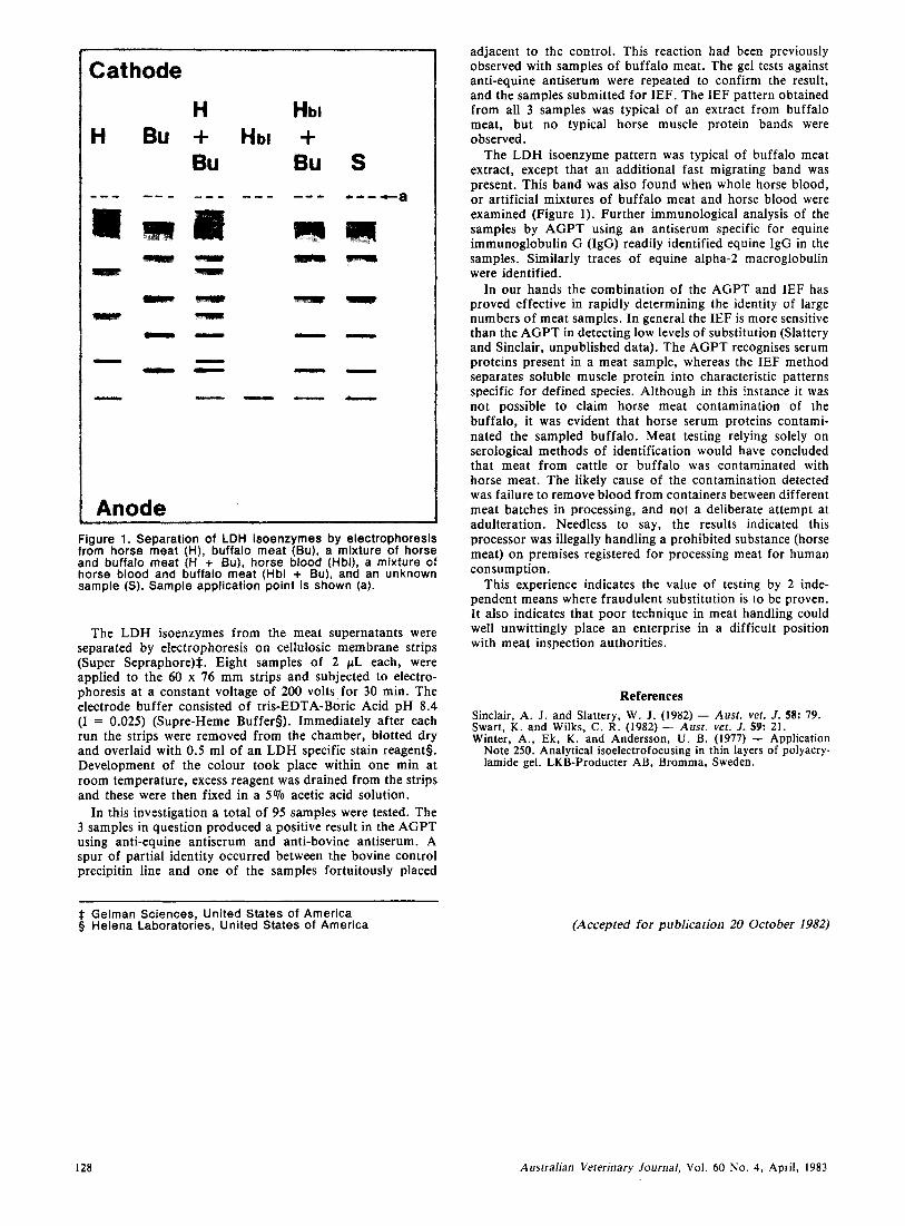
Figure 1. Separation of LDH isoenzymes by electrophoresis from horse meat (H), buffalo meat (Bu), a mixture of horse and buffalo meat (H + Bu), horse blood (Hbl), a mixture of horse blood and buffalo meat (Hbl + Bu), and an unknown sample (S). Sample application point is shown (a).
The LDH isoenzymes from the meat supernatants were separated by electrophoresis on cellulosic membrane strips (Super Sepraphore)*. Eight samples of 2 pL each, were applied to the 60 x 76 mm strips and subjected to electro- phoresis a t a constant voltage of 200 volts for 30 min. The electrode buffer consisted of tris-EDTA-Boric Acid pH 8.4 (I = 0.025) (Supre-Heme Buffers). Immediately after each run the strips were removed from the chamber, blotted dry and overlaid with 0.5 ml of a n LDH specific stain reagents. Development of the colour took place within one min at room temperature, excess reagent was drained from the strips and these were then fixed in a 5 % acetic acid solution.
In this investigation a total of 95 samples were tested. The 3 samples in question produced a positive result in the AGPT using anti-equine antiserum and anti-bovine antiserum. A spur of partial identity occurred between the bovine control precipitin line and one of the samples fortuitously placed
adjacent to the control. This reaction had been previously observed with samples of buffalo meat. The gel tests against anti-equine antiserum were repeated to confirm the result, and the samples submitted for IEF. The IEF pattern obtained from all 3 samples was typical of an extract from buffalo meat, but n o typical horse muscle protein bands were observed.
The LDH isoenzyme pattern was typical of buffalo meat extract, except that an additional fast migrating band was present. This band was also found when whole horse blood, or artificial mixtures of buffalo meat and horse blood were examined (Figure 1). Further immunological analysis of the samples by A G P T using a n antiserum specific for equine immunoglobulin G (IgG) readily identified equine IgG in the samples. Similarly traces of equine alpha-2 macroglobulin were identified.
In our hands the combination of the AGPT and IEF has proved effective in rapidly determining the identity of large numbers of meat samples. In general the IEF is more sensitive than the AGPT in detecting low levels of substitution (Slattery and Sinclair, unpublished data). The AGPT recognises serum proteins present in a meat sample, whereas the IEF method separates soluble muscle protein into characteristic patterns specific for defined species. Although in this instance it was not possible to claim horse meat contamination of the buffalo, it was evident that horse serum proteins contami- nated the sampled buffalo. Meat testing relying solely on serological methods of identification would have concluded that meat from cattle or buffalo was contaminated with horse meat. The likely cause of the contamination detected was failure to remove blood from containers between different meat batches in processing, and not a deliberate attempt at adulteration. Needless to say, the results indicated this processor was illegally handling a prohibited substance (horse meat) on premises registered for processing meat for human consumption.
This experience indicates the value of testing by 2 inde- pendent means where fraudulent substitution is to be proven. It also indicates that poor technique in meat handling could well unwittingly place an enterprise in a difficult position with meat inspection authorities.
References Sinclair, A. J. and Slattery, W. J. (1982) - Aust. vef. J. 58: 79. Swart, K. and Wilks, C. R. (1982) - Anst. vet. J. 59: 21. Winter, A., Ek, K. and Anderson, U. 8. (1977) - Application
Note 250. Analytical isoelectrofocusing in thin layers of polyacry- lamide gel. LKB-Producter AB, Bromma, Sweden.
* Gelman Sciences, United States of America 5 Helena Laboratories, United States of America (Accepted for publication 20 October 1982)
128 Australian Vererinary Journal, Vol. 60 No. 4, April, 1983