somatosensory findings in postherpetic neuralgiasomatosensoryfindings inpostherpetic neuralgia 137...
TRANSCRIPT

135Journal of Neurology, Neurosurgery, and Psychiatry 1990;53:135-141
Somatosensory findings in postherpetic neuralgia
Turo Nurmikko, David Bowsher
AbstractSomatic sensory perception thresholds(warm, cold, hot pain, touch, pinprick,vibration, two-point discrimination),allodynia and skin temperature were
assessed in the affected area of42 patientswith unilateral postherpetic neuralgia(PHN) and 20 patients who had hadunilateral shingles not followed by PHN(NoPHN), and in the mirror-image area
on the other side. There was no differencebetween the two groups for age or lengthof time after the acute herpes zosterinfection. The PHN group showed sig-nificant changes in all sensory thresholdmeasurements when the affected area wascompared with the mirror-image area on
the unaffected side, while the NoPHNgroup exhibited no threshold changes.Mechanical allodynia was present in 87%of the PHN group; half of the 12 patientswith ophthalmic PHN showed extensionofallodynia to the maxillary distribution.No differences in skin temperature were
recorded between affected and unaffectedregions in either group. Our findingsshow a deficit of sensory functionsmediated by both large and smallprimary afferent fibres and also suggestmajor central involvement in the patho-physiology of the condition. If PHN doesnot occur following acute herpes zoster,recovery of neural functions appears tobe good.
Pain ReliefFoundation andCentre for Pain Relief,Walton Hospital,LiverpoolT NurmikkoD BowsherCorrespondence to: Dr TNurmikko, Department ofNeurology, UniversityCentral Hospital (TAYKS),Teiskontie 35, 33520Tampere, Finland.
Received 21 February 1989and in revised form10 August 1989.Accepted 7 September 1989
Postherpetic neuralgia (PHN) has been definedas pain in the area ofacute shingles persisting orrecurring 30 or more days after the acuteinfection.' Several recent reviews concur thatits pathogenesis remains largely unknown.25 Itis generally accepted that in the acute stages ofshingles inflammatory changes occur through-out the sensory nerve from the periphery to thedorsal root. Inflammation is most intense in thedorsal root ganglion where local haemorrhagemay occur.6 Spinal cord changes and anteriorroot involvement are rarely present.7
In contrast, only a few pathological studiesexist on postherpetic neuralgia. In 1900, Headand Campbell reported on 21 cases of herpeszoster, 13 of which were examined between 57and 790 days after the eruption. They des-cribed fibrosis of the dorsal root ganglion andsecondary sclerosis of the dorsal root andperipheral nerve. They also reported ipsilateraldegeneration of the posterior column which"disappeared between five and nine months".
However, apart from one vivid description, it isdifficult to know whether these cases actuallyexperienced pain or not. Recently, Watson et alreported atrophy of the dorsal horn in one caseof PHN; atrophic changes extended severalsegments outside the affected nerve root.8 Towhat extent acute changes are reversible inpatients recovering from shingles has not beensystematically studied. Neither is there anyagreement over the actual mechanisms of paininvolved. Noordenbos has suggested that painis associated with a preferential loss of largediameter fibres and relative preservation ofsmall fibres,9 basing his hypothesis on necropsystudies of four patients. Conversely, Zacksnoted eventual degeneration of both small andlarge fibres.'0 Watson et al noticed a reductionof both myelinated and unmyelinated fibres inthe dorsal root although they recognise theintegrity of at least some unmyelinated fibreinput by the lack of change in substance Pstaining.8 At present, we acknowledge thatalthough both peripheral and central degen-erative changes are present in PHN, themechanism which leads to relentless pain andnot just simple sensory loss, remains obscure.The clinical picture of postherpetic neuralgia
is unique among the non-traumatic neuro-pathies. Both positive and negative sensorysigns can be recognised. There is usuallyevidence of sensory impairment in one or moredermatomes but there is also inappropriatesensitivity to tactile stimuli, broadly referred toas allodynia, so that lightly stroking the affectedskin produces pain whereas firm pressure in thesame region does not.4 Hyperpathia and radia-tion of pain outside the affected dermatome arewell-known features.5 On the McGill Ques-tionnaire" patients described the pain as burn-ing, pricking, lancinating and aching, and itch-ing was also frequently underlined. The word"tender" was usually chosen to describe theallodynia characteristic of the condition (seebelow).'2 The pain differs from that generatedby nociceptor stimulation in being relativelyresistant to analgesics including narcotics andother treatments."' " Shingles is one of thecommonest neurological diseases, its incidencerate ranging from 1 31 to 3-4 per 1000 person-years.7"' Estimates of the risk of PHN lasting aminimum of six months after the rash hasdisappeared currently range from 2 9",, to21*400")16 However, elderly patients seem tobe especially at risk; 50)( of those at age 60 and75"), of those at age 70 develop PHN.5To study the long term sensory impairment
in the area involved by zoster infection weemployed simple noninvasive quantitative
on March 24, 2020 by guest. P
rotected by copyright.http://jnnp.bm
j.com/
J Neurol N
eurosurg Psychiatry: first published as 10.1136/jnnp.53.2.135 on 1 F
ebruary 1990. Dow
nloaded from

Nurmikko, Bowsher
somatosensory perception tests suitable for useon an outpatient population.
Material and methodsPatientsTwo groups of patients were investigated;patients with PHN and patients who had hadshingles in the past but had not developedPHN(NoPHN). All PHN patients attended theCentre for Pain Relief, Walton Hospital, Liver-pool, between September 1987 and May 1988.During that time, 42 consecutive patients wereadmitted to the study. There were no refusals.The diagnosis was arrived at by clinical means(history, typical scars) and agreed upon by atleast two specialists. The length of time sincethe eruption was at least three months.The NoPHN group of20 patients was collec-
ted from a list supplied by three generalpractitioners in Liverpool and the localophthalamological hospital where a recentherpes zoster survey had been carried out.'7One hundred and five letters were sent to thepatients enquiring about the sequelae ofthe acute shingles. Those who answered werecontacted by telephone to ask whether they hadpain or not. Twenty four patients volunteeredto participate in the study. Four of them wereexcluded. In two cases there was some doubt asto the actual diagnosis of shingles and notypical scars; in two further cases (thoracic) thediagnosis of shingles seemed tenable but therewere no scars at all to guide the examiner. Aswith PHN patients, the time that had elapsedafter the eruption of shingles was at least threemonths. The final number of patients in thisgroup was 20.
Patients were considered as having PHN ifthey had spontaneous pain or allodynia, with orwithout spontaneous pain. Patients with itch-ing only were not considered as PHN subjects.Of the NoPHN group four reported havingexperienced slight bouts of itching occasion-ally; they had not consulted their doctor orreceived any treatment for that and wereincluded in the study. Excluded from bothgroups were patients with dementia, knownperipheral or central nervous system disease,major metabolic diseases, malignancies, alco-holism and subjects who had been subjected toneuroablative procedures for the treatment ofPHN.The aim of the study and the nature of the
tests were explained to the patients according
Table I Demographic and clinical data of testedgroups
PHN NoPHN
N 42 20female:male 21/21 14/6mean age (SD) 69 6 (9 6) 69-5 (7 5)mean duration (SD)' 32-6 (25 4) 35 5 (27-0)
medication2none 14 20AC + AD 12 -AD + analg 7 -
AC + analg 3 -
analg only 6 -
Ivalues in months; 'AC = anticonvulsant, AD = tricyclicantidepressant, analg = analgesic medication
Table 2 Distribution of zoster in PHN and NoPHNpatients
PHN NoPHN
Ophthalmic 12 10Cervical 10 0Upper thoracic 7 4Lower thoracic 10 5Lumbar 2 1Sacral 1 0Total 42 20
to the Helsinki Declaration. An oral or writtenconsent was obtained from all participants.Demographic and clinical data for the two
groups are shown in table 1.Location ofzoster in the two groups is shown
in table 2. All cases were unilateral, more or lessevenly distributed between right and left (asdescribed by Campbell et al,'2 18) and usuallyaffected one or two dermatomes.
MethodsAll the tests were carried out at the Pain ReliefFoundation, Liverpool. Each patient was clin-ically examined to rule out any overtneurological disease or dementia. Each patientfilled in a pain questionnaire on the history andquality of pain, past treatment, complications,and psychosocial aspects. These data are notconsidered further in the present report.
Location of PHN or original shingles (in thecase ofNoPHN) was determined by visible scartissue which was usually quite prominent inpatients with PHN and far less conspicuous inNoPHN subjects. All the measurements weremade within the affected zone although in somePHN patients pain was reported beyond thelimits of the scar area.Care was taken to avoid measuring directly
over scar tissue. In essence, tests were carriedout over the scarless skin within the affecteddermatome. Even with the use of a thermode(see below) this proved possible in all testedcases. Wherever possible, the area ofmaximumpain intensity was determined andmeasurements were carried out within it. Anattempt was also made to define a pain-free areawithin the affected dermatome(s), but this wasonly found in three out of 42 cases. It did,however, prove possible to outline an area ofminimum pain intensity within each affecteddermatome and this was used for the secondmeasurement in each case.
In all cases, the exact counterparts of the testsites on the contralateral (mirror) side weredetermined and the same tests performed. Thetesting sequence alternated and in half of thecases the first test carried out was on the mirrorside and in half on the affected side. All thepatients entered into the study had unilateralzoster only.
Before testing, each patient received a shortexplanation of the methods used and a shortrehearsal was conducted on the dorsal aspect ofthe forearm. In NoPHN cases only one spot oneither side was measured. Testing took approx-imately 60 minutes to complete.Warm and cold thresholds were determined
136
on March 24, 2020 by guest. P
rotected by copyright.http://jnnp.bm
j.com/
J Neurol N
eurosurg Psychiatry: first published as 10.1136/jnnp.53.2.135 on 1 F
ebruary 1990. Dow
nloaded from

137Somatosensoryfindings in postherpetic neuralgia
by a commercially available device (Thermo-test, Somedic AB, Stockholm, Sweden) whichworks on the Peltier principle.'" Briefly, a
thermocouple 10 cm2 in area (for cervical,thoracic and lumbar regions) or 3-6 cm2 (forfacial regions) is warmed up or cooled down at a
rate of change of 0-5-1-5 C/s. The testingmethod is a modification of the Marstocktechnique.2 Measurements started from an
adapting skin temperature of 30 C after thecomplete disappearance of any thermal sensa-
tion caused by the application of the thermode.The patients responded to each sensation ofwarm, cold, hot pain or cold pain by pressing a
switch which reversed the direction of tem-
perature change and allowed the thermode to
return to the initial temperature. A cut off pointfor the warm threshold was determined at
50 0 C and for the cold threshold at 5 C. Eachmeasurement was carried out 4-6 times inconsecutive order and the average value was
calculated. In cases where cut off points were
reached without the patient recognising any
thermal sensation, only one repetition was
performed for fear of injuring the skin. Asimilar procedure was carried out on the con-
tralateral side. In seven patients, a slightlydifferent method was employed. From theadaptation temperature of 30"C the thermodewas warmed and cooled alternately; each timethe patient perceived either sensation he rever-
sed the current of the thermode (from warm to
cold and vice versa). Plateau levels of boththermal thresholds were obtained within a fewminutes; they proved to be 2-3 C higher thanwith the other method.
Hot pain was measured using the same
instrumentation. The probe temperature was
increased until the patient perceived pain andpressed the switch. This procedure was
repeated once and the average of the two
calculated. In 22 patients, the thermode was
allowed to cool until the patient either per-
ceived pain or the lower limit of the equipment(5 C) was reached. Cold pain was tested onlyonce.
Tactile sensation was measured with von
Frey filaments, which were applied in a
descending and ascending order of magnitudeto assess both the disappearance andappearance thresholds (at least three applica-tions). Care was taken to avoid stroking the skinwith the hair and only to do an indentation.The force required to bend each filament (ingrams) was converted to log units.
Pinprick sensation was tested with weightedneedles. The needles (25 mm 24G needlesweighing from 0 2 to 5 2 g) were on plungers of5 ml plastic syringes moving freely. By holdingthe syringe the examiner could guide the needleof any weight perpendicularly to the area
tested; in practice it was only the weight of theneedle that determined the force and con-
sequent pinprick sensation (or lack of it).Similarly to tactile stimulation, this test was
carried out in a descending and ascendingmanner (method of limits) and the thresholddetermined by the average of three applica-tions.Two point discrimination was tested con-
ventionally with Weber's compasses, movingthe instrument gently over the skin, a methodwhich has been shown to produce moreaccurate results than simply applying it on theskin.2' Three ascending and descending seriesin increments of 5 mm (face) and 10 mm (neck,trunk) were administered in alternating order.
Vibration was tested over a bony part in 14patients using a hand held fixed-frequencyvariable-amplitude vibrameter with weightcontroller (Somedic AB, Stockholm, Sweden).With this method, the threshold is expressed interms of amplitude of stimulator movement.22Both the appearance and disappearance thres-holds were determined (in pim) and the averagecalculated.
Skin temperature was measured using freethermocouples attached to the skin (DigitronInstruments, model 4071, Hertford, UnitedKingdom). Each patient stayed in the inves-tigation room for at least 20 minutes toacclimatise before the measurement.23 Liquidcrystal contact thermography was also carriedout in the affected and nonaffected regions.The presence of allodynia was assessed by
gently applying a battery driven toothbrush(Braun D 3, FRG) on the affected skin area.The brush was found to move back and forth 53times a minute, with an amplitude of 1 5 mm.We found it easier to hold the handle of thebrush without exerting a force in excess of 8 gon the skin surface. The patient reportedchange in subjective pain sensation, using avisual analogue scale (VAS) for pain24 with amobile slider enabling the constant responsesto change in intensity to be registered, rangingfrom 0 to 100 mm. VAS before stimulation andat the height of pain (peak VAS) were recorded,as well as the time taken to reach the peak. Theduration of hyperpathia was also recorded.Allodynia was considered to be present if theVAS scale reached 100 mm (whatever theinitial level) or if a reading of at least 25 mmabove the initial (pretest) level was recorded.We also studied allodynia in cases ofophthal-
mic PHN by testing the effect of moving one tothree of the stiff hairs in the eyebrow. Theresponse was recorded as painful (allodynic) ornon-painful (non-allodynic). Also, using thebrush method, maxillary and mandibular div-isions on the affected side were tested for thepresence of allodynia in these 12 cases.
Hyperalgesia to skin stretch was evaluated inanother 12 patients by manually pulling theskin from outside the affected region. Again,the patient's response was recorded as painfulor non-painful.
Statistical analysisThe Wilcoxon matched-pairs test was used fora comparison of results between the zosteraffected dermatomes and the contralateral der-matomes in both groups. A value of p < 0-05(two-tailed) was considered to be statisticallysignificant.
In addition, we tested 20 healthy subjects(mean age 68 5 (SD) 9 2 years, female:male12:8) collected from the spouses and relatives ofthe patients for normative data. In these
on March 24, 2020 by guest. P
rotected by copyright.http://jnnp.bm
j.com/
J Neurol N
eurosurg Psychiatry: first published as 10.1136/jnnp.53.2.135 on 1 F
ebruary 1990. Dow
nloaded from
Nurmikko, Bowsher
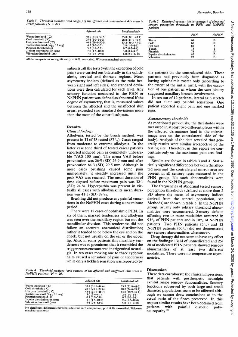
Table 3 Threshold medians (and ranges) of the affected and contralateral skin areas inPHN patients (N = 42)
Affected side Unaffected side
Warm threshold ("C) 40.0 (336-50 5) 35 0 (321-43 1)Cold threshold ("C) 25 3 (50-34 0) 28 8 (233-35 5)Hot pain threshold ('C) 46-1 (396-50-0) 43 2 (363-47-7)Tactile threshold (log,0 0.1 mg) 4 3 (1 7-67) 2 8 (1 7-49)Pinprick threshold (g) 5 2 (0 2-5 2) 0 7 (0-2-44)2-point discrimination (cm) 7-0 (1 5-10-0) 3 0 (0 9-9-0)Vibration threshold (pm) 7 8 (28-79 0) 6 8 (20-37-0)
All the comparisons are significant (p < 0-01, two-tailed; Wilcoxon matched-pairs test)
subjects, all the tests (with the exception ofcoldpain) were carried out bilaterally in the ophth-almic, cervical and thoracic regions. Meanasymmetry indices (defined as the ratio bet-ween right and left sides) and standard devia-tions were then calculated for each level. Anysensory function measured in the PHN orNoPHN patient was defined as abnormal if thedegree of asymmetry, that is, measured valuesbetween the affected and the unaffected skinareas, exceeded two standard deviations morethan the mean of the control subjects.
ResultsClinicalfindingsAllodynia, tested by the brush method, waspresent in 33 of 38 tested (87"%). Cases rangedfrom moderate to extreme allodynia. In thelatter case (one third of tested cases) patientsreported induced pain as completely unbeara-ble (VAS 100 mm). The mean VAS beforeprovocation was 26 5 (SD) 26-9 mm and afterprovocation 64 3 (SD) 29 5 mm. Although inmost cases brushing caused some painimmediately, it steadily increased until thepeak VAS was reached. The mean duration oftime elapsed before maximum pain was 32 1(SD) 24 8s. Hyperpathia was present in vir-tually all cases with allodynia; its mean dura-tion was 41 5 (SD) 58 9s.
Brushing did not produce any painful sensa-tions in the NoPHN cases during a one minuteperiod.There were 12 cases of ophthalmic PHN. In
six of them, marked tenderness and allodyniawas seen over the maxillary region but not themandibular division. This tenderness did notfollow an accurate anatomical distribution;rather it tended to be below the eye and on thecheek, but not usually on the ear or the upperlip. Also, in some patients this maxillary ten-derness was so prominent that it resembled thetrigger zones encountered in trigeminal neural-gia. In ten cases moving one to three eyebrowhairs caused a sensation of pain or tendernesswhile only a ticklish sensation was reported (by
Table 4 Threshold medians (and ranges) of the affected and unaffected skin areas inNoPHN patients (N = 20)
Affected side Unaffected side
Warm threshold ( C) 33.4 (31 8-44 6) 33-7 (318-41-2)Cold threshold ( C) 28-8 (230-29-6) 28.8 (246-29 7)Hot pain threshold ( C) 43.8 (319-46 7) 44.8 (389-47 1)Tactile threshold (log,,, 0.1 mg) 3-0 (2-4-4 1) 2 6 (1 7-3 6)Pinprick threshold (g) 0 7 (0 2-3 8) 0 7 (0-2-2 8)2-point discrimination (cm) 3.8 (15-10 0) 2 6 (1 5-10 0)Vibration threshold (pm) 5-4 (28-52 0) 5 8 (18-23 0)
No significant differences between sides (for each comparison, p < 010, two-tailed; Wilcoxonmatched-pairs test)
Table S Relativefrequency (in percentages) ofabnormalsensory perception thresholds in PHN and NoPHNpatients
PHN NoPHN
Warm 68 10Cold 60 10Hot pain 60 5Touch 73 5Pinprick 78 102-point discrimination 62 13Vibration 27 13
the patient) on the contralateral side. Thesepatients had previously been diagnosed ashaving ophthalmic zoster only (according tothe extent of the initial rash), with the excep-tion of one patient in whom the case historysuggested maxillary branch involvement.
In ten out of 12 patients, lateral skin stretchdid not elicit any painful sensations. Onepatient reported slight pain and one markedpain.
Somatosensory thresholdsAs mentioned previously, the thresholds weremeasured in at least two different places withinthe affected dermatome (and in the mirror-image area on the contralateral side of thebody). Analysis of the data revealed that gen-erally results were similar irrespective of thetesting site. Therefore, in this report we con-centrate only on the maximum pain area find-ings.
Results are shown in tables 3 and 4. Statis-tically significant differences between the affec-ted area and the contralateral mirror area arepresent in all sensory tests measured in thePHN group. No such abnormalities werefound in the NoPHN group.The frequencies of abnormal tested sensory
perception thresholds (defined as more than 2SD above the mean of asymmetry indicesderived from the control population, seeMethods) are shown in table 5. In the NoPHNgroup, usually only solitary threshold abnor-malities were encountered. Sensory deficitsaffecting two or more modalities occurred in93", of PHN patients and in 100, of NoPHNpatients. Two PHN patients (5°0) and 12NoPHN patients (600,) did not demonstrateany sensory abnormalities whatsoever.Drug therapy did not seem to have any effect
on the findings: 13/14 of unmedicated and 25/28 of medicated PHN patients showed sensoryabnormalities of at least two differentmodalities. There were no temperature asym-metries.
DiscussionThese data corroborate the clinical impressionsthat patients with postherpetic neuralgiaexhibit major sensory abnormalities. Sensoryfunctions subserved by both large and smalldiameter populations seem to be affected alth-ough we cannot draw conclusions as to theactual ratio of the fibres preserved. In thisrespect similar results have been obtained frompatients with painful diabetic poly-neuropathy.25
138
on March 24, 2020 by guest. P
rotected by copyright.http://jnnp.bm
j.com/
J Neurol N
eurosurg Psychiatry: first published as 10.1136/jnnp.53.2.135 on 1 F
ebruary 1990. Dow
nloaded from
Somatosensory findings in postherpetic neuralgia
It seems evident that people who recoverfrom shingles without major pain or itch (alltested subjects in the NoPHN group hadrecovered in the first three months) are left withno or minor subclinical local neuropathy. Thisagrees with Noordenbos' view that as a rulesensory changes are lacking in patients whorecover from shingles.9 (Noordenbos did notstate the number of patients he had observed).Similarly, Head and Campbell did not find anymorphological changes in two of their cases(cases 20, 21) and correlate findings in theDRG, dorsal root and peripheral nerve to theseverity of the eruption.6 In turn, Higa et alfound a relationship between antibody respon-ses to varicella-zoster virus and the severity ofskin lesions.' The obvious clinical conclusionone wishes to draw from the present data is thatpatients who complain about pain after shin-gles should have sensory changes ifpain is to berelated to postherpetic neuralgia; if none arefound, other reasons for pain (such asmusculoskeletal) should be looked for.The dual nature ofpain associated with PHN
has been recognised.'2 14 For example, there ismore or less constant pain present (described asburning, nagging, aching and stabbing by thepatients)'2 and this obviously reflects spontan-eous abnormal barrages somewhere along thesomatosensory pathways. As both peripheraland central interruption of pain pathwaysindependently abolish pain in PHN, althoughtemporarily,'42728 one must assume dysfunc-tion at both levels, but the pathophysiologicalmechanisms involved remain elusive.
Alternatively, more may be speculated aboutallodynia, referred to by patients as "soreness"or "tenderness".'2 Our finding of87 ",, showingallodynia in response to toothbrush applicationclosely resembles that of Watson et al.29 Byrubbing the skin with a cotton-tipped stick theyfound "hyperesthesia, dysesthesia or allodyniato light stroking" in 65'"(, of their PHNpatients. Our higher figures may simply reflecta more vigorous method of stimulation. Theresults are underlined by the additional findingof hair follicle movements producing pain inthe tested patients. In healthy subjects, lowfrequency stimulation predominantly activatesrapidly adapting low threshold mechanorecep-tors and hair receptors.30 In contrast, lateralskin stretch and maintained skin compressionare associated with the activation of slowlyadapting mechanoreceptors.3' In this study,skin stretch failed to produce any pain in mostpatients that were tested. We also noticed thefairly common situation whereby patientsspend much of their time pressing a hand overthe painful region, or wearing an extra tightgarment. 14
These observations suggest that mechanicalstimuli known to activate slowly adaptingmechanoreceptors in healthy subjects do notprovoke allodynia in postherpetic neuralgiawhereas those stimuli which activate rapidlyadapting mechanoreceptors do. Couldallodynic pain be produced by non-nociceptivestimulation of other receptors than mechano-receptors? Noordenbos has argued that allstimuli if applied long enough elicit pain in
PHN.9 In his study, multiple pinpricks caused,after a delay, an outbreak of pain spreadingover large areas and slowly wearing off.Similarly, a hot test tube which did not causeany discomfort on normal skin produced, whenapplied to the zoster area, a steady increase overa minute or so in perceived sensation fromfaintly warm to hot and finally painful.9 In theirstudy Watson et al noted that pinching the skinresulted in hyperalgesia in 58% of their PHNpatients.29 It seems possible therefore that inPHN innocuous stimulation of A deltanociceptors may result in pain, similar tohyperalgesia after mechanical irritation.
In this study, however, we found no suchbroad-spectrum hyperalgesia. The patients didnot usually find pinprick testing painful in spiteof repeated needling. Cooling the affected skinslowly down to 5°C resulted in a pain responsein 12 of 22 tested; but only four patientsreported pain at a higher temperature than onthe healthy side. Similarly, during heat induc-tion only three of40 patients felt pain at a lowertemperature on the zoster area than on thecontralateral side. In contrast, there wereraised thresholds for warm or hot stimuli in58% and 45% respectively, and loweredthresholds for cold in 50% of our cases.Lindblom and Verrillo describe in 11 patientswith mainly post-traumatic neuralgias whoshowed two different responses to thermalstimuli: hypersensitive and hyposensitive.32Hypersensitivity was considered present ifheatpain thresholds were low and cold pain thre-sholds raised compared with the intact skin.With this definition, our results show that inPHN the hyposensitive reaction to thermalstimuli is much more frequent than the hyper-sensitive reaction. Whereas hyperalgesia tothermal stimuli as a consequence of nervetrauma is reported to be common,32 33 posther-petic neuralgia evidently represents an entity ofits own with the characteristic feature ofextreme hyperalgesia to mechanical stimuli butless so to thermal ones.The question remains whether this mechan-
ical allodynia is provoked by activation ofintactRA I mechanoreceptors or whether it is thesign of other afferent receptors being sensitisedto mechanical stimuli. Two facts in our studyfavour the mechanoreceptor theory: 1) even incases where thermal thresholds were extremelypathological, being elevated to the tissue injurylevel, there was still mechanoreceptor sen-sitivity, 2) in a group ofPHN patients, selectiveblocking of large diameter fibres abolishedallodynia whereas that of small fibres did not(Nurmikko et al, submitted). Similar observa-tions have been published in selective nerveinjuries.34One can also conjecture a role for the sym-
pathetic system because of its postulated abilityto sensitise mechanoreceptors in injured nerveendings.35 Allodynia is a prominent feature ofvarious sympathetically dependent pain condi-tions, notably reflex sympathetic dystrophy.'5Is allodynia in PHN also, at least in partsympathetically maintained? Certainly,patients withPHN do report increase in pain inresponse to anxiety and exposure to cold. They
139
on March 24, 2020 by guest. P
rotected by copyright.http://jnnp.bm
j.com/
J Neurol N
eurosurg Psychiatry: first published as 10.1136/jnnp.53.2.135 on 1 F
ebruary 1990. Dow
nloaded from

Nurmikko, Bowsher
seem to experience bouts of cold and hot withinthe affected area, suggesting local autonomicinstability. Also, sympathetic blocks have beenreported to be of benefit even in PHN of morethan one year36 although not all authors agree.23Our results fail to show any sympathetic hyper-function in patients with PHN, and, indeed,reflex sympathetic dystrophy is extremelyrarely documented in postherpetic neuralgia.37This is not to say that the sympathetic systemhas no role in PHN. It is quite possible that itsphysiological fluctuations may modify pain orallodynia in PHN. Loh and Nathan noted thatfor a sympathetic block to be effective inchronic neuralgias, signs of sympathetic abnor-mality did not have to be present.38 Recently,we have assessed the role of the sympatheticsystem by means of differential blocks and theresults will be reported in a separate study(Nurmikko et al submitted).Animal studies show that as a consequence of
peripheral nerve injury there is a change of thereceptive fields of the cut nerve in the dorsalhorn. Those neurons which previously respon-ded only to the territory of the cut nerve beginto respond to nearby intact nerves.394" Whetherthis phenomenon, recently named somatotopicremodelling, happens in the sensory domain isnot known.4" From our findings we suggest thatmechanical allodynia represents a state wheresecond order neurons that primarily respond tonoxious stimuli start responding to inputstravelling in the A beta fibres associated withRA I receptors. This central reorganisationalso offers a plausible explanation for thediscovery of extension of hyperesthesia outsidethe territory of the ophthalmic nerve in six ofthe twelve trigeminal PHN patients. In theacute stages of shingles, these patients, withone exception, were judged by primary caredoctors and ophthalmologists to have ophthal-mic involvement only. In epidemiologicalstudies, maxillary involvement in acute statesof shingles is rare6 12 and it is unlikely that ourpatients had subclinical maxillary branchinfection without eruption. In 1949 Russell etal observed that in ophthalmic zoster, blockadeof the ipsilateral occipital nerve, as well as thecontralateral ophthalmic or supratrochlearnerves, alleviated hyperesthesia,42 an idea alsosuggesting that allodynia may actually extendoutside the area innervated by the affectedophthalmic nerve. This cannot be explained byoverlapping of dermatomes; there is very littleoverlap between the three divisions of thetrigeminal nerve.43 Somatotopic remodellingappears to be the most likely mechanism,although accurate repeated measurementsfrom the beginning of the infection to the endstage of PHN would clarify this. Additionalcentral contributions may arise from atrophicchanges in the trigeminal nucleus which havebeen recently verified in pathological examin-ations.44Our findings suggest that the physiopath-
ological changes in PHN are likely to be in theCNS as well as in the PNS. This would placethis narcotic-resistant non-nocigenic painfulcondition in the same category as otherneurogenic pains of both central and peripheral
origin, although its autonomic characteristicscertainly differentiate it from some of them.Further investigation of acute herpes zoster isneeded to discover if there are patho-physiological factors which will predictwhether a given case is likely to recover withoutsensory deficit and without PHN, or to persistwith sensory deficit and with the PHN whichappears ineluctably to accompany it.
This study was supported by grants from theAcademy of Finland and Tampere BrainResearch Centre (Tatke), Finland.
1 Hope-Simpson, RE. Post-herpetic neuralgia. J Royal CollGen Pract 1975;25:571-5.
2 Loeser J. Herpes zoster and postherpetic neuralgia. Pain1986;25: 149-64.
3 Portenoy RK, Duma C, Foley KM. Acute herpetic andpostherpetic neuralgia: clinical review and current man-agement. Ann Neurol 1986;20:651-4.
4 Lobato RD, Madrid JL. Clinical and pathophysiologicalmechanisms of postherpetic neuralgia. Clin J Pain1987;2:253-7.
5 Watson PN, Evans RJ. Postherpetic neuralgia. A review.Arch Neurol 1986;43:836-40.
6 Head H, Campbell AW. The pathology of herpes zoster andits bearing on sensory localization. Brain 1900;23:353-523.
7 Ragozzino MW, Melton LJ, Kurland LT, Chu CP, PerryRO. Population-based study of herpes zoster and itssequelae. Medicine 1982;61:310-6.
8 Watson CPN, Morshead C, Van der Kooy D, Deck J, EvansRJ. Post-herpetic neuralgia: post-mortem analysis of acase. Pain 1988;34:129-38.
9 Noordenbos W. Pain. Problems pertaining to the transmissionof nerve impulses which give rise to pain. Amsterdam:Elsevier, 1959;4.-10:68-80.
10 Zacks SL, Langfitt TW, Elliott FA. Herpetic neuritis: a lightand electron microscopic study. Neurology 1964;14:744-50.
11 Melzack R. The McGill Pain questionnaire: Major proper-ties and scoring methods. Pain 1975;1:277-99.
12 Bhala BB, Ramamoorthy C, Bowsher D, Yelnoorker KN.Shingles and postherpetic neuralgia. Clin J Pain1988;4:169-74.
13 Bowsher D. Pain as a neurological emergency. In: BowsherD, ed, Neurological Emergencies in Medical Practice.Beckenham: Croom Helm, 1988:218-36.
14 Lipton S. Relief of pain in clinical practice. Oxford: Black-well, 1979:231-48.
15 Hope-Simpson RE. The nature of herpes zoster: along-termstudy and a new hypothesis. Proc R Soc Med 1965;58:9-20.
16 Burgoon CF, Burgoon JS, Baldridge GD. The naturalhistory of herpes zoster. JAMA 1957;164:265-9.
17 Harding SP, Lipton JR, Wells JCD. Natural history ofherpes zoster ophthalmicus: predictors of postherpeticneuralgia and ocular involvement. Brit J Ophthalmol1987;71:353-8.
18 Campbell JA, Lahuerta J, Bowsher D. Pain laterality inrelation to site of pain and diagnosis. Pain 1985;23:61-6.
19 Hansson P, Ekblom A, Lindblom U, Marchettini P. Doesacute intraoral pain alter cutaneous sensibility? J NeurolNeurosurg Psychiatry 1988;51:1031-6.
20 Fruhstorfer H, Lindblom U, Schmidt WG. Method forquantitative estimation of thermal thresholds in patients.JNeurol Neurosurg Psychiatry 1976;39:1071-5.
21 Louis DS, Greene TL, Jacobson KE, Rasmussen C, Kol-owich P, Goldstein SA. Evaluation of normal values forstationary and moving two point discrimination in thehand.J Hand Surg 1984;9A:552-5.
22 Goldberg JM, Lindblom U. Standardised method of deter-ming vibratory and perception thresholds in patients. JNeurol Neurosurg Psychiatry 1979;42:793-803.
23 UematsuS, Edwin DH, Jankel WR, Kozikowski J, TrattnerM. Quantification of thermal asymmetry. Part 1: Normalvalues and reproducibility.J Neurosurg 1988;69:552-5.
24 Bond MR, Pilowsky I. The subjective assessment of painand its relationship to the administration of analgesicsin patients with advanced cancer. J Psychosom Res1966;10:203.
25 Ziegler D, Mayer P, Wiefels K, Gries FA. Assessment ofsmall and large fiber function in long-term I (insulin-dependent) diabetic patients with and without painfulneuropathy. Pain 1988;34:1-10.
26 Higa K, Dan K, Manabe H, Noda B. Factors influencing theduration of treatment of acute herpetic pain with sympath-etic block; importance of severity of herpes zoster assessedby the maximum antibody titers to varicella-zoster virus inotherwise healthy patients. Pain 1988;32: 147-57.
27 Sweet WH. Deafferentation pain after posterior rhizotomy,trauma to a limb and herpes zoster. Neurosurgery1984;15:928-32.
28 Friedman AH, Nashold BS. DREZ lesions for postherpeticneuralgia. Neurosurgery 1 984;iS:969-70.
140 on M
arch 24, 2020 by guest. Protected by copyright.
http://jnnp.bmj.com
/J N
eurol Neurosurg P
sychiatry: first published as 10.1136/jnnp.53.2.135 on 1 February 1990. D
ownloaded from

Somatosensory findings in postherpetic neuralgia
29 Watson CPN, Evans RJ, Watt VR, Birkett N. Post-herpeticneuralgia: 208 cases. Pain 1988;35:289-97.
30 Vallbo AB, Olsson KA, Westberg K-G, Clark FJ.Microstimulation of single tactile afferents from thehuman hand. Brain 1984;107:727-49.
31 Roberts WJ. A hypothesis on the physiological basis forcausalgia and related pains. Pain 1986;24:297-31 1.
32 Lindblom U, Verrillo RT. Sensory functions in chronicneuralgia. J Neurol Neurosurg Psychiatry 1979;42:422-35.
33 Frost SA, Raja SN, Campbell JN, Meyer RA, Khan AA.Does hyperalgesia to cooling stimuli characterize patientswith sympathetically maintained pain (reflex sympatheticdystrophy)? In: Dubner R, Gebhart GF, Bond MR, eds.Proc Vth World Congress on Pain. Amsterdam: Elsevier,1988:151-6.
34 Campbell JN, Raja SN, Meyer RA, MacKinnon SE.Myelinated afferents signal the hyperalgesia associatedwith nerve injury. Pain 1988;32:89-94.
35 Janig W, Kollman W. The involvement of the sympatheticnervous system in pain. Possible neuronal mechanisms.Arzneimittel-Forschung 1984;34:1066-73.
36 Milligan NS, Nash TP. Treatment of post-herpetic neural-gia. A review of77 consecutive cases. Pain 1985;23:381-6.
37 Grosslight KR, Rowlingson JC, Boaden RW. Herpes zoster
and reflex sympathetic dystrophy. Anesth Analg1984;65:309-1 1.
38 Loh L, Nathan PW. Painful peripheral states and sympath-etic blocks. J Neurol Neurosurg Psychiatry 1978;41:664-71.
39 Devor M, Wall PD. Plasticity in the spinal cord sensory map
following peripheral nerve injury in rats. J Neurosci1981 ;7:676-84.
40 Devor M, Wall PD. The effect of peripheral nerve injury inreceptive fields of cells in the cat spinal cord. J CompNeurol 1981;199:277-91.
41 Devor M. Central changes mediating neuropathic pain. In:Dubner R, Gebhart GF, Bond MR, eds. Proc Vth WorldCongress on Pain. Amsterdam: Elsevier, 1988:114-28.
42 Russell WR, Espir MLE, Morganstern FS. Treatment ofpostherpetic neuralgia. Lancet 1957;i:242-5.
43 Fromm GH. Anatomy and physiology of the trigeminalsystem. In: Fromm GH, ed. The medical and surgicalmanagement of trigeminal neuralgia. Mount Kisco: Futura,1987:17-30.
44 Reske-Nielsen E, Oster S, Pedersen B. Herpes zosterophthalmicus and the mesencephalic nucleus. Aneuropathological study. Acta Pathol Microbiol ImmunolScand 1986;94:263-9.
141
on March 24, 2020 by guest. P
rotected by copyright.http://jnnp.bm
j.com/
J Neurol N
eurosurg Psychiatry: first published as 10.1136/jnnp.53.2.135 on 1 F
ebruary 1990. Dow
nloaded from