solid-state 33s mas nmr of inorganic sulfates
TRANSCRIPT

www.elsevier.com/locate/jmr
Journal of Magnetic Resonance 170 (2004) 336–344
Solid-state 33S MAS NMR of inorganic sulfates
Todd A. Wagler, William A. Daunch, Matthew Panzner, Wiley J. Youngs,Peter L. Rinaldi*
Knight Chemical Laboratory, Department of Chemistry, The University of Akron, Akron, OH 44325-3601, USA
Received 6 May 2004; revised 19 July 2004
Available online 23 August 2004
Abstract
Solid-state 33S MAS NMR spectra of a variety of inorganic sulfates have been obtained at magnetic field strengths of 4.7, 14.1,
17.6, and 18.8 T. Some of the difficulties associated with obtaining natural abundance 33S NMR spectra have been overcome by
using a high magnetic field strength and magic angle spinning (MAS). Multiple factors were considered when analyzing the spectral
linewidths, including magnetic field inhomogeneity, dipolar coupling, chemical shift anisotropy, chemical shift dispersion, and quad-
rupolar coupling. In most of these sulfate samples, quadrupolar coupling was the dominant line broadening mechanism. Nuclear
electric quadrupolar coupling constants (Cq) as large as 2.05 MHz were calculated using spectral simulation software. Spectral infor-
mation from these new data are compared with X-ray measurements and GAUSSIAN 98W calculations. A general correlation was
observed between the magnitude of the Cq and the increasing difference between S–O bond distances within the sulfate groups. Sol-
id-state 33S spin–lattice (T1) relaxation times were measured and show a significant reduction in T1 for the hydrated sulfates. This is
most likely the result of the modulation of the time-dependent electric field gradient at the nuclear site by motion of water molecules.
This information will be useful in future efforts to use 33S NMR in the compositional and structural analysis of sulfur containing
materials.
� 2004 Elsevier Inc. All rights reserved.
1. Introduction
The only isotope of sulfur detectable by NMR, 33S,
happens to be among the first NMR-active nuclei to
be studied. In 1951 Dharmatti and Weaver [1] deter-
mined the magnetic moment of 33S using the observed
signal from a pure sample of CS2. Unfortunately, since
then fewer than 200 scientific papers have been pub-lished on 33S NMR, with much of the work focused
heavily on sulfur compounds in solution. Most of those
solution studies have been performed on SIV and SVI
species, which exist in environments with high electronic
symmetry around the sulfur nucleus [2–5]. It has only
been recently that unsymmetric sulfur species have been
studied in solution using high magnetic fields [6,7].
1090-7807/$ - see front matter � 2004 Elsevier Inc. All rights reserved.
doi:10.1016/j.jmr.2004.07.011
* Corresponding author. Fax: +1 330 972 5256.
E-mail address: [email protected] (P.L. Rinaldi).
There are very few solid-state 33S NMR studies de-
scribed in the literature, and they are briefly summarized
in the four published reviews of 33S NMR [8–11]. The
most comprehensive study to date was published in
1986 [12]. In that work, which was performed with hard-
ware available at the time, the authors examined chem-
ical shifts, linewidths, and second order quadrupolar
broadening effects on the static lineshapes of the central(�1/2M +1/2) transition of a number of sulfur contain-
ing inorganic compounds. In the time since this publica-
tion, very little new solid-state 33S NMR work has been
reported in the literature [13–15].
The dearth of 33S NMR work is rather discouraging,
since NMR has proven to be an invaluable tool for most
chemists and the fact that sulfur, and more notably sul-
fate, compounds are important to both the biochemicaland chemical industries. Large quantities of sulfates are
consumed every year in the manufacturing of wallboard
(gypsum), in the tanning and dyeing (alums) industries,
T.A. Wagler et al. / Journal of Magnetic Resonance 170 (2004) 336–344 337
and in the petroleum industry. It has also been shown
that sulfates play an important role in mediating disso-
lution and precipitation of potentially toxic contami-
nants in our environment. Moreover, and probably
most important, sulfate is known to be an essential elec-
trolyte which is required for the proper growth anddevelopment of living organisms [16].
There are several reasons why so little 33S NMR
work has been published. 33S has a small gyromag-
netic ratio (1/13 that of 1H) and a low natural abun-
dance (0.75%), making it among the most difficult
nuclei to study by NMR. Its receptivity, a measure
of the absolute signal strength from a fixed number
of nuclei, is 1.7 · 10�5 relative to that of 1H, and9.72 · 10�2 relative to that of 13C [17]. Furthermore,
unlike the more commonly studied 1H and 13C nuclei,33S is a quadrupolar nucleus with a nuclear spin I = 3/2and a relatively large quadrupole moment(Q= �0.0678 b) [18,19]. The large quadrupole momentresults in rapid T2 relaxation leading to extremely broadlines in chemical species having unsymmetrical sulfurenvironments.
Even with these inherent difficulties, there are very
compelling reasons to use 33S NMR. Significant chemi-
cal structure information can be obtained by directly
observing 33S. Its chemical shift range exceeds
800 ppm, which is approximately four times that of13C and nearly 16 times that of 1H. Additionally, be-
cause sulfur is often directly involved in many of the
chemical transformations of interest, its NMR proper-ties are more sensitive than those of nuclei such as 1H
and 13C which are only indirectly affected.
Until recently, it has been exceedingly difficult to ex-
tract information from the NMR spectra of quadrupo-
lar nuclei like 33S. Fortunately, higher magnetic field
strengths and a variety of new experimental techniques
can now be used to average the effect of quadrupolar
broadening, and improve both sensitivity and resolution[20]. In addition, line narrowing can be attained through
partial averaging of the second order quadrupolar inter-
action using high speed sample rotation at the ‘‘magic
angle,’’ h = 54.7�. Theoretical investigations of the
quadrupolar spin interaction tensors have shown that
complete averaging can be achieved by mechanical
means (e.g., dynamic angle spinning [21–24] and double
rotation [25,26]), or by pulsed NMR techniques likemultiple quantum excitation [27,28].
To determine the degree of resolution and sensitivity
enhancement achievable with current instrumental capa-
bilities, this paper compares solid-state 33S NMR spec-
tra of a variety of inorganic sulfates obtained at 4.7,
11.7, 14.1, 17.6, and 18.8 T. Spectral information from
this new data are compared with X-ray analysis and
GAUSSIAN 98W (G98W) [29] calculations to deter-mine the source of the complex NMR lineshapes
observed.
2. Experimental
A variety of inorganic sulfates were obtained from
commercial sources, and most were used as received.
(NH4)2SO4, NH4Al(SO4)2 Æ 12H2O, K2SO4, KAl(SO4)2Æ 12H2O, and CsAl(SO4)2 Æ 12H2O were recrystallizedfrom water for X-ray crystallography measurements.
For all other samples, space group and unit cell param-
eters were taken from the literature. Table 1 shows the
sample supplier, space group, unit cell parameters and
purity of the samples studied. All X-ray data, except
for (NH4)2SO4, which was studied at 100 and 298 K,
were collected at 100 K using MoKa (0.71073 A) radia-
tion on a Bruker APEX CCD diffraction system. Unitcell parameters were obtained by indexing the peaks of
the first 60 frames, and refined using the entire data
set. All frames were integrated using the Bruker Saint
program, and structures were solved using direct
methods. The coordinate data obtained by X-ray
crystallography measurements were used in G98W to
calculate the electric field gradient around the sulfur
nucleus. The Hartree–Fock method with a basis set of6-311+G(3df) was used in all calculations. G98W calcu-
lations were run on a dual processor 2.8 GHz Xeon
Gateway PC (note: only one CPU can be utilized with
the windows version of G98W).33S NMR data were collected on a variety of instru-
ments including Varian Unityplus 200, 600, and 750,
and Bruker AMX 800 MHz spectrometers operating at
4.7, 14.1, 17.6, and 18.8 T, respectively. Samples wereground and packed as powders into silicon nitride or zir-
conia ceramic rotors and sealed with Kel-F, Vespel, or
Aurum end caps. All samples underwent MAS with
rates ranging from 6 to 10 kHz. A Doty Scientific, wide
bore, broadband MAS probe, optimized for observing13C at 4.7 T was tuned to the lower 33S resonance fre-
quency by adding additional capacitance to the probes
X channel tuning circuit. A second Doty Scientific, nar-row bore, MAS probe was optimized in a similar fashion
for experiments performed at 14.1 T. A Varian/Chemag-
netics T3 two channel probe, optimized for observing33S, was used with a Unityplus 750 MHz (17.6 T) spec-
trometer. One Bruker, single-channel probe, optimized
for observing low frequencies was employed at 18.8 T.
Magnetic field homogeneity was adjusted by shim-
ming on the deuterium or proton signal from 99%D2O. At 4.7 and 17.6 T the 1H linewidths at half height
from the residual HDO were 4 and 12 Hz, respectively.
The B0 field inhomogeneity contribution to the 33S line-
width was estimated to be 61 Hz. Chemical shift refer-
encing and 90� pulse widths were determined using
primary and secondary 33S standards. 33S signals were
referenced relative to external 1 M aqueous Cs2SO4
(at 333 ppm relative to CS2). The 90� pulse widthswere determined using either 1 M aqueous Cs2SO4 or
solid CaS (cubic symmetry), both samples gave the same

Table 1
Inorganic sulfate compounds studied
Compound Supplier Space group a (A) b (A) c (A) Ref. Purity
Na2SO4 Aldrich Fddd 9.829 12.302 5.868 [44] 99.99%
(NH4)2SO4 Aldrich 7.747 10.593 5.977 [45] 99.99%
at 100 K Pna21 7.8932(13) 10.5080(18) 5.9364(10) —b
at 298 K Pnma 7.709(4) 5.940(3) 10.532(5) —b
NH4Al(SO4)2 Æ 12H2O Aldrich Pa�3 12.248 12.248 12.248 [46] 99.99%
12.2281(4) 12.2281(4) 12.2281(4) —b
K2SO4 Fisher Sci. Pnma 7.476 5.763 10.07 [47] Reagent grade
7.4256(11) 5.7317(8) 10.0031(15) —b
KAl(SO4)2 Æ 12H2O Mallinckrodt Pa�3 12.157 12.157 12.157 [48] Reagent grade
12.1136(4) 12.1136(4) 12.1136(4) —b
Rb2SO4 Pfaltz and Bauer Pnma 7.814 10.422 5.975 [49] Technical grade
Cs2SO4 Aldrich Pnma 8.239 10.937 6.256 [49] 99.99%
CsAl(SO4)2 Æ 12H2O Aldrich Pa�3 12.3019(3) 12.3019(3) 12.3019(3) —b 99.99%
MgSO4 EM Science Rhombic 4.82 6.72 8.35 [50] >98%
MgSO4 Æ 6H2O —a C2/c 10.0014(7) 7.2077(5) 24.2810(18) —b
CaSO4 Aldrich Amma 6.993 6.995 6.245 [51] 99%
CaSO4 Æ 2H2O Aldrich I2/c 5.679 15.202 6.522 [52] 98%
BaSO4 Aldrich Pbnm 7.154 8.879 5.454 [40] 99%
Pnma 8.884(4) 5.458(3) 7.153(3) [41]
a MgSO4 recrystallized from water.b X-ray data obtained in-house.
338 T.A. Wagler et al. / Journal of Magnetic Resonance 170 (2004) 336–344
result. Typical 90� (liquid) pulse width values were 5 and
8 ls at 17.6 and 4.7 T, respectively.
A one-pulse experiment was used at 17.6 T to obtain
spectra for chemical shift and linewidth measurements.
Because a baseline roll from acoustic probe ringing
was observed at low field, the ring down elimination
(RIDE) pulse sequence [10] was used to acquire spectra
at 4.7 T for those samples that gave broad resonances.Spin–lattice relaxation (T1) measurements were ob-
tained using the inversion recovery sequence (delay–p–s–p/2–acquire). The Varian STARS software package
[30–32] was used to determine the relative magnitude
of the quadrupole coupling constant (Cq), asymmetry
parameter (gq), and chemical shift. Lineshape simula-
tions were performed on a Sun Ultra Sparc workstation.
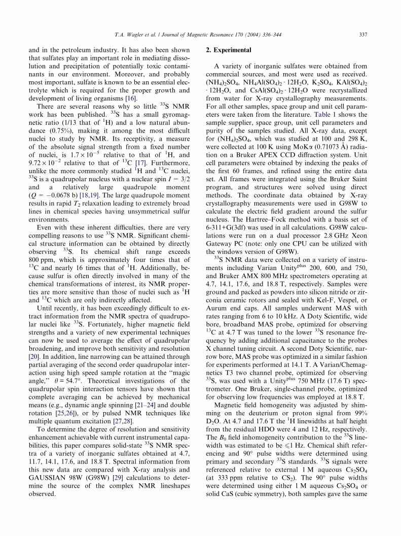
Fig. 1. Solid-state 33S MAS NMR spectra of KAl(SO4)2 Æ 12H2O: (A)
acquired at 4.7 T in 2 days; and (B) acquired at 17.6 T in 1 h.
3. Results and discussion
Typically solid-state 33S NMR spectral linewidths are
dominated by the broadening from quadrupolar interac-
tions. In the solid state, the outer (�3/2 M �1/2 and + 1/
2 M +3/2) spectral transitions are usually broadened be-
yond detection by quadrupolar interactions. However,the central (�1/2M +1/2) transition, to a first approxi-
mation, is unaffected by these same interactions and
can produce relatively narrow resonances. In cases
where the quadrupolar interaction is comparable to
the resonance frequency (e.g., in unsymmetrical struc-
tures), second order quadrupolar effects can lead to sig-
nificant broadening of the central transition. For
quadrupolar broadened resonances the spectral line-width is inversely proportional to the strength of the
applied magnetic field. Therefore, the spectral linewidth
can be reduced by using a stronger magnetic field.
The benefits of using a high magnetic field, including
improved sensitivity and resolution, are illustrated in
Fig. 1 which shows spectra of KAl(SO4)2 Æ 12H2O ob-
tained at 4.7 and 17.6 T. The spectrum shown in Fig.
1A required 2 days of signal averaging at 4.7 T using a
7 mm rotor with a 215 ll sample volume. However, only1 h of signal averaging was needed at 17.6 T to acquire
an adequate spectrum using a 5 mm rotor with a 90 llsample volume. Another benefit of using a high mag-
netic field strength was the reduction of acoustic ringing.

T.A. Wagler et al. / Journal of Magnetic Resonance 170 (2004) 336–344 339
Several contributions were considered when analyz-
ing the 33S NMR spectral linewidths, including: dipolar
coupling, magnetic field inhomogeneity, chemical shift
anisotropy (CSA), chemical shift dispersion (CSD),
and quadrupolar coupling. Dipolar coupling is depen-
dent on the internuclear distance and the gyromagneticratios (cIcS⁄/r3) of the interacting nuclei. Most of the
NMR active nuclei under consideration have a small
gyromagnetic ratio. 33S has a gyromagnetic ratio of
2.055685 · 107 rad/T s. Therefore, the dipolar couplings
are expected to be small and are effectively averaged by
MAS, even at modest spin rates of a few kHz. Magnetic
field inhomogeneity was also found to be small, and
contributes 6 1 Hz to the width of the 33S resonances.Spinning at frequencies greater than the static linewidth
will eliminate broadening from CSA, dipolar coupling,
and first order quadrupolar coupling. Therefore, second
order quadrupolar coupling and CSD (from variations
in sample morphology) must be the dominant factors
influencing linewidth. The linewidth contribution from
CSD is proportional to the magnetic field strength
[33]. Thus, a fourfold increase in field strength will resultin a corresponding increase in CSD contributions to the
linewidth. The quadrupolar coupling�s contribution to
the spectral linewidth is a function of the Cq and the
magnetic field strength, defined by [34–36]
A ¼C2
q
m z� CI ; ð1Þ
where CI = 3/64 for I = 3/2 and mz ¼ cB0
2p . Thus, increasing
the field strength should result in a proportional de-
crease in linewidth if quadrupolar coupling is the pri-mary contributor.
Table 2 summarizes the linewidths measured in this
work and for which solid-state 33S NMR data have ap-
peared in the literature. As mentioned above, increasing
the field strength should result in a proportional de-
crease in linewidth if quadrupolar coupling is the domi-
Table 2
Summary of 33S NMR linewidth data
Compound m1/2 (Hz) at 11.7 T m1/2 (Hz) at 4.7 T
Statica MAS = 6 kHz
Na2SO4 2300
(NH4)2SO4 1200 600
NH4Al(SO4)2 Æ 12H2O 950 12
K2SO4 4300
KAl(SO4)2 Æ 12H2O 2100 850
Rb2SO4 3500
Cs2SO4 3200
CsAl(SO4)2 Æ 12H2O 950
MgSO4 —
MgSO4 Æ 6H2O —
CaSO4 3500
CaSO4 Æ 2H2O 2000
BaSO4 18000
a Data obtained from [12].
nant line broadening mechanism. This phenomenon is
clearly seen in the 33S spectral linewidths of KAl
(SO4)2 Æ 12H2O obtained at 4.7, 11.7, 14.1 17.6, and
18.8 T (Table 2, row 5). Na2SO4 and BaSO4 show a sim-
ilar effect. The decrease in linewidth between the static
and MAS spectra, even at low fields, shows that MAShas essentially eliminated the broadening due to dipolar
coupling and CSA. In most cases, approximately an or-
der of magnitude reduction in linewidths were observed
when comparing the 33S NMR spectra measured from
static samples at 11.7 T [12], with the spectra measured
at 17.6 T with MAS. For NH4Al(SO4)2 Æ 12H2O the line-
width actually increases with increasing field strength
(Table 2, row 3). This is similar to the effect seen in sym-metric metal sulfides studied earlier, where it was shown
that CSD was the dominant line broadening mechanism
[15]. Cs2SO4 and (NH4)2SO4 show a combined influence
from both CSD and Cq. For those samples that gave
linewidths greater than about 300 Hz at 17.6 T it was
not possible to obtain spectra at 4.7 T, even after signal
averaging for several days. Time constraints and instru-
ment availability prevented some measurements at 4.7and 14.1 T.
Fig. 2 shows the 57.6 MHz 33S NMR spectra of sev-
eral inorganic sulfates. Most of these spectra show fea-
tures characteristic of second order quadrupolar
broadening effects. This is consistent with an unsymmet-
rical electronic environment around sulfur in these com-
pounds (see Tables 1 and 4). For many sulfur
compounds, complete averaging of the second orderquadrupolar interaction would require sample spinning
around two different axes or special pulse sequences.
However, MAS at very high field provided adequate line
narrowing to obtain relatively good spectra in a reason-
able amount of time. By using high magnetic fields, sig-
nificant gains in sensitivity and resolution are achieved.
Table 3 compares the chemical shifts and quadrupo-
lar coupling constants obtained at 17.6 T (with MAS)
m1/2 (Hz) at 14.1 T m1/2 (Hz) at 17.6 T m1/2 (Hz) at 18.8 T
MAS = 10 kHz MAS = 6 kHz MAS = 10 kHz
310 240
140 145
18 18
600
270 220 210
500
460 400
50
2200
450
500
105
2250 1600 1600

Fig. 2. Solid-state 33S MAS NMR spectra obtained at 57.6 MHz (17.6 T) of the following samples: (A) Na2SO4, 30� pulse width, 0.2 s relaxation
delay, 214 k scans, 17.5 h; (B) (NH4)2SO4, 20� pulse width, 0.3 s relaxation delay, 262 k scans, 24 h; (C) NH4Al(SO4)2 Æ 12H2O, 45� pulse width, 1.0 srelaxation delay, 44 k scans, 13 h; (D) K2SO4, 15� pulse width, 0.5 s relaxation delay, 348 k scans, 53 h; (E) KAl(SO4)2 Æ 12H2O, 90� pulse width, 0.1 srelaxation delay 268 k scans, 11 h; (F) Rb2SO4 15� pulse width, 0.2 s relaxation delay, 377 k scans, 24 h; (G) Cs2SO4, 45� pulse width, 0.5 s relaxationdelay, 65 k scans, 11 h; (H) CsAl(SO4)2 Æ 12H2O, 15� pulse width, 0.5 s relaxation delay, 122 k scans, 20 h; (I) MgSO4, 30� pulse width, 0.2 s
relaxation delay, 262 k scans, 21 h; (J) CaSO4, 10� pulse width, 0.04 s relaxation delay, 1491 k scans, 21 h; (K) CaSO4 Æ 2H2O, 30� pulse width, 0.5 srelaxation delay, 131 k scans, 22 h; and (L) BaSO4, 30� pulse width, 0.2 s relaxation delay, 262 k scans, 18 h. The liquid 90� pulse width was 5.5 ls.
340 T.A. Wagler et al. / Journal of Magnetic Resonance 170 (2004) 336–344
with literature values obtained at 11.7 T (without
MAS). In all cases, the quadrupolar coupling constantsobtained at 17.6 T are lower than those obtained at
11.7 T. As noted in [12], quadrupolar coupling con-
stants obtained without MAS are to be considered
approximate values, since CSA and dipolar coupling
also contribute to the linewidths in spectra from static
samples. Table 3 also shows the gq and T1�s obtained at
17.6 T. Due to much lower sensitivity, it was not pos-
sible to perform relaxation experiments for most of thesamples at 4.7 T. Although a complete understanding
of the spin–lattice relaxation behavior would require
temperature- or field-dependent spin–lattice relaxationstudies, some conclusions can be made from the data
provided. For instance, the T1�s of the hydrated sam-
ples are significantly lower than the anhydrous sam-
ples. This is attributed to the motion of the water
molecules in the crystal lattice, which increases the T1
relaxation efficiency. Haase et al. [37–39] showed that
relaxation of 27Al nuclei in hydrated zeolites were
overwhelmingly dominated by modulation of thetime-dependent electric field gradient at the nuclear site

Table 3
Summary of NMR parameters and D(S–O)m
Compound d (ppm)
11.7 Ta
d (ppm)
17.6 T
Cq (MHz)
11.7 Ta
Cq (MHz)
17.6 Tb
Cq (MHz)
G98W
gq 17.6 Tb D(S–O)m (A) T1 (s)
Na2SO4 330 341 0.82 0.66 0.13 30
(NH4)2SO4 328 332 0.59 0.58 0.56 at 298 K
0.84 at 100 K
0.75 0.008
0.020
1
NH4Al(SO4)2 Æ 12H2O 333 331 0.53 c 0.08 c 0.001 0.27
K2SO4 334 336 1.13 0.97 0.76 0.50 0.015 16
KAl(SO4)2 Æ 12H2O 327 332 0.79 0.64 0.26 (78%) 0.16 0.004 0.03
3.08 (22%) 0.060
Rb2SO4 329 337 1.01 0.93 0.42 29
Cs2SO4 335 336 0.97 0.83 0.46 16
CsAl(SO4)2 Æ 12H2O 331 330 0.53 0.29 0.29 0.55 0.005 8
MgSO4 325 2.05 0.10
MgSO4 Æ 6H2O 331 0.89 0.70 0.24 0.015 1.4
CaSO4 326 327 1.0 0.90 0.40 45
CaSO4 Æ 2H2O 337 327 0.77 0.70 1.00 12
BaSO4 328 2.3 1.70 1.75 0.72 0.034d
2.87 0.070e
a Data obtained from [12].b Determined using Varian STARS lineshape simulation package.c Cq is very small. Unable to determine using STARS.d Determined from [40,42].e Determined from [41,42].
T.A. Wagler et al. / Journal of Magnetic Resonance 170 (2004) 336–344 341
due to motion of water molecules and hydrated cations
in zeolite pores. There was no relationship observed be-
tween the quadrupole coupling constant and T1�s in the
anhydrous samples. This could be a result of impurities
within the crystal lattice or structural point defects
which can enhance relaxation.
To gain a better understanding of the factors contrib-
uting to the lineshapes observed in the spectra of thesecompounds, six samples were chosen for X-ray crystal-
lography and G98W studies. Table 4 shows S–O bond
distances and bond angles for the six compounds stud-
ied by X-ray crystallography in our lab, and 2 structures
of BaSO4 found in the literature [40–42]. X-ray analysis
of the six compounds showed that (NH4)2SO4, NH4Al
(SO4)2 Æ 12H2O, K2SO4, CsAl(SO4)2 Æ 12H2O, MgSO4 Æ6H2O, and BaSO4 have sulfate groups that are equiva-lent within their respective unit cells. However, the
X-ray data obtained for KAl(SO4)2 Æ 12H2O revealed
that within the orthorhombic crystal lattice, the sulfate
groups are disordered and distribute themselves into
two separate arrangements. Most of the sulfates (78%)
have their oxygen atoms more symmetrically arranged
around the central sulfur atom than the second arrange-
ment of sulfates (see Table 4), which include only 22% ofthe structures. This data indicates that the 33S NMR
spectrum of KAl(SO4)2 Æ 12H2O may actually contain
two distinct resonances (see Fig. 2E).
In an attempt to determine if two overlapping reso-
nances were present in the NMR spectrum of KAl
(SO4)2 Æ 12H2O, solid-state 33S NMR data at an even
higher magnetic field strength was collected. Compari-
son of the spectra obtained at 4.7 and 18.8 T revealed
that no further chemical shift resolution could be ob-
served and that the pattern only appeared to narrow
with increasing field strength (see Table 2, row 5). The
overall linewidth decreased by a factor of four, as ex-
pected from the diminished second order quadrupolar
effects at increased field strength. This signifies that the
observable pattern is solely due to second order quadru-polar broadening, and that any chemical shift informa-
tion is either obscured by this pattern or the second
resonance is so broad that it is not detected.
S–O bond distances and bond angles of the sulfate
groups measured experimentally by X-ray crystallogra-
phy were directly entered into G98W to calculate the
EFG tensors. Cq�s were calculated using the largest
traceless value of the EFG, given by
CqðMHzÞ ¼ 234:96QðbÞqzzða:u:Þ; ð2Þ
where Q = �0.0678 b and qzz = largest traceless value of
the EFG. Unfortunately, it was too computationally
expensive to simulate a large crystal unit where a cen-
tralized sulfate is coordinated to an inner sphere of ca-
tions, which in turn are coordinated to a second
sphere of sulfate groups. However, the most significant
contribution to the asymmetric EFG around the sulfurnucleus is provided by the closest atoms, and a general
correspondence can be seen between the calculated
and experimentally determined Cq�s shown in Table 3,
column 5 and 6. In all cases, except KAl(SO4)2 Æ 12H2O,
the calculated Cq�s are within 25% of the experimentally
determined values.

Fig. 3. Plot of Cq vs. D(S–O)m. The open circles (s) are based on the
Cq�s determined by simulation of the NMR spectra, and the openble
4
nddistancesandbondanglesofselected
sulfates
nddistance
andbondangle
(NH
4) 2SO
4NH
4Al(SO
4) 2Æ12H
2O
K2SO
4KAl(SO
4) 2Æ12H
2O
CsA
l(SO
4) 2Æ12H
2O
MgSO
4Æ6H
2O
BaSO
4a
BaSO
4b
298K
100K
78%
22%
O(1)
1.443(3)
1.4657(14)
1.4727(16)
1.4676(19)
1.470(2)
1.433(7)
1.473(3)
1.4687(9)
1.456(3)
1.472
O(2)
1.4472(17)
1.4787(15)
1.4727(16)
1.4809(13)
1.4743(13)
1.493(4)
1.4781(13)
1.4778(9)
1.467(2)
1.448
O(3)
1.4472(17)
1.4824(16)
1.4727(16)
1.4809(13)
1.4743(13)
1.493(4)
1.4781(13)
1.4806(9)
1.490(2)
1.518
O(4)
1.451(3)
1.4854(13)
1.474(3)
1.4829(18)
1.4743(13)
1.493(4)
1.4781(13)
1.4837(9)
1.490(2)
1.518
O(1)–S–O(2)
108.83(10)
109.75(9)
109.61(8)
109.83(7)
109.56(6)
111.14(17)
109.76(6)
110.32(5)
112.3(4)
111.8
O(1)–S–O(3)
108.83(10)
110.17(9)
109.61(8)
109.83(7)
109.56(6)
111.14(17)
109.76(6)
109.88(5)
109.7(2)
110.3
O(2)–S–O(3)
110.47(15)
108.87(9)
109.61(8)
109.38(11)
109.39(6)
107.75(18)
109.18(6)
109.74(5)
108.6(2)
109.7
O(1)–S–O(4)
110.30(17)
110.23(8)
109.33(8)
110.44(11)
109.56(6)
111.14(17)
109.76(6)
109.91(6)
109.7(2)
110.3
O(2)–S–O(4)
109.20(10)
108.97(9)
109.33(8)
108.67(7)
109.39(6)
107.75(18)
109.18(6)
108.50(5)
108.6(2)
109.7
O(3)–S–O(4)
109.20(10)
108.82(9)
109.33(8)
108.67(7)
109.39(6)
107.75(18)
109.18(6)
108.45(5)
107.8(4)
104.7
aData
obtained
from
[40,42].
bData
obtained
from
[41,42].
342 T.A. Wagler et al. / Journal of Magnetic Resonance 170 (2004) 336–344
Ta
Bo
Bo
S–
S–
S–
S–
\ \ \ \ \ \
Close inspection of the X-ray crystallography data re-
vealed that a correlation existed between the S–O bond
distances, d(S–O), and Cq. It was observed that as the
disparity in S–O bond distances within the sulfate group
grew, the Cq also became larger. The disparity in d(S–O)
was characterized best by the difference between thelargest and smallest S–O bond distances, given by
DðSOÞm ¼ dðSOÞl � dðSOÞs ð3Þwhere d(S–O)l is the longest S–O bond distance and d(S–
O)s is the shortest S–O bond distance within the sulfate
group. Fig. 3 shows a plot of Cq vs. D(S–O)m where theopen circles (s) represent the Cq�s determined by simu-
lation of the NMR spectra, and the open squares (h)
represent the Cq�s determined by ab initio calculations.
A linear regression fit of the calculated values gave Cq
= 44.0 · D(S–O)m + 0.1. This fit can be used as a predic-
tive tool for optimizing NMR acquisition parameters if
X-ray data is available. Table 3 and Fig. 3 show D(S–O)m for two BaSO4 crystal structures found in the liter-ature [40,41]. This data shows that the BaSO4 sample
more closely resembles the Pbnm structure of the mate-
rial studied by Jacobsen et al. [40].
The two sulfate arrangements in KAl(SO4)2 Æ 12H2O
have a D(S–O)m of 0.004 A for the major (78%) form
and 0.060 A for the minor (22%) form of the sulfate
groups. This leads to a calculated Cq of 0.26 MHz for
sulfate groups of the major component and 3.08 MHzfor sulfate groups of the minor component in KAl
(SO4)2 Æ 12H2O. Thus, it is highly probable that 22% of
the sulfate groups in KAl(SO4)2 Æ 12H2O are not de-
tected by NMR even at 18.8 T. The large Cq and lower
abundance of that sulfate structure reduces the intensity
and broadens the resonance so that it is buried in the
baseline. Spectral simulation of a two component sys-
tem, having 78% sulfate groups with a Cq = 0.64 MHzand gq = 0.16 and 22% sulfate groups with a Cq = 3.08
squares (h) are based on the Cq�s calculated by G98W.

T.A. Wagler et al. / Journal of Magnetic Resonance 170 (2004) 336–344 343
and gq = 0.16, did indeed show a broad signal from the
minor component which was about 50 times weaker
than the signal from the major component.
All X-ray crystallography measurements were run at
100 K to reduce thermal motion and to obtain higher
resolution. However, it was found that (NH4)2SO4
exhibits a phase change at reduced temperature. Ahmed
et. al. [43] showed that the lattice parameters and unit
cell volume of (NH4)2SO4 change as a function of tem-
perature and that it undergoes a transition from a para-
electric phase to a ferroelectric phase at 223 K. Thus, it
was necessary to repeat the X-ray crystallography mea-
surements under the same conditions as the NMR
experiments. Columns 2 and 3 of Table 4 contain theS–O bond distances and bond angles of (NH4)2SO4 ob-
tained at 298 and 100 K. It shows that the D(S–O)m at
100 K is 0.013 A resulting in a calculated Cq of
0.84 MHz (see Table 3). At 298 K the D(S–O)m is only
0.004 A which leads to a significantly smaller Cq of
0.56 MHz, which is much closer to the experimentally
determined value of 0.58 MHz.
4. Conclusions
Solid-state 33S NMR has been largely neglected over
the past 50 years mostly because of the difficulty associ-
ated with collecting suitable spectra. However, with the
increasing number of high field NMR spectrometers
available, it should be easier to obtain such spectra. Enor-mous gains in sensitivity and resolution can be achieved
by collecting 33S NMR spectra at a high magnetic field
strength. An order of magnitude reduction in linewidth
was observed between static spectra obtained at 11.7 T
andMAS spectra obtained at 17.6 T, with a concomitant
increase in peak height and signal-to-noise. Furthermore,
spectra obtained at high fields (high frequencies) lacked
acoustic ringing that complicated lower field measure-ments. Spin–lattice relaxation measurements showed sig-
nificantly reduced T1�s for the hydrated samples. This
resulted from the modulation of the time-dependent elec-
tric field gradient at the nuclear site due tomotionofwater
molecules. G98W calculations and X-ray measurements
proved helpful in understanding the complex NMR line-
shapes of these samples. These techniques also revealed a
general correlation between D(S–O)m andCq that may behelpful in future studies of sulfates.
Acknowledgments
The authors would like to thank Ann Bolek for her
efforts in obtaining much of the crystal structure litera-
ture, Simon Stakleff for maintaining the NMR equip-ment used, the Kresge Foundation and the donors
to the Kresge Challenge Program at the University of
Akron for funds to purchase the NMR instruments used
in this work, and partial support of this research by the
Petroleum Research Fund administered by the Ameri-
can Chemical Society (26776-AC). We also wish to
thank H. Foerster of Bruker Instruments, B. Bluemich,
and the staff of the NMR lab at RWTH, Aachen forassistance in obtaining 800 MHz data.
References
[1] S.S. Dharmatti, H.E. Weaver, Magnetic moment of sulfur-33,
Phys. Rev. 83 (1951) 845.
[2] R. Musio, O. Sciacovelli, Detection of taurine in biological tissues
by 33S NMR spectroscopy, J. Magn Reson. 153 (2) (2001) 259–
261.
[3] R. Gawinecki, E. Kolehmainen, A. Zakrzewski, K. Laihia, B.
Osmialowski, R. Kauppinen, Predominance of inductive overre-
sonance substituent effect on 33S NMR chemical shifts of 4-
substituted phenyl-40-methylphenacyl sulfones, Magn. Reson.
Chem. 37 (6) (1999) 437–440.
[4] B. Berke, C. Cheze, J. Vercauteren, G. Deffieux, Bisulfite addition
to anthocyanins: revisited structures of colorless adducts, Tetra-
hedron Lett. 39 (32) (1998) 5771–5774.
[5] R. Musio, O. Sciacovelli, 33S NMR Spectroscopy. 2. Substituent
effects on 33S chemical shifts and nuclear quadrupole coupling
constants in 3- and 4-substituted benzenesulfonates. Correlation
between chemical shifts and nuclear quadrupole coupling con-
stants, J. Org. Chem. 62 (26) (1997) 9031–9033.
[6] A. Perjessy, E. Kolehmainen, W.M.F. Fabian, M. Ludwig, K.
Laihia, J. Kulhanek, Z. Sustekova, Structure-reactivity-spectra
correlations for substituted benzenesulfonamides, Sulfur Lett. 25
(2) (2002) 71–78.
[7] R.A. Aitken, S. Arumugam, S.T.E. Mesher, F.G. Riddell, Natural
abundance 33S NMR spectroscopy. The first spectra of several
major compound types, J. Chem. Soc. Perkin Trans. 2 (2) (2002)
225–226.
[8] V.M. Bzhezovsky, G.A. Kalabin, Chemistry of Organosulfur
Compounds, Horwood, Chichester, 1990.
[9] J.F. Hinton, Sulfur-33 NMR spectroscopy, Annu. Rep. NMR
Spectrosc. 19 (1987) 1–34.
[10] P.S. Belton, I.J. Cox, R.K. Harris, Experimental sulfur-33 nuclear
magnetic resonance spectroscopy, J. Chem. Soc. Faraday Trans. 2
(81) (1985) 63.
[11] G. Barbarella, Sulfur-33 NMR, Prog. NMR Spectrosc. 25 (1993)
317.
[12] H. Eckert, J.P. Yesinowski, Sulfur-33 NMR at natural abundance
in solids, J. Am. Chem. Soc. 108 (1986) 2140.
[13] T.J. Bastow, NMR study of the II-VI semiconductors ZnX
(X = O, S, Se, Te), Mater. Australas. 19 (1987) 12.
[14] T.J. Bastow, S.N. Stuart, NMR study of the zinc chalcogenides
(ZnX, X = O, S, Se, Te), Phys. Status Solidi B 145 (1988) 719.
[15] T.A. Wagler, W.A. Daunch, P.L. Rinaldi, A.R. Palmer, Solid
state 33S NMR of inorganic sulfides, J. Magn. Reson. 161 (2)
(2003) 191.
[16] D. Markovich, Physiological roles and regulation of mammalian
sulfate transporters, Physiol. Rev. 81 (4) (2001) 1499.
[17] C. Brevard, P. Granger, Handbook of High Resolution Multinu-
clear NMR, Wiley, New York, 1981.
[18] P. Pyykko, The nuclear quadrupole moments of the 20 first
elements: high-precision calculations on atoms and small mole-
cules, Z. Naturforsch. A: Phys. Sci. 47 (1–2) (1992) 189.
[19] D. Sundholm, J. Olsen, Nuclear quadrupole moments of sulfur-33
and sulfur-35, Phys. Rev. A: At. Mol. Opt. Phys. 42 (3) (1990)
1160.

344 T.A. Wagler et al. / Journal of Magnetic Resonance 170 (2004) 336–344
[20] G. Maciel, High-resolution nuclear magnetic resonance of solids,
Science 226 (1984) 282.
[21] G.C. Chingas, K.T. Mueller, A. Pines, J. Stebbins, Y. Wu, J.W.
Zwanziger, Dynamic-angle spinning of quadrupolar nuclei, J.
Magn. Reson. 86 (1990) 470.
[22] S. Ganapathy, S. Schramm, E. Oldfield, Variable-angle sample-
spinning high resolution NMR of solids, J. Chem. Phys. 77 (1982)
4360.
[23] E. Oldfield, R.J. Kirkpatrick, High-resolution nuclear magnetic
resonance of inorganic solids, Science 227 (1985) 1537.
[24] K.T. Mueller, B.Q. Sun, G.C. Chingas, J.W. Zwanziger, T.
Terao, A. Pines, Dynamic-angle spinning of quadrupolar nuclei,
J. Magn. Reson. 86 (1990) 470.
[25] A. Samoson, A. Pines, Double rotor for solid-state NMR, Rev.
Sci. Instrum. 60 (1989) 3239.
[26] E.W. Wooten, K.T. Mueller, A. Pines, New angles in nuclear
magnetic resonance sample spinning, Acct. Chem. Res. 25 (1992)
209.
[27] L. Frydman, J.S. Harwood, Isotropic spectra of half-integer
quadrupolar spins from bidimensional magic-angle spinning
NMR, J. Am. Chem. Soc. 117 (1995) 5367.
[28] A. Medek, J.S. Harwood, L. Frydman, Multiple-quantum magic-
angle spinning NMR: a new method for the study of quadrupolar
nuclei in solids, J. Am. Chem. Soc. 117 (1995) 12779.
[29] M.J. Frisch, G.W. Trucks, H.B. Schlegel, G.E. Scuseria, M.A.
Robb, J.R. Cheeseman, V.G. Zakrzewski, J.A. Montgomery, Jr.,
R.E. Stratmann, J.C. Burant, S. Dapprich, J.M. Millam, A.D.
Daniels, K.N. Kudin, M.C. Strain, O. Farkas, J. Tomasi, V.
Barone, M. Cossi, R. Cammi, B. Mennucci, C. Pomelli, C.
Adamo, S. Clifford, J. Ochterski, G.A. Petersson, P.Y. Ayala, Q.
Cui, K. Morokuma, N. Rega, P. Salvador, J.J. Dannenberg, D.K.
Malick, A.D. Rabuck, K. Raghavachari, J.B. Foresman, J.
Cioslowski, J.V. Ortiz, A.G. Baboul, B.B. Stefanov, G. Liu, A.
Liashenko, P. Piskorz, I. Komaromi, R. Gomperts, R.L. Martin,
D.J. Fox, T. Keith, M.A. Al-Laham, C.Y. Peng, A. Nanayakkara,
M. Challacombe, P.M.W. Gill, B. Johnson, W. Chen, M.W.
Wong, J.L. Andres, C. Gonzalez, M. Head-Gordon, E.S. Repl-
ogle, J.A. Pople, GAUSSIAN 98, Revision A.11.2 (Gaussian,
Inc., Pittsburgh PA, 2001)..
[30] J. Skibsted, N.C. Nielsen, H. Bildsøe, H.J. Jakobsen, Satellite
transitions in MAS NMR spectra of quadrupolar nuclei, J. Magn.
Reson. 95 (1991) 88.
[31] J. Skibsted, N.C. Nielsen, H. Bildsøe, H.J. Jakobsen, Vanadium-51
MASNMR spectroscopy: determination of quadrupole and aniso-
tropic shielding tensors, including the relative orientation of their
principal-axis systems, Chem. Phys. Lett. 188 (1992) 405.
[32] J. Skibsted, N.C. Nielsen, H. Bildsøe, H.J. Jakobsen, Magnitudes
and relative orientation of vanadium-51 quadrupole coupling and
anisotropic shielding tensors in metavanadates and potassium
vanadium oxide (KV3O8) from vanadium-51 MAS NMR spectra.
Sodium-23 quadrupole coupling parameters for a- and b-NaVO3,
J. Am. Chem. Soc. 115 (1993) 7351.
[33] T.C. Farrar, E.D. Becker, Pulse and Fourier Transform NMR,
Academic Press, New York, 1971.
[34] M.H. Cohen, F. Reif, Quadrupole effects in nuclear magnetic
resonance studies of solids, Solid State Phys. 5 (1957) 321.
[35] A.P.M. Kentgens, A practical guide to solid-state NMR of half-
integer quadrupolar nuclei with some applications to disordered
systems, Geoderma 80 (1997) 271.
[36] D. Freude, J. Haase, Quadrupole effects in solid-state nuclear
magnetic resonance, NMR 29 (1993) 1–90.
[37] J. Haase, K.D. Park, K. Guo, H.K.C. Timken, E. Oldfield,
Nuclear magnetic resonance spectroscopic study of spin–lattice
relaxation of quadrupolar nuclei in zeolites, J. Phys. Chem. 95 (18)
(1991) 6996.
[38] J. Haase, H. Pfeifer, W. Oehme, J. Klinowski, Logitudinal NMR
relaxation of 27Al nuclei in zeolites, Chem. Phys. Lett. 150 (1988)
189.
[39] J. Haase, H. Pfeifer, W. Oehme, J. Klinowski, Longitudinal
N.M.R. relaxation of aluminium-27 in zeolites is governed by
quadrupole interactions with adsorbed polar molecules and
exchangeable cations, J. Chem. Soc. Chem. Commun. 17 (1988)
1142.
[40] S.D. Jacobsen, J.R. Smyth, R.J. Swope, Rigid-body character of
the SO4 groups in Celestine, anglesite and barite, Can. Miner. 36
(1998) 1053.
[41] A.A. Colville, K. Staudhammer, A refinement of the structure of
barite from Cow Green Mine, Am. Miner. 52 (1967) 1877.
[42] American Mineralogist Crystal Structure Database. Available at:
<http://www.geo.arizona.edu/AMS/>.
[43] S. Ahmed, A.M. Shamah, R. Kamel, Y. Badr, Structural changes
of (NH4)2SO4 crystals, Phys. Stat. Sol. A 99 (1987) 131.
[44] F.C. Hawthorne, R.B. Ferguson, Anhydrous sulphates. I. Refine-
ment of the crystal structure of celestite with an appendix on the
structure of thenardite, Can. Miner. 13 (1975) 181.
[45] K. Hasebe, Studies of the crystal structure of ammonium sulfate
in connection with its ferroelectric phase transition, J. Phys. Soc.
Japan 50 (1981) 1266.
[46] A.M. Abdeen, G. Will, W. Schaefer, A. Kirfel, M.O. Bargouth,
K. Recker, A. Weiss, X-ray and neutron diffraction study of
alums: III. The crystal structure of ammonium aluminum alum, Z.
Kristallogr. 157 (1981) 147.
[47] S. Lidin, A.K. Larsson, A structural description of b-potassiumsulfate, Acta Chem. Scand. 45 (1991) 856.
[48] A.C. Larson, D.T. Cromer, Refinement of the alum structures:
III. X-ray study of the a-alums, K, Rb, and NH4Al(SO4)2(H2O)12,
Acta Crystallogr. 22 (1967) 793.
[49] D. Liu, H.M. Lu, J.R. Hardy, F.G. Ullman, Raman scattering
and lattice-dynamical calculations of alkali-metal sulfates, Phys.
Rev. B: Condens. Matter. 44 (1991) 7387.
[50] F. Hammel, Anhydrous sulfates of the magnesian series, Hebd.
Seances Acad. Sci. 202 (1936) 57.
[51] F.C. Hawthorne, R.B. Ferguson, Anhydrous sulfates. II. Refine-
ment of the crystal structure of anhydrite, Can. Miner. 13 (1975)
289.
[52] B.F. Pedersen, D. Semmingsen, Neutron diffraction refinement of
the structure of gypsum, CaSO4(H2O)2, Acta Crystallogr. B 38
(1982) 1074.