small animal toxicology essentials (poppenga/small animal toxicology essentials) || metals and...
TRANSCRIPT
273
29 Metals and Minerals
INTRODUCTION
Metals are intrinsic to nature and are ubiquitous in the environment of animals. While the form or valence of any given metal can be changed, metals themselves cannot be destroyed. The activities of man have resulted in the large - scale redistribution of metals in the environment. Perhaps the best example of this relates to lead, which originates as lead ore that is processed for numerous uses. The prior use of lead as an antiknock compound in gasoline and pigment in paints contributed to widespread environmen-tal contamination before such uses were banned. Despite curtailment of lead use, it still poses signifi cant risks for intoxication. Lead in old paints continues to cause adverse health effects in people and pets.
Metals can be categorized as those that play an essential role in biological processes (e.g., those that are nutrition-ally necessary such as iron, copper, zinc and selenium, among others) and those that do not (e.g., lead and mercury). It is important to point out that disease syn-dromes caused by defi ciencies of nutritionally important metals are different in pathogenesis and clinical manifesta-tions than those caused by exposure to excessive amounts of the same metal.
The toxicity of metals is sometimes harder to defi ne than for other toxicants. One reason for this is that the form of a metal plays a critical role in how readily it is systemi-cally absorbed resulting in intoxication. For example, elemental mercury, when ingested, is essentially unavail-able for systemic absorption. In contrast, organic forms of
mercury such as methyl mercury are highly bioavailable. The interaction of metals also plays an important role in the determination of toxicity. For example, high dietary calcium can decrease absorption of lead because lead uses calcium transport mechanisms for its uptake into the body. Other factors that can infl uence toxicity include animal age, gender, and the capacity of an individual for biotrans-formation of a metal.
The distribution and accumulation of metals in certain tissues in the body can mitigate damage. Lead can accumulate in bone where it is not available to affect target tissues such as the CNS. Also, metals can be sequestered in the body by cystine - rich proteins called metallothioneins . Metallothioneins are highly inducible by metals and are essential for metal homeostasis and detoxifi cation of metals (Liu et al. 2008 ). The interac-tion of metals with sulfhydryl - containing groups like cystine is the basis for the effi cacy of metal chelators, such as succimer or dimercaprol, which contain numer-ous sulfhydryl groups.
Although the periodic table is full of potentially toxic metals, the following discussion is limited to those few metals that most commonly intoxicate, or pose an intoxi-cation risk for, companion animals. We do not discuss metals that are more frequently involved in livestock or wildlife intoxications such as selenium and mercury nor discuss copper accumulation in susceptible dog breeds. However, keep in mind that most metals, under appropri-ate circumstances, can poison animals and people.
Small Animal Toxicology Essentials, First Edition. Edited by Robert H. Poppenga, Sharon Gwaltney-Brant.© 2011 John Wiley and Sons, Inc. Published 2011 by John Wiley and Sons, Inc.
Robert H. Poppenga
274 Section 3 / Specifi c Toxicants
olized in the liver by the addition of methyl groups prior to its elimination.
Toxicity
The toxicity of arsenic is quite variable and depends on its form, purity, solubility, particle size, valence, and species exposed, and the condition of the exposed individual (Neiger 2004 ). For example, trivalent arsenicals are 4 to 10 times more toxic than pentavalent arsenicals. The cat is considered to be one of the more sensitive species. A lethal oral dose of sodium arsenite is reported to be less than 5 mg/kg body weight. More generally, the range of lethal oral doses for sodium arsenite is 1 to 25 mg/kg body weight. Weak and debilitated animals are more susceptible to intoxication (Neiger 2007 ).
Mechanism of Action
The mechanism of toxic action is dependent on the form of arsenic. Trivalent inorganic arsenicals (i.e., arsenites) inhibit cellular respiration. They bind to sulfhydryl com-pounds, especially lipoic acid and α - keto oxidases. Lipoic acid, a tissue respiratory enzyme cofactor, plays an essen-tial role in the tricarboxylic acid cycle (TCA). Tissues with high oxidative energy requirements (e.g., actively dividing cells such as those of the intestinal epithelium, kidney, liver, skin, and lungs) are more severely affected. Trivalent arsenic affects capillary integrity by an unknown mecha-nism. The gastrointestinal tract is most affected; capillary dilatation is followed by transudation of plasma into the gastrointestinal tract resulting in submucosal congestion and edema.
In contrast, pentavalent inorganic arsenicals (i.e., arse-nates) appear to substitute for phosphate in oxidative phos-phorylation. Uncoupling of oxidative phosphorylation produces a cellular energy defi cit. Elevated body tempera-ture is not characteristic of pentavalent arsenical poisoning as it is in poisonings by other oxidative uncouplers (e.g., nitrophenols).
The organic arsenicals also vary in terms of their mech-anism of toxic action. Trivalent organic arsenicals appear to have a mechanism of toxic action similar to the inor-ganic arsenicals. However, pentavalent organic arsenicals (e.g., used as livestock feed additives) act on nerves by an unknown mechanism. They produce demyelination and eventual axonal degeneration, leading some researchers to believe that pentavalent organic arsenicals interfere with the B vitamins essential for maintenance of nervous tissue.
Although chronic arsenic exposure has been associated with various cancers in people, its ability to cause cancer is animals is uncertain (Neiger 2004 ). In humans, chronic
Arsenic
Sources/Formulations
Historically, various forms of arsenic have been used as herbicides, insecticides, wood preservatives, growth promotants, and therapeutic agents. Arsenic is found in various valences and forms. There are trivalent and pentavalent inorganic and organic arsenic forms. The tri-valent inorganic arsenic trioxide was formerly used as an insecticide and herbicide and as a source for other arseni-cals (Ensley 2004 ). Pentavalent (arsenate, H 3 AsO 4 ) and trivalent (arsenite, H 3 AsO 3 ) forms of arsenic include sodium, potassium, and calcium salts. Paris green (copper acetoarsenite) and lead arsenate have been used as insec-ticides, and trivalent inorganic arsenic forms such as monosodium methanarsentate (MSMA) and disodium methanearsonate (DMSA) are used as herbicides. Pentava-lent organic arsenicals such as sodium arsenilate and 3 - nitro, 4 - hydroxyphenylarsonic acid are used as feed additives for some livestock species.
Although arsenic is still utilized in some products, it is not as ubiquitous as it once was due to restrictions on its use and the availability of newer, more targeted pesticides. For example, its use as a wood preservative in the form of chromated copper arsenic (CCA) has been banned for resi-dential use by the USEPA ( http://www.epa.gov/oppad001/reregistration/cca/ ). Thiacetarsamide, a phenylarsenoxide previously used as a heartworm adulticide, is no longer commercially available in the U.S. although another organic arsenical, melarsomine, is available (Plumb 2005 ). According to the ASPCA Animal Poison Control Center, the highest number of arsenic - related calls involves the exposure of dogs and cats to ant or roach baits, which are typically inorganic forms of arsenic (Neiger 2004 ).
Kinetics
As for most metals, the bioavailability of arsenic is vari-able and depends on its form (organic vs. inorganic, solu-tion vs. powder). The arsenic in solutions of organic arsenicals is almost completely absorbed from the GI tract (Neiger 2004 ). Alternatively, arsenic compounds of low solubility such as arsenic trioxide are absorbed less effi -ciently. Once absorbed, arsenic is widely distributed in the body and reaches its highest concentrations in the liver, spleen, kidney, lungs, and GI tract. Persistent residues are found in keratinized tissues such as skin, hair, and nails. Clearance of arsenic from the blood is multiphasic with the fi rst phase having a 1 - to - 2 hour half - life. Forty to 70% of an absorbed dose is eliminated via the urine within the fi rst 48 hours. To a large extent, inorganic arsenic is metab-
Chapter 29 / Metals and Minerals 275
early as possible an important consideration. Most veteri-nary diagnostic laboratories can test for arsenic. It is important to point out that low - level concentrations of arsenic in blood or urine are not unusual and do not indi-cate arsenic intoxication. The interpretation of a given concentration of arsenic depends on an evaluation of the entire case and the timing of sample collection in relation to exposure.
Postmortem
Postmortem analysis of stomach contents, liver, kidney, or urine can confi rm exposure. Again, the signifi cance of detected concentrations, especially relatively low concen-trations, needs to be interpreted in conjunction with other case variables. Postmortem lesions might be absent in cases of rapid death. Otherwise, severe gastrointestinal lesions consisting of reddening of the gastric mucosa and proximal small intestine, watery GI contents, and blood and sloughed mucosa in the feces are commonly noted. Other organ lesions are not specifi c for arsenic.
Management of Exposures
The rapidity of onset of clinical signs such as vomiting often precludes initiating decontamination procedures. Activated charcoal is unlikely to adsorb signifi cant amounts of arsenic, although with signifi cant exposures its administration is recommended (Ford 2006 ). Symptomatic animals need to be stabilized fi rst. This might entail treat-ment for circulatory shock and hypotension and mainte-nance of body temperature. Renal and liver failure and electrolyte abnormalities might need to be addressed. Glucose and glycogen stores should be maintained paren-terally with dextrose or parenteral alimentation solutions (Ford 2006 ).
Chelation therapy is typically indicated. Historically, dimercaprol (BAL or British anti - Lewisite) was the rec-ommended chelator. It is a sulfhydryl - containing chelator that binds to arsenic and allows the arsenic - chelator complex to be eliminated. An alternative chelator, suc-cimer, has several theoretical advantages over dimer-caprol, including less toxicity and oral (vs. injectable) dosing form. It has been theorized that because succimer is hydrophilic it might not remove arsenic that has escaped the extracelluar space as effi ciently as dimer-caprol (Neiger 2004 ). However, available studies suggest that succimer is equal to or even superior to dimercaprol in increasing arsenic elimination (Ford 2006 ). If dimer-caprol is used, monitoring for adverse reactions included pain at the injection site, vomiting, tremors, and seizures is required.
exposure to arsenic induces characteristic changes in the skin including hyper - or hypopigmentation and hyperkeratosis.
Clinical Effects
Typically, arsenic intoxication in small animals is an acute illness. Signs can occur within minutes of exposure, with death occurring within several hours. Intoxication from chronic arsenic exposure is not well described in compan-ion animals. However, there is one case report describing dermatitis in a German shorthaired pointer chronically exposed to arsenic (Evinger and Blakemore 1984 ). The source of exposure was from a container of 44% sodium arsenate that had been leaking into the dog ’ s house.
Signs
Signs associated with acute arsenic intoxication generally develop in the following progression: intense abdominal pain, salivation, vomiting, staggering gait, diarrhea (often bloody), rapid and weak pulse, hypothermia, collapse, and death. Experimentally, long - term exposure of dogs to inor-ganic arsenic caused nonspecifi c signs such as anorexia and weight loss secondary to reduced food consumption (Neiger and Osweiler 1989 ).
Laboratory
There are no specifi c abnormalities noted from CBC or serum chemistry tests. Because arsenic can damage mul-tiple organs, changes associated with liver or kidney dys-function can be present if the animal survives for several days. Protein, red blood cells, and casts can be noted in the urine.
Differential Diagnoses
Other disease etiologies to consider include intoxication from other metals or metal salts (e.g., lead, copper, or zinc salts), ingestion of acid or alkaline caustics, ingestion of irritating plants (e.g., those containing insoluble calcium oxalates, castor bean), and infectious agents such as parvovirus.
Diagnostics
Antemortem
An antemortem diagnosis relies on a history of ingestion of arsenic, the occurrence of consistent clinical signs, and the detection of toxic concentrations of arsenic in vomitus or stomach contents, whole blood, or urine. Suspect sources of exposure can also be tested to confi rm the pres-ence of arsenic. The rapid elimination of the metal from the body makes the collection of appropriate samples as
276 Section 3 / Specifi c Toxicants
products, possibly as a result of enhanced absorption of the chelated iron.
Kinetics
The kinetics of iron absorption are complex. Iron body stores are regulated at the site of absorption from the GI tract because the body is not able to actively excrete iron. From the GI tract, iron must fi rst enter duodenal mucosal cells, possibly by a carrier - mediated process. Next the iron is either lost as the mucosal cells slough into the GI lumen or is bound to ferritin for later transfer to transferrin, a serum iron - binding transport protein. Serum transferrin concentrations greatly exceed amounts necessary to bind iron under normal physiological processes (normal binding capacity is 3 to 4 times the serum iron concentration). However, during intoxications the binding capacity is exceeded, allowing free iron to cause cell damage.
Toxicity
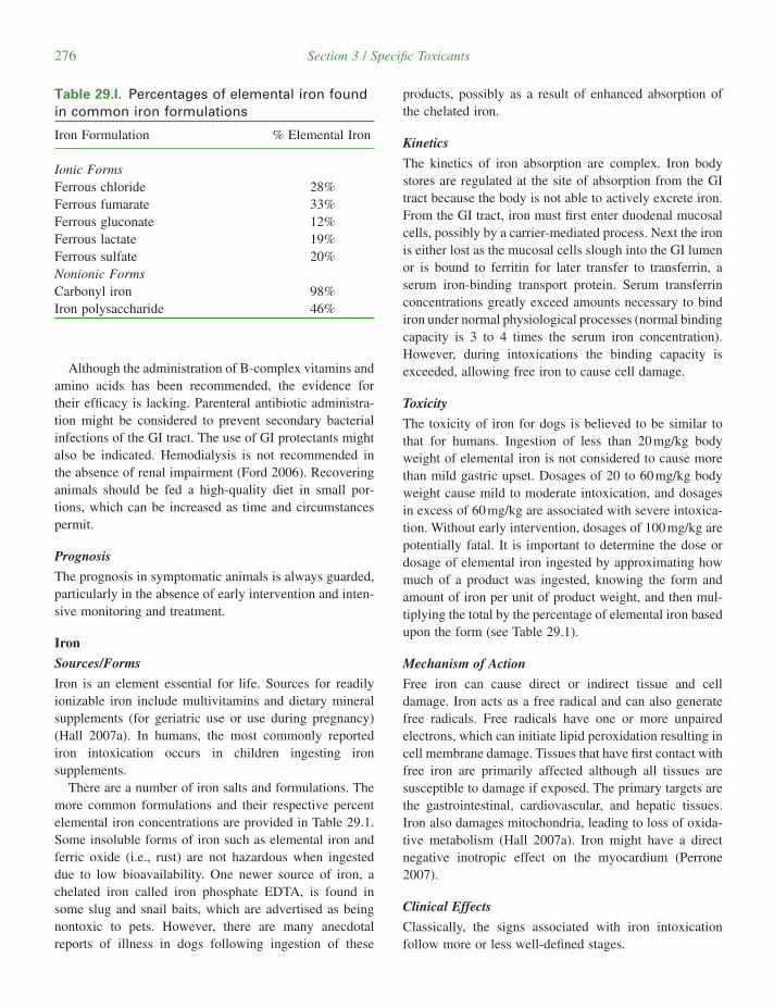
The toxicity of iron for dogs is believed to be similar to that for humans. Ingestion of less than 20 mg/kg body weight of elemental iron is not considered to cause more than mild gastric upset. Dosages of 20 to 60 mg/kg body weight cause mild to moderate intoxication, and dosages in excess of 60 mg/kg are associated with severe intoxica-tion. Without early intervention, dosages of 100 mg/kg are potentially fatal. It is important to determine the dose or dosage of elemental iron ingested by approximating how much of a product was ingested, knowing the form and amount of iron per unit of product weight, and then mul-tiplying the total by the percentage of elemental iron based upon the form (see Table 29.1 ).
Mechanism of Action
Free iron can cause direct or indirect tissue and cell damage. Iron acts as a free radical and can also generate free radicals. Free radicals have one or more unpaired electrons, which can initiate lipid peroxidation resulting in cell membrane damage. Tissues that have fi rst contact with free iron are primarily affected although all tissues are susceptible to damage if exposed. The primary targets are the gastrointestinal, cardiovascular, and hepatic tissues. Iron also damages mitochondria, leading to loss of oxida-tive metabolism (Hall 2007a ). Iron might have a direct negative inotropic effect on the myocardium (Perrone 2007 ).
Clinical Effects
Classically, the signs associated with iron intoxication follow more or less well - defi ned stages.
Although the administration of B - complex vitamins and amino acids has been recommended, the evidence for their effi cacy is lacking. Parenteral antibiotic administra-tion might be considered to prevent secondary bacterial infections of the GI tract. The use of GI protectants might also be indicated. Hemodialysis is not recommended in the absence of renal impairment (Ford 2006 ). Recovering animals should be fed a high - quality diet in small por-tions, which can be increased as time and circumstances permit.
Prognosis
The prognosis in symptomatic animals is always guarded, particularly in the absence of early intervention and inten-sive monitoring and treatment.
Iron
Sources/Forms
Iron is an element essential for life. Sources for readily ionizable iron include multivitamins and dietary mineral supplements (for geriatric use or use during pregnancy) (Hall 2007a ). In humans, the most commonly reported iron intoxication occurs in children ingesting iron supplements.
There are a number of iron salts and formulations. The more common formulations and their respective percent elemental iron concentrations are provided in Table 29.1 . Some insoluble forms of iron such as elemental iron and ferric oxide (i.e., rust) are not hazardous when ingested due to low bioavailability. One newer source of iron, a chelated iron called iron phosphate EDTA, is found in some slug and snail baits, which are advertised as being nontoxic to pets. However, there are many anecdotal reports of illness in dogs following ingestion of these
Table 29.l. Percentages of elemental iron found in common iron formulations
Iron Formulation % Elemental Iron
Ionic Forms Ferrous chloride 28% Ferrous fumarate 33% Ferrous gluconate 12% Ferrous lactate 19% Ferrous sulfate 20% Nonionic Forms Carbonyl iron 98% Iron polysaccharide 46%
Chapter 29 / Metals and Minerals 277
larly with large ingestions, emergency gastrotomy has been suggested (Hall 2007a ). Although not used much in veterinary medicine currently, in human exposures, whole bowel irrigation using polyethylene glycol electrolyte lavage solution has shown some promise for removing iron tablets or pills. Activated charcoal does not bind iron and is not indicated.
Patient stabilization is a priority in symptomatic animals. Treating circulatory shock and metabolic acidosis is criti-cal. Sucrafate might be useful in order to provide some GI protection.
Deferoxamine mesylate is an effective iron chelator. It combines with iron to form ferrioxamine, which is subse-quently eliminated via the kidneys (Perrone 2007 ).
Deferoxamine given intravenously too rapidly can cause cardiac arrhthymias; it is a teratogen and should be used in pregnant animals only if the potential benefi ts outweigh the risks.
Prognosis
The prognosis is variable depending on the dosage ingested and availability of appropriate monitoring. In animals that recover from acute intoxication, owners should be instructed to watch for evidence of GI obstruction several weeks after discharge.
LEAD
The true incidence of lead intoxication of household pets is unknown but is likely to be of decreasing prevalence due to curtailment of lead paint use and resulting decreases in exposure. At least historically, a higher prevalence of intoxication is reported during warmer months. There is also a higher prevalence in young animals, most likely due to their increased susceptibility as a result of greater bio-availability of lead and a more permeable blood - brain barrier (Poppenga 2007 ). Additionally, the prevalence is higher in pets housed in older homes or buildings. Low socioeconomic status of a pet - owning family is more likely to be associated with a high blood - lead concentra-tion in pets, although housing undergoing renovations in urban areas of “ gentrifi cation ” is potentially hazardous. In one review of lead intoxication in cats, 84% of the cases involved exposure of the cats to old paint, most often as a result of home renovations (Knight and Kumar 2003 ).
Sources/Forms
Lead is used in an impressive array of products including tank linings, piping, radiation shielding, paint pigments, inks, lead ammunition (shot and bullets), ballast and weights, solder, linoleum, wine bottle foil, lubricants,
Signs
Initial clinical signs are due to GI upset (within 6 hours after ingestion). Bloody vomitus or stool can occur during this stage. The next stage (approximately 6 to 24 hours postexposure) mimics an apparent recovery as GI signs subside. The apparent recovery stage is followed by a worsening of signs that include vomiting, diarrhea, depres-sion, GI hemorrhage, abdominal pain, circulatory shock, tremors, metabolic acidosis, coagulopathy, and in some cases, death. The last stage, 2 to 6 weeks after exposure, is characterized by GI obstruction secondary to fi brosing repair of prior GI damage.
Laboratory
Leukocytosis, hyperglycemia, metabolic acidosis, and normal - to - high AST, ALT, ALP, and serum bilirubin con-centrations can occur. Iron - containing tablets or pills might be visualized by abdominal imaging.
Differential Diagnoses
A variety of diseases with a signifi cant GI involvement needs to be considered. Such diseases include garbage intoxication, toxic plant ingestion (e.g., castor bean) gastric torsion, caustic ingestions, snake bite, heat prostra-tion, and infectious (bacterial or viral) enteritis.
Diagnostics
Antemortem
A diagnosis relies on a history of exposure and occurrence of compatible clinical signs. Serum iron in excess of the total serum iron - binding capacity (TIBC) is consistent with exposure to excessive iron (Hall 2007b ). If possible, TIBC should be determined at 2 to 3 hours and again at 5 to 6 hours postexposure. Analysis of vomitus or stomach contents might suggest a high iron exposure.
Postmortem
Postmortem tissue iron concentrations might help support a diagnosis of iron intoxication. Lesions in the liver tend to have a rather distinct periportal distribution because periportal regions of the liver are the fi rst to be exposed to excessive free iron absorbed from the GI tract.
Management of Exposures
Early after exposure (within the fi rst 2 hours), emesis can be induced or gastric lavage considered. Keep in mind that pill bezoars can form that makes gastric evacuation more challenging. Also many patients present with signifi cant prior vomiting; in such cases induction of emesis is not warranted. In cases where pill bezoars might form, particu-
278 Section 3 / Specifi c Toxicants
long - term storage site. The half - life of lead is multiphasic due to redistribution within various compartments of the body (Gwaltney - Brant 2004 ). For example, the half - life of lead in whole blood is approximately 35 days, whereas in brain it is approximately 2 years. Lead can persist in bone for years. Enhanced bone remodeling can release lead into the blood and cause adverse effects. Elimination of absorbed lead can occur via sloughing of renal tubular epithelial cells, via the bile or via pancreatic secretions (Gwaltney - Brant 2004 ).
Toxicity
Few studies have determined the acute or chronic toxicity of lead for many species, especially for pet birds. The risk for lead intoxication is infl uenced not only by the amount and form of lead ingested (see “ Kinetics ” ), but species exposed, dietary factors, size of ingested lead particles and, in birds, the amount of grit in the ventriculus. The length of retention of lead particles in the GI tract varies among individuals within a given species and between species. Given the large number of variables that can affect the toxicity of lead, the availability of precise toxic or lethal doses is limited.
Mechanism of Action
Most cellular damage due to lead is caused by the ability of lead to substitute for a variety of polyvalent cations, especially calcium and zinc, in their binding sites (Garza et al. 2006 ). The role that metal ions play in biological systems are numerous and diverse. They serve as charge carriers, intermediates in catalyzed reactions, and struc-tural elements in the maintenance of protein conformation. Metal transport, energy metabolism, apoptosis, ionic con-duction, cell adhesion, inter - and intracellular signaling, diverse enzymatic processes, protein maturation, and genetic regulation can all be affected. Lead produces oxi-dative damage to lipids and proteins as a result of release of iron, disruption of antioxidant mechanisms, and direct oxidative damage.
The neurotoxicity of lead is most likely due to such diverse mechanisms as lipid peroxidation, excitotoxicity (i.e., cell damage secondary to receptor overstimulation due to excitatory neurotransmitters such as glutamate), alterations in neurotransmitter synthesis, storage and release, alterations in expression and functioning of recep-tors, interference with mitochondrial metabolism, interfer-ence with second messenger systems, and damage to astroglia and oligodendroglia.
The mechanism of lead - induced altered GI motility is not entirely clear, but it does not appear to be related to an
bearings, alloys, storage batteries, ceramics, plastics, elec-tronic devices, leaded glass, stained - glass framing, fi shing gear, jewelry and small toys (Casteel 2004 ; Poppenga 2007 ). Wrappers used for imported candies have been found to contain high concentrations of lead as a result of the use of lead - contaminated inks (Medlin 2004 ). Animals can be intoxicated from lead - contaminated soils. For example, cats and birds can ingest lead - contaminated soil via grooming or foraging behavior, respectively. Pet and aviary birds are unlikely to be exposed to many sources of lead due to their being kept in home or cage environments. The more common sources of exposure for such birds are likely to be from paint (either from direct ingestion of lead - based paint fl akes or secondary to paint dust contami-nating the environment) or from ingestion of small lead - containing objects.
Because of the toxicity of lead for children and wild bird species, several former uses of lead have been eliminated or curtailed (e.g., in paints, gasoline, and ammunition). Lead has not intentionally been added to most paint since 1978, although it has been estimated by the Centers for Disease Control that 74% of privately owned housing in the United States built before 1980 still contains hazardous quantities of lead paint (Casteel 2004 ). Thus, animals kept in older homes have an increased risk of lead exposure from paint.
Kinetics
As for other metals, the bioavailability of lead following ingestion depends on its form. Elemental lead is less bio-available than inorganic lead salts such as lead acetate or organic lead such as tetraethyl lead. Elemental lead is relatively insoluble in hard, basic water, but more soluble in acidic water. Therefore, elemental lead is more soluble, and therefore more bioavailable, in the acidic fl uids of the proventriculus or ventriculus of birds and the stomach of mammals. Lead is actively transported across the GI tract using the same transport mechanisms used for calcium absorption. This absorption mechanism explains the greater bioavailability of lead in immature, rapidly growing animals with an increased need for calcium. Irre-spective of the form of lead, a signifi cant amount of ingested lead is excreted in the feces without being absorbed.
Approximately 90% of absorbed lead is found in red blood cells, with small amounts bound to albumin or found in plasma as free lead. Within red blood cells, lead is associated with the cell membrane, hemoglobin and pos-sibly other cell components (Liu et al. 2008 ). Lead is widely distributed in soft tissues and bone serves as a
Chapter 29 / Metals and Minerals 279
polychromasia, poikilocytosis, target cells, hypochroma-sia, and basophilic stippling. The latter is often diffi cult to detect. A neutrophilic leukocytosis might be noted. In cats, elevated AST and ALP values are reported. Urinalysis can refl ect mild nonspecifi c renal damage, glucosuria, and hemoglobinuria.
Diagnostics
Antemortem
As mentioned, clinical signs associated with lead intoxica-tion can be nonspecifi c, making a diagnosis more diffi cult. Radiographs might identify metallic objects in the GI tract. Obviously, detecting a metal density does not identify it as to type of metal. The absence of metal densities does not rule out metal exposure, because metal objects may have been passed or exposure to lead was from a nonra-diodense form.
Diagnosis of lead exposure or intoxication is most directly made by measurement of lead in whole blood samples. In contrast to the diagnosis of zinc intoxication, serum or plasma are not appropriate samples for lead analysis, because lead associates with the red blood cell. Lead analyses are widely available through veterinary diagnostic laboratories. Fortunately, small sample sizes can be used; blood samples as small as 20 μ l are often suitable. This is important when obtaining blood from small animals such as many caged birds. In general, any anticoagulant, including EDTA can be used to prevent samples from clotting, although there may be exceptions to this general rule; it is best to consult with the laboratory conducting the testing prior to sample collection. Whole blood lead concentrations consistent with lead exposure and/or intoxication are generally 0.20 ppm or greater (20 μ g/dl or greater). There are no “ normal ” background blood lead concentrations in animals. Measurement of ALAD activity, blood zinc protoporphyrin, or free eryth-rocyte protoporphyrin concentrations are also good bio-markers of lead exposure, but these tests are not widely available.
Postmortem
Postmortem diagnosis relies on an antemortem history of compatible clinical signs, detection of metallic particles or other forms of lead in the GI tract, and measurement of liver or kidney lead concentrations. Reported diagnostic liver or kidney concentrations are variable, but values of 4 ppm wet weight or greater in either tissue are likely to be signifi cant. There can be signifi cant differences in liver and kidney tissue concentrations in the same animal; as a consequence it is often advisable to test both tissues.
effect of lead on peripheral nerves or calcium fl ux. Lead - induced relaxation may be due to stimulation of adenylate cyclase activity resulting in an increase in intracellular cyclic AMP (Boyer et al. 1985 ).
Lead causes anemia by increasing erythrocyte fragility, delaying erythrocyte maturation, and inhibiting heme synthesis. Heme synthesis is impaired as a result of aminolevulinic acid synthetase, δ - aminolevulinic acid dehydratase (ALAD), coproporphyrinogen decarboxylase, and ferrochelatase inhibition (Henritig 2002 ).
Clinical Effects
Signs
As discussed, the primary organ systems affected are the gastrointestinal, nervous, and hematopoietic. The most obvious and common signs refl ect GI and CNS effects. Gastrointestinal signs often precede CNS signs and are predominant with chronic, low - level exposure. CNS signs are more frequently observed following acute exposures and are more common in younger animals. Common signs include vomiting, diarrhea, anorexia, abdominal pain, regurgitation due to megaesophagus, lethargy, hysteria, seizures, and blindness. In intoxicated cats central vestibu-lar abnormalities such as vertical nystagmus and ataxia are reported (Poppenga 2007 ). In a review of lead intoxication of cats, the most common clinical signs reported were anorexia, vomiting, and seizures (Knight and Kumar 2003 ).
In pet birds, signs of intoxication can be nonspecifi c and limited to anorexia, weakness, and weight loss. Signs related to nervous system impairment include lethargy, weakness manifested as wing droop, leg paresis or paraly-sis, changes in phonation, head tilt, ataxia, blindness, cir-cling, head tremors, and seizures (Locke and Thomas 1996 ). Gastrointestinal signs include regurgitation and decreased motility of the upper GI tract (esophagus, pro-ventriculus, and ventriculus) resulting in impaction and greenish diarrhea, which stains feathers around the vent (Locke and Thomas 1996 ; Dumonceaux and Harrison 1994 ).
It is important to note that blood lead concentrations do not correlate with the occurrence or severity of clinical signs.
Laboratory
Diagnostic laboratory abnormalities are noted most fre-quently when the hematopoietic system is affected. The presence of 5 to 40 nucleated RBCs/100 WBCs without anemia strongly suggests lead exposure. However, the absence of nucleated RBC changes does not rule out the diagnosis. Red blood cell changes include anisocytosis,
280 Section 3 / Specifi c Toxicants
If animals present with neurologic symptoms such as seizures, control with a benzodiazepine can be tried. Seizure activity might suggest the presence of cerebral edema. This should be treated using mannitol and dexamethasone.
Chelation therapy is universally recommended. The two chelators most commonly used are calcium disodium EDTA (CaNa 2 EDTA) and succimer. In human medicine, succimer has largely replaced the use of CaNa 2 EDTA due to its effi cacy, lack of adverse side effects and ability to be given orally.
If using EDTA, only the calcium salt should be used to avoid calcium chelation and resulting hypocalcemia (Casteel 2004 ; Poppenga 2007 ). There are several signifi -cant disadvantages to the use of CaNa 2 EDTA. It is poten-tially nephrotoxic, which can either induce renal dysfunction or exacerbate concurrent lead - induced renal impairment. It has to be administered parenterally because oral administration of CaNa 2 EDTA enhances the absorp-tion of lead from the GI tract. Repeated IM injections in birds can cause signifi cant muscle damage. Additionally, CaNa 2 EDTA chelates important endogenous minerals such as zinc. Prolonged use of CaNa 2 EDTA is generally inter-rupted by intervals of no therapy to avoid adverse effects. Treatment periods of 5 – 10 days followed by a 3 – 5 - day “ rest ” period are recommended. Assessment of renal func-tion every 2 – 3 days during chelation therapy is also recommended.
Succimer (DMSA, dimercaptosuccinic acid) is a newer chelating agent that has several advantages over CaNa 2 EDTA. It can be given orally, which avoids the need for repeated IM injections and potentially allows for in - home treatment, it does not increase elimination of other essential minerals such as zinc and it is not nephrotoxic. Succimer is more effective at removing lead from soft tissues compared to CaNa 2 EDTA. Suc-cimer decreases CNS lead concentrations more rapidly than CaNa 2 EDTA (Gwaltney - Brant 2004 ). In pet birds, succimer can be given orally by gavage or other direct means, although it has been effective when sprinkled on food (Hoogesteijn et al. 2003 ). As with CaNa 2 EDTA use, the length of treatment should be based on clinical improvement and determination of blood lead concentra-tions. Whole blood lead should be determined following chelation to assess the need for additional chelation therapy. However, 3 to 5 days should be allowed for remaining lead to reequilibrate to obtain an accurate assessment of lead status. Succimer has been used suc-cessfully in dogs and cats (Ramsey et al. 1996 ; Knight et al. 2001 ).
Necropsy fi ndings can include the observation of paint chips or lead objects in the gastrointestinal tract. Intranu-clear inclusion bodies in hepatocytes or renal tubular epi-thelial cells (intracellular storage form of lead) are considered highly suggestive. Cerebrocortical lesions can include spongiosis, vascular hypertrophy, gliosis, neuronal necrosis, and demyelination.
Differential Diagnoses
With the exception of CBC changes, clinical signs associ-ated with lead intoxication are nonspecifi c and a number of differential diagnoses need to be considered. In dogs, the following diseases can mimic lead intoxication: canine distemper, infectious encephalitides, epilepsy, bromethalin, methylxanthine or tremorgenic mycotoxin toxicoses, NSAID toxicosis, heatstroke, intestinal parasitism, intus-susception, foreign body, pancreatitis, and infectious canine hepatitis. In cats, degenerative or storage diseases, hepatic encephalopathy, infectious encephalitides, and organophosphorous/carbamate, bromethalin, or methylx-anthine toxicoses need to be considered.
Management of Cases
Case management focuses on evacuation of the GI tract, stabilization of the patient, and reduction of lead body burden. Typically, animals intoxicated by lead have not been exposed to a single dose of lead or developed signs immediately after a single exposure. Thus, gastric evacu-ation is not always indicated. The exception would be in those cases in which a metallic object is detected in the stomach. In those cases, gastric lavage or endoscopy can be considered. Because a signifi cant amount of lead might remain in the GI tract beyond the stomach, decontamina-tion of the GI tract can be useful. This is accomplished using saccharide or saline cathartics such as sorbitol or sodium or magnesium sulfate. The advantage of using sodium or magnesium sulfate lies in the potential to form lead sulfate, which has relatively low bioavailability. However, use of sodium sulfate in combination with chela-tors such as CaNa 2 EDTA or succimer has not been shown to be more effective than using a chelator alone. In pet birds, decontamination approaches include the use of emollient laxatives such as mineral oil, bulk laxatives such as psyllium, or cathartics such as sodium sulfate. Admin-istration of 3 to 5 appropriately sized pieces of grit has been reported to aid in the passage of metal objects from the ventriculus (Dumonceaux and Harrison 1994 ). Saline lavage has been successful in removing lead particles from the proventriculus or ventriculus of lead - intoxicated birds (Loudis 2004 ).
Chapter 29 / Metals and Minerals 281
There is evidence in mammals that the effi cacy of chela-tion is improved when thiamine or antioxidants such as ascorbic acid are used in conjunction with chelators. Such combinations have not been investigated in birds.
Prognosis
The prognosis is favorable with treatment, although it is guarded in animals presenting with uncontrollable sei-zures. Signs should improve signifi cantly within 24 to 48 hours of initiating chelation therapy.
Zinc
Zinc intoxication is most commonly reported in small dogs and caged birds. Although cats are occasionally intoxi-cated based upon anecdotal reports, intoxication appears to be relatively uncommon in this species.
Sources/Forms
Metallic zinc is commonly used to galvanize metals, such as iron and steel, to provide protective coating. Until 1982, pennies consisted mainly of copper (95%) and zinc (4%),
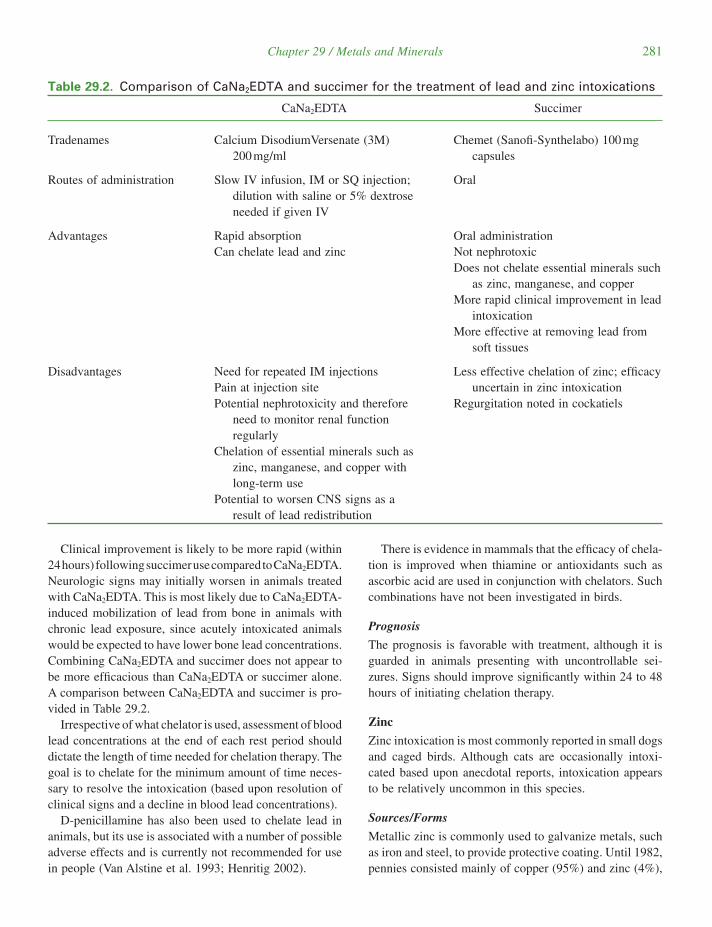
Clinical improvement is likely to be more rapid (within 24 hours) following succimer use compared to CaNa 2 EDTA. Neurologic signs may initially worsen in animals treated with CaNa 2 EDTA. This is most likely due to CaNa 2 EDTA - induced mobilization of lead from bone in animals with chronic lead exposure, since acutely intoxicated animals would be expected to have lower bone lead concentrations. Combining CaNa 2 EDTA and succimer does not appear to be more effi cacious than CaNa 2 EDTA or succimer alone. A comparison between CaNa 2 EDTA and succimer is pro-vided in Table 29.2 .
Irrespective of what chelator is used, assessment of blood lead concentrations at the end of each rest period should dictate the length of time needed for chelation therapy. The goal is to chelate for the minimum amount of time neces-sary to resolve the intoxication (based upon resolution of clinical signs and a decline in blood lead concentrations).
D - penicillamine has also been used to chelate lead in animals, but its use is associated with a number of possible adverse effects and is currently not recommended for use in people (Van Alstine et al. 1993 ; Henritig 2002 ).
Table 29.2. Comparison of C a N a 2 EDTA and succimer for the treatment of lead and zinc intoxications
CaNa 2 EDTA Succimer
Tradenames Calcium DisodiumVersenate (3M) 200 mg/ml
Chemet (Sanofi - Synthelabo) 100 mg capsules
Routes of administration Slow IV infusion, IM or SQ injection; dilution with saline or 5% dextrose needed if given IV
Oral
Advantages Rapid absorption Can chelate lead and zinc
Oral administration Not nephrotoxic Does not chelate essential minerals such
as zinc, manganese, and copper More rapid clinical improvement in lead
intoxication More effective at removing lead from
soft tissues
Disadvantages Need for repeated IM injections Pain at injection site Potential nephrotoxicity and therefore
need to monitor renal function regularly
Chelation of essential minerals such as zinc, manganese, and copper with long - term use
Potential to worsen CNS signs as a result of lead redistribution
Less effective chelation of zinc; effi cacy uncertain in zinc intoxication
Regurgitation noted in cockatiels
282 Section 3 / Specifi c Toxicants
cells, and brain, although specifi c mechanisms for many of these effects have not elucidated. In acute cases of zinc poisoning, particularly when zinc salts are ingested, local corrosive effects occur in the gastrointestinal tract fol-lowed by damage to the liver, kidney, and pancreas. The hemolytic anemia associated with zinc intoxication might be due to a direct damaging effect of zinc on red blood cell membranes. In birds, a major concern is chronic zinc toxicosis with resulting anemia. Anemia might be second-ary to functional iron or copper defi ciencies. Zinc has been shown to cause acute pancreatic, hepatic, and renal failure, although underlying specifi c pathophysiological mecha-nisms have not been described. Zinc toxicosis has been associated with brain damage that is most likely a combi-nation of hypoxic and direct toxic effects.
Clinical Effects
Signs
In intoxicated dogs, the most common signs include anorexia, vomiting, diarrhea, lethargy, depression, pale mucous membranes, icterus, and orange - tinged feces. Often metallic objects are noted on abdominal radio-graphs, although the absence of such fi ndings does not rule out zinc intoxication. Arrhythmias and ST - segment abnor-malities have been reported (Talcott 2004 ).
In birds clinical signs of intoxication are variable and nonspecifi c. They include lethargy, anorexia, regurgita-tion, polyuria, polydipsia, hematuria, hematochezia, pallor, dark or bright green diarrhea, foul - smelling feces, paresis, seizures, and sudden death (Puschner and Poppenga 2009 ). However, zinc toxicosis was associated with sudden death in 7 of 21 psittacine birds evaluated in one study (Puschner et al. 1999 ). Therefore, any acute death in a caged bird needs to be evaluated for possible zinc poisoning. Exces-sive zinc exposure as a cause of feather picking is questionable.
Laboratory
In dogs, fi ndings indicative of an oxidant - induced hemo-lytic anemia predominate. These include Heinz body for-mation, target cells, spherocytosis, hemoglobinemia, hemoglobinuria, and bilirubinemia. Regenerative changes such as nucleated RBCs, basophilic stippling, and poly-chromasia might be noted.
Diagnostics
Antemortem
Antemortem diagnosis relies on a history of ingestion of zinc, the occurrence of compatible clinical signs (e.g.,
but the copper - clad pennies minted after 1982 contain 97% zinc and 2.5% copper (Barceloux 1999 ). Additionally, zinc is found in soil and may be present at high enough con-centrations to result in avian poisonings. Zinc is also used in a variety of medical formulations, pigments, wood pre-servatives, insecticides, and rubber. Zinc oxide ointments and creams can be licked from the skin following topical application or cause intoxication if the products are chewed and the contents swallowed (Talcott 2004 ).
Kinetics
The rate of absorption depends on the amount and form of ingested zinc. The acidity of the stomach provides an excellent environment for the quick release and dissolu-tion of zinc from metallic objects (Talcott 2004 ). Once absorbed, zinc is distributed widely to tissues, including pancreas, liver, kidney, bone, muscle, brain, retina, and skin. In tissues, especially the pancreas, liver, kidney, intestinal mucosa, and brain, zinc is bound to metallothio-nein. Metallothionein is a low molecular weight, cysteine - rich protein that has potent metal - binding capabilities. Zinc has a high binding affi nity for metallothionein, which may play an integral role in zinc metabolism. The major route of excretion of zinc is via the feces.
Toxicity
Zinc is an essential metal, and animals and humans have the ability to regulate zinc effectively. Zinc is relatively nontoxic in mammals as judged by their tolerance to dietary concentrations greater than 100 times the minimum recommended daily zinc requirement (Leonard and Gerber 1989 ). If dietary exposure is excessive and homeostatic mechanisms fail, zinc toxicity can occur. Defi nite data on the toxicity of zinc in caged birds is lacking, although there is limited information available for certain avian and mam-malian species. In dogs, the ingestion of one or two pennies is likely suffi cient to cause intoxication. One report esti-mated that the toxic dose of zinc in the form of zinc oxide for dogs was 108 grams of zinc (Breitschwerdt et al. 1986 ).
Mechanism of Action
Zinc is required for a large number of physiological pro-cesses including bone formation, immunity, keratogenesis, reproduction, growth, vision, wound healing, brain devel-opment, normal functioning of the central nervous system, and many other physiological processes (Talcott 2004 ). Major pathophysiological mechanisms of zinc intoxica-tion are attributed to direct and indirect toxic effects on the gastrointestinal tract, liver, kidney, pancreas, red blood
Chapter 29 / Metals and Minerals 283
Prognosis
The prognosis is good to guarded. In one retrospective study in dogs (N = 19), 17 dogs receiving treatment survived (one was euthanized without treatment and one was discharged but returned the next day in severe respi-ratory distress) (Gurnee and Drobatz 2007 ). Rapid removal of the source of zinc should result in progres-sive improvement over 2 to 3 days. The mean hospital stay in the retrospective study was 2 days. Multiple organ failure, DIC, pancreatic disease, renal failure, and car-diopulmonary arrest are potential complications (Talcott 2004 ).
REFERENCES
Barceloux , D.G. 1999 . Zinc . J Toxicol — Clin Toxicol 37 : 279 – 292 .
Boyer , I.J. , Cory - Slechta , D.A. , DiStefano , V. 1985 . Lead induction of crop dysfunction in pigeons through a direct action on neural or smooth muscle components of crop tissue . J Pharmacol Exp Ther 234 : 607 – 615 .
Breitschwerdt , E.B. , Armstrong , P.J. , Robinette , C.L. 1986 . Three cases of acute zinc toxicosis in dogs . Vet Hum Toxicol 28 : 109 – 117 .
Casteel , S.W. 2004 . Lead . In Peterson , M.E. , Talcott , P.A. , eds. Small Animal Toxicology , 2nd ed. St. Louis , Saunders Elsevier , pp. 795 – 805 .
Dumonceaux , G. and Harrison , G.H. 1994 . Toxins . In Ritchie , B.W. , Harrison , G.J. , Harrison , L.R. , eds. Avian Medicine: Principles and Application . Delray Beach, Florida : Wingers Publishing , pp. 1030 – 1052 .
Ensley , S. 2004 . Arsenic . In Plumlee , K.H. , ed. Clinical Vet-erinary Toxicology . Mosby , St. Louis , pp. 193 – 195 .
Evinger , J.V. and Blakemore , J.C. 1984 . Dermatitis in a dog associated with exposure to an arsenic compound . J Am Vet Med Assoc 84 ( 10 ): 1281 – 1282 .
Ford , M. 2006 . Arsenic . In Flomenbaum , N.E. , Howland , M.A. , Goldfrank , L.R. et al., eds. Goldfrank ’ s Toxicologic Emergencies . New York , McGraw - Hill , pp. 1251 – 1264 .
Garza , A. , Vega , R. , Soto , E. 2006 . Cellular mechanisms of lead neurotoxicity . Med Sci Monit 12 : RA57 – 65 .
Gurnee , C.M. , Drobatz , K.J. 2007 . Zinc intoxication in dogs: 19 cases (1991 – 2003) . J Am Vet Med Assoc 230 ( 8 ): 1174 – 1179 .
Gwaltney - Brant , S. 2004 . Lead . In Plumlee , K.H. , ed. Clini-cal Veterinary Toxicology . Mosby , St. Louis , pp. 204 – 210 .
Hall , J.O. 2007a . Iron . In Peterson , M.E. , Talcott , P.A. , eds. Small Animal Toxicology , 2nd ed. St. Louis , Saunders Else-vier , pp. 777 – 784 .
— — — . 2007b . Iron toxicity . In Tilley , L.P. , Smith , F.W.K. , eds. Blackwell ’ s Five Minute Veterinary Consult , 4th ed. Ames, Iowa : Blackwell Publishing , p. 771 .
hemolytic anemia in conjunction with a metallic object in the GI tract), and measurement of zinc in serum samples. Serum zinc concentrations typically exceed 5 ppm and are often much higher (for dogs and cats a serum zinc refer-ence range is 0.7 to 2.0 ppm). Because many rubbers can leach zinc, it is important to use appropriate serum tubes (i.e., royal blue top) for sample collection and submission or scrupulously avoid contact of the blood/serum samples with rubber (e.g., a plastic - capped vial can be used to store and ship serum). Once exposure to zinc is stopped, follow - up determinations of serum zinc concentrations might be useful.
Postmortem
A postmortem diagnosis relies on antemortem fi ndings and measurement of zinc concentrations in appropriate tissue samples. Liver is the most commonly tested sample, although kidney and pancreatic tissue samples can also be used.
Differential Diagnoses
Differential diagnoses include immune - mediated hemo-lytic anemia, Babesia , onion/garlic, naphthalene mothball, some mushroom and acetaminophen intoxications, snake and brown recluse spider bites, caval syndrome, overhy-dration, skunk spray, and numerous causes of gastrointes-tinal signs.
Management of Exposures
The most important intervention is rapid removal of any identifi ed zinc object by endoscopy or laparotomy. Severe hemolytic anemias might require blood transfu-sions. In one case series of 19 dogs with zinc intoxica-tion, all animals received either packed red blood cells or an oxygen - carrying solution (Oxyglobin ® ) (Gurnee and Drobatz 2007 ). Fluid therapy and diuresis are impor-tant to both maintain hydration and minimize renal damage from hemoglobinuria. Although CaNa 2 EDTA and D - penicillamine can chelate zinc, their routine use is questionable, given the relatively rapid elimination of zinc. Once exposure to zinc is stopped, tissue and serum zinc concentrations drop rapidly. H 2 - receptor blockers might help reduce stomach acidity and the rate of release of zinc from zinc objects, although rapid removal of the zinc object might make their use unnecessary.
Frequent evaluation of PCV and ECGs is warranted. Periodic assessment of renal function might also be prudent.
284 Section 3 / Specifi c Toxicants
Neiger , R.D. and Osweiler , G.D. 1989 . Effect of subacute low level dietary sodium arsenite on dogs . Fund Appl Toxicol 13 : 439 – 451 .
Perrone , J. 2007 . Iron . In Flomenbaum , N.E. , Howland , M.A. , Goldfrank , L.S. et al., eds. Goldfrank ’ s Toxicologic Emer-gencies . New York , McGraw - Hill , pp. 629 – 637 .
Plumb , D.C. 2005 . Thiacetarsamide sodium . In Plumb ’ s Vet-erinary Drug Handbook , 5th ed. Ames , Blackwell Publish-ing , pp. 747 – 749 .
Poppenga , R.H. 2007 . Lead poisoning . In Tilley , L. P., Smith , F.W.K. , eds. Blackwell ’ s Five Minute Veterinary Consult , 4th ed. Ames , Blackwell Publishing , pp. 796 – 797 .
Puschner , B. and Poppenga , R.H. 2009 . Lead and zinc intoxi-cation in companion birds . Compend Contin Educ Vet 31 ( 1 ): E1 – 12 .
Puschner , B. , St. Leger , J. , Galey , F.D. 1999 . Normal and toxic zinc concentrations in serum/plasma and liver of psit-tacines with respect to genus differences . J Vet Diagn Invest 11 : 522 – 527 .
Ramsey , D.R. , Casteel , S.W. , Fagella , A.M. et al. 1996 . Use of orally administered succimer (meso - 2,3 - dimercaptosuccinic acid) for treatment of lead poisoning in dogs . J Am Vet Med Assoc 208 ( 3 ): 371 – 375 .
Talcott , P.A. 2004 . Zinc . In Peterson , M.E. , Talcott , P.A. , eds). Small Animal Toxicology , 2nd ed. St. Louis , Saunders Else-vier , pp. 1094 – 1100 .
Van Alstine , W.G. , Wickliffe , L.W. , Everson , R.J. et al. 1993 . Acute lead toxicosis in a household of cats . J Vet Diagn Invest 5 : 496 – 498 .
Henritig , F.M. 2002 . Lead . In Goldfrank , L.R. , Flomenbaum , N.E. , Lewin , N.A. , eds., Goldfrank ’ s Toxicologic Emergen-cies . Mew York : McGraw - Hill pp. 1200 – 1238 .
Hoogesteijn , A.L. , Raphael , B.L. , Callem P. et al. 2003 . Oral treatment of avian lead intoxication with meso - 2,3 - dimercaptosuccinic acid . J Zoo Wildl Med 34 : 82 – 87 .
Knight , T.E. , Kent , M. , Junk , J.E. 2001 . Succimer treatment of lead toxicosis in two cats . J Am Vet Med Assoc 218 ( 12 ): 1946 – 1948 .
Knight , T.E. , Kumar , M.S.A. 2003 . Lead toxicosis in cats — A review . J Feline Med and Surg 5 : 249 – 255 .
Leonard , A. , Gerber , G.B. 1989 . Zinc toxicity — Does it exist? J Am Coll Toxicol 8 : 1285 – 1290 .
Liu , J. , Goyer , R.A. , Waalkes , M.P. 2008 . Toxic effects of metals . In Klaassen , C.D. , ed. Casarett and Doull ’ s Toxicol-ogy: The Basic Science of Poisons . New York : McGraw Hill , pp. 931 – 979 .
Locke , L.N. , Thomas , N.J. 1996 . Lead poisoning of water-fowl and raptors . In Fairbrother , A. , Locke , L.N. , Hoff , G.L. , eds. Noninfectious Diseases of Wildlife . Ames : Iowa State University Press, Ames , pp. 108 – 117 .
Loudis , B. 2004 . Endoscope assisted gastric lavage for foreign body retrieval . Association of Avian Veterinarians 83 – 88 .
Medlin , J. 2004 . Sweet candy, bitter poison . Environ Health Perspect 112 : A803 .
Neiger , R.D. 2004 . Arsenic . In Peterson , M.E. , Talcott , P.A. (eds). Small Animal Toxicology , 2nd ed. St. Louis , Saunders Elsevier , pp. 592 – 602 .
— — — . 2007 . Arsenic . In Tilley , L. P., Smith , F.W.K. , eds. Blackwell ’ s Five Minute Veterinary Consult , 4th ed. Ames , Blackwell Publishing , p. 101 .