slm microscope
TRANSCRIPT

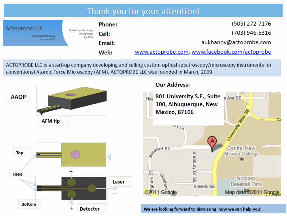
Actoprobe Scanning Laser Confocal Microscope

SLM Overview
Designed using top class off-the-shelf components Fully automated Fast Imaging capabilities:4 fp/sec to 30 fp/sec Optical sectioning available up to 100 µm Field of scan up to 280 µms Suitable for wavelengths from 300 to 700 nm Compatible with most AFMs Compatible with upright and inverted microscope configurations
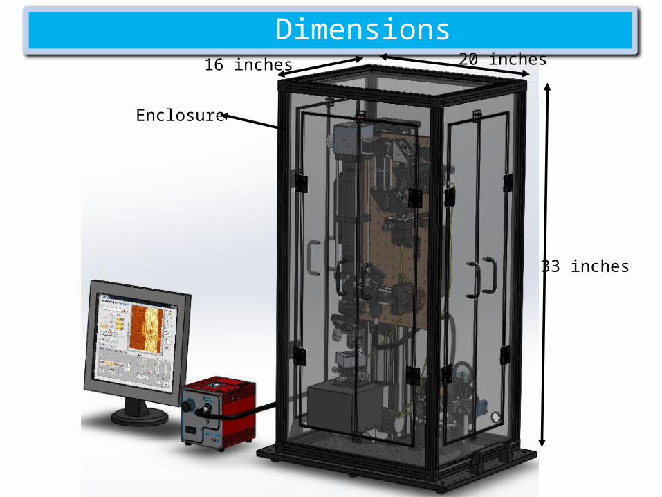
Dimensions
33 inches
20 inches16 inches
Enclosure

Enclosure
System offers very durable and sturdy
enclosure to protect the microscope from airborne noise and
dust
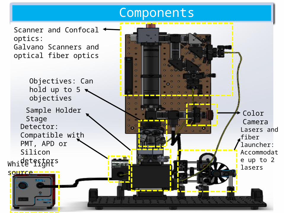
ComponentsScanner and Confocal optics:Galvano Scanners and optical fiber optics
Color Camera
Objectives: Can hold up to 5 objectives
Sample Holder Stage
Lasers and fiber launcher: Accommodate up to 2 lasers
Detector: Compatible with PMT, APD or Silicon detectors
White light source

Confocal System
1kHz or 8kHz Scanners 4 fp/sec to 30fp/sec acquisition rate at 512X512 pixel resolution Confocal aperture diameters up to 1.8 µm Fiber coupled: Suitable for wavelengths from 300 to 700 nm Suitable for objectives from 10X to 100X Suitable for PMT, APD or Silicon detectors Field of View up to 280X280 µm Lateral resolution up to 270 nm and axial resolution up to 370 nm
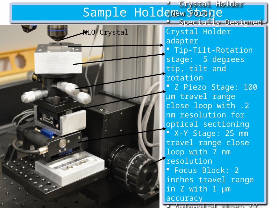
Sample Holder Stage
Crystal Holder (New Port) Specially Designed Crystal Holder adapter Tip-Tilt-Rotation stage: 5 degrees tip, tilt and rotation Z Piezo Stage: 100 µm travel range close loop with .2 nm resolution for optical sectioning X-Y Stage: 25 mm travel range close loop with 7 nm resolution Focus Block: 2 inches travel range in Z with 1 µm accuracy Automated stage (X-Y-Z) controlled through JoyStick
NLO Crystal
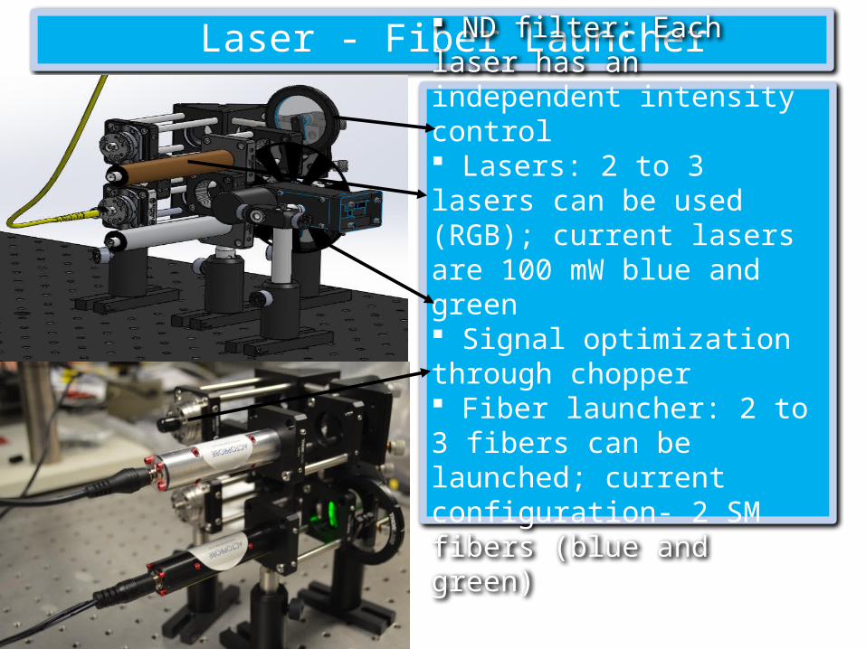
Laser - Fiber Launcher
ND filter: Each laser has an independent intensity control Lasers: 2 to 3 lasers can be used (RGB); current lasers are 100 mW blue and green Signal optimization through chopper Fiber launcher: 2 to 3 fibers can be launched; current configuration- 2 SM fibers (blue and green)

Software: Data Acquisition GUI

Sample Holder Stage GUI

Camera GUI

c Data Analysis GUI

Quick Data Plotter GUI

Optical sectioning of a laser burn on mirror surface
Camera Image
3D Image constructed from individual slices
Slice at the top surface
Slice at 1.5 µm depthSlice at 1 µm depth
Slice at 2.5 µm depthSlice at 2 µm depth
Slice at 500nm depth
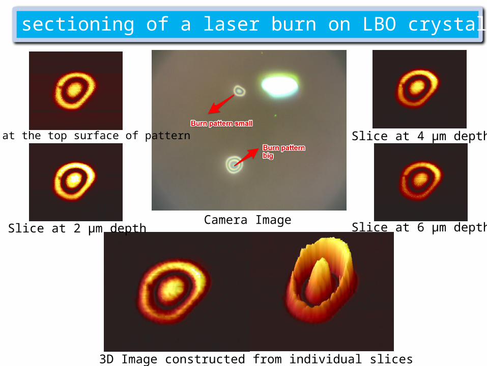
Optical sectioning of a laser burn on LBO crystal surface
Camera Image
3D Image constructed from individual slices
Slice at the top surface of pattern Slice at 4 µm depth
Slice at 2 µm depth Slice at 6 µm depth

Optical sectioning of a laser burn on LBO crystal surface
Camera Image
3D Image constructed from individual slices
Slice at the top surface of pattern Slice at 600 nm depth
Slice at 400 nm depth Slice at 800 nm depth