skin histology
TRANSCRIPT
Dr Prennie VidieraFr Muller Homoeopathic Medical College & Hospital
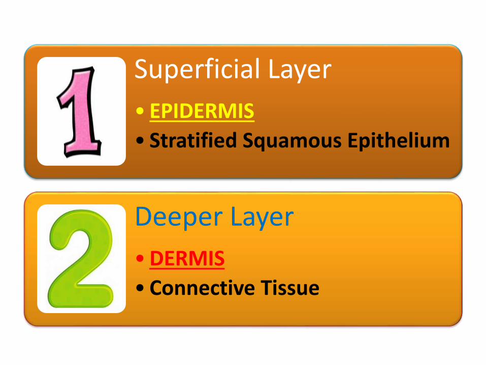
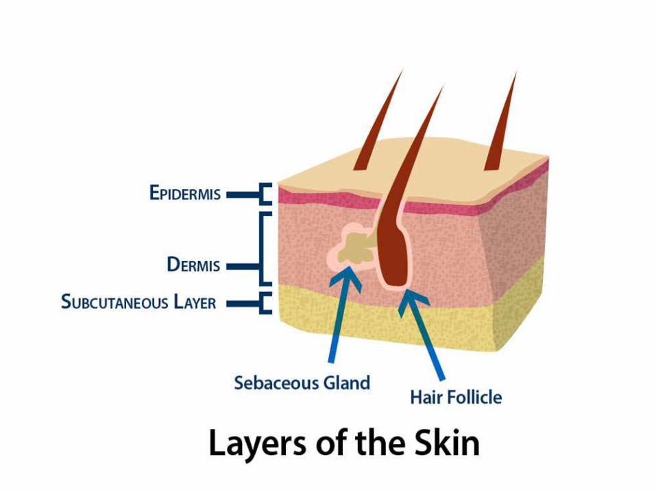
Superficial Layer
• EPIDERMIS
• Stratified Squamous Epithelium
Deeper Layer
•DERMIS
• Connective Tissue
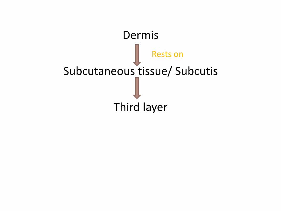
Dermis
Rests on
Subcutaneous tissue/ Subcutis
Third layer
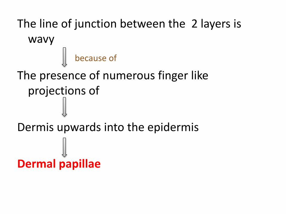
The line of junction between the 2 layers is wavy
because of
The presence of numerous finger like projections of
Dermis upwards into the epidermis
Dermal papillae
The downward projections of the epidermis
Epidermal papillae
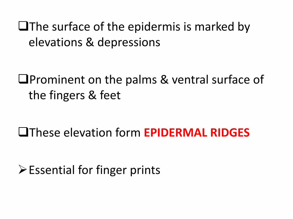
The surface of the epidermis is marked by elevations & depressions
Prominent on the palms & ventral surface of the fingers & feet
These elevation form EPIDERMAL RIDGES
Essential for finger prints
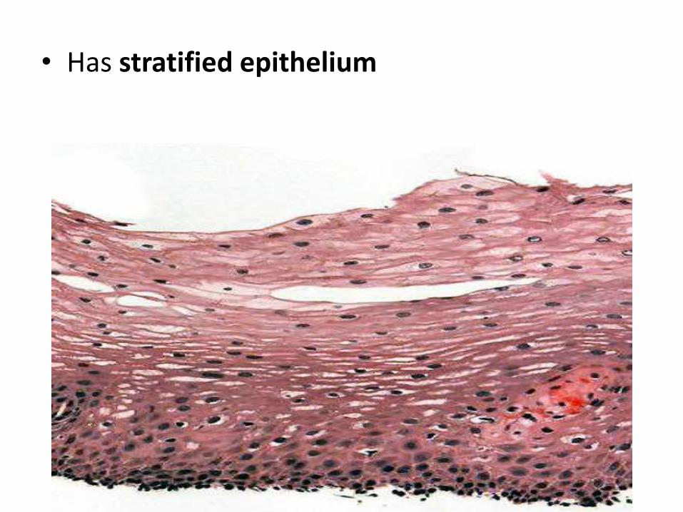
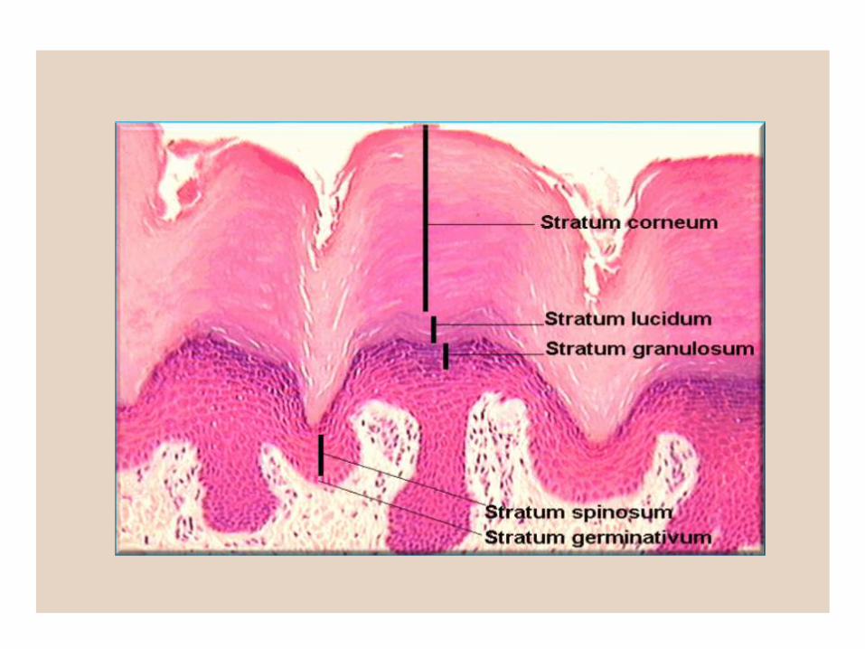
• Has stratified epithelium
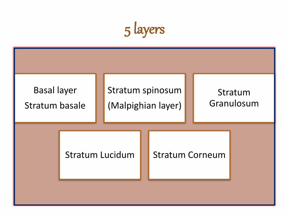
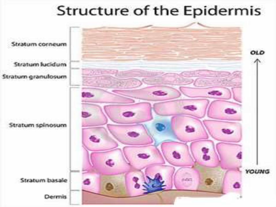
5 layers
Basal layer
Stratum basale
Stratum spinosum
(Malpighian layer)
Stratum Granulosum
Stratum Lucidum Stratum Corneum
Stratum Basale
• Deepest
• Single layer of columnar cells
• Undergo mitosis & give off cells called Keratinocytes
also known as
Germinal layer/ Stratum Germinativum
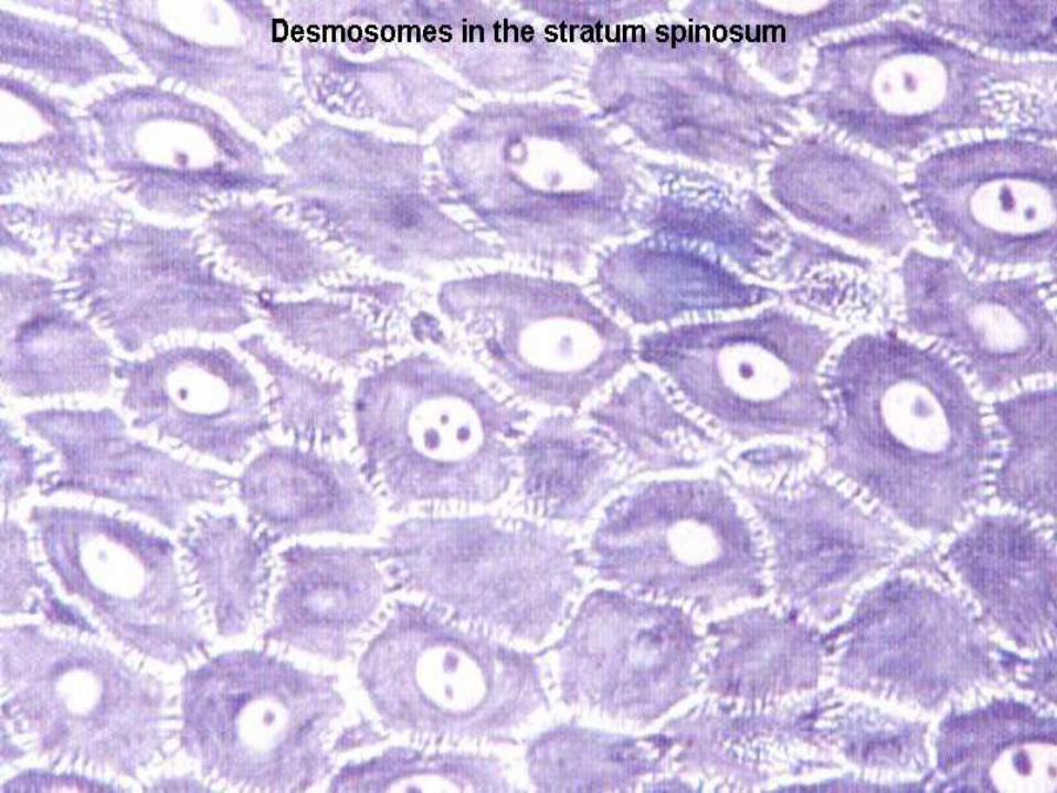
Stratum spinosum
• Several layers of polygonal keratinocytes
• Cells are attached to one another by numerous Desmosomes
• Some mitosis may occur in the deeper cells
• Germinative zone
Stratum Granulosum
• Overlies the stratum spinosa
• Few (1-5) layers of flattened cells (deeply staining)
• The granules consists of a protein called keratohyalin.
Stratum Lucidum
• Lucid=Clear
• Appears homogenous
• Cell boundaries extremely indistinct
• Traces of flattened nuclei
Stratum Corneum
• Superficial layer
• Acellular
• Made up of flattened scale like elements containing keratin filaments embedded in protein
• held together by a glue like material containing lipids & carbohydrates
• Resistant to permeability
• The thickness of this layer is greatest where the skin is exposed to maximal friction
• Eg:- Soles and palms
• The superficial layer constantly sheds off & replaced by proliferation of cells in deeper layers
Zone of Keratinization(Cornified Zone)
• Stratum Corneum
• Stratum Lucidum
• Stratum Granulosum
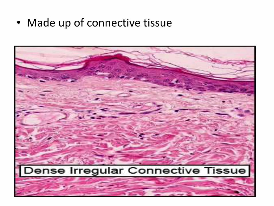
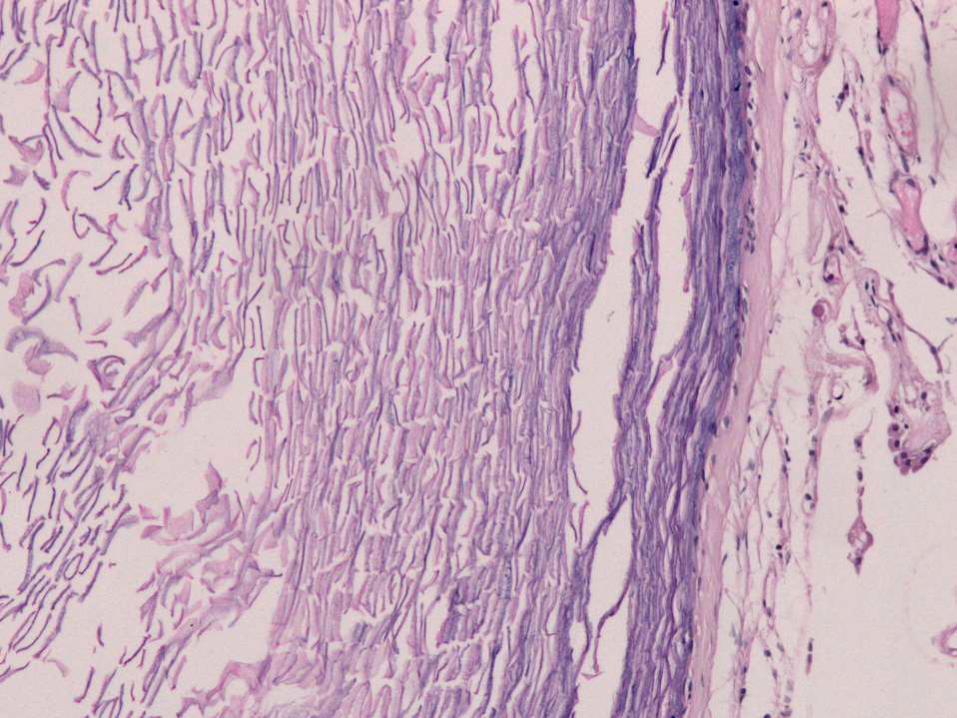
• Made up of connective tissue
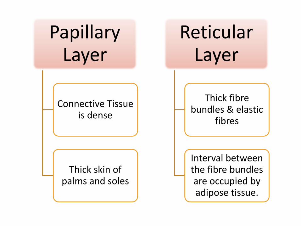
Papillary Layer
Connective Tissue is dense
Thick skin of palms and soles
Reticular Layer
Thick fibre bundles & elastic
fibres
Interval between the fibre bundles are occupied by adipose tissue.
Appendages of the skin
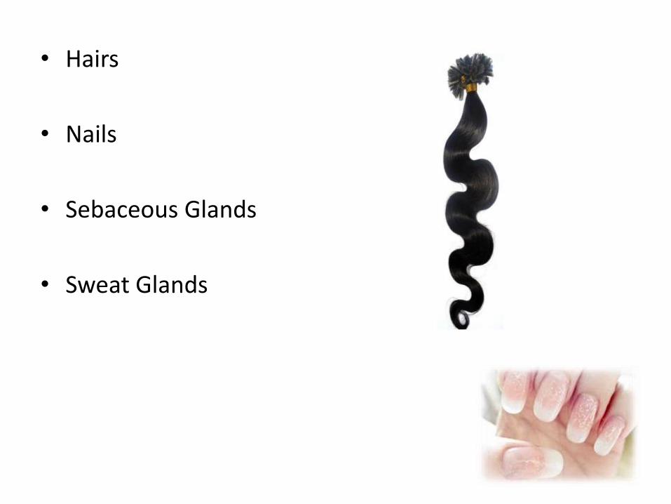
• Hairs
• Nails
• Sebaceous Glands
• Sweat Glands
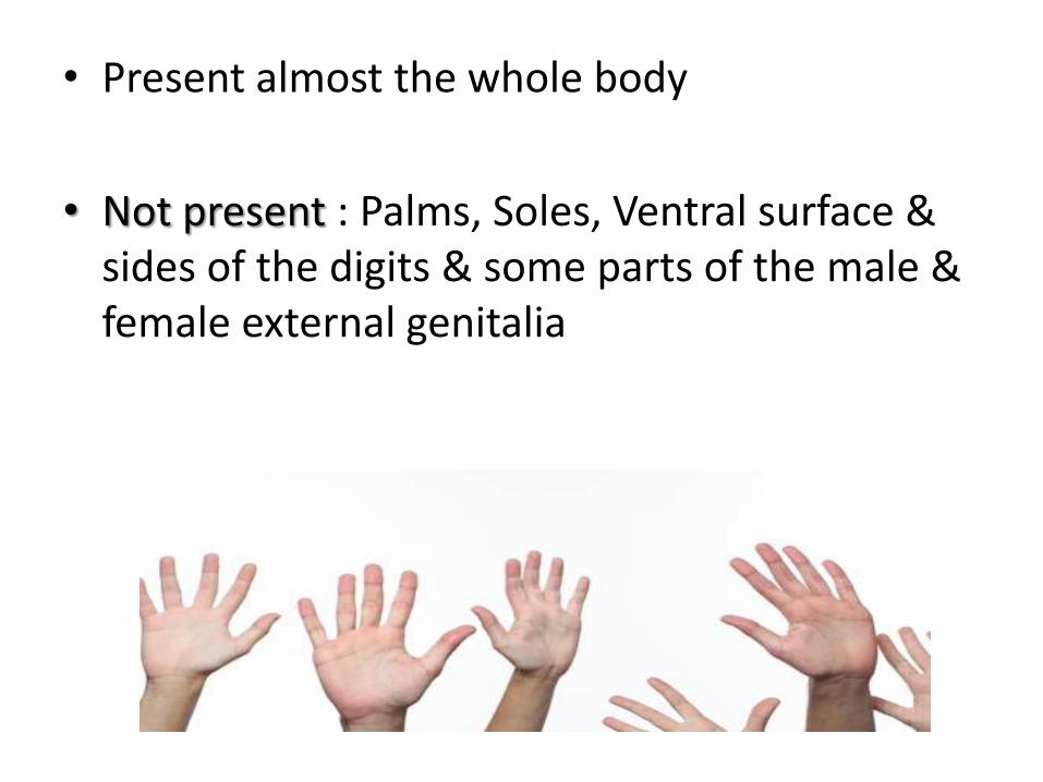
• Present almost the whole body
• Not present : Palms, Soles, Ventral surface & sides of the digits & some parts of the male & female external genitalia
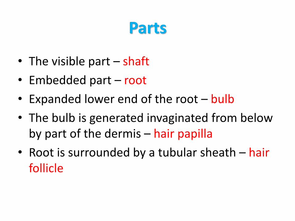
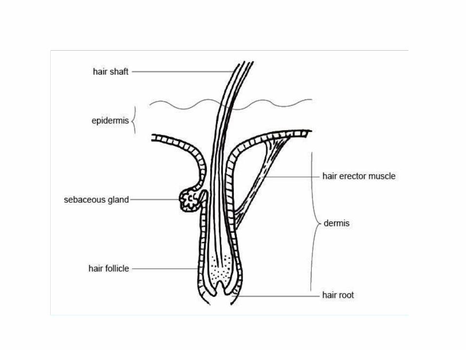
Parts
• The visible part – shaft
• Embedded part – root
• Expanded lower end of the root – bulb
• The bulb is generated invaginated from below by part of the dermis – hair papilla
• Root is surrounded by a tubular sheath – hair follicle
Structure of hair shaft
Hair – Modified part of stratum corneum
Has outer cortex and inner medulla in large hairs, no medulla in thin hairs
Cortex is acellular & is made up of keratin
Medulla consists of cornified cells of irregular shapes
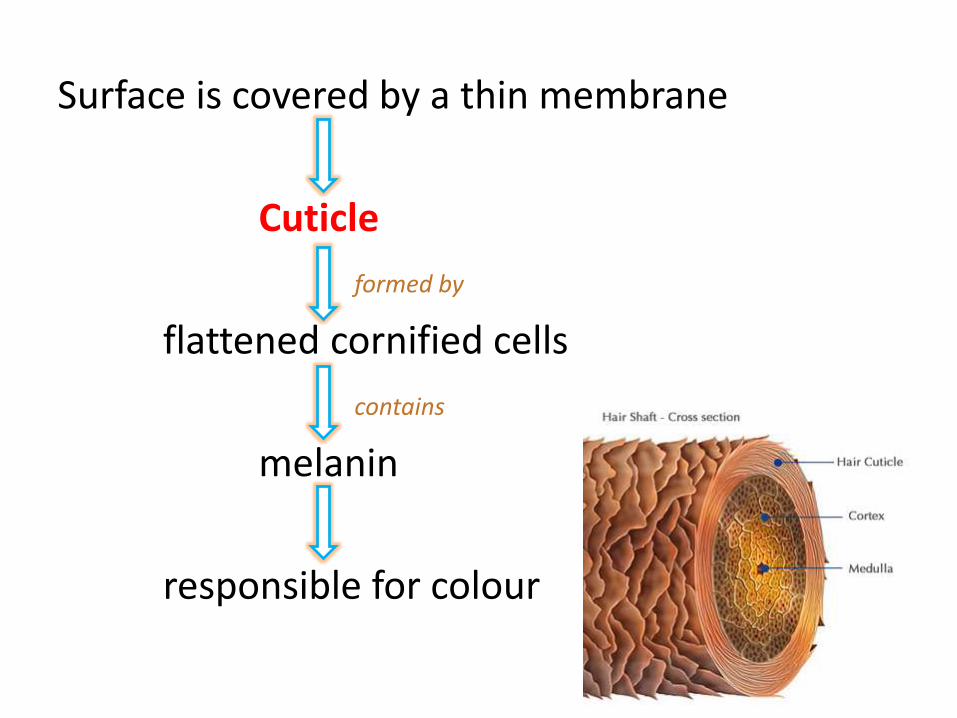
Surface is covered by a thin membrane
Cuticle
formed by
flattened cornified cells
contains
melanin
responsible for colour
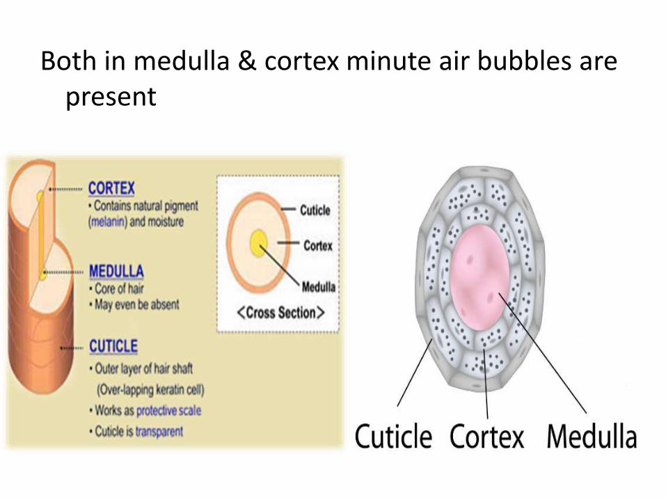
Both in medulla & cortex minute air bubbles are present

Structure of Hair Follicle
• Part of the epidermis that has been invaginated into the dermis around the hair root
• Its innermost layer continues with the surface of the skin & outermost layer continues with the dermis
Arrector Pili Muscles
• These are bands of smooth muscles attached at one end to the dermis, just below the dermal papilla &
• at the other end to the connective tissue sheath of the hair follicle

• Contraction of arector pilorum muscle results in erection of hair shaft
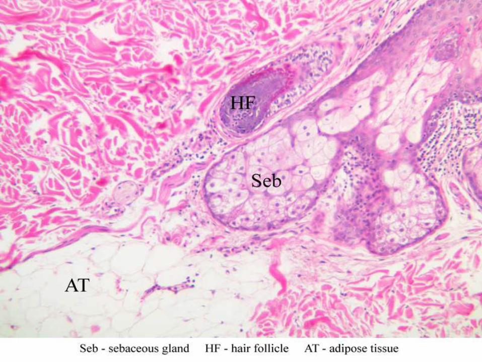
• Found in dermis of the skin
• Ducts open into the hair follicle
• Holocrine gland
• Secretes sebum: antifungal & anti bacterial. Contains lipids & cholesterol– Oily in nature
– Prevents dryness
– Resistant to moisture
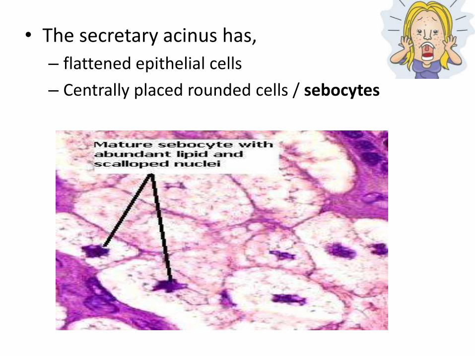
• The secretary acinus has,
– flattened epithelial cells
– Centrally placed rounded cells / sebocytes

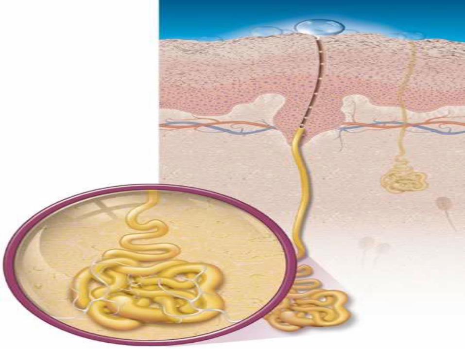
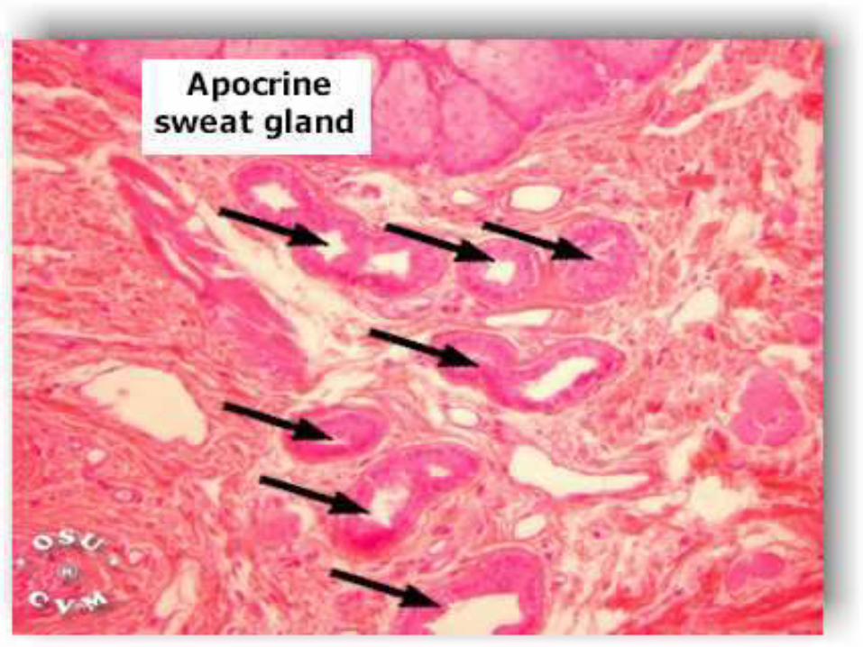
Sweat Glands
• Found in deeper parts of the dermis
The part of the duct present in the dermis is straight
Stratified cuboidal epithelium
the part passing through the dermis is coiled
epidermal cells

2 Types
Eccrine Glands
Apocrine Glands
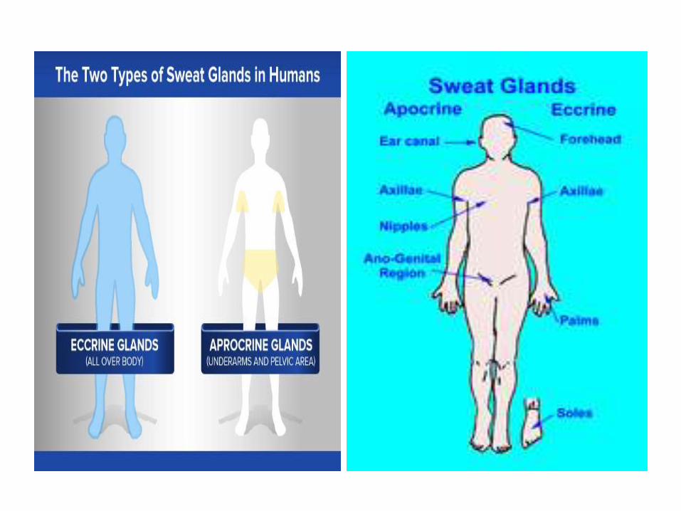
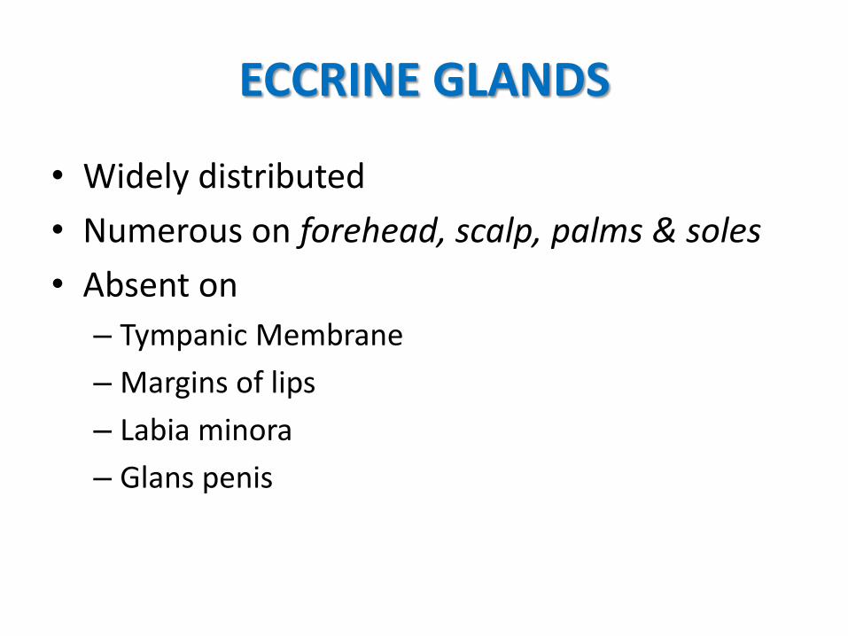
ECCRINE GLANDS
• Widely distributed
• Numerous on forehead, scalp, palms & soles
• Absent on
– Tympanic Membrane
– Margins of lips
– Labia minora
– Glans penis
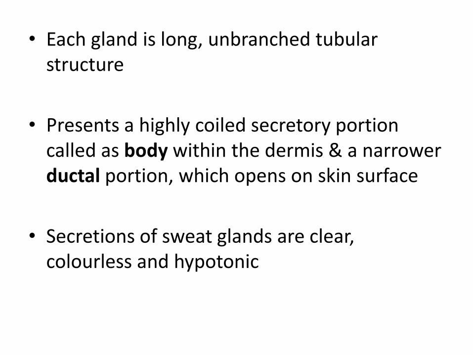
• Each gland is long, unbranched tubular structure
• Presents a highly coiled secretory portion called as body within the dermis & a narrower ductal portion, which opens on skin surface
• Secretions of sweat glands are clear, colourless and hypotonic
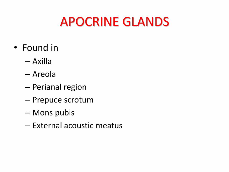
APOCRINE GLANDS
• Found in
– Axilla
– Areola
– Perianal region
– Prepuce scrotum
– Mons pubis
– External acoustic meatus
• These glands secrete a protein rich, milky fluid which is initially odourless but acquires a distinct odour due to bacterial decomposition
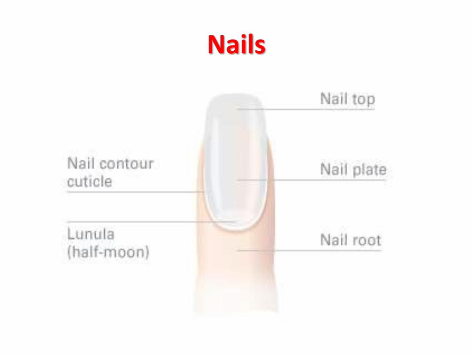
Nails
• Nails are plates of keratinised epithelial cells on dorsal surface of distal phalynx
• Consists of 3 parts
– Proximal part or root
– Exposed part or body
– Free distal border
• Consists of dead anucleate keratin
• Body rests on nail bed which is composed of stratum basale & stratum spinosum