skin cancer incidence
DESCRIPTION
Skin Cancer Incidence. Over 1 million new cases of BCC & SCC Basal Cell Carcinoma 4 to 5 times more common than Squamous Cell Carcinoma 2004 - Estimated 55,000 new cases of Melanoma Someone dies of Melanoma every hour. Why the rapid increase ?. Sun exposure habits - PowerPoint PPT PresentationTRANSCRIPT
Skin Cancer Incidence
• Over 1 million new cases of BCC & SCC
• Basal Cell Carcinoma 4 to 5 times more common than Squamous Cell Carcinoma
• 2004 - Estimated 55,000 new cases of Melanoma
• Someone dies of Melanoma every hour
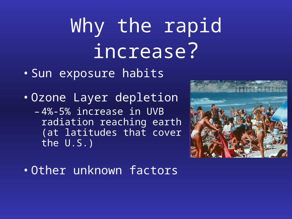
Why the rapid increase?
• Sun exposure habits
• Ozone Layer depletion– 4%-5% increase in UVB
radiation reaching earth (at latitudes that cover the U.S.)
• Other unknown factors

Causes of Skin Cancer
• Ultraviolet radiation– UVB (290nm - 320nm) most important– UVA (320nm - 400nm) more penetrating
• Ionizing radiation (X-rays)
• Chemicals (arsenic, coal tars)
Causes of Skin Cancer
• “Marjolin’s ulcers”
• Immunosuppression – Organ transplant patients – 10%-45% of transplant patients develop skin cancers– 2 to 3 times more SCCs than BCCs
• Human papillomavirus - HPV 16
• Inherited diseases - XP, BCNS, albinism
Basal Cell Carcinoma• Most common cancer in America• Usually seen in the middle-aged and elderly• Usually due to solar radiation • Most common locations
• Face - nose, cheeks, forehead, periocular• Ears• Neck• Trunk• Extremities
• Frequently develop another within 5 years
Basal Cell Carcinoma
• Subtypes– Nodulo-ulcerative (most common)– Pigmented– Morpheaform (sclerosing, infiltrative)– Micronodular– Metatypical (basosquamous)– Superficial (“multicentric”)
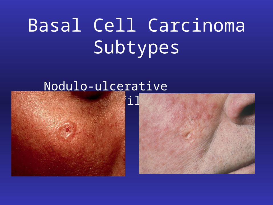
Basal Cell Carcinoma Subtypes
Nodulo-ulcerative Morpheaform/Infiltrative

Basal Cell Carcinoma
• Subtypes– Superficial “Multicentric”– Can be misdiagnosed as psoriasis or eczema
Basal Cell Carcinoma
• Course
– Slow progressive growth
– Bleeding, ulceration
– Enlarges over months to years
– Is capable of extensive tissue destruction
(invading into muscle, cartilage, and bone)
Basal Cell Carcinoma
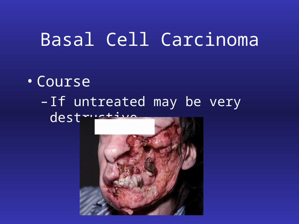
• Course– If untreated may be very destructive
Basal Cell Carcinoma
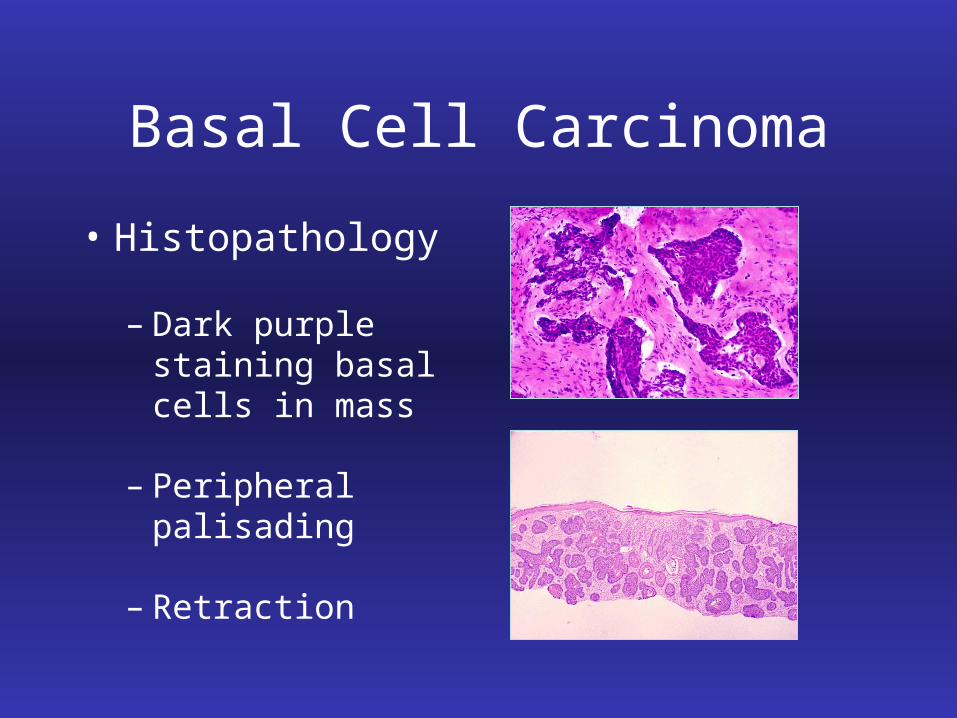
• Histopathology
– Dark purple staining basal cells in mass
– Peripheral palisading
– Retraction
Basal Cell Carcinoma
The Proverbial “Tip of the Iceberg”
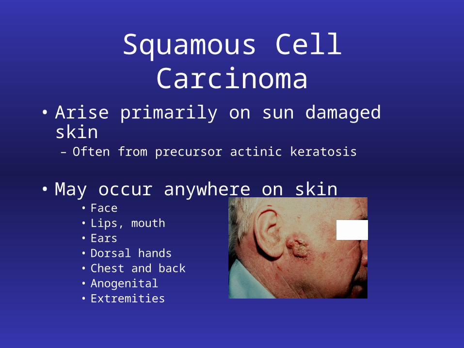
Squamous Cell Carcinoma
• Arise primarily on sun damaged skin– Often from precursor actinic keratosis
• May occur anywhere on skin• Face• Lips, mouth• Ears• Dorsal hands• Chest and back• Anogenital• Extremities
Squamous Cell Carcinoma
• Cases where SCCs > BCCs
– Immunocompromised patients
– Black patients
– On lips and dorsal hands
– PUVA treatment patients
Squamous Cell Carcinoma
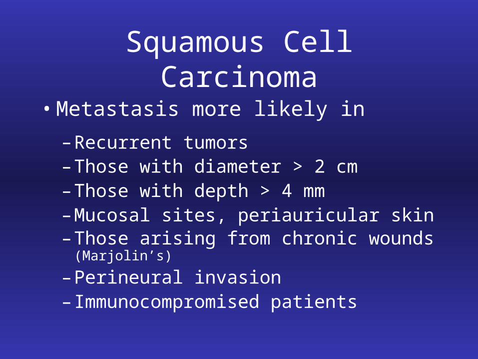
• Metastasis more likely in
– Recurrent tumors– Those with diameter > 2 cm– Those with depth > 4 mm– Mucosal sites, periauricular skin– Those arising from chronic wounds (Marjolin’s)
– Perineural invasion– Immunocompromised patients
Squamous Cell Carcinoma
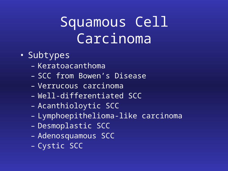
• Subtypes– Keratoacanthoma– SCC from Bowen’s Disease– Verrucous carcinoma– Well-differentiated SCC– Acanthioloytic SCC– Lymphoepithelioma-like carcinoma– Desmoplastic SCC– Adenosquamous SCC– Cystic SCC
Squamous Cell Carcinoma
• Keratoacanthoma• Initial rapid growth
• Exophytic nodule with central keratin-filled
crater
• Remains stable for a few months
• May spontaneously resolve
Squamous Cell Carcinoma
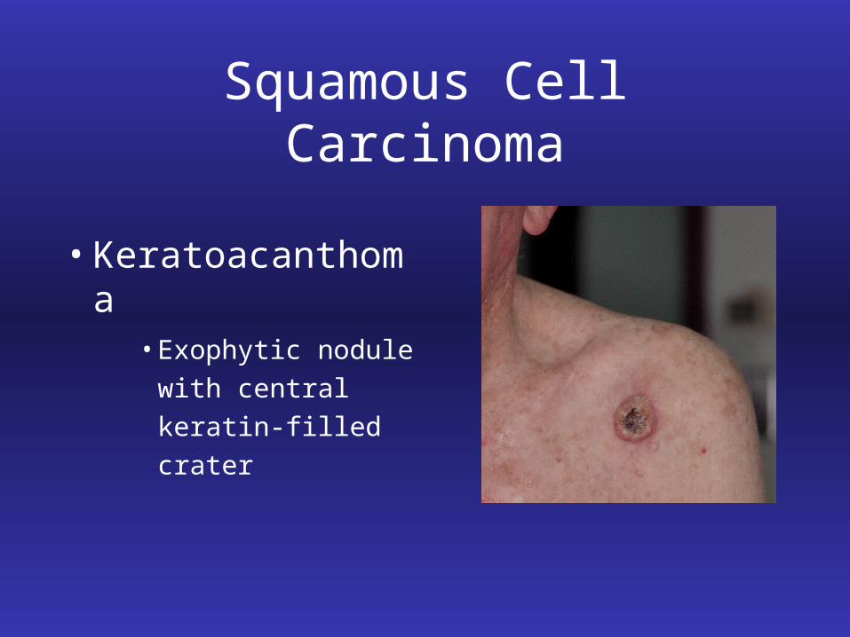
• Keratoacanthoma• Exophytic nodule
with central
keratin-filled crater
Squamous Cell Carcinoma
• Bowen’s Disease• Squamous cell carcinoma in-situ
• Thin, erythematous, scaling plaques
• Often progress into, and/or coincide with
invasive SCCs
• Can be misdiagnosed as psoriasis or eczema
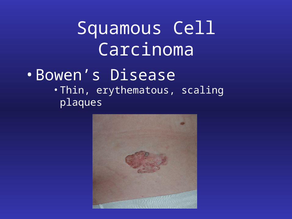
Squamous Cell Carcinoma
• Bowen’s Disease• Thin, erythematous, scaling plaques
Squamous Cell Carcinoma
• Verrucous Carcinoma• Exophytic, verrucous, or fungating tumor • Usually in genital or oral regions but also found
on the sole of the foot
• May be related to human papillomavirus
Treatment of BCC/SCC
• Electrodessication & Curettage
• Cryotherapy
• Radiation Therapy
• Surgical Excision
• Mohs Micrographic Surgery
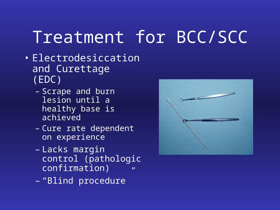
Treatment for BCC/SCC• Electrodesiccation
and Curettage (EDC)– Scrape and burn lesion
until a healthy base is achieved
– Cure rate dependent on experience
– Lacks margin control (pathologic confirmation)
– “Blind procedure”
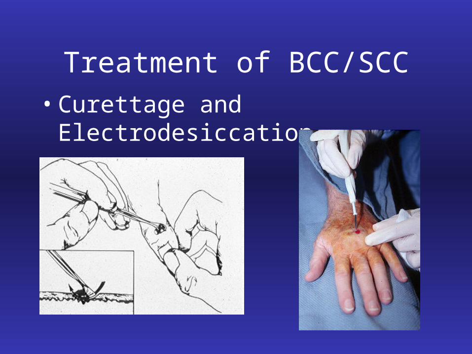
Treatment of BCC/SCC• Curettage and Electrodesiccation
Treatment of BCC/SCC
• Cryotherapy– Liquid nitrogen – Used frequently to destroy benign or
premalignant (AKs)
– May be used to treat malignancies
– Lacks margin control
– Method of blind destruction
Treatment of BCC/SCC
• Radiation Therapy– May be very effective in certain areas– Primary vs. adjuvant role (with surgery)– Requires multiple treatments over 4 to 8 weeks– Tumor may recur in more aggressive form– Used in certain patients, such as those unable to
tolerate surgery– Malignancies may develop w/in irradiated skin
Treatment of BCC/SCC
• Surgical Excision– Traditional excision with safety margins
• 3 mm to 5 mm margin
– Make ellipse and close in linear fashion– Larger lesions may require flaps or grafts– Common method of removing skin cancers– Approximately 90% cure rate
Treatment of BCC/SCC
• Mohs Micrographic Surgery– Highest cure rate (97% - 99%)– Spares healthy tissue– Evaluates the entire surgical margin
microscopically– Standard of care when:
• tumor is in critical location (cosmetic or functional)
• tumor is recurrent• tumor has ill-defined margins• tumor is large (> 2 cm) or aggressive
Mohs Micrographic Surgery
• Used on tumors with contiguous growth
• Precise microscopic margin control of tumor margins
• 100% of peripheral & deep margin examined
– Traditional vertical sections examine less than 1%
• Highest cure rate (97% - 99%)
• Most tissue conservation
Mohs Micrographic Surgery• Recurrent Tumors
– Tumors that have recurred after prior treatment
– Can be more aggressive than original tumor• More difficult to cure• Have even higher subsequent recurrence• More ill-defined• Have higher metastatic potential
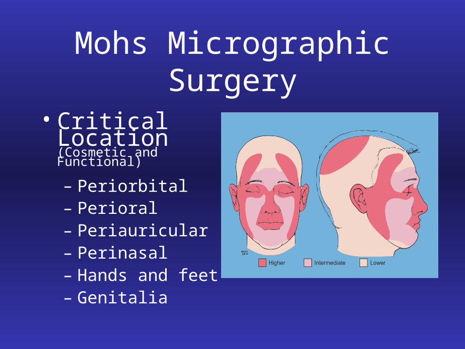
Mohs Micrographic Surgery • Critical Location
(Cosmetic and Functional)
– Periorbital– Perioral– Periauricular– Perinasal– Hands and feet– Genitalia
Mohs Micrographic Surgery
• Aggressive Histology– Infiltrating BCC– Micronodular BCC– Morpheaform BCC – Metatypical BCC– Perineural invasion– Poorly differentiated SCC– Acantholytic SCC
Mohs Micrographic Surgery
• Other Cutaneous Tumors– Dermatofibrosarcoma protuberans (DFSP)– Atypical fibroxanthoma (AFX)– Sebaceous carcinoma– Merkel cell carcinoma– Microcystic aonexal carcinoma– Verrucous carcinoma– Angiosarcoma
Mohs Surgery Advantages
• Highest Cure Rate– 97% - 99% for primary tumors
– 94% for recurrent tumors
– Entire margin evaluated microscopically
– Cure rates of other methods
• Standard excision 89.9 %
• Destruction 81% - 96%
• Radiation 91 %
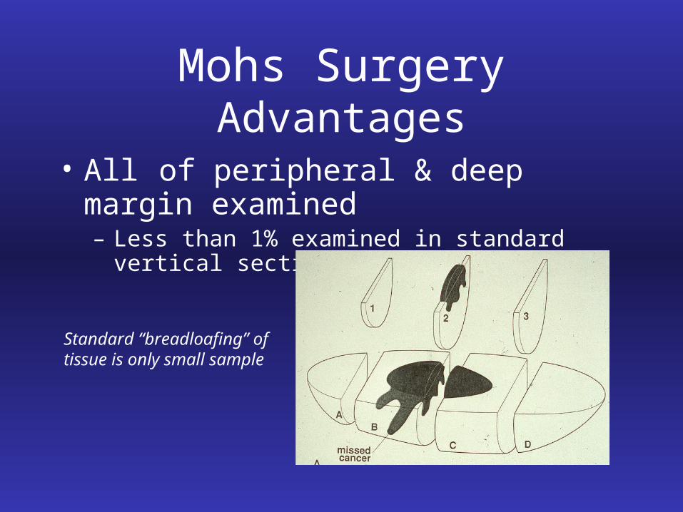
Mohs Surgery Advantages
• All of peripheral & deep margin examined– Less than 1% examined in standard vertical sections
Standard “breadloafing” of tissue is only small sample
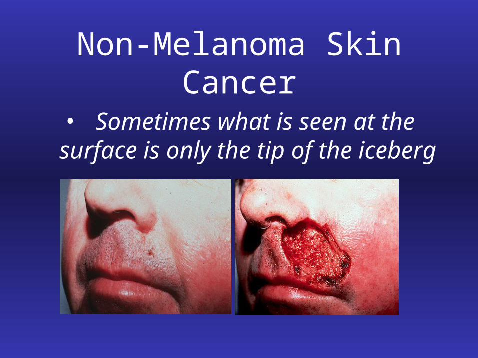
Non-Melanoma Skin Cancer
• Sometimes what is seen at the surface is only the tip of the iceberg
Mohs Surgery Advantages
• Tissue Conservation– All tumor roots are traced and removed– Preserves maximal amount of healthy skin– Smallest surgical defect possible – Smallest margin but greatest confidence– Standard excision takes guess at margins and excises additional tissue (3 mm - 5mm in each direction)
Mohs Surgery Advantages
• Cost Effective– Outpatient office setting, not OR
– Pathology reading included
– Local anesthesia rather than general
– Lowest recurrence rate
Mohs Surgery Advantages
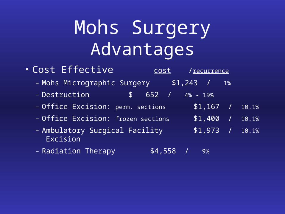
• Cost Effective cost /recurrence
– Mohs Micrographic Surgery $1,243 / 1%
– Destruction $ 652 / 4% - 19%
– Office Excision: perm. sections $1,167 / 10.1%
– Office Excision: frozen sections $1,400 / 10.1%
– Ambulatory Surgical Facility $1,973 / 10.1%
Excision
– Radiation Therapy $4,558 / 9%
Mohs Surgery Procedure
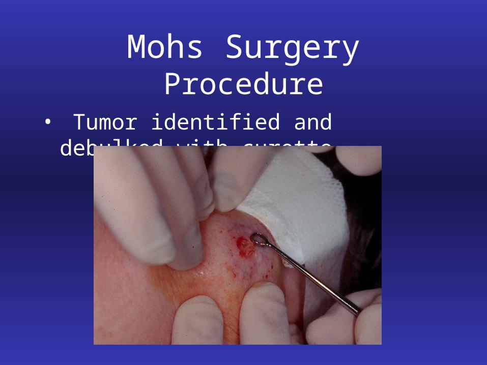
1) Tumor identified and debulked
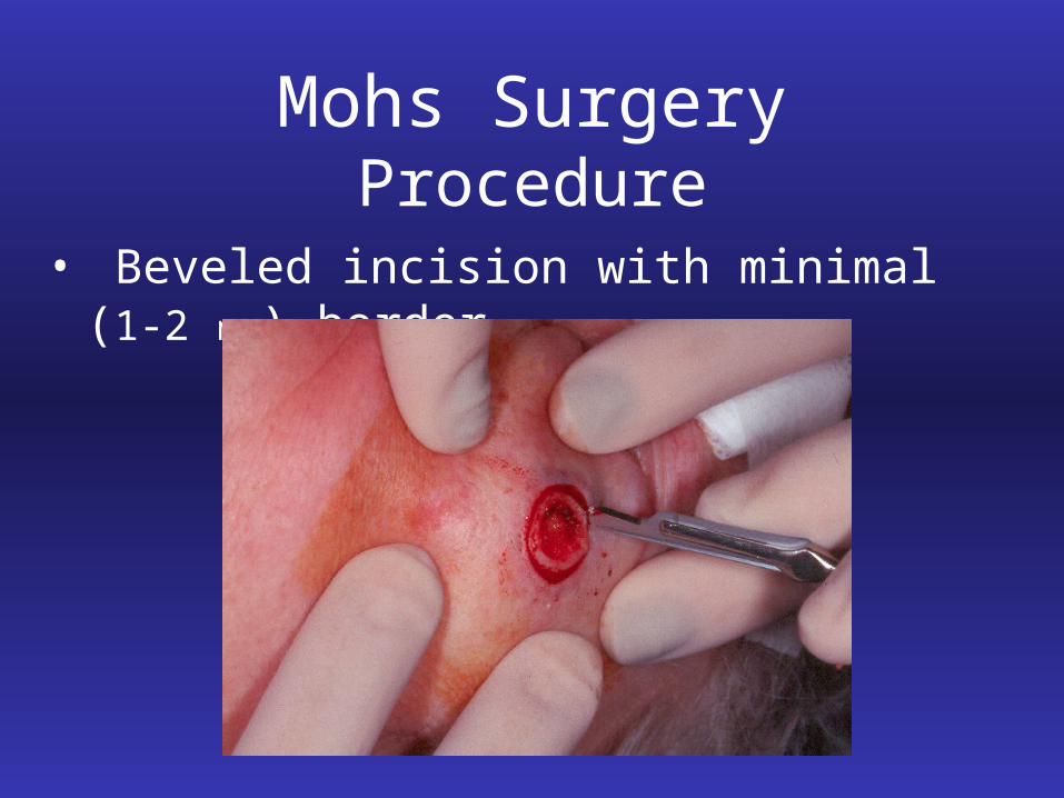
2) Saucer-shaped piece of tissue is excised with 1 mm – 2 mm margin around and underneath
curetted borders
3) The skin is marked for orientation
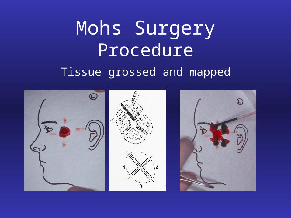
4) Excised tissue is color-coded and mapped by sections for orientation
Mohs Surgery Procedure
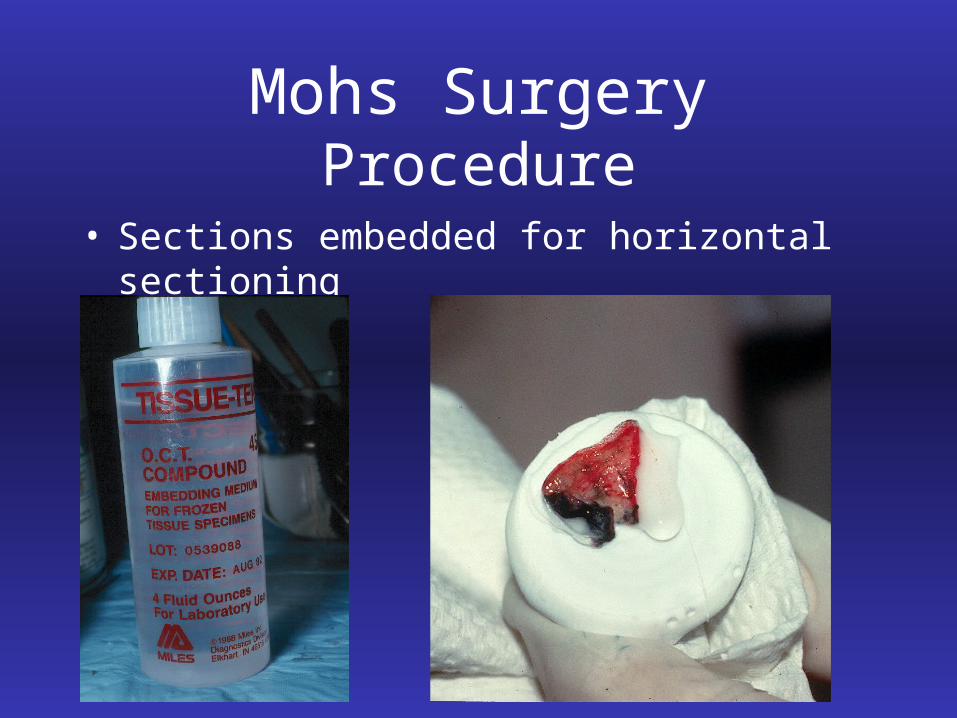
5) Tissue sections processed with frozen horizontal technique
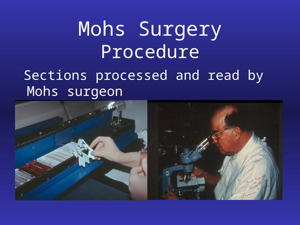
6) Mohs surgeon evaluates slides for residual tumor
Mohs Surgery Procedure
7) If residual tumor found, it is marked on map with proper orientation
8) Second Mohs layer taken only in positive area
9) Process repeated until margins clear
10) Defect repaired with appropriate technique
Mohs Surgery Procedure
• Tumor identified and debulked with curette
Mohs Surgery Procedure
• Beveled incision with minimal (1-2 mm) border
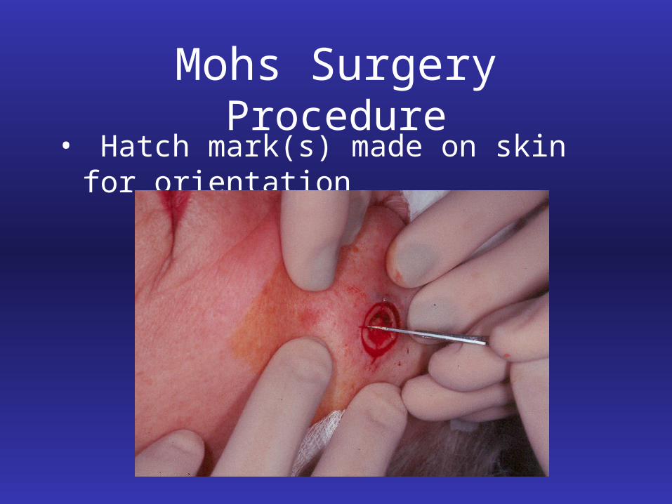
Mohs Surgery Procedure• Hatch mark(s) made on skin for orientation
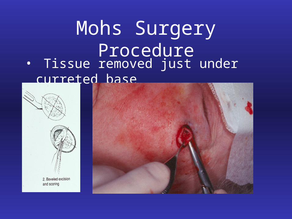
Mohs Surgery Procedure• Tissue removed just under curreted base
Mohs Surgery Procedure
Tissue grossed and mapped
Mohs Surgery Procedure
• Sections embedded for horizontal sectioning
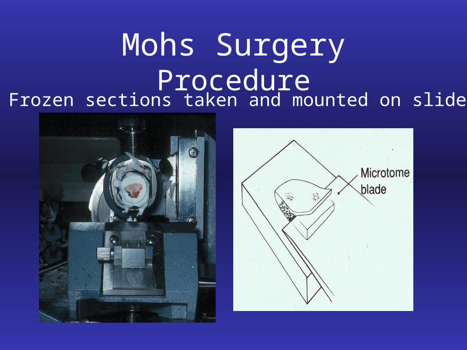
Mohs Surgery Procedure Frozen sections taken and mounted on slide
Mohs Surgery Procedure
Sections processed and read by Mohs surgeon
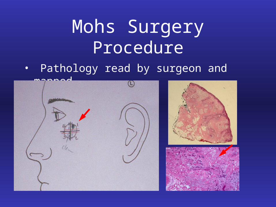
Mohs Surgery Procedure
• Pathology read by surgeon and mapped
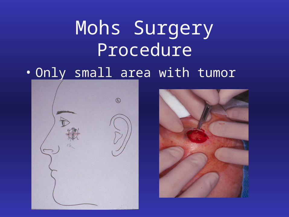
Mohs Surgery Procedure
• Only small area with tumor re-excised
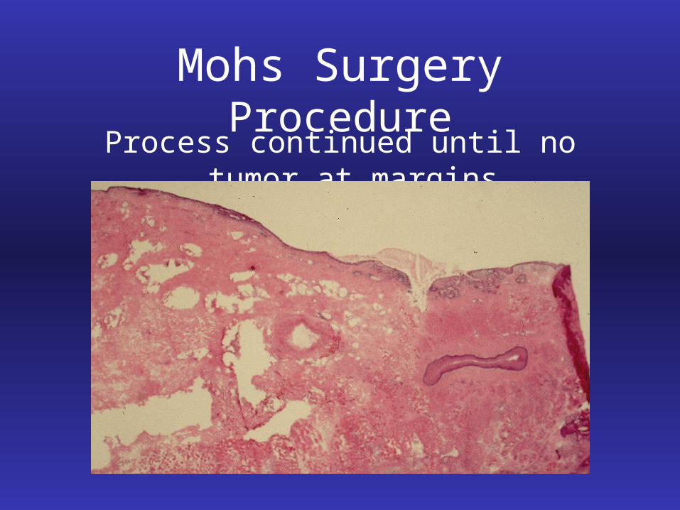
Mohs Surgery ProcedureProcess continued until no tumor at margins
Mohs Surgery Advantages
• Extremely high cure rate gives Mohs surgeon confidence to repair with most appropriate technique– Second intention healing– Simple or complex linear closures– Local flaps– Full and split thickness skin grafts
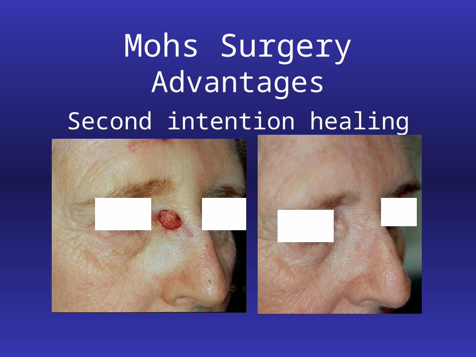
Mohs Surgery Advantages
Second intention healing
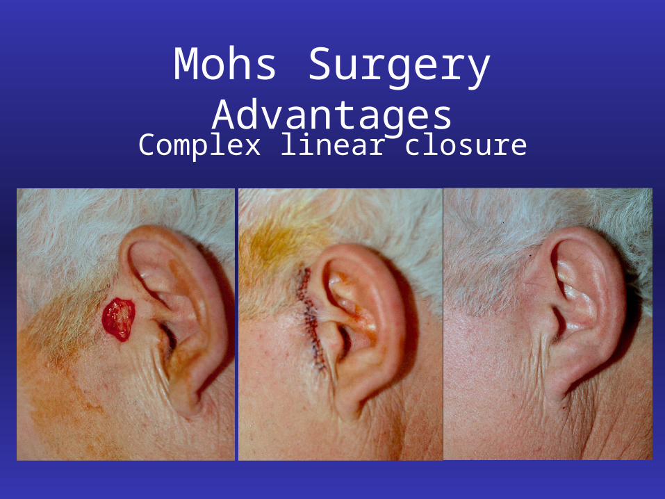
Mohs Surgery AdvantagesComplex linear closure
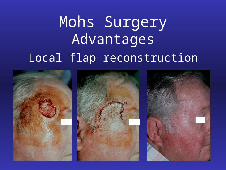
Mohs Surgery Advantages
Local flap reconstruction
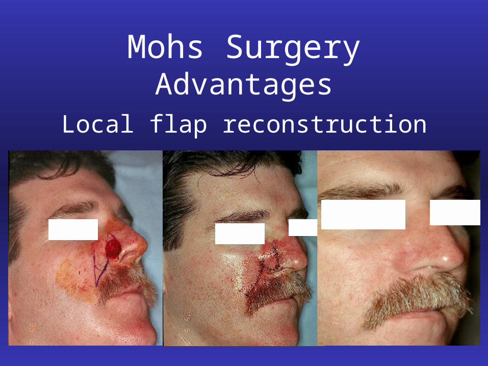
Mohs Surgery Advantages
Local flap reconstruction
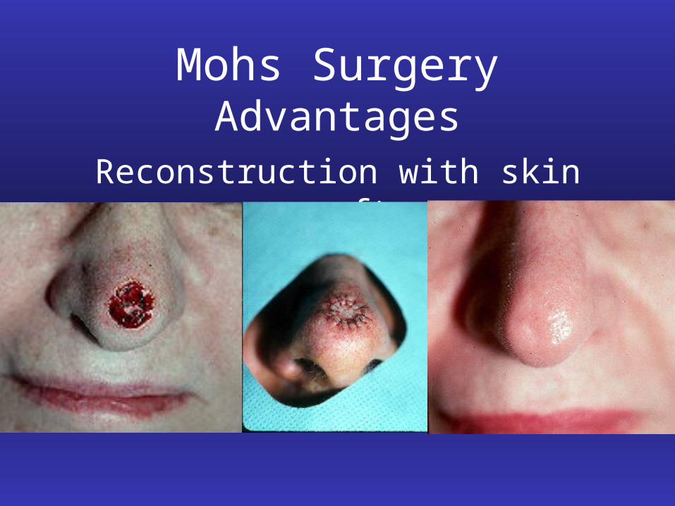
Mohs Surgery Advantages
Reconstruction with skin grafts
Mohs Surgery: Summary
• Highest cure rate (97-99%)– Entire margin evaluated– Fewer recurrences
• Leaves the smallest surgical defect possible– Preserves maximal amount of tissue– Increases the chance of a good aesthetic result
• Most cost effective treatment of select tumors– Outpatient setting