skeletal system your bones manufacture blood cells. our bones are held by our muscles the smallest...
TRANSCRIPT
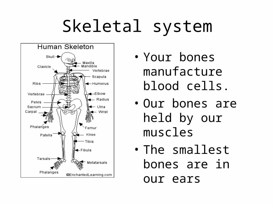
Skeletal system
• Your bones manufacture blood cells.
• Our bones are held by our muscles
• The smallest bones are in our ears

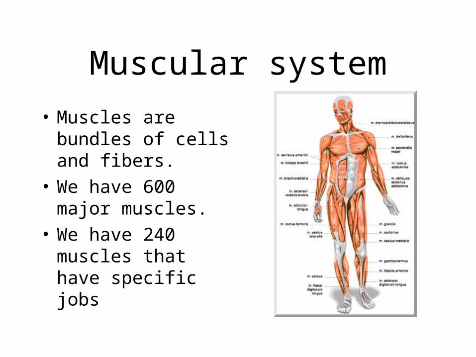
Muscular system
• Muscles are bundles of cells and fibers.
• We have 600 major muscles.
• We have 240 muscles that have specific jobs

TMJ

Structure of Bone

Anatomy of a Long Bone
• Diaphysis• Medullary Cavity• Nutrient Art & Vein• 2 Epiphyses• Epiphyseal Plates• Epiphyseal Art & Vein• Periosteum
– Outer: Dense irregular CT– Inner: Osteoblasts, osteoclasts– Does not cover epiphyses– Attaches to bone matrix via collagen fibers
• Endosteum– Osteoblasts, osteoclasts– Covers trabeculae, lines medullary cavity
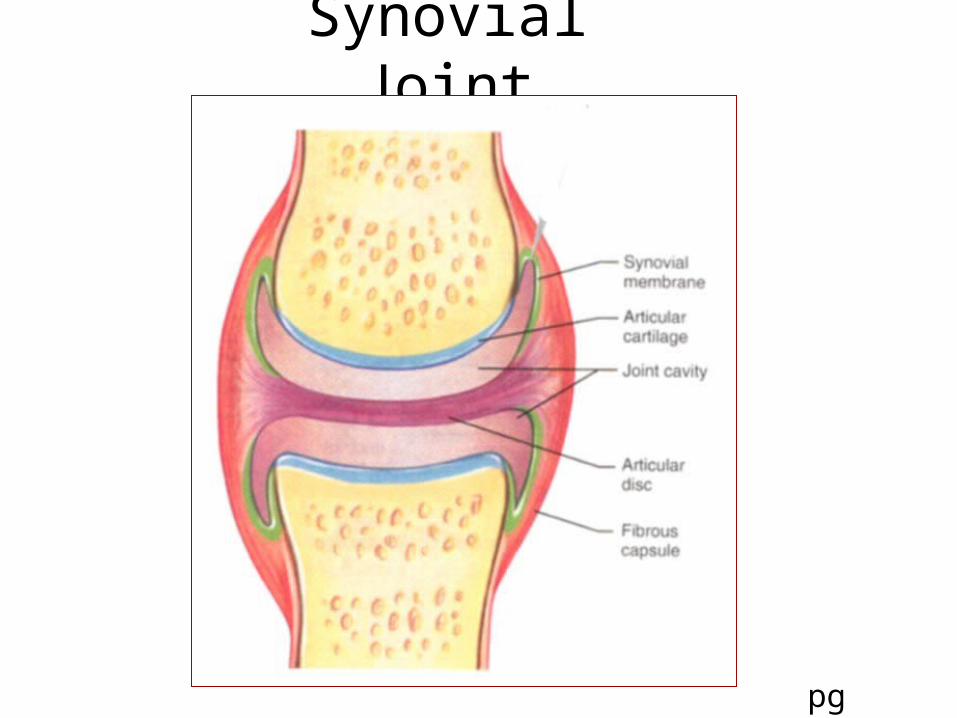
Synovial Joint
pg 215
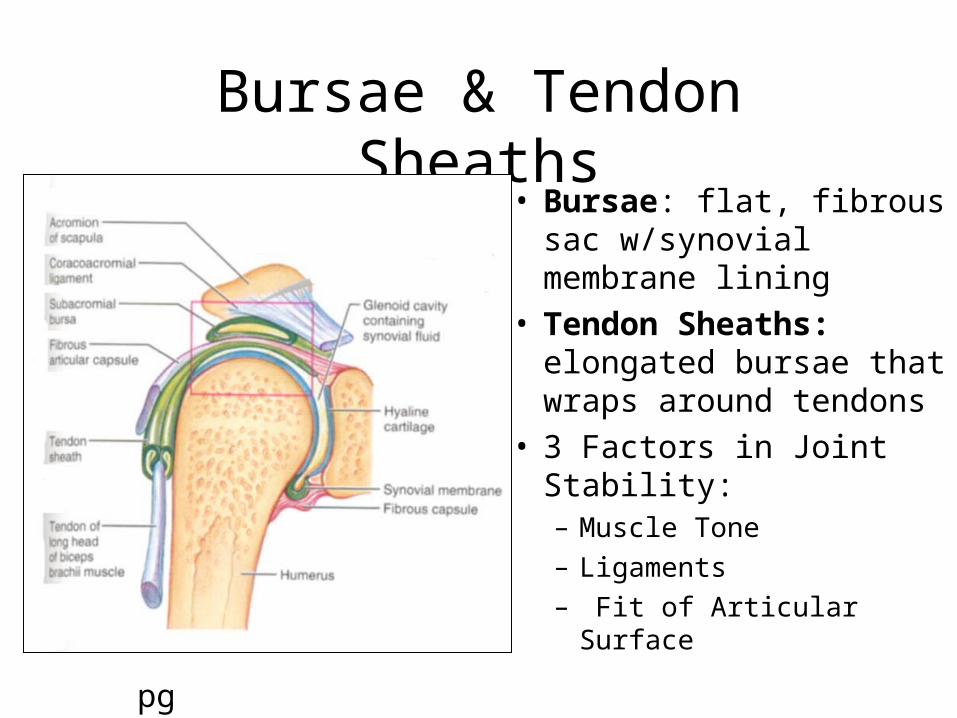
Bursae & Tendon Sheaths• Bursae: flat, fibrous sac
w/synovial membrane lining
• Tendon Sheaths: elongated bursae that wraps around tendons
• 3 Factors in Joint Stability:– Muscle Tone
– Ligaments
– Fit of Articular Surfacepg 219

Joint Shapes
• Hinge: cylindrical end of 1 bone fits into trough shape of other– angular movement-1 plane (eg)
elbow, ankle, interphalangal
• Plane: articular surface in flat plane– Short gliding movement
– (eg) intertarsal, articular processes of vertebrae
pg 224
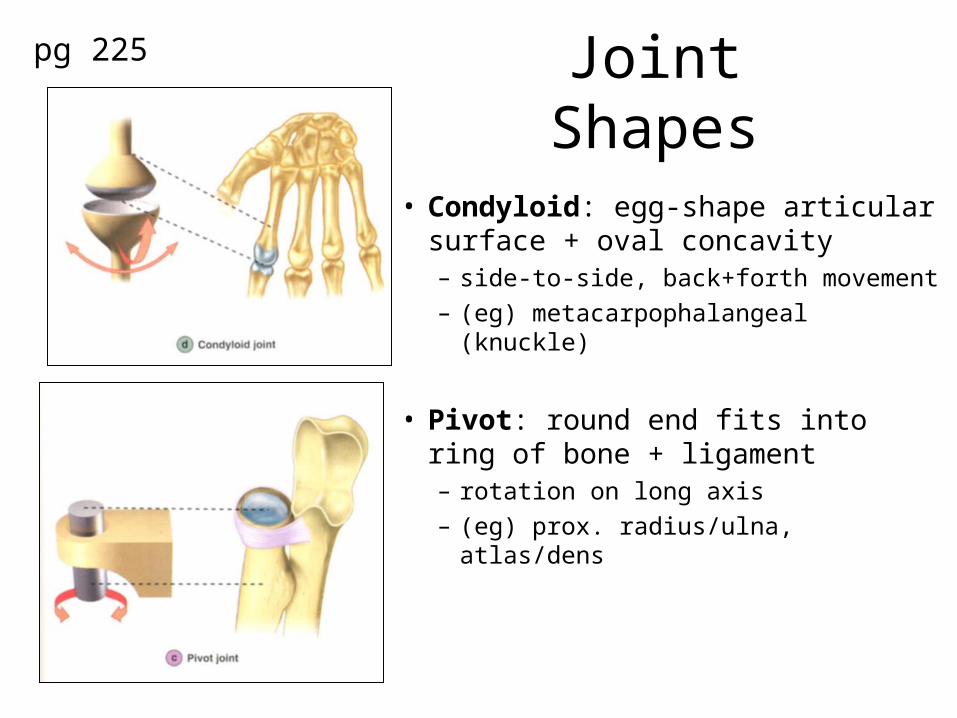
Joint Shapes
• Condyloid: egg-shape articular surface + oval concavity– side-to-side, back+forth movement
– (eg) metacarpophalangeal (knuckle)
• Pivot: round end fits into ring of bone + ligament– rotation on long axis
– (eg) prox. radius/ulna, atlas/dens
pg 225

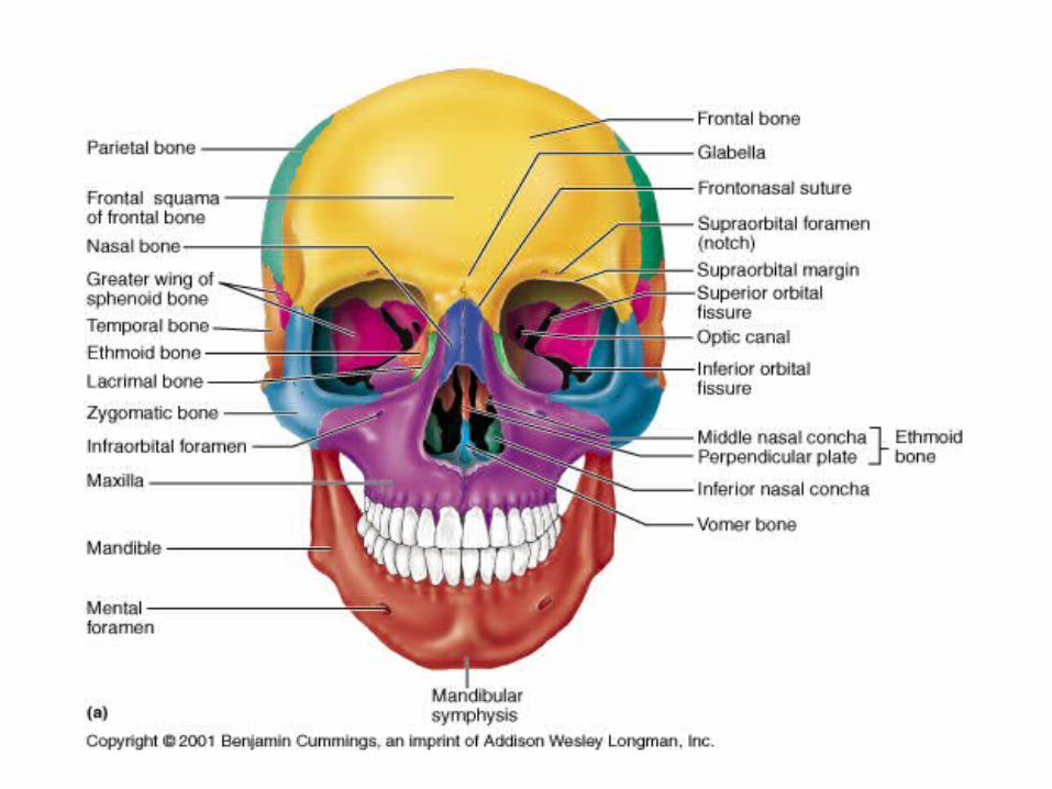
Anterior Skull
frontal bonefrontal bone
supraorbisupraorbital tal foramenforamenzygomatic bonezygomatic bone
maxillamaxillary bonery bone
alveolar fossaalveolar fossa
infraorbitinfraorbital al
foramenforamen
glabella
mental foramenmental foramenmandiblemandible
mandibulmandibular ar symphysissymphysis

Anterior Skull
nasal nasal boneboneperpendicularperpendicular
plateplate
middle nasalmiddle nasalconchaconcha
vomer bonevomer bone
superior superior orbital fissureorbital fissureinferior nasalinferior nasal concha boneconcha bone

Paranasal Sinuses
frontal sinusfrontal sinus
ethmoid ethmoid sinussinusmaxilary sinusmaxilary sinus
sphenoid sinussphenoid sinus

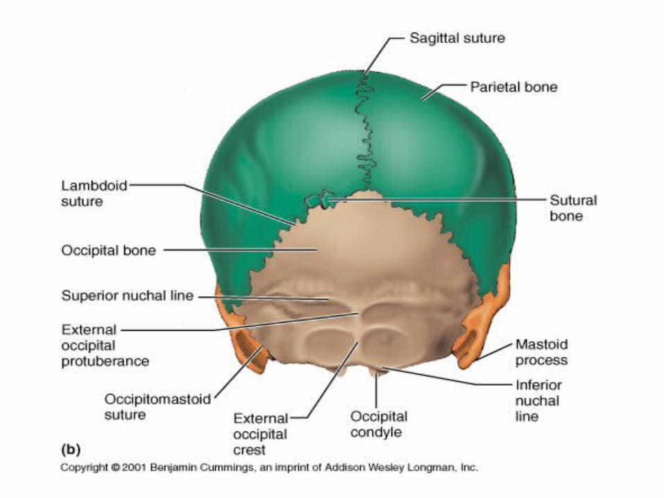
Craniumfrontal bonefrontal bone
parietal boneparietal bone
occipital boneoccipital bone
lambdoidallambdoidal suturesuture
sagittal suturesagittal suture
coronal suturecoronal suture

Ventral Skullpalatine processpalatine process
palatine bonepalatine bone
vomer bonevomer bone
mastoid processmastoid process
styloid processstyloid process
external occipitalexternal occipitalprotuberanceprotuberance
sphenoid bonesphenoid bone
temporal temporal bonebone
occipital boneoccipital bone
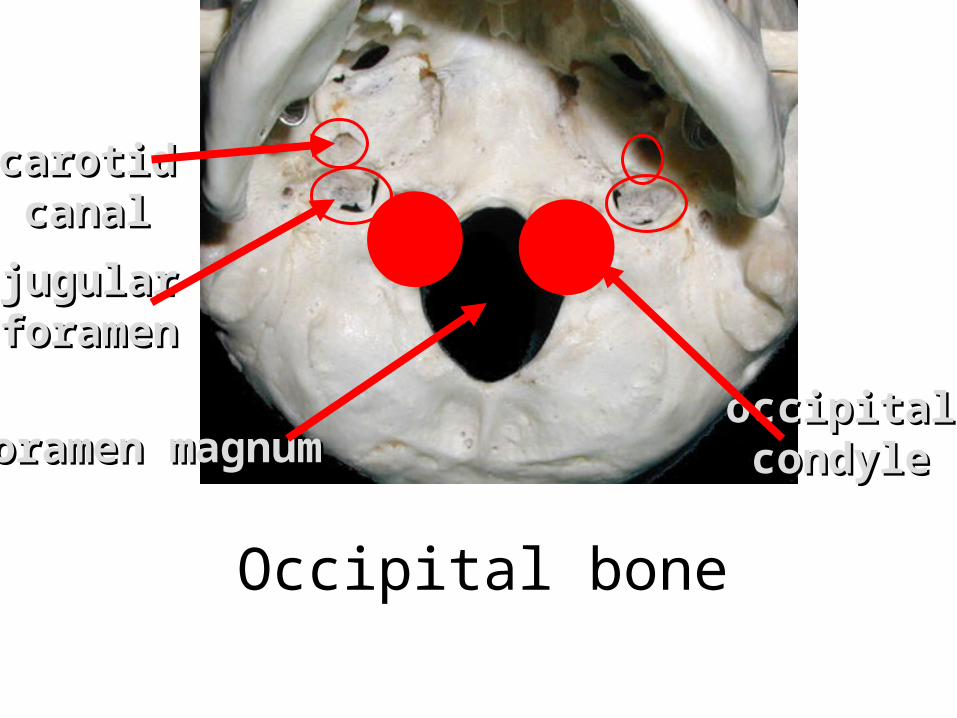
Occipital bone
occipitaloccipitalcondylecondyle
jugularjugularforamenforamen
carotidcarotidcanalcanal
foramen magnumforamen magnum
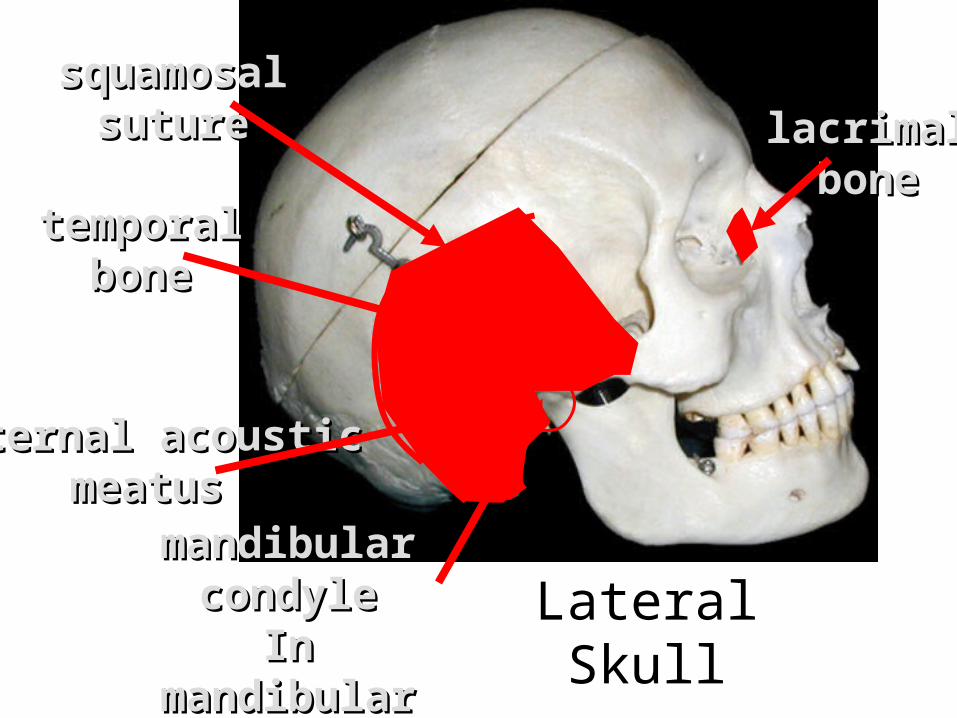
Lateral Skull
lacrimallacrimalbonebone
temporaltemporalbonebone
squamosalsquamosalsuturesuture
mandibular mandibular condylecondyle
In mandibular In mandibular fossafossa
(TMJ joint)(TMJ joint)
external acousticexternal acousticmeatusmeatus
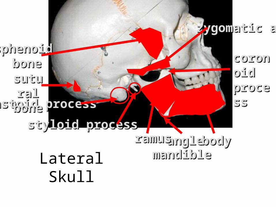
angleangle
coronoicoronoid d processprocess
zygomatic archzygomatic arch
mastoid processmastoid process
styloid processstyloid process
sphenoid sphenoid bonebone
bodybodyramusramusmandiblemandible
Lateral Skull
suturasutural bonel bone

crista gallicrista gallicribriborm platecribriborm plate
intenal intenal acoustic acoustic meatusmeatus
greater winggreater winglesser winglesser wing
optic canaloptic canalsella turcicasella turcica
jugular foramenjugular foramen
Internal Skull

Hyoid boneHyoid bone
temmporaltemmporalmandibularmandibular
jointjoint
external external acousticacousticmeatusmeatus
Hyoid +


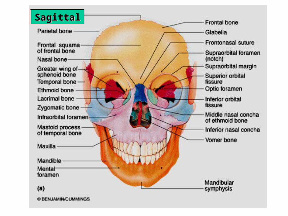
________________________________________________SagittalSagittal
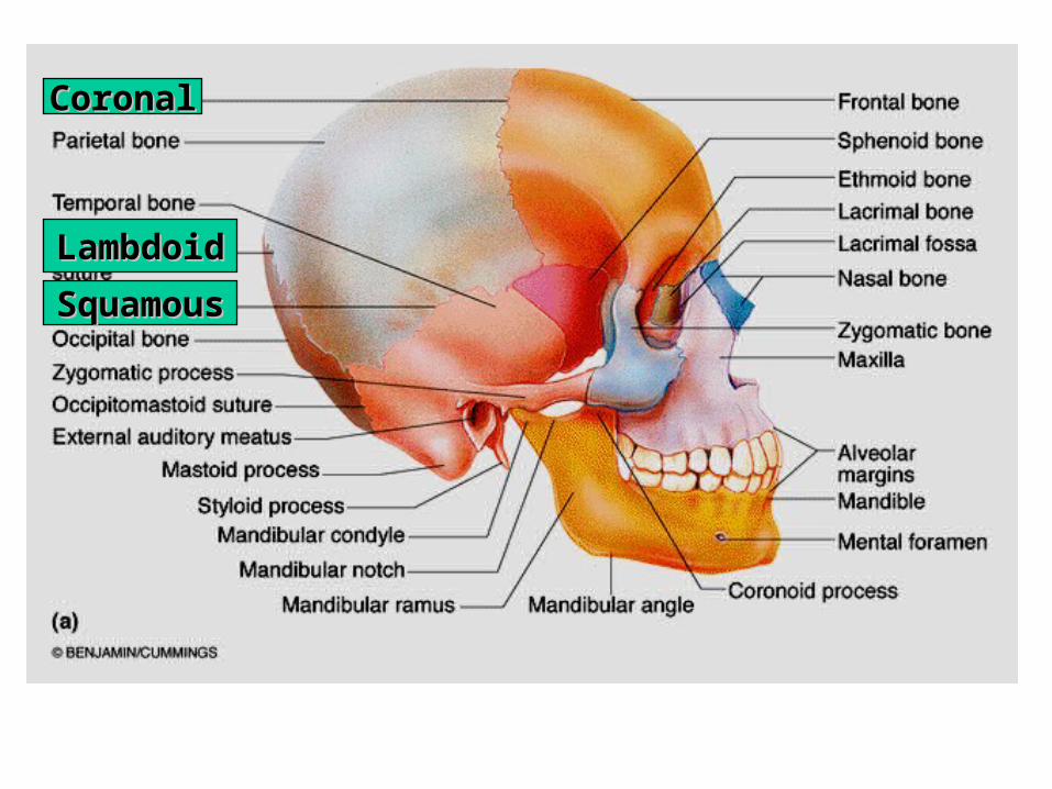
CoronalCoronal
LambdoidLambdoid
SquamousSquamous
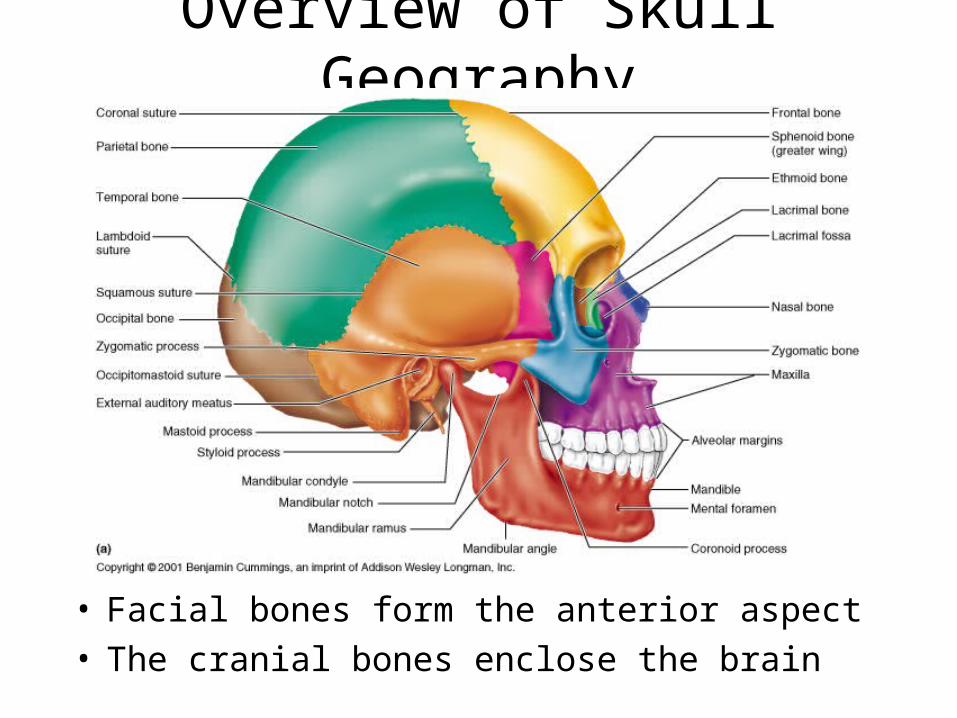
Overview of Skull Geography
• Facial bones form the anterior aspect• The cranial bones enclose the brain

Vault• The cranial vault
or calvaria forms the superior, lateral, and posterior aspects of skull
• The cranial base forming the inferior aspect of skull

Cranial Base• Cranial base forms
the skull’s inferior aspect
• Three prominent ridges divide the base into fossae
• The brain rests on these cranial fossae completely enclosed by the cranial vault
• The brain occupies the cranial cavity
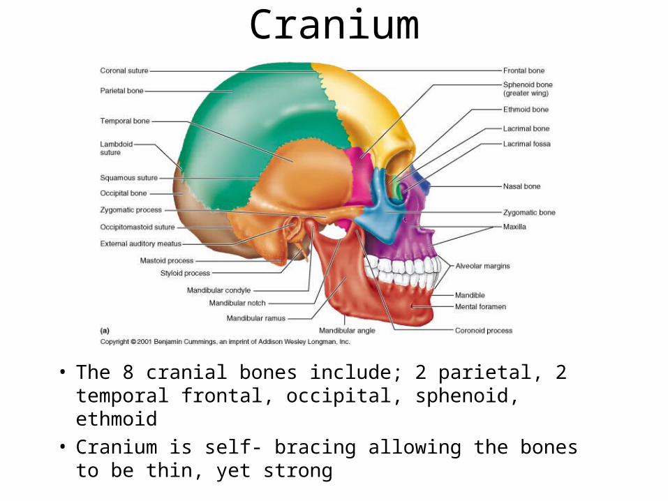
Cranium
• The 8 cranial bones include; 2 parietal, 2 temporal frontal, occipital, sphenoid, ethmoid
• Cranium is self- bracing allowing the bones to be thin, yet strong
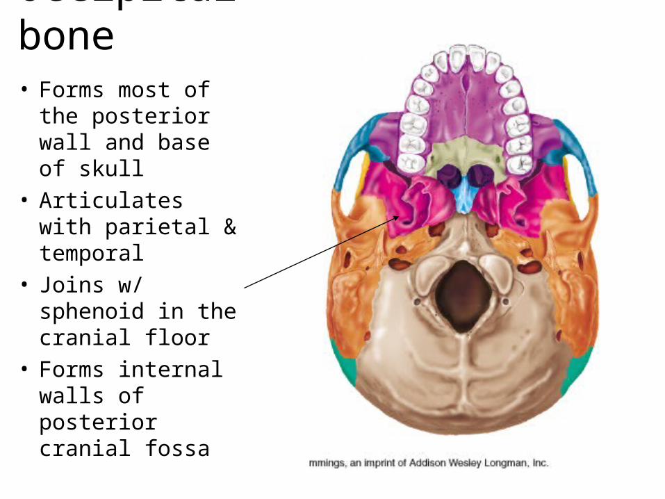
Occipital bone• Forms most of the
posterior wall and base of skull
• Articulates with parietal & temporal
• Joins w/ sphenoid in the cranial floor
• Forms internal walls of posterior cranial fossa

Occipital bone - Int. landmarks
• Hypoglossal canal, Posterior cranial fossa

Temporal Bone
• Forms the infero-lateral aspects of the skull
• Parts of the cranial floor
• Divided into four regions; squamous tympanic, mastoid, and petrous-(int)
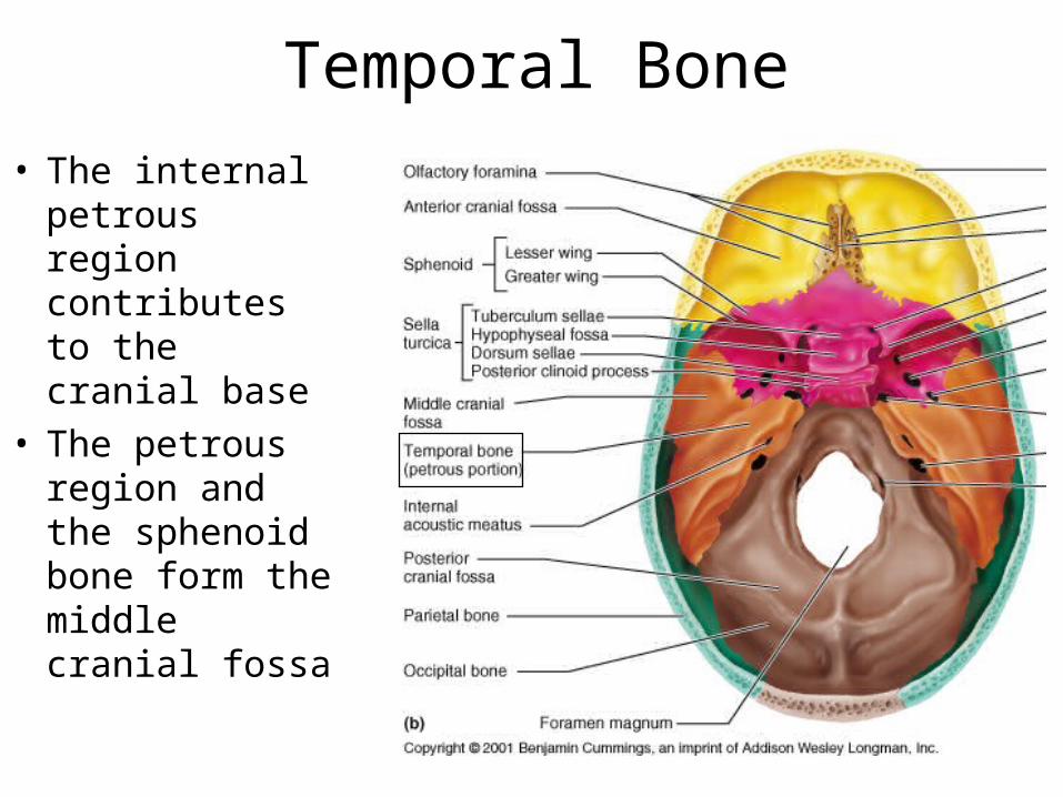
Temporal Bone
• The internal petrous region contributes to the cranial base
• The petrous region and the sphenoid bone form the middle cranial fossa
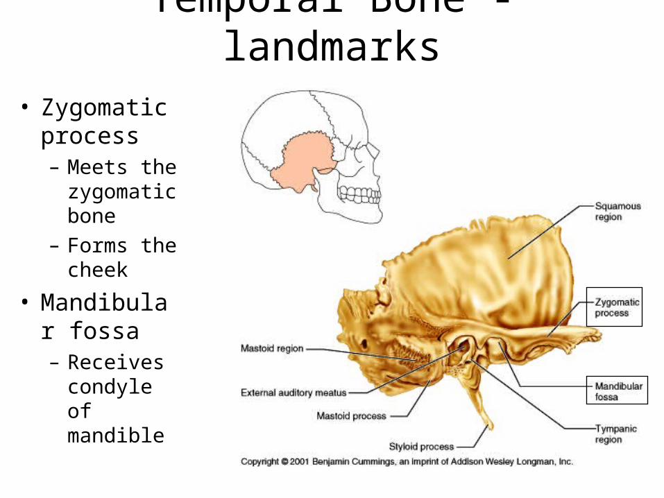
Temporal Bone - landmarks• Zygomatic
process– Meets the
zygomatic bone
– Forms the cheek
• Mandibular fossa– Receives
condyle of mandible
Temporal bones - landmarks
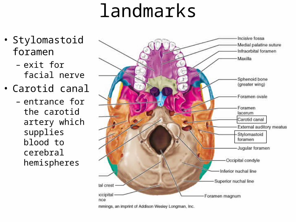
• Stylomastoid foramen– exit for facial
nerve
• Carotid canal– entrance for the
carotid artery which supplies blood to cerebral hemispheres
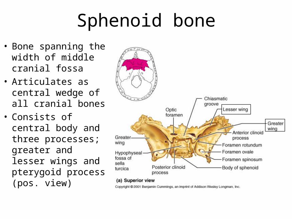
Sphenoid bone• Bone spanning the
width of middle cranial fossa
• Articulates as central wedge of all cranial bones
• Consists of central body and three processes; greater and lesser wings and pterygoid process (pos. view)
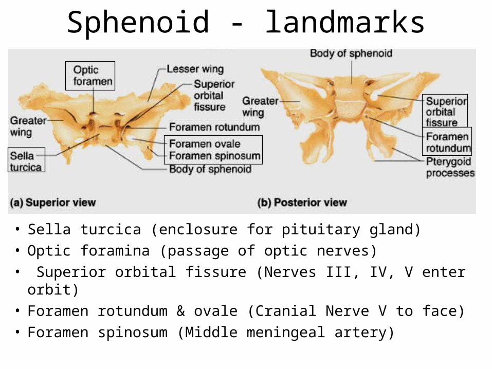
Sphenoid - landmarks
• Sella turcica (enclosure for pituitary gland)• Optic foramina (passage of optic nerves)• Superior orbital fissure (Nerves III, IV, V enter orbit)• Foramen rotundum & ovale (Cranial Nerve V to face) • Foramen spinosum (Middle meningeal artery)

Ethmoid bone• Forms most of the
area between the nasal cavity & orbits of eyes
• Lies between nasal bones & sphenoid
• Complex shape gives rise to nasal septum, sinuses and cribiform plate

Ethmoid bone - landmarks• Cribiform plates
– Forms roof of nasal cavity
• Olfactory formina– Olfactory nerves
enter brain
• Crista galli– Attachment of the
dura mater which secures brain in cavity
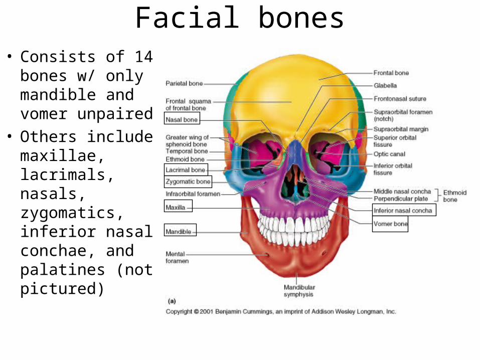
Facial bones• Consists of 14
bones w/ only mandible and vomer unpaired
• Others include maxillae, lacrimals, nasals, zygomatics, inferior nasal conchae, and palatines (not pictured)
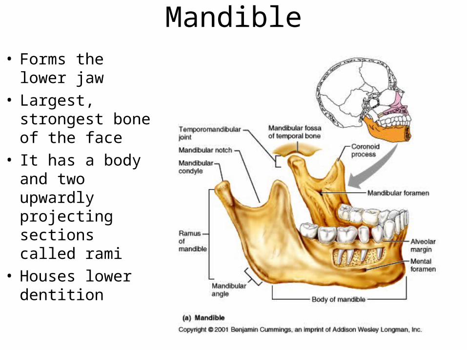
Mandible• Forms the lower
jaw• Largest, strongest
bone of the face• It has a body and
two upwardly projecting sections called rami
• Houses lower dentition

Mandible - landmarks• Mandibular angle• Mandibular notch• Coronoid process• Mandibular
condyle• Alveolar margin• Mandible formina• Mental formina• Ramus of mandible
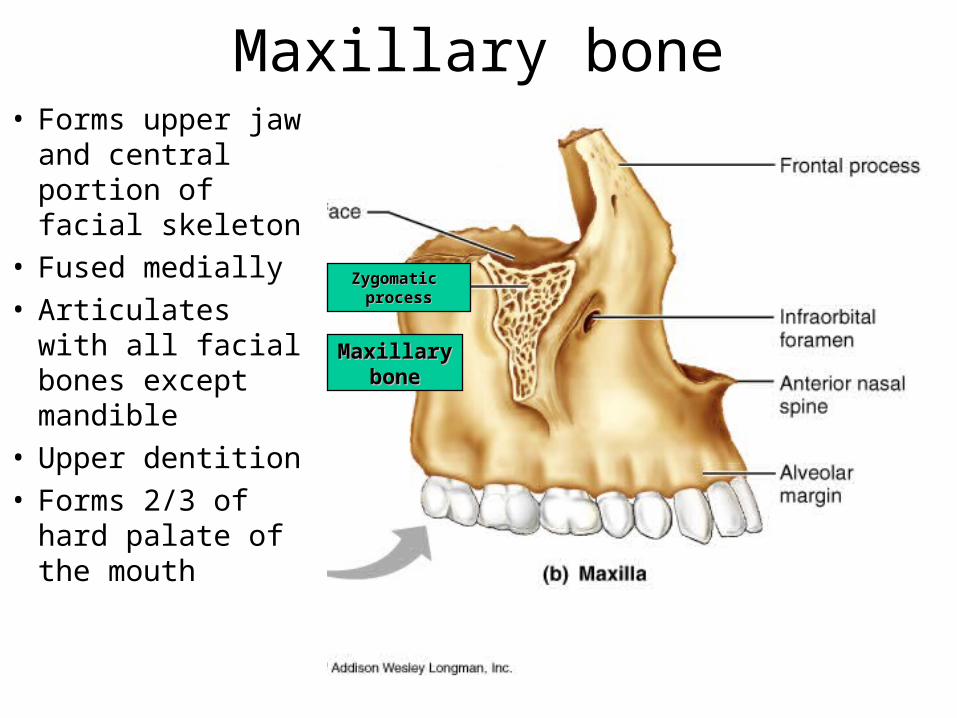
Maxillary bone• Forms upper jaw
and central portion of facial skeleton
• Fused medially• Articulates with
all facial bones except mandible
• Upper dentition• Forms 2/3 of
hard palate of the mouth
MaxillaryMaxillarybonebone
Zygomatic Zygomatic processprocess

Maxillary bones - landmarks• Alveolar margin
– Upper dentition
• Frontal process– Forms lateral
aspects of nose
• Zygomatic process– Articulates with
zygomatic bone
• Maxillary sinuses – (Fig. 7.11)

Palatine bones• The horizontal
plates forms the posterior portion of hard palate
• Vertical plate forms part of the posterolateral wall of nasal cavity and a small portion of orbit
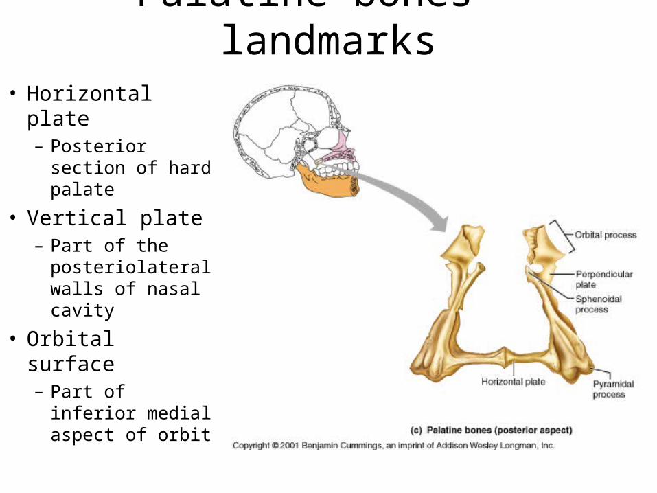
Palatine bones - landmarks• Horizontal plate
– Posterior section of hard palate
• Vertical plate– Part of the
posteriolateral walls of nasal cavity
• Orbital surface– Part of inferior
medial aspect of orbit
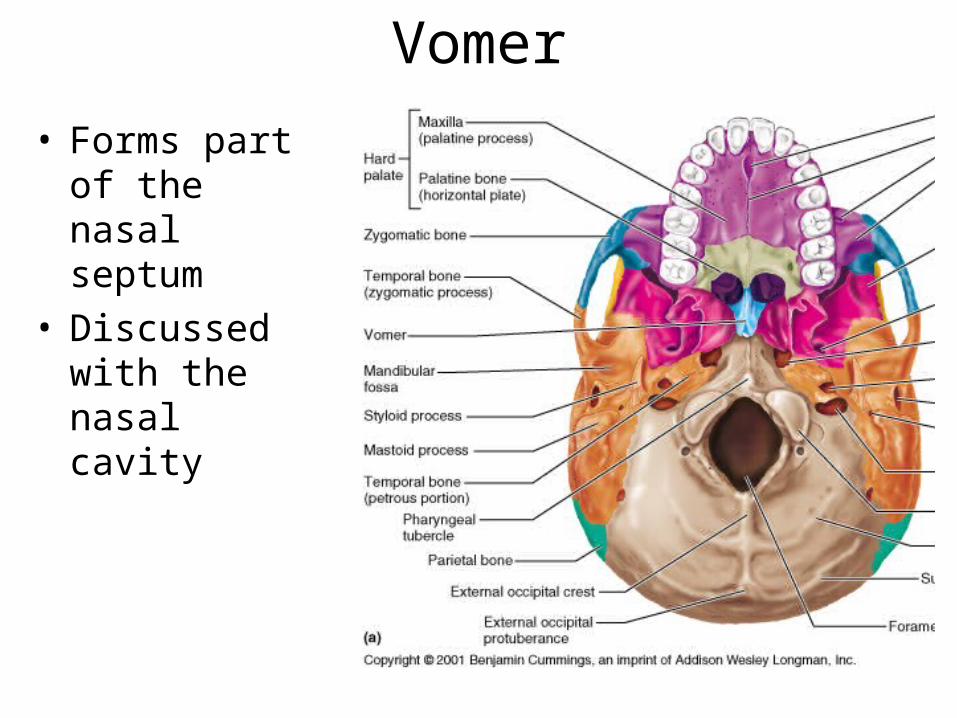
Vomer
• Forms part of the nasal septum
• Discussed with the nasal cavity

Vomer - landmarks
• Plow shape– Divides nasal
septum into right and left parts
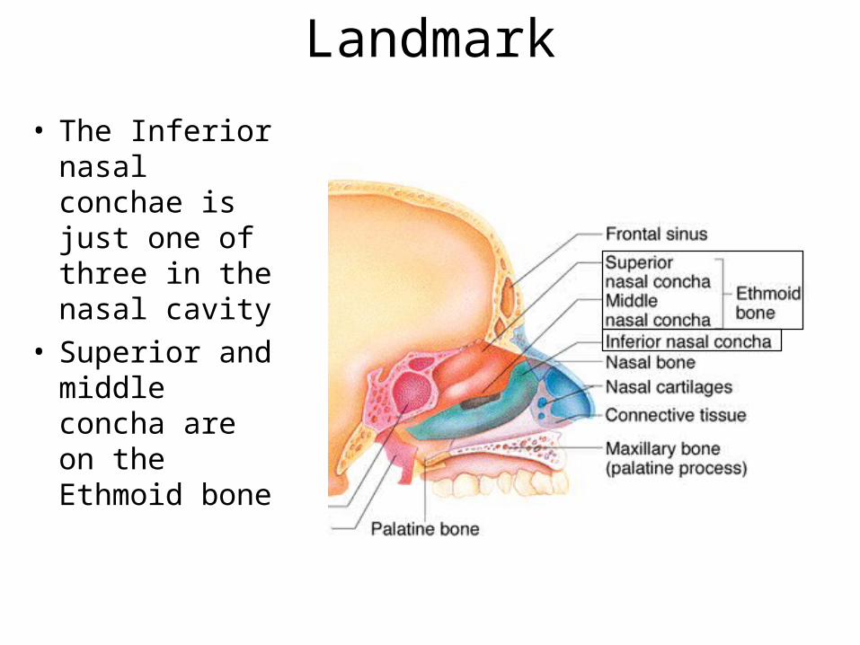
Inferior Nasal Conchae - Landmark
• The Inferior nasal conchae is just one of three in the nasal cavity
• Superior and middle concha are on the Ethmoid bone
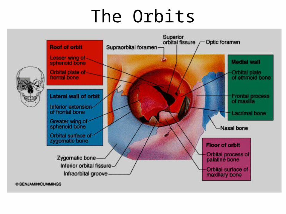
The Orbits
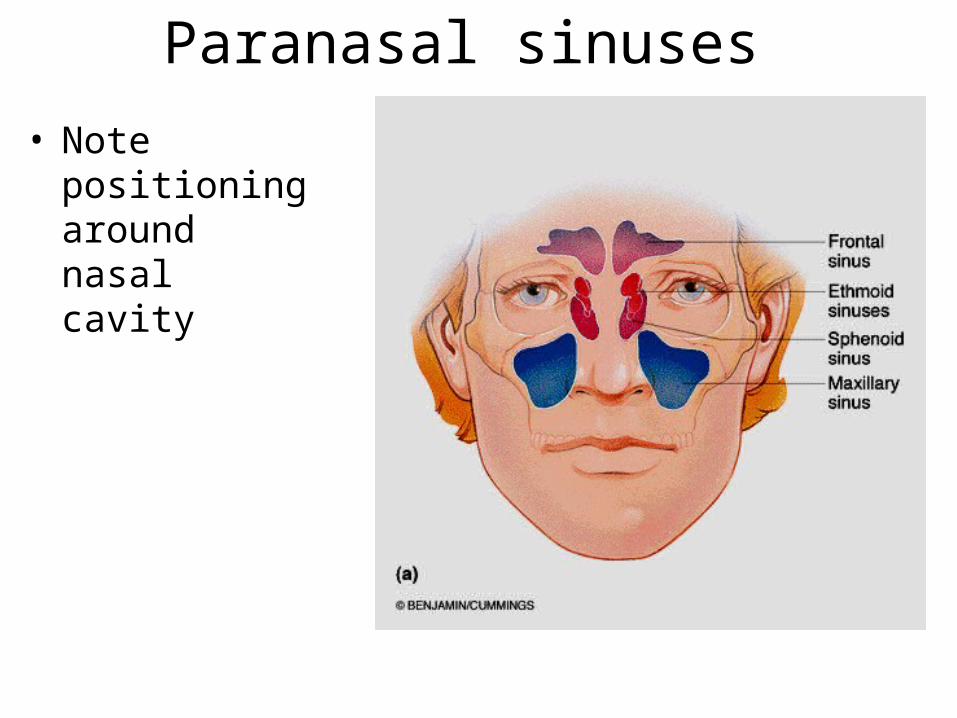
Paranasal sinuses
• Note positioning around nasal cavity

Paranasal sinuses
• Sphenoid sinus• Frontal sinus• Ethmoid sinus• Maxillary sinuses
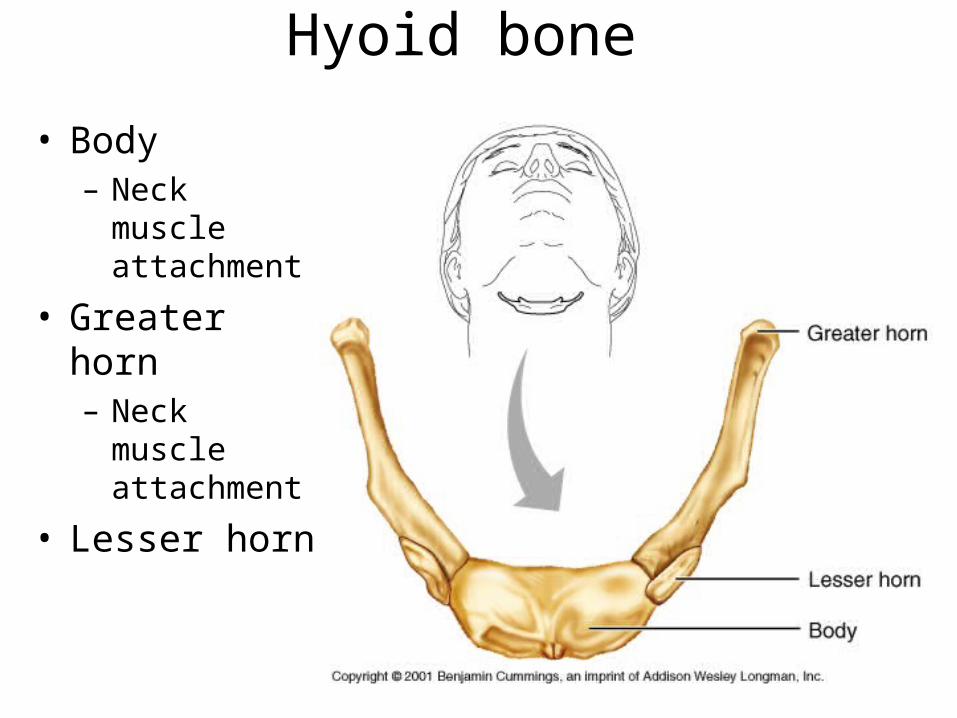
Hyoid bone
• Body– Neck muscle
attachment
• Greater horn– Neck muscle
attachment
• Lesser horn
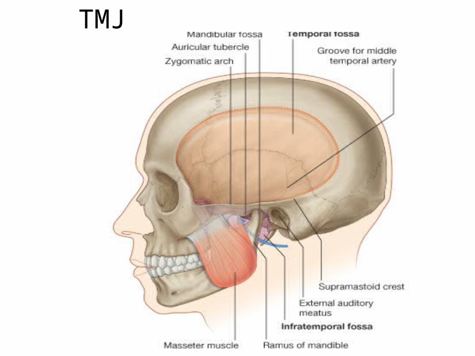
TMJ

TMJ Capsule

TMJ Capsule

TMJ Motions

TMJ Motions

Characteristics - Ligaments• Ligaments hold the
vertebral column in an upright position– The broad Anterior
Longitudinal Ligament prevents hyperextension and is quite strong
– The cord like Posterior Longitudinal Ligament prevents hyperflexion and is relatively weak

Characteristics - Ligaments• Ligaments
also connect specific vertebra and support disc position– Supraspinos
ligament
– Ligamentum flavum
– Interspinous ligament

Intervertebral Discs• Intervertebral discs are cushion like pads
interposed between vertebra
• The discs provide elasticity and compressibility
• Compression flattens discs
• Discs are thickest in the cervical and lumbar to provide flexibility
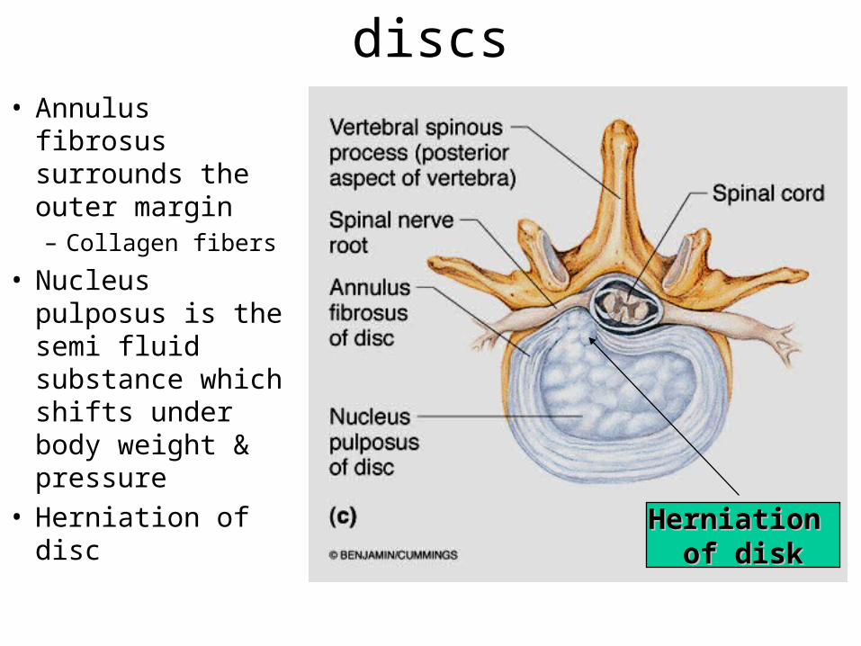
Characteristics - discs• Annulus fibrosus
surrounds the outer margin– Collagen fibers
• Nucleus pulposus is the semi fluid substance which shifts under body weight & pressure
• Herniation of discHerniation Herniation
of diskof disk

General structure of vertebrae• Common pattern
– Body or centrum
– Vertebral arch• lamina
• pedicle
– Vertebral foramen
– Spinous process• Muscles attach
– Transverse process
• Muscles attach
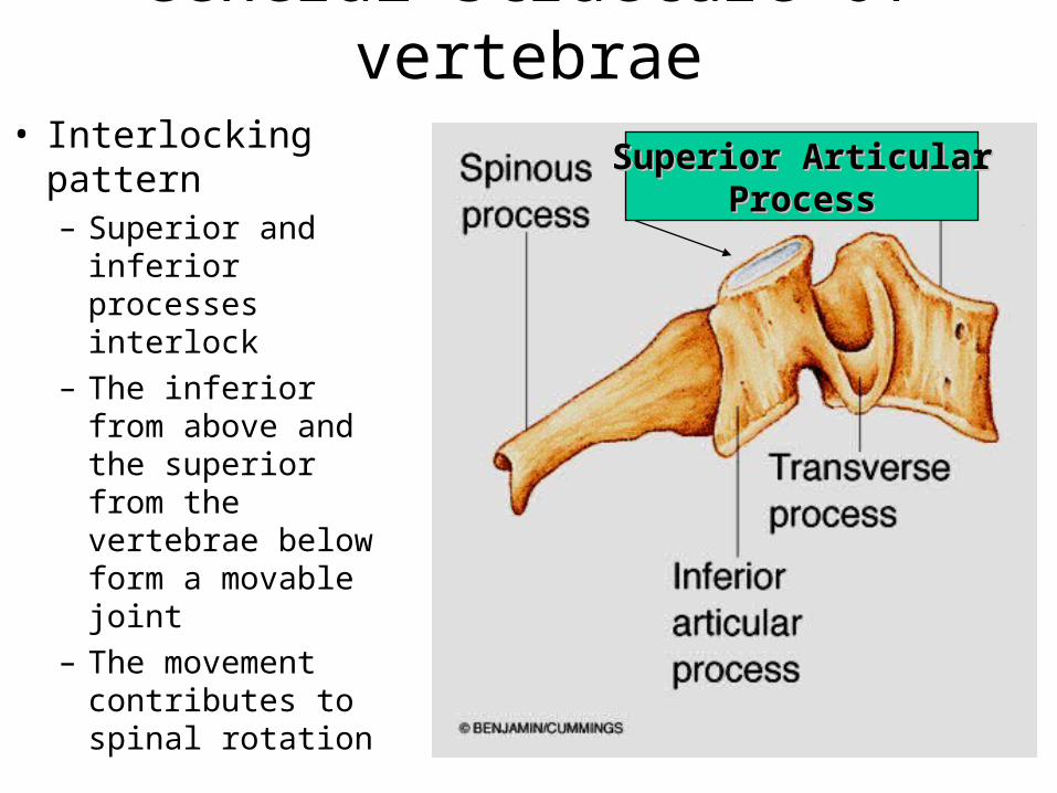
General structure of vertebrae• Interlocking pattern
– Superior and inferior processes interlock
– The inferior from above and the superior from the vertebrae below form a movable joint
– The movement contributes to spinal rotation
Superior ArticularSuperior ArticularProcessProcess
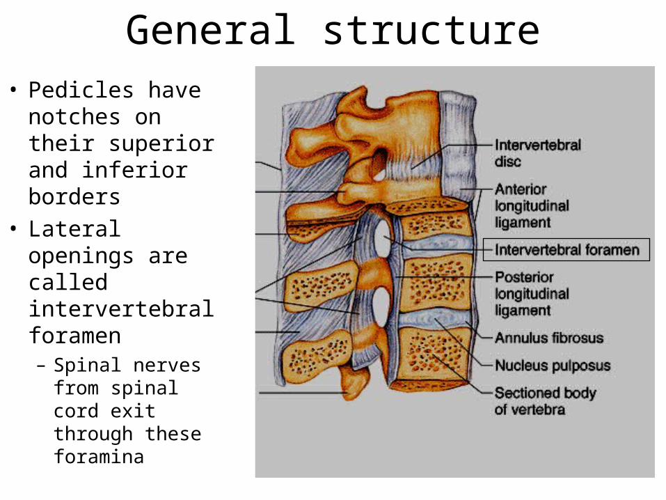
General structure• Pedicles have
notches on their superior and inferior borders
• Lateral openings are called intervertebral foramen– Spinal nerves
from spinal cord exit through these foramina

Regional Characteristic: Cervical• Body is oval, but wide
side to side C3 - C7• Spinous process is
short and bifid (split) except in C7
• Vertebral foramen is triangular
• Transverse processes contain foramina for blood vessels leading to brain
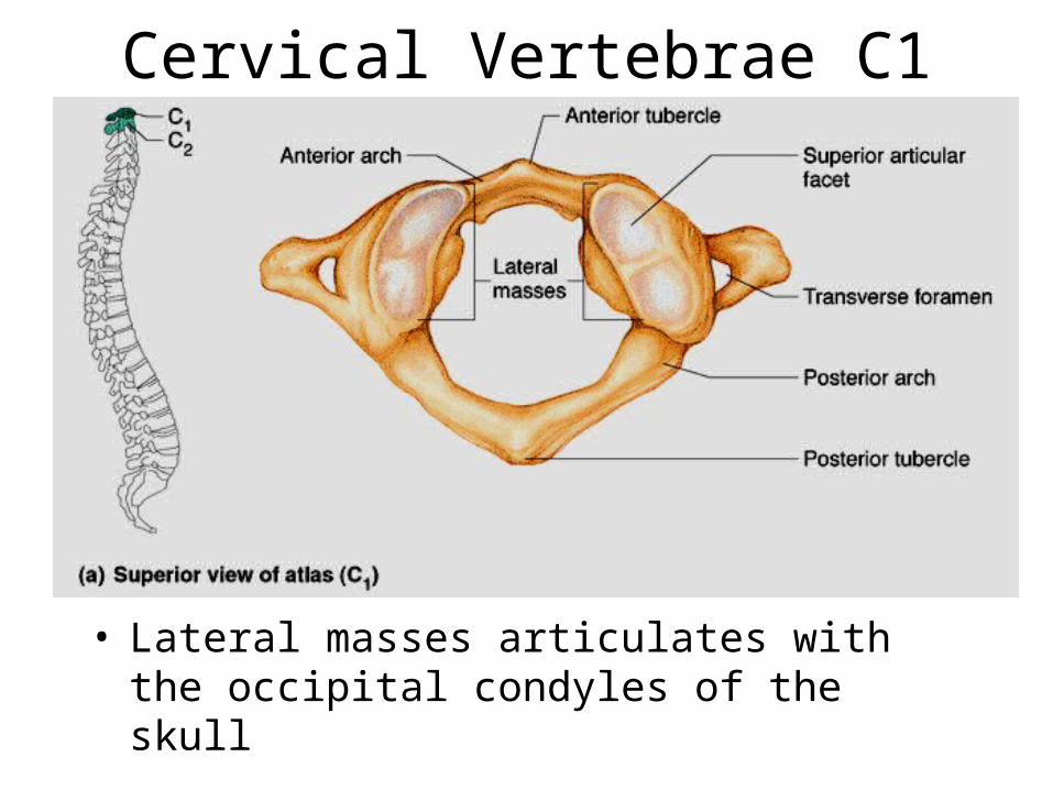
Cervical Vertebrae C1
• Lateral masses articulates with the occipital condyles of the skull
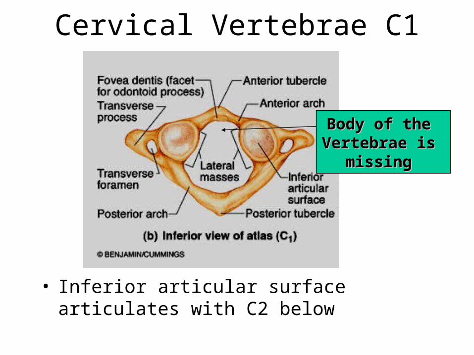
Cervical Vertebrae C1
• Inferior articular surface articulates with C2 below
Body of the Body of the Vertebrae is Vertebrae is
missing missing

Cervical Vertebrae C2• The axis has the
odontoid process or dens is its unique feature
• The dens is the missing body of the atlas which fuses with the atlas during embryonic development

Regional Characteristic: Cervical • Spinous processes
project directly posteriorly
• Superior facets directed superoposteriorly
• Inferior facets directed inferoanteriorly
• Flexion/extension, lateral flexion and rotation

Regional Characteristic: Thoracic
• Body is larger than cervical; heart shaped
• Spinous process is long and sharp
• Vertebral foramen is circular
• Transverse processes project posteriorly and bear facets for ribs

Regional Characteristic: Lumbar
• Body is massive and kidney shaped
• Spinous processes are short and blunt
• Vertebral foramen is triangular
• Transverse processes are perpendicular to spinous process but has no special features
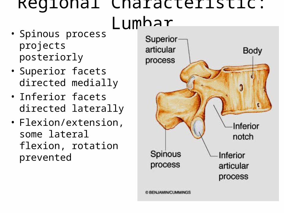
Regional Characteristic: Lumbar• Spinous process
projects posteriorly• Superior facets
directed medially• Inferior facets directed
laterally• Flexion/extension,
some lateral flexion, rotation prevented
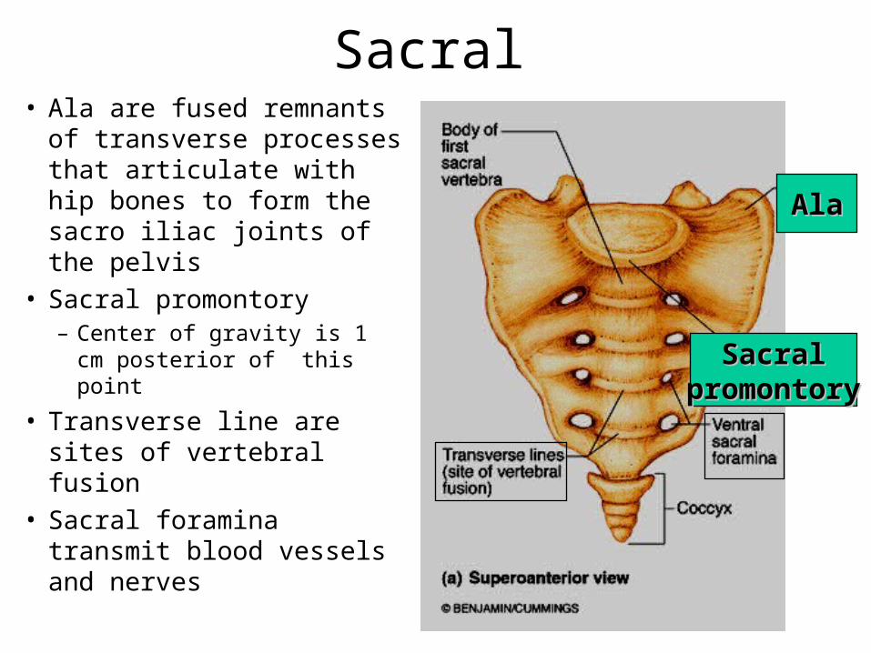
Sacral• Ala are fused remnants
of transverse processes that articulate with hip bones to form the sacro iliac joints of the pelvis
• Sacral promontory – Center of gravity is 1 cm
posterior of this point
• Transverse line are sites of vertebral fusion
• Sacral foramina transmit blood vessels and nerves
SacralSacralpromontorypromontory
AlaAla

Sacral• On the posterior aspect
median sacral crest are fused spinous processes
• The vertebral canal continues inside the sacrum as the sacral canal
• Sacral hiatus is at the inferior end of the sacral canal
• Superior articular surface form a joint with the spinal column
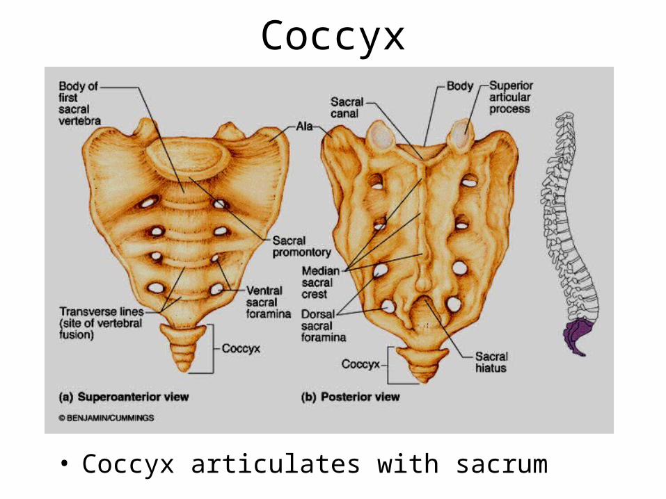
Coccyx
• Coccyx articulates with sacrum
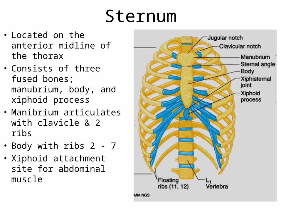
Sternum• Located on the anterior
midline of the thorax • Consists of three fused
bones; manubrium, body, and xiphoid process
• Manibrium articulates with clavicle & 2 ribs
• Body with ribs 2 - 7• Xiphoid attachment site
for abdominal muscle
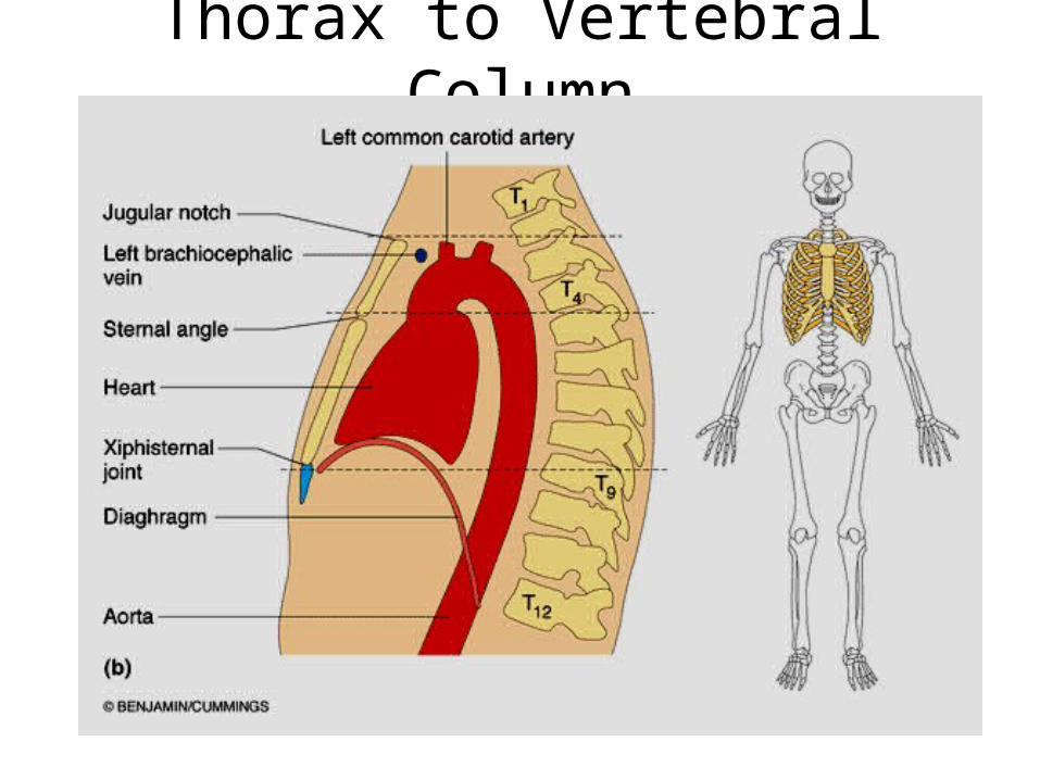
Thorax to Vertebral Column
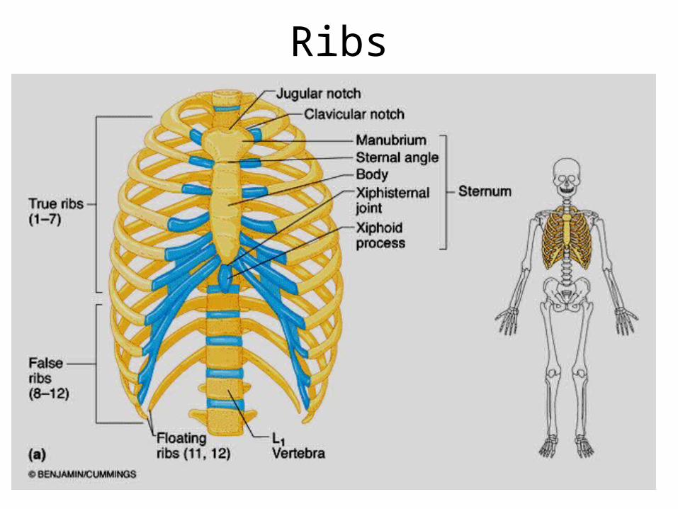
Ribs
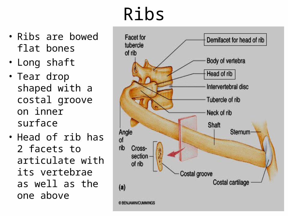
Ribs• Ribs are bowed
flat bones• Long shaft• Tear drop shaped
with a costal groove on inner surface
• Head of rib has 2 facets to articulate with its vertebrae as well as the one above

Ribs• Tubercle of rib
articulates with transverse process
• Ligaments secure rib to transverse process
• Note how the transverse processes of thoracic vertebrae are angled posteriorly
*Вступ до курсу анатомії людини.*Загальне вчення про кісткову систему. *Види з’єднання кісток.*Кістки черепа.*Краніометрія. Черепні показники.*Скронево-нижньощелепний суглоб
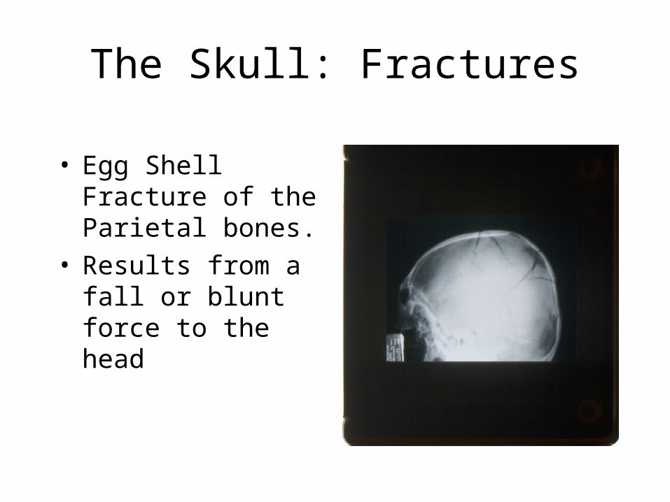
The Skull: Fractures
• Egg Shell Fracture of the Parietal bones.
• Results from a fall or blunt force to the head
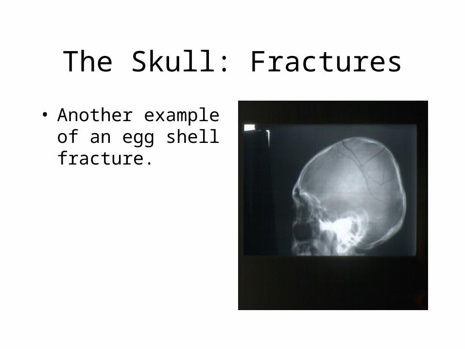
The Skull: Fractures
• Another example of an egg shell fracture.

Knife in Skull Above OrbitAP Projection