skeletal system. the structures of the skeletal system include: bones, joints, and ligaments
TRANSCRIPT

SKELETAL SYSTEMSKELETAL SYSTEM
SKELETAL SYSTEMSKELETAL SYSTEM
THE STRUCTURES OF THE SKELETALTHE STRUCTURES OF THE SKELETAL
SYSTEM INCLUDE:SYSTEM INCLUDE:
BONES, JOINTS, AND LIGAMENTS.BONES, JOINTS, AND LIGAMENTS.
SKELETAL SYSTEMSKELETAL SYSTEM
FUNCTIONS OF THE SKELETAL SYSTEMFUNCTIONS OF THE SKELETAL SYSTEM
1.1. SUPPORT SUPPORT
2.2. PROTECTION PROTECTION
3.3. MOVEMENT MOVEMENT
4.4. MINERAL STORAGE MINERAL STORAGE
5.5. BLOOD CELL FORMATION BLOOD CELL FORMATION
CLASSIFICATION OF BONES BY CLASSIFICATION OF BONES BY POSITIONPOSITION
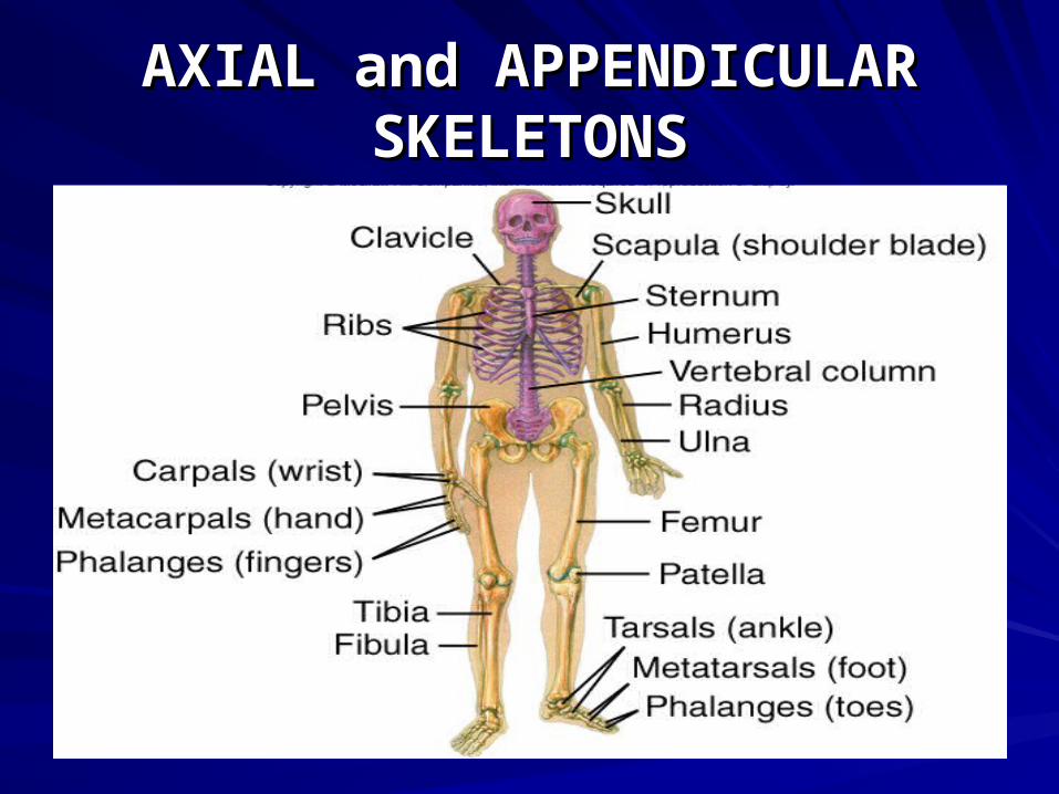
THE THE 206206 BONES OF THE HUMAN BONES OF THE HUMAN
BODY ARE GROUPED INTOBODY ARE GROUPED INTO
THE THE AXIAL AND THE AXIAL AND THE
APPENDICULAR SKELETONS.APPENDICULAR SKELETONS.
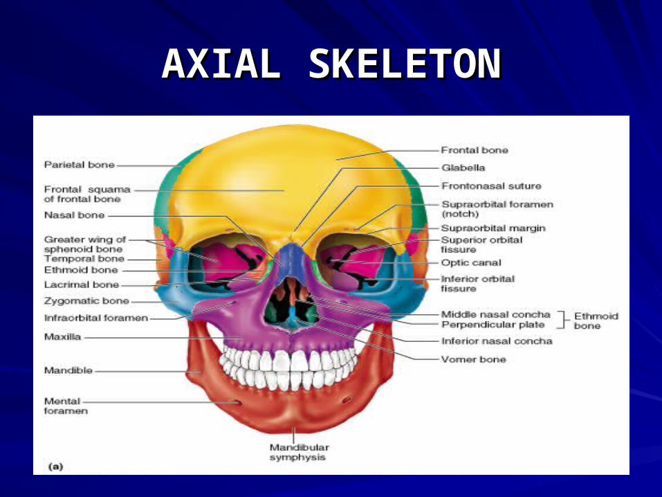
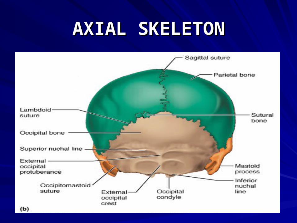
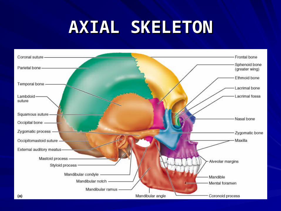
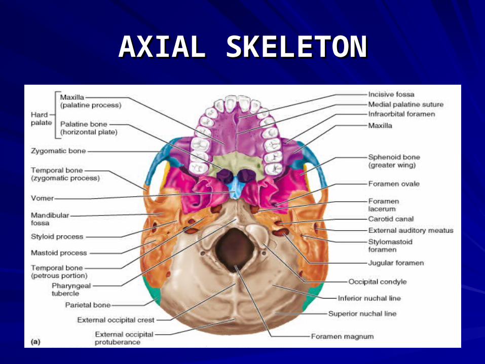
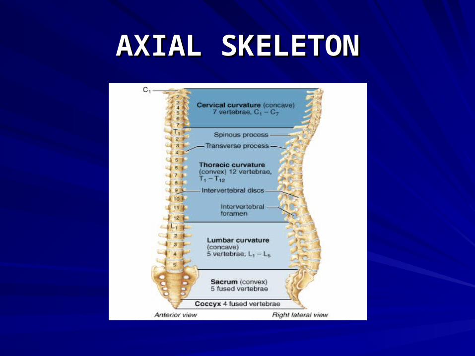
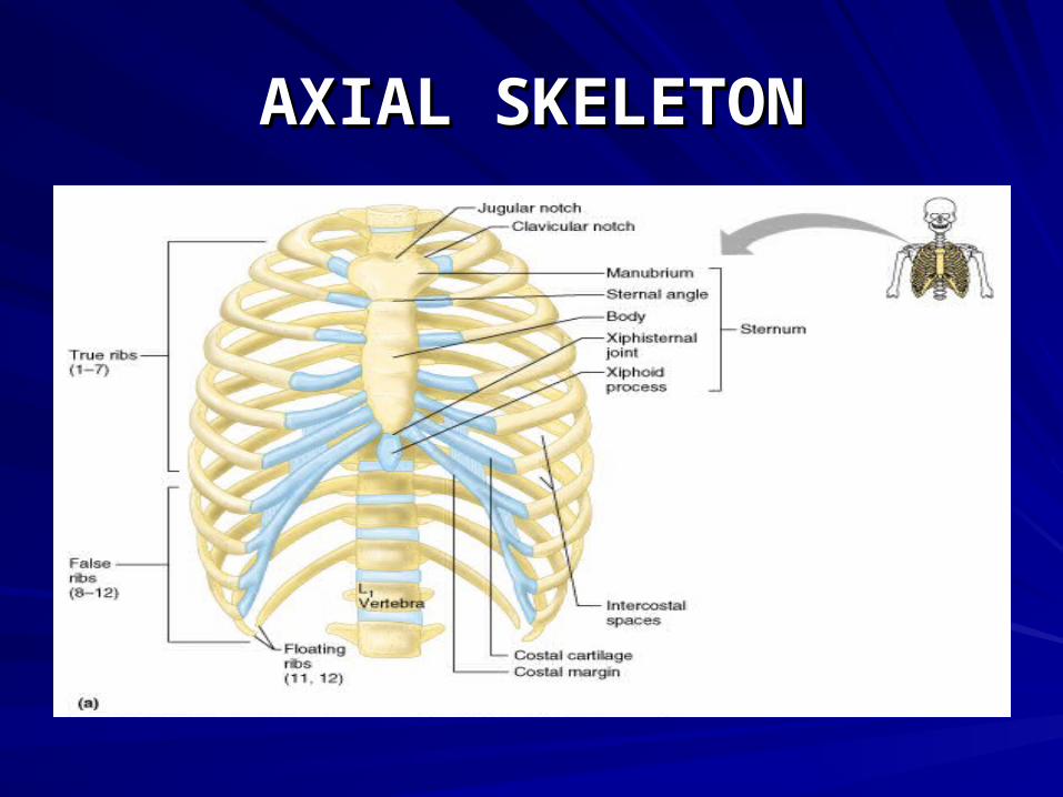
AXIAL SKELETONAXIAL SKELETON
THE THE AXIAL SKELETONAXIAL SKELETON
FORMS THE LONG AXIS OF THEFORMS THE LONG AXIS OF THE
BODY AND INCLUDES THE BODY AND INCLUDES THE
BONES OF THE SKULL, VERTEBRALBONES OF THE SKULL, VERTEBRAL
COLUMN, AND THE RIB CAGE.COLUMN, AND THE RIB CAGE.
AXIAL SKELETONAXIAL SKELETON
GENERALLY THESE BONES ARE MOSTGENERALLY THESE BONES ARE MOST
INVOLVED IN PROTECTING, ANDINVOLVED IN PROTECTING, AND
SUPPORTING. SUPPORTING.
AXIAL SKELETONAXIAL SKELETON
AXIAL SKELETONAXIAL SKELETON
AXIAL SKELETONAXIAL SKELETON
AXIAL SKELETONAXIAL SKELETON
AXIAL SKELETONAXIAL SKELETON
AXIAL SKELETONAXIAL SKELETON
APPENDICULAR SKELETONAPPENDICULAR SKELETON
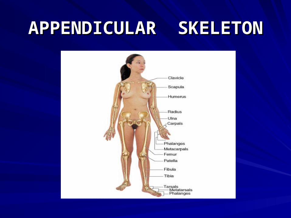
THE THE APPENDICULAR SKELETONAPPENDICULAR SKELETON
CONSISTS OF THE BONES OF THECONSISTS OF THE BONES OF THE
UPPER AND LOWER LIMBS,UPPER AND LOWER LIMBS,
AND THE GIRDLES THATAND THE GIRDLES THAT
ATTACH THE LIMBS TO THEATTACH THE LIMBS TO THE
AXIAL SKELETON.AXIAL SKELETON.
APPENDICULAR SKELETONAPPENDICULAR SKELETON
THE APPENDICULAR SKELETONTHE APPENDICULAR SKELETON
CONSISTS OF CONSISTS OF 126126 BONES. IT BONES. IT
FUNCTIONS TO HELP IN MOVEMENT.FUNCTIONS TO HELP IN MOVEMENT.
APPENDICULAR SKELETONAPPENDICULAR SKELETON
AXIAL and APPENDICULARAXIAL and APPENDICULARSKELETONSSKELETONS
CLASSIFICATION OF BONE BY CLASSIFICATION OF BONE BY SHAPESHAPE
THE BONES OF THE HUMANTHE BONES OF THE HUMAN
SKELETON COME IN MANY SIZESSKELETON COME IN MANY SIZES
AND SHAPES. BONES CAN BE AND SHAPES. BONES CAN BE
CLASSIFIED BY SHAPE INTO:CLASSIFIED BY SHAPE INTO:
LONG; SHORT; FLAT; IRREGULAR.LONG; SHORT; FLAT; IRREGULAR.
LONG BONESLONG BONES
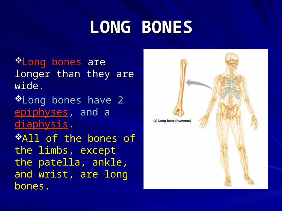
Long bonesLong bones are are longer than they are longer than they are wide.wide.Long bones have 2 Long bones have 2 epiphysesepiphyses, and a , and a diaphysisdiaphysis..All of the bones of All of the bones of the limbs, except the the limbs, except the patella, ankle, and patella, ankle, and wrist, are long bones.wrist, are long bones.
SHORT BONESSHORT BONES
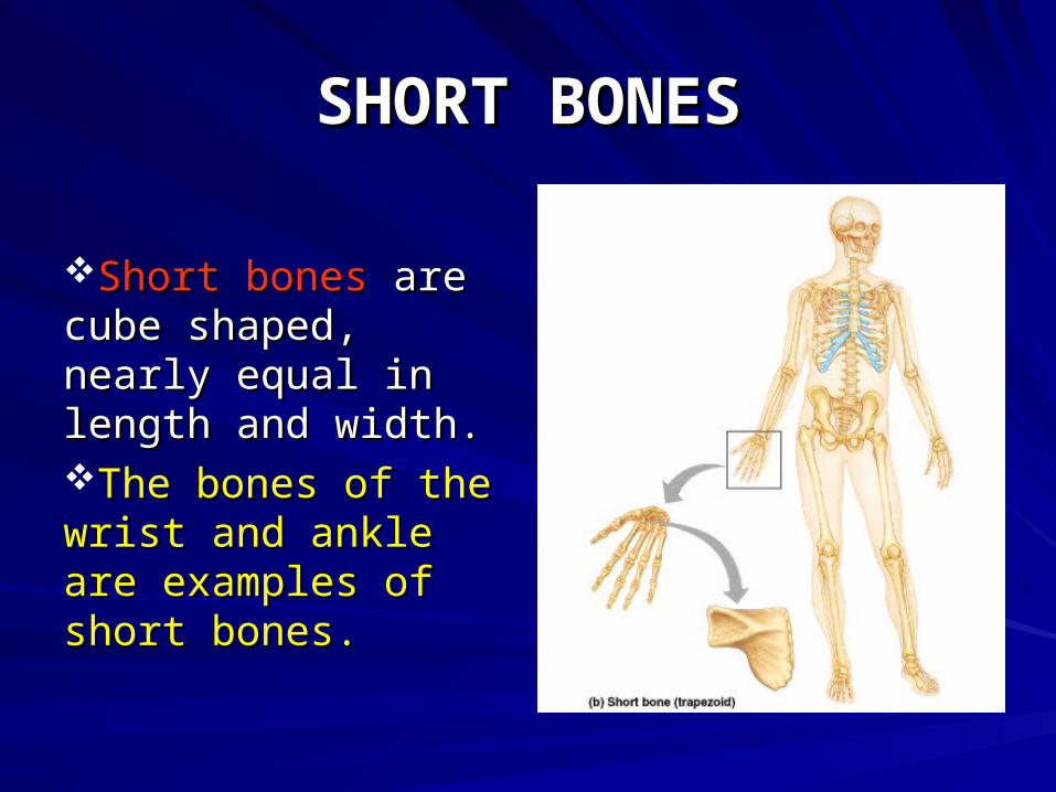
Short bonesShort bones are are cube shaped, nearly cube shaped, nearly equal in length and equal in length and width.width.The bones of the The bones of the wrist and ankle are wrist and ankle are examples of short examples of short bones.bones.
SHORT BONESSHORT BONES
A SPECIAL TYPE OF SHORT A SPECIAL TYPE OF SHORT
BONE IS A BONE IS A SESAMOND BONE.SESAMOND BONE.
THIS TYPE OF BONE IS A THIS TYPE OF BONE IS A
SHORT BONE WHICH FORMS SHORT BONE WHICH FORMS
WITHIN A TENDON. AN EXAMPLE WITHIN A TENDON. AN EXAMPLE
IS THE PATELLA, AND THE PISIFORM.IS THE PATELLA, AND THE PISIFORM.
FLAT BONESFLAT BONES
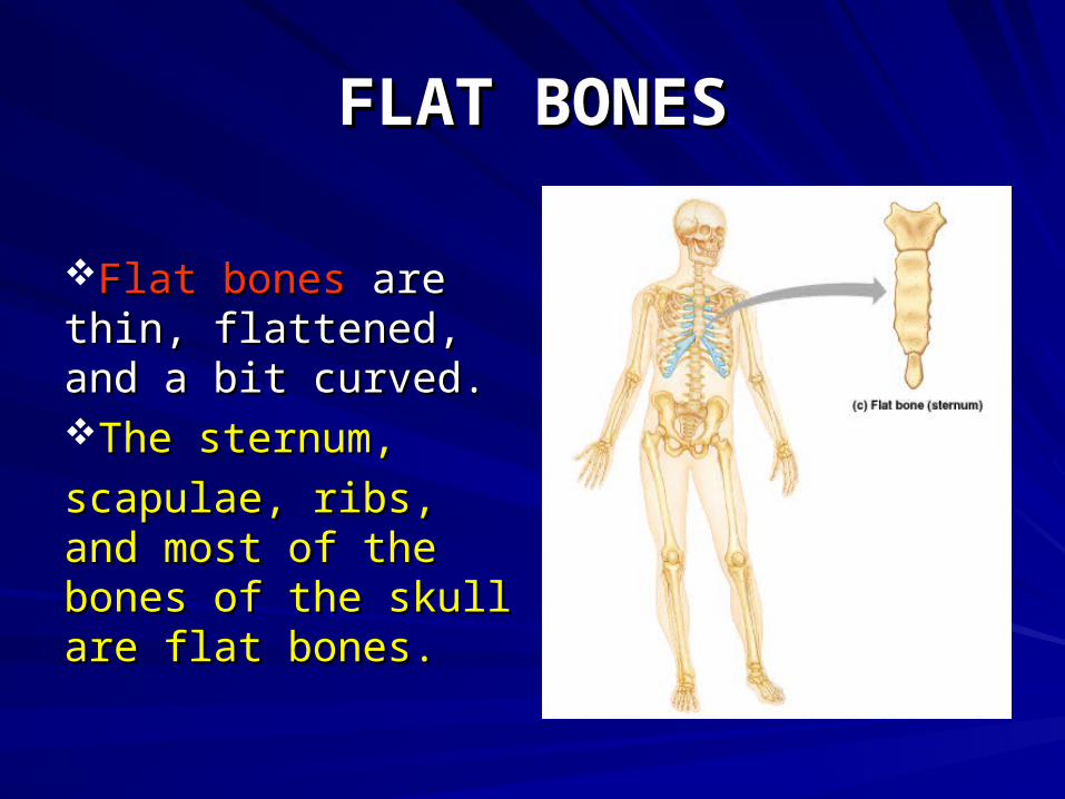
Flat bonesFlat bones are thin, are thin, flattened, and a bit flattened, and a bit curved. curved. The sternum, The sternum,
scapulae, ribs, and scapulae, ribs, and most of the bones of most of the bones of the skull are flat the skull are flat bones.bones.
IRREGULAR BONESIRREGULAR BONES
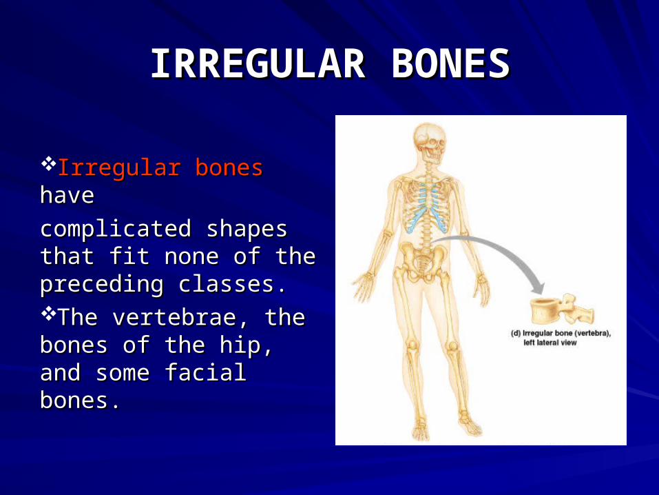
Irregular bonesIrregular bones havehave
complicated shapes complicated shapes that fit none of the that fit none of the preceding classes.preceding classes.The vertebrae, the The vertebrae, the bones of the hip, and bones of the hip, and some facial bones.some facial bones.
GROSS ANATOMY OF AGROSS ANATOMY OF A LONG BONE LONG BONE
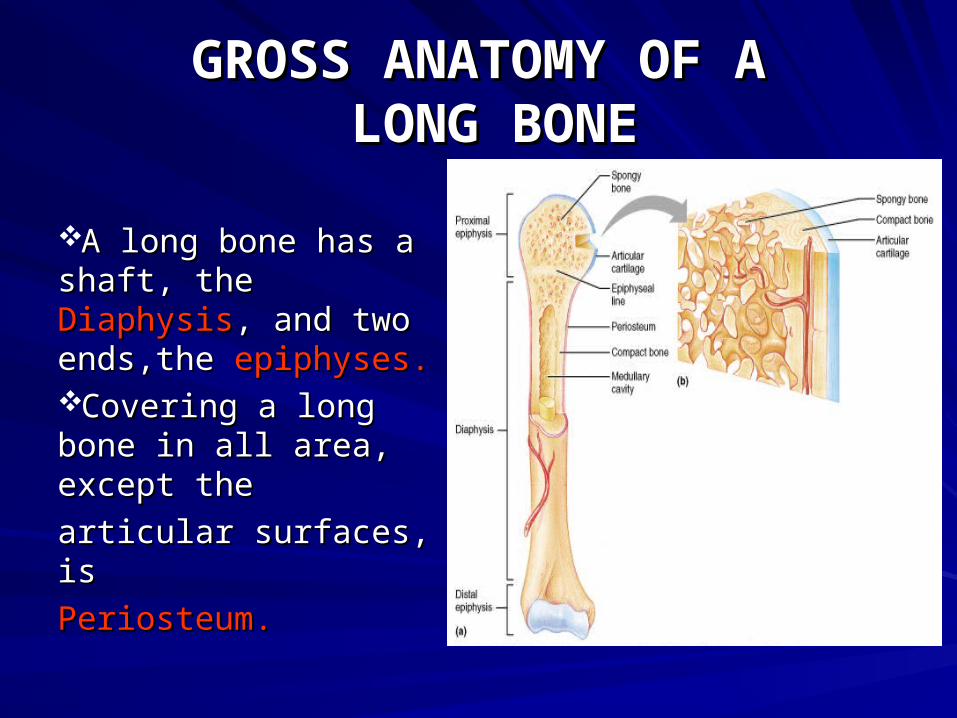
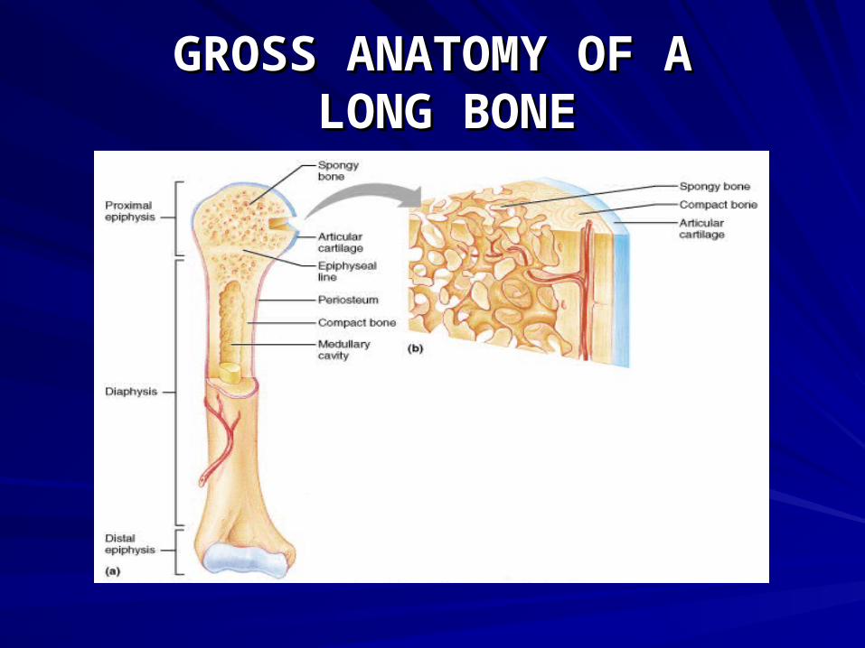
A long bone has a A long bone has a shaft, the shaft, the DiaphysisDiaphysis, , and two ends,the and two ends,the epiphyses.epiphyses.Covering a long Covering a long bone in all area, bone in all area, except the except the
articular surfaces, is articular surfaces, is
PeriosteumPeriosteum..
GROSS ANATOMY OF AGROSS ANATOMY OF A LONG BONE LONG BONE
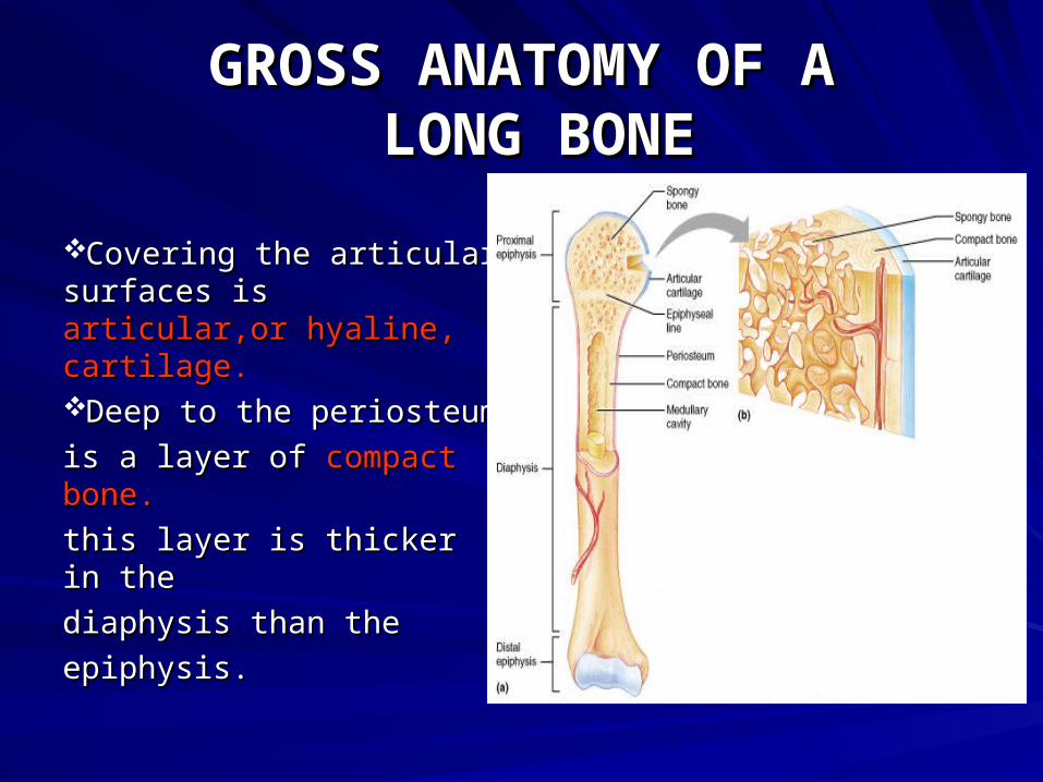
Covering the articular Covering the articular surfaces is surfaces is articular,or articular,or hyaline, cartilage.hyaline, cartilage.Deep to the periosteum Deep to the periosteum
is a layer of is a layer of compact compact bone.bone.
this layer is thicker in thethis layer is thicker in the
diaphysis than thediaphysis than the
epiphysis.epiphysis.
GROSS ANATOMY OF AGROSS ANATOMY OF A LONG BONE LONG BONE
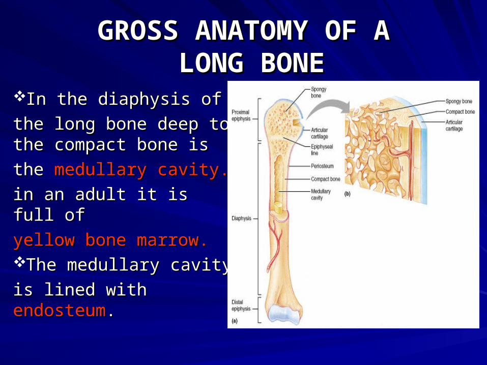
In the diaphysis of In the diaphysis of
the long bone deep to the long bone deep to the compact bone is the compact bone is
the the medullary cavity.medullary cavity.
in an adult it is full of in an adult it is full of
yellow bone marrow.yellow bone marrow.The medullary cavityThe medullary cavity
is lined with is lined with endosteumendosteum..
GROSS ANATOMY OF AGROSS ANATOMY OF A LONG BONE LONG BONE
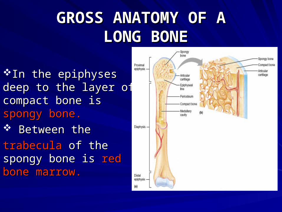
In the epiphyses deep In the epiphyses deep to the layer of compact to the layer of compact bone is bone is spongy bone.spongy bone. Between theBetween the
trabeculatrabecula of the spongy of the spongy bone is bone is red bone red bone marrow.marrow.
GROSS ANATOMY OF AGROSS ANATOMY OF A LONG BONE LONG BONE
MICROSCOPIC STRUCTUREMICROSCOPIC STRUCTURE OF COMPACT BONE OF COMPACT BONE
THE STRUCTURAL UNIT OF THE STRUCTURAL UNIT OF
COMPACT BONE IS THE COMPACT BONE IS THE OSTEON,OSTEON,
OR HAVERSIAN SYSTEM.OR HAVERSIAN SYSTEM. EACH OSTEON EACH OSTEON
IS AN ELONGATED CYLINDER IS AN ELONGATED CYLINDER
ORIENTED PARALLEL TO THE ORIENTED PARALLEL TO THE
LONG AXIS OF THE BONE.LONG AXIS OF THE BONE.
MICROSCOPIC STRUCTUREMICROSCOPIC STRUCTURE OF COMPACT BONE OF COMPACT BONE
MICROSCOPIC STRUCTUREMICROSCOPIC STRUCTURE OF COMPACT BONE OF COMPACT BONE
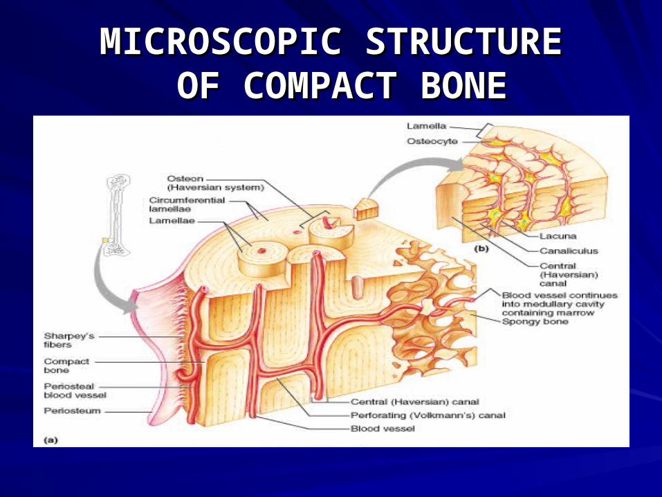
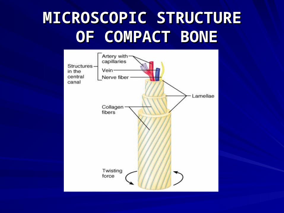
AN OSTEON IS A GROUP OF HOLLOWAN OSTEON IS A GROUP OF HOLLOW
TUBES OF BONE MATRIX,TUBES OF BONE MATRIX,
ONE PLACED OUTSIDE THE NEXTONE PLACED OUTSIDE THE NEXT
LIKE THE GROWTH RINGS OF A LIKE THE GROWTH RINGS OF A
TREE TRUNK. EACH OF THE MATRIXTREE TRUNK. EACH OF THE MATRIX
TUBES IS A TUBES IS A LAMELLA.LAMELLA.
MICROSCOPIC STRUCTUREMICROSCOPIC STRUCTURE OF COMPACT BONE OF COMPACT BONE
THE COLLAGEN FIBERS IN ATHE COLLAGEN FIBERS IN A
PARTICULAR LAMELLA RUN IN PARTICULAR LAMELLA RUN IN
A SINGLE DIRECTION. A SINGLE DIRECTION.
MICROSCOPIC STRUCTUREMICROSCOPIC STRUCTURE OF COMPACT BONE OF COMPACT BONE
MICROSCOPIC STRUCTUREMICROSCOPIC STRUCTURE OF COMPACT BONE OF COMPACT BONE
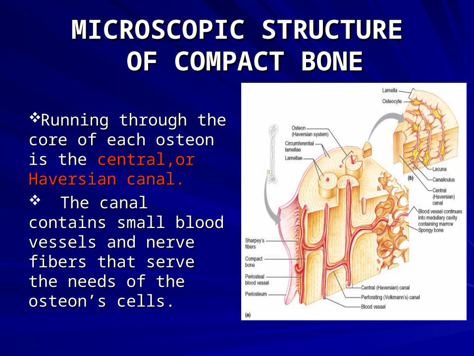
Running through the Running through the core of each osteon is the core of each osteon is the central,or Haversian canal.central,or Haversian canal. The canal contains The canal contains small blood vessels and small blood vessels and nerve fibers that serve the nerve fibers that serve the needs of the osteon’s cells.needs of the osteon’s cells.

MICROSCOPIC STRUCTUREMICROSCOPIC STRUCTURE OF COMPACT BONE OF COMPACT BONE
Spider shaped Spider shaped osteocytes osteocytes occupy small cavities occupy small cavities called called lacunaelacunae at the at the junctions of the lamellae.junctions of the lamellae.Hair like canals called Hair like canals called
canaliculi canaliculi connect the connect the
lacunae to each other.lacunae to each other.The space between these The space between these
structures is occupied by structures is occupied by bony bony matrix.matrix.

MICROSCOPIC STRUCTUREMICROSCOPIC STRUCTURE OF COMPACT BONE OF COMPACT BONE
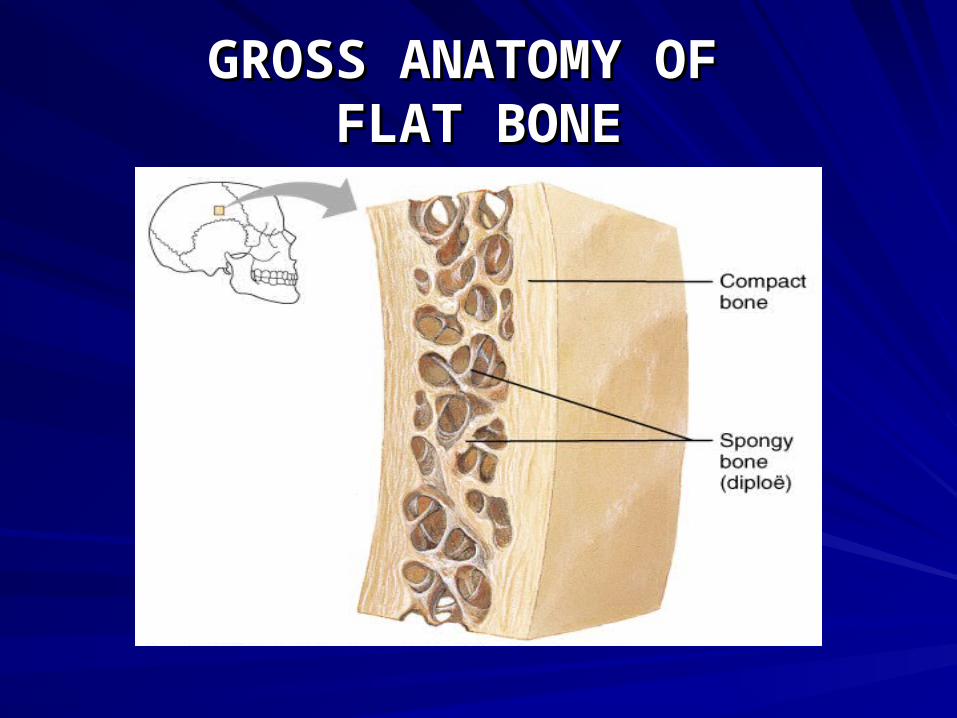
GROSS ANATOMY OF GROSS ANATOMY OF FLAT BONEFLAT BONE
OSSIFICATIONOSSIFICATION
OSSFICATION OR OSTEOGENESISOSSFICATION OR OSTEOGENESIS
IS THE PROCESS OF BONE FORMATION.IS THE PROCESS OF BONE FORMATION.
THERE ARE 2 MECHANISM THERE ARE 2 MECHANISM
WHICH FORM BONE:WHICH FORM BONE:
1. INTRAMEMBRANOUS1. INTRAMEMBRANOUS
2. ENDOCHONDRAL2. ENDOCHONDRAL
OSSIFICATIONOSSIFICATION
INTRAMEMBRANOUS OSSIFICATIONINTRAMEMBRANOUS OSSIFICATION
RESULTS IN THE FORMATION RESULTS IN THE FORMATION
OF THE CRANIAL BONES AND OF THE CRANIAL BONES AND
THE CLAVICLES.THE CLAVICLES.
OSSIFICATIONOSSIFICATION
ENDOCHONDRAL OSSIFICATIONENDOCHONDRAL OSSIFICATION
RESULTS IN THE FORMATION OF THERESULTS IN THE FORMATION OF THE
BONES BELOW THE BONES BELOW THE
SKULL, WITH THE EXCEPTION OF SKULL, WITH THE EXCEPTION OF
THE CLAVICLES.THE CLAVICLES.
OSSIFICATIONOSSIFICATION
THREE TYPES OF CELLS ARE INVOLVEDTHREE TYPES OF CELLS ARE INVOLVED
IN BOTH MECHANISM OF OSSIFICATION:IN BOTH MECHANISM OF OSSIFICATION:
1. OSTEOBLASTS1. OSTEOBLASTS
2. OSTEOCLASTS2. OSTEOCLASTS
3. OSTEOCYTES3. OSTEOCYTES
STEPS OF INTRAMEMBRANOUS STEPS OF INTRAMEMBRANOUS OSSIFICATIONOSSIFICATION
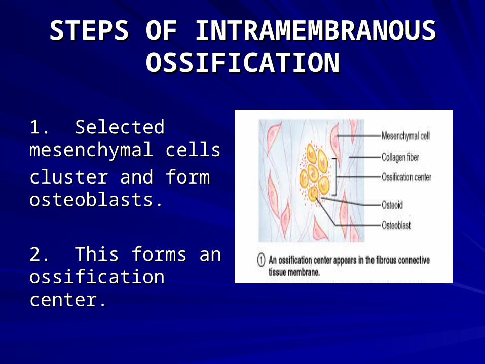
1. Selected 1. Selected mesenchymal cellsmesenchymal cells
cluster and form cluster and form osteoblasts.osteoblasts.
2. This forms an 2. This forms an ossification center.ossification center.
STEPS OF INTRAMEMBRANOUS STEPS OF INTRAMEMBRANOUS OSSIFICATIONOSSIFICATION
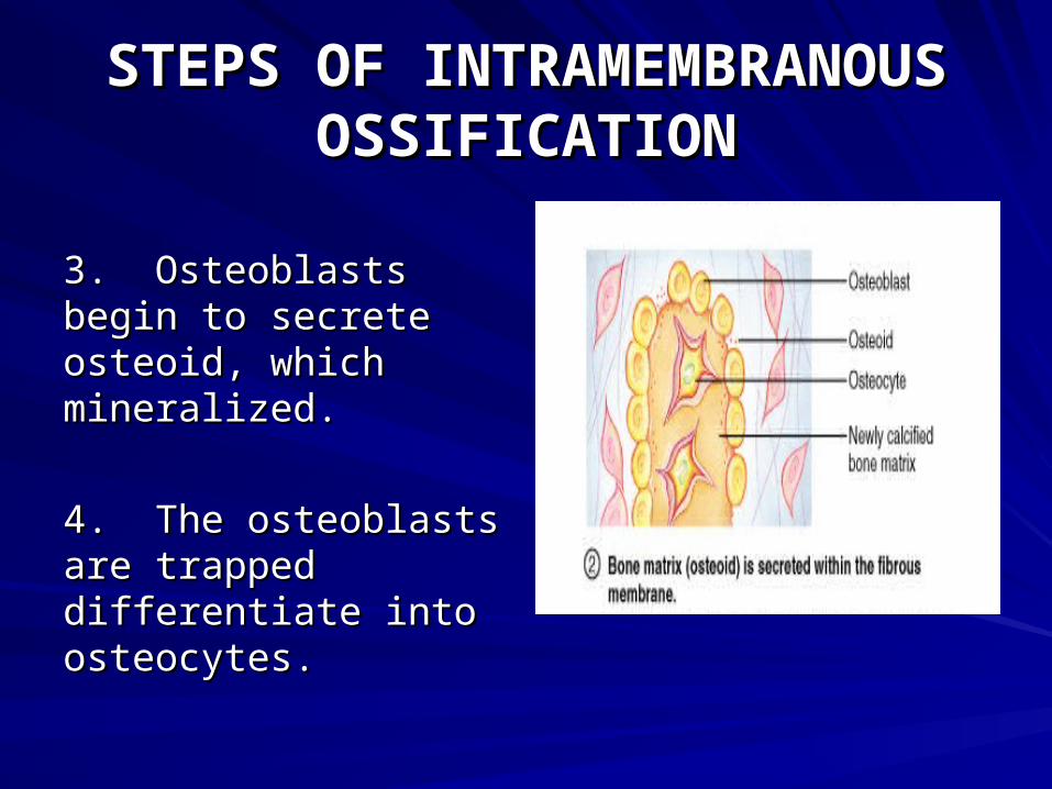
3. 3. Osteoblasts begin Osteoblasts begin to secrete osteoid, to secrete osteoid, which mineralized.which mineralized.
4. The osteoblasts 4. The osteoblasts are trapped are trapped differentiate into differentiate into osteocytes.osteocytes.
STEPS OF INTRAMEMBRANOUS STEPS OF INTRAMEMBRANOUS OSSIFICATIONOSSIFICATION
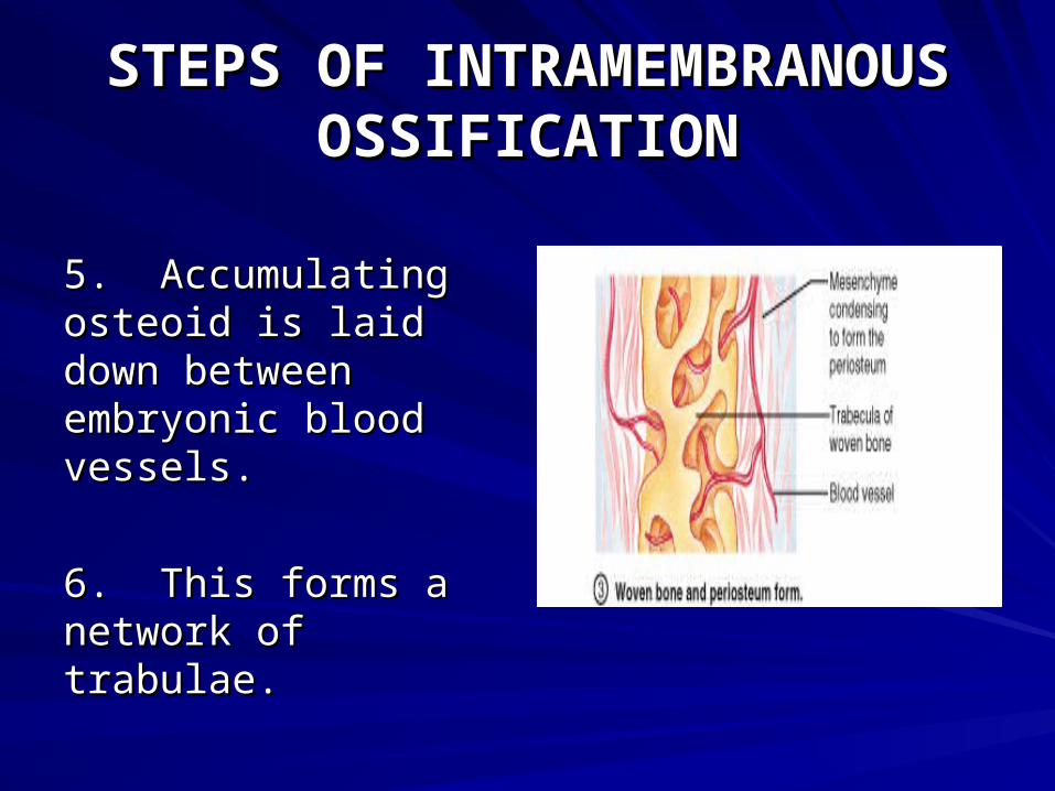
5. Accumulating 5. Accumulating osteoid is laid down osteoid is laid down between embryonic between embryonic blood vessels.blood vessels.
6. This forms a 6. This forms a network of trabulae.network of trabulae.
STEPS OF INTRAMEMBRANOUS STEPS OF INTRAMEMBRANOUS OSSIFICATIONOSSIFICATION
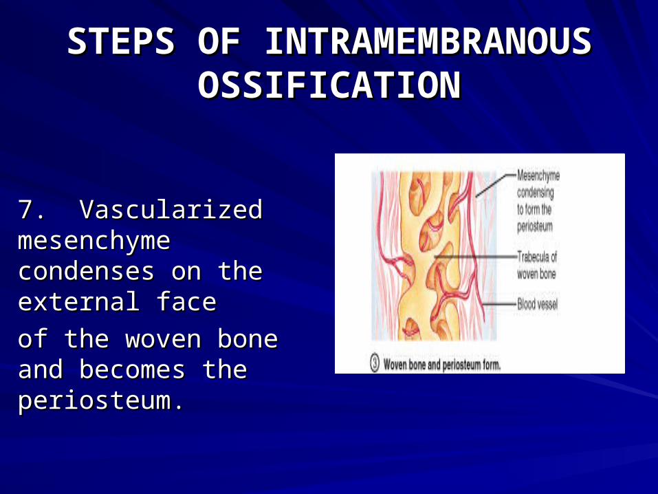
7. Vascularized 7. Vascularized mesenchyme mesenchyme condenses on the condenses on the external face external face
of the woven bone and of the woven bone and becomes the becomes the periosteum.periosteum.
STEPS OF INTRAMEMBRANOUS STEPS OF INTRAMEMBRANOUS OSSIFICATIONOSSIFICATION
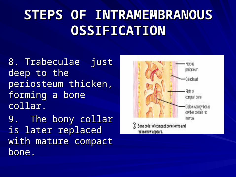
8. Trabeculae just 8. Trabeculae just deep to the deep to the periosteum thicken, periosteum thicken, forming a bone collar.forming a bone collar.
9. The bony collar is 9. The bony collar is later replaced with later replaced with mature compact bone.mature compact bone.
STEPS OF INTRAMEMBRANOUS STEPS OF INTRAMEMBRANOUS OSSIFICATIONOSSIFICATION
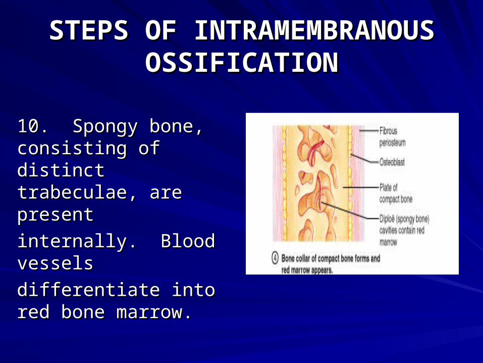
10. Spongy bone, 10. Spongy bone, consisting of distinct consisting of distinct trabeculae, are trabeculae, are presentpresent
internally. Blood internally. Blood vessels vessels
differentiate into red differentiate into red bone marrowbone marrow..
STEPS OF ENDOCHONDRAL STEPS OF ENDOCHONDRAL OSSIFICATIONOSSIFICATION
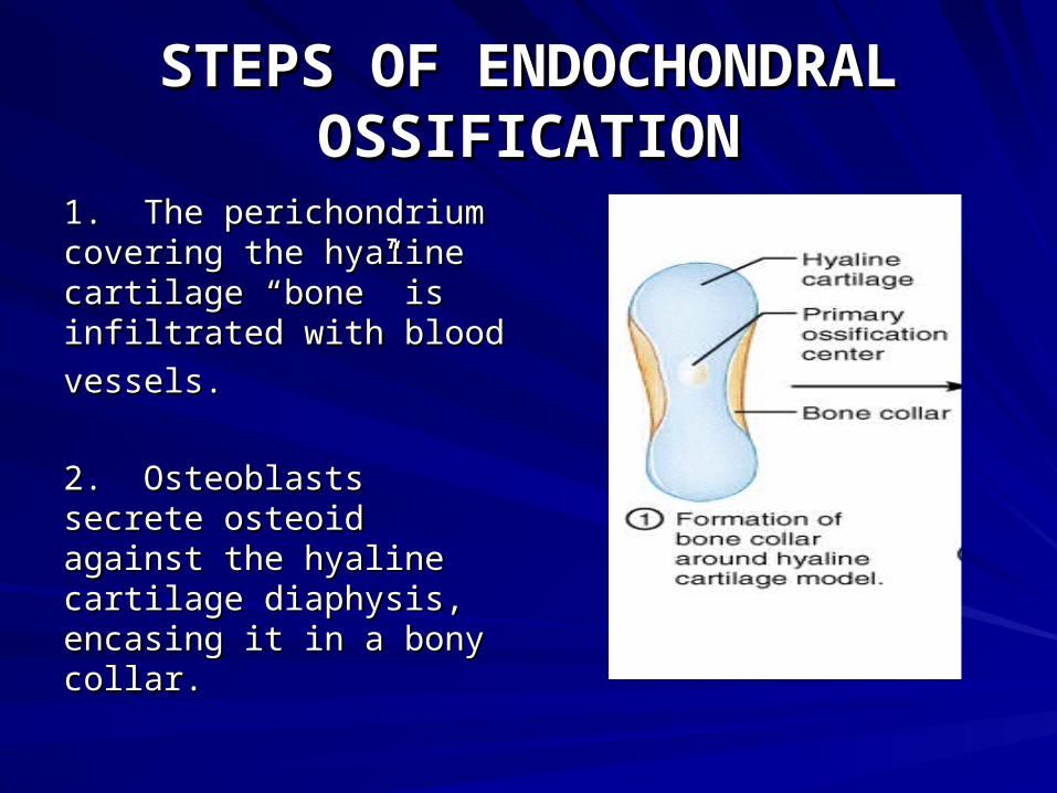
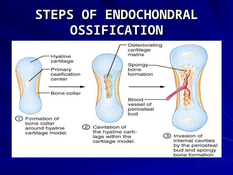
1. The perichondrium 1. The perichondrium covering the hyaline covering the hyaline cartilage “bone” is cartilage “bone” is infiltrated with bloodinfiltrated with blood
vessels.vessels.
2. Osteoblasts secrete 2. Osteoblasts secrete osteoid against the osteoid against the hyaline cartilage hyaline cartilage diaphysis, encasing it in diaphysis, encasing it in a bony collar.a bony collar.
STEPS OF ENDOCHONDRAL STEPS OF ENDOCHONDRAL OSSIFICATIONOSSIFICATION
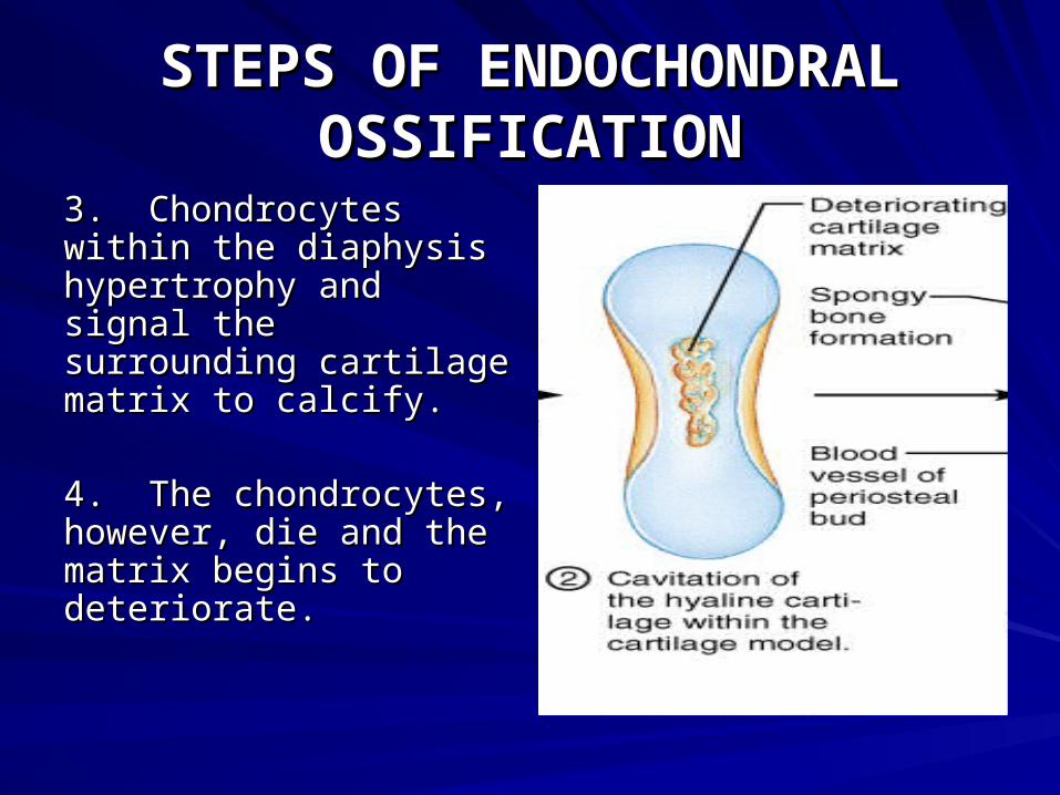
3. Chondrocytes 3. Chondrocytes within the diaphysis within the diaphysis hypertrophy and hypertrophy and signal the signal the surrounding cartilage surrounding cartilage matrix to calcify.matrix to calcify.
4. The chondrocytes, 4. The chondrocytes, however, die and the however, die and the matrix begins to matrix begins to deteriorate.deteriorate.
STEPS OF ENDOCHONDRAL STEPS OF ENDOCHONDRAL OSSIFICATIONOSSIFICATION
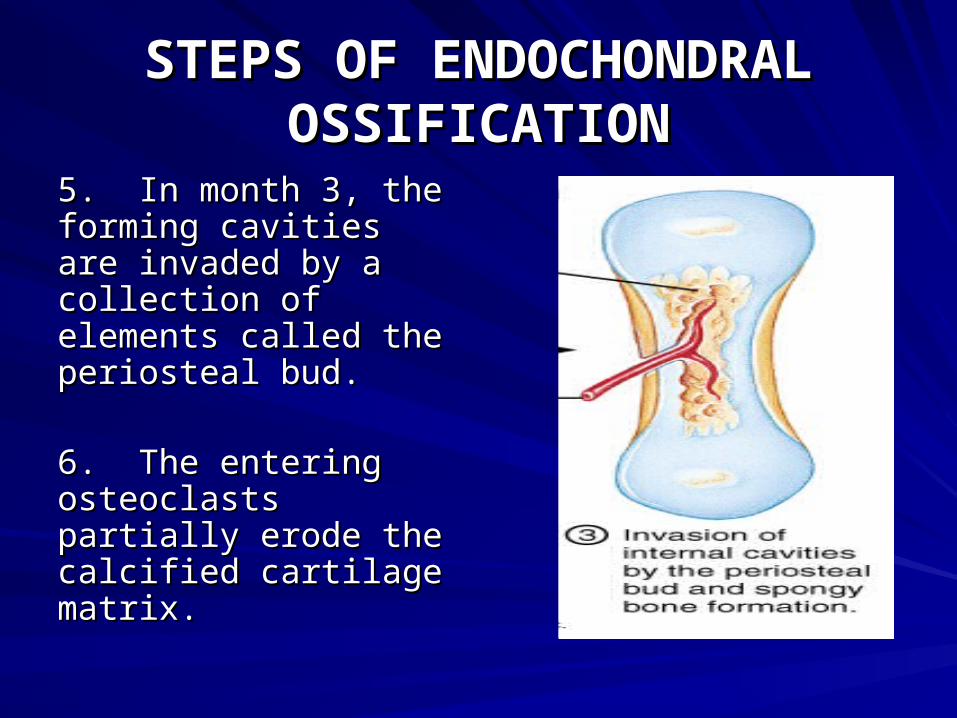
5. In month 3, the 5. In month 3, the forming cavities are forming cavities are invaded by a invaded by a collection of collection of elements called the elements called the periosteal bud.periosteal bud.
6. The entering 6. The entering osteoclasts partially osteoclasts partially erode the calcified erode the calcified cartilage matrix.cartilage matrix.

STEPS OF ENDOCHONDRAL STEPS OF ENDOCHONDRAL OSSIFICATIONOSSIFICATION
STEPS OF ENDOCHONDRAL STEPS OF ENDOCHONDRAL OSSIFICATIONOSSIFICATION
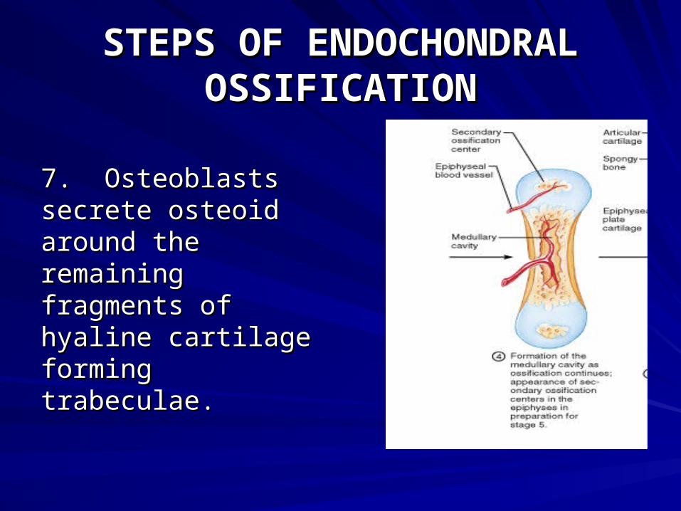
7. Osteoblasts 7. Osteoblasts secrete osteoid secrete osteoid around the around the remaining fragments remaining fragments of hyaline cartilage of hyaline cartilage forming trabeculae.forming trabeculae.
STEPS OF ENDOCHONDRAL STEPS OF ENDOCHONDRAL OSSIFICATIONOSSIFICATION
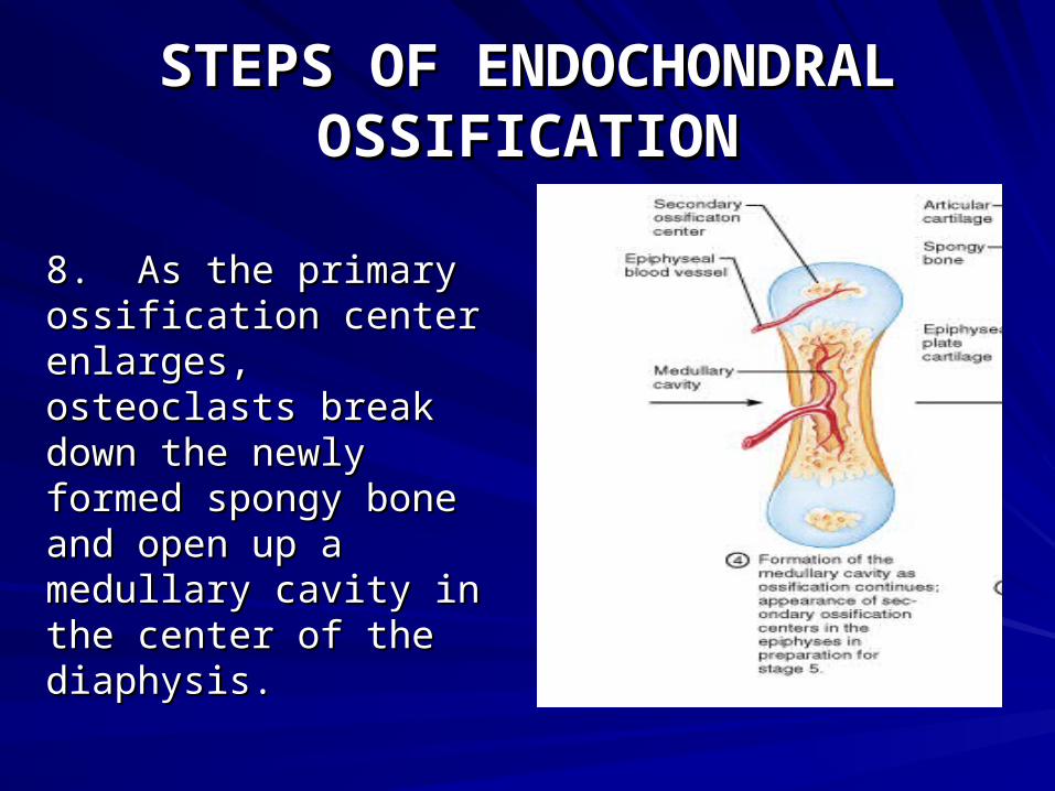
8. As the primary 8. As the primary ossification center ossification center enlarges, osteoclasts enlarges, osteoclasts break down the newly break down the newly formed spongy bone formed spongy bone and open up a and open up a medullary cavity in medullary cavity in the center of the the center of the diaphysis.diaphysis.
STEPS OF ENDOCHONDRAL STEPS OF ENDOCHONDRAL OSSIFICATIONOSSIFICATION
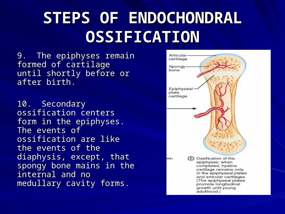
9. The epiphyses remain 9. The epiphyses remain formed of cartilage until formed of cartilage until shortly before or after shortly before or after birth.birth.
10. Secondary 10. Secondary ossification centers form ossification centers form in the epiphyses. The in the epiphyses. The events of ossification are events of ossification are like the events of the like the events of the diaphysis, except, that diaphysis, except, that spongy bone mains in the spongy bone mains in the internal and no medullary internal and no medullary cavity forms.cavity forms.
STEPS OF ENDOCHONDRAL STEPS OF ENDOCHONDRAL OSSIFICATIONOSSIFICATION

STEPS OF ENDOCHONDRAL STEPS OF ENDOCHONDRAL OSSIFICATIONOSSIFICATION
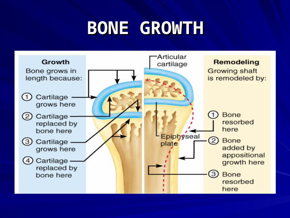
BONE GROWTHBONE GROWTH
THERE ARE 2 TYPES OF THERE ARE 2 TYPES OF
BONE GROWTH:BONE GROWTH:
1.1. LONGITUDINAL--LENGTH LONGITUDINAL--LENGTH
2. APPOSITIONAL--DIAMETER 2. APPOSITIONAL--DIAMETER
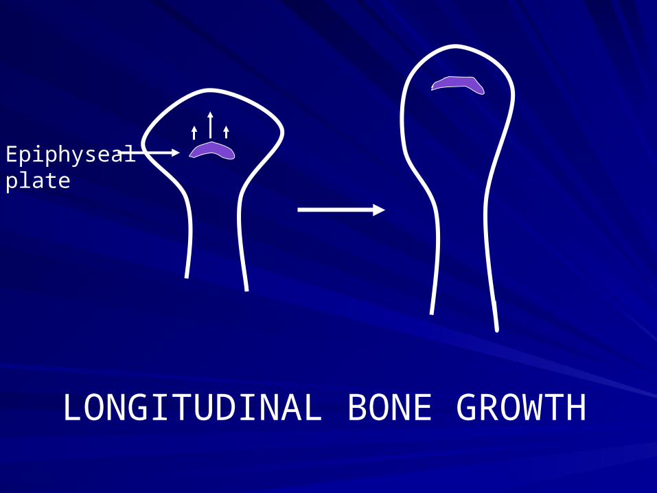
LONGITUDINAL BONE GROWTH
Epiphysealplate
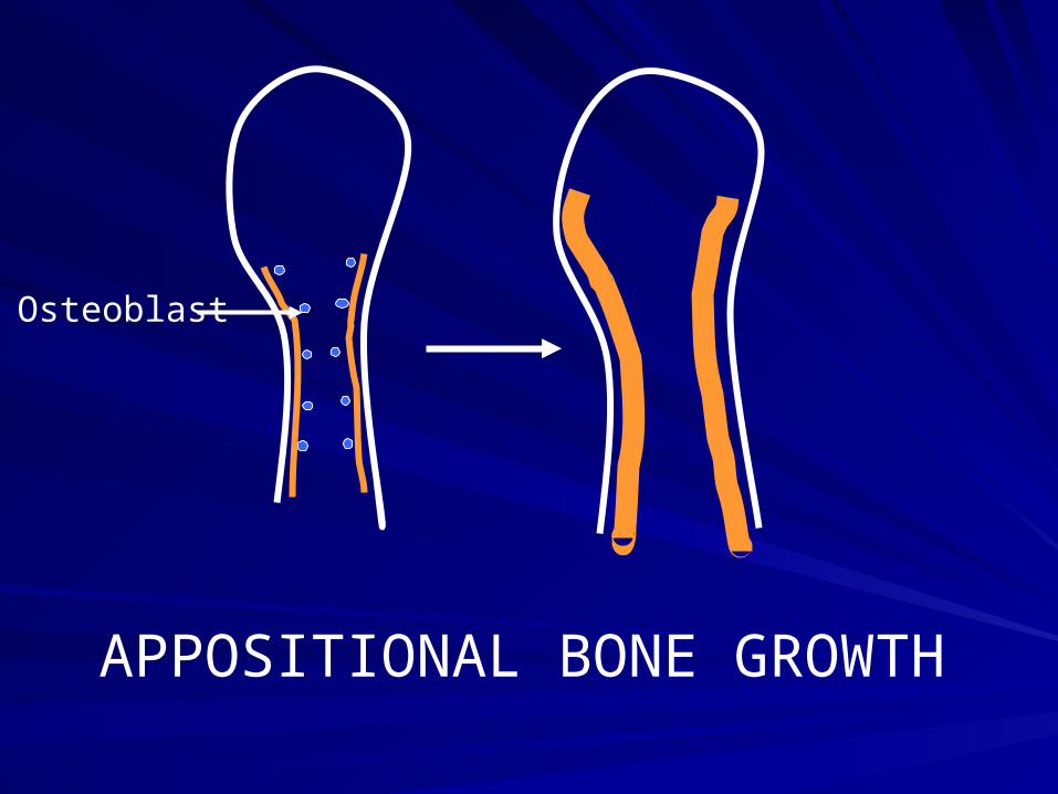
APPOSITIONAL BONE GROWTH
Osteoblast
BONE GROWTHBONE GROWTH
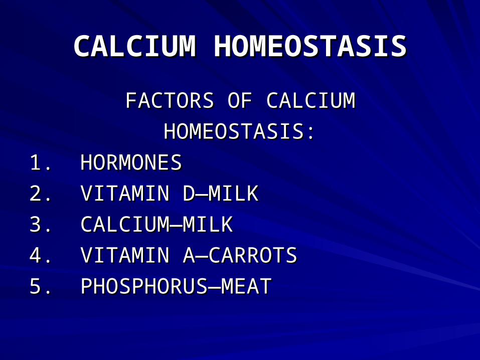
CALCIUM HOMEOSTASISCALCIUM HOMEOSTASIS
FACTORS OF CALCIUMFACTORS OF CALCIUM
HOMEOSTASIS:HOMEOSTASIS:
1. HORMONES1. HORMONES
2. VITAMIN D—MILK2. VITAMIN D—MILK
3. CALCIUM—MILK3. CALCIUM—MILK
4. VITAMIN A—CARROTS4. VITAMIN A—CARROTS
5. PHOSPHORUS—MEAT 5. PHOSPHORUS—MEAT
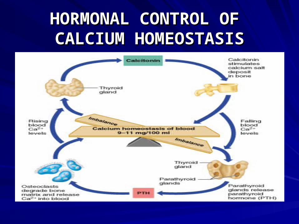
HORMONAL CONTROL OF HORMONAL CONTROL OF CALCIUM HOMEOSTASISCALCIUM HOMEOSTASIS
CALCIUM HOMEOSTASISCALCIUM HOMEOSTASIS
OTHER FACTORS IN CALCIUMOTHER FACTORS IN CALCIUM
HOMEOSTASIS:HOMEOSTASIS:
1. VITAMIN D—AIDS IN THE ABSORPTION1. VITAMIN D—AIDS IN THE ABSORPTION
OF BOTH CALCIUM AND PHOSPHORUS.OF BOTH CALCIUM AND PHOSPHORUS.
2. VITAMIN A—HELPS THE OSTEOBLASTS2. VITAMIN A—HELPS THE OSTEOBLASTS
PRODUCE BONY MATRIX.PRODUCE BONY MATRIX.
CALCIUM HOMEOSTASISCALCIUM HOMEOSTASIS
3. TESTOSTERONE AND ESTROGEN—3. TESTOSTERONE AND ESTROGEN—
STIMULATES BONE DEPOSITION OF STIMULATES BONE DEPOSITION OF
CALCIUM STARTING AT PUBERTY.CALCIUM STARTING AT PUBERTY.
HOMEOSTATIC IMBALANCES HOMEOSTATIC IMBALANCES OF THE SKELETAL SYSTEMOF THE SKELETAL SYSTEM
RICKETSRICKETS
1. DISEASE OF CHILDREN DUE TO1. DISEASE OF CHILDREN DUE TO
LACK OF VITAMIN D.LACK OF VITAMIN D.
2. CALCIUM IS NOT DEPOSITED.2. CALCIUM IS NOT DEPOSITED.
3. BOWING OF THE BONES.3. BOWING OF THE BONES.
HOMEOSTATIC IMBALANCES HOMEOSTATIC IMBALANCES OF THE SKELETAL SYSTEMOF THE SKELETAL SYSTEM
OSTEOMALCIAOSTEOMALCIA
1. RICKETS IN ADULTS1. RICKETS IN ADULTS
2. DUE TO A LACK OF VITAMIN D2. DUE TO A LACK OF VITAMIN D
3. CALCIUM IS NOT DEPOSITED IN3. CALCIUM IS NOT DEPOSITED IN
BONE.BONE.
4. MAIN SYMPTOM IS PAIN WHEN WEIGHT IS PUT ON THE AFFECTED BONE.

HOMEOSTATIC IMBALANCES HOMEOSTATIC IMBALANCES OF THE SKELETAL SYSTEMOF THE SKELETAL SYSTEM
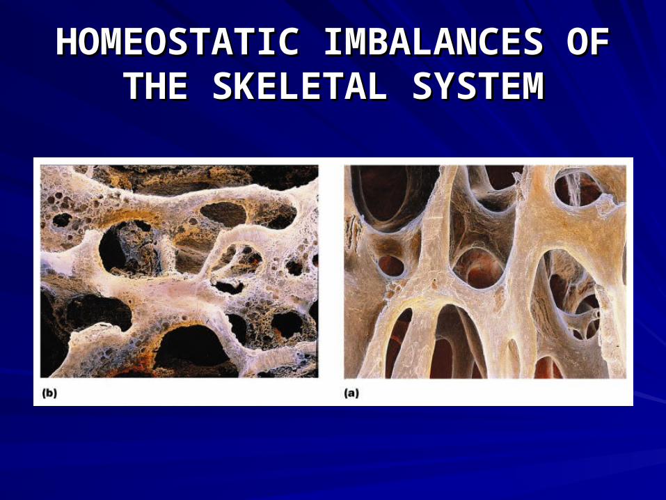
OSTEOPOROSISOSTEOPOROSIS
1. BONE REABSORPTION IS GREATER 1. BONE REABSORPTION IS GREATER
THAN BONE DEPOSITION.THAN BONE DEPOSITION.
2. CAUSES:2. CAUSES:
A. LACK OF ESTROGEN A. LACK OF ESTROGEN
B. LACK OF EXERCISEB. LACK OF EXERCISE
C. INADEQUATE INTAKEC. INADEQUATE INTAKE
D. LACK OF VITAMIN DD. LACK OF VITAMIN D
HOMEOSTATIC IMBALANCES HOMEOSTATIC IMBALANCES OF THE SKELETAL SYSTEMOF THE SKELETAL SYSTEM
OSTEOPOROSISOSTEOPOROSIS
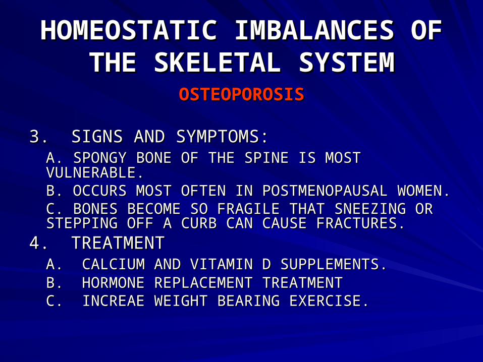
3. SIGNS AND SYMPTOMS:3. SIGNS AND SYMPTOMS:A. SPONGY BONE OF THE SPINE IS MOST VULNERABLE.A. SPONGY BONE OF THE SPINE IS MOST VULNERABLE.B. OCCURS MOST OFTEN IN POSTMENOPAUSAL B. OCCURS MOST OFTEN IN POSTMENOPAUSAL WOMEN.WOMEN.C. BONES BECOME SO FRAGILE THAT SNEEZING OR C. BONES BECOME SO FRAGILE THAT SNEEZING OR STEPPING OFF A CURB CAN CAUSE FRACTURES.STEPPING OFF A CURB CAN CAUSE FRACTURES.
4. TREATMENT4. TREATMENTA. CALCIUM AND VITAMIN D SUPPLEMENTS.A. CALCIUM AND VITAMIN D SUPPLEMENTS.B. HORMONE REPLACEMENT TREATMENTB. HORMONE REPLACEMENT TREATMENTC. INCREAE WEIGHT BEARING EXERCISE.C. INCREAE WEIGHT BEARING EXERCISE.
HOMEOSTATIC IMBALANCES HOMEOSTATIC IMBALANCES OF THE SKELETAL SYSTEMOF THE SKELETAL SYSTEM