simple and rapid dot-enzyme immunoassay for visual detection of rinderpest antibodies in bovine and...
TRANSCRIPT

Trop. Anita. Hlth Prod. (1986) lg, 209-216
SIMPLE AND RAPID DOT-ENZYME IMMUNOASSAY FOR VISUAL DETECTION OF RINDERPEST
ANTIBODIES IN BOVINE AND CAPRINE SERA
A. A~HAR and D. J. MYERS
Animal Diseases Research Institute, PO Box 11300, Station H, Nepean, Ontario, K2H 81"9, Canada
SUMMARY
A modified solid phase enzyme immunoassay (EIA) is described for visual detection of anti-rinderpest virus (RPV) antibodies in cattle and goat sera. Dots of RPV antigens were adsorbed to nitrocellulose (NC) paper (hence Dot-EIA) and the adsorptive reactive sites were blocked with skim milk powder. After immersion in bovine or caprine test serum bound antibodies were reacted with a peroxidasr anti-bovine or anti-caprine IgG (H & L), respectively. Positive reactions were easily visualized as red-brown dots after enzyme degradation of a substrate containing hydrogen peroxide and amino- ethylcarbazole (AEC). The Dot-EIA was comparable to the serum neutralisa- tion (SN) test in fts ability to detect antibody in bovine sera seven or ten days after experimental infection (DPI) with live attenuated Kabete "0'" (RBOK) strain of RPV (grown in Vero cells) by a combination of subcutaneous (s/c), intravenous (i/v) or intranasal (i/n) routes. Early (seven DPI) RPV antibodies were detected in a serum sample from one goat experimentally infected with RPV by combined s /c- i /v routes but not in another goat only infected intranasally. The specificity of the Dot-EIA was equal to that of the SN test, as serum samples, collected from these experimental animals and those inoculated with non infected Vero cell culture fluid, with SN titres of 0.3 or lower were all negative by Dot-EIA. The Dot-EIA may have potential application as a rapid, simple and economical field test in diagnosis of rinderpest, vaccination surveillance and other seroepidemiological studies.
INTRODUCTION
The re-emergence of rinderpest (RP), a highly contagious and usually fatal disease of cattle and buffalo in Africa, the Middle East and Asia (Scott, 1985) has prompted many investigators to develop and apply new diagnostic tests which have been considered suitable for the detection of antibodies to RP virus (RPV) in cattle sera. Of these techniques, the enzyme-linked immunosorbent assay (ELISA) has proved to be highly sensitive and capable of assaying a large number of samples in sero-epidemiological studies (Anderson, Rowe and Taylor, 1983). However, until now the application of ELISA, like other serodiagnostic tests, has been limited to well-equipped laboratories with expertise in this method. A simple, inexpensive and reliable serological test that could be conducted without specialised facilities and .techniques has been sought for screening field sera for the presence of anti-RPV antibodies. Recently, based on the general principles of indirect enzyme immunoassay (EIA) we described and evaluated a simple and rapid Dot-EIA technique for visual detection of antibodies to pseudorabies virus in swine serum (Afshar, Wright and Dulac, 1986). The present communication describes the development of a similar Dot-EIA test for detection of antibodies to RPV in sera from experimentally infected cattle and goats and compares its performance to that of the micro-serum neutralisation (MSN) test.
2O9
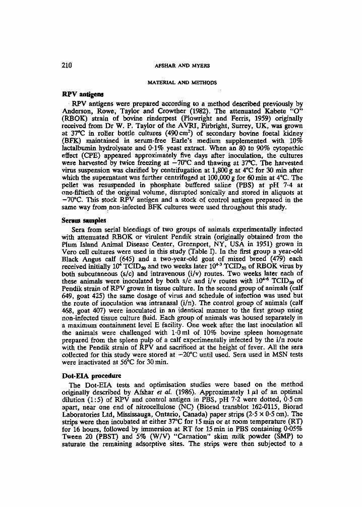
210 AFSHAR AND MYERS
MATERIAL AND METHODS
RPV aaligem RPV antigens were prepared according to a method described previously by
Anderson, Rowe, Taylor and Crowther (1982). The attenuated Kabete "O" (RBOK) strain of bovine rinderpest (Plowright and Ferris, 1959) originally received from Dr W. P. Taylor of the AVRI, Pirbright, Surrey, UK, was grown at 37"C in roller bottle cultures (490 em 2) of secondary bovine foetal kidney (BFK) maintained in serum-free Earle's medium supplemented with 10% lactalbumin hydrolysate and 0.1% yeast extract. When an 80 to 90% cytopathic effect (CPE) appeared approximately five days after inoculation, the cultures were harvested by twice freezing at -70"C and thawing at 37"C. The harvested virus suspension was clarified by centrifugation at 1,800 g at 4"C for 30 rain after which the supernatant was further centrifuged at 100,000 g for 60 rain at 4"C. The pellet was resuspended in phosphate buffered saline (PBS) at pH 7-4 at one-fiftieth of the original volume, disrupted sonically and stored in aliquots at -70"C. This stock RPV antigen and a stock of control antigen prepared in the same way fromnon-infected BFK cultures were used throughout this study.
Serum samples Sera from serial bleedings of two groups of animals experimentally infected
with attenuated RBOK or virulent Pendik strain (originally obtained from the Plum Island Animal Disease Center, Greenport, NY, USA in 1951) grown in Vero cell cultures were used in this study (Table I). In the first group a year-old Black Angus calf (645) and a two-year-old goat of mixed breed (479) each received initially 104 TCIDso and two weeks later 10 4.3 TCIDso of RBOK virus by both subcutaneous (s/c) and intravenous (i/v) routes. Two weeks later each of these animals were inoculated by both s/e and i/v routes with 104 .4 TCIDso of Pendik strain of RPV grown in tissue culture. In the second group of animals (calf 649, goat 425) the same dosage of virus and schedule of infection was used but the route of inoculation was intranasal (i/n). The control group of animals (calf 468, goat 407) were inoculated in an identical manner to the first group using non-infected tissue culture fluid. Each group of animals was housed separately in a maximum containment level E facility. One week after the last inoculation all the animals were challenged with 1.Oral of 10% bovine spleen homogenate prepared from the spleen pulp of a calf experimentally infected by the i/n route with the Pendik strain of RPV and sacrificed at the height of fever. All the sera collected for this study were stored at -20~ until used. Sera used in MSN tests were inactivated at 56~ for 30 min.
Dot-EIA procedure The Dot-EIA tests and optimisation studies were based on the method
originally described by Afshar et al. (1986). Approximately 1/~1 of an optimal dilution (1:5) of R~'V and control antigen in PBS, pH 7.2 were dotted, 0-5 cm apart, near one end of nitrocellulose (NC) (Biorad transblot 162-0115, Biorad Laboratories Ltd, Mississauga, Ontario, Canada) paper strips (2.5 x 0.5 cm). The strips were then incubated at either 37"C for 15 rain or at room temperature (RT) for 16 hours, followed by immersion at RT for 15 rain in PBS containing 0.05% Tween 20 (PBST) and 5% (W/V) "Carnation" skim milk powder (SMP) to saturate the remaining adsorptive sites. The strips were then subjected to a
R I N D E R P ~ DOT-EIA 211
. I
~
II1
e@ " -
~ m
".=8 z"G"
~ua
v v v v ~ v v v v v
I I I I I I I I I I
I I I I I I I I I I
v
v v v v v v v v v
I I I I I I I I I
I I I I I I I I I
V V V V V V 6 6 6 ~ ~ I I I I I I + + 1 + + + + + +
I I I I I I I I I § 2 4 7
I I I ~ + + + + + ~ + +
I I I + + + + + § 2 4 7 2 4 7
I I + + + + § 2 4 7 2 4 7 2 4 7
I I + + + + + + + + + + §
I I + + + + + + + + + + + + +
~ ~ �9
0 O 0 ~ 0

212 AFSHAR AND MYERS
15 min wash in three changes of PBST with 3% SMP. The strips were immersed in a predetermined dilution (1:200) in PBST of the test serum sample and incubated at RT for 45 rain. Following another 15 rain wash (two changes of PBST with 3% SMP and one final change of PBST) the strips were immersed in a 1:160 dilution in PBST of horseradish peroxidase labelled rabbit anti-bovine IgG (Cappel Laboratories, Malvern, PA, U.S.A.) or anti-goat IgG (Miles Laboratory Ltd, Rexdale, Ontario, Canada) for test sera from cattle or goats respectively and incubated at RT for 45 rain. Following a final 15 min wash in three changes of PBST the strips were immersed at RT in substrate solution, prepared freshly by dissolving 10 mg of 3 amino 9 ethylcarbazole (AEC) in 3.0 ml of N,N-diethyl- formamide (Aldrich Chemical Co, Milwaukee, Wisconsin, USA) then slowly adding to 50 ml of 0.5 acetate/acetic acid buffer, pH 5.0 and 0-2 ml of 3% H202. The enzymatic reaction was stopped by three washes in tap water and the strips were dried and visually inspected for colour development.
MSN test Serum samples were tested for the presence of neutralising antibodies
according to a micro-test procedure described by Mushi and Plowdght (1979) using serial doubling dilutions. An estimated RBOK rinderpest virus dose of 100 to 300 TCIDso per well was added to the serum dilutions and the mixture incubated at RT for 90 min. After this time one volume (25 ~1) of BFK cell suspension (300,000 cell/ml) in Earle's maintenance medium containing 5% foetal bovine serum and antibiotics was added to each well. The microplates were incubated in a humidified atmosphere of 5% CO2 and 95% air at 37~ for 10 days at which time final observation for CPE was made. The neutralising antibody titres were calculated by the method of K~irber (1931) and are expressed as the log 10 of the reciprocal serum dilution which protected 50% of the BFK cells. Titres of 0-3 and under were considered as negative, of 0-6 and above as positive and in between as trace positive.
RESULTS
Examples of some typical positive and negative Dot-EIA reaction with the same bovine and caprine sera are shown in Fig. 1. Positive immunobinding reactions between RPV antibody and RPV antigen gave clear red-brown dots while the negative control antigen gave no colour. The results of the screening Dot-EIA and SN titres for the serum samples from experimentally infected cattle and goats are shown in Table I. RPV antibody was demonstrable by the Dot-EIA at seven days post-infection (DPI) in the sera of calf 645 and goat 479, inoculated by both s/c and i/v routes and at 10 DPI in serum from calf 649 inoculated by the i/n route alone. RPV antibody was not demonstrable by the Dot-EIA in serum samples collected from goat 425 until 14 days post challenge (DPC) with the virulent Pendik strain of RPV. Based on an established positive SN titre of 0-6 (Anderson et aL, 1983) the Dot-EIA results were in agreement with SN results except in two serum samples collected from goat 425 on 28 and 34 DPI when RPV antibody was demonstrable by SN but not Dot-EIA (Table I). The SN results indicated that unlike the other infected animals this goat did not seroconvert after the initial inoculation of RBOK strain of RPV and only developed RPV antibody 14 days after the second dose of RBOK virus inoculum. RPV antibody was not demonstrable by Dot-EIA and SN tests in any of the sera

RINDERPEST DOT-EIA 213
Panel A Panel B 1 1
I 2 2 __
3
4 4
I I
5 5
6 6
7 ' 7
8 8
9
llI 10
11
i 9
10
11
t t t t ~> ~>
rr or-
Cattle Goat sera sera
FIG. 1. Detection of cattle (Panel A) and goat (Panel B) antibody to rinderpest virus (RPV) by Dot-EIA. Representative serum samples from animals experimentally infected with attenuated Kabete "O", RBOK, strain of RPV, group I (s/c & i/v), group II (i/n) and group III (control animals, inoculated s/c & i/v with non-infected tissue culture fluid).
Positive reactions are strips: 2,6 (14 DPI); 3,7 (48 DPI); 4,8 (153 DPI) in Panel A and 2 (14 DPI); 3,7 (48 DPI); 4,8 (153 DPI) in Panel B.
Negative reactions are strips: 1,5,9 (pre-inoculation); 10 (14 DPI); 11 (28 DPI) in Panel A and 1 (2 DPI); 5,9 (pre-inoculation); 6,10 (14 DPI); 11 (28 DPI) in Panel B. Arrows indicate placement of RPV and cell control (CC) antigens on each strip.
collected from control animals (Table I). All the animals in group I and II with positive Dot-EIA results resisted the challenge dose of virulent Pendik strain of RPV and remained healthy throughout the experiment. The control calf and goat with negative Dot-EIA- results succumbed to rinderpest and were necropsied on eight and 14 DPC respectively.
DISCUSSION
The standard indirect EIA for detection of rinderpest virus antibodies has been documented to be the method of choice for large scale epidemiological studies (Anderson et al., 1983). Based on the same principle (VoUer, Bidwell and

214 AFSHAR AND MYERS
Bartlett, 1980) we have developed a simple, specific and sensitive solid phase Dot-EIA for screening bovine and caprine sera for RPV antibodies. In the Dot-EIA nitrocellulose paper is used as a solid support for immobilising RPV antigen which is reacted with antibody present in a test serum sample. The bound antibody is then detected with peroxidase conjugate anti-bovine or anti-caprine immunoglobulin. A positive reaction is easily visualized as a red-brown dot after enzyme degradation of a precipitating chromogen substrate. Unlike standard EIA this visually read technique is cost effective, antigen and reagent conservative and does not require sophisticated equipment and expertise for data manipulation.
The efficient adsorption of RPV antigen to NC paper allowed us to react a small volume (1 pl) of the antigen and to use a higher screening dilution of 1: 200 of test serum. This dilution factor also eliminates the occurrence of non-specific reactions as previously reported by Anderson et al. (1982) in detection of IgM to RPV in bovine sera. Furthermore for the preparation of a 1:200 dilution we required only a drop (2/~1) of test serum. Such a small quantity of serum may be eluted from whole blood collected and dried on filter paper (Whatman) as described by Hamblin and Hedger (1982), Lana, Marquardt and Snyder (1983) and Banks (1985) for standard EIA.
Other potential advantages of the Dot-EIA include the incorporation of non infected cell control antigen or other viral antigens on the same NC paper strip for screening of a single serum for the presence of antibodies to several agents sim~lltaneously. It is also possible that serum could be tested against both RPV antigen and a marker protein incorporated into the RPV vaccine for distinguish- ing between a vaccinated or a naturally RP exposed animal.
The ability of the Dot-EIA to detect anti-RPV antibodies at an early stage of infection with attenuated RBOK strain was comparable to that of the SN test in both calves infected by i/n or both s/c and i/v routes (Table I). In animals inoculated parenterally with RBOK strain antibodies were detectable as early as seven days post-infection by both tests. RPV antibodies were also detectable 10 days after intranasal infection in one calf but not in the goat (425) inoculated in a similar manner. Goats are known to be generally less susceptible than cattle to natural RPV infection (Scott, 1985). Following the second inoculation of the same goat with another dose of RBOK virus, low levels of detectable neutralising antibodies were developed which were not detectable by Dot-EIA or indirect immunofluorescent antibody tests (unpub.). When repeat inoculation of cattle and goats by i/n or both of s/c and i/v routes resulted in SN titres of ~> 0-9, comparable to those reported by Plowright (1984) after RBOK vaccination, the Dot-EIA results were invariably positive. The specificity of the Dot-EIA results were invariably positive. The specificity of the Dot-EIA test was equal to that of the SN test in that all the serum samples with SN titres of 0.3 or lower were negative by Dot-EIA. The performance of the Dot-EIA would suggest that this rapid, inexpensive and simple assay would be an effective field test for detection of bovine and caprine antibodies to RPV in vaccination surveillance or other sero-epidemiologic~l studies. Furthermore similar to the immunoperoxidase technique used in histochemical studies (Kurstak, Tijssen and Kurstak, 1977) the resultant colour reaction in Dot-EIA is stable and provides a permanent visual record, an added advantage for retrospective sero-epidemiological surveys. The diagnostic sensitivity and specificity of Dot-EIA for detection of RPV antibodies in bovine sera remains to be evaluated by testing a large number of field sera from countries where rinderpest vaccination is carried out.

RINDERPEST DOT-EIA 215
ACKNOWLEDGEMENTS
The authors arc grateful to Kathy Wilkins for excellent technical help.
Accepted for publication June 1986
REFERENCES
AVSRAR, A., WRIGHT, P. F. & DULAC, (}. C. (1986). Journal of Clinical Microbiology, 7.3, 563-567. ANDERSON, J., ROWE, L. W., TAYLOR, W. P. & CROWTHER, J. R. (1982). Research in Veterinary
Science, 32, 242-247. ANDERSON, J., ROWE, L. W., TAYLOR, W. P. (1983). Research in Veterinary Science, 34, 77-81. BANKS, M. (1985). Journal of Virological Methods, 12, 41-45. HAMnLr~, C. & HEDGER, R. S. (1982). Veterinary Record, 111, 460-461. KARBER, G. (1931). Archive fiir Experimentelle Pathologie und Pharmakologie, 162, 480. KURSTAK, E., TUSSEN, P. & KURSTAK, C. (1977). Comparative Diagnosis of Viral Diseases, (Eds. E.
Kurstak, aitd C. Kurstak) Vol. II Academic Press, NY, pp 403-448. LANA, D. P., MARQUARDT, W. W. & SNYDER, D. B. (1983). Avian Diseases, 27, 813-821. MUSHI, E. Z. & PLOWRIGHT, W. (1979). Research in Veterinary Science, 27, 230-232. PLOWRIOHT, W. (1984). Journal of Hygiene (Cambridge), 92, 285-296. PLOWRIGHT, W. & FERRIS, R. D. (1959). Journal of Comparative Pathology and Therapeutics, 69,
152-172. Scorr, G. R. (1985). Progress in Veterinary Microbiology and Immnnology. Vol. 1, S. Karger, Basel,
Switzerland, pp 145-174. VOLLER, A., BIDWELL, D. E. & BARTLETt, A. (1980). Manual of Clinical Microbiology, (Eds. N. R.
Rose and H. Friedman), Washington, DC, pp 359-371.
UN TEST IMMUNO-ENZYMATIQUE EN POINTS, SIMPLE El" RAPIDE, POUR LA DI~TEC'rION VISUELLE DES ANTICORPS ANTIBOVIPESTIQUES DANS LES SI~RUMS DE
BOVINS El" DE CHI~VRES R~smn6--On decrit un test immuno-enzymatique (TIE) modifi6 en phase solide, permettant la d~tection visuelle des anticorps antibovipestiques dans les s~rums de bovins et de ch~vres. Des antis~ncs antibovipcstiques rcpartis par points (d ou Point-TIE) sont adsorbes sur un papicr de nitrocellulose (NC) et les sites d'absorption des rdactifs sont bloquds avec du lait ~ir~md en poudre. Apr~s immersion dans des sdrums dc bovins ou dc ch~vres a tester, Ics anticorps fix6s sont mis en evidence par unc IgG, respectivement anti-boeuf ou anti-ch~vre, conjugude a une peroxydase. Apr~s ddgradation cnzyrnatique d'un substrat contenant du peroxyde d'hydrosene et de l'aminoethylcarbazole (AEC), on visualisc aisement les reactions positives comme des points brun-rouge~tres. Le Point-TIE est comparable au test de sdro-ncutralisation (SN) dans sa capacit6 de ddtcction des anticorps dans les sdrums de bovins sept ou 10 jours apr~s ['infection (JPI) cxpdrimental de la souche bovipcstiquc attenu~c Kabcte 'O' (RBOK) (cultiv~c cn CcUulcs Vero) par combinaison des voles sous-cutande (SC), intravcineusc (IV) et intranasale. Dcs anticorps antibovipestiques precoccs (sept JPI) ont dte ddtcct~s dans un dchantillon de serum dc ch~vre infectde exl~rimentale- ment par le virus bovipestique par une combinaison de voies SC-IV, mais pas d'une autre ch~vre infectdc par voie nasale. La spdcificit6 du test Point-TIE est dgal a ccUc du test SN car les ~chantillons dc sdrums de ces animaux d'expcrience et de ccux inocul~s avec un liquidc dc culture de ccllules Vero non infectds, et posscdant des titres SN de 0.3 ou infcricurs, se sont tous montrds ncgatifs en Point TIE. Lc Point-TIE pout avoir des applications potenciclles car c'est un test dc terrain rapide, simple et 6conomique pour le diagnostic de la pcste bovine, la survcillancc de la vaccination et d'autres dtudcs s~ro-~pidemiologiques:
INMUNOENSAYO ENZIMATICO DE PUNTO UNA PRUEBA DIAGNOSTICA SIMPLE Y RAPIDA PARA LA DETECCION VISUAL DE ANTICUERPOS DE RINDERPEST EN
SUERO BOVINO Y CAPRINO Resumen=-Se describe un inmunoensayo de fase s61ida modifieada, para la detecci6n visual de anticuerpos contra el virus de rinderpest, en suero bovino y caprino. Pequefias cantidades de

216 AFSHAR AND M Y ~
anflgenos en forma de puntos, del virus de rinderpest, se adsorbieron en papel nitrocelulosa, siendo las zonas adsortivas reactivas bloqueadas con leche descremada. Despues de la inmersi6n en suero bovino o caprino, los anticuerpos adheridos se hicieron reaccionar con un conjugado de peroxidasa IgG, antibovino y anficaprino respectivamente. Las reacciones positivas se visualizaron f~ilmente, como puntos cafe-rojizos, despues de la degradaci6n del substrato el cual contenla per6xido de hidr6geno y amino-etilcarbazol. El inmunoensayo enzim~itico de punt(3 rue comparable a Is prueba de seroneutralizaci6n, en la abilidad para detectar anticuerpos en suero bovino, siete a diez dfas despues de la infecci6n experimental con cepas vivas atenuadas del virus de rinderpest (Kabete 'O') cultivado en c61ulas Vero, infecci6n Uevada a cabo mediante la combinaciSn de las rutas subcuuinea, intranasal e intravenosa. Siete dIas post-inoculaci6n experimental, se detectaron anticuerpos del virus de rinderpest en el suero de una cabra que habfa sido infectada mediante la combinaci6n de las rutas subcut,4nea e intravenosa. No se encontraron anticuerpos en el suero de otra cabra inoculada via intranasal. La especificidad del inmunoensayo enzim~tico de punto, rue similar a la obtenida con la prueba de seroneutralizaci6n, debido a que las muestras de suero colectadas de los animales arriba mencionados y de otros que habfan sido inoculados con cultivos de c~lulas Vero no infectados, con fltulos de seroneutralizaci6n de 0.3 o menores, fueron todos negativos a la prueba de inmunoensayo enzim~tico. Esta ~tima, puede tener aplicaci6n potencial como herramienta r~pida, simple y econ6mica.en el campo, para el diagn6stico de rinderpest. Tambien podria tener aplicaci6n en campafias de vigilancia vacunal y cualquier otto estudio seroepidemiol6gico.