“signal transduction biochemistry: a field afflicted with many facts and blessed with only a few...
TRANSCRIPT
“Signal transduction biochemistry: a field afflicted with many facts and blessed with only a few unifying principles.”
R. A. Weinberg
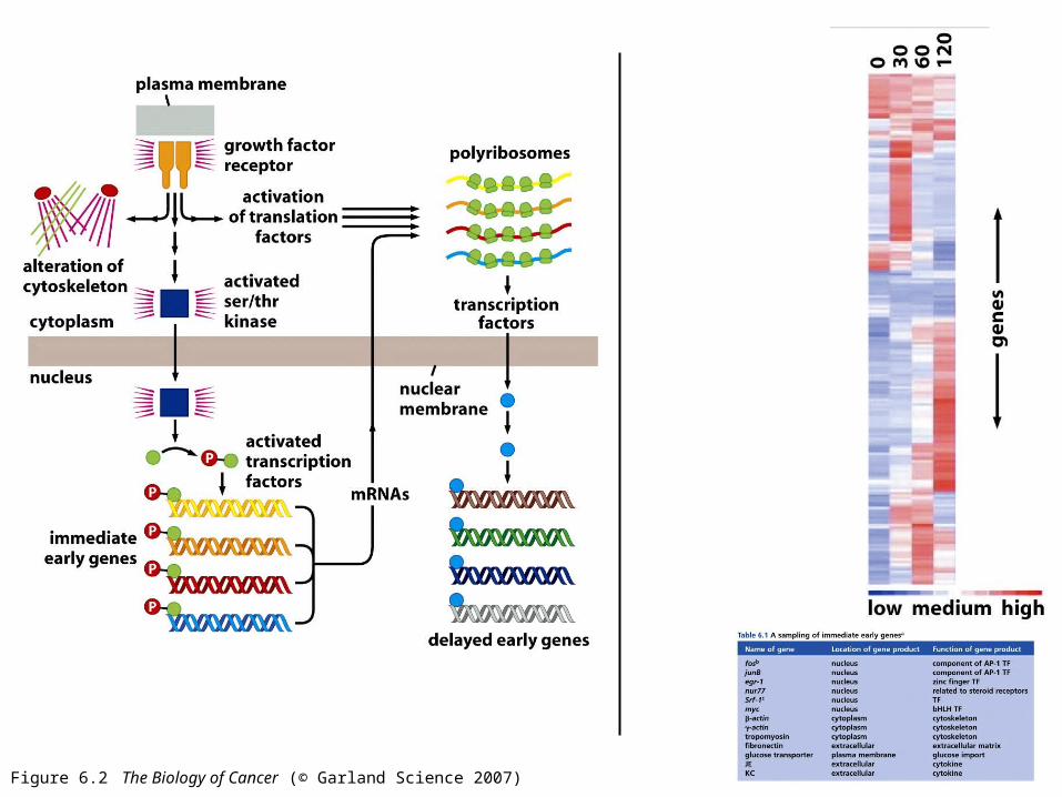
Figure 6.2 The Biology of Cancer (© Garland Science 2007)
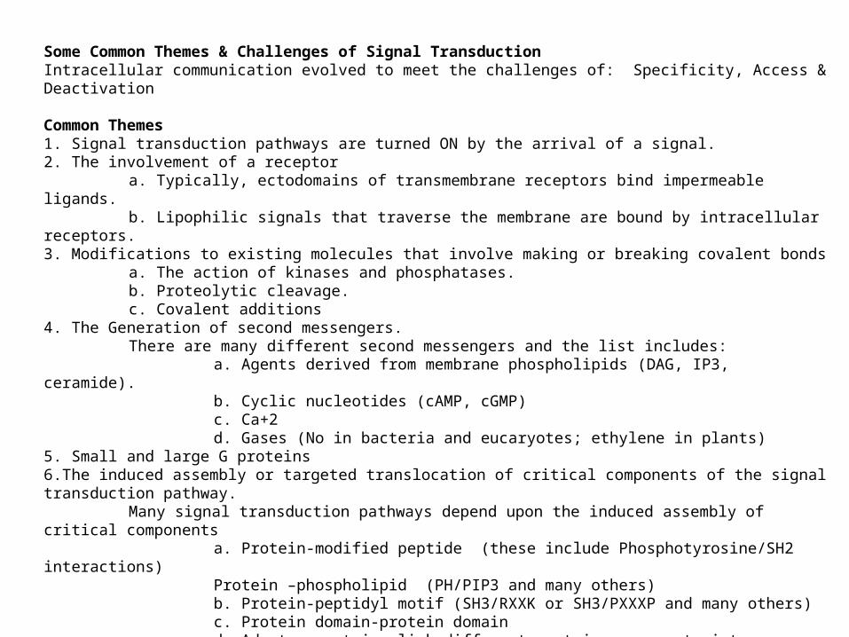
Some Common Themes & Challenges of Signal TransductionIntracellular communication evolved to meet the challenges of: Specificity, Access & Deactivation
Common Themes 1. Signal transduction pathways are turned ON by the arrival of a signal.2. The involvement of a receptor
a. Typically, ectodomains of transmembrane receptors bind impermeable ligands. b. Lipophilic signals that traverse the membrane are bound by intracellular receptors.3. Modifications to existing molecules that involve making or breaking covalent bonds
a. The action of kinases and phosphatases. b. Proteolytic cleavage. c. Covalent additions
4. The Generation of second messengers. There are many different second messengers and the list includes:
a. Agents derived from membrane phospholipids (DAG, IP3, ceramide).b. Cyclic nucleotides (cAMP, cGMP)c. Ca+2 d. Gases (No in bacteria and eucaryotes; ethylene in plants)
5. Small and large G proteins6.The induced assembly or targeted translocation of critical components of the signal transduction pathway.
Many signal transduction pathways depend upon the induced assembly of critical componentsa. Protein-modified peptide (these include Phosphotyrosine/SH2 interactions)Protein –phospholipid (PH/PIP3 and many others)b. Protein-peptidyl motif (SH3/RXXK or SH3/PXXXP and many others)c. Protein domain-protein domaind. Adaptor proteins link different protein components into signaling complexes.
7. Cascades to amplify and relay signals8. Compartmentation. 9. Signal transdution pathways interact with each other (‘crosstalk’ is the rule rather than the exception).10. The default setting for signal transduction pathways is OFF
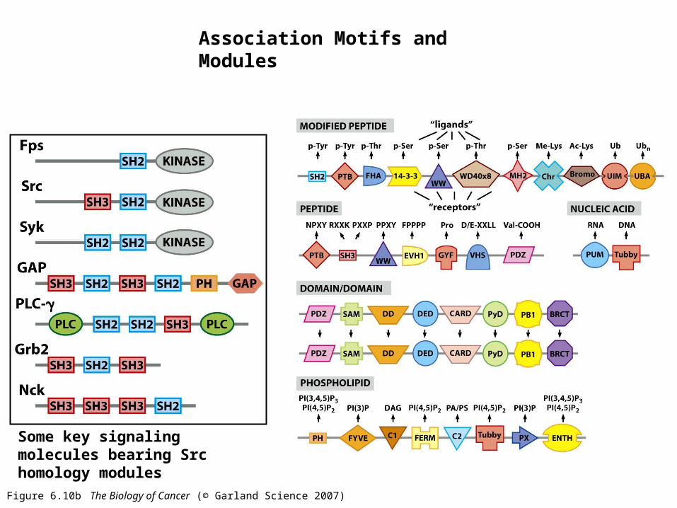
Figure 6.10b The Biology of Cancer (© Garland Science 2007)
Association Motifs and Modules
Some key signaling molecules bearing Src homology modules
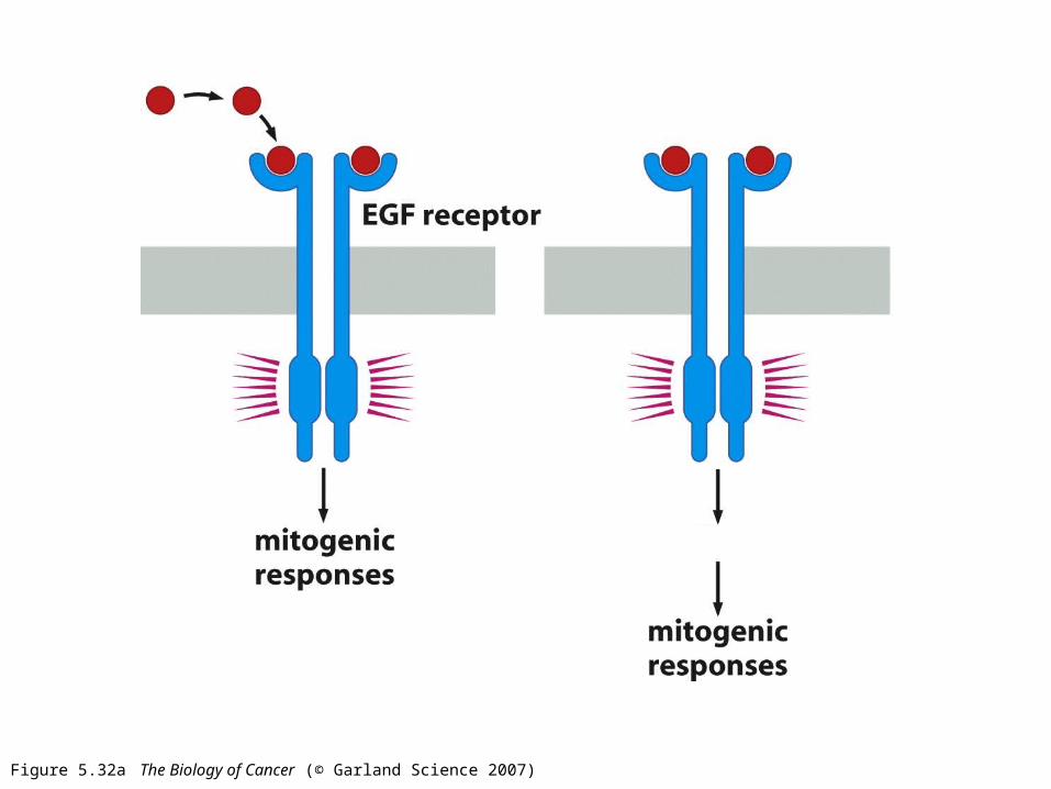
Figure 5.32a The Biology of Cancer (© Garland Science 2007)
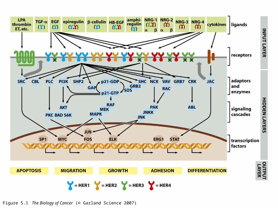
Figure 5.1 The Biology of Cancer (© Garland Science 2007)
Lessons From Src
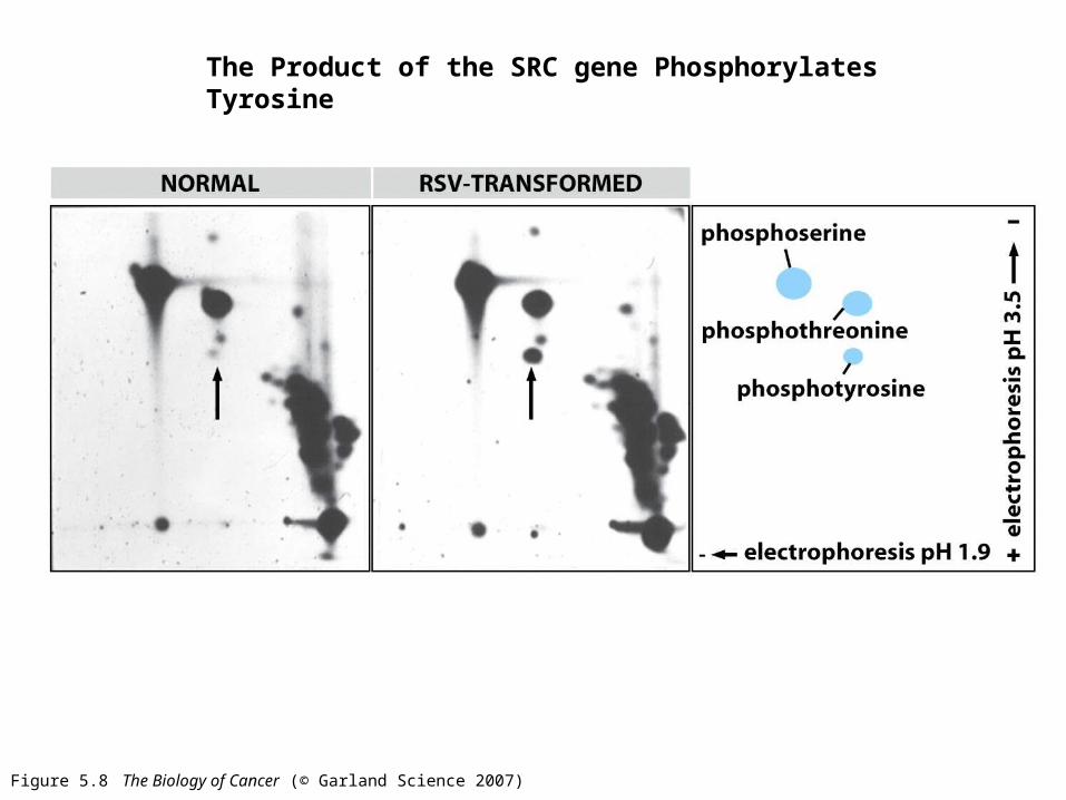
Figure 5.8 The Biology of Cancer (© Garland Science 2007)
The Product of the SRC gene Phosphorylates Tyrosine
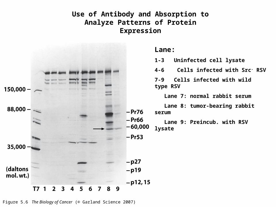
Figure 5.6 The Biology of Cancer (© Garland Science 2007)
Use of Antibody and Absorption to Analyze Patterns of Protein Expression
Lane:
1-3 Uninfected cell lysate
4-6 Cells infected with Src- RSV
7-9 Cells infected with wild type RSV
Lane 7: normal rabbit serum
Lane 8: tumor-bearing rabbit serum
Lane 9: Preincub. with RSV lysate
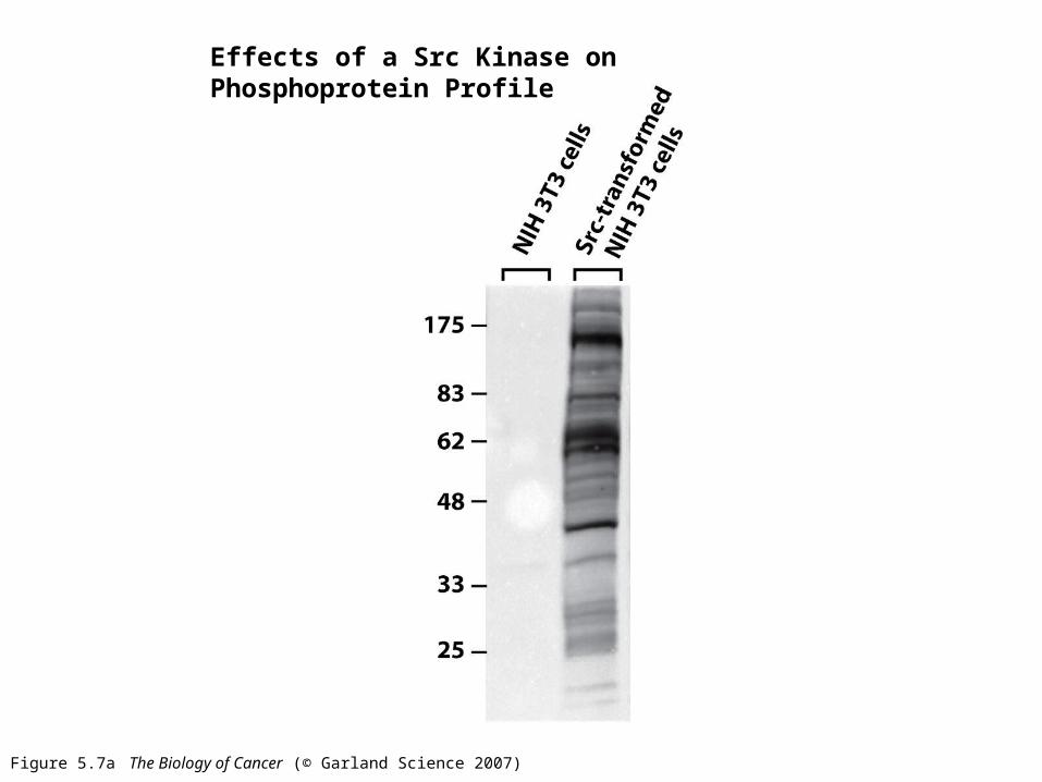
Figure 5.7a The Biology of Cancer (© Garland Science 2007)
Effects of a Src Kinase on Phosphoprotein Profile
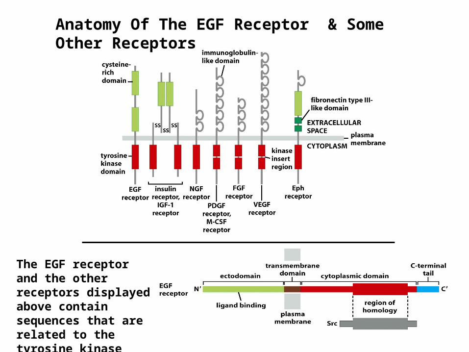
Anatomy Of The EGF Receptor & Some Other Receptors
The EGF receptor and the other receptors displayed above contain sequences that are related to the tyrosine kinase domain of Src.

Figure 6.7a The Biology of Cancer (© Garland Science 2007)
3-D Structure of Src
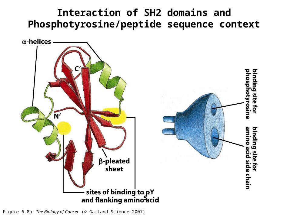
Figure 6.8a The Biology of Cancer (© Garland Science 2007)
s
Interaction of SH2 domains and Phosphotyrosine/peptide sequence context
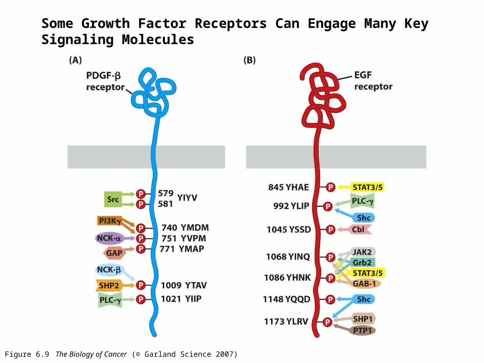
Figure 6.9 The Biology of Cancer (© Garland Science 2007)
Some Growth Factor Receptors Can Engage Many Key Signaling Molecules
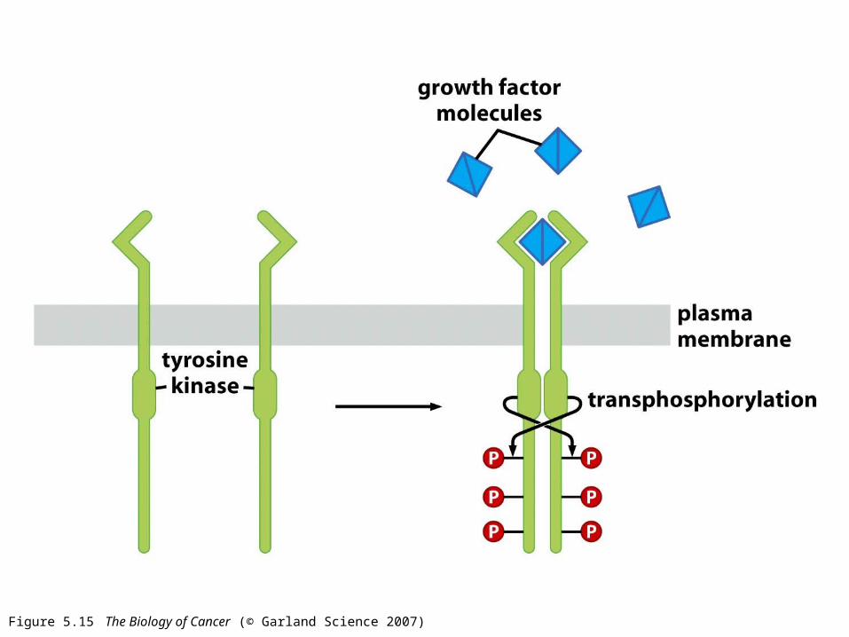
Figure 5.15 The Biology of Cancer (© Garland Science 2007)
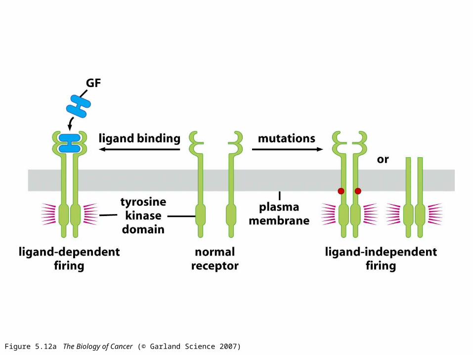
Figure 5.12a The Biology of Cancer (© Garland Science 2007)
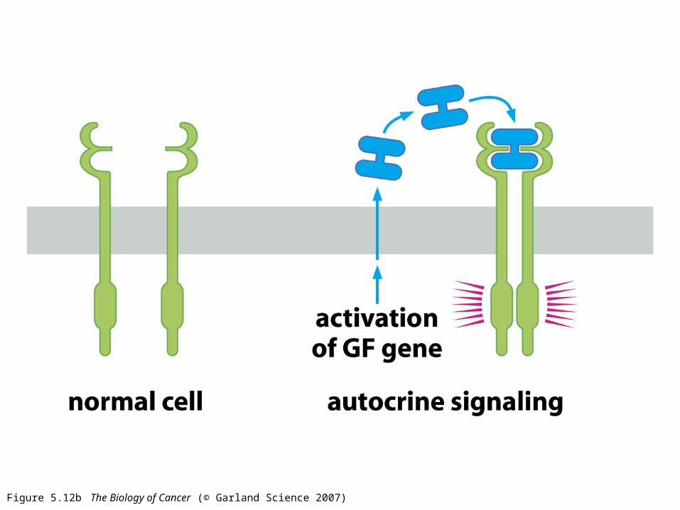
Figure 5.12b The Biology of Cancer (© Garland Science 2007)
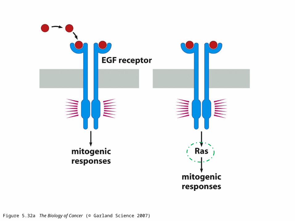
Figure 5.32a The Biology of Cancer (© Garland Science 2007)
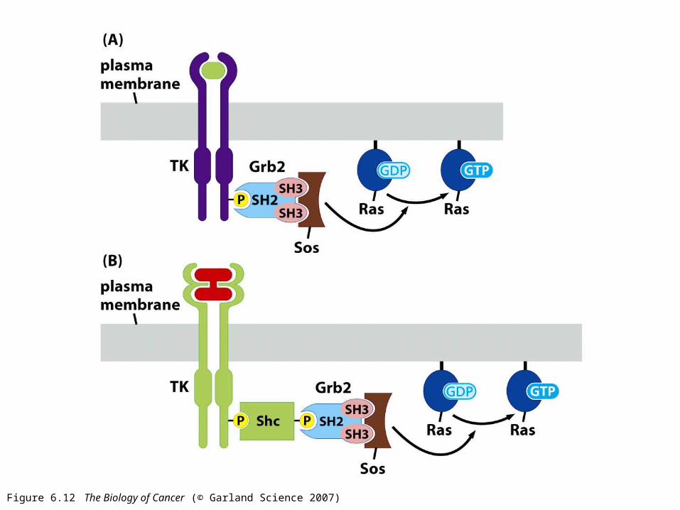
Figure 6.12 The Biology of Cancer (© Garland Science 2007)
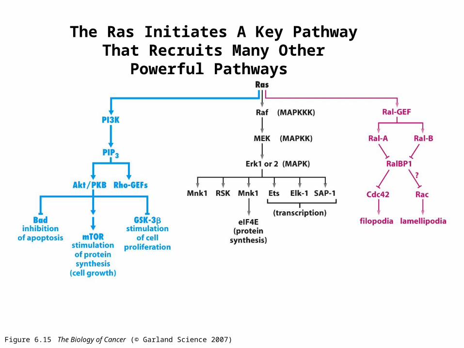
Figure 6.15 The Biology of Cancer (© Garland Science 2007)
The Ras Initiates A Key Pathway That Recruits Many Other Powerful Pathways
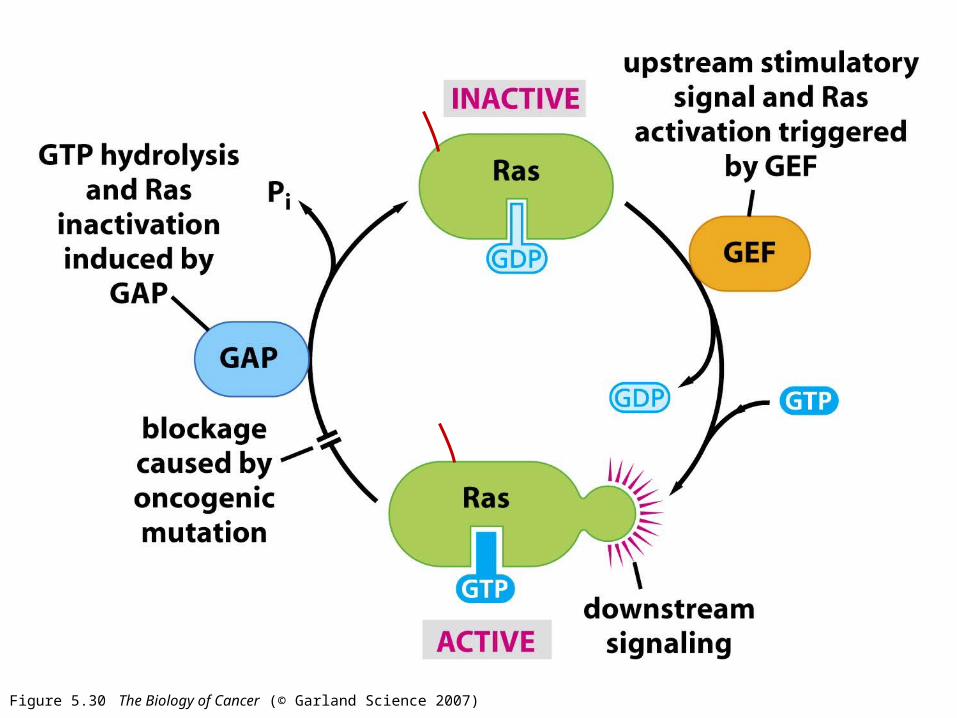
Figure 5.30 The Biology of Cancer (© Garland Science 2007)
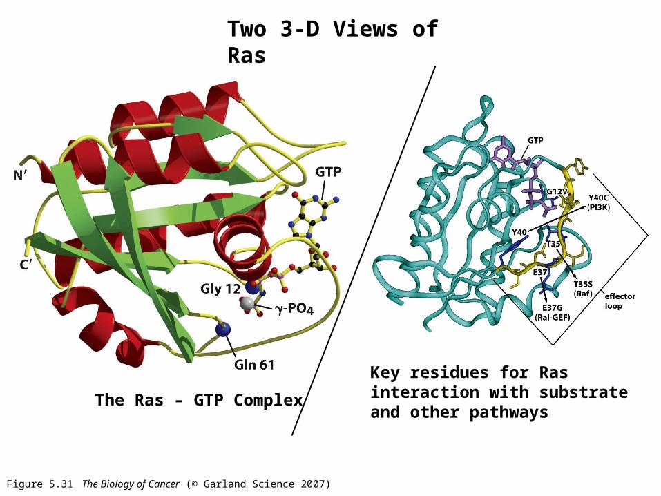
Figure 5.31 The Biology of Cancer (© Garland Science 2007)
Two 3-D Views of Ras
The Ras – GTP Complex
Key residues for Ras interaction with substrate and other pathways
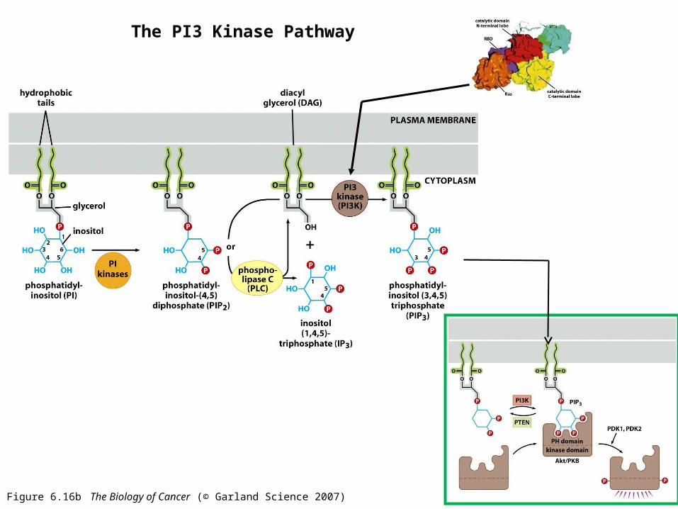
Figure 6.16b The Biology of Cancer (© Garland Science 2007)
The PI3 Kinase Pathway
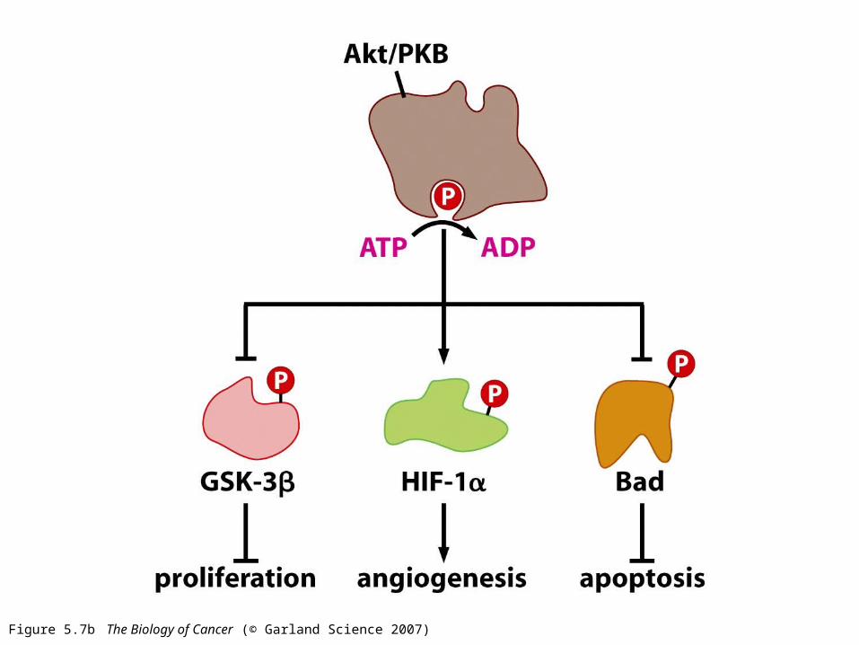
Figure 5.7b The Biology of Cancer (© Garland Science 2007)
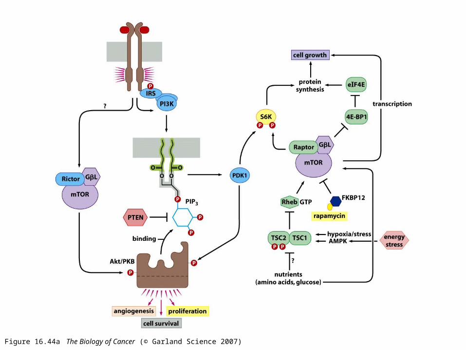
Figure 16.44a The Biology of Cancer (© Garland Science 2007)
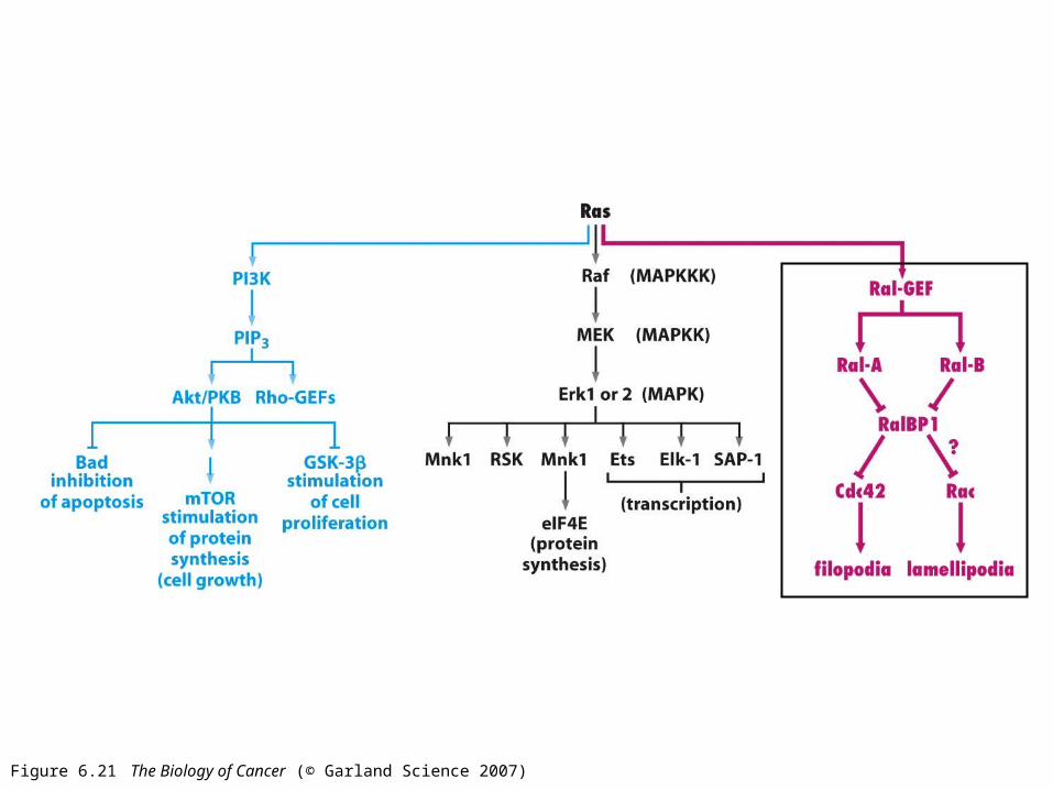
Figure 6.21 The Biology of Cancer (© Garland Science 2007)
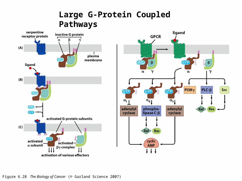
Figure 6.28 The Biology of Cancer (© Garland Science 2007)
Large G-Protein Coupled Pathways
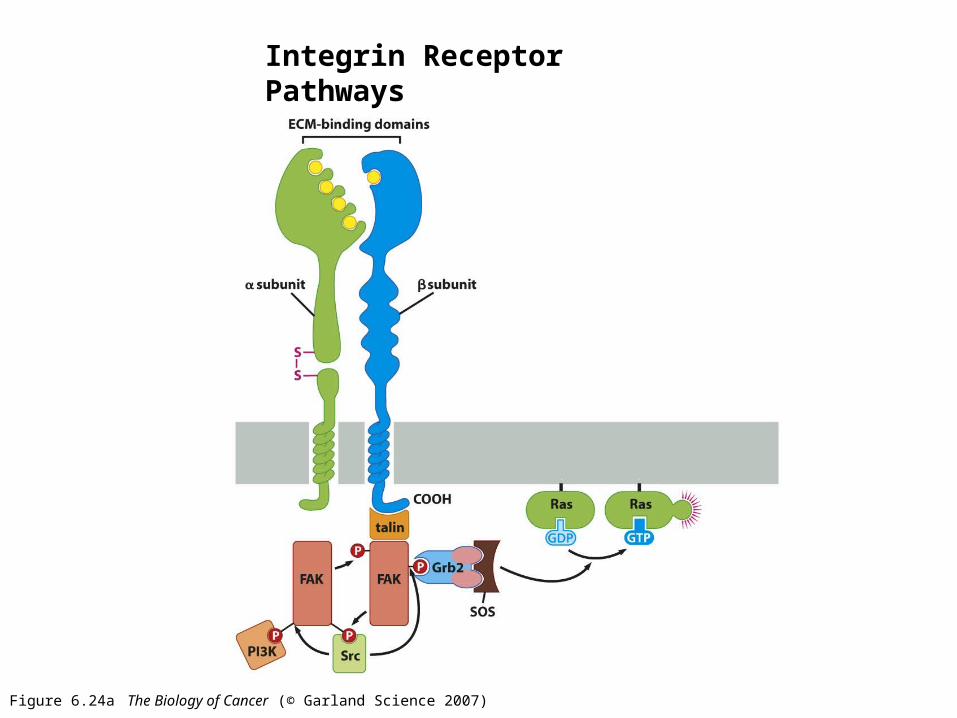
Figure 6.24a The Biology of Cancer (© Garland Science 2007)
Integrin Receptor Pathways
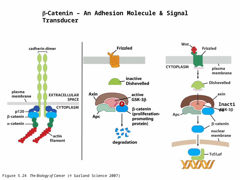
Figure 5.24 The Biology of Cancer (© Garland Science 2007)
Catenin – An Adhesion Molecule & Signal Transducer
Inactive
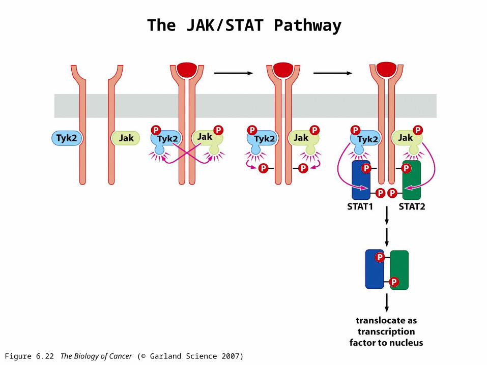
Figure 6.22 The Biology of Cancer (© Garland Science 2007)
The JAK/STAT Pathway
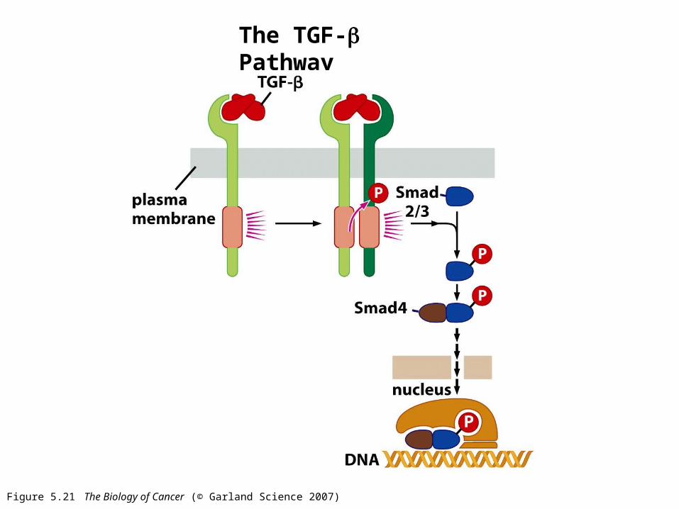
Figure 5.21 The Biology of Cancer (© Garland Science 2007)
The TGF- Pathway
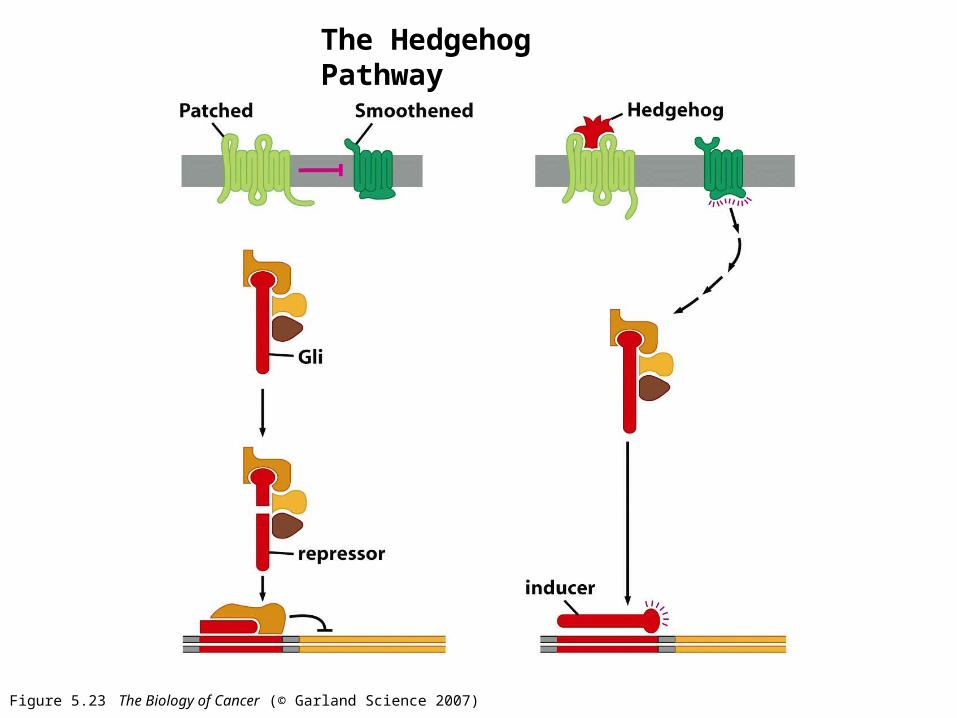
Figure 5.23 The Biology of Cancer (© Garland Science 2007)
The Hedgehog Pathway
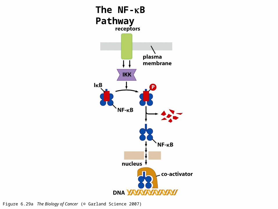
Figure 6.29a The Biology of Cancer (© Garland Science 2007)
The NF-B Pathway
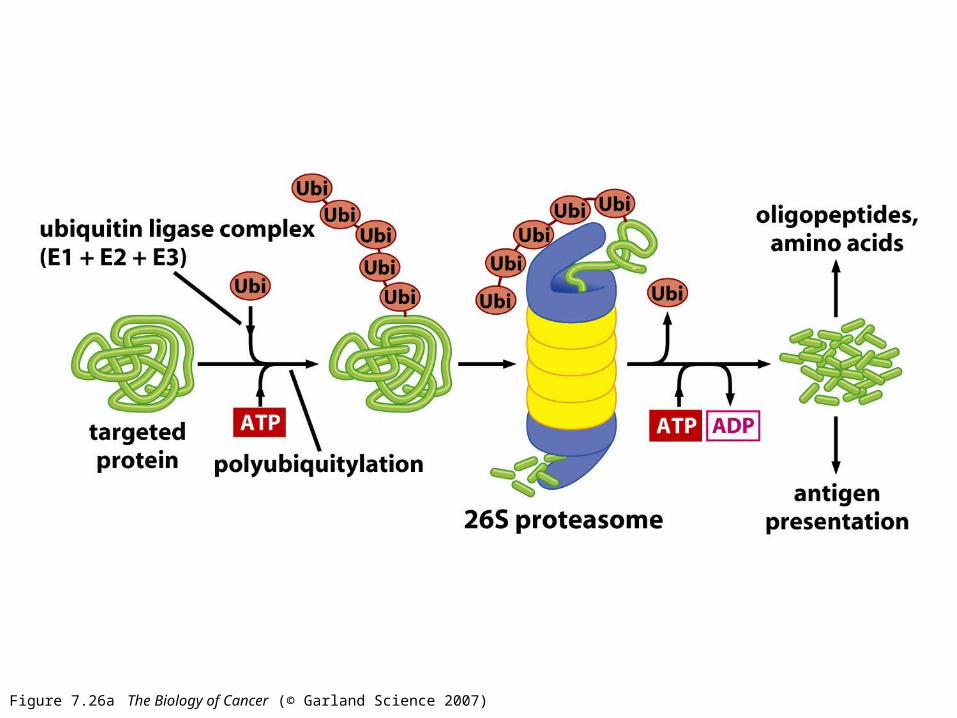
Figure 7.26a The Biology of Cancer (© Garland Science 2007)
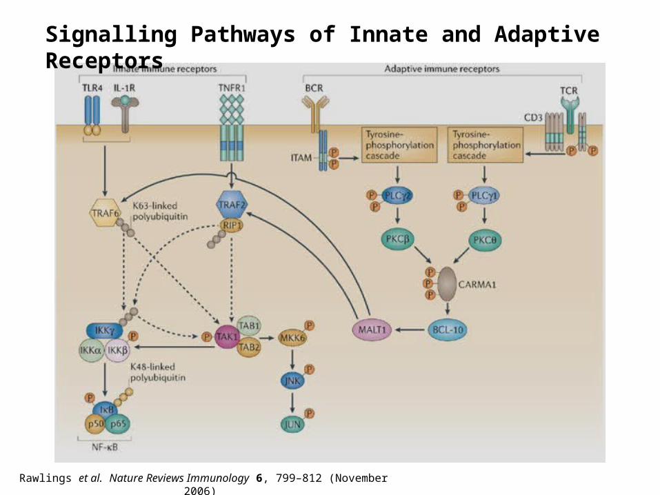
Rawlings et al. Nature Reviews Immunology 6, 799–812 (November 2006)
Signalling Pathways of Innate and Adaptive Receptors
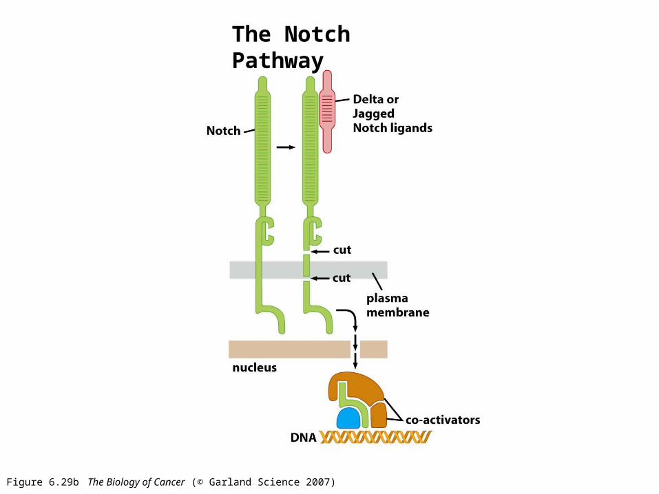
Figure 6.29b The Biology of Cancer (© Garland Science 2007)
The Notch Pathway
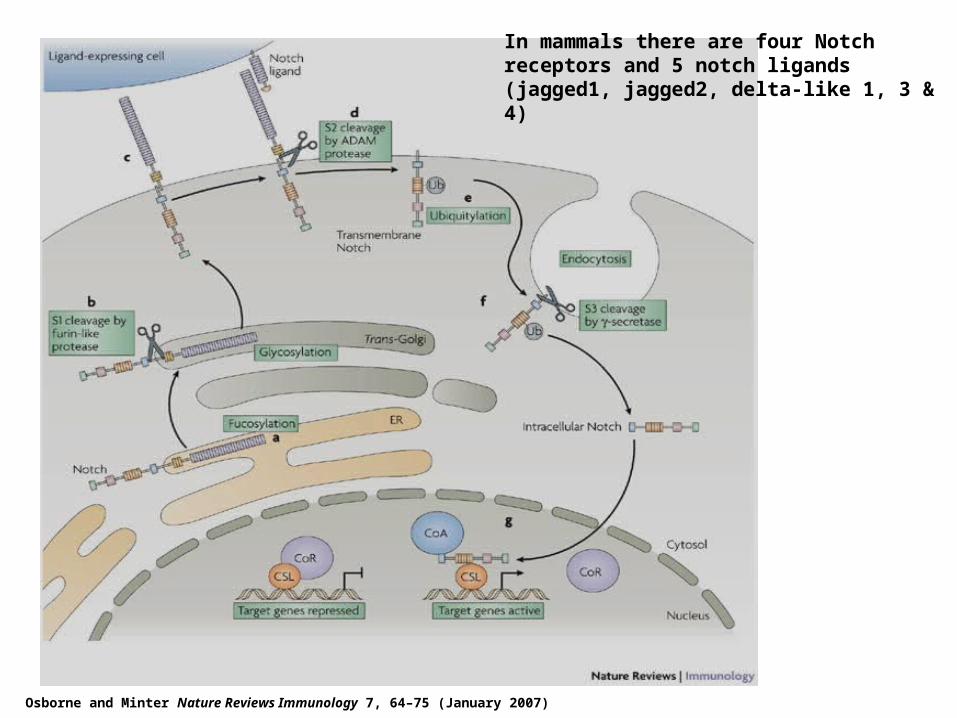
Osborne and Minter Nature Reviews Immunology 7, 64–75 (January 2007)
In mammals there are four Notch receptors and 5 notch ligands (jagged1, jagged2, delta-like 1, 3 & 4)
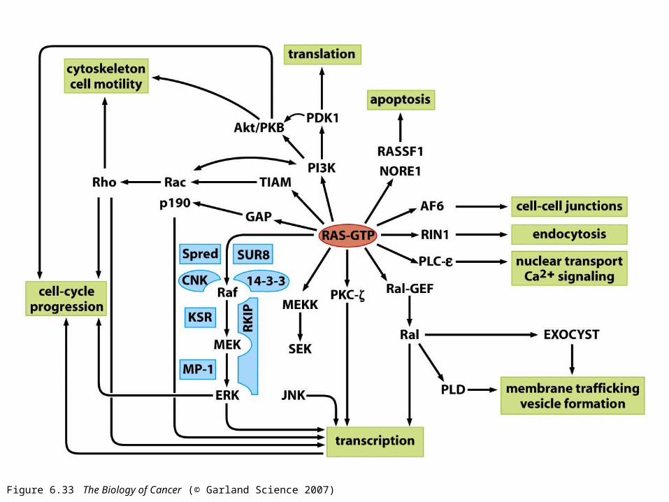
Figure 6.33 The Biology of Cancer (© Garland Science 2007)
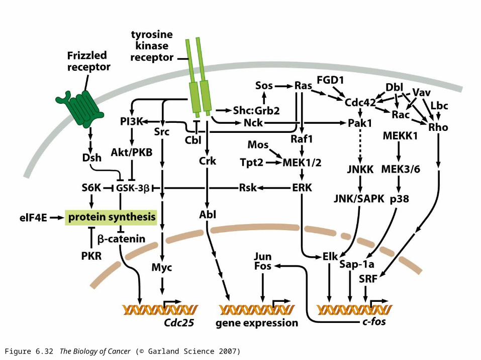
Figure 6.32 The Biology of Cancer (© Garland Science 2007)